Xanthomonas oryzae pv. oryzae - Purdue University
14
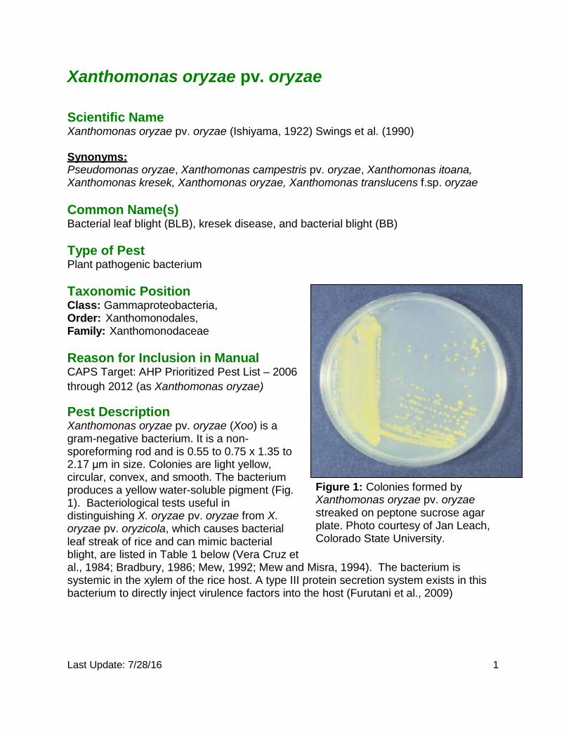
Last Update: 7/28/16 1 Xanthomonas oryzae pv. oryzae Scientific Name Xanthomonas oryzae pv. oryzae (Ishiyama, 1922) Swings et al. (1990) Synonyms: Pseudomonas oryzae, Xanthomonas campestris pv. oryzae, Xanthomonas itoana, Xanthomonas kresek, Xanthomonas oryzae, Xanthomonas translucens f.sp. oryzae Common Name(s) Bacterial leaf blight (BLB), kresek disease, and bacterial blight (BB) Type of Pest Plant pathogenic bacterium Taxonomic Position Class: Gammaproteobacteria, Order: Xanthomonodales, Family: Xanthomonodaceae Reason for Inclusion in Manual CAPS Target: AHP Prioritized Pest List – 2006 through 2012 (as Xanthomonas oryzae) Pest Description Xanthomonas oryzae pv. oryzae (Xoo) is a gram-negative bacterium. It is a non- sporeforming rod and is 0.55 to 0.75 x 1.35 to 2.17 μm in size. Colonies are light yellow, circular, convex, and smooth. The bacterium produces a yellow water-soluble pigment (Fig. 1). Bacteriological tests useful in distinguishing X. oryzae pv. oryzae from X. oryzae pv. oryzicola, which causes bacterial leaf streak of rice and can mimic bacterial blight, are listed in Table 1 below (Vera Cruz et Figure 1: Colonies formed by Xanthomonas oryzae pv. oryzae streaked on peptone sucrose agar plate. Photo courtesy of Jan Leach, Colorado State University. al., 1984; Bradbury, 1986; Mew, 1992; Mew and Misra, 1994). The bacterium is systemic in the xylem of the rice host. A type III protein secretion system exists in this bacterium to directly inject virulence factors into the host (Furutani et al., 2009)
Transcript of Xanthomonas oryzae pv. oryzae - Purdue University
Last Update: 7/28/16 1
Xanthomonas oryzae pv. oryzae Scientific Name Xanthomonas oryzae pv. oryzae (Ishiyama, 1922) Swings et al. (1990)
Synonyms: Pseudomonas oryzae, Xanthomonas campestris pv. oryzae, Xanthomonas itoana, Xanthomonas kresek, Xanthomonas oryzae, Xanthomonas translucens f.sp. oryzae
Common Name(s) Bacterial leaf blight (BLB), kresek disease, and bacterial blight (BB)
Type of Pest Plant pathogenic bacterium
Taxonomic Position Class: Gammaproteobacteria, Order: Xanthomonodales, Family: Xanthomonodaceae
Reason for Inclusion in Manual CAPS Target: AHP Prioritized Pest List – 2006 through 2012 (as Xanthomonas oryzae)
Pest Description Xanthomonas oryzae pv. oryzae (Xoo) is a gram-negative bacterium. It is a non- sporeforming rod and is 0.55 to 0.75 x 1.35 to 2.17 μm in size. Colonies are light yellow, circular, convex, and smooth. The bacterium produces a yellow water-soluble pigment (Fig. 1). Bacteriological tests useful in distinguishing X. oryzae pv. oryzae from X. oryzae pv. oryzicola, which causes bacterial leaf streak of rice and can mimic bacterial blight, are listed in Table 1 below (Vera Cruz et
Figure 1: Colonies formed by Xanthomonas oryzae pv. oryzae streaked on peptone sucrose agar plate. Photo courtesy of Jan Leach, Colorado State University.
al., 1984; Bradbury, 1986; Mew, 1992; Mew and Misra, 1994). The bacterium is systemic in the xylem of the rice host. A type III protein secretion system exists in this bacterium to directly inject virulence factors into the host (Furutani et al., 2009)
Last Update: 7/28/16 2
A pathovar is a bacterial strain (or set of strains) with similar characteristics that are usually distinguished by a different host range. In this case, Xanthomonas oryzae has two pathovars (Xanthomonas oryzae pv. oryzae and X. oryzae pv. oryzicola) that affect the same host but have strong differences in symptomatology on the same host, which allows for different pathovar designations.
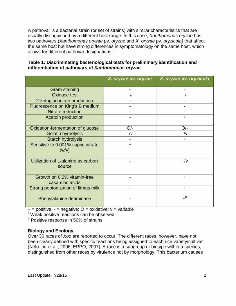
Table 1: Discriminating bacteriological tests for preliminary identification and differentiation of pathovars of Xanthomonas oryzae.
X. oryzae pv. oryzae X. oryzae pv. oryzicola
Gram staining - - Oxidase test -a -a
2-ketoglucontate production - - Fluorescence on King’s B medium - -
Nitrate reduction - - Acetoin production - +
Oxidation-fermentation of glucose O/- O/- Gelatin hydrolysis -/v -/v Starch hydrolysis - +
Sensitive to 0.001% cupric nitrate (w/v)
+ -
Utilization of L-alanine as carbon source
- +/v
Growth on 0.2% vitamin-free casamino acids
- +
Strong peptonization of litmus milk - +
Phenylalanine deaminase - +b
+ = positive; - = negative; O = oxidative; v = variable a Weak positive reactions can be observed. b Positive response in 50% of strains.
Biology and Ecology Over 30 races of Xoo are reported to occur. The different races, however, have not been clearly defined with specific reactions being assigned to each rice variety/cultivar (Niño-Liu et al., 2006; EPPO, 2007). A race is a subgroup or biotype within a species, distinguished from other races by virulence not by morphology. This bacterium causes
Last Update: 7/28/16 3
bacterial blight by invading the vascular tissue. Bacterial blight is a very serious disease in rice (Niño-Liu et al., 2006).
Xanthomonas oryzae pv. oryzae survives primarily in rice stubble and on weed hosts, notably Leersia oryzoides, Zizania latifolia Leptocholoa chinensis, L. panicea, and Cyperus rotundis. In Australia, the bacterium is known to survive in wild Oryza species (O. rufipogon and O. australiensis). Xoo can also survive for short periods on infected seed and in soil, but these have not been demonstrated to be important sources of inoculum. In tropical areas, the bacterium may also survive in irrigation water (Mew, 1992).
Xanthomonas oryzae pv. oryzae enters the rice leaf typically through the hydathodes at the leaf tip and leaf margin. Bacterial cells on the leaf surface may become suspended in guttation fluid as it exudes at night and then enter the plant by swimming, or passively as the fluid is withdrawn into the leaf in the morning. Bacteria multiply in the intercellular spaces of the underlying epithelial cells. It then enters and spreads into the plant through the xylem. Xoo may also gain access to the xylem through wounds or openings caused by emerging roots at the base of the leaf sheath. Within the xylem, Xoo presumably interacts with the xylem parenchyma cells. The pathogen moves vertically through the leaf via the primary veins. It may progress laterally through commissural veins. Within a few days, bacterial cells and EPS (extracellular polysaccharide) fill the xylem vessels and ooze out from hydathodes, forming beads or strands of exudate on the leaf surface, a characteristic sign of the disease and a source of secondary inoculum (Niño-Liu et al., 2006). As the bacterium develops in the plant it can also spread into the mesophyll (Niño-Liu et al., 2005).
The ‘kresek’ syndrome is generally associated with seedling infection that occurs through wounds made during transplanting operations. The roots of seedlings are often damaged when they are removed from the nurseries, and in many areas, the tops of the leaves are clipped off before transplanting. The severity of kresek is dependent on the time of infection; the earlier seedlings are infected, the more severe is the syndrome.
Genetic resistance against X. oryzae pv. oryzae has been found in some rice cultivars/varieties. Host resistance is currently being used to as a disease management tool to control the disease (Niño-Liu et al., 2005).
Bacterial leaf blight is favored by warm temperatures (25 to 30°C, 77 to 86°F), high humidity, rain, and deep water. The disease is more prevalent in wetland areas where these conditions often occur. Winds severe enough to cause wounds and excess nitrogen also favor the disease. The severity of the disease is in part dependent on the virulence of the bacterial isolates present. Bacterial blight is severe in susceptible rice varieties under high nitrogen fertilization (IRRI, 2004; 2010).The bacterium can be disseminated by irrigation water, by splashing or windblown rain, by plant-to-plant
Last Update: 7/28/16 4
contact, by trimming tools used in transplanting, and by handling plants during transplanting (Mew, 1992).
A
B
C
D
E
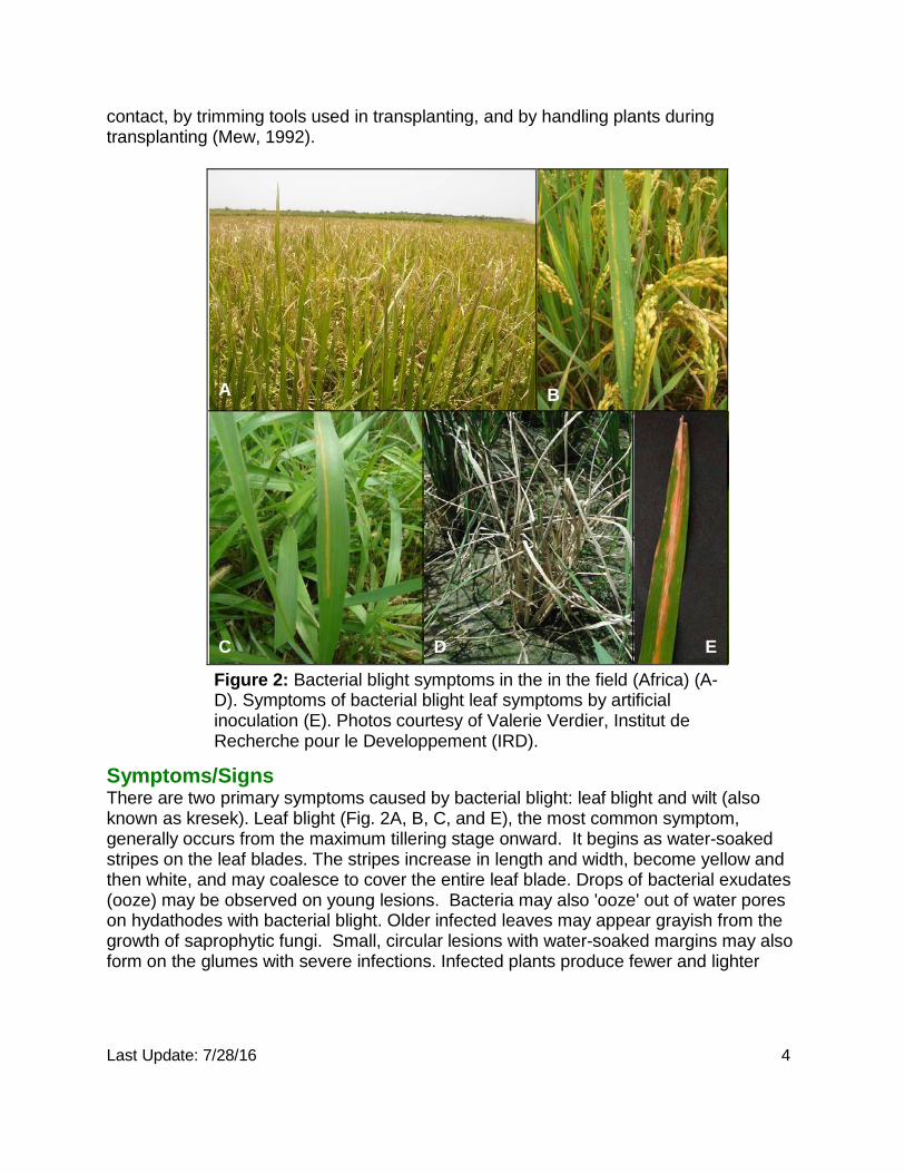
Figure 2: Bacterial blight symptoms in the in the field (Africa) (A- D). Symptoms of bacterial blight leaf symptoms by artificial inoculation (E). Photos courtesy of Valerie Verdier, Institut de Recherche pour le Developpement (IRD).
Symptoms/Signs There are two primary symptoms caused by bacterial blight: leaf blight and wilt (also known as kresek). Leaf blight (Fig. 2A, B, C, and E), the most common symptom, generally occurs from the maximum tillering stage onward. It begins as water-soaked stripes on the leaf blades. The stripes increase in length and width, become yellow and then white, and may coalesce to cover the entire leaf blade. Drops of bacterial exudates (ooze) may be observed on young lesions. Bacteria may also 'ooze' out of water pores on hydathodes with bacterial blight. Older infected leaves may appear grayish from the growth of saprophytic fungi. Small, circular lesions with water-soaked margins may also form on the glumes with severe infections. Infected plants produce fewer and lighter
Last Update: 7/28/16 5
grains, and the grain is of poor quality. Seed may be discolored and poorly filled with bacterial blight.
The wilt syndrome, known as kresek, is the most destructive manifestation of the disease; it occurs in the tropics from the seedling to the early tillering stage. Leaves of infected plants wilt, roll up, and turn a grayish green color (Fig. 2D). The leaves then turn yellow to straw-colored and wither. The entire plant generally dies. Plants that do survive are stunted and yellow in color. Total crop failure is not uncommon with kresek.
A third symptom associated with bacterial blight is called yellow leaf or pale yellow. The youngest leaf of the plant becomes uniformly pale yellow or has a broad yellow stripe. With yellow leaf, the bacteria are not present in the leaf itself but can be found in the internodes and crowns of affected stems.
Pest Importance Bacterial blight is one of the most serious diseases of rice worldwide and is found in both tropical and temperate regions. It was first recorded in Japan in 1884. In the 1960’s, bacterial blight became prevalent in other rice-growing regions of Asia with the introduction of “improved” cultivars such as TN1 and IR8, which were susceptible to the disease. In addition to Asia, bacterial blight occurs in Australia, Africa, Latin America, and the Caribbean. Economically, it has had the greatest impact in Asia, where several epidemics have occurred in the past two decades, and in the West Africa, particularly in Niger, where irrigated rice was extensively damaged in 1982. Yield losses of 10 to 50% from leaf blight have been reported (Mew, 1992). The study of bacterial leaf blight allows a better understanding of plant pathogen interaction and tissue vulnerability in plant-pathogen interactions.
Known Hosts Major Hosts: Oryza sativa (rice), Oryza spp.
Minor/Other Reported Hosts: Alopecurus aequalis (shortawn foxtail), Bracharia mutica (para grass), Cenchrus ciliaris (buffelgrass), Cynodon dactylon (bermudagrass), Cyperaceae (sedges), Cyperus difformis (variable flatsedge, small flowered nutsedge), Cyperus rotundus (nutgrass), Digitaria sanguinalis (hairy crabgrass), Echinochloa crus- galli (barnyardgrass), Isachne globosa (swamp millet-England), Leersia japonica, Leersia oryzoides (rice cutgrass), Leptochloa chinensis (Chinese sprangletop), Leptochloa panicea (mucronate sprangletop), Oryza rufipogon (brownbeard rice), Paspalum distichum (knotgrass), Paspalum scrobiculatum (kodomillet), Phalaris arundinacea (reed canarygrass), Phragmites communis (common reed), Plantago major (common plantain), Poaceae (grasses), Oryza australiensis, Oryza rufipogon (brownbeard rice), Setaria viridis (green bristlegrass), Urochloa mutica (tall panicum), Zizania aquatica (annual wildrice), Zizania latifolia (Manchurian wildrice), Zizania palustris (northern wild rice), and Zoysia japonica (zoysiagrass) (Aldrick et al., 1973;
Last Update: 7/28/16 6
Dye and Lelliott, 1974; Reddy and Nayak, 1974; Duan et al., 1979; Leyns et al., 1984; Li et al.,1985; Mew, 1992; Zhong et al.,1998; EPPO, 2007; CABI, 2011)
Hosts from Artificial Inoculation: Leersia hexandra (southern cutgrass), Panicum maximum (guineagrass) (Chattopadhyay and Mikherjee, 1968; Reddy and Nayak, 1974; Valluvaparidasan and Mariappan, 1989).
Note: The level of susceptibility and symptom expression varies significantly among hosts tested by artificial inoculation.
Known Vectors (or associated insects) Bacteria may be disseminated in irrigation water, as well as by humans, insects and birds (Niño-Liu et al., 2006).
Known Distribution Bacterial blight is one of the most serious diseases of rice worldwide and is found in both tropical and temperate regions (Mew, 1992). The disease is known to occur in Asia, parts of West Africa, Australia, Latin America, and the Caribbean (Niño-Liu et al., 2006). PCR and RFLP markers were used to detect that strains are moving within Asia (George et al., 1997). In the United States, although an apparent mild outbreak of bacterial blight was reported in the late 1980s (Jones et al., 1990), it was later determined that the bacterium associated with the disease was not Xoo (Niño-Liu et al., 2006).
Asia: Bangladesh, Cambodia, China, India, Indonesia, Iran, Japan, Korea, Laos, Malaysia, Myanmar, Nepal, Pakistan, Philippines, Sri Lanka, Taiwan, Thailand, and Vietnam. Africa: Benin, Burkina Faso, Cameroon, Egypt, Gabon, Gambia, Mali, Niger, Senegal, Tanzania, and Togo. North America: Mexico, United States (see note below). Central America: Costa Rica, El Salvador, Honduras, Panama, Saba. South America: Bolivia, Columbia, Ecuador, and Venezuela. Oceania: Australia (Choi et al., 1998; Niño-Liu et al., 2006; CABI, 2011).
Note: X. oryzae pv. oryzae was detected in Louisiana and Texas (Jones et al., 1989). Strains were of low virulence compared to Asian strains. Discussion with the authors of this paper indicated that this find should be a new pathovar and not Xanthomonas oryzae pv. oryzae (Jan Leech, Personal Communication, 2010). Therefore, the pathogen is not known to occur in the United States. The low virulence pathovar has not been found again in either location since they stopped planting susceptible hosts.
Pathway Outbreaks of bacterial leaf blight are more likely to occur during the monsoon season of the south-east Asian and Indian oceans (from June to September) than at other times of the year (Niño-Liu et al., 2006; Mew, 1992). Wind and rain, as well as contaminated rice
Last Update: 7/28/16 7
stubble from previous crop seasons, spread the bacterium from infected rice plants and other hosts. This is the most important source of primary inoculum.
Severe epidemics often occur following typhoons. The fierce winds, wind-blown rain and hail wound rice plants and disperse bacteria. The bacterium can spread from plant-to- plant contact and water that has been in contact with the bacterium (Stall et al., 1993). The bacterium is also known to spread by seed, but scientists disagree about the role the method in transmission (Sakthivel et al, 2001).
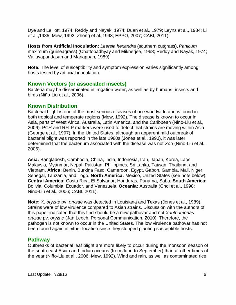
Potential Distribution within the United States Surveys should be focused where the greatest risk for pest establishment occurs. A recent rice acreage map developed by USDA-APHIS-PPQ-CPHST (Fig. 3) indicates that California and southern states in the Mississippi river valley have the highest concentration of rice cultivation in the United States.
Figure 3. Rice commodity acreage. Map courtesy of USDA-APHIS-PPQ-CPHST.
Last Update: 7/28/16 8
Survey CAPS-Approved Method: Visual survey is the approved survey method for Xanthomonas oryzae pv. oryzae. For visual survey, collect symptomatic leaf samples. For a preliminary indications of seed infection, look for bacterial streaming (Singh and Rao, 1977). *For the most up-to-date methods for survey and identification, see Approved Methods on the CAPS Resource and Collaboration Site, at http://caps.ceris.purdue.edu/. Literature-Based Methods: Survey for Xanthomonas oryzae pv. oryzae consists of visual inspection for symptoms, tissue sampling, and pathogen isolation.
Key Diagnostics/Identification CAPS-Approved Method: Morphological and Serological methods are approved methods for Xanthomonas oryzae pv. oryzae.
Morphological: Colony morphology: The pathogen is difficult to isolate directly from plant material and seed due to slow growth of bacterium and overgrowth by other organisms. Contaminants may also be of a yellow color (e.g., Pantoea agglomerans and Xanthomonas-like saprophytic bacteria). Isolation from symptomatic material should be completed as soon as possible after collection of samples. Plant parts showing fresh symptoms preferably with bacterial exudates should be selected for isolation if available. See EPPO (2007) for additional detail about pathogen isolation from plant material and seeds.
Isolation of Xanthomonas from symptomatic material can be performed using peptone sucrose agar (PSA), nutrient broth yeast extract medium (NBY), growth factor (GF) agar, modified Wakimoto’s agar, and various semi-selective media (Agarwal et al., 1989; Mew and Mistra, 1994; Sakthivel et al., 2001). Growth is very slow on nutrient agar (EPPO, 2007).
Ming et al. (1991) developed a semi-selective medium, called XOS, to isolate both Xanthomonas oryzae pathovars from rice seed.
Yuan (1990) describes a new culture medium for culturing Xanthomonas oryzae pv. oryzae from rice seed.
Last Update: 7/28/16 9
Gnanamanickam et al. (1994) tested three strains for growth on TZC, WF-P, YCM, YAT, MXO, and XOS semi-selective media. Results varied for each isolate used, but worked best when using monoclonal antibodies to confirm the genus and pathovar.
Serological: Monoclonal antibodies: Genus and pathovar specific antibodies can be used in an ELISA reaction on presumptive positives (Alvarez et al., 1985; Benedict et al., 1989).
Literature-Based Methods: Pathogenicity: Isolates can be tested for pathogenicity on susceptible rice cultivars. For X. oryzae pv. oryzae use 30 to 45 day-old IR24 or IR8 (International Rice Institute) or local popular varieties with known susceptibility to bacterial blight. Leaf clipping and spray inoculation methods are available for inoculations (Kauffman et al., 1973; Cottyn et al., 1994b; EPPO, 2007). Niño-Liu et al. (2005) inoculated plants by dipping them in bacterial mixture and incubating in a growth chamber. Symptoms developed a 6-day period.
Fatty Acid Profiles: Fatty acid profiles allow identification at the genus level only (Swings et al., 1990), so this analysis is not recommended a diagnostic method.
Molecular: PCR: Vera Cruz et al. (1995, 1996) compared Rep-PCR using repetitive DNA sequences with Restriction Fragment Length Polymorphism (RFLP).
Leach et al. (1990) used a repetitive DNA sequence (pJEL 101) to distinguish X. oryzae pv. oryzae from other pathovars and species of Xanthomonas.
PCR assays using primers that amplify internal fragments IS 1112 and IS 1113 of X. oryzae pv. oryzae (Cottyn et al., 1994a; Alvarez et al., 1997) have been used as well.
Sakthivel et al. (2001) developed a PCR technique to detect X. oryzae pv. oryzae in rice seed. A combined biological and enzymatic amplification (BIO-PCR) technique was used to detect the pathogen in naturally infected seed.
Gonzalez et al. (2007) found that X. oryzae pv. oryzae African and Asian strains were genetically different from each other by using RFLP, fluorescent amplified fragment length polymorphism (FAFLP) and repetitive sequence-based polymerase chain reaction.
Real-time PCR: Zhao et al. (2007) developed a real-time PCR to detect X. oryzae pv. oryzae and can distinguish it from X. oryzae pv. oryzicola.
Liao et al. (2003) developed a real-time PCR that can distinguish the two pathovars.
Last Update: 7/28/16 10
Computational Genomics/Multiplex PCR: Lang et al. (2010) used a computational genomics pipeline to compare sequenced genomes of Xanthomonas species to identify regions for development of highly specific diagnostic markers. A suite of primers were selected to monitor diverse loci and to distinguish the rice bacterial blight and leaf streak pathogens. A subset of primers were combined into a multiplex PCR to accurately distinguish the two rice pathogens in a geographically diverse collection from other xanthomonads and other plant pathogenic and plant-or seed associated bacteria.
Easily Confused Pests In the early stage of disease, the symptoms are similar to narrow brown leaf spot. Physiological disorders can also cause similar symptoms to bacterial blight.
Xanthomonas oryzae pv. oryzae (bacterial blight) and Xanthomonas oryzae pv. oryzicola (bacterial leaf streak) symptoms are easily differentiated in the early stages of disease and reflect the different modes of infection by each pathogen. Foliar symptoms of bacterial blight usually become evident at the tillering stage as small, green water- soaked spots at the tips and margins of fully developed leaves. The spots expand along the veins, merge, and become chlorotic and then necrotic, forming opaque, white to gray colored lesions that typically extend from the leaf tip down along the leaf veins and margins. Bacterial leaf streak symptoms, by contrast, begin with small, water-soaked lesions anywhere along the leaf between the veins. Veins act as barriers as infected areas expand and coalesce lengthwise, resulting in the symptom for which the disease is named. Streaks are translucent and typically yellow. At later stages infected leaves turn grayish white and die. When infection results from entry through breaks in the leaf as might occur due to high wind, symptoms may extend across the leaf break and expand lengthwise killing most or all of the leaf (Niño-Liu et al., 2006)
At the later stage, when the streaks have coalesced, symptoms of bacterial blight and bacterial leaf streak are similar. The shape of the edges of the lesions differs; straight in leaf streak and wavy in leaf blight. X. oryzae pv. oryzicola may be distinguished from X. oryzae pv. oryzae by colony morphology in typical isolates, strong starch and gelatin hydrolysis, and by biochemical and molecular methods.
Glossary Exudates: Liquid excreted or discharged from diseased tissues, from roots and leaves, or by fungi and bacteria.
Glumes: Small dry membranous bract found in inflorescences of Graminaceae and Cyperaceae.
Guttation: Exudation of watery, sticky liquid from hydathodes, especially along leaf margins.

Last Update: 7/28/16 11
Hydathodes: Epidermal leaf structure specialized for secretion or exudation of water; leaf opening at terminus of vein.
Internode: The portion of a stem or other structure between two nodes.
Saprophyte (adj. Saprophytic): An organism that obtains nourishment from nonliving organic matter.
Tiller: A lateral shoot, culm, or stalk arising from a crown bud; common in grasses; to put forth new shoots from the root, or round the bottom of the original stalk.
Tillering: To produce tillers
References Agarwal, P.C., Mortensen, C.N., and Mathur, S.B. 1989. Seedborne disease and seed health testing of rice. Technical Bulletin No. 3/Phytopathological Papers No. 30. Danish Government Institute of Seed Pathology for Developing Countries and CAB International Mycological Institute.
Aldrick, S.J. 1973. The occurrence of bacterial leaf blight in wild and cultivated rice in northern Australia. Australian Journal Agricultural Research 24 (2): 219-227.
Alvarez, A.M., Benedict, A.A., and Mizumoto, C.Y. 1985. Identification of xanthomonads and grouping of strains of Xanthomonas campestris pv. campestris with monoclonal antibodies. Phytopathology 75: 722-728.
Alvarez, A.M., Rehman, F.U., and Leach, J.E. 1997. Comparison of serological and molecular methods for detection of Xanthomonas oryzae pv. oryzae in rice seed. In: Seed Health Testing: Progress Towards the 21st Century. J.D. Hutchins and J.C. Reeves (Eds.). pp. 175-183.
Benedict, A.A., Alvarez, A.M., Berestecky, J., Imanaka, W., Mizumoto, C.Y., Pollard, L.W., Mew, T.W., and Gonzalez, C.F. 1989. Pathovar-specific monoclonal antibodies for Xanthomonas campestris pv. oryzae and for Xanthomonas campestris pv. oryzicola. Phytopathology 79: 322-328.
Bradbury, J.F. 1986. Guide to Plant Pathogenic Bacteria. CAB International Mycological Institute, Kew, Surrey.
CABI. 2011. Crop Protection Compendium. http://www.cabi.org/cpc/
Chattopadhyay, S., Mukherjee, N. 1968. Occurrence in nature of collateral hosts (Cyperus rotundus and C. difformis) of Xanthomonas oryzae, incitant of bacterial blight of rice. Current Science 15: 441-442.
Choi, S.H., Vera Cruz, C.M., and Leach, J. 1998. Distribution of Xanthomonas oryzae pv. oryzae DNA modification systems in Asia. Applied and Environmental Microbiology 64(5): 1163-1668.
Cottyn, B., Bautista, A.T., Nelson, R.J., Leach, J.E., Swings, J., and Mew, T.W. 1994a. Polymerase chain reaction amplification of DNA from bacterial populations of rice using specific oligonucleotide primers. International Rice Research Note 19: 30-32.

Last Update: 7/28/16 12
Cottyn, B., Cerez, M.T., and Mew, T.W. 1994b. Chapter 7: Bacteria. In: A Manual of Seed Health Testing. T.W. Mew and J.K. Mistra (Eds.). pp. 322-328. IRRI, Manila.
Duan, Y., Wang, Y., and Lu, X. 1979. Studies on the weed carriers of bacterial leaf blight of rice. Journal of Plant Protection. 03. English Abstract.
Dye, D.W., and Lelliott, R.A. 1974. Genus II. Xanthomonas Dowson 1939, 187. Pages 243-249. In R. E. Buchanan and N. E. Gibbons (eds.), Bergey's manual of determinative bacteriology, ed. 8. Williams and Wilkins Co., Baltimore, Maryland.
EPPO. 2007. Xanthomonas oryzae: Diagnostics. Volume 37 (3), pages 543-553.
Furutani, A., Takaoka, M., Sanada, H., Noguchi, Y., Oku, T., Tsuno, K., Ochiai, H., and Tsuge, S. 2009. Identification of novel type III secretion effectors in Xanthomonas oryzae pv. oryzae. Molecular Plant-Microbe Interaction 22(1):9 6-106.
George, M.L.C., Bustamam, M., Cruz, W.T., Leach, J.E., and Nelson, R.J. 1997. Movement of Xanthomonas oryzae pv. oryzae in southeast Asia detected using PCR-based DNA fingerprinting. Phytopathology 87(3): 302-309.
Gnanamanickam, S.S., Shigaki, T., Medalla, E.S., Mew, T.W., and Alvarez, A.M. 1994. Problems in detection of Xanthomonas oryzae pv. oryzae in rice seed and potential for improvement using monoclonal antibodies. Plant Disease 78: 173-178.
Gonzalez, C., Szurek, B., Marceau, C., Mathieu, T., Sére, Y., and Verdier, V. 2007. Molecular and pathotypic characterization of new Xanthomonas oryzae strains from west Africa. Molecular Plant- Microbe Interactions 20(5): 534-546.
IRRI. 2004. International Rice Research Institute, Rice Fact Sheet, Bacterial Leaf Streak. September 6, 2004.
IRRI. 2010. International Rice Research Institute, Rice Fact Sheet, Bacterial Blight. March 2010.
Jones, R.K., Barnes, L.W., Gonzalez, C.F., Leach, J.E., Alvarez, A.M., and Benedict, A.A. 1989. Identification of low-virulence strains of Xanthomonas campestris pv. oryzae in the United States. Phytopathology 79(9): 984-990.
Kauffman, H.E., Reddy, A.P.K., Hsieh, S.P.Y., and Merca, S.D. 1973. An improved technique for evaluating resistance in rice varieties to Xanthomonas oryzae. Plant Disease Reporter 57: 537-541.
Lang, J.M., Hamilton, J.P., Diaz, M.G.Z., Van Sluys, M.A., Burgos, M.R.G., Vera Cruz, C.M., Buell, C.R. Tisserat, N.A., and Leach, J.E. 2010. Genomics-based diagnostic marker development for Xanthomonas oryzae pv. oryzae and X. oryzae pv. oryzicola. Plant Disease 94: 311-319.
Leach, J.F., White, F.F., Rhoads, M.L., and Leung, H. 1990. Repetitive DNA sequence differentiates Xanthomonas campestris pv. oryzae from other pathovars of X. campestris. Molecular Plant-Microbe Interactions 3(4): 238-246.
Leyns, F., De Cleene, M., Swings, J., De Ley, J. 1984. The host range of genus Xanthomonas. Botanical Review 50 (3): 308-356.
Last Update: 7/28/16 13
Li, Z.Z., Zhao, H. and Ying, X.D. 1985. The weed carriers of bacterial leaf blight of rice. Acta Phtopathologica Sinica 15 (4):246-248. English Abstract.
Liao, X., Zhu, S., Zhao, W., Luo, K., and Qi, Y. 2003. Detection and identification of Xanthomonas oryzae pv. oryzae and Xanthomonas oryzae pv. oryzicola by real-time fluorescent PCR. Wei Sheng Wu Xue Bao 43(5): 626-634. English Abstract
Mew, T. W. 1992. Bacterial Blight. Compendium of Rice Diseases. Robert K. Webster and Pamela S. Gunnell (Eds.). St. Paul: American Phytopathological Society, 1992. 10-11.
Mew, T.W., and Misra, J.K. 1994. A Manual of Rice Seed Health Testing. IRRI. Manila.
Ming, D., Ye, H.Z., Schaad, N.W., and Roth, D.A. 1991. Selective recovery of Xanthomonas spp. from rice seed. Phytopathology 81(11): 1358-1363.
Niño-Liu D.O., Ronald P.C., and Bogdanove A.J. 2005.A Simple method of mass inoculation of rice effective for both pathovars of Xanthomonas oryzae, and the construction of comparable sets of host cDNA libraries spanning early stages of bacterial leaf blight and bacterial leaf streak. Journal of Phytopathology 153: 500-504.
Niño-Liu D.O., Darnielle L., and Bogdanove A.J. 2006. Xanthomonas oryzae pathovars: model pathogens of a model crop. Molecular Plant Pathology 7(5):303-324.
Reddy, P.R., and Nayak, P. 1974. A new host for bacterial leaf blight pathogen of rice. Current Science 43(4): 116-117.
Sakthivel, N., Mortensen, C.N., and Mathur, S.B. 2001. Detection of Xanthomonas oryzae pv. oryzae in artificially inoculated and naturally infected rice seeds and plants by molecular techniques. Appl. Microbiol. Biotechnol. 56: 435-441.
Singh, R.A., and Rao, M.H.S. 1977. A simple technique for detecting Xanthomonas oryzae in rice seeds. Seed Science and Technology 5: 123-127.
Stall, R. E., Gottwald, T. R., Koizumi, M., and Schaad, N. C. Xanthomonas. Ed. J. G. Swings and E. L. Civerolo. London: Chapman & Hall, 1993. 288-89. Print.
Swings, J., Van Den Mooter, M., Vauterin, L., Hoste, B., Gillis, M., and Mew, T.W. et al. 1990. Reclassification of the causal agents of bacterial blight (Xanthomonas campestris pv. oryzae) and bacterial leaf streak (Xanthomonas campestris pv. oryzicola) of rice as pathovars of Xanthomonas oryzae ex Ishiyama 1922) sp. nov., nom. rev. International Journal of Systematic Bacteriology 40: 301-311.
Valluvapridasan, V., and Mariappan, V. 1989. Alternate hosts of rice bacterial blight (BB) pathogen Xanthomonas campestris pv. oryzae. International Rice Research Newsletter 14 (5): 27-28.
Vera Cruz, C.M., Gossele, F., Kersters, K., Segers, P., Van Den Mooter, M., Swings, J. et al., 1984. Differentiation between Xanthomonas campestris pv. oryzae, Xanthomonas campestris pv. oryzicola and the bacterial ‘brown blotch’ pathogen on rice by numerical analysis of phenotypic features and protein gel electrophoresis. Journal of General Microbiology 130: 2983-2999.
Vera Cruz, C.M., Halda-Alija, L., Louws, F.J., Skinner, D.Z., George, M.L., Nelson, R.J., de Bruijn, F.J., Rice, C.W., and Leach, J.E. 1995. Repetitive sequence-based polymerase chain reaction of Xanthomonas oryzae pv. oryzae and Pseudomonas species. International Rice Research Note 20: 23-24.
Last Update: 7/28/16 14
Vera Cruz, C.M., Ardales, E.Y., Skinner, D.Z., Talag, J., Nelson, R.J., Louws, F.J., Leung, H., Mew, T.W., and Leach, J.E. 1996. Measurement of haplotype variation in Xanthomonas oryzae pv. oryzae within a single field by Rep-PCR and RFLP analyses. Phytopathology 86: 1352-1359.
Yuan, W.Q. 1990. Culture medium for Xanthomonas campestris pv. oryzae. Journal of Applied Bacteriology 69: 798-805.
Zhao, W.J., Zhu, S.F., Liao, X.L., Chen, H.Y., and Tan, T.W. 2007. Detection of Xanthomonas oryzae pv. oryzae in seeds using a specific Taqman probe. Molecular Biotechnology 36: 119-127.
Zhong, Z., Wang, C., Lu, X., Xian, F., Yu, Y., and Zhen, Y. 1998. The comparing study of weed hosts on bacterial leaf stripe and leaf blight. Fujian Journal of Agricultural sciences. English Abstract
This datasheet was developed by USDA-APHIS-PPQ-CPHST staff. Cite this document as:
Sullivan, M., Daniells, E., and Southwick, C. 2011. CPHST Pest Datasheet for Xanthomonas oryzae pv. oryzae. USDA-APHIS-PPQ-CPHST. Draft log July, 2016: Updated mapping information