WRITTEN BY: Nisreen & Marwa · 2020-01-22 · Nisreen & Marwa. The lymphatic system plays a key...
26
NOTE 22 WRITTEN BY: Nisreen & Marwa
Transcript of WRITTEN BY: Nisreen & Marwa · 2020-01-22 · Nisreen & Marwa. The lymphatic system plays a key...
NOTE 22
WRITTEN BY:
Nisreen & Marwa

The lymphatic system plays a key role in controlling interstitial fluid protein concentration, volume, and pressure: It is already clear that the lymphatic system functions as an “overflow mechanism” to return excess proteins and excess fluid volume from the tissue spaces to the circulation. Therefore, the lymphatic system also plays a central role in controlling (1) The concentration of proteins in the interstitial fluids, (2) The volume of interstitial fluid, and (3) The interstitial fluid pressure. Let us explain how these factors interact. First, remember that small amounts of proteins leak continuously out of the blood capillaries into the interstitium. Only minute amounts, if any, of the leaked proteins return to the circulation by way of the venous ends of the blood capillaries. Therefore, these proteins tend to accu-mulate in the interstitial fluid, which in turn increases the colloid osmotic pressure of the interstitial fluids. Second, the increasing colloid osmotic pressure in the interstitial fluid shifts the balance of forces at the blood capillary membranes in favor of fluid filtration into the interstitium. Therefore, in effect, fluid is trans-located osmotically outward through the capillary wall by the proteins and into the interstitium, thus increasing both interstitial fluid volume and interstitial fluid pressure. Third, the increasing interstitial fluid pressure greatly increases the rate of lymph flow, which carries away the excess interstitial fluid volume and excess protein that has accumulated in the spaces. Thus, once the interstitial fluid protein concentration reaches a certain level and causes comparable increases in interstitial fluid volume and pressure, the return of protein and fluid by way of the lymphatic system becomes great enough to balance the rate of leakage of these into the interstitium from the blood capillaries. Therefore, the quantitative values of all these factors reach a steady state, and they will remain balanced at these steady state levels until something changes the rate of leakage of proteins and fluid from the blood capillaries.

إسعبع ب رجم ؾبخ lymphاؾذ٠ش ػ اذس ازظ١ الؽظخ:
( زش ١٠ب.4-2رشعغ ب مذاس )اغائ اجشر١بد رؾبي أ
ف أصبء شس م اجشر١بد داخ اشؼ١شاد ٠ؾص ب رغش٠ت رزم إ
interstitial fluid ال رغزط١غ اؼدح ػ غش٠ك األسدح ثغجت فشق اعغػ
٠زجؼ ص٠بدح interstitium صئجم ثبزب ٠ؾص ب رشاو داخ 7از مذاس
ثبزب ازمبي اغبئ interstitial colloid osmotic pressureف مذاس
ثبزب ص٠بدح interstitiumثؾغت اخبص١خ األعص٠خ اشؼ١شاد إ داخ
interstitial hydrostatic osmoticؽغ اغبئ از ٠زجؼ ص٠بدح ف ل١خ
pressure
ز اض٠بدح اىج١شح ف اعغغ رزغجت ف ص٠بدح رذفك اغبئ ا١ف از ٠أخز ؼ
اغائ اجشر١بد اضائذح ػ ؽبعخ اخال٠ب
ز اؼ١خ غزشح دائخ ؾفبظ ػ غز اغائ اجشر١بد
الؽظخ :
pressure osmotic pressureص٠بدح اجشر١بد ......ص٠ب
hydrostatic osmotic pressure اغائ .....ص٠بدح ص٠بدح
Effect of Interstitial Fluid Pressure on Lymph Flow. Note that normal lymph flow is very little at interstitial fluid pressures more negative than the normal value of −6 mm Hg. Then, as the pressure rises to 0 mm Hg (atmospheric pressure), flow increases more than 20-fold. Therefore, any factor that increases interstitial fluid pressure also increases lymph flow if the lymph vessels are functioning normally. Such factors include the following: • Elevated capillary hydrostatic pressure • Decreased plasma colloid osmotic pressure • Increased interstitial fluid colloid osmotic pressure • Increased permeability of the capillaries All of these factors cause a balance of fluid exchange at the blood capillary membrane to favor fluid .movement into the interstitium, thus increasing interstitial fluid volume, interstitial fluid pressure, and lymph flow all at the same time.

When the interstitial fluid pressure becomes 1 or 2 mm Hg greater than atmospheric pressure (>0 mm Hg), lymph flow fails to rise any further at still higher pressures. This results from the fact that the increasing tissue pressure not only increases entry of fluid into the lymphatic capillaries but also compresses the outside surfaces of the larger lymphatics, thus impeding lymph flow. At the higher pressures, these two factors balance each other almost exactly, so lymph flow reaches a maximum flow rate. This maximum flow rate is illustrated by the upper level plateau
interstitial fluid&lymph flowزا اؾ اج١ب ٠ظؼ اؼاللخ ب ث١
١ظ مذاس ٠01غت از٠ إ أ ؾ رذفك اغبئ ا١ف غج أ أ سل
٠ؼ ص٠بدح ازذفك 21أظؼبف وزه اشل 01ازذفك إب ٠ؼ أ ازذفك اصداد
شح. 21
Interstitial fluid osmotic pressure ٠ى مذاس ثبغبت زا از ٠زغجت
ثبزصبق اغذ ثبألغغخ زا األش از ٠غبػذ اغشاؽ١ خالي ػ١بد صساػخ
اغذ
( رغججذ ف ص٠بدح رذفك اغبئ 1إ 6- ) interstitial fluidص٠بدح ل١خ
اعغػ ٠ضداد ازذفك شح : وب ٠ضداد 21ا١ف
٠ؤصش ػ Interstitial fluid osmotic pressureػ١ أ ػب ٠ؤصش ػ
Lymphatic flow

األضخ ػ ز اؼا :
• 1) capillary hydrostatic pressure • 2) Plasma colloid osmotic pressure • 3) Interstitial fluid colloid osmotic pressure • 4) Permeability of the capillaries
ز اؼا فظ ام١ ف ؼبدخ عزبسظ ثبغجخ ز ام١ إ وبذ عبج
ؽص ب ص٠بدح فغزم اؾصخ إ وبذ عجخ اصدادد فغزضداد اؾصخ
2=3-5ضبي :
أل ابرظ 0ع١ى ابرظ 4صجؼ ضال ١( 3اصداد اشل اغبت ) إرا
أوجش ابرظ 4ع١ى ابرظ 7( ١صجؼ ضال 5اصداد اشل ) إراث١ب األص
األص
"و شء عت رض٠ذ ٠ض٠ذ و شء عبت رض٠ذ ٠م"
ص٠بدر 2( عزض٠ذ اؾصخ ث١ب ػب سل 0،3،4ص٠بدح ل١خ و اؼا )
ع١زظ ػب رم١ ام١خ
ص٠بدر عزغجت ص٠بدح ف ام١خ ١ظ أ إالعبت 3ػ اشغ أ ػب
ص٠بدر إرامصبب ره ػذ فه األلاط عشة اغبت ف عبت ف١صجؼ عت
ع١زظ ػب ص٠بدح ف ام١خ اؾصخ .
ع١ؾذس ب ٠: 2لب ل١خ اؼب 0،3،4صدب ل١ إراا٢
أ(رضداد فبر٠خ اغبئ
ة(ص٠بدح اؾغ
ط(ص٠بدح اعغػ
lymphatic flow د(ص٠بدح
دح اعغػ ػ اصفش الؽع ا اغضء ا٢خش اؾ ص٠ب إا٢ زم
ب ٠غ فف اش٠بظ١بد أ ٠ؾص lymphatic flow ل١خ ص٠بدح فأ رغجتال
plateau :رغ١ش ل١ أؽذ اؾبس غ صجبد ل١خ اؾس ا٢خش ثغجت

أ٠عب lymphatic flowرضا٠ذ اعغػ ف داخ األغغخ ثغجت ص٠بدح وج١شح ف
ػ١ب ٠ؼ ا ال رمذس ػ اعز١ؼبة lymph vesselsظغػ ػ ٠غجت ص٠بدح
flowاؾذ األلص إظغػ أوضش رى لذ صذ
Lymphatic Capillary Pump. The terminal lymphatic capillary is also capable of pumping lymph, in addition to the pumping by the larger lymph vessels. The walls of the lymphatic capillaries are tightly adherent to the surrounding tissue cells by means of their anchoring filaments. Therefore, each time excess fluid enters the tissue and causes the tissue to swell, the anchoring filaments pull on the wall of the lymphatic capillary and fluid flows into the terminal lymphatic capillary through the junctions between the endothelial cells. Then, when the tissue is compressed, the pressure inside the capillary increases and causes the overlapping edges of the endothelial cells to close like valves. Therefore, the pressure pushes the lymph forward into the collecting lymphatic instead of backward through the cell junctions. The lymphatic capillary endothelial cells also contain a few contractile actomyosin filaments. In some animal tissues (e.g., the bat’s wing), these filaments have been observed to cause rhythmical contraction of the lymphatic capillaries in the same rhythmic way that many of the small blood vessels and larger lymphatic vessels contract. Therefore, it is probable that at least part of lymph pumping results from lymph capillary endothelial cell contraction in addition to contraction of the larger muscular lymphatics.
زج إ أ دسح اغبئ أ ا lymphا٢ عف ذسط آ١خ ػ
ا١ف ال رشج اذسح اذ٠خ فبذسح اذ٠خ دسح ربخ ال ٠ى رؾذ٠ذ ثذا٠زب
terminalب٠زب أب دسح اغبئ ا١ف فب ثذا٠خ ب٠خ اجذا٠خ
lymphatics اب٠خ غمخ
anchoring filamentsأ٠عب ٠عذ endotheliumالؽع أ ٠عذ خال٠ب
ف داخ اغ١ظ lymphaticػجبسح ػ أ١بف رضجذ
فزفزؼ anchoring filamentsرغؾت lymphaticsرغؼذ اغائ ؽي إرا
اجاثبد

الؽع ا ٠عذ فشاؽ ث١ اخال٠ب ازغبسح رى أ١ز ف أ ػذ عؾت
lymphatics خال رزئ ألب زا افشاؽ ٠ىجش رؼجش اغائ
lymphatics ثبغائ
Lymphatic Capillary Pump. The terminal lymphatic capillary is also capable of pumping lymph, in addition to the pumping by the larger lymph vessels. The walls of the lymphatic capillaries are tightly adherent to the surrounding tissue cells by means of their anchoring filaments. Therefore, each time excess fluid enters the tissue and causes the tissue to swell, the anchoring filaments pull on the wall of the lymphatic capillary and fluid flows into the terminal lymphatic capillary through the junctions between the endothelial cells. Then, when the tissue is compressed, the pressure inside the capillary increases and causes the overlapping edges of the endothelial cells to close like valves. Therefore, the pressure pushes the lymph forward into the collecting lymphatic instead of backward through the cell junctions. The lymphatic capillary endothelial cells also contain a few contractile actomyosin filaments. In some animal tissues (e.g., the bat’s wing), these filaments have been observed to cause rhythmical contraction of the lymphatic capillaries in the same rhythmic way that many of the small blood vessels and larger lymphatic vessels contract. Therefore, it is probable that at least part of lymph pumping results from lymph capillary endothelial cell contraction in addition to contraction of the larger muscular lymphatics.
الؽع أب شج١خ ربب ثزشو١جخ lymphatic fluidػذ دساعخ رشو١جخ
interstitial fluid ف اجذا٠خ أب غ رمذ داخlymphatic فغززغ١ش ز
ازشو١جخ ثغجت إظبفخ اجشر١بد اذ
أب ػذ ص٠بدح غجخ اغائ وض١شا فغ١زظ ػب زا ب ٠ؾذس ف اؾبخ اطج١ؼ١خ
ز اؾبخ lymphaticsإغالق جاثبد ثبزب ٠زلف دخي اغائ إ
plateauاز ػجشب ػب عبثمب ة

األب إ lymphaticsص٠بدح ؽغ اغبئ ع١عغػ ػ١ب ٠ؼصشب ثبزب رمذ
ثبزب ١lymphaticsىب١ى١خ خبصخ ؽز رذفغ اغائ ف رؼزجش أيز
circulation إإسعبػ
أب ا١ىب١ى١خ اضب١خ ف عدح ػذ ثؼط أاع اؾ١ابد ض اغاغ ١غذ
ؽ١ش أ ٠مف ؼبوغب ؽ١ش ٠ى سأع ألعف لذب ألػ اإلغبعدح ػذ
زشاد غ٠خ عذا طم١ب ز اظؼ١خ عزغجت رغغ ٠غزش ػ ز اظؼ١خ ف
أ ػذ ؽ١ا اغاغ ال إال األسظ١خغائ ف طمخ اشأط ثغجت اغبرث١خ
اخال٠ب اطالئ١خ داخ ٠actomyosin filamentsؾذس زا ثغجت راعذ
endothelium رؼ وأب ػعالد ؽ١ش رغجت ػصشlymphatic رؾش٠ى
اشأطغغ اغائ ف ثبزب غ ر
إ terminal lymphaticsثب١٢ز١ اغبثمز١ ػب ػ إسعبع اغائ
large lymphatics
Lymphatic Pump Increases Lymph Flow. Valves exist in all lymph channels.
When a collecting lymphatic or larger lymph vessel becomes stretched
with fluid, the smooth muscle in the wall of the vessel automatically
contracts. Furthermore, each segment of the lymph vessel between
successive valves functions as a separate automatic pump. That is,
even slight filling of a segment causes it to contract, and the fluid is
pumped through the next valve into the next lymphatic segment. This
fluid fills subsequent segment and a few seconds later it, too, contracts,
the process continuing all along the lymph vessel until the fluid is finally
emptied into the blood circulation. In a very large lymph vessel such as
the thoracic duct, this lymphatic pump can generate pressures as great
as 50 to 100 mm Hg

از رفزؼ رغؼ غائ أ رزمذ ٠valvesعذ lymphaticsرشو١جخ داخ
ػعالد دفغ اغائ ٠ؤد إي رعغ عذساب از ٠عذ ث ا٢رغ سعػب
large lymphaticsداخ lymphغبء ز ١ىب١ى١خ ازمبي
أ اطمخ اؾصسح ث١ صب١ رؼ وأب لطؼخ اؽذح إ٠غت االزجب
أوجش لبح thoracic duct إع١خ ذفغ اغبئ إ األب إ أ رص
ف ز امبح ٠ى circulationإ١ف عدح ف اغغ از رؼ١ذ
صئجم 011-51اعغػ شرفغ عذا عذا ؽ١ش رزشاػ ل١ز ب ث١
Pumping causes by external intermittent compression of lymphatics In addition to the pumping caused by intrinsic intermittent contraction of the lymph vessel walls, any external factor that intermittently com-presses the lymph vessel also can cause pumping. In order of their importance, such factors are as follows: • Contraction of surrounding skeletal muscles • Movement of the parts of the body • Pulsations of arteries adjacent to the lymphatics • Compression of the tissues by objects outside the body The lymphatic pump becomes very active during exercise, often increasing lymph flow 10- to 30-fold. Conversely, during periods of rest, lymph flow is sluggish (almost zero).
ب دس ثغ١ػ terminal & large lymphaticsإسعبع اغبئ ا١ف ف آ١بد
االػزبد األوجش ػ ا١ىب١ى١خ اعدح ؽي lymphaticعذا ف ػ١خ إسعبع
األغغخ ا١فب٠خ ز األ رش :
جبض اؼعالد اغبء اؾ١طخ ؽ١ش أ امجبظب ٠غجت ظغػ أ(رمص ام
ا١فب٠خ ف١ؼصشب ٠ذفؼب ألب األغغخػ
:0 ضبي

ػذ اغفش فزشاد غ٠خ ػذ اعزخذا اؼعالد ٠ؾص ب اسرخبء أ
ؽذس رس ف lymphaticsاجغبغ ثبزب رغغ اغائ ػذ رؾش٠ه
اشع١
:2ضبي
بسعخ اش٠بظخ ٠ؾذس اعزخذا ؼعالد ثشى وج١ش عذا زا ٠ؼ ػذ
ظؼف 31ػ ص٠بدح رذفك اغبئ ا١ف ثمذاس وج١ش عذا ٠ص إ
ة( رؾش٠ه أعضاء اغغ
ط(رمص عذسا اششا١٠ امت
د( اعغػ ػ أغغخ اغغ خالي ػا خبسع١خ
١lymphaticsىب١ى١خ ازار١خ الؽظخ : ز اؼا األسثؼخ أ ا
األ١خ شرجخ ؽغت
Edema
Edema refers to the presence of excess fluid in the body tissues.
Intracellular Edema
Two conditions are especially prone to cause intracellular swelling:
(1) Lack of adequate nutrition to the cells and Depression of the
metabolic systems of the tissues:
For example, when blood flow to tissues decreased, the delivery of
oxygen and nutrients is reduced. If the blood flow becomes too low to
maintain normal tissue metabolism, the cell membrane ionic pumps
become depressed. When this occurs, sodium ions that normally leak
into the interior of the cell can no longer be pumped out of the cells,
and the excess sodium ions inside the cells cause osmosis of water
into the cells.

Sometimes this can increase intracellular volume of a tissue area (even
of an entire ischemic leg, for example)to two to three times normal.
When this occurs, it is usually a prelude to death of the tissue.
Importance of the Na+-K+ Pump for Controlling Cell Volume.
One of the most important functions of the Na+-K+ pump is to control
the volume of each cell. Without function of this pump, most cells of the
body would swell until they burst.
The mechanism for controlling the volume is as follows:
Inside the cell are large numbers of proteins and other organic
molecules that cannot escape from the cell. Most of these are
negatively charged and therefore attract large numbers of
potassium,sodium,and other positive ions as well. All these molecules
and ions then cause osmosis of water to the interior of the cell. Unless
this is checked, the cell will swell indefinitely until it bursts. The normal
mechanism for preventing this is the Na+-K+ pump.
Note again that this device pumps three Na+ ions to the outside of the
cell for every two K+ ions pumped to the interior.Also, the membrane is
far less permeable to sodium ions than to potassium ions, so that once
the sodium ions are on the outside, they have a strong tendency to stay
there. Thus, this represents a net loss of ions out of the cell, which
initiates osmosis of water out of the cell as well. If a cell begins to swell
for any reason, this automatically activates the Na+-K+ pump,moving
still more ions to the exterior and carrying water with them. Therefore,
the Na+-K+ pump performs a continual surveillance role in maintaining
normal cell volume.
(2)Intracellular edema can occur in inflamed tissues:
Inflammation usually has a direct effect on the cell membranes to
increase their permeability, allowing sodium and other ions to diffuse

into the interior of the cell, with subsequent osmosis of water into the
cells.
(3)Hyponatrimia meaning there is less sodium or more water in extracellular fluid. When there is this condition, the water moves from compartment of low concentration of solute to the compartment with high concentration of solute that is inside the cell causing swelling of the cell. Example brain edemas.
رغغ اغائ رؼ: از edemaعزى ػ اظع األ ا٢
داخ اغغ ثمذاس أوجش اؾذ اطج١ؼ
ػ غ رغغ اغائ داخ الؽظب أ ٠عذ اصخ دل١مخ عذا رؼ
interstitium
رى خبسعب ثذا٠خ عذسط أرى ف داخ اخال٠ب ى أى
edema داخ اخال٠ب ص اع ا٢خش
داخ اخال٠ب ؟! edemaب عجت ؽذس
(tissue ischemia)(مصب اذ ازغز٠خ إ األغغخ 0
(االزبثبد2
(مص رشو١ض أ٠بد اصد3٠

(tissue ischemia)(مصب اذ ازغز٠خ إ األغغخ 0
ا٢ااد اغزائ١خ األوغغ١مص و١خ اذ ااصخ غ١ظ ٠ؼ مص
ثبزب مصب لذسح اخ١خ ػ إزبط ischemia إاغ١ظ رؼشض
مصب ز (خ طبلخ ف اخ١ اصذس األعبع) ATPعض٠ئبد
ثربع١ ػ اؼ ظ١فخ -رؼط عخخ صد٠ إاغض٠ئبد ٠ؤد
ا٢ز اعخخ إخشاط أ٠بد اصد٠ إدخبي أ٠بد اجربع١
رؼط ز اعخخ ٠ؤد إ دخي اصد٠ از ٠زجؼ دخي بء أ
edemaؽذس
ثربع١ ال رى أ١زب فمػ ف غشد – ب غززظ أ عخخ صد٠
إب أ٠عب ف غ دخ فبذف إثمبء أ٠بد أ٠بد اصد٠
داخ اخال٠ب إازمبي بء ال ٠ؾذساصد٠ ف اخبسط ؽز
٠خ ػجبسح ػ اغذاد اغالق ششا١٠ ثبزب مصب االزبثبد:ا ضبي:
ischemia of the cardiacاؼعخ امج١خ ٠مبي إو١خ اذ ااسدح
muscle
(ؽذس االزبثبد : ف ؽبخ ؽذس االزبة ضال رصجؼ ا١ذ ؽشاء زفخخ 2
edemaثغجت
(مصب رشو١ض اصد٠ ف اذ 3
إثؾغت اخبص١خ األعص٠خ فأ ابء ٠زم طمخ رشو١ض اصد٠ ف١ب ل١
طمخ رشو١ض اصد٠ ف١ب أػ
ػذب ٠ؾذس مصب زشو١ض اصد٠ ف اذ ع١صجؼ رشو١ض داخ اخال٠ب ثبزب
edema أػ ثبزب رؾشن ابء ثبرغب اخال٠ب ؽذس
٠شع از١١ض ث١ اصطؾبد ازب١خ :
hypoxemiaمص ف غجخ األوغغ١ فمػ
tissue ischemiaمص ف غجخ األوغغ١ ااد اغزائ١خ ؼب
hyponatrimiaف رشو١ض أ٠بد اصد٠ مص

Extracellular Edema
Extracellular fluid edema occurs when there is excessfluid
accumulation in the extracellular spaces. Thereare two general causes
of extracellular edema:
(1) Abnormal leakage of fluid from the plasma to the interstitial spaces
across the capillaries, and
The most common clinical cause of interstitial fluid accumulation is
excessive capillary fluid filtration.
(2) Failure of the lymphatic to return fluid from the interstitium
back into the blood.
النوع الثان هو االتتفاخ الذي كون سببه تراكم الماء ف السائل بن الخلوي له
سببان
االول : زادة نفاذة السوائل الى داخل االنسجة :
الثان :عدم لدرة السائل اللمفاوي على اعادة هذه السوائل الزائدة
Lymphatic Blockage Causes Edema
When lymphatic blockage occurs, edema can become especially
severe because plasma proteins that leak into the interstitium have no
other way to be removed. The rise in protein concentration raises the
colloid osmotic pressure of the interstitial fluid, which draws even more
fluid out of the capillaries.
(1) Blockage of lymph flow can be especially severe with infections of
the lymph nodes, such as occurs with infection by filaria nematodes.
(2)Blockage of the lymph vessels can occur in certain types of cancer
or after surgery in which lymph vessels are removed or obstructed. For

example, large numbers of lymph vessels are removed during radical
mastectomy
؟؟ متى حصل السبب الثان
:نف حالت
الفالرا حث حدث انسداد المنوات اللمفاوة بواسطة دودة نأممكن ««األولى
وضها ف المنوات ثم تنمو داخل المنوات وتسبب تنتمل عن طرك البعوض وتضع ب
وحركة السائل اللمفاوي إعادةانسدادها وتسبب خلل ف
ف حالة وأثناء العملات الجراحة الت تتسبب ف تلف الغدد اللمفاوة أ««الثانة
زالة الغدد إزالة الثدي بسبب سرطان الثدي تم إثناء عملة أو أشعاع التعرض لإل
بط والت تكون مهمتها جمع السوائل من الد والمنوات اللمفاوة الموجودة تحت اإل
.لى انتفاخهاإد مما ؤدي زالتها تتجمع السوائل ف الإ...عند

Safety Factors That Normally Prevent Edema
The reason the abnormality must be severe is that three major safety factors prevent excessive fluid accumulation in the interstitial spaces: (1) Low compliance of the interstitium when interstitial fluid pressure is in the negative pressure range, (2) The ability of lymph flow to increase 10-to 50-fold, and (3) “Wash-down” of interstitial fluid protein concentration, which reduces interstitial fluid colloid osmotic pressure as capillary filtration increases.
Importance of interstitial gel in preventing fluid accumulation in the interstitium In normal tissues with negative interstitial fluid pressure, virtually all the fluid in the interstitium is in gel form. That is, the fluid is bound in a proteoglycan meshwork so that there are virtually no “free” fluid spaces larger than a few hundredths of a micrometer in diameter. The importance of the gel is that it prevents fluid from flowing easily through the tissues because of impediment from the “brush pile” of trillions of proteoglycan filaments. Also, when the interstitial fluid pressure falls to very negative values, the gel does not contract greatly because the meshwork of proteoglycan filaments offers an elastic resistance to compression. In the negative fluid pressure range, the interstitial fluid volume does not change greatly, regardless of whether the degree of suction is only a few millimeters of mercury negative pressure or 10 to 20 mm Hg negative pressure. In other words, the compliance of the tissues is very low in the negative pressure range.

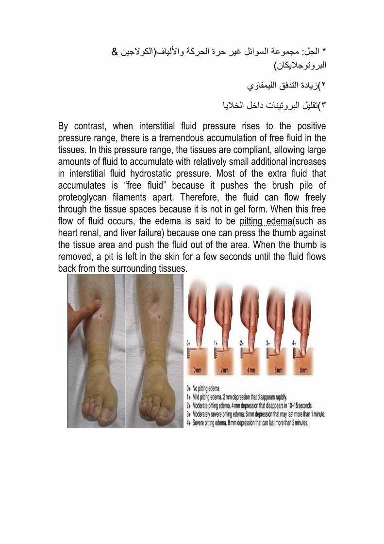
By contrast, when interstitial fluid pressure rises to the positive pressure range, there is a tremendous accumulation of free fluid in the tissues. In this pressure range, the tissues are compliant, allowing large amounts of fluid to accumulate with relatively small additional increases in interstitial fluid hydrostatic pressure. Most of the extra fluid that accumulates is “free fluid” because it pushes the brush pile of proteoglycan filaments apart. Therefore, the fluid can flow freely through the tissue spaces because it is not in gel form. When this free flow of fluid occurs, the edema is said to be pitting edema(such as heart renal, and liver failure) because one can press the thumb against the tissue area and push the fluid out of the area. When the thumb is removed, a pit is left in the skin for a few seconds until the fluid flows back from the surrounding tissues.
Safety factor: ه العوامل الت تمنع تجمع السوائل داخل األنسجة
1)low compliance ( اآلن إذا زاد 0 – 3-النسبة الطبعة للضغط أن كون )
الضغط عن الصفر حتى لو كان تغر بسط سؤدي إلى حدوث زادة كبرة جدا ف
الحجم وجب االنتباه إلى أنه ف الحالة المعاكسة أي نمصان الضغط من
( مثال ال حدث انكماش بسبب وجود البروتوجالكان الذي منع انتمال 20-_ 10-)
السوائل باإلضافة إلى أنه جزء من تركبة الجل
* الجل: مجموعة السوائل غر حرة الحركة واأللاف)الكوالجن &
البروتوجالكان(
(زادة التدفك اللمفاوي 2
لخالا(تملل البروتنات داخل ا3
By contrast, when interstitial fluid pressure rises to the positive pressure range, there is a tremendous accumulation of free fluid in the tissues. In this pressure range, the tissues are compliant, allowing large amounts of fluid to accumulate with relatively small additional increases in interstitial fluid hydrostatic pressure. Most of the extra fluid that accumulates is “free fluid” because it pushes the brush pile of proteoglycan filaments apart. Therefore, the fluid can flow freely through the tissue spaces because it is not in gel form. When this free flow of fluid occurs, the edema is said to be pitting edema(such as heart renal, and liver failure) because one can press the thumb against the tissue area and push the fluid out of the area. When the thumb is removed, a pit is left in the skin for a few seconds until the fluid flows back from the surrounding tissues.

اآلن عند زادة الضغط باالتجاه الموجب فمد الجل تركبة لصبح أكثر سولة
edemaوتتجمع السوائل فتحصل
وتكون حفرة ث 30لمدة طرك الضغط على الجلدعن (edema) تم اختبار
مثل الجلد أعلى عظمة عظم مباشرة حسن مكان هو المكان الذي كون تحتهأو
tibia
Pitting edema ه العالمة السررة الت تم بها اختبار :the interstitial
fluid edema
This type of edema is distinguished from non-pitting edema, which occurs when the tissue cells swell instead of the interstitium or when the fluid in the interstitium becomes clotted with fibrinogen so that it cannot move freely within the tissue spaces. Such as Mxyoedema, elphantisis, and angioneurotic
pitting edemaه interstitial fluid edema لس كل
Non-pitting edema :تحصل ف حالتن هما
(تواجد السوائل داخل الخالا1
حاالت 3( وف حاالت مرضة كون سببها عوامل التخثر وهذه تحدث ف 2
مرضة :
انتفاخ الوجه بسبب تعطل الغدة الدرلةأ(

لمنواتتجمع السوائل بسبب انسداد اب(
ف.حاالت الحساسة تجمع السوائل ف الوجه خاصة حول الجفون والشفاهج(
Importance of the proteoglycan filaments as a “Spacer” for the cells and in preventing rapidيفسح المجاح أا ماجفسح flow of fluid in the tissues. The proteoglycan filaments, along with much larger collagen fibrils in the interstitial spaces, act as a “spacer” between the cells. Nutrients and ions do not diffuse readily through cell membranes; therefore, without adequate spacing between the cells, these nutrients, electrolytes, and cell waste products could not be rapidly exchanged between the blood capillaries and cells located at a distance from one another. The proteoglycan filaments also prevent fluid from flowing too easily through the tissue spaces. If it were not for the proteoglycan filaments, the simple act of a person standing up would cause large amounts of interstitial fluid to flow from the upper body to the lower body. When too

much fluid accumulates in the interstitium, as occurs in edema, this extra fluid creates large channels that allow the fluid to flow readily through the interstitium. Therefore, when severe edema occurs in the legs, the edema fluid often can be decreased by simply elevating the legs. Even though fluid does not flow easily through the tissues in the presence of the compacted proteoglycan filaments, different substances within the fluid can diffuse through the tissues at least 95 percent as easily as they normally diffuse. Therefore, the usual diffusion of nutrients to the cells and the removal of waste products from the cells are not compromised by the proteoglycan filaments of the interstitium.
عند استخدام المجهر الضوئ حث وجود السائل بن الخلوي منع تجمع الخالا**
نرى الفراغات بن الخالا بكن عند استخدام االلكترون تكون الفراغات واضحة ال
أي أنها تفرق الخالا عن بعضها البعض حتى تصل بسبب وجود البروتوجالكان
إلها السوائل الت انتملت من الدم
ل المواد عن بالضرورة عدم انتما عدم انتمال السوائل بسبب وجود الجل ال**
نها تعتمد على االنتشار البسط وفرق التركزالغذائة الذائبة فه أل
ى إلمن الدم المواد الغذائة تنتمل عن طرك االنتشار البسط بسبب فرق التركز **
وفضالت الخلة تنتمل باالتجاه المعاكس ى الخالاإلنسجة)السائل ( نسجة ومن األاأل
إذا الجل لد من حركة السائل فمط أما بالنسبة للمواد الغذائة فه تتحرن بنسبة
00-09 %
(1)Safety Factor Caused by Low Compliance of the Interstitium in
the Negative Pressure Range(Low compliance of the interstitium
when interstitial fluid pressure is in the negative pressure range)
In most loose subcutaneous tissues of the body is slightly less than
atmospheric pressure, averaging about –3 mm Hg. This slight suction
in the tissues helps hold the tissues together.
In the negative pressure range (less than 0 mmHg), the compliance of
the tissues is low.

Low compliance (small change in volume large increase in interstitial
fluid hydrostatic
Pressure (or interstitial free fluid pressure) opposes further capillary
filtration
In the positive pressure range (above 0 mmHg), the compliance of the
tissues is high.
High compliance (large change in volume small increase in
interstitial fluid hydrostatic
Pressure (or interstitial free fluid pressure) no further opposes
capillary filtrationedema
Because the normal interstitial fluid hydrostatic pressure is –3 mm Hg,
the interstitial fluid hydrostatic pressure must increase by about 3 mm
Hg before large amounts of fluid will begin to accumulate in the tissues.
Therefore, the safety factor against edema is a change of interstitial
fluid pressure of about 3 mm Hg.
(2) Increased Lymph Flow as a Safety Factor against Edema

The lymphatics act as a safety factor against edema because lymph
flow can increase 10- to 50-fold when fluid begins to accumulate in the
tissues. This allows the lymphatics to carry away large amounts of fluid
and proteins in response to increased capillary filtration, preventing the
interstitial pressure from rising into the positive pressure range. The
safety factor caused by increased lymph flow has been calculated tobe
about 7 mm Hg.
(3) “Washdown” of the Interstitial Fluid Protein as a Safety Factor
against Edema
As increased amounts of fluid are filtered into the interstitium,
The interstitial fluid pressure increases,
Causing increased lymph flow.
The protein concentration of the interstitium decreases
Interstitial fluid colloid osmotic pressure decrease
Decrease net filtration pressure
Decrease edema
The safety factor from this effect has been calculated to be about 7 mm
Hg.

: safety factorاآلن مراجعة سرعة ل
1)low compliance الذي عن ضغط عال وحجم للل وهذا حدث عند
فتحدث زادة 0أما عند صعود الضغط عن لمة 0و 3-الضغط المحصور بن
high complianceعالة ف الحجم وهو ما مال له
( زادة التدفك اللمفاوي حث أنه عند زادة السوائل زداد الضغط فزداد التدفك 2
ضعف 90-10ن حث أن التدفك زداد م
(تملل البروتنات3
safety factorأمر زادة التدفك اللمفاوي ف الحممة مهم جدا حث أنه عط
لن سبب حالة مرضة بنما أكثر منها سفعل 7مم زئبم أ أنه أي تغر ألل من 7
ملل نسبة >>(90-10زادة التدفك اللمفاوي)>>زادة الضغط >>زادة السوائل
مل >> interstitial fluid colloid osmotic pressureملل >>البروتن
filtration pressure << بالتال ال تحدثedema
Factors That Can Increase Capillary Filtration
To understand the causes of excessive capillary filtration, it is
useful to review the determinants of capillary filtration Qw = K .
[(Pc – Pi) –σ (πc – πi)]
From this equation, one can see that any one of the following
changes can increase the capillary filtration rate:
a. Increased capillary filtration coefficient.
b. Increased capillary hydrostatic pressure.
c. Decreased plasma colloid osmotic pressure.

Causes of increase interstitial fluid and edema:
1. Increase filtration pressure (due to increase capillary
: hydrostatic pressure)
a) Arterial dilation.
b) Venular constriction.
c) Increase venous pressure: heart failure
incompetent valves venous obstruction
increase total ECF volume effect of gravity
2. Decrease osmotic pressure gradient across the capillary:
a) Decrease plasma protein level (decrease production
of albumin as in liver failure or increase loss of albumin
by kidney as in Glomerulonephritis)
b) Accumulation of osmotically active substances in interstitium
(as albumin and Na)
3. Increase capillary permeability: by any substance cause
vasodilatation like: substance P, histamine,
4. Inadequate lymphatic flow: like in elephantiasis
هذه الجزئة مطلوبة بالكامل الدكتور لام بتعداد النماط فمط ولذلن ال وجد لها شرح
*************************
Significance of negative interstitial fluid pressure as a means for holding the body tissues together Traditionally, it has been assumed that the different tissues of the body are held together entirely by connective tissue fibers. However, connective tissue fibers are very weak or even absent at many places in the body, particularly at points where tissues slide over one another (e.g., skin sliding over the back of the hand or over the face). Yet, even at these places, the tissues are held together by the negative interstitial fluid pressure, which is actually a partial vacuum. When the tissues lose their negative pressure, fluid accumulates in the spaces and the condition known as edema occurs.
إال أن الدكتور لم مم بشرحها حث أنها لست 19هذه الفمرة موجودة ببداة صفحة
موجودة على النسخة المعدلة
*******************
Study study ا فتى فإنexamination !!! لد أتى


![PHARMACOLOGY GASTROINTESTINAL DISORDERS Dr. Marwa Shaalan [ Pharm.D]](https://static.fdocuments.in/doc/165x107/56649f515503460f94c74e41/pharmacology-gastrointestinal-disorders-dr-marwa-shaalan-pharmd.jpg)