Wound Healing. Skin Haemostasis Meet the cells Inflammation Migration Proliferation Maturation.
42
Wound Healing
-
Upload
julius-walton -
Category
Documents
-
view
222 -
download
1
Transcript of Wound Healing. Skin Haemostasis Meet the cells Inflammation Migration Proliferation Maturation.

Wound Healing
Wound Healing• Skin• Haemostasis• Meet the cells• Inflammation• Migration• Proliferation• Maturation
Integumentary system
• What is the integumentary system? Outer covering• What organ(s) are involved?Skin • What structures are involved? Hair, nails, receptors, glands
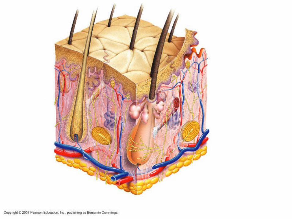
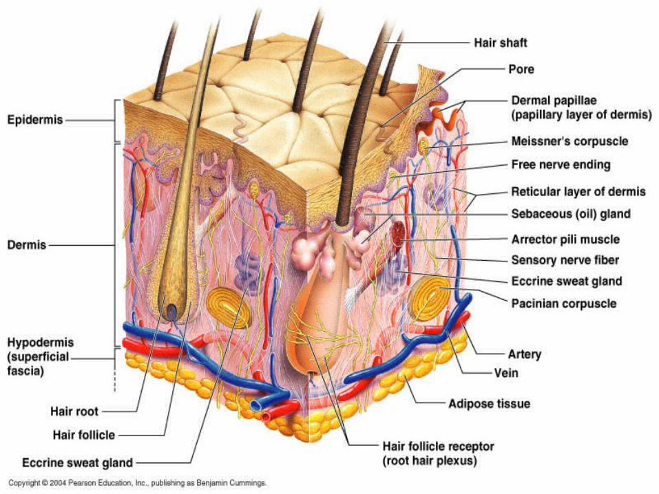
Integumentary system• Skin structure:3 layers; epidermis, dermis,
hypodermis or subcutaneous.
• Epidermis: Outer layers (4-5) of closely packed cells.• Stratum basale, Stratum spinosum, Stratum
granulosum, Stratum lucidum, Stratum corneum• Contains keratinocytes & melanocytes, Merkel cells
& Langerhans (dendritic) cells.
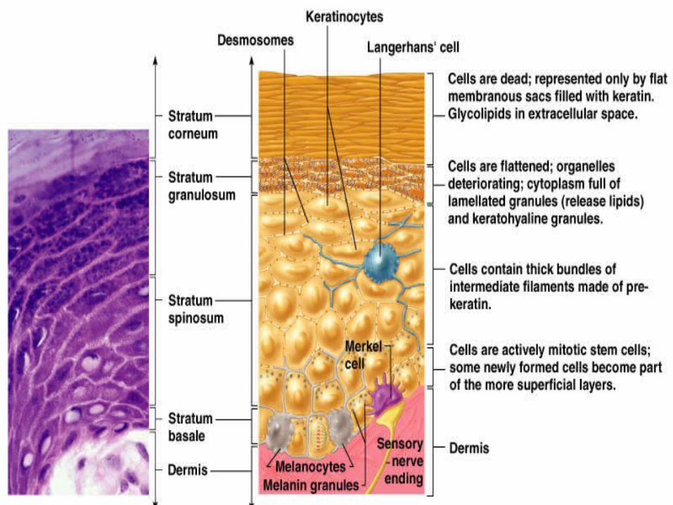

Epidermis
• Stratum basale is base layer where cells (keratinocytes) multiply. New cells push old cells away from blood supply. They fill with keratin, flatten & die in the corneum. Millions are shed daily.
• Melanocytes are in the stratum basale & produce melanin pigment
• Merkel cells at the epidermis-dermis junction function in touch
• Langerhans (dendritic) cells are phagocytes (immune system cells).
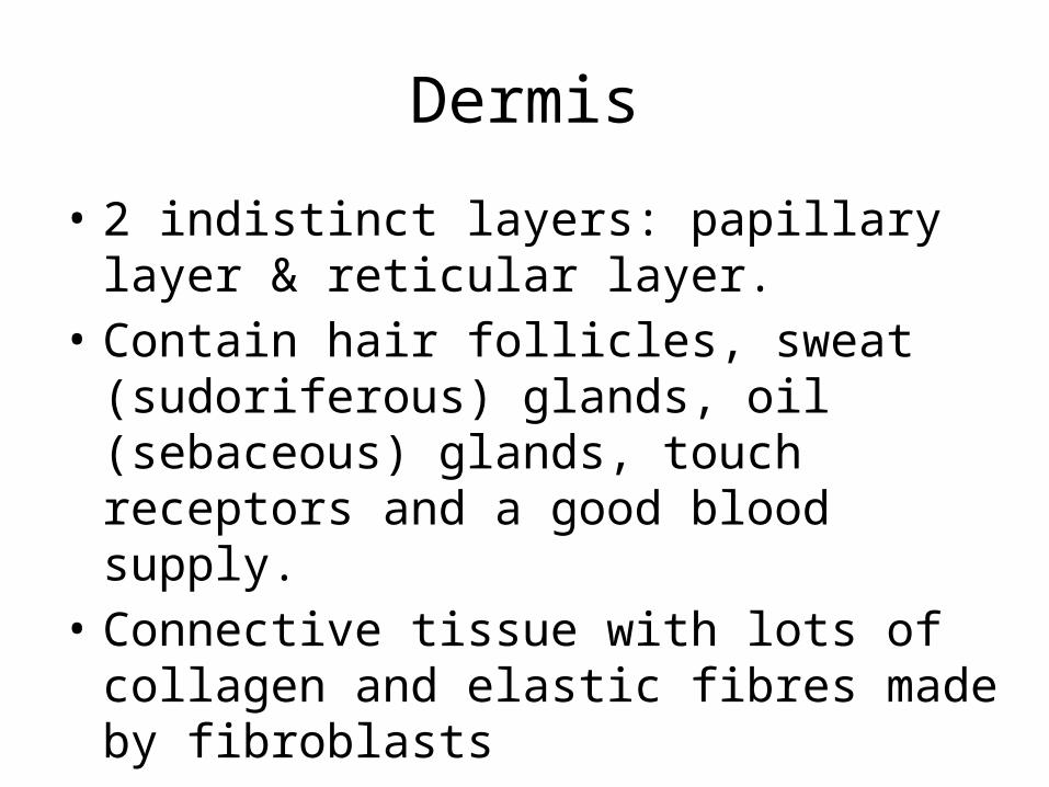
Dermis
• 2 indistinct layers: papillary layer & reticular layer.
• Contain hair follicles, sweat (sudoriferous) glands, oil (sebaceous) glands, touch receptors and a good blood supply.
• Connective tissue with lots of collagen and elastic fibres made by fibroblasts
Hypodermis
• Subcutaneous or superficial fascia anchors skin to underlying muscle.
• contains fat

Integumentary key functions
• Provides external protection• Regulates body temperature• Sensory organ• Excretion• Vitamin D synthesis• Immunity
Functions 1. • Regulation of body temperature primarily by
sweating and changing superficial vein diameter (& blood flow)
• Heat conservation occurs when vasoconstriction of superficial veins re-routes warm blood deeper into the body.
Regulation of body temperature
• Cooling occurs when • (a) Vasodilation of superficial veins brings
blood to the body surface and heat is lost to the environment by radiation convection & conduction.
• (b) An increase in perspiration results in increased evaporation cooling the skin but effectiveness is dependant on the humidity.
Functions
• 2. Protection from mechanical damage (keratin), microbial invasion, dehydration & UV radiation (melanin)
• 3. Excretion for loss of some ions, water & nitrogen containing waste
Functions 4.
• Sensations from sensory receptors• Free nerve endings: temperature, touch, pressure &
pain • Hair receptors : hair movement• Merkels disc: Light pressure• Meissner’s corpuscle: Light pressure • Ruffini’s endings: Deep pressure, stretch • Pacinian corpuscle: Deep pressure, stretch
Functions• 5. Langerhans cells of the epidermis are immune
system cells• 6. Blood reservoir carrying 8-10% of total blood
flow and is highly variable.• 7. Synthesis of Vitamin D when UV rays from sun
promote production of a Vitamin D precursor by the epidermis
• 8. Stores fat & fat soluble vitamins (A, D, E & K)• 9. Cutaneous absorption and secretion• 10. Social interaction


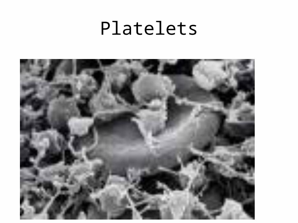
Platelets
Clot Retraction
• Contractile proteins within the platelets pull on the fibrin
• This pulls the edges of the broken blood vessel together
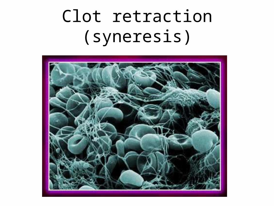
Clot retraction (syneresis)
Clot Destruction (Fibrinolysis)
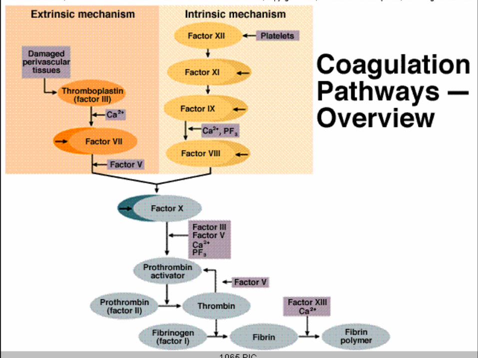
• Plasminogen is converted to Plasmin
• Plasmin breaks down the fibrin in the clot
Meet the cells
• Labile cells– Regenerate readily, involved in wound repair
• Stable cells– Don’t normally divide in adult, but may
• Permanent cells– Unable to divide, replaced by scar tissue
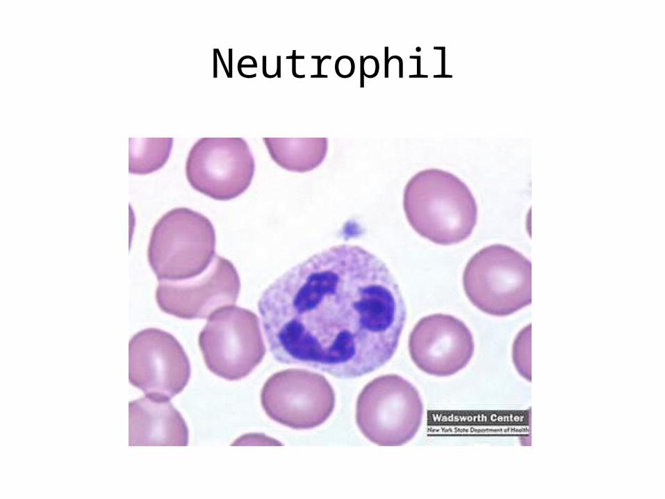
Neutrophil
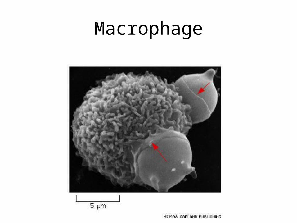
Macrophage

Epithelial cells
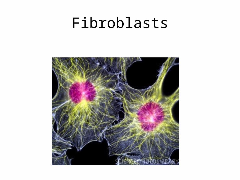
Fibroblasts

Repair
• Replacement of destroyed tissue by scar tissue• Doesn’t perform original function• Process:– fill in the wound– cover or seal the wound– shrink the wound
• 4 stages:
1) Inflammatory phase
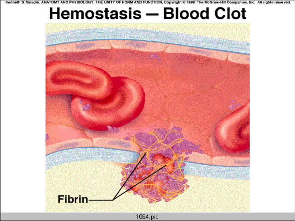
• Haemostasis• Inflammation• Vasodilation & permeability of blood vessels• Phagocytotic cells (neutrophils &
macrophages) eat up cell debris & bacteria• Immediate to 2 - 5 days
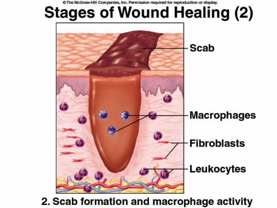

2) Migratory phase
• Clot loses fluid and hardens forming a scab• Capillaries grow into damaged area
2) Migratory phase
• Fibroblasts make collagen to fill in the wound
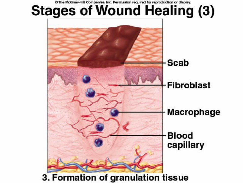
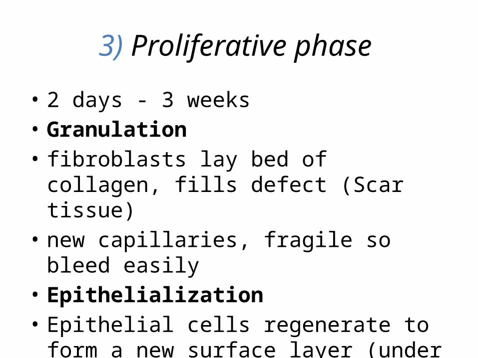
3) Proliferative phase
• 2 days - 3 weeks• Granulation• fibroblasts lay bed of collagen, fills defect (Scar
tissue)• new capillaries, fragile so bleed easily• Epithelialization• Epithelial cells regenerate to form a new
surface layer (under the scab)

Mitosis
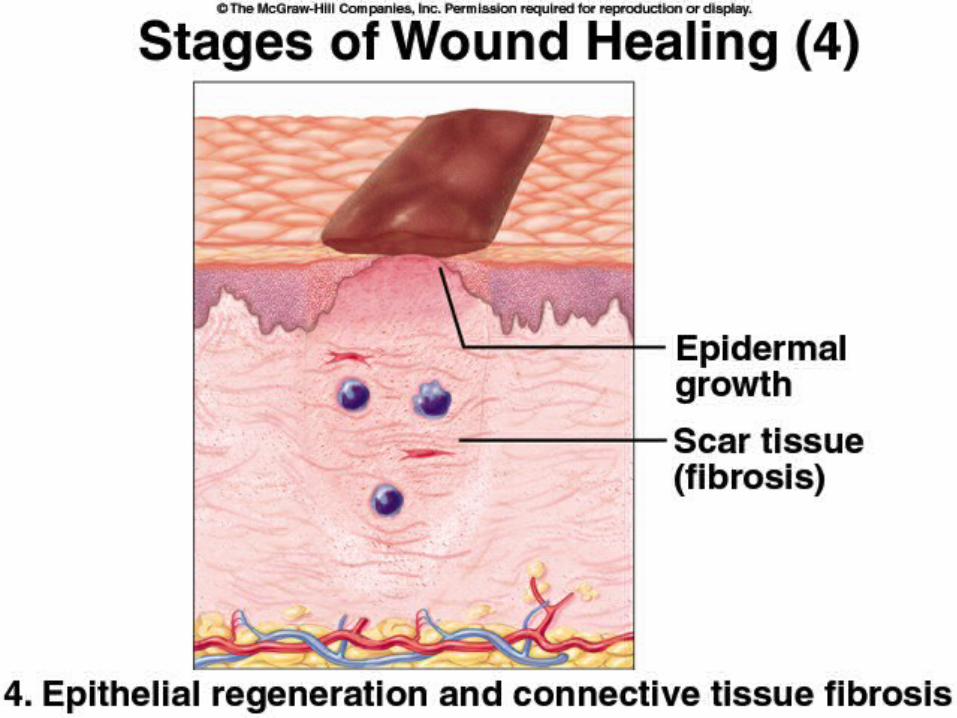
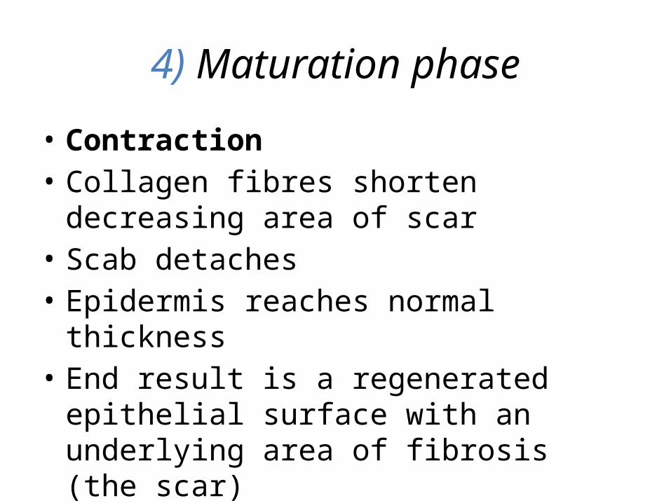
4) Maturation phase
• Contraction• Collagen fibres shorten decreasing area of scar • Scab detaches• Epidermis reaches normal thickness• End result is a regenerated epithelial surface
with an underlying area of fibrosis (the scar)
Primary Intention
• minimal tissue loss• e.g. clean, sutured incision

Secondary Intention
• much more tissue replacement• takes longer• e.g. stage IV decubitus ulcer
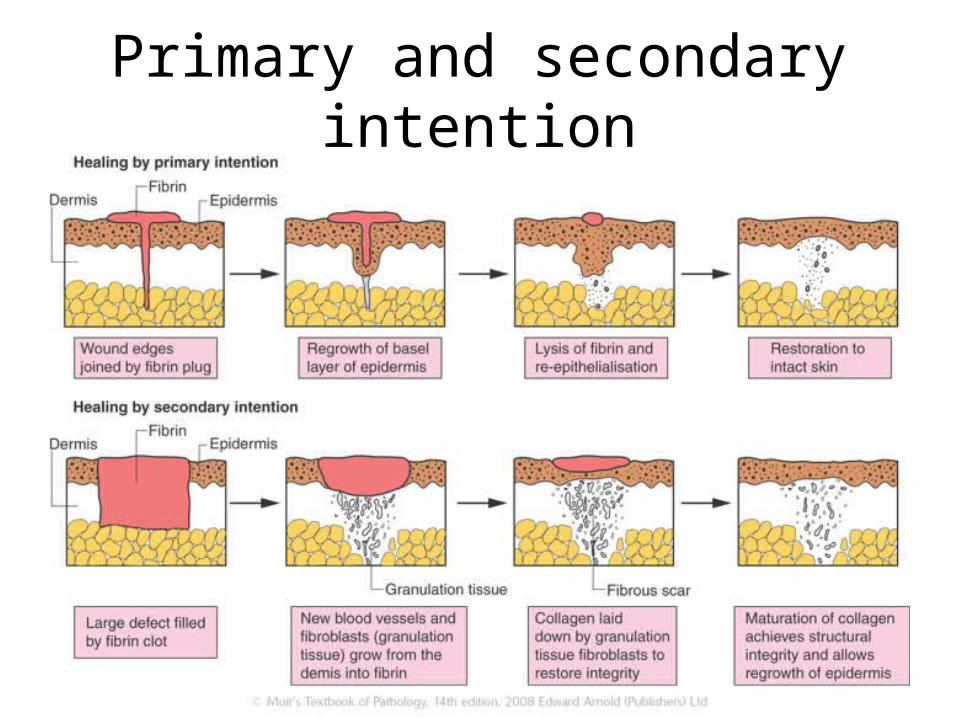
Primary and secondary intention