WOUND EXAMINATION. PATIENT HISTORY WOUND HISTORY DURATION ATTRIBUTING EVENT SYMPTOMS PAIN...
49
WOUND EXAMINATION
-
Upload
katrina-ball -
Category
Documents
-
view
224 -
download
2
Transcript of WOUND EXAMINATION. PATIENT HISTORY WOUND HISTORY DURATION ATTRIBUTING EVENT SYMPTOMS PAIN...

WOUND EXAMINATION

PATIENT HISTORY
WOUND HISTORY DURATION ATTRIBUTING EVENT SYMPTOMS PAIN PARESTHESIA/ANESTHESIA

HISTORY (cont.)
DOES PAIN CHANGE WITH POSITION elevation decreases pain = venous dependency increases pain in venous
lesions pain with rest - severe occlusive
disease intermittent pain with ambulation =
claudication
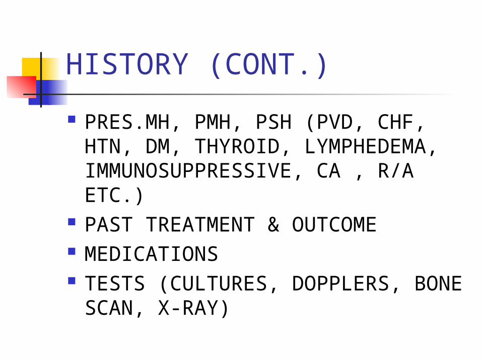
HISTORY (CONT.)
PRES.MH, PMH, PSH (PVD, CHF, HTN, DM, THYROID, LYMPHEDEMA, IMMUNOSUPPRESSIVE, CA , R/A ETC.)
PAST TREATMENT & OUTCOME MEDICATIONS TESTS (CULTURES, DOPPLERS,
BONE SCAN, X-RAY)

HISTORY (CONT.)
ADDITIONAL STUDIES (ARTERIOGRAM, VENOGRAM, ABI)
SOCIAL HX VOCATIONAL HISTORY HOBBIES

OBJECTIVE EVALUATION
Test & Measures

LOCATION
HYPERTENSIVE-posterio/lateral leg, onset with infarction, very severe pain hypertension
VENOUS-distal leg, medial aspect, red base, wet, periwound skin staining, no pain, mild insufficiency

Chronic Venous Insufficiency:“champagne bottle”“piano leg” appearance
Atrophie blanche

LOCATION (CONT.)
ARTERIAL-DISTAL LOWER EXREMITY, LATERAL ASPECT, TOES & FEET, PALE BASE, ATROPHIC SKIN, DRY WOUND, SEVERE PAIN, ARTERIOSCLEROSIS
NEUROTROPHIC-PLANTAR SURFACE OF FOOT, SMALL OR DEEP, PERIWOUND CALLOUS, INFECTION, NO PAIN POSSIBLE DM

SIZE
LENGTH, WIDTH, AREA, DEPTH, VOLUME - IF REMOVE ESCHAR WOUND WILL APPEAR BIGGER
MEASURE FROM WOUND EDGE USE CONSISTENT TOOL & UNITS
OF MEASUREMENT PHOTOGRAPHY, TRACING,
VOLUME, SYRINGE

UNDERMINING
ALSO KNOWN AS RIMMING OR TUNNELING
TISSUE DESTRUCTION UNDERLYING INTACT SKIN ALONG THE WOUND MARGINS (HYPOGRANULATION)
MEASURE USING THE O’CLOCK SYSTEM, HEMISPHERES

GIRTH
EDEMA, ATROPHY MEASURE WITH REFERENCE TO BONY
LANDMARKS USING TAPE MEASURE VOLUMETRIC DISPLACEMENT
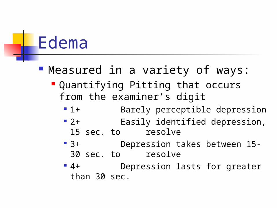
Edema Measured in a variety of ways:
Quantifying Pitting that occurs from the examiner’s digit
1+ Barely perceptible depression 2+ Easily identified depression, 15
sec. to resolve 3+ Depression takes between 15-
30 sec. to resolve 4+ Depression lasts for greater
than 30 sec.

SHAPE
TRIANGULAR- SKIN TEAR ROUND- ARTERIAL IRREGULAR-VENOUS

SLOPES
ANGLES OF MARGINATION DEPICT GRANULATION VERY IMPORTANT MEASUREMENT

Staging of Wounds
Stage I-IV Pressure Wounds Wounds other than Pressure
Superficial Partial Thickness -epidermal layer,
superficial layer of dermis Full-Thickness- epidermis, dermis,
subcutaneous , may also involve muscle and bone
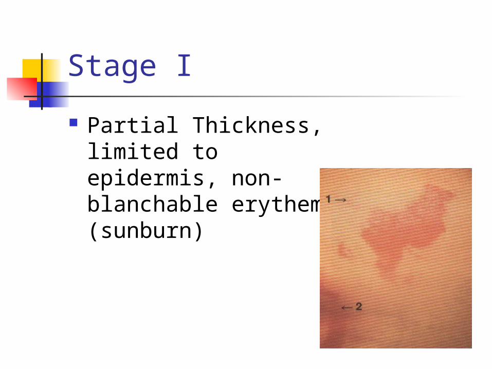
Stage I
Partial Thickness, limited to epidermis, non-blanchable erythema (sunburn)

Stage II
Partial Thickness Skin Loss,
involves both epidermis and dermis
(abrasion, blister, shallow crater)

Stage III
Full Thickness Skin Loss
Damage or Necrosis of Subcutaneous Tissue
May Extend to Fascia
(deep crater, with or without undermining)

Stage IV
Full-Thickness Skin Loss
Extensive Destruction
Necrosis Damage to
Muscle, Tendon, Joint Capsule, Bone

Wagner Ulcer Classification Diabetic Ulcers
Grade 0 Intact Skin 1 Superficial Ulcer 2 Deep Ulcer 3 Deep Infected Ulcer 4 Partial Foot Gangrene 5 Full Foot Gangrene


Tissue Composition
RED WOUNDS- clean healthy granulating wounds
YELLOW WOUNDS-may contain fibrous tissue, hydrated necrotic tissue, or dead tissue, referred to as slough
BLACK WOUNDS-dried eschar, leathery

Tissue Found in Wounds
Eschar Granulation Adipose Fascia Muscle Tendon Bone

Foreign Debris & Necrotic Tissue Remove as Soon as Possible
This will prevent bacterial colonization and infection

Peri-Wound
Trophic Changes (dry skin, brittle nails, hair loss) indicates poor arterial nutrition

Peri-Wound
Change in skin color cyanotic = Arterial Compromise Pigmentation (hemosiderin staining),
pigment is deposited from RBC = Venous
Ring of Redness or Halo of erythema around the wound may indicate infection

Drainage
Inactive found on dressing, at time of observation no
drainage is found in or near the wound

Drainage
Active Free flowing, able to be milked from
the wound

Characteristics of Drainage
Transudate (Serous): clear, watery contains: H20, salts and proteins
Serosanguineous: tinged red/brown watery, thin contains: serum, blood
Exudate: creamy, yellowish moderately thick contains: proteins, WBC

Characteristics of Drainage (cont.) Purulent/Pus: yellowish/brownish
Thick contains: WBC, necrotic debris
Infected Pus yellow, green/blue thick contains: pathogens
describe amount:none, min, mod, max

Odor
Pseudomonas-sweet smell (fruity) Garbage- rotten= infection Proteus- ammonia
describe; absent, mild, moderate, foul smelling

Temperature
systemic v. localized measured
touch thermistor thermography radiometer
measure infrared radiation from the body

Indications for culture
Clinical Signs of Local Infection by Linholm edema, erythema, purulent or foul
smelling drainage, increased pain, induration, heat around the wound; IFEE
Signs of systemic infection fever, abnormal CBC
Bone Involvement (osteomyelitis)
Non-Healing Wounds (silent infection)

Aerobic swab culture technique. The culturetteIs rotated while moving in a 10-point pattern.Gentle pressure to express fluid is required.
From: Myers, B.A. Wound Management: Principles and Practice. Prentice Hall, Upper Saddle River, NJ. 2004: p. 94

Vasculature Examinations
Pulses(2+Normal, 1+Diminished, 0 Absent)
Auscultation (swishing sound, only heard in abnormal artery)
Venous Exam (venous doppler)
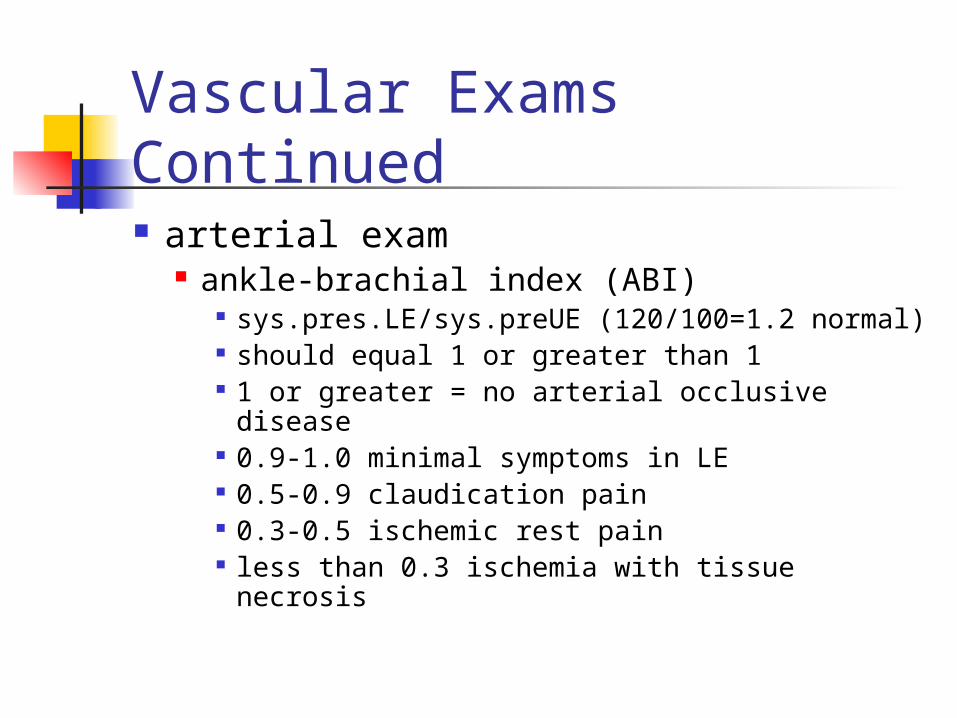
Vascular Exams Continued
arterial exam ankle-brachial index (ABI)
sys.pres.LE/sys.preUE (120/100=1.2 normal) should equal 1 or greater than 1 1 or greater = no arterial occlusive disease 0.9-1.0 minimal symptoms in LE 0.5-0.9 claudication pain 0.3-0.5 ischemic rest pain less than 0.3 ischemia with tissue necrosis

Normal ABI
Heart LevelSBP100 mmHg
ABI120 mmHg/100mmHg= 1.2
AnkleSBP120mmHg
DopplerDoppler

ABI When ABI value is <.9
95% sensitive 99% specific For angiographically significant PVD

ABI Change of 0.15 correlates with
disease and symptomology

Measuring ABI
Tissue Oxygen Tension
tc-Po2 transcutaneous oximmetry
Rubor of Dependency Test
assess arterial flow by evaluating skin color changes during elevation and dependency
leg elevation at 60 degrees for 1 min. normally no significant change in color lower the leg, record time for color return arterial insufficiency may take longer
than 30 sec. color will be bright red (hyperemic)

VENOUS FILLING TIME
assess arterial flow by evaluating time veins take to fill after emptying
elevate LE for 1 min. to 60 degrees lower the leg, record time that veins
on the dorsum of the foot take to refill with arterial insufficiency may take 30
sec. or longer
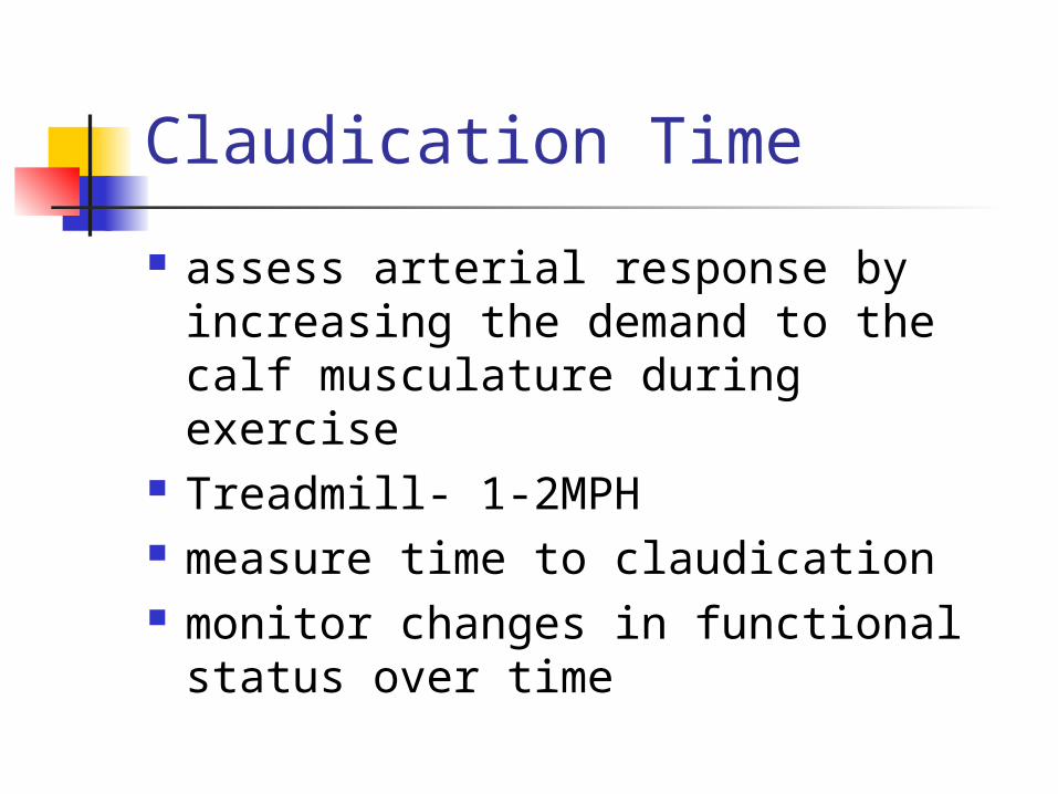
Claudication Time
assess arterial response by increasing the demand to the calf musculature during exercise
Treadmill- 1-2MPH measure time to claudication monitor changes in functional
status over time

Test for DVT
Homan’s Sign squeeze calf while dorsiflexing the ankle,
with the knee held in an extended position
tenderness with increased firmness may suggest DVT
confirm using blood pressure cuff pt. unable to tolerate 40mmHg if DVT present normally able to tolerate much higher
pressures

Test for Cutaneous Sensitivity perception of light touch
use cotton ball perception to temperature
warm, cool 2-point discrimination
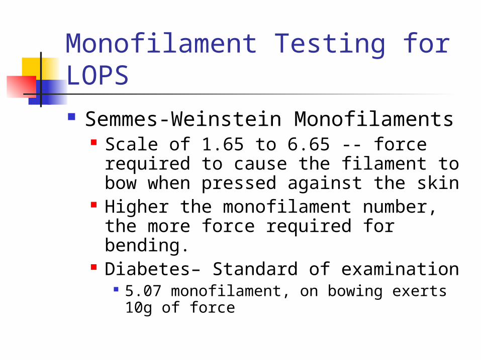
Monofilament Testing for LOPS Semmes-Weinstein Monofilaments
Scale of 1.65 to 6.65 -- force required to cause the filament to bow when pressed against the skin
Higher the monofilament number, the more force required for bending.
Diabetes– Standard of examination 5.07 monofilament, on bowing exerts 10g
of force