Wound Care and Hyperbaric Medicine Miguel G. Madariaga, MD.
54
Wound Care and Wound Care and Hyperbaric Hyperbaric Medicine Medicine Miguel G. Madariaga, MD Miguel G. Madariaga, MD
-
Upload
brook-norris -
Category
Documents
-
view
217 -
download
0
Transcript of Wound Care and Hyperbaric Medicine Miguel G. Madariaga, MD.

Wound Care and Wound Care and Hyperbaric MedicineHyperbaric Medicine
Miguel G. Madariaga, MDMiguel G. Madariaga, MD

What is the skin good for?What is the skin good for?
The epidermisThe epidermis
The epidermis is the tough, The epidermis is the tough, leathery outer surface of the skinleathery outer surface of the skinIt has five layers of cells and It has five layers of cells and appendages appendages It helps us by:It helps us by: Providing a barrierProviding a barrier Regulating fluidsRegulating fluids Providing light touch sensationProviding light touch sensation Controlling temperatureControlling temperature Excretes toxinsExcretes toxins Produces vitamin DProduces vitamin D Cosmetic apperanceCosmetic apperance
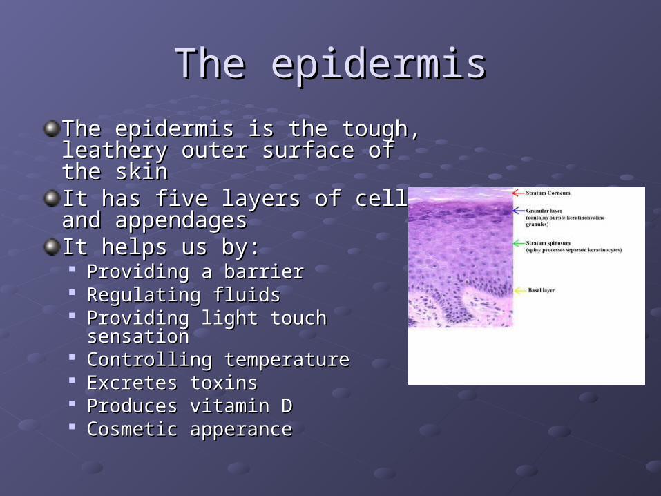
The dermisThe dermis
It is a 2-4 mm thick layer It is a 2-4 mm thick layer below the epidermisbelow the epidermis
It helps us by:It helps us by: Supporting and nourishing the Supporting and nourishing the
epidermisepidermis Housing skin appendages Housing skin appendages
(nails, hair, glands)(nails, hair, glands) Fighting against infectionFighting against infection Controlling temperatureControlling temperature Providing sensationProviding sensation

What is a wound?What is a wound?

How does a wound heal?How does a wound heal?

What is a chronic wound?What is a chronic wound?
Chronic wounds: the burden of Chronic wounds: the burden of diseasedisease
Chronic wounds represent a silent epidemic (6.5 Chronic wounds represent a silent epidemic (6.5 million in the US)million in the US)
The amount of money spent on wound care, the The amount of money spent on wound care, the loss of productivity for afflicted individuals and loss of productivity for afflicted individuals and the families and their diminished quality of life the families and their diminished quality of life come at great cost to our societycome at great cost to our society
It is claimed that an excess of US$25 billion is It is claimed that an excess of US$25 billion is spent annually on treatment of chronic woundsspent annually on treatment of chronic wounds
Sen, Wound Repair Regen, 2009

Causes of chronic woundsCauses of chronic wounds
DiabetesDiabetes
Arterial insufficiencyArterial insufficiency
Venous diseaseVenous disease
PressurePressure
RadiationRadiation
OthersOthers

Diabetic ulcersDiabetic ulcers
Diabetes is the sixth leading cause of death in Diabetes is the sixth leading cause of death in the United Statesthe United States
7% of the American population have diabetes 7% of the American population have diabetes and several millions (6) are undiagnosedand several millions (6) are undiagnosed
Over 85% of all diabetic related lower extremity Over 85% of all diabetic related lower extremity amputations are preceded by an ulcerationamputations are preceded by an ulceration
Risk factors for diabetic ulcers include: Risk factors for diabetic ulcers include: neuropathy, deformity/limited joint mobility, neuropathy, deformity/limited joint mobility, vascular disease and pivotal traumavascular disease and pivotal trauma

Why do diabetic ulcers occur? Why do diabetic ulcers occur?
NeuropathyNeuropathy Motor: weakness, muscle atrophy (deformity)Motor: weakness, muscle atrophy (deformity) Sensory: diminished sensation, painSensory: diminished sensation, pain Autonomic: dry, fissured skinAutonomic: dry, fissured skin
Vascular diseaseVascular disease

Complications of diabetic ulcersComplications of diabetic ulcers

Venous ulcersVenous ulcers
These are the most common cause of ulcers These are the most common cause of ulcers
1-2% of the population has venous insufficiency1-2% of the population has venous insufficiency
14% of people with venous insufficiency develop 14% of people with venous insufficiency develop ulcersulcers
Women are three times more likely than men to Women are three times more likely than men to have venous ulcershave venous ulcers
The risk of ulceration is 7.5 times greater in The risk of ulceration is 7.5 times greater in people older than 65people older than 65

Why do venous ulcers occur?Why do venous ulcers occur?

Venous ulcers: characteristicsVenous ulcers: characteristics
Pressure ulcersPressure ulcers
Pressure ulcers are localized areas of dead tissue that Pressure ulcers are localized areas of dead tissue that develop when soft tissues is compressed between a firm develop when soft tissues is compressed between a firm surface and a bony prominencesurface and a bony prominenceThe overall prevalence among hospitalized patients is The overall prevalence among hospitalized patients is 15%15%Patients at risk: hospitalized patients, individuals in long Patients at risk: hospitalized patients, individuals in long term care and patients with spinal cord injuryterm care and patients with spinal cord injuryPressure ulcer complications may be life-threateningPressure ulcer complications may be life-threateningThe cost of caring for each ulcer can be up to 70,000 per The cost of caring for each ulcer can be up to 70,000 per ulcerulcer

Pressure ulcer stagesPressure ulcer stages
What is done at the Wound What is done at the Wound Center?Center?
Adequate perfusionPresence of nonviable tissueSigns of infection or inflammationPresence of edemaConduciveness of wound healing environmentOptimization of tissue growthAppropriateness of pressure offloadingControllability of pain Optimization of host factors

Confirm adequate perfusionConfirm adequate perfusion

Eliminate non-viable tissueEliminate non-viable tissue

Different types of debridementDifferent types of debridement

Control infection/inflammationControl infection/inflammation

A word about MRSAA word about MRSA
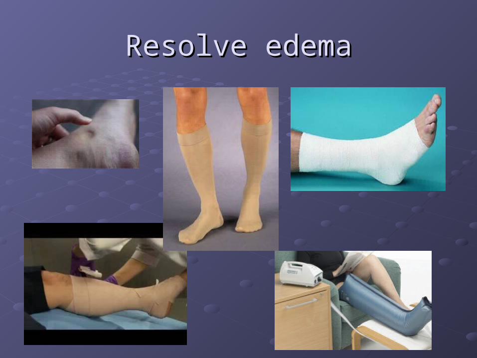
Resolve edemaResolve edema

Optimize wound bedOptimize wound bed

Enhance tissue growthEnhance tissue growth
Graft
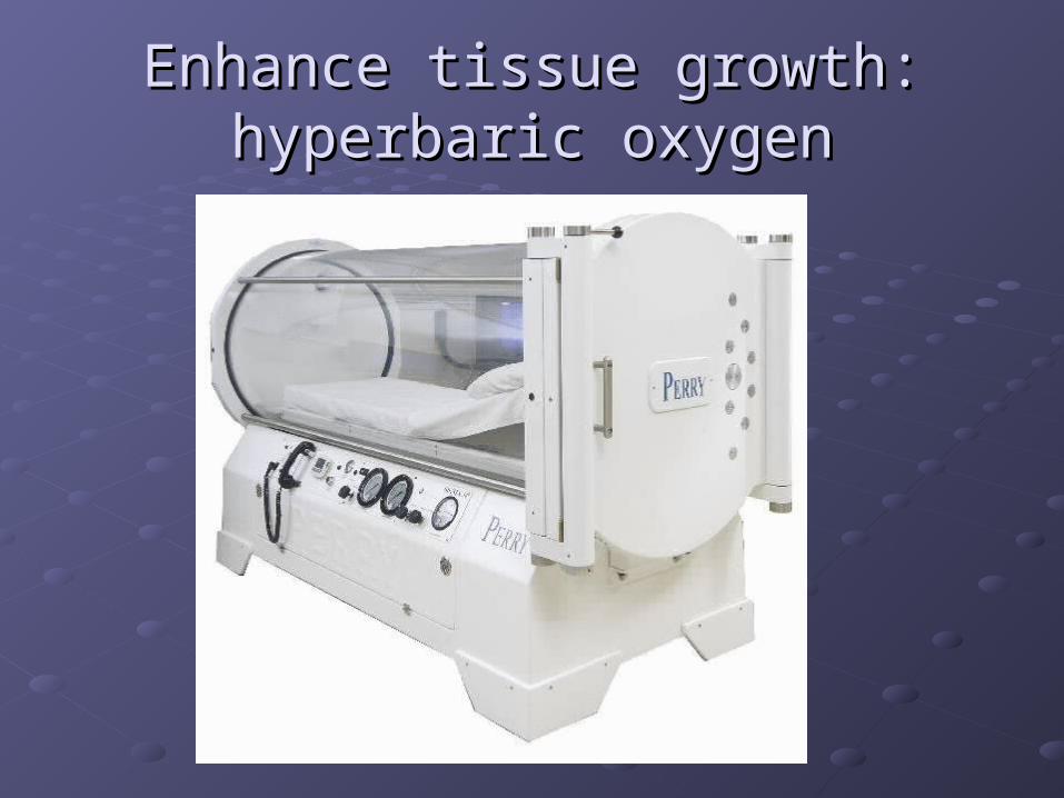
Enhance tissue growth: hyperbaric Enhance tissue growth: hyperbaric oxygenoxygen
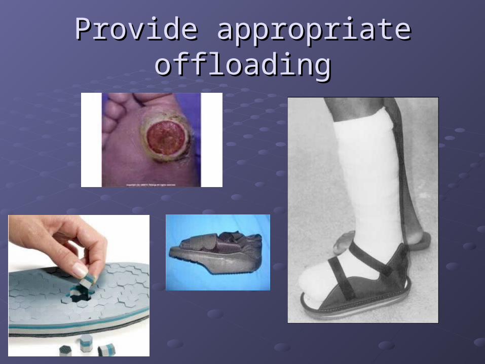
Provide appropriate offloadingProvide appropriate offloading

Control painControl pain

Optimize host factorsOptimize host factors
Diabetes mellitusDiabetes mellitus
Renal dysfunctionRenal dysfunction
Ischemic heart disease\SmokingIschemic heart disease\Smoking
COPDCOPD
MalnutritionMalnutrition
Mobility impairmentMobility impairment
AddictionAddiction
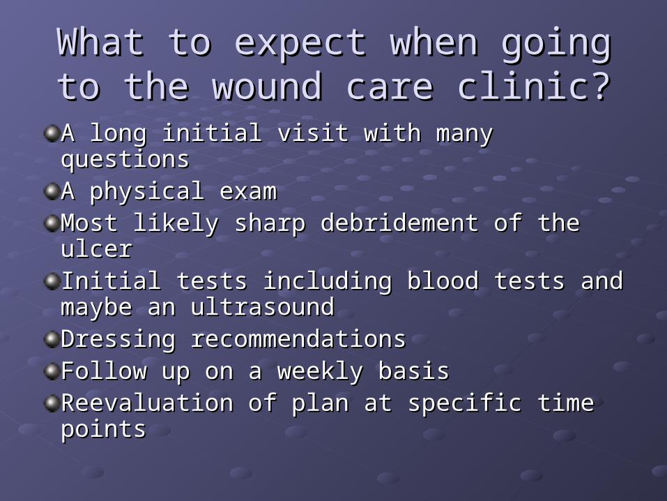
What to expect when going to the What to expect when going to the wound care clinic?wound care clinic?
A long initial visit with many questionsA long initial visit with many questionsA physical examA physical examMost likely sharp debridement of the ulcerMost likely sharp debridement of the ulcerInitial tests including blood tests and Initial tests including blood tests and maybe an ultrasoundmaybe an ultrasoundDressing recommendationsDressing recommendationsFollow up on a weekly basisFollow up on a weekly basisReevaluation of plan at specific time pointsReevaluation of plan at specific time points

Hyperbaric MedicineHyperbaric Medicine
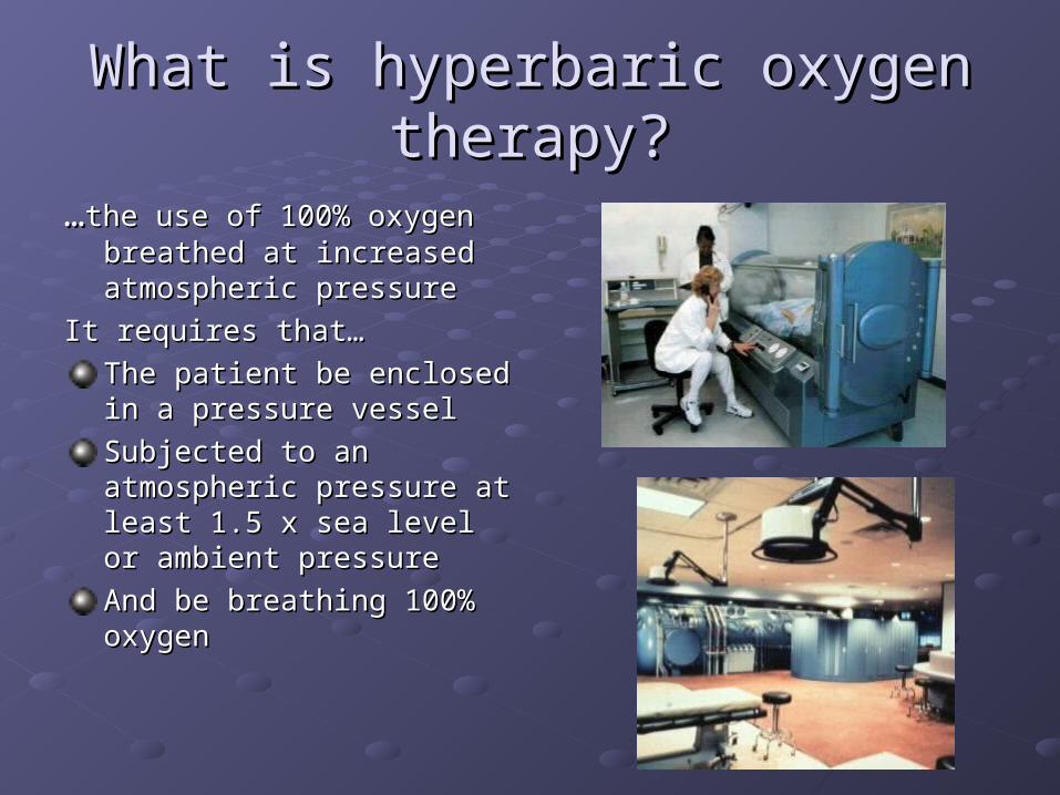
What is hyperbaric oxygen What is hyperbaric oxygen therapy?therapy?
……the use of 100% oxygen the use of 100% oxygen breathed at increased breathed at increased atmospheric pressure atmospheric pressure
It requires that…It requires that…
The patient be enclosed in a The patient be enclosed in a pressure vesselpressure vessel
Subjected to an atmospheric Subjected to an atmospheric pressure at least 1.5 x sea pressure at least 1.5 x sea level or ambient pressurelevel or ambient pressure
And be breathing 100% And be breathing 100% oxygen oxygen

The “bends” The “bends”

HBO: Boyle’s lawHBO: Boyle’s law

HBO and carbon monoxide HBO and carbon monoxide poisoningpoisoning

HBO mechanism of action in HBO mechanism of action in wound carewound care

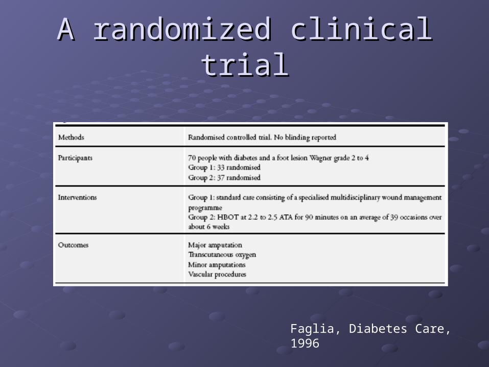
A randomized clinical trialA randomized clinical trial
Faglia, Diabetes Care, 1996
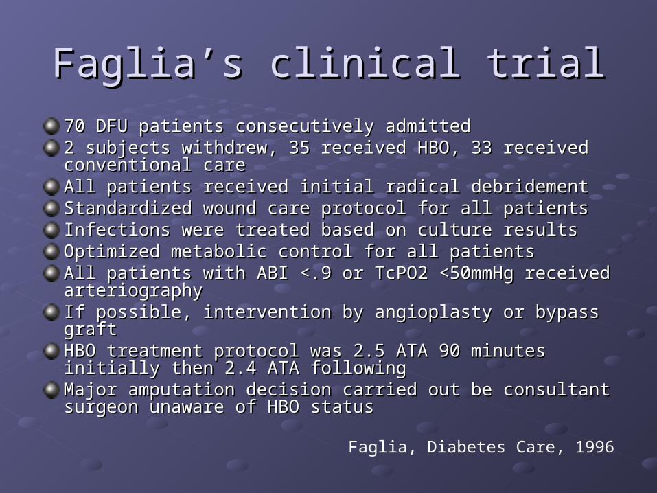
Faglia’s clinical trialFaglia’s clinical trial
70 DFU patients consecutively admitted70 DFU patients consecutively admitted2 subjects withdrew, 35 received HBO, 33 received conventional 2 subjects withdrew, 35 received HBO, 33 received conventional carecareAll patients received initial radical debridementAll patients received initial radical debridementStandardized wound care protocol for all patientsStandardized wound care protocol for all patientsInfections were treated based on culture results Infections were treated based on culture results Optimized metabolic control for all patientsOptimized metabolic control for all patientsAll patients with ABI <.9 or TcPO2 <50mmHg received arteriographyAll patients with ABI <.9 or TcPO2 <50mmHg received arteriographyIf possible, intervention by angioplasty or bypass graftIf possible, intervention by angioplasty or bypass graftHBO treatment protocol was 2.5 ATA 90 minutes initially then 2.4 HBO treatment protocol was 2.5 ATA 90 minutes initially then 2.4 ATA followingATA followingMajor amputation decision carried out be consultant surgeon Major amputation decision carried out be consultant surgeon unaware of HBO statusunaware of HBO status
Faglia, Diabetes Care, 1996
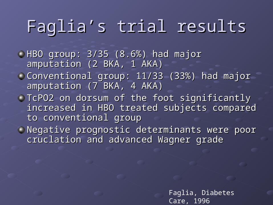
Faglia’s trial resultsFaglia’s trial results
HBO group: 3/35 (8.6%) had major amputation (2 BKA, 1 HBO group: 3/35 (8.6%) had major amputation (2 BKA, 1 AKA)AKA)Conventional group: 11/33 (33%) had major amputation Conventional group: 11/33 (33%) had major amputation (7 BKA, 4 AKA) (7 BKA, 4 AKA) TcPO2 on dorsum of the foot significantly increased in TcPO2 on dorsum of the foot significantly increased in HBO treated subjects compared to conventional groupHBO treated subjects compared to conventional groupNegative prognostic determinants were poor cruclation Negative prognostic determinants were poor cruclation and advanced Wagner gradeand advanced Wagner grade
Faglia, Diabetes Care, 1996

FDA approved usage of HBO based on UHMS FDA approved usage of HBO based on UHMS Report, 2008Report, 2008
• Decompression illness, gas embolism
• Carbon monoxide, cyanide poisoning
• Blood loss anemia
• Clostridial myonecrosis
• Other necrotizing soft tissue infections
• Refractory osteomyelitis
• Prep and preservation of compromised skin grafts and flaps
• Crush injury, compartment syndrome
• Other acute traumatic ischemias
• Central retinal artery and vein occlusion
• Other wounds with demonstrated periwound hypoxia
• Acute thermal burns
• Osteoradionecrosis
• Soft tissue radionecrosis
Bubble compression,toxin displacement,temporary Increase in oxygen deliver
Enhanced host immuneresponse, resolution of infection
Reversal of hypoxia, woundregeneration effects, tissuegrowth
Wound regeneration effects, tissue growth, reversal offibrosis

HBO techniqueHBO technique
Single occupancy chambers are most appropriate for the Single occupancy chambers are most appropriate for the treatment of chronic medical conditions in stable patientstreatment of chronic medical conditions in stable patientsAcute therapy may require only one or two treatments, Acute therapy may require only one or two treatments, while chronic medical conditions may warrant up to 30 or while chronic medical conditions may warrant up to 30 or more sessionsmore sessionsTreatment is given 3-5 times a week, usually once a day Treatment is given 3-5 times a week, usually once a day for stable patientsfor stable patientsChamber pressure is usually maintained between 2.0 Chamber pressure is usually maintained between 2.0 and 2.5 ATM, with treatment lasting 90 to 120 minutes and 2.5 ATM, with treatment lasting 90 to 120 minutes depending upon the indicationdepending upon the indicationAir breaks are given every 30 mins to prevent Air breaks are given every 30 mins to prevent complicationscomplications

Most common complication: Most common complication: barotraumabarotrauma
Observe TM Observe TM movement during movement during valsalvavalsalva
If unable to If unable to demonstrate auto demonstrate auto inflation refer to ENT inflation refer to ENT for myringotomyfor myringotomy

Other complications of HBOOther complications of HBO
Reversible myopia due to direct toxicity to the Reversible myopia due to direct toxicity to the lens; recovers in weeks to monthslens; recovers in weeks to monthsPulmonary barotrauma is unusualPulmonary barotrauma is unusualPulmonary oxygen toxicity (chest tightness, Pulmonary oxygen toxicity (chest tightness, cough, reversible decline of pulmonary function), cough, reversible decline of pulmonary function), occurs in patients receiving multiple treatments occurs in patients receiving multiple treatments or previously exposed to high oxygen levels or previously exposed to high oxygen levels Seizures are a rare but dramatic consequence Seizures are a rare but dramatic consequence of HBO treatment; estimates of incidence range of HBO treatment; estimates of incidence range from 1 in 11,000 to 2.4 per 100,000 treatments from 1 in 11,000 to 2.4 per 100,000 treatments Decompression sickness may occur in patients Decompression sickness may occur in patients breathing compressed air that contains nitrogenbreathing compressed air that contains nitrogen
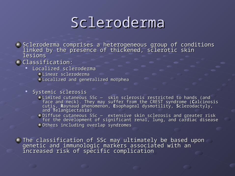
SclerodermaSclerodermaScleroderma comprises a heterogeneous group of conditions linked by the Scleroderma comprises a heterogeneous group of conditions linked by the presence of thickened, sclerotic skin lesions presence of thickened, sclerotic skin lesions Classification:Classification:
Localized sclerodermaLocalized sclerodermaLinear sclerodermaLinear sclerodermaLocalized and generalized morpheaLocalized and generalized morphea
Systemic sclerosisSystemic sclerosisLimited cutaneous SSc — skin sclerosis restricted to hands (and face and neck). They Limited cutaneous SSc — skin sclerosis restricted to hands (and face and neck). They may suffer from the CREST syndrome (may suffer from the CREST syndrome (CCalcinosis cutis, alcinosis cutis, RRaynaud phenomenon, aynaud phenomenon, EEsophageal dysmotility, sophageal dysmotility, SSclerodactyly, and clerodactyly, and TTelangiectasia)elangiectasia)Diffuse cutaneous SSc — extensive skin sclerosis and greater risk for the Diffuse cutaneous SSc — extensive skin sclerosis and greater risk for the development of significant renal, lung, and cardiac diseasedevelopment of significant renal, lung, and cardiac diseaseOthers including overlap syndromesOthers including overlap syndromes
The classification of SSc may ultimately be based upon genetic and The classification of SSc may ultimately be based upon genetic and immunologic markers associated with an increased risk of specific immunologic markers associated with an increased risk of specific complicationcomplication

Scleroderma picturesScleroderma pictures
Linear scleroderma
Morphea

Skin involvement in SclerodermaSkin involvement in Scleroderma
Skin involvement is a nearly universal feature of SSc. It Skin involvement is a nearly universal feature of SSc. It is characterized by variable extent and severity of skin is characterized by variable extent and severity of skin thickening and hardening. The fingers, hands, and face thickening and hardening. The fingers, hands, and face are generally the earliest areas of the body involved. are generally the earliest areas of the body involved. Edematous swelling and erythema may precede skin Edematous swelling and erythema may precede skin induration.induration.Other prominent skin manifestations include:Other prominent skin manifestations include:
Itching in the early stagesItching in the early stages Edema in the early stagesEdema in the early stages SclerodactylySclerodactyly Digital ulcersDigital ulcers Pitting at the fingertipsPitting at the fingertips TelangiectasiaTelangiectasia Calcinosis cutisCalcinosis cutis

Scleroderma skin involvementScleroderma skin involvement
Ulcers Sclerodactily
TelangiectasiasCalcinosis
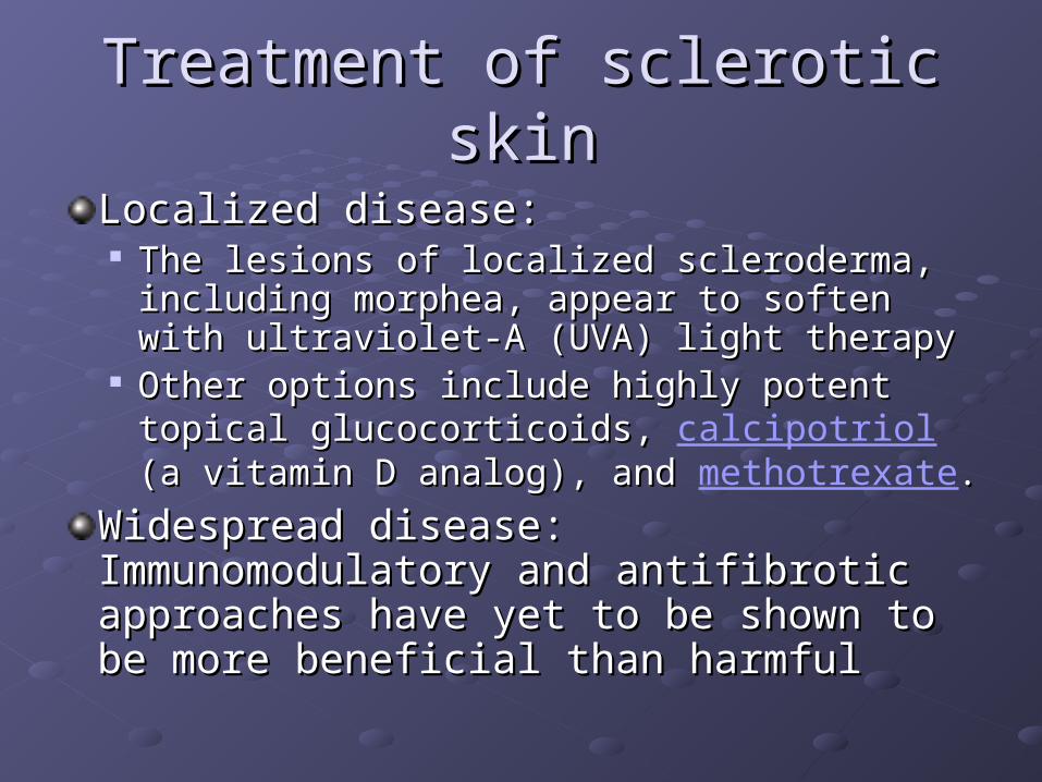
Treatment of sclerotic skinTreatment of sclerotic skin
Localized disease: Localized disease: The lesions of localized scleroderma, The lesions of localized scleroderma,
including morphea, appear to soften with including morphea, appear to soften with ultraviolet-A (UVA) light therapyultraviolet-A (UVA) light therapy
Other options include highly potent topical Other options include highly potent topical glucocorticoids, glucocorticoids, calcipotriol (a vitamin D (a vitamin D analog), and analog), and methotrexate. .
Widespread disease: Immunomodulatory Widespread disease: Immunomodulatory and antifibrotic approaches have yet to be and antifibrotic approaches have yet to be shown to be more beneficial than harmfulshown to be more beneficial than harmful

Treatment of other symptomsTreatment of other symptomsPruritus:Pruritus:
Antihistamines may help, but can cause drowsinessAntihistamines may help, but can cause drowsiness Maintaining adequate lubrication of the skin is essentialMaintaining adequate lubrication of the skin is essential Low-dose oral glucocorticoids are sometimes effective, topical steroids are rarely Low-dose oral glucocorticoids are sometimes effective, topical steroids are rarely
helpfulhelpfulTelangiectasia:Telangiectasia:
Green foundation make-upGreen foundation make-up Laser or other light therapy may be useful for large lesionsLaser or other light therapy may be useful for large lesions
Calcinosis:Calcinosis: Medications are unhelpfulMedications are unhelpful Suitably located lesions can be removed surgically, sometimes by using a dental Suitably located lesions can be removed surgically, sometimes by using a dental
drill; this technique causes less tissue damage than conventional methodsdrill; this technique causes less tissue damage than conventional methodsRaynaud phenomenon:Raynaud phenomenon:
Avoidance of cold, stress, nicotine, caffeine, and decongestants Avoidance of cold, stress, nicotine, caffeine, and decongestants Medications help for symptomatic relief: diltiazem, ACE inhibitors, ketanserin, Medications help for symptomatic relief: diltiazem, ACE inhibitors, ketanserin,
fluoxetinefluoxetine Different forms of sympathectomy (surgery) have been tried in an attempt to treat Different forms of sympathectomy (surgery) have been tried in an attempt to treat
severe Raynaud phenomenon. severe Raynaud phenomenon.
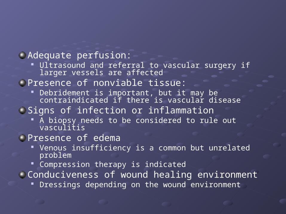
Adequate perfusion: Ultrasound and referral to vascular surgery if larger vessels are
affected
Presence of nonviable tissue: Debridement is important, but it may be contraindicated if there
is vascular disease
Signs of infection or inflammation A biopsy needs to be considered to rule out vasculitis
Presence of edema Venous insufficiency is a common but unrelated problem Compression therapy is indicated
Conduciveness of wound healing environment Dressings depending on the wound environment

Optimization of tissue growth Hyperbaric oxygen therapy is not indicated for
scleroderma, but it may be considered if there is a coexistent disease or evidence of poor oxygenation in the wound
Byosinthetic substitutes may be of use
Appropriateness of pressure offloading Usually not an issue
Controllability of pain
Optimization of host factors

The most important message: The most important message: preventionprevention
Wash skin daily using warm water and a mild Wash skin daily using warm water and a mild soapsoapUse a moisturizer daily (lactic acid, urea, Use a moisturizer daily (lactic acid, urea, dimethicone). Avoid perfumed moisturizersdimethicone). Avoid perfumed moisturizersWear wide comfortable shoesWear wide comfortable shoesPrevent triggering Raynaud’s syndromePrevent triggering Raynaud’s syndromeConsult a podiatrist for nail care and callus Consult a podiatrist for nail care and callus removalremovalDo not attempt to remove calcinosis by yourself. Do not attempt to remove calcinosis by yourself. Consider wearing orthoticsConsider wearing orthoticsSeek prompt attention for skin openingsSeek prompt attention for skin openings


![Hyperbaric Oxygen Therapy (HBOT)1].pdf · Hyperbaric Oxygen Therapy (HBOT) for Tissue Damage, Including Wound Care and Treatment of Central Nervous System (CNS) Conditions ... patients](https://static.fdocuments.in/doc/165x107/5c79ad9809d3f2bd0e8b960a/hyperbaric-oxygen-therapy-hbot-1pdf-hyperbaric-oxygen-therapy-hbot-for.jpg)