World Journal of - Microsoft · The World Journal of Gastrointestinal Pathophysiology Editorial...
18
World Journal of Gastrointestinal Pathophysiology World J Gastrointest Pathophysiol 2017 February 15; 8(1): 1-10 ISSN 2150-5330 (online) Published by Baishideng Publishing Group Inc
Transcript of World Journal of - Microsoft · The World Journal of Gastrointestinal Pathophysiology Editorial...

World Journal of Gastrointestinal PathophysiologyWorld J Gastrointest Pathophysiol 2017 February 15; 8(1): 1-10
ISSN 2150-5330 (online)
Published by Baishideng Publishing Group Inc
EDITOR-IN-CHIEFThomas Y Ma, Albuquerque
ASSOCIATE EDITORNaohisa Yoshida, Kyoto
GUEST EDITORIAL BOARD MEMBERSJia-Ming Chang, TaipeiWai-Keung Chow, TaichungChien-Wei Hsu, KaohsiungMing-Tsan Lin, TaipeiBor-Shyang Sheu, TainanJin-Town Wang, Taipei
MEMBERS OF THE EDITORIAL BOARD
ArgentinaBernabé M Quesada, Buenos AiresMarcelo G Roma, Rosario
AustraliaChris R Abbiss, JoondalupGuy D Eslick, SydneyMontri Gururatsakul, South AustraliaChandana B Herath, VictoriaMichael Horowitz, AdelaideMustafa Khasraw, VictoriaShu-Chuen Li, New South WalesMichael HJ Maes, GeelongNam Q Nguyen, South AustraliaKulmira Nurgali, VictoriaNicholas J Spencer, South Australia
Deborah J Verran, NSWShufeng Zhou, Victoria
AustriaCord Langner, GrazDietmar Ofner-Velano, Salzburg
Belgium
Kathleen Blondeau, LeuvenIlse M Hoffman, LeuvenTheodoor A Niewold, HeverleeXavier Sagaert, LeuvenJean-Marie Vanderwinden, BrusselsKristin Verbeke, BelgiumMathieu Vinken, Brussels
BrazilUilian Andreis, BotucatuEverson LDA Artifon, Sao PauloJoao B Calixto, TrindadeNiels OS Camara, Cidade UniversitáriaJúlio MF Chebli, Juiz de ForaClélia A Hiruma-Lima, BotucatuMarcel CC Machado, Sao PauloJuarez AS Quaresma, BelemWagner Vilegas, Araraquara
Brunei Darussalam
Vui H Chong, Bandar Seri Begawan
Canada
Jean-Francois Beaulieu, SherbrookeFrancois Boudreau, SherbrookeGeorge A Bubenik, GuelphWangxue Chen, OttawaFernando Fornari, MontréalJan D Huizinga, PuslinchRuben Hummelen, DundasKusum K Kharbanda, OmahaWolfgang A Kunze, HamiltonJianjun Li, OttawaRoderick J MacLeod, KingstonMichele Molinari, HalifaxNathalie Rivard, SherbrookeNathalie Rivard, SherbrookeKirill Rosen, HalifaxManuela M Santos, MontrealCaroline Saucier, QuébecJean Sévigny, QuébecEldon A Shaffer, CalgaryAlan BR Thomson, EdmontonPierre H Vachon, Sherbrooke
China
Kai-Xing Ai, ShanghaiZhao-Xiang Bian, Hong Kong Minhu Chen, GuangzhouChi-Hin Cho, Hong KongZhong-Hong Gao, WuhanJun-Ming Guo, NingboJing-Yan Han, BeijingJian-Dong Huang, Hong KongJia-Fu Ji, Beijing
I
Editorial Board2016-2019
The World Journal of Gastrointestinal Pathophysiology Editorial Board consists of 503 members, representing a team of worldwide experts in gastrointestinal pathophysiology. They are from 45 countries, including Argentina (2), Australia (13), Austria (2), Belgium (7), Brazil (9), Brunei Darussalam (1), Canada (21), China (30), Croatia (1), Czech Repoublic (2), Denmark (3), Egypt (1), Estonia (1), Finland (1), France (8), Germany (24), Greece (7), Hungary (5), India (12), Indonesia (1), Iran (2), Ireland (2), Israel (8), Italy (38), Japan (45), Lebanon (3), Malaysia (1), Mexico (2), Netherlands (7), Norway (1), Poland (4), Portugal (1), Romania (1), Russia (1), Singapore (4), South Korea (12), Spain (24), Sweden (11), Switzerland (3), Thailand (2), Turkey (7), Ukraine (1), United Kingdom (10), United States (161) and Venezuela (1).
February 23, 2016WJGP|www.wjgnet.com
World Journal ofGastrointestinal PathophysiologyW J G P

Zhan-Ju Liu, ShanghaiShi Liu, WuhanZhen-Ning Wang, ShenyangXiao-Hong Wang, BeijingWei Wei, HefeiDong-Ping Xie, ShanghaiWen-Xie Xu, ShanghaiXiao Yang, BeijingHua Yang, ChongqingWei-Zhen Zhang, BeijingHua-Chuan Zheng, ShenyangMin-Sheng Zhu, NanjingJinxia Zhu, BeijingDa-Ling Zhu, HarbinYong-Liang Zhu, Hangzhou
Croatia
Alen Protic, Rijeka
Czech Republic
Pavel Hladik, SemilyMartin Vokurka, Prague
Denmark
Lars Arendt-Nielsen, AalborgFrank V Schiodt, CopenhagenJonas Worsoe, Aarhus
Egypt
Mahmoud Aboelneen Khattab, Minia
Estonia
Enn Seppet, Tartu
Finland
Pauli Antero Puolakkainen, Turku
France
Bruno Bonaz, GrenoblePierre M Dechelotte, RouenJean-Paul Lallès, Saint-GillesCharles-Henri Malbert, Saint-GillesThierry Piche, NicePascale Plaisancié, LyonMichelina Plateroti, LyonVéronique Vitton, Marseille
Germany
Hans G Beger, Ulm Carsten Bergmann, IngelheimElke Cario, EssenArno J Dormann, KolnNikolaus Gassler, AachenAndreas Geier, Wuerzburg
Werner Hartwig, HeidelbergMarion Hewicker-Trautwein, HannoverJens Hoeppner, FreiburgTobias Keck, FreiburgJorg Kleeff, MunichPeter Malfertheiner, MagdeburgOliver Mann, HamburgChristoph W Michalski, MunichAndreas K Nussler, MunichChristian Pehl, VilsbiburgPeter Schemmer, HeidelbergManuel A Silva, PenzbergMarc Stemmler, FreiburgFrank Tacke, AachenSya N Ukena, BraunschweigBrigitte Vollmar, RostockThomas M Wex, MagdeburgMargot Zoller, Heidelberg
Greece
Stelios F Assimakopoulos, PatrasGeorge N Dalekos, LarissaAlkiviadis Efthymiou, thessalonikiMaria Gazouli, AthensIoannis E Koutroubakis, HeraklionGerassimos J Mantzaris, AthensGeorge V Papatheodoridis, Athens
Hungary
Mária Bagyánszki, SzegedMihály Boros, SzegedLaszlo Czako, SzegedPal Miheller, BudapestZoltan Rakonczay, Szeged
India
Anil K Agarwal, DelhiUday Bandyopadhyay, KolkataSriparna Basu, VaranasiChandra K Chakraborti, RourkelaNilesh M Dagia, MumbaiRajeev Garg, PunjabAkhtar Mahmood, ChandigarhVeerareddy P Reddy, Andhra PradeshChandra P Sharma, ThiruvananthapuramShailesh V Shrikhande, Mumbai Virendra Singh, ChandigarhNicholas J Skill, Indianapolis
Indonesia
Laurentius A Lesmana, Jakarta
Iran
Gholamreza Roshandel, GorganShahram Shahabi, Urmia
Ireland
Billy Bourke, DublinStephen J Keely, Dublin
Israel
Yosefa Avraham, JerusalemYaron Bar-Dayan, HolonShomron Ben-Horin, Tel-HashomerBoris Kirshtein, Beer ShevaStephen Malnick, RehovotYaakov Maor, Tel-HashomerRifaat Safadi, JerusalemNachum Vaisman, Tel Aviv
Italy
Rosaria Acquaviva, CataniaAlessandro Antonelli, PisaGiacosa Attilio, GenovaSalavtore Auricchio, NaplesGuido Basilisco, MilanAntonio Basoli, RomeMassimo Bellini, PisaLuigi Bonavina, MilanoAlfio Brogna, CataniaGiuseppe Calamita, BariRaffaele Capasso, NaplesIgnazio Castagliuolo, PadovaFrancesco Cresi, TorinoRosario Cuomo, NapoliSalvatore Cuzzocrea, GazziMario M D'Elios, FlorenceCinzia Domeneghini, MilanLuca Elli, MilanoWalter Fries, MessinaEugenio Gaudio, RomeMarco Gobbetti, BariFabio Grizzi, RozzanoEnzo Grossi, MilaneseEnzo Ierardi, FoggiaPietro Invernizzi, MonzaAngelo A Izzo, NaplesAnna Kohn, RomeGiovanni Latella, L'AquilaMassimo Marignani, RomeSergio Morini, RomeRaffaele Pezzilli, BolognaCristiano Rumio, MilanGiovanni Sarnelli, NaplesEdoardo V Savarino, PaduaPierpaolo Sileri, RomaAnnamaria Staiano, NaplesGiacomo C Sturniolo, PaduaClaudio Tiribelli, Trieste
Japan
Hirotada Akiho, FukuokaAkihiro Asakawa, KagoshimaHisashi Aso, SendaiYasu-Taka Azuma, OsakaShotaro Enomoto, WakayamaMikihiro Fujiya, HokkaidoTakahisa Furuta, HamamatsuAkira Hokama, NishiharaRyota Hokari, SaitamaYuichi Hori, HyogoMasahiro Iizuka, AkitaShoji Ikuo, KobeMotohiro Imano, Osaka
II February 23, 2016WJGP|www.wjgnet.com
III February 23, 2016WJGP|www.wjgnet.com
Hajime Isomoto, NagasakiTatehiro Kagawa, IseharaHaruki Kitazawa, SendaiShigeru BH Ko, TokyoXiao-Kang Li, TokyoNoriaki Manabe, OkayamaAtsushi Masamune, SendaiHiroyuki Matsubayashi, SuntogunKazuyuki Matsushita, Inohana Chuo-ku Reiko Miyazawa, GunmaKazunari Murakami, OitaHikaru Nagahara, TokyoYuji Naito, KyotoAtsushi Nakajima, YokohamaShoji Natsugoe, Kagoshima CityTsutomu Nishida, SuitaKoji Nomoto, TokyoShouji Shimoyama, Tokyo Goshi Shiota, YonagoHidekazu Suzuki, TokyoHitoshi Takagi, GunmaToru Takahashi, OkayamaYoshihisa Takahashi, TokyoKan Uchiyama, ChibaYoshiyuki Ueno, SendaiTakato Ueno, FukuokaHisayuki Uneyama, KwasakiMitsunori Yamakawa, YamagataTakayuki Yamamoto, MieYutaka Yata, Maebashi-cityNaohisa Yoshida, KyotoHitoshi Yoshiji, Kashihara
Lebanon
Costantine F Daher, ByblosAssaad M Soweid, BeirutJulnar Usta, Beirut
Malaysia
Andrew CS Boon, Ipoh
Mexico
José M de la Roca-Chiapas, LeonMaria-Raquel Huerta-Franco, Leon
Netherland
Wouter J de Jonge, AmsterdamAldo Grefhorst, GroningenDaniel Keszthelyi, MaastrichtCornelis FM Sier, ZA LeidenPieter J Tanis, AZ AmsterdamLuc JW van der Laan, RotterdamSander van der Marel, Leiden
Norway
Anne M Bakke, Oslo
Poland
Stanistaw A Hac, Gdansk
Stanislaw J Kowalski, KrakowAgata Mulak, WroclawNapoleon Waszkiewicz, Choroszcz
Portugal
Ricardo Marcos, Porto
Romania
Mihai Ciocirlan, Bucharest
Russia
Ludmila Filaretova, Petersburg
Singapore
Madhav Bhatia, SingaporeBrian K Goh, SingaporeKhek-Yu Ho, SingaporeCliff KS Ong, Singapore
South Korea
Jae Hee Cheon, SeoulMyung-Haing Cho, SeoulKi-Baik Hahm, SeongnamHO Jae Han, GwangjuChang-Duk Jun, GwangjuSang Geon Kim, SeoulHong Joo Kim, SeoulJin-Kyung Kim, Gyeongsan-SiWon-Jae Lee, SeoulSung-Joo Park, IksanSeung Ha Park, Gangwon DoKwan-Kyu Park, Daegu
Spain
Raquel Abalo, MadridJuan G Abraldes, BarcelonaDario Acuna-Castroviejo, ArmillaAgustin Albillos, MadridMaria-Angeles Aller, MadridFernando Azpiroz, BarcelonaRamón Bataller, BarcelonaMarco Bustamante-Balén, ValenciaAndres Cardenas, BarcelonaJoan Claria, BarcelonaPere Clavé, BarcelonaManuel B de Acosta, SantiagoManuel Giner, MadridBorja Hernandez-Breijo, MadridAngel I Lanas, ZaragozaMaite T Martin, BarcelonaVicente Martinez, BellaterraJose M Mates, MalagaJulio Mayol, MadridMarcal Pastor-Anglada, BarcelonaMaría E Sáez, SevilleYolanda Sanz, BurjassotCarlos Taxonera, Madrid
Maria D Yago, Granada
Sweden
Marco Del Chiaro, StockholmFrida Fak, GothenburgGunnar FA Flemstrom, UppsalaEvangelos Kalaitzakis, GothenburgKristina Lamas, UmeaBob R Olsson, GoteborgSara M Regnér, MalmoPeter T Schmidt, StockholmXiao-Feng Sun, LinkopingHenrik Thorlacius, MalmoCurt Tysk, Orebro
Switzerland
Jyrki J Eloranta, ZurichRemy Meier, LiestalCatherine M Pastor, Geneva
Thailand
Thawatchai Akaraviputh, BangkokWeekitt Kittisupamongkol, Bangkok
Turkey
Mehmet Bektas, AnkaraUgur Duman, BursaMukaddes Esrefoglu, IstanbulAhmet Guven, AnkaraMuammer Karadeniz, Bornova-IzmirElvan Ozbek, SakaryaIlhami Yuksel, Ankara
Ukraine
Oksana S Zayavhkivska, Lviv
United Kingdom
Geoffrey Burnstock, LondonJanice E Drew, AberdeenGirish L Gupte, BirminghamDavid C Hay, EdinburghNusrat Husain, ManchesterMichael L Lucas, GlasgowJamie Murphy, LondonVadim Sumbayev, Chatham MaritimeWing-Kin Syn, BirminghamAndrea Varro, Liverpool
United States
Sami R Achem, FloridaTauseef Ali, Oklahoma CityShrikant Anant, OklahomaMohammed S Anwer, North GraftonAndrew Aronsohn, ChicagoToms Augustin, Sayre
IV February 23, 2016WJGP|www.wjgnet.com
Gyorgy Baffy, BostonMichael T Bailey, ColumbusKim E Barrett, San DiegoMarc D Basson, Grand ForksRobert L Bell, Berkeley HeightsDavid H Berger, TexasUrs A Boelsterli, StorrsMichael W Bradbury, ErieQiang Cai, AtlantaWeibiao Cao, ProvidenceSubhash C Chauhan, Sioux FallsTao-Sheng Chen, MemphisJian-De Chen, GalvestonChiang YL Chiang, RootstownMashkoor A Choudhry, MaywoodParimal Chowdhury, Little RockEric Cohen, BostonRobert T Cormier, DuluthEdwin A Deitch, NewarkSharon DeMorrow, TexasDan A Dixon, ColumbiaJames P Dolan, PortlandHenry H Dong, PittsburghHui Dong, La JollaAshkan Farhadi, IrvineBin Feng, PittsburghJenifer Fenton, East LansingAlessandro Fichera, ChicagoMitchell P Fink, PittsburghLeo R Fitzpatrick, Rancho CordovaRobert A Forse, OmahaGlenn T Furuta, AuroraJuan F Gallegos-Orozco, ScottsdalePandu R Gangula, NasvhilleTimothy B Gardner, LebanonShannon S Glaser, TempleBeverley Greenwood-Van Meerveld, Oklahoma CityJohn R Grider, RichmondYan-Fang Guan, CincinnatiGregory M Holmes, Baton RougeRichard Hu, Los AngelesHartmut Jaeschke, Kansas CityRobert T Jensen, BethesdaSreenivasa S Jonnalagadda, LouisMichel Kahaleh, CharlottesvilleAndreas M Kaiser, Los AngelesRandeep S Kashyap, RochesterRichard Kellermayer, HoustonChris Kevil, ShreveportSandeep Khurana, BaltimorePawel R Kiela, TucsonTammy L Kindel, CincinnatiGordana Kosutic, DurhamDavid Kravetz, San Diego
Ashok Kumar, DetroitAnthony Kumar, Los AngelesJohn H Kwon, ChicagoMuriel Larauche, Los AngelesAi-Xuan Le Holterman, ChicagoI. Michael Leitman, New YorkFelix W Leung, SepulvedaSuthat Liangpunsakul, IndianapolisFeng-Xin Lu, BostonPauline K Lund, Chapel HillGuang-Xiang Luo, LexingtonJay Luther, Ann ArborRam I Mahato, MemphisAkhil Maheshwari, BirminghamKenneth Maiese, NewarkAdhip PN Majumdar, DetroitJosé E Manautou, StorrsCraig J McClain, LouisvilleDermot PB McGovern, Los AngelesDouglas S Merrell, BethesdaMurielle Mimeault, OmahaEmiko Mizoguchi, BostonHuan-Biao Mo, DentonAdam J Moeser, RaleighRamzi M Mohammad, DetroitSatdarshan SP Monga, PittsburghRoger K Moreira, New YorkSandeep Mukherjee, OmahaKarnam S Murthy, RichmondMichael J Nowicki, JacksonShuji Ogino, BostonMary F Otterson, WisconsinChung Owyang, Ann ArborHelieh S Oz, LexingtonMarco G Patti, ChicagoTimothy M Pawlik, BaltimoreSara Peleg, HoustonLi-Ya Qiao, RichmondChao Qin, OklahomaParvaneh Rafiee, MilwaukeeSigrid A Rajasekaran, WilmingtonVazhaikkurichi Rajendran, MorgantownJean P Raufman, BaltimoreRamesh M Ray, MemphisArie Regev, IndianapolisYehuda Ringel, Chapel HillRichard A Rippe, RockvilleChantal A Rivera, BossierAndrea Romani, ClevelandPraveen K Roy, MarshfieldPaul A Rufo, BostonDavid B Sachar, New YorkBimaljit S Sandhu, RichmondSanjaya K Satapathy, New Hyde Park
Ron Schey, Iowa CityMuhammad Y Sheikh, FresnoBo Shen, ClevelandLe Shen, ChicagoFrank A Simmen, Little RockSteven M Singer, WashingtonShailinder J Singh, WashingtonAdam J Smolka, CharlestonNed Snyder, HoustonZhen-Yuan Song, ChicagoGagan K Sood, HoustonRhonda Souza, DallasStuart J Spechler, DallasSubbaramiah Sridhar, AugustaCatia Sternini, Los AngelesVeedamali S Subramanian, Long BeachJun Sun, RochesterYvette Taché, Los AngelesXiao-Di Tan, ChicagoPaul D Terry, AtlantaMa Thomas, AlbuquerqueJennifer S Tirnauer, FarmingtonAndrea Todisco, Ann ArborGeorge C Tsokos, BostonVic Velanovich, DetroitRaj Vuppalanchi, IndianapolisEstela Wajcberg, CranfordArnold Wald, MadisonLi-Xin Wang, Los AngelesHorst C Weber, BostonGuo-Yao Wu, TexasChristian Wunder, BethesdaZuo-Liang Xiao, ClevelandGuang-Yin Xu, GalvestonGuo-Rong Xu, East OrangeYoshio Yamaoka, HoustonGuang-Yu Yang, ChicagoJay A Yelon, ValhallaShao-Yong Yu, HersheyYana Zavros, CincinnatiJoerg Zehetner, Los AngelesJian X Zhang, CharlotteZhi Zhong, CharlestonHui-Ping Zhou, RichmondZhan-Xiang Zhou, KannapolisQing Zhu, BethesdaYao-Hui Zhu, Stanford
Venezuela
Fabian Michelangeli, Caracas

Contents
February 15, 2017|Volume 8|Issue 1|WJGP|www.wjgnet.com I
Quarterly Volume 8 Number 1 February 15, 2017
FIELD OF VISION1 Impairedinactivationofdigestiveproteases:Thepossiblekeyfactorforthehighsusceptibilityofgerm-free
andantibiotic-treatedanimalstogutepithelialinjury
Qin X
ORIGINAL ARTICLE Observational Study
3 Polypdetectionrateandpathologicalfeaturesinpatientsundergoingacomprehensivecolonoscopy
screening
Asadzadeh Aghdaei H, Nazemalhosseini Mojarad E, Ashtari S, Pourhoseingholi MA, Chaleshi V, Anaraki F, Haghazali M,
Zali MR
EditorialBoardMemberofWorld JournalofGastrointestinalPathophysiology ,Guang-YuYang,MD,PhD,Professor,DepartmentofPathology,NorthwesternUniversity,Chicago,IL60611,UnitedStates
World Journal of Gastrointestinal Pathophysiology (World J Gastrointest Pathophysiol, WJGP, online ISSN 2150-5330, DOI: 10.4291), is a peer-reviewed open access academic journal that aims to guide clinical practice and improve diagnostic and therapeutic skills of clinicians.
WJGP is to report rapidly the most recent results in basic and clinical research on gastrointestinal pathophysiology, including all aspects of normal or abnormal function of the gastrointestinal tract, hepatobiliary system, and pancreas. WJGP specifically covers growth and development, digestion, secretion, absorption, metabolism and motility relative to the gastrointestinal organs, as well as immune and inflammatory processes, and neural, endocrine and circulatory control mechanisms that affect these organs. This journal will also report new methods and techniques in gastrointestinal pathophysiological research. We encourage authors to submit their manuscripts to WJGP. We will give priority to manuscripts that are supported by major national and international foundations and those that are of great basic and clinical significance.
World Journal of Gastrointestinal Pathophysiology is now indexed in PubMed, PubMed Central.
I-IV EditorialBoard
ContentsWorld Journal of Gastrointestinal Pathophysiology
Volume 8 Number 1 February 15, 2017
FLYLEAF
EDITORS FOR THIS ISSUE
NAMEOFJOURNALWorld Journal of Gastrointestinal Pathophysiology
ISSNISSN 2150-5330 (online)
LAUNCHDATEApril 15, 2010
FrequencyQuarterly
EDITOR-IN-CHIEFThomas Y Ma, MD, PhD, Professor, Chief, Division of Gastroenterology and Hepatology, University of New Mexico, MSC10 5550, 1 UNM, Albuquerque, NM 87131, United States
EDITORIALBOARDMEMBERSAll editorial board members resources online at http://www.wjgnet.com/2150-5330/editorialboard.htm
EDITORIALOFFICEXiu-Xia Song, DirectorWorld Journal of Gastrointestinal PathophysiologyBaishideng Publishing Group Inc8226 Regency Drive, Pleasanton, CA 94588, USATelephone: +1-925-2238242Fax: +1-925-2238243E-mail: [email protected] Desk: http://www.wjgnet.com/esps/helpdesk.aspxhttp://www.wjgnet.com
PUBLISHERBaishideng Publishing Group Inc8226 Regency Drive, Pleasanton, CA 94588, USATelephone: +1-925-2238242Fax: +1-925-2238243E-mail: [email protected] Desk: http://www.wjgnet.com/esps/helpdesk.aspxhttp://www.wjgnet.com
PUBLICATIONDATEFebruary 15, 2017
COPYRIGHT© 2017 Baishideng Publishing Group Inc. Articles pub-lished by this Open Access journal are distributed under the terms of the Creative Commons Attribution Non-commercial License, which permits use, distribution, and reproduction in any medium, provided the original work is properly cited, the use is non commercial and is otherwise in compliance with the license.
SPECIALSTATEMENTAll articles published in journals owned by the Baishideng Publishing Group (BPG) represent the views and opinionsof their authors, and not the views, opinions or policies of the BPG, except where other-wise explicitly indicated.
INSTRUCTIONSTOAUTHORShttp://www.wjgnet.com/bpg/gerinfo/204
ONLINESUBMISSIONhttp://www.wjgnet.com/esps/
ABOUT COVER
February 15, 2017|Volume 8|Issue 1|WJGP|www.wjgnet.com II
AIM AND SCOPE
INDEXING/ABSTRACTING
Responsible Assistant Editor: Xiang Li Responsible Science Editor: Fang-Fang JiResponsible Electronic Editor: Dan Li Proofing Editorial Office Director: Xiu-Xia Song Proofing Editor-in-Chief: Lian-Sheng Ma
Xiaofa Qin
FIELD OF VISION
� February �5, 20�7|Volume 8|Issue �|WJGP|www.wjgnet.com
Impaired inactivation of digestive proteases: The possible key factor for the high susceptibility of germ-free and antibiotic-treated animals to gut epithelial injury
Xiaofa Qin, GI Biopharma Inc, Westfield, NJ 07090, United States
Author contributions: Qin X contributes solely to this paper.
Conflict-of-interest statement: There is no conflict of interest.
Open-Access: This article is an open-access article which was selected by an in-house editor and fully peer-reviewed by external reviewers. It is distributed in accordance with the Creative Commons Attribution Non Commercial (CC BY-NC 4.0) license, which permits others to distribute, remix, adapt, build upon this work non-commercially, and license their derivative works on different terms, provided the original work is properly cited and the use is non-commercial. See: http://creativecommons.org/licenses/by-nc/4.0/
Manuscript source: Invited manuscript
Correspondence to: Xiaofa Qin, MD, PhD, GI Biopharma Inc, 918 Willow Grove Road, Westfield, NJ 07090, United States. [email protected]: +1-908-4637423
Received: October 9, 2016Peer-review started: October 11, 2016First decision: December 13, 2016Revised: December 16, 2016Accepted: January 2, 2017Article in press: January 3, 2017Published online: February 15, 2017
AbstractRecent study shows that germ-free and antibiotic-treated animals are highly susceptible to gut epithelial injury. This paper addresses that impaired inactivation of digestive proteases may be the key factor for the increased susceptibility.
Key words: Digestive proteases; Germ-free; Antibiotics; Gut microbiota; Gut epithelial injury
© The Author(s) 2017. Published by Baishideng Publishing Group Inc. All rights reserved.
Core tip: This paper addresses that impaired inactivation of digestive proteases may be the possible key factor for the high susceptibility of germ-free and antibiotic-treated animals to gut epithelial injury.
Qin X. Impaired inactivation of digestive proteases: The possible key factor for the high susceptibility of germ-free and antibiotic-treated animals to gut epithelial injury. World J Gastrointest Pathophysiol 2017; 8(1): 1-2 Available from: URL: http://www.wjgnet.com/2150-5330/full/v8/i1/1.htm DOI: http://dx.doi.org/10.4291/wjgp.v8.i1.1
COMMENTARY ON HOT TOPICSI read with great interest the paper by Hernández-Chirlaque et al[1] regarding the high susceptibility of germ-free and antibiotic-treated mice to epithelial injury. It is found that germ-free mice treated with dextran sulfate sodium (DSS) showed only minimal inflammation (no colonic thickening, lower myeloperoxidase activity, IL-6, IL-17, TNF-alpha and IFN-gamma secretion by splenocytes and mesenteric cell cultures, etc.), but enhanced hemorrhage, epithelial injury and mortality along with weakened intestinal barrier. Animals treated with antibiotics also showed similar but less severe changes with intermediate effects. That paper discussed the interaction among gut bacteria, immune cells, and epithelial cells through inflammatory mediators, cyto-kines, bioactive molecules, receptors, ligands, tran-
World J Gastrointest Pathophysiol 20�7 February �5; 8(�): �-2ISSN 2�50-5330 (online)
© 20�7 Baishideng Publishing Group Inc. All rights reserved.
Submit a Manuscript: http://www.wjgnet.com/esps/Help Desk: http://www.wjgnet.com/esps/helpdesk.aspxDOI: �0.429�/wjgp.v8.i�.�

2 February �5, 20�7|Volume 8|Issue �|WJGP|www.wjgnet.com
Qin X. Impaired inactivation of digestive proteases in gut epithelial injury
scription factors, and pathways, but failed to provide a coherent mechanistic explanation. Here I suggest that impaired inactivation of digestive proteases may be the key factor for the observed increased susceptibility. Studies have well documented that large amount of digestive proteases can be found in the large intestine of germ-free or antibiotics-treated animals but not in animals raised at conventional condition[2-7]. As we know, the gut is mainly protected by a layer of mucus secreted from goblet cells. Mucin, the structural molecule of mucus, contains a central core of peptide and side carbohydrate branches that can constitute up to 85% of the molecule. These carbohydrate side chains greatly retarded degradation of the protective mucus layer and damage of the underlying gut tissue by the digestive proteases being existed in germ-free or antibiotics-treated animals. However, the great destructive potency of these digestive proteases would be released and manifested once the gut barrier is broken as occurred either shortly after treatment with high concentrations of DSS or prolonged treatment with lower dose of DSS, resulting in the severe damage of epithelial cells at gut surface and then blood vessels in the mucosa, leading to excessive blood loss and enhanced mortality, even in the absence of remarkably inflammation. This would be similar to the so-called tryptic hemorrhagic necrosis of the gut as seen in animals under shock, which can be greatly prevented by previous inactivation of digestive proteases or ligation of pancreatic duct[8]. In fact, evidences I collected during the last fifteen years make me to believe that impaired inactivation of digestive proteases due to reduction in gut bacteria along with the improved hygiene and inhibition by some dietary chemicals such as saccharin and sucralose may have also played critical causative role in the pathogenesis of inflammatory bowel disease (IBD) in human[9-11]. Thus, impaired inactivation of digestive proteases in colitis and
IBD would be worthwhile for further study.
REFERENCES1 Hernández-Chirlaque C, Aranda CJ, Ocón B, Capitán-Cañadas F,
Ortega-González M, Carrero JJ, Suárez MD, Zarzuelo A, Sánchez de Medina F, Martínez-Augustin O. Germ-free and Antibiotic-treated Mice are Highly Susceptible to Epithelial Injury in DSS Colitis. J Crohns Colitis 2016; 10: 1324-1335 [PMID: 27117829 DOI: 10.1093/ecco-jcc/jjw096]
2 Norin KE. Influence of antibiotics on some intestinal microflora associated characteristics. Anaerobe 1997; 3: 145-148 [PMID: 16887579 DOI: 10.1006/anae.1997.0091]
3 Genell S, Gustafsson BE. Impaired enteric degradation of pancreatic endopeptidases in antibiotic-treated rats. Scand J Gastroenterol 1977; 12: 801-809 [PMID: 594651 DOI: 10.3109/00365527709181723]
4 Genell S, Gustafsson BE, Ohlsson K. Quantitation of active pancreatic endopeptidases in the intestinal contents of germfree and conventional rats. Scand J Gastroenterol 1976; 11: 757-762 [PMID: 1006149]
5 Gustafsson BE. The physiological importance of the colonic microflora. ScandJ Gastroenterol Suppl 1982; 77: 117-131 [PMID: 6750828]
6 Midtvedt T, Carlstedt-Duke B, Höverstad T, Midtvedt AC, Norin KE, Saxerholt H. Establishment of a biochemically active intestinal ecosystem in ex-germfree rats. Appl Environ Microbiol 1987; 53: 2866-2871 [PMID: 3124742]
7 Reddy BS, Pleasants JR, Wostmann BS. Pancreatic enzymes in germfree and conventional rats fed chemically defined, water-soluble diet free from natural substrates. J Nutr 1969; 97: 327-334 [PMID: 5773333]
8 Bounous G. Metabolic changes in the intestinal mucosa during hemorrhagic shock. Can J Surg 1965; 8: 332-339 [PMID: 5294426]
9 Qin XF. Impaired inactivation of digestive proteases by decon-jugated bilirubin: the possible mechanism for inflammatory bowel disease. Med Hypotheses 2002; 59: 159-163 [PMID: 12208202 DOI: 10.1016/S0306-9877(02)00243-8]
10 Qin X. Etiology of inflammatory bowel disease: a unified hypo-thesis. World J Gastroenterol 2012; 18: 1708-1722 [PMID: 22553395 DOI: 10.3748/wjg.v18.i15.1708]
11 Qin X. Publications by Xiaofa Qin. [accessed 2016 Oct 8]. Available from: URL: http://www.ncbi.nlm.nih.gov/pubmed/?term=xiaofa qin
P- Reviewer: Matowicka-Karna J, Yang CH, Zhu YL S- Editor: Qi Y L- Editor: A E- Editor: Li D
Hamid Asadzadeh Aghdaei, Ehsan Nazemalhosseini Mojarad, Sara Ashtari, Mohmad Amin Pourhoseingholi, Vahid Chaleshi, Fakhrosadat Anaraki, Mehrdad Haghazali, Mohammad Reza Zali
ORIGINAL ARTICLE
� February 15, 2017|Volume 8|Issue 1|WJGP|www.wjgnet.com
Polyp detection rate and pathological features in patients undergoing a comprehensive colonoscopy screening
Observational Study
Hamid Asadzadeh Aghdaei, Basic and Molecular Epidemiology of Gastrointestinal Disorders Research Center, Research Institute for Gastroenterology and Liver Diseases, Shahid Beheshti University of Medical Sciences, Tehran 19835-178, Iran
Ehsan Nazemalhosseini Mojarad, Sara Ashtari, Mohmad Amin Pourhoseingholi, Vahid Chaleshi, Mehrdad Haghazali, Mohammad Reza Zali, Gastroenterology and Liver Diseases Research Center, Research Institute for Gastroenterology and Liver Diseases, Shahid Beheshti University of Medical Sciences, Tehran 19835-178, Iran
Fakhrosadat Anaraki, Colorectal Division of Department of Surgery, Taleghani Hospital, Shahid Beheshti University of Medical Sciences, Tehran 19835-178, Iran
Author contributions: Asadzadeh Aghdaei H, Haghazali M and Zali MR performed all colonoscopies; Asadzadeh Aghdaei H drafted the initial manuscript; Nazemalhosseini Mojarad E and Anaraki F were the guarantors and designed the study; Chaleshi V gathered the data; Ashtari S participated in the acquisition, analysis, and interpretation of the data, and wrote and revised the last version of manuscript; Pourhoseingholi MA as PhD of biostatistics revised the article for statistical materials and methods.
Supported by Gastroenterology and Liver Diseases Research Center, Research Institute for Gastroenterology and Liver Diseases, Shahid Beheshti University of Medical Sciences, Tehran, Iran.
Institutional review board statement: The study was reviewed and approved by the Research Institute for Gastroenterology and Liver Diseases, Shahid Beheshti University of Medical Sciences, Tehran, Iran.
Informed consent statement: All study participants, or their legal guardian, provided informed written consent prior to study enrollment.
Conflict-of-interest statement: There are no conflicts of interest to report.
Data sharing statement: No additional data are available.
Open-Access: This article is an open-access article which was selected by an in-house editor and fully peer-reviewed by external reviewers. It is distributed in accordance with the Creative Commons Attribution Non Commercial (CC BY-NC 4.0) license, which permits others to distribute, remix, adapt, build upon this work non-commercially, and license their derivative works on different terms, provided the original work is properly cited and the use is non-commercial. See: http://creativecommons.org/licenses/by-nc/4.0/
Manuscript source: Invited manuscript
Correspondence to: Sara Ashtari, MSc of Epidemiology in Research Institute of Gastroenterology and Liver Diseases, Gastroenterology and Liver Diseases Research Center, Research Institute for Gastroenterology and Liver Diseases, Shahid Beheshti University of Medical Sciences, Tabnak St, Yaman Ave, Velenjak, Tehran 19835-178, Iran. [email protected]: +98-21-22432515Fax: +98-21-22432517
Received: September 13, 2016 Peer-review started: September 14, 2016 First decision: October 21, 2016 Revised: November 23, 2016 Accepted: December 7, 2016Article in press: December 9, 2016Published online: February 15, 2017
AbstractAIMTo identify the prevalence, and clinical and pathologic characteristic of colonic polyps among Iranian patients undergoing a comprehensive colonoscopy, and deter-mine the polyp detection rate (PDR) and adenoma detection rate (ADR).
World J Gastrointest Pathophysiol 2017 February 15; 8(1): �-10ISSN 2150-5��0 (online)
© 2017 Baishideng Publishing Group Inc. All rights reserved.
Submit a Manuscript: http://www.wjgnet.com/esps/Help Desk: http://www.wjgnet.com/esps/helpdesk.aspxDOI: 10.4291/wjgp.v8.i1.�

4 February 15, 2017|Volume 8|Issue 1|WJGP|www.wjgnet.com
Asadzadeh Aghdaei H et al . Prevalence and characteristics of colonic polyps
METHODSIn this cross-sectional study, demographics and epide-miologic characteristics of 531 persons who underwent colonoscopies between 2014 and 2015 at Mehrad gastrointestinal clinic were determined. Demographics, indication for colonoscopy, colonoscopy findings, number of polyps, and histopathological characteristics of the polyps were examined for each person.
RESULTSOur sample included 295 (55.6%) women and 236 (44.4%) men, with a mean age of 50.25 ± 14.89 years. Overall PDR was 23.5% (125/531). ADR and colorectal cancer detection rate in this study were 12.8% and 1.5%, respectively. Polyps were detected more significantly frequently in men than in women (52.8% vs 47.2%, P < 0.05). Polyps can be seen in most patients after the age of 50. The average age of patients with cancer was significantly higher than that of patients with polyps (61.3 years vs 56.4 years, P < 0.05). The majority of the polyps were adenomatous. More than 50% of the polyps were found in the rectosigmoid part of the colon.
CONCLUSIONThe prevalence of polyps and adenomas in this study is less than that reported in the Western populations. In our patients, distal colon is more susceptible to developing polyps and cancer than proximal colon.
Key words: Adenoma detection; Polyp detection; Iran; Colonoscopy; Screening
© The Author(s) 2017. Published by Baishideng Publishing Group Inc. All rights reserved.
Core tip: One of the major reasons for colonoscopy is detection of colon polyps, such as adenomas. Early diagnosis and endoscopic removal of adenomatous polyps is one of the main objectives for screening and prevention of colorectal cancer (CRC). Given that, only few studies are available in the national literature regarding the assessment of colorectal polyps, but none has explicitly noted the rate of polyp detection. Nevertheless, our study provides comprehensive information about clinical and epidemiological features of colorectal polyps. Therefore, the results of this study can provide a good infrastructure for the next preventive program and have clinical implications for CRC screening.
Asadzadeh Aghdaei H, Nazemalhosseini Mojarad E, Ashtari S, Pourhoseingholi MA, Chaleshi V, Anaraki F, Haghazali M, Zali MR. Polyp detection rate and pathological features in patients undergoing a comprehensive colonoscopy screening. World J Gastrointest Pathophysiol 2017; 8(1): 3-10 Available from: URL: http://www.wjgnet.com/2150-5330/full/v8/i1/3.htm DOI:
http://dx.doi.org/10.4291/wjgp.v8.i1.3
INTRODUCTIONColorectal cancer (CRC) is the third most common malignant disease in both men and women worldwide, accounting for more than 8% of mortality in the world with approximately 1.4 million new cases a year[14].
CRC is also the third most common cancer in men and women in Asia[5,6]. In the AsiaPacific region, the incidence varies between regions, with high incidence in Australia, and Eastern Asia, and low incidence in Southcentral Asia[7]. According to the International Agency for Research on Cancer, the incidence of CRC in many Asian countries is similar to that in many Western countries[8]. CRC is also the third most common cancer in Iranians, after excluding skin cancer, and it occurs at younger ages with an increasing trend similar to that in the Asia-Pacific countries[1,9]. These increasing rates may result from the young age structure and low rates of colon cancer in older people of these countries[6,10,11].
Almost all CRCs develop from colorectal polyps. Over a period of ten years, most of adenomatous polyps can be converted to colon carcinoma[12,13]. Given that the process of conversion of colorectal adenomas into adenocarcinoma is very long and slow[14], early detection and endoscopic removal of these precancerous lesions are very effective in reducing the incidence and mortality rate of CRC[1517].
CRC is a suitable disease for screening[18]. However, due to a lack of comprehensive screening strategy and public acceptance, this program is not implemented in many countries. Nevertheless, access to the CRC screening is an important key to reducing the burden of CRC. Endoscopic screening is comprised of four techniques including sigmoidoscopy, colonoscopy, barium enema, and computed tomographic colonography[19]. Colonoscopy is a highly specific and the most effective screening tool for detecting colonic polyps and CRC[20].
Limited data are available in the national literature regarding the assessment of colorectal polyps[2125]. Understanding of the prevalence of colorectal polyps especially adenomas in the general population would help clarify the efficacy of a CRC screening program. Therefore, updating the current knowledge in the scope of colorectal polyps and CRC is essential. Hence, identifying the features of colon polyps (e.g., distribution, location, and histology type) has great implications for developing national screening guidelines for CRC[26,27]. In this study, we aimed to determine the baseline polyp and adenoma prevalence in persons who underwent colonoscopies for various indications as well as opportunistic screening for CRC. We also assessed the polyp detection rate (PDR) and adenoma detection rate (ADR), and evaluated the clinical and histological
5 February 15, 2017|Volume 8|Issue 1|WJGP|www.wjgnet.com
characteristics of colorectal polyps in Iranian patients and Iranian volunteers for CRC screening.
MATERIALS AND METHODSStudy designIn this crosssectional study, all data were extracted from a colonoscopy database and pathology reports maintained by Mehrad gastrointestinal clinic in Iran. We included all persons aged 15 to 85 years, who underwent their first time colonoscopy during 2014-2015. Patients who had previously been identified with colon polyp or colorectal malignancies including CRC, colonic resection, active colitis, active diverticulitis and familial adenomatous polyposis were excluded from the study. We collected the data on demographic variables, indications for colonoscopy, and family history of colorectal malignancies. Family history was defined as having a first degree relative with CRC. For all colorectal lesions, data on clinical and pathological features (i.e., number, size, site, and grade of dysplasia) were obtained. The study was approved by the Research Institute for Gastroenterology and Liver Diseases, Shahid Beheshti University of Medical Sciences.
Polyp classificationAll polyps identified during colonoscopy were biopsied or removed endoscopically and submitted for histopathology. The overall PDR was defined as the proportion of procedures in which at least one polyp was detected over the total number of colonoscopies. ADR was defined as the number of colonoscopies in which one or more adenomas was detected, divided by the total number of colonoscopies performed by the endoscopist[28].
Pathological features of colorectal lesions were determined using the World Health Organization criteria[28]. Hyperplastic and adenomatous polyps were classified as nonneoplastic polyps and neoplastic polyps, respectively. Microscopically, adenomas were categorized architecturally as serrated, tubular, tubularvillous, and villous.
The locations of the polyps were defined as proximal colon including the transverse colon, hepatic flexure, ascending colon, and cecum, and distal colon including the rectum, sigmoid, descending colon, and splenic flexure.
The polyp size was classified as small (< 5 mm), medium (59 mm), or large (> 10 mm). Estimation of polyp size was performed by the endoscopist using the diameter of the open biopsy forceps, which is about 8 mm. In the event of multiple polyps, only the size of the largest was considered for the purposes of analysis. Degrees of dysplasia observed in the adenomas were graded as low (mild and moderate) or high grade (severe). Patients with no polyps were regarded as normal.
Statistical analysisCategorical variables are expressed as numbers and percentages. χ2 or Fisher’s exact test, where appropriate, was used for analysis of categorical variables. Continuous variables are expressed as medians, or as means and standard deviation, and 95%CI as appropriate. The Student’s ttest was used for comparisons of means. All analyses were performed using SPSS version 21.0 (SPSS INC, Chicago, IL, United States). A twotailed P < 0.05 was considered statistically significant.
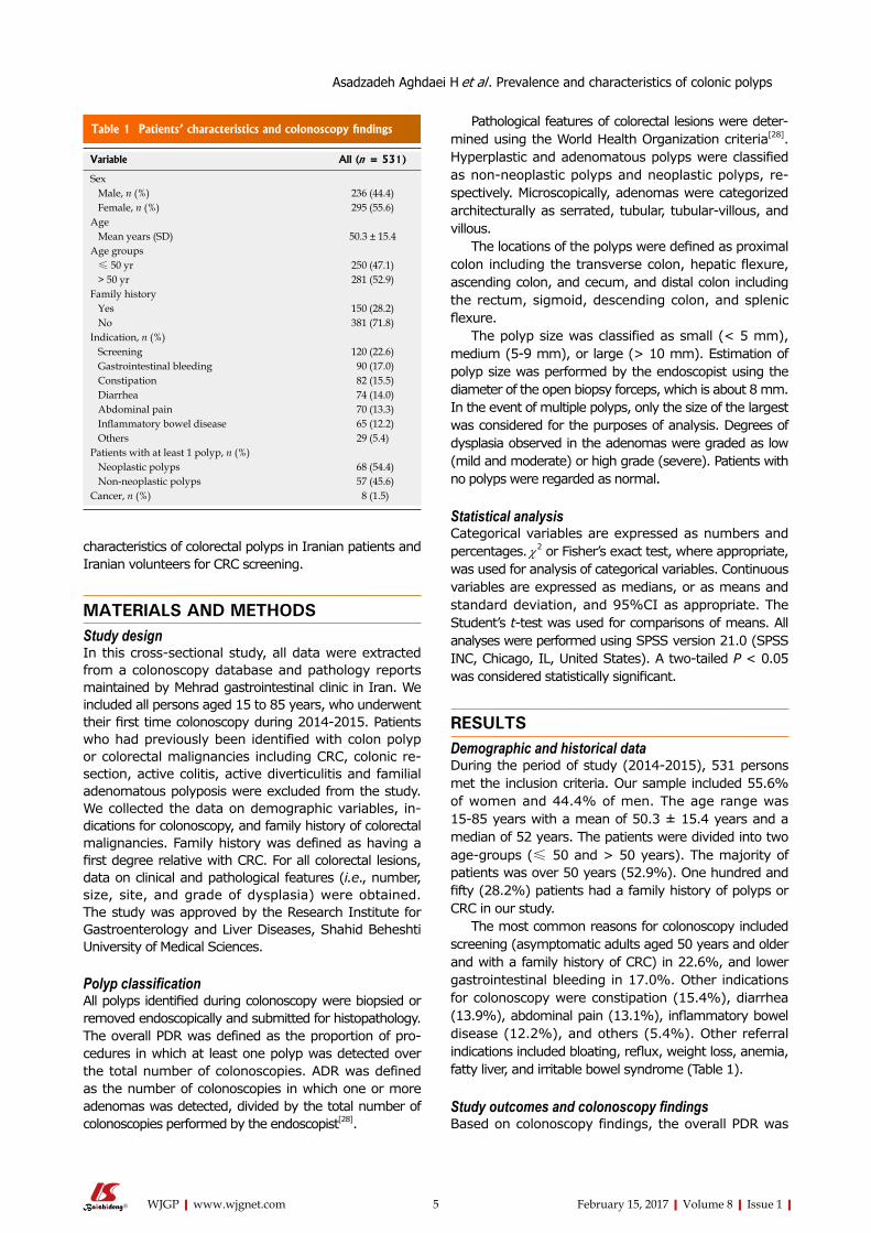
RESULTSDemographic and historical dataDuring the period of study (20142015), 531 persons met the inclusion criteria. Our sample included 55.6% of women and 44.4% of men. The age range was 1585 years with a mean of 50.3 ± 15.4 years and a median of 52 years. The patients were divided into two agegroups (≤ 50 and > 50 years). The majority of patients was over 50 years (52.9%). One hundred and fifty (28.2%) patients had a family history of polyps or CRC in our study.
The most common reasons for colonoscopy included screening (asymptomatic adults aged 50 years and older and with a family history of CRC) in 22.6%, and lower gastrointestinal bleeding in 17.0%. Other indications for colonoscopy were constipation (15.4%), diarrhea (13.9%), abdominal pain (13.1%), inflammatory bowel disease (12.2%), and others (5.4%). Other referral indications included bloating, reflux, weight loss, anemia, fatty liver, and irritable bowel syndrome (Table 1).
Study outcomes and colonoscopy findingsBased on colonoscopy findings, the overall PDR was
Variable All (n = 531)
Sex Male, n (%) 2�6 (44.4) Female, n (%) 295 (55.6)Age Mean years (SD) 50.� ± 15.4Age groups ≤ 50 yr 250 (47.1) > 50 yr 281 (52.9)Family history Yes 150 (28.2) No �81 (71.8)Indication, n (%) Screening 120 (22.6) Gastrointestinal bleeding 90 (17.0) Constipation 82 (15.5) Diarrhea 74 (14.0) Abdominal pain 70 (1�.�) Inflammatory bowel disease 65 (12.2) Others 29 (5.4)Patients with at least 1 polyp, n (%) Neoplastic polyps 68 (54.4) Non-neoplastic polyps 57 (45.6)Cancer, n (%) 8 (1.5)
Table 1 Patients’ characteristics and colonoscopy findings
Asadzadeh Aghdaei H et al . Prevalence and characteristics of colonic polyps

6 February 15, 2017|Volume 8|Issue 1|WJGP|www.wjgnet.com
23.5% (125/531). According to histopathology results from 125 patients with at least one polyp, 54.4% of the lesions were neoplastic polyps and 45.6% were hyperplastic polyps (nonneoplastic polyps). The overall ADR in this study was 12.8% (68/531). The percentage of male patients with polyps was significantly higher than that of female patients (52.8% vs 47.2%, P < 0.05). CRC was detected in 1.5% (8/531) of the total population (men, 62.5%; women, 37.5%) (Table 2). The mean age of patients with polyp was 56.4 ± 13.5 years. Polyps can be seen in most patients after the age of 50 (69.6%). CRC was more frequently observed in patients aged from 60 to 80 years. The average age of patients with cancer was significantly higher than that of those with polyps (61.3 ± 19.7 years vs 56.4 ± 13.5 years, P < 0.05) (Table 3). In addition, the relationship between PDR and family history of CRC or polyps was assessed and this was found not to be statistically significant (P > 0.05), while all patients with CRC in this study had a family history (Table 4).
Histopathological characteristics of the polypsTotally 138 polyps were removed by colonoscopy in this
study. Among the 138 polyps, 56.5% were neoplastic and the others were nonneoplastic. All nonneoplastic polyps were hyperplastic. Among the 78 neoplastic polyps, the common histologic types were tubular adenomas (48.7%), tubulevillous adenomas (28.2%), villous adenomas (12.8%), and serrated adenomas (10.2%).
The prevalence of polyps in distal colon was higher than that in proximal colon (68.1% vs 31.9%, P < 0.05). Accordingly, most of cancers were located in the distal colon compared with the proximal colon (75% vs 25%, P < 0.05) (Table 5). Overall polyps were frequently detected in the rectum (32.0%), sigmoid (24.6%), transverse colon (16%), and ascending and descending colon (10.1%), and the others located in the cecum (5%), and splenic and hepatic flexure (2.1%) (Table 6).
Data about the size of polyps were available for only 75 polyps; 33.3% were smaller than 5 mm as small size, 40% were between 59 mm as medium size and 26.7% were more than 10 mm as large size (Table 7). According to the degree of dysplasia observed in the adenomas, most of patients (52.6%) had mild grade, 24.4% had moderate, and 23% had severe grade of dysplasia (Table 8).
Histologic type of polyps Gender Total
Male (%) Female (%)Neoplastic Tubular 18 (5�.0) 16 (47.0) �4 (100) Tubulo-villous 1� (68.4) 6 (�1.6) 19 (100) Villous 4 (40.0) 6 (60.0) 10 (100) Serrated � (60.0) 2 (40.0) 5 (100)Adenomatous polyps �8 (55.9) �0 (44.1) 68 (100)Non-neoplastic Hyperplastic 28 (40.6) 29 (45.�) 57 (100)Total PDR 66 (52.8) 59 (47.2) 125 (100)Cancer 5 (62.5) � (�7.5) 8 (100)Total 69 (51.9) 64 (48.1) 1�� (100)
Table 2 Detection rates of different histologic types of polyps and cancer by gender
PDR: Polyp detection rate.
Histologic type of polyps Age-groups Total≤ 50 yr > 50 yr
Neoplastic Tubular 6 (17.6) 28 (82.4) �4 (100) Tubulo-villous 7 (�6.8) 12 (6�.2) 19 (100) Villous � (�0.0) 7 (70.0) 10 (100) Serrated � (60.0) 2 (40.0) 5 (100)Adenomatous polyps 19 (28.0) 49 (72.0) 68 (100)Non-neoplastic Hyperplastic 19 (��.�) �8 (66.7) 57 (100)Total PDR �8 (�0.4) 87 (69.6) 125 (100)Cancer 1 (12.5) 7 (87.5) 8 (100)Total �9 (29.�) 94 (70.7) 1�� (100)
Table 3 Detection rates of different histologic types of polyps and cancer by age
PDR: Polyp detection rate.
Histologic type of polyps Family history Total
No (%) Yes (%)Neoplastic Tubular 26 (76.4) 8 (2�.6) �4 (100) Tubulo-villous 1� (68.4) 6 (�1.6) 19 (100) Villous 5 (50.0) 5 (50.0) 10 (100) Serrated 2 (40.0) � (60.0) 5 (100)Adenomatous polyps 46 (67.6) 22 (�2.4) 68 (100)Non-neoplastic Hyperplastic 46 (80.7) 11 (19.�) 57 (100)Total PDR 92 (7�.6) �� (26.4) 125 (100)Cancer 0 (0) 8 (100) 8 (100)Total 92 (69.2) 41 (�0.8) 1�� (100)
Table 4 Detection rates of different histologic types of polyps and cancer by family history
PDR: Polyp detection rate.
Histologic type of polyps Location TotalProximal colon
(%)Distal colon
(%)
Neoplastic Tubular 10 (26.�) 28 (7�.7) �8 (100) Tubulo-villous 9 (40.9) 1� (59.1) 22 (100) Villous � (�0.0) 7 (70.0) 10 (100) Serrated � (�7.5) 5 (62.5) 8 (100)Adenomatous polyps 25 (�2.0) 5� (68.0) 78 (100)Non-neoplastic Hyperplastic 19 (�1.7) 41 (68.�) 60 (100)Total PDR 44 (�1.9) 94 (68.1) 1�8 (100)
Table 5 Detection rates of different histologic types of polyps by location
PDR: Polyp detection rate.
Asadzadeh Aghdaei H et al . Prevalence and characteristics of colonic polyps

7 February 15, 2017|Volume 8|Issue 1|WJGP|www.wjgnet.com
DISCUSSIONThe PDR and ADR rates obtained in this study are low, when compared to the figures from most Western and some Asian countries. In a large multicenter study from Italy, the median detection rate for polyps was 35%[29]. A large colonoscopy series from Spain reported a PDR of 45.8%[30]. Similar studies from Mayo Clinic in the United States and France reported PDR of 49% and 35.5%, and ADR of 31% and 17.7%, respectively[31,32]. In some Asian countries like Korea, China and Thailand, PDR and ADR were similar to those in Europeans and Americans[3336]. However, our findings are similar to reports from Kuwait, Malaysia and Oman where PDR of 20% and ADR of 10%, 11.5% and 12.1% were reported, respectively[3739]. While in African countries like Nigeria these rates were reported to be lower than our results (PDR, 16.1%; ADR, 6.8%)[40]. The mean age of the studied population was relatively young (50.3 years) and it might decrease the ADR because adenomas have been demonstrated to be more frequent in those older than 50 years of age.
The overall estimate for PDR in our study was 23.5%, while ADR was 12.8%. The percentage of male patients with polyps was significantly higher than that of female patients (52.8% vs 47.2%, P < 0.05). These data are consistent with the other reports that support
gender differences in the prevalence of colon polyps and cancer[27,40,41].
Older age is the most important predictor for the prevalence of adenomas and cancer. In our study, the PDR and cancer prevalence reached a peak in the 6th decade of life. Nonetheless, the average age of patients with cancer was significantly higher than that of patients with polyps (61.3 years vs 56.4 years, P < 0.05). Studies from the Middle East and the Western countries also mentioned a significant increase in the risk of CRC, in particular after the age of 50 years[24,28,42].
Based on increasing prevalence of CRC in the sixth decade of life, regular screening should begin at the age of 50. However, people at higher risk of developing CRC should begin screening at a younger age[43,44].
The tubular type was the most common histological feature of adenomas in the present study, in accordance with the results of other reports[21,27,45]. Polyps were detected significantly in distal (left sided) colon, comparable with results from Asian and the Western countries[46,47]. Nonetheless, because of the significance of adenomatous polyps and hyperplastic polyps present in proximal colon, the location of polyps is not helpful for distinguishing between neoplastic and nonneoplastic polyps. So, complete colonoscopy is recommended in screening guidelines for colon cancer[43,48]. In addition, this study showed that only 56.6% of the polyps were found in the rectum and sigmoid region. Our study did not find any association between the age and location of polyps. This is in contrast with previous studies showing that the incidence of right sided polyps increased with increasing age[44].
With regard to the size of polyps, we observed that the hyperplastic polyps and tubular adenoma were always smaller than 10 mm, while tubulovillous and villous adenoma were always bigger than 10 mm. So,
Polyps Location Total
Ascending colon Transverse colon Descending colon Sigmoid Rectum Cecum Hepatic flexure Splenic flexureTubular � (7.9) 5 (1�.2) 4 (10.5) 7 (18.4) 16 (42.1) 1 (2.6) 1 (2.6) 1 (2.6) �8 (100)Tubulo-villous 4 (18.2) � (1�.6) 2 (9.1) 6 (27.�) 5 (22.7) 2 (9.1) 0 (0) 0 (0) 22 (100)Villous 0 (0) 1 (10) 0 (0) 2 (20) 5 (50) 2 (20) 0 (0) 0 (0) 10 (100)Serrated 0 (0) � (�7.5) 1 (12.5) � (�7.5) 1 (12.5) 0 (0) 0 (0) 0 (0) 8 (100)Hyperplastic 7 (11.7) 10 (16.7) 7 (11.7) 16 (26.7) 17 (28.�) 2 (�.�) 0 (0) 1 (1.7) 60 (100)Total 14 (10.1) 22 (16.0) 14 (10.1) �4 (24.6) 44 (�2.0) 7 (5.1) 1 (0.7) 2 (1.4) 1�8 (100)
Table 6 Detection rates of different histologic types of polyps by colonic segments
Size Histologic type Total
Hyperplastic Tubular Tubul-villous villous SerratedSmall 11 (44.0) 0 (0) 14 (56.0) 0 (0) 0 (0) 25 (100)Medium 14 (46.7) 0 (0) 12 (40.0) 0 (0) 4 (1�.�) �0 (100)Large 0 (0) 8 (40.0) � (15.0) 9 (45.0) 0 (0) 20 (100)Unknown �5 (55.6) 2 (�.2) 9 (14.�) 1� (20.6) 4 (6.�) 6� (100) Total 60 (4�.5) 10 (7.2) �8 (27.5) 22 (16.0) 8 (5.8) 1�8 (100)
Table 7 Detection rates of different histologic types of polyps by size
Data available for only 75 polyps.
Grade Histologic type Total
Tubular Tubul-villous Villous SerratedMild 21 (51.2) 11(26.9) 6 (14.6) � (7.�) 41 (100)Moderate 11 (57.9) � (15.8) � (15.8) 2 (10.5) 19 (100)Severe 6 (��.�) 8 (44.5) 1 (5.5) � (16.7) 18 (100)Total �8 (48.7) 22 (28.2) 10 (12.9) 8 (10.2) 78 (100)
Table 8 Detection rates of different histologic types of polyps by degree of dysplasia
Asadzadeh Aghdaei H et al . Prevalence and characteristics of colonic polyps

8 February 15, 2017|Volume 8|Issue 1|WJGP|www.wjgnet.com
removing the polyps which are larger than 10 mm is recommended[36,49].
The study faced some limitations. First, this study was not populationbased, therefore, the selection bias of the study population must be kept in mind. Second, our sample included mostly symptomatic patients, in which the estimates may be different from screening studies with asymptomatic individuals. Nevertheless, the results of this study can provide a good infrastructure for the next preventive program and have clinical implications for CRC screening.
In conclusion, PDR, ADR and CRC detection rate in this study were 23.5%, 12.8% and 1.5%, respectively. Most of the polyps and CRC were identified in patients aged 50 years or older. The majority of the polyps were adenomatous. More than 50% of the polyps were found in the rectosigmoid part of the colon. Finally, our study did not find any association between the family history and PDR.
COMMENTSBackgroundColorectal cancer (CRC) is the third most common malignant disease in both men and women worldwide, accounting for more than 8% of cancer-related death in the world with approximately 1.4 million new cases a year. Almost all CRCs develop from colorectal polyps. CRC largely can be prevented by the detection and removal of adenomatous polyps, and survival is significantly better when CRC is diagnosed while still localized. When CRC is found at an early stage before it has spread, the 5-year relative survival rate is about 90%. But only about 4 out of 10 CRCs are found at this early stage. When cancer has spread outside the colon or rectum, survival rates are lower.
Research frontiersEarly diagnosis and endoscopic resection of adenomatous polyps is the main approach for screening and prevention of CRC. This study aimed at identifying the prevalence, and clinical and pathologic characteristic of colonic polyps among Iranian patients undergoing a comprehensive colonoscopy and determine the polyp detection rate (PDR) and adenoma detection rate (ADR).
Innovations and breakthroughsOnly few studies are available in the national literature regarding the assess-ment of colorectal polyps, but none has explicitly noted the rate of polyp detection. Nevertheless, the study provides comprehensive information about clinical and epidemiological features of colorectal polyps.
ApplicationsOlder age is the most important predictor for the prevalence of adenomas and cancer. Based on the results of this study, the PDR and cancer prevalence reached a peak in the 6th decade of life. Given the increased prevalence of CRC in the sixth decade of life, regular screening beginning at the age of 50 is the key to preventing CRC.
TerminologyThe overall PDR was defined as the proportion of procedures in which at least one polyp was detected over the total number of colonoscopies. ADR was defined as the number of colonoscopies in which one or more adenomas was detected, divided by the total number of colonoscopies performed by the endoscopist.
Peer-reviewAuthors report in this paper the detection rates by colonoscopy for cancer and adenoma in an Iranian population.
REFERENCES1 Jemal A, Center MM, DeSantis C, Ward EM. Global patterns of
cancer incidence and mortality rates and trends. Cancer Epidemiol Biomarkers Prev 2010; 19: 1893-1907 [PMID: 20647400 DOI: 10.1158/1055-9965.epi-10-0437]
2 Edwards BK, Ward E, Kohler BA, Eheman C, Zauber AG, Anderson RN, Jemal A, Schymura MJ, Lansdorp-Vogelaar I, Seeff LC, van Ballegooijen M, Goede SL, Ries LA. Annual report to the nation on the status of cancer, 1975-2006, featuring colorectal cancer trends and impact of interventions (risk factors, screening, and treatment) to reduce future rates. Cancer 2010; 116: 544-573 [PMID: 19998273 DOI: 10.1002/cncr.24760]
3 Ferlay J, Autier P, Boniol M, Heanue M, Colombet M, Boyle P. Estimates of the cancer incidence and mortality in Europe in 2006. Ann Oncol 2007; 18: 581-592 [PMID: 17287242 DOI: 10.1093/annonc/mdl498]
4 Joseph DA, King JB, Miller JW, Richardson LC. Prevalence of colorectal cancer screening among adults--Behavioral Risk Factor Surveillance System, United States, 2010. MMWR Suppl 2012; 61: 51-56 [PMID: 22695464]
5 Pourhoseingholi MA. Increased burden of colorectal cancer in Asia. World J Gastrointest Oncol 2012; 4: 68-70 [PMID: 22532878 DOI: 10.4251/wjgo.v4.i4.68]
6 Yiu HY, Whittemore AS, Shibata A. Increasing colorectal cancer incidence rates in Japan. Int J Cancer 2004; 109: 777-781 [PMID: 14999789 DOI: 10.1002/ijc.20030]
7 Pourhoseingholi MA. Epidemiology and burden of colorectal cancer in Asia-Pacific region: what shall we do now? Transl Gastrointest Cancer 2014; 3: 169-173 [DOI: 10.3978/j.issn.2224-4778.2014.08.10]
8 Ferlay J, Soerjomataram I, Dikshit R, Eser S, Mathers C, Rebelo M, Parkin DM, Forman D, Bray F. Cancer incidence and mortality worldwide: sources, methods and major patterns in GLOBOCAN 2012. Int J Cancer 2015; 136: E359-E386 [PMID: 25220842 DOI: 10.1002/ijc.29210]
9 Hossein Somi M, Mirinezhad K, Farhang S, Jazayeri E, Sani A, Seif-Farshadi M, Golzari M, Kashef S, Sadegy M. Gastrointestinal cancer occurrence in East Azarbaijan: a five year study from North Western Iran. Asian Pac J Cancer Prev 2006; 7: 309-312 [PMID: 16839228]
10 Mousavi SM, Gouya MM, Ramazani R, Davanlou M, Hajsadeghi N, Seddighi Z. Cancer incidence and mortality in Iran. Ann Oncol 2009; 20: 556-563 [PMID: 19073863 DOI: 10.1093/annonc/mdn642]
11 Ansari R, Mahdavinia M, Sadjadi A, Nouraie M, Kamangar F, Bishehsari F, Fakheri H, Semnani S, Arshi S, Zahedi MJ, Darvish-Moghadam S, Mansour-Ghanaei F, Mosavi A, Malekzadeh R. Incidence and age distribution of colorectal cancer in Iran: results of a population-based cancer registry. Cancer Lett 2006; 240: 143-147 [PMID: 16288832 DOI: 10.1016/j.canlet.2005.09.004]
12 Noffsinger AE. Serrated polyps and colorectal cancer: new pathway to malignancy. Annu Rev Pathol 2009; 4: 343-364 [PMID: 19400693 DOI: 10.1146/annurev.pathol.4.110807.092317]
13 Levine JS, Ahnen DJ. Clinical practice. Adenomatous polyps of the colon. N Engl J Med 2006; 355: 2551-2557 [PMID: 17167138 DOI: 10.1056/NEJMcp063038]
14 Huang CS, Farraye FA, Yang S, O’Brien MJ. The clinical sig-nificance of serrated polyps. Am J Gastroenterol 2011; 106: 229-240; quiz 241 [PMID: 21045813 DOI: 10.1038/ajg.2010.429]
15 García Sánchez J. [Colonoscopic polypectomy and long-term prevention of colorectal cancer deaths]. Rev Clin Esp 2012; 212: 408 [PMID: 22937540 DOI: 10.1016/j.rce.2012.05.002]
16 Espey DK, Wu XC, Swan J, Wiggins C, Jim MA, Ward E, Wingo PA, Howe HL, Ries LA, Miller BA, Jemal A, Ahmed F, Cobb N, Kaur JS, Edwards BK. Annual report to the nation on the status of cancer, 1975-2004, featuring cancer in American Indians and Alaska Natives. Cancer 2007; 110: 2119-2152 [PMID: 17939129 DOI: 10.1002/cncr.23044]
COMMENTS
Asadzadeh Aghdaei H et al . Prevalence and characteristics of colonic polyps

9 February 15, 2017|Volume 8|Issue 1|WJGP|www.wjgnet.com
17 Atkin WS, Edwards R, Kralj-Hans I, Wooldrage K, Hart AR, Northover JM, Parkin DM, Wardle J, Duffy SW, Cuzick J. Once-only flexible sigmoidoscopy screening in prevention of colo-rectal cancer: a multicentre randomised controlled trial. Lancet 2010; 375: 1624-1633 [PMID: 20430429 DOI: 10.1016/s0140-6736(10)60551-x]
18 Inadomi JM, Vijan S, Janz NK, Fagerlin A, Thomas JP, Lin YV, Muñoz R, Lau C, Somsouk M, El-Nachef N, Hayward RA. Adherence to colorectal cancer screening: a randomized clinical trial of competing strategies. Arch Intern Med 2012; 172: 575-582 [PMID: 22493463 DOI: 10.1001/archinternmed.2012.332]
19 Elmunzer BJ, Hayward RA, Schoenfeld PS, Saini SD, Deshpande A, Waljee AK. Effect of flexible sigmoidoscopy-based screening on incidence and mortality of colorectal cancer: a systematic review and meta-analysis of randomized controlled trials. PLoS Med 2012; 9: e1001352 [PMID: 23226108 DOI: 10.1371/journal.pmed.1001352]
20 Iravani S, Nazemalhosseini-Mojarad E, Kashfi SM, Azimzadeh P. Screening of colorectal diseases among individuals without family history in a private hospital, Tehran, Iran from 2011 to 2013. Transl Gastrointest Cancer 2014; 3: 165-168 [DOI: 10.3978/j.issn.2224-4778.2014.08.04]
21 Mirzaie AZ, Abolhasani M, Moghaddam RM, Kabivar M. The Frequency of gastrointestinal polyps in Iranian population. Iran J Pathol 2012; 7: 183-189
22 Bafandeh Y, Khoshbaten M, Eftekhar Sadat AT, Farhang S. Clinical predictors of colorectal polyps and carcinoma in a low prevalence region: results of a colonoscopy based study. World J Gastroenterol 2008; 14: 1534-1538 [PMID: 18330943 DOI: 10.3748/wjg.14.1534]
23 Bafandeh Y, Daghestani D, Esmaili H, Aharizad S. Distribution of cancer and adenomatous polyps in the colorectum: study in an Iranian population. Asian Pac J Cancer Prev 2006; 7: 65-68 [PMID: 16629518]
24 Bafandeh Y, Daghestani D, Esmaili H. Demographic and anato-mical survey of colorectal polyps in an Iranian population. Asian Pac J Cancer Prev 2005; 6: 537-540 [PMID: 16436008]
25 Khatibzadeh N, Ziaee SA, Rahbar N, Molanie S, Arefian L, Fanaie SA. The indirect role of site distribution in high-grade dysplasia in adenomatous colorectal polyps. J Cancer Res Ther 2005; 1: 204-207 [PMID: 17998654 DOI: 10.4103/0973-1482.19587]
26 Eshghi MJ, Fatemi R, Hashemy A, Aldulaimi D, Khodadoostan M. A retrospective study of patients with colorectal polyps. Gastroenterol Hepatol Bed Bench 2011; 4: 17-22 [PMID: 24834150]
27 Delavari A, Mardan F, Salimzadeh H, Bishehsari F, Khosravi P, Khanehzad M, Nasseri-Moghaddam S, Merat S, Ansari R, Vahedi H, Shahbazkhani B, Saberifiroozi M, Sotoudeh M, Malekzadeh R. Characteristics of colorectal polyps and cancer; a retrospective review of colonoscopy data in iran. Middle East J Dig Dis 2014; 6: 144-150 [PMID: 25093062]
28 Heitman SJ, Ronksley PE, Hilsden RJ, Manns BJ, Rostom A, Hemmelgarn BR. Prevalence of adenomas and colorectal cancer in average risk individuals: a systematic review and meta-analysis. Clin Gastroenterol Hepatol 2009; 7: 1272-1278 [PMID: 19523536 DOI: 10.1016/j.cgh.2009.05.032]
29 Ricci E, Hassan C, Petruzziello L, Bazzoli F, Repici A, Di Giulio E. Inter-centre variability of the adenoma detection rate: a prospective, multicentre study. Dig Liver Dis 2013; 45: 1022-1027 [PMID: 23816699 DOI: 10.1016/j.dld.2013.05.009]
30 Lucendo AJ, Guagnozzi D, Angueira T, González-Castillo S, Fernández-Fuente M, Friginal-Ruiz AB, Tenias JM. The relationship between proximal and distal colonic adenomas: is screening sigmoidoscopy enough in the presence of a changing epidemiology? Eur J Gastroenterol Hepatol 2013; 25: 973-980 [PMID: 23571611 DOI: 10.1097/MEG.0b013e3283614b57]
31 Boroff ES, Gurudu SR, Hentz JG, Leighton JA, Ramirez FC. Polyp and adenoma detection rates in the proximal and distal colon. Am J Gastroenterol 2013; 108: 993-999 [PMID: 23567353 DOI: 10.1038/ajg.2013.68]
32 Barret M, Boustiere C, Canard JM, Arpurt JP, Bernardini D, Bulois P, Chaussade S, Heresbach D, Joly I, Lapuelle J, Laugier R, Lesur G, Pienkowski P, Ponchon T, Pujol B, Richard-Molard
B, Robaszkiewicz M, Systchenko R, Abbas F, Schott-Pethelaz AM, Cellier C. Factors associated with adenoma detection rate and diagnosis of polyps and colorectal cancer during colonoscopy in France: results of a prospective, nationwide survey. PLoS One 2013; 8: e68947 [PMID: 23874822 DOI: 10.1371/journal.pone.0068947]
33 Chung SJ, Kim YS, Yang SY, Song JH, Park MJ, Kim JS, Jung HC, Song IS. Prevalence and risk of colorectal adenoma in asymptomatic Koreans aged 40-49 years undergoing screening colonoscopy. J Gastroenterol Hepatol 2010; 25: 519-525 [PMID: 20370730 DOI: 10.1111/j.1440-1746.2009.06147.x]
34 Choi YS, Suh JP, Lee DS, Youk EG, Lee IT, Lee SH, Kim DS, Lee DH. Colonoscopy screening for individuals aged 40-49 years with a family history of stomach cancer in Korea. Int J Colorectal Dis 2010; 25: 443-447 [PMID: 20012440 DOI: 10.1007/s00384-009-0855-3]
35 Leung WK, Tang V, Lui PC. Detection rates of proximal or large serrated polyps in Chinese patients undergoing screening colonoscopy. J Dig Dis 2012; 13: 466-471 [PMID: 22908972 DOI: 10.1111/j.1751-2980.2012.00621.x]
36 Aswakul P, Prachayakul V, Lohsiriwat V, Bunyaarunnate T, Kachintorn U. Screening colonoscopy from a large single center of Thailand - something needs to be changed? Asian Pac J Cancer Prev 2012; 13: 1361-1364 [PMID: 22799332]
37 Al-Enezi SA, Alsurayei SA, Ismail AE, Aly NY, Ismail WA, Abou-Bakr AA. Adenomatous colorectal polyps in patients referred for colonoscopy in a regional hospital in Kuwait. Saudi J Gastroenterol 2010; 16: 188-193 [PMID: 20616414 DOI: 10.4103/1319-3767.65194]
38 Rajendra S, Ho JJ, Arokiasamy J. Risk of colorectal adenomas in a multiethnic Asian patient population: race does not matter. J Gastroenterol Hepatol 2005; 20: 51-55 [PMID: 15610446 DOI: 10.1111/j.1440-1746.2004.03522.x]
39 Ashktorab H, Brim H, Al-Riyami M, Date A, Al-Mawaly K, Kashoub M, Al-Mjeni R, Smoot DT, Al-Moundhri M, Al-Hashemi S, Ganguly SS, Raeburn S. Sporadic colon cancer: mismatch repair immunohistochemistry and microsatellite instability in Omani subjects. Dig Dis Sci 2008; 53: 2723-2731 [PMID: 18299982 DOI: 10.1007/s10620-007-0189-3]
40 Alatise OI, Arigbabu AO, Agbakwuru AE, Lawal OO, Sowande OA, Odujoko OO, Adegoke O, Ojo O. Polyp prevalence at colono-scopy among Nigerians: A prospective observational study. Niger J Clin Pract 2014; 17: 756-762 [PMID: 25385915 DOI: 10.4103/1119-3077.144391]
41 Omranipour R, Doroudian R, Mahmoodzadeh H. Anatomical distribution of colorectal carcinoma in Iran: a retrospective 15-yr study to evaluate rightward shift. Asian Pac J Cancer Prev 2012; 13: 279-282 [PMID: 22502685]
42 Nam JH, Yang CH. [Clinical characteristics and risk factors of colon polyps in gyeongju and pohang area]. Korean J Gastroenterol 2008; 52: 142-149 [PMID: 19077510]
43 Sung JJ, Ng SC, Chan FK, Chiu HM, Kim HS, Matsuda T, Ng SS, Lau JY, Zheng S, Adler S, Reddy N, Yeoh KG, Tsoi KK, Ching JY, Kuipers EJ, Rabeneck L, Young GP, Steele RJ, Lieberman D, Goh KL. An updated Asia Pacific Consensus Recommendations on colorectal cancer screening. Gut 2015; 64: 121-132 [PMID: 24647008 DOI: 10.1136/gutjnl-2013-306503]
44 Okamoto M, Shiratori Y, Yamaji Y, Kato J, Ikenoue T, Togo G, Yoshida H, Kawabe T, Omata M. Relationship between age and site of colorectal cancer based on colonoscopy findings. Gastrointest Endosc 2002; 55: 548-551 [PMID: 11923770 DOI: 10.1067/mge.2002.122335]
45 Khodadoostan M, Fatemi R, Maserat E, Hooshang A, Alizade M, Molaie M, Mashaiekhy R, Moaddab Y, Poor SY, Hashemy A, Zali MR. Clinical and pathological characteristics of colorectal polyps in Iranian population. East Afr J Public Health 2010; 7: 157-159 [PMID: 21413595]
46 Almadi MA, Alharbi O, Azzam N, Wadera J, Sadaf N, Aljebreen AM. Prevalence and characteristics of colonic polyps and adenomas in 2654 colonoscopies in Saudi Arabia. Saudi J Gastroenterol 2014; 20: 154-161 [PMID: 24976278 DOI: 10.4103/1319-3767.132986]
47 Kim JH, Choi YJ, Kwon HJ, Park SJ, Park MI, Moon W, Kim SE. Simple colonoscopy reporting system checking the detection
Asadzadeh Aghdaei H et al . Prevalence and characteristics of colonic polyps

10 February 15, 2017|Volume 8|Issue 1|WJGP|www.wjgnet.com
rate of colon polyps. World J Gastroenterol 2015; 21: 9380-9386 [PMID: 26309364 DOI: 10.3748/wjg.v21.i31.9380]
48 Mehran A, Jaffe P, Efron J, Vernava A, Liberman A. Screening colonoscopy in the asymptomatic 50- to 59-year-old population. Surg Endosc 2003; 17: 1974-1977 [PMID: 14569451 DOI:
10.1007/s00464-003-8807-4]49 Hassan C, Pickhardt PJ, Rex DK. A resect and discard strategy
would improve cost-effectiveness of colorectal cancer screening. Clin Gastroenterol Hepatol 2010; 8: 865-869 [PMID: 20621680 DOI: 10.1016/j.cgh.2010.05.018]
P- Reviewer: Rey JF, Sali L S- Editor: Ji FF L- Editor: Wang TQ E- Editor: Li D
Asadzadeh Aghdaei H et al . Prevalence and characteristics of colonic polyps

© 2017 Baishideng Publishing Group Inc. All rights reserved.
Published by Baishideng Publishing Group Inc8226 Regency Drive, Pleasanton, CA 94588, USA
Telephone: +1-925-223-8242Fax: +1-925-223-8243
E-mail: [email protected] Desk: http://www.wjgnet.com/esps/helpdesk.aspx
http://www.wjgnet.com