Workshop. The ABC of Pediatric ECG,M. Hamdan - … ABC of...+ BAE LAD RBBB Always after surgery:...
100
ECG Workshop: The ABC of Pediatric ECG M. Hamdan, MD
Transcript of Workshop. The ABC of Pediatric ECG,M. Hamdan - … ABC of...+ BAE LAD RBBB Always after surgery:...
ECG Workshop:The ABC of Pediatric ECG
M. Hamdan, MD

2
Normal Values
3

4
GOLDEN RULE..!

5
Forget Internal Medicine
6
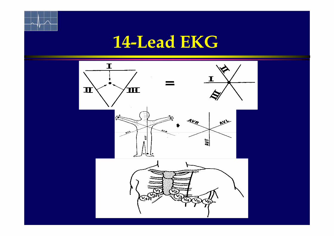
14-Lead EKG

7
Nomenclature
Right leadsaVR, V1, V3R, V4R
Left Leads I, aVL, V6
Inferior Leads II, III, aVF
Transitional LeadsV2-V5 IGNORE

8
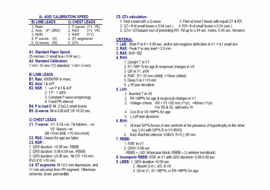
Practical Approach
B) LIMB LEADS C) CHEST LEADS
1. Rate 1. T waves (V1 & V6)
2. Axis (P& QRS) 2. R & S (V1 & V6)
3. NSR 3. RSR’ (V1)
4. P waves & PR interval (II) 4. ST Segments
5. Q waves (III) 5. QTc
A) Age/ Calibration/ Speed

9
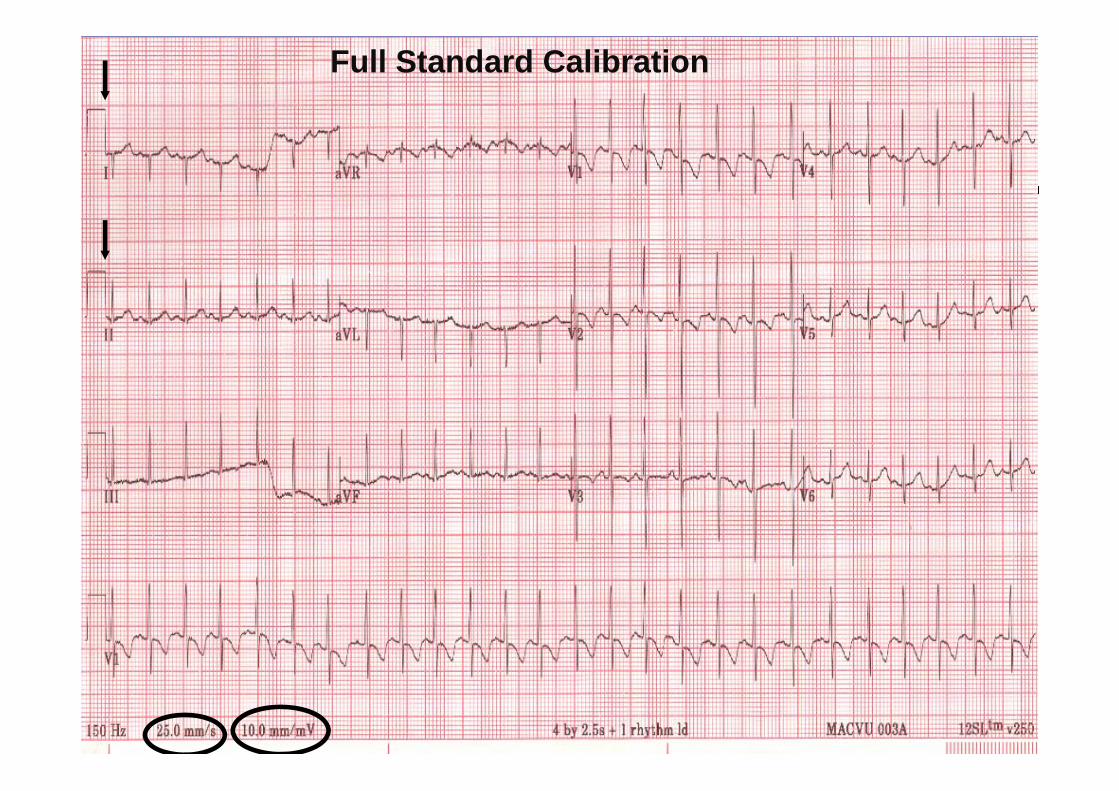
A) Age/ Calibration/ Speed
A.1) Speed:25 mm/sec 1 small box = 0.04 sec (or 40 msec) 1 large box = 0.2 sec (or 200 msec)
A.2) Calibration:1 mV = 10 mm 1 small box = 1 mm (0.1 mV)
½ standard: 1 mV = 5 mm 1 small box = 0.2 mV (voltages x 2)
10
Full Standard Calibration

11
1/2 Standard Calibration

12
Electronic Interpretation
GOOD for: rate, axis, intervals, RVH
OK for: LVH
BAD for: ST changes, Q waves, rhythm, QTc

13
10 yrs

14
B) Limb Leads
1. Rate2. Axis (P&QRS) in I & aVF3. NSR4. P-waves & PR interval (II)5. Q-waves
15
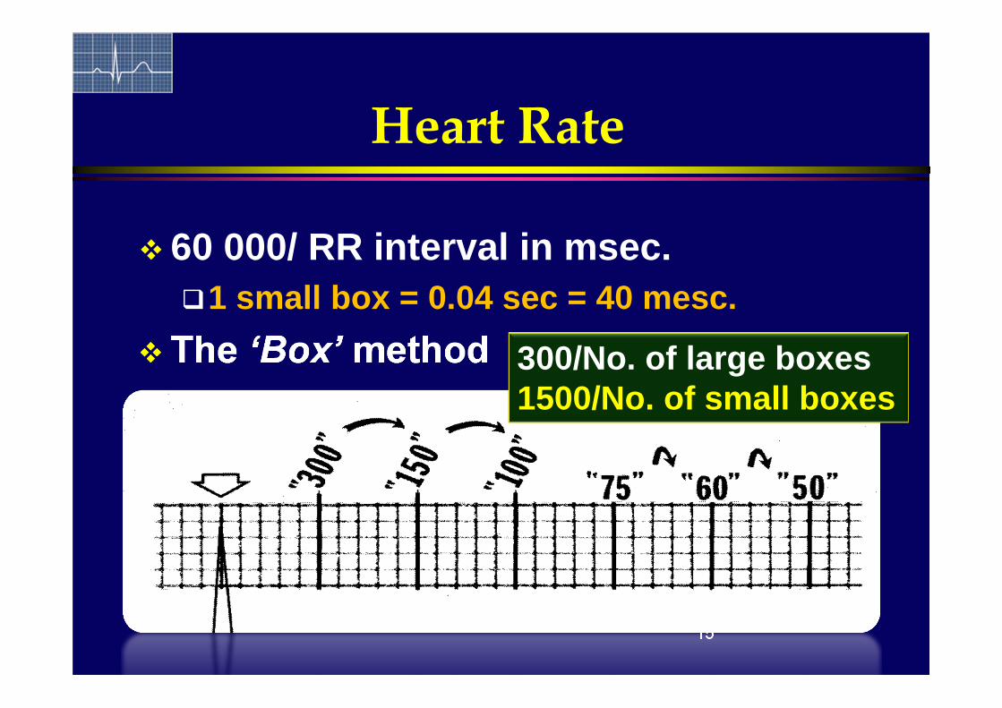
Heart Rate
60 000/ RR interval in msec.1 small box = 0.04 sec = 40 mesc.
The ‘Box’ method 300/No. of large boxes1500/No. of small boxes

16
B) Limb Leads
1. Rate2. Axis (P&QRS) in I & aVF3. NSR4. P-waves & PR interval (II)5. Q-waves

17
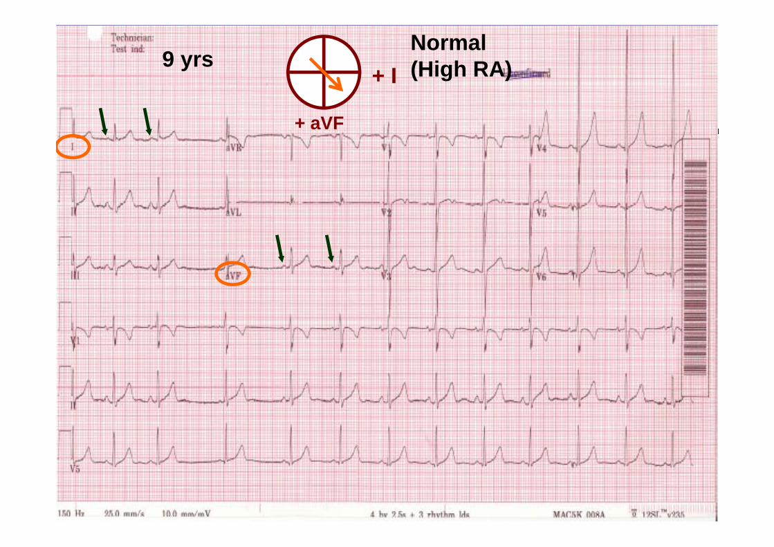
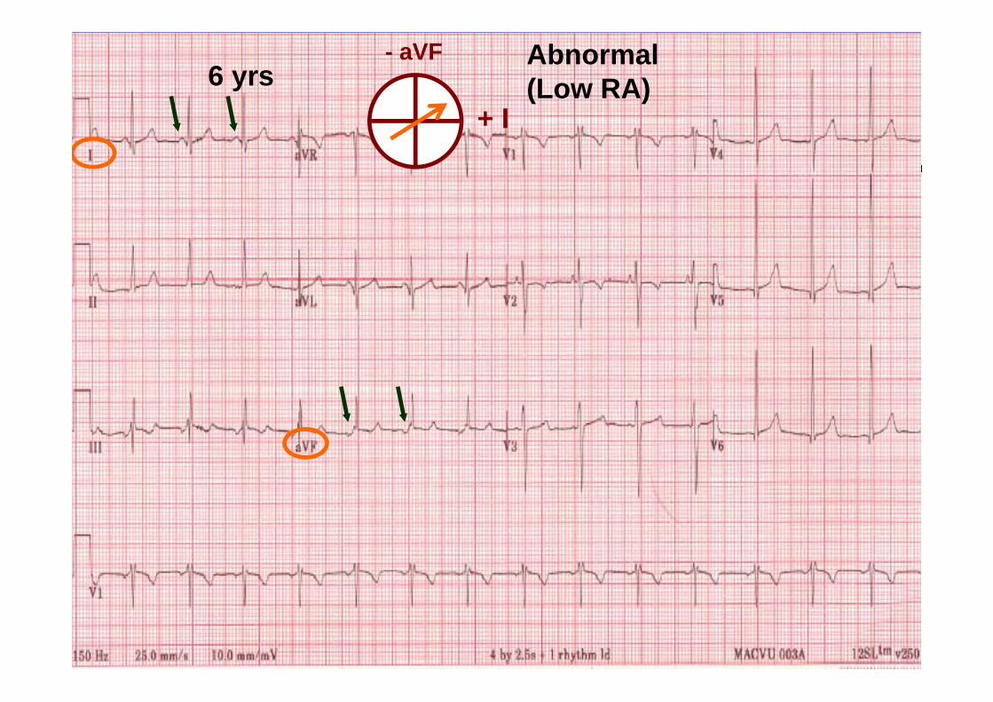
B2) P-wave Axis
0+180
-90
P in Lead I
P in Lead aVF+90
NormalHigh RA Rhythm
LowRA Rhythm
LowLA Rhythm
HighLA Rhythm
18
9 yrs+ I
+ aVF
Normal(High RA)

19
6 yrs
20
6 yrs+ I
- aVF Abnormal(Low RA)

21
B2) QRS Axis

QRS Axis
NormalRAD
LADLAD
RAD
0
+120
+180
-90
Lead I
Lead aVF+90
NW AxisSuperior Axis
Normal

Left Axis Deviation is
NEVER normal
(When QRS is –ve in aVF)
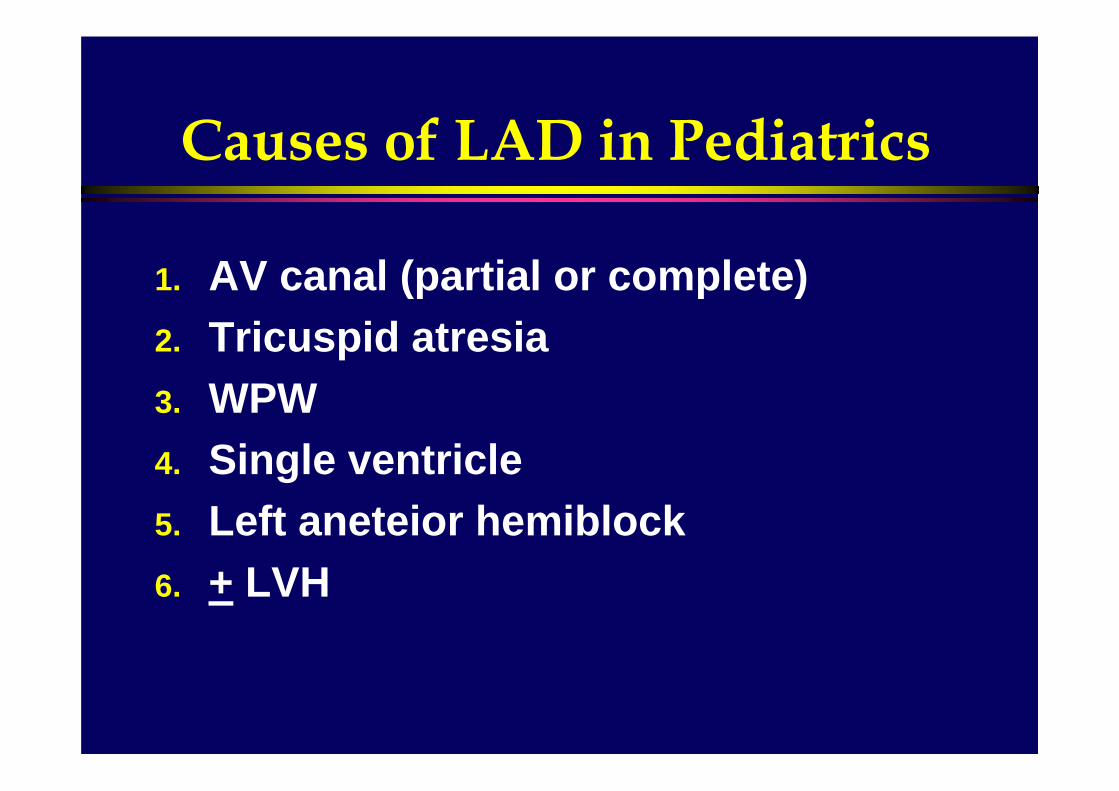
Causes of LAD in Pediatrics
1. AV canal (partial or complete)2. Tricuspid atresia3. WPW4. Single ventricle5. Left aneteior hemiblock6. + LVH
25
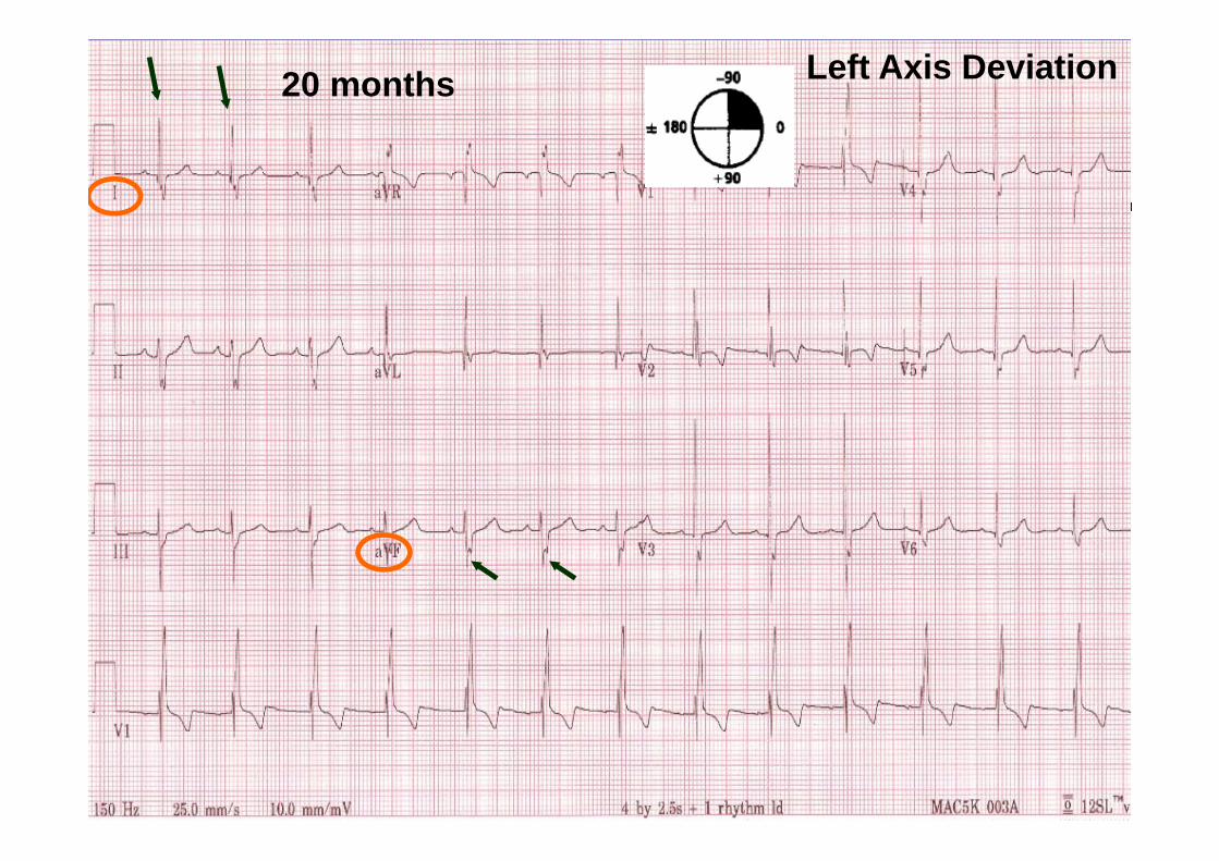
20 months Left Axis Deviation

26
5 yrs Left axis deviationNW-axis

B) Limb Leads
1. Rate2. Axis (P&QRS) (I & aVF)
3. NSR (Normal Sinus Rhythm)4. P-waves (II)5. Q-waves

Normal Sinus Rhythm
1. Normal P-wave axis +ve in I & aVF
2. One P : One QRS3. Fixed P-wave morphology4. Fixed PR interval
I (+)
aVF (+)

29
2 yrs Normal sinus rhythm
30
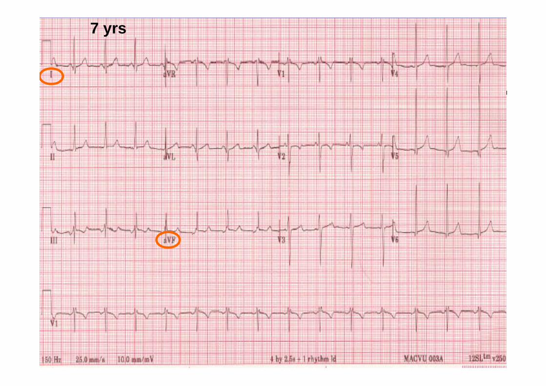
7 yrs

31
7 yrs
+ I
- aVFLow RA rhythm

32
11 yrs

33
11 yrs
- aVF
+ I
Different P-wave axisDifferent P-wave morphology
Wandering Pacemaker

B) Limb Leads
1. Rate2. Axis (P&QRS) (I & aVF)3. NSR 4. P-waves & PR interval (II)5. Q-waves

35
B4) P-Waves
Lead II Normal:
2.5 X 2.5 boxes 0.09 s X 2.5 mm
Abnormal: RAE (P-pulmonale) LAE (P-mitrale)

36
2 month

37
2 yrs

B4) PR Interval
Regardless the age:Normal 0.08-0.16 (2-4 small squares)
Prolonged PR1st- degree AV block: No clinical significance Common in: ASD, repaired TOF
Short PR interval:WPWPompe

39
B) Limb Leads
1. Rate2. Axis (P&QRS) (I & aVF)3. NSR 4. P-waves & PR interval (II)5. Q-waves

40
B5) Q-Waves
Not infarction Ventricular septum
Look for lead III value for age Normal:
Inferior leads, aVR & V6 Up to 1 box wide X 5 boxes height
(0.04 sec X 5 mm) Abnormal:
Leads I & aVL (ALCAPA) Wide (> small square = 0.04 sec.) (Infarction) Deep ((> 95th percentile for age) (LVH)

41
2 years2 yrs
42
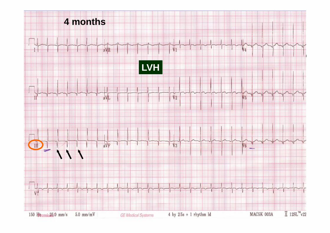
4 months
LVH
43
7 months

44
C) Chest Leads
1. T-waves (V1 & V6)2. RSR’3. R & S (V1 & V6)4. ST segments5. QTc

45
C) Chest Leads
1. T-waves (V1 & V6)2. RSR’3. R & S (V1 & V6)4. ST segments5. QTc

46
C1) T-Waves
AGE V1 V6
< 1 wk + +
1 wk- adolescence - +
> adolescence + +

47
1 day old1 day
Normal T-waves
48
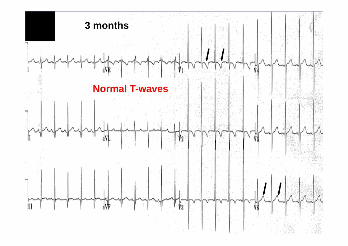
3 months
Normal T-waves

49
6 years
Normal T-waves

50
T-Waves
3 wks
15 yrs
Normal T-waves

51
3 wks

52
3 wks Flat T-waves in V1: RVH

53
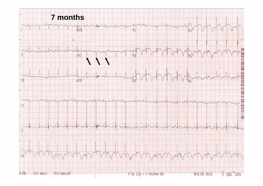
7 mo.

54
7 mo. +ve T-waves in V1: RVH

55
C) Chest Leads
1. T-waves (V1 & V6)2. RSR’ (V1)3. R & S (V1 & V6)4. ST segments5. QTc

56
C2) RSR’
Common in pediatrics: V1
Can be:NormalRVHRBBB
R
S
R’

57
C2) RSR’ in V1
Normal
< 10 mm
RVH
>= 10 mm
Height of R' ?
<= 0.08 sec
Incomplete RBBB
0.08-0.09 sec
RBBB
> 0.09 sec
QRS duration
R
S
R’
58
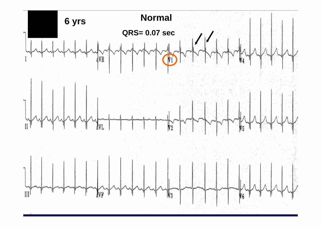
6 yrsQRS= 0.07 sec
Normal

59
16 mo. QRS= 0.05 secRVH

60
3 yrs RBBB

61
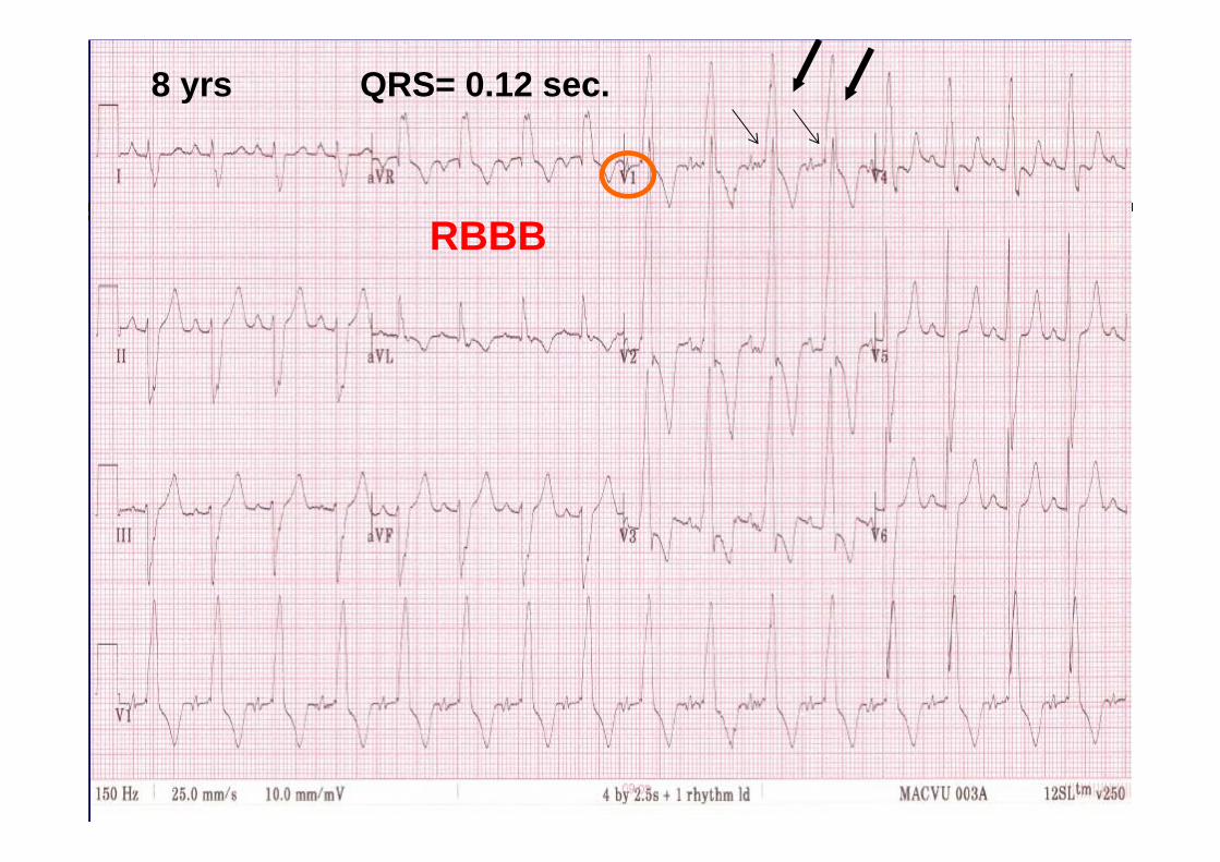
8 yrs QRS= 0.12 sec.
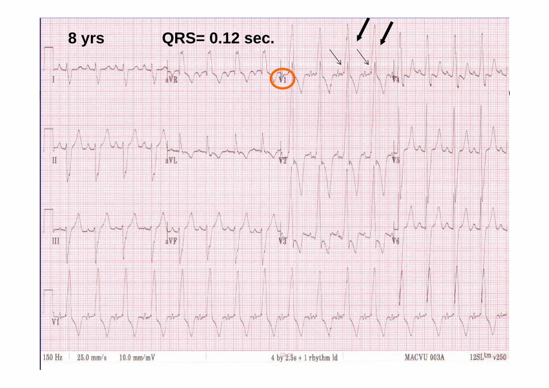
62
8 yrs QRS= 0.12 sec.
63
8 yrs QRS= 0.12 sec.
RBBB

64
C) Chest Leads
1. T-waves (V1 & V6)2. RSR’ (V1)
3. R & S (V1 & V6)4. ST segments5. QTc

65
R & S Waves8 yrs
S1 &/or R6 >98%: LVH
66
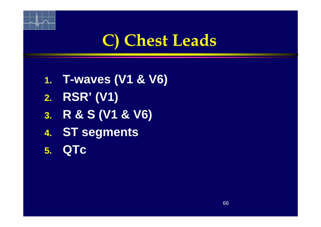
C) Chest Leads
1. T-waves (V1 & V6)2. RSR’ (V1)3. R & S (V1 & V6)4. ST segments5. QTc

67
2 days ST depression or elevation >1mm

68
C) Chest Leads
1. T-waves (V1 & V6)2. RSR’ (V1)3. R & S (V1 & V6)4. ST segments
5. QTc
69
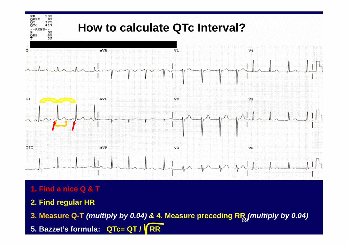
1. Find a nice Q & T
2. Find regular HR
3. Measure Q-T (multiply by 0.04) & 4. Measure preceding RR (multiply by 0.04)
5. Bazzet’s formula: QTc= QT / RR
How to calculate QTc Interval?

70
Step-Wise Approach
B) Limb Leads1. Rate2. Axis (P&QRS)3. NSR 4. P waves & PR
interval (II)5. Q waves (III)
C) Chest Leads1. T waves2. RSR’ (V1)3. R & S in (V1 & V6)4. ST segments5. QTc
A) Age/ Calibration/ Speed

Workshop

72
#1: 16 months
NSRRADBorderline RAERVH
TOF, Sec. ASD, PS, Pulm. HTN

73
#2: 2 months
NSRLADRVH
Tri atresia, AV canal

74
#3: 4 yrs
NSRLAD (NW axis)RBBBLAD+RBBB= Bi-fasicular block
Always after surgery for: TOF, Truncus, VSD, AV canal

75
#4: 7 yrs
Low RA rhythm Normal variation, PAC’s, Sinus venosus ASD

76
#5: 8 yrs
NSR with 1st degree AVB+ BAELADRBBB
Always after surgery: TOF, Truncus, AV canal

77
#6: 10 yrs
NSR with suns arrhythmia Normal

78
#7: 5 months
NSRBVH with strainShort PR
Classical Pompe EKG

79
#8: 1 yr
NSRBVH VSD, Cardiomyopathy

80
#9: 12 yrs
NSR Long QT syndrome

81
#10: 5 yrs
NSRLADShort PRDelta wavesWide QRSAbsent Q in V6
WPW

82
#11: 11 yrs
Wandering pacemaker Normal, PAC’s

83
#12: 2 yrs
Complete AV blockJunctional escape rhytm Complete AVB

STOP
85
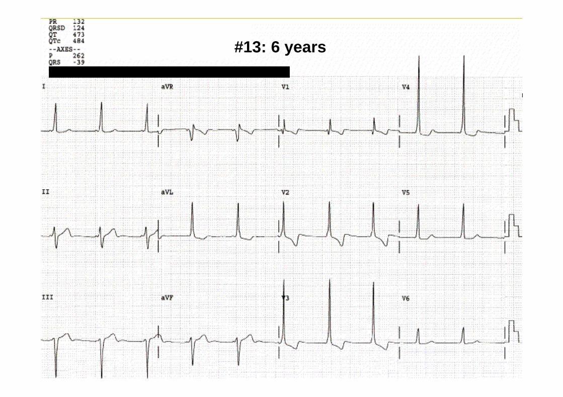
What’s this ?#13: 6 years

86
What’s this ?
NSRLADShort PRDelta wavesAbsent Q in V6
WPW)
#13: 6 years

87
#1: 3 months

88
Example 1: Tricuspid Atresia
HR 135 NSR LAD RAE RVH LVH BVH
3 months

89
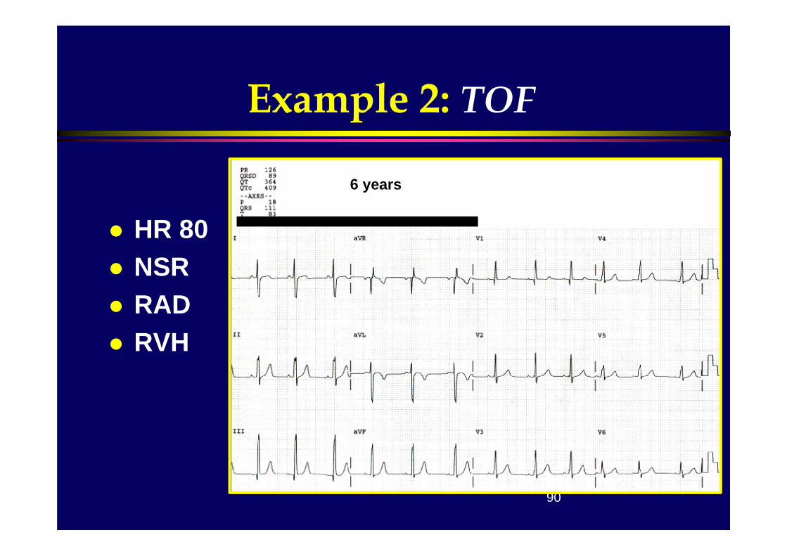
# 2: 6 years
90
Example 2: TOF
HR 80 NSR RAD RVH
6 years

91
# 3: 6 months

92
Example 3: VSD
HR 150 NSR LVH
15 years# 3: 6 months
93
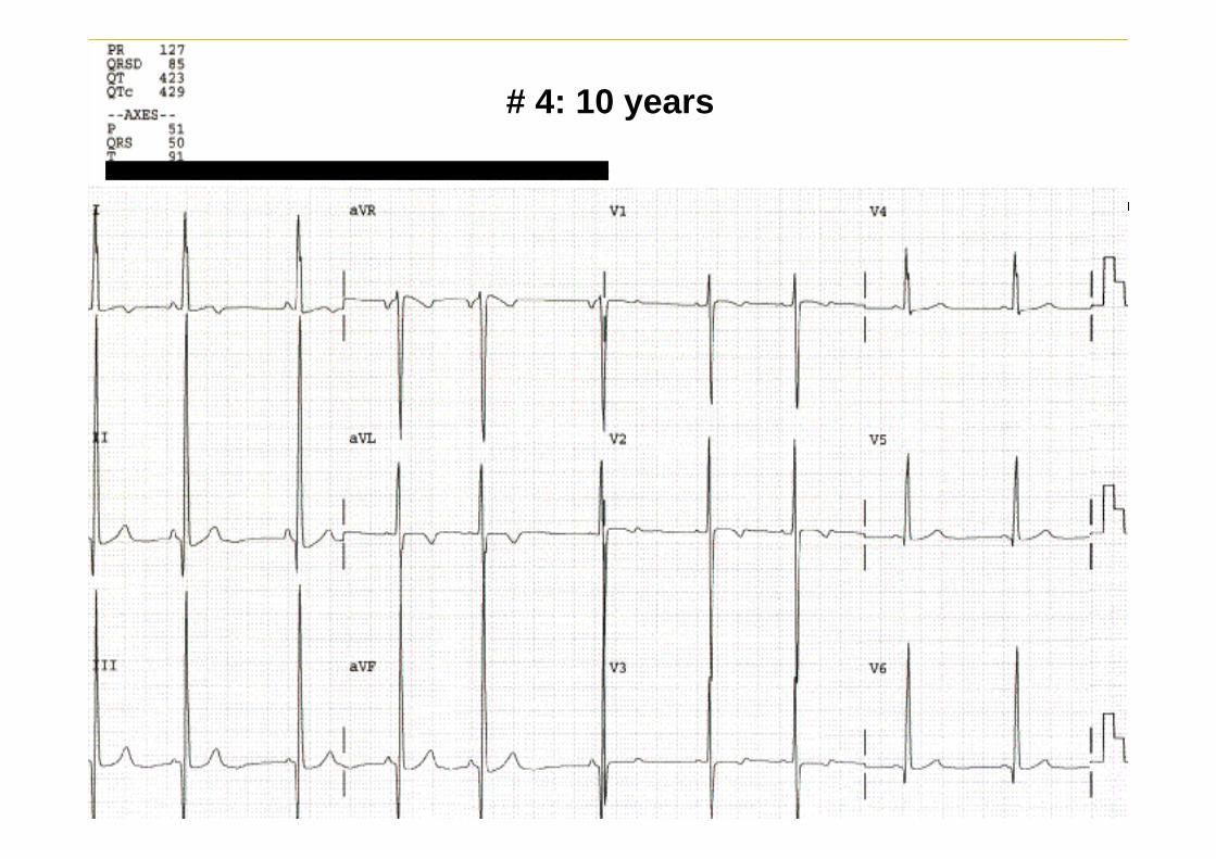
# 4: 10 years

94
Example 4: HCM
HR 60 NSR RVH LVH BVH ‘Strain’
10 years

95
# 5: 3 years

96
Example 5: LQTS
HR 94 NSR BVH QTc 0.54
5 years

97
#6: 2 years

98
Example 6: TOF s/p repair
HR 130 LAD RBBB
10 years2 years

99
3 yrs
NSRRAELVH

100
8 yrs
NSRLAD (NW axis)RAE