WORKING MEMORY: DEVELOPMENT, DISORDERS AND TRAINING
103
DEPARTMENT OF WOMEN AND CHILD HEALTH Karolinska Institutet, Stockholm, Sweden WORKING MEMORY: DEVELOPMENT, DISORDERS AND TRAINING Helena Westerberg Stockholm 2004
Transcript of WORKING MEMORY: DEVELOPMENT, DISORDERS AND TRAINING

DEPARTMENT OF WOMEN AND CHILD HEALTH
Karolinska Institutet, Stockholm, Sweden
WORKING MEMORY: DEVELOPMENT,
DISORDERS AND TRAINING
Helena Westerberg
Stockholm 2004

All previously published papers were reproduced with permission from the publisher. Paper: I- JOCN, Berkeley, II. & III. Swets & Zetlinger, VI. Nature Publishing Group All figures on the training software were reproduced with permission from the copyright owner; Cogmed Cognitive Medical Systems AB, Stockholm, Sweden. Proofreading Charlotta Thiringer. Published and printed by Karolinska University Press Box 200, SE-171 77 Stockholm, Sweden © Helena Westerberg, 2004 ISBN 91-7349-881-5

1
ABSTRACT Working memory: Development, Disorders and Training Working memory (WM) is the ability to keep information online during a short period of time. Brain regions underlying WM functioning are found in the frontal and parietal cortices. It is largely unknown to what extent the neural substrates underlying WM are susceptible to training induced change. Here we investigate the development of WM capacity, if improvement by training is possible and explore the neuronal correlates for training induced change.
In Study I we used functional magnetic resonance imaging (fMRI) to investigate the neural correlates of the developmental change in WM capacity during childhood. We found that during performance of a visuo-spatial working memory test (VSWM), there was a significantly higher activity in the superior frontal and intraparietal cortex in subjects with higher capacity. Thus, the development of these areas may underlie the development of VSWM during childhood. In Study II we used the VSWM test in children with and without ADHD and found that the test differentiated between these groups (P<.01). This supports the hypothesis that the WM deficit is a core deficit in ADHD. We proceeded by investigating if it was possible to improve WM capacity by training on WM tasks and secondly, if training effects could generalize to other cognitive tasks and areas of behaviour. These hypotheses were tested by designing a computerized program for WM training which was implemented in children with ADHD in two consecutive studies. In Study III (N=14), we saw significant improvements in the treatment group as compared to the control group on the trained WM task (P < .001), on the non-trained WM tests (span-board P < .001, and digit span P < .001), as well as on other non-trained tasks; Stroop (P < .01) and Raven’s matrices (P < .001). In Study IV the effects on the cognitive tests were replicated at a significance level of .01, with a randomized, double-blind, placebo-controlled multi-center design (N=44). In this study we also found generalisation of training effects to the behavioural level as evaluated by parents and teachers (inattention P < .01) and parent-rated hyperactivity/impulsivity P < .05). In Study V we investigated if stroke patients with significant WM deficits also could benefit from training. Participants suffering stroke one to three years earlier gained significant improvements in WM capacity (digit span p < .005, span board P < .05, and PASAT P < .001), and attention (RUFF 2&7 p < .005), as well as on cognitive symptoms in daily life, as measured by a self rating questionnaire (P < .005). Study VI was undertaken to explore the neuronal correlates of WM improvement. Healthy young adults underwent fMRI before and after WM training. Task specific increases in brain activity were found in prefrontal and parietal cortices. These regions are known to underlie WM functioning.
In summary: WM shows a prolonged developmental course in humans.
WM deficits are prominent in ADHD and following brain injury. WM can be improved by training and the training effect also generalizes to other cognitive tasks. The increase in WM capacity during childhood as well as after training is associated with increased brain activity in the prefrontal and parietal cortices.

2
LIST OF PUBLICATIONS
I. Klingberg T, Forssberg H, Westerberg H
Increased Brain Activity in Frontal and Parietal Cortex Underlies the Development
of Visuospatial Working Memory Capacity during Childhood
Journal of Cognitive Neuroscience, 2002, 14:1, pp. 1–10
II. Westerberg H, Hirvikoski T, Forssberg H, Klingberg T
Visuo-spatial working memory span: a sensitive measure of cognitive deficits in
children with ADHD
Child Neuropsychology, in press
III. Klingberg T, Forssberg H, Westerberg H
Training of Working Memory in Children With ADHD
Journal of Clinical and Experimental Neuropsychology, 2002, 24:6, pp. 781-791
IV. Klingberg T, Fernell E, Olesen P, Johnson M, Gustafsson P, Dahlström K, Gillberg CG,
Forssberg H, Westerberg H
Computerized Training of Working Memory in Children with Attention-
Deficit/Hyperactivity Disorder – a Controlled, Randomized, Double-blind, Trial
Submitted
V. Westerberg H, Jacobaeus H, Hirvikoski T, Clevberger P, Östensson M-L, Bartfai A,
Klingberg T
Computerized Working Memory Training - A Method of Cognitive Rehabilitation
after Stroke
Submitted
VI. Olesen PJ, Westerberg H, Klingberg T
Increased prefrontal and parietal activity after training of working memory
Nature Neuroscience, 2004, 7:1, pp. 75-79

3
CONTENTS Abstract ................................................................................................................1
Working memory: Development, Disorders and Training................1 List of Publications ..............................................................................................2 Contents................................................................................................................3
List of abbreviations ......................................................................................4 Acknowledgements..............................................................................................5 1 General introduction......................................................................................6
1.1 Definition of working memory ..........................................................6 1.2 Neuronal mechanisms underlying working memory ........................9
1.2.1 Working memory at the cellular level .................................10 1.2.2 Working memory at the macro level ...................................10
1.3 Multiple domain or multi-modal working memory?.......................12 1.3.1 Verbal working memory ......................................................12 1.3.2 Non-verbal working memory...............................................13 1.3.3 Supramodal working memory areas ....................................13 1.3.4 Content or process-specific organization in the prefrontal cortex ...............................................................................................14
1.4 Working Memory at a functional level and its relation to other cognitive constructs .....................................................................................15
1.4.1 Short term versus working memory ....................................15 1.4.2 Long term versus working memory.....................................16 1.4.3 Working memory in attention, inhibition and executive control ...............................................................................................16
1.5 On cognitive development................................................................20 1.6 On cognitive training ........................................................................21
1.6.2 Attention Deficit Hyperactivity Disorder ............................27 DSM IV Criteria of ADHD...............................................................28 1.6.3 Working Memory deficits and ADHD................................28
1.7 hypothesis .........................................................................................30 2 Methods........................................................................................................31
2.1.1 Psychometrics -tests of WM and executive functions ........31 2.1.2 Training method ...................................................................38 2.1.3 Functional brain imaging, linking biology to behaviour.....41
3 Results and discussion.................................................................................44 3.1.1 Study I – Increased Brain Activity in Frontal and Parietal Cortex Underlies the Development of Visuospatial Working Memory Capacity during Childhood ...............................................44 3.1.2 Study II - Visuo-spatial working memory span: a sensitive measure of cognitive deficits in children with ADHD....................48 3.1.3 Study III-Training of Working Memory in Children with ADHD ...............................................................................................53 3.1.4 Study IV - Computerized Training of Working Memory in Children with Attention- Deficit/Hyperactivity Disorder – a Controlled, Randomized, Double-blind, Trial .................................56

4
3.1.5 Study V- Computerized Working Memory Training - A Method of Cognitive Rehabilitation after Stroke............................ 59 3.1.6 Study VI- Increased prefrontal and parietal activity after training of working memory............................................................ 62
4 General Discussion ......................................................................................65 References ..........................................................................................................71 LIST OF ABBREVIATIONS ADHD Attention Deficit Hyperactivity Disorder BOLD Blood Oxygen Level Dependent CNS Central Nervous System CPT Continuous Performance Test CRT Choice Reaction Time DMS Delayed Match to Sample DLPFC Dorso-lateral Prefrontal Cortex DSM-IV Diagnostic and Statistical Manual of mental disorders, 4th ed. DTI Diffusion Tensor Imaging EEG Electro-Encephalography ES Effect Size fMRI functional Magnetic Resonance Imaging MRI Magnetic Resonance Imaging PET Positron Emission Tomography PFC Pre Frontal Cortex SPM Statistical Parametric Mapping STM Short Term Memory TBI Traumatic Brain Injury TMS Trans cranial Magnetic Stimulation WM Working Memory VSWM Visuo Spatial Working Memory

5
ACKNOWLEDGEMENTS People directly involved in the studies are either authors or mentioned in the acknowledgement section in each paper. Anyhow, I would like to thank the clinicians who have been helpful in the recruitment of patients and also express my gratitude towards the participants themselves. I also want to mention some significant colleagues: David Skoglund and Jonas Beckeman, the professional programmers who turned the idea about WM training into reality by creating the training software. Tatja Hirvikoski, psychologist, for sharing the interest in neuropsychology. My co-doctoral students, Sara Bengtsson, Zoltan Nagy and Pernille Olesen. I would also like to take the opportunity to express my gratitude to some other significant persons. These are persons who have provided me with the opportunity to reach new areas of interest and higher levels of individual and professional maturation. These are: Carl-Johan Marnell who in 1987 introduced me to the field of psychology by employing me as a gardener. Margareta Hilpertshauser who introduced me to the field of neuropsychiatry. Liselotte Maurex and Birgitta Böhm who guided me into becoming a professional clinician in the field of neuropsychology. Hans Forssberg who welcomed me to, and encouraged me to stay and work in his research group. Torkel Klingberg, my main supervisor who has put a lot of effort into my scientific education, encouraged critical thinking and promoted high standards in research. In my personal life significant persons are: Louise and Oswald Westerberg, my parents. Anders, Katarina och Samuel, my brother and his family. Eva Nilsson, long lasting friend. And Stefan Berglund my life companion. Thank you for being there. Financial support: Frimurarna Barnahuset Stockholm, Föreningen för Rörelsehindrade Barn och Ungdomar, Institutionen för kvinnors och barns hälsa (interna doktorandmånader), Karolinska Institutet (Rörlig doktorandtjänst), Majblommans Riksförbund, Märta och Gunnar Philipsons stiftelse, Stiftelsen Samariten, Stiftelsen Solstickan, Strokeförbundet, Sällskapet Barnavård.

6
1 GENERAL INTRODUCTION
1.1 DEFINITION OF WORKING MEMORY There are many definitions of WM stemming from different areas of research, from
psychology (Alan Baddeley) to neuroscience (Patricia Goldman-Rakic). Since our
work is in the field of cognitive neuroscience we have used definitions close to
Goldman-Rakic´s (1987) definition; “Working memory is the ability to keep
information online during a short period of time”. However, our view of WM also
agrees on a broader description of WM, which includes operations on the information
held on-line.
1.1.1.1 Background
Working memory is close to and sometimes partly overlapping with other cognitive
constructs, as for example short term memory (STM), attention, executive
functioning and interference control (see these paragraphs respectively for a more
detailed description). To put it short; WM includes the selection of relevant
information to attend to and the filtering out of irrelevant information, functions that
also can be described as Control of Attention, (Engle RW, Kane JM & Tuholski SW,
1999).
The term “working memory” was introduced into the literature of
cognitive psychology by Miller, Galanter and Pribram in their book Plans and
structure of behaviour, 1960. Their definition is astonishingly modern; they did
foresee the goal directed and executive components of WM:
“When we have decided to execute some particular plan, it
is probably put in some special state or place where it can be remembered while it is
being executed. Particularly if it is a transient, temporary kind of plan that will be
used today and never again, we need some special place to store it.”…”…we should
like to speak of the memory we use for the execution of our plans as a kind of quick-
access, working memory.” (p.65)

7
They did not stop with a cognitive definition of working memory, but
went on and proposed an anatomical localisation of these functions in the brain:
“This most forward position of the primate frontal lobe
appears to us to serve as “working memory” where plans can be retained
temporarily when they are being formed, or transformed, or executed.” (p. 207)
According to my own opinion the functional purpose of WM is to guide goal-
directed thoughts and behaviour. The ability to maintain sensory representations no
longer present in the environment, and piece together relationships between these and
internal information is a prerequisite for goal-directed and adaptive behaviour.
Since the fifties there have been a number of definitions of WM and STM,
often describing the same cognitive construct (see §1.4.1 Short term versus working
memory). The term STM was more often used earlier while the term WM has
successively become more common and are the most frequently used today. These
concepts have been incorporated in such disparate areas as psychology and computer
programming (Shute VJ, 1991). In spite of the lack of an agreement on a core-
definition of WM, shared in different nomenclatures, most definitions of WM include
components of temporary storage of internal representations (maintenance),
operational processes (manipulation) upon these, and some definitions also include
(executive) control aspects. Furthermore, WM has been fractionated into different
basic subcomponents. Terms often encountered are listed below.
Content The representations temporarily held on-line during the delay of WM tasks
consists of either external information from the sensory system, or internally stored
information, retrieved from long-term memory (LTM).
Process The content in WM is not only maintained (which is more in line with the
definition of STM) but can be manipulated by different operations, with the
prospective aim of facilitating goal directed behaviour (external as motor execution or
internal such as decision making).

8
Rehearsal One way to actively maintain the information in WM is to rehearse it,
(repetitively direct attention to it). Rehearsal supports WM, protects the information
from fading and possibly also from interference from competing stimuli (Raye CL,
Johnson MK, Mitchell KJ, Reeder JA & Greene EJ, 2002 ).
Decay The content in WM is retained transiently (for seconds) and is not stored in
LTM. If not actively maintained (repeatedly attended to), the content vanishes.
Interference control The ability to inhibit attending to irrelevant information
influences how well the relevant representations can preserve against passive decay
and distracters.
Capacity Capacity is a quantitative value that describes the maximum amount, i.e.
the number of items of information that can be held active in parallel in WM. WM
capacity corresponds to an individual’s current ability or to the average ability of a
defined population such as an age group. The capacity of WM is limited and a
popular view is that seven (±2) items of information can be processed simultaneously
in WM. Although, one item can constitute a ´chunk´ of information; a chunk is a
cluster of logically connected items, maintained as one and thus extending the
perimeter of seven. A concept closely related to WM capacity is WM Span which
denotes the upper limit an individual manage to reach on a particular WM test.
Load effect Quantifies the relative processing resources required to perform a WM
task. The load on WM is not only about the number of items to be maintained, but
can also denote the need for elevated processing in for example performance of dual
tasks (see §1.4.3).

9
1.2 NEURONAL MECHANISMS UNDERLYING WORKING MEMORY
Research in the field of WM is quite unique in that it spans from sub cellular
mechanisms such as the function of dopamine receptors in neurons, via neuronal
networks, over cognition to the behaviour level (Goldman-Rakic 1995, 1996;
Castellanos FX & Tannock R, 2002). The neural substrates of WM in both primates
and humans is found in the prefrontal cortex (PFC); recorded at the neuronal level
during delay-specific activity in primates (Funahashi, Bruce, & Goldman-Rakic,
1989; Fuster & Alexander, 1971; Tsujimoto S & Sawaguchi T, 2004) and
documented by neuro-imaging methods at the cortical level in humans performing
WM tasks (Jonides J, Smith EE, Koeppe RA, Awh E, Minoshima S & Mintun MA,
1993; Courtney SM, Petit L, Maisog JM, Ungerleider LG & Haxby JV, 1998a;
Paulesu E, Frith CD & Frackowiak RS, 1993; Cohen JD, Pearstein WM, Braver TS,
Nystrom LE, Noll DC, Jonides J & Smith EE, 1997). In the search for the neuronal
mechanisms that underlie WM, tasks that tap vision are most commonly used in
primates since they lack language. A frequently used paradigm in animal research is
the delayed match to sample (DMS) task, in which for example a piece of food is
placed in a container by an experimenter while the monkey watches. During the delay
a cover is applied which prevents the monkey from visually fixate the containers.
After the delay the cover is removed and the monkey indicates in which container it
thinks the food is. Also in humans a great deal of research utilizing brain imaging
techniques has focused on non-verbal WM. One reason is the intent to compare and
extend the findings from animal studies. Another reason is that at least magnetic
resonance imaging (MRI) is noisy and can interfere with auditory WM tasks. In
human’s, tasks similar to but more demanding than the DMS has been used, for
example the N-back test, in which the subject has to match and respond to stimuli that
appeared one, two or three items back in a series. The delay specific activity seen in
electrophysiological single cell recordings in the PFC of primates (Funahashi, Bruce,
& Goldman-Rakic, 1989) are thought to be analogous to the delay-specific activity
found in functional brain imaging studies in the PFC of humans (Cohen JD, et al.,
1997; Courtney SM, et al., 1998a; Rowe JB, Toni I, Josephs O, Frackowiak RS &
Passingham RE, 2000)

10
1.2.1 Working memory at the cellular level Work with electrophysiological recordings in awake monkeys shows that neurons
normally change their rate of firing when a distracting stimulus that interfere with
ongoing activity appears (Miller EK, Erickson CA & Desimone R, 1996). But there
are some areas of the brain, situated in the PFC where neurons maintain their rate of
activity, induced by holding a target stimulus on-line during a delay, even after
exposure to distracting stimuli (Miller EK, Erickson CA & Desimone R, 1996). These
neurons are thought to underlie the maintenance of internal representations (content)
that are not available to the sensory organs anymore. Indeed, results from
electrophysiological recordings performed on neurons in the primates PFC, show that
there are neurons with sustained delay specific activity throughout the entire retention
period (but not necessarily before and after this period) (Fuster JM & Alexander GE,
1971; Kubota & Niki 1971; Funahashi, S., Bruce, C. & Goldman-Rakic, PS., 1989;
Goldman-Rakic PS., 1996; Hasegawa R, Sawaguchi T & Kubota K, 1998; Tsujimoto
S & Sawaguchi T, 2004).
1.2.2 Working memory at the macro level Brain activity studies in humans as well as in primates have shown that prefrontal
areas are bilaterally active during performance of WM tasks (and that there also are
parietal cortex and the basal ganglia activations) (in humans: D’Esposito M, Aguirre
GK, Zarahn E, Ballard D, Shin RK & Lease J, 1998a; D’Esposito M, Postle BR &
Rypma B, 2000a; Smith EE & Jonides J, 1998a; Lewis SJG, Dove A, Robbins TW,
Barker RA & Owen, AM, 2004) (In animals: Goldman-Rakic, P. S, 1995; Inoue M,
Mikami A, Ando I & Tsukada H, 2004). This sustained activity is thought to be the
mechanism mediating WM functioning; either by maintaining stimuli specific
information temporarily stored in the prefrontal neuronal network per se, or by
driving networks with connections to posterior neuronal assemblies that store
modality specific information. In humans delay specific activity is not necessarily
restricted to maintenance of an earlier sensory stimulation, but can represent
processing of a mental representation, for example an instruction of prospective
action. Thus information held in WM is thought to be; either transiently stored in
PFC, alternatively controlled from the PFC and parts of the parietal cortex (Goldman-

11
Rakic P, 1987). In the latter view, interactions between PFC and stimulus-specific
sensory areas mediate WM.
My own opinion is in line with this interpretation. WM processes probably
don’t take place in one spot in the brain; i.e. the anatomical substrate that underlie
WM whereto representations are copied and transiently buffered. But more
reasonably, is carried out by interacting networks whose activity is regulated from the
PFC. WM may be operating by top-down control, recurrently directing attention to
relevant representations, which are (probably) stored in more posterior regions of the
brain. (Desimone R & Duncan J, 1995; Miller EK & Cohen JD, 2001; Smith EE &
Jonides J, 1999; Courtney, SM et al. 1998a; D’Esposito, M., Ballard D, Zarahn E &
Aguirre GK, 2000b). How well these cortical areas are connected to and
communicate with other areas in the brain that store relevant representations (i.e
sensory areas) affects the effectiveness of WM. Given that top-down signals from
PFC enhance internal representations of sensory stimuli in posterior parts of the brain,
efferent signals from the PFC may have different functional implications, depending
on which cortical region it communicates with. For example, sensory areas for
perceptual processing of visual or auditory stimuli in the posterior cortex store
different kinds of information, but both can be under the influence of top-down
control from the PFC. This theory is supported by studies which show that prefrontal
cooling remove neurons in more posterior areas the ability to retain stimuli in WM
(Quintana & Fuster, 1993). Moreover others (Fuster JM, 1985; Chafee and Goldman-
Rakic, PS., 2000) have found that deactivation of the lateral PFC cortex decrease the
response of visual cortical neurons to a relevant stimulus. In order to be in the
position to influence other areas of the brain, connections within and from the PFC
are widespread, the PFC projects to many other areas in the cortex (Petrides M &
Pandya DN, 1994; Ongur D & Price JL, 2000).
12
1.3 MULTIPLE DOMAIN OR MULTI-MODAL WORKING MEMORY?
Is WM one unitary system processing different kinds of information, or is WM a
number of different systems specialized in processing different content as Patricia
Goldman-Rakic has proposed? The most well known model, that of Baddeley and
Hitch (1974), is somewhere in between. According to this model which stems from
cognitive psychology, WM has a domain-specific organization with specialized sub-
stores that are regulated by a third separate entity that operates as a central executive.
The executive unit is thought to manage the allocation of attentional control to the
slave systems, which stores short term representations of diverse content, either
verbal or visual (see also § 1.4.3 Working memory in attention, inhibition and
executive control).
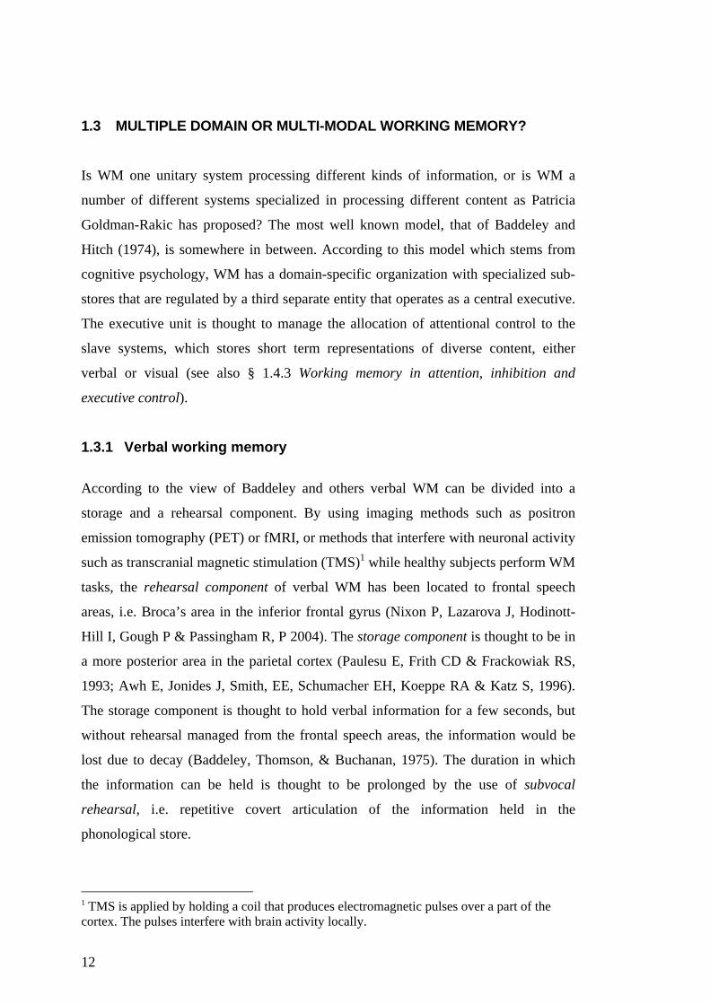
1.3.1 Verbal working memory According to the view of Baddeley and others verbal WM can be divided into a
storage and a rehearsal component. By using imaging methods such as positron
emission tomography (PET) or fMRI, or methods that interfere with neuronal activity
such as transcranial magnetic stimulation (TMS)1 while healthy subjects perform WM
tasks, the rehearsal component of verbal WM has been located to frontal speech
areas, i.e. Broca’s area in the inferior frontal gyrus (Nixon P, Lazarova J, Hodinott-
Hill I, Gough P & Passingham R, P 2004). The storage component is thought to be in
a more posterior area in the parietal cortex (Paulesu E, Frith CD & Frackowiak RS,
1993; Awh E, Jonides J, Smith, EE, Schumacher EH, Koeppe RA & Katz S, 1996).
The storage component is thought to hold verbal information for a few seconds, but
without rehearsal managed from the frontal speech areas, the information would be
lost due to decay (Baddeley, Thomson, & Buchanan, 1975). The duration in which
the information can be held is thought to be prolonged by the use of subvocal
rehearsal, i.e. repetitive covert articulation of the information held in the
phonological store.
1 TMS is applied by holding a coil that produces electromagnetic pulses over a part of the cortex. The pulses interfere with brain activity locally.

13
1.3.2 Non-verbal working memory There is data from both animal and human studies on the neuronal substrate for non-
verbal WM. Electrophysiological studies in primates have shown that there is
position specific cells in the PFC which are activated selectively during spatial WM
tasks (Goldman-Rakic PS, 1995; 1996; Tsujimoto & Sawaguchi, 2004). Functional
imaging shows that the PFC is activated also in humans during performance of non-
verbal WM tasks together with activations in the parietal cortex (Owen AM, Evans
AC & Petrides M, 1996; Smith EE, Jonides J & Koeppe RA, 1996; Klingberg T,
O’Sullivan BT & Roland PE, 1997). There are some indications on asymmetrical
activation with relatively greater left hemispheric recruitment in verbal WM tasks and
right hemispheric involvement in non-verbal WM (Smith EE, Jonides J & Koeppe
RA, 1996). However, an increase in bilateral activation can be seen with increasing
task demand, suggesting functional recruitment from the corresponding contra-lateral
areas (Smith EE, Jonides J & Koeppe RA, 1996; Klingberg T, O’Sullivan BT &
Roland PE, 1997).
Visuospatial information can, hypothetically be processed by an equivalent to
the verbal rehearsal loop, i.e. through repetitive rehearsal of locations, mediated by
covert rehearsal of eye-movements, this suggestion is supported by the activation in
or close to the frontal eye-fields (FEF) during tasks of spatial WM (Courtney SM et
al., 1998a; Awh E, Anllo-Vento L & Hillyard SA, 2000; see also Study I).
1.3.3 Supramodal working memory areas Regardless of the type of WM task to be performed; there is a widely distributed WM
network activated, including prefrontal and parietal regions (Klingberg T, 1998;
Duncan J & Owen AM, 2000; Sylvester, CYC, Wager TD, Lacey SC, Hernandez L,
Nichols TE, Smith EE & Jonides J, 2003). There is some support from neuro-imaging
studies on the distribution of different WM networks. These networks though, are not
entirely separated but partly overlapping. There are areas in frontal premotor cortex
(Broca’s area) predominantly activated during performance of verbal WM and a
counterpart for visuo-spatial information is found in a fronto -parietal neuronal
network. However the major parts of regional activation, evoked by performance of
different WM tasks, are overlapping. The intersections are found in prefrontal and

14
parietal association areas, which implies that these areas mediate supra modal
function (Klingberg T, Roland PE, & Kawashima, R. 1996) (see also §1.4.3.1).
1.3.4 Content or process-specific organization in the prefrontal cortex Some functional imaging studies support a regional distinction between the
processing of spatial and object information in WM (Belger A, Puce A, Krystal JH,
Gore JC, Goldman-Rakic P. & McCarthy G, 1998; Courtney SM, Ungerleider LG,
Keil K & Haxby JV, 1996; Sala JB, Rämä P & Courtney SM, 2003), whereas as
others have suggested that these activation patterns are better accounted for by
differences in the executive demands of the task (Petrides M, Alivisatos B, Evans AC
& Meyer E, 1993). Others again (D’Esposito M et al., 1998a) has not found any
evidence for a dorso-ventral subdivision of prefrontal cortex depending on the type of
material held in WM. Those who advocate a process-specific organization suggest
that that ventrolateral frontal cortex is more activated during the maintenance of
information, while the dorsolateral frontal cortex is more active in manipulating
information (Petrides M et al., 1993). The PFC in humans is involved in higher
executive functions such as manipulation of information in WM but these areas do
also show task related activity when only maintenance of a single item is required but
memory load is high, such as during prolonged delay (Cohen J, 1997). Moreover,
electrophysiological studies show that there are neurons in the prefrontal cortex of
monkeys that are sensitive to both the object identity and spatial location of objects
(Miller EK & Cohen J, 2001). These findings imply that the PFC is involved both
during maintenance and processing of content in WM and supports the idea that
different modalities of WM is managed by overlapping neuronal substrates.

15
1.4 WORKING MEMORY AT A FUNCTIONAL LEVEL AND ITS RELATION TO OTHER COGNITIVE CONSTRUCTS
1.4.1 Short term versus working memory
The concept of WM, earlier referred to as STM, has undergone some modification
since it was introduced. STM was thought to have two functions: (1) storing material
on-line for a few seconds, as when we rehearse a phone number until we dial it, and
(2) providing a gateway to LTM (Atkinson R & Shiffrin R, 1968).
While cognitive scientists in the psychological tradition continue to consider
the simple storage as one component in a multi level system, the theory of a gateway
function has been discarded since there are patients impaired on STM tasks, but
performing normally on LTM tasks (Shallice T & Warrington EK, 1970).
Conversely, patients suffering from Korsakoff Syndrome perform normally on WM
tests, but their LTM is poor. In modern cognitive neurosciences the term ‘working
memory’ is most common and the concept of WM has been widened to include, not
only maintenance of information, but higher cognitive functioning as reasoning and
problem solving. If considering the storing part of WM as a memory for the short-
term, this function relies on strategies such as rehearsal and chunking while WM is
more complex and consists of both storing, manipulation and attention components
(Engle RW, Kane JM & Tuholski SW, 1999). Moreover, the extent to which a task
demands WM is determined by the extent to which it requires active maintenance of
information that otherwise should be lost due to interference (Lustig C, May CP &
Hasher L, 2001).
As there is no clear-cut distinction between STM and WM, there are no
‘‘pure’’ measures of either STM or WM (Cowan N, 1995; Engle RW, Kane JM &
Tuholski SW, 1999). Conway et al (Conway ARA, Cowan N, Bunting MF, Therriault
DJ & Minkoff SRB, 2002) has argued that the more a task forces the participant to
engage in controlled effortful processing rather than automatized skills, the more the
task tap WM capacity, and the less STM (Conway ARA, et al., 2002). Consistent
with this, there is no objective measure that separates between STM and WM. It all
depends on the individual’s capacity; a STM task for an adult can be considered a
WM task for a child. The latter is also applicable for individual differences in
capacity within the same age-group (Engle RW, Kane JM & Tuholski SW, 1999).

16
1.4.2 Long term versus working memory
LTM and WM relies on different anatomical substrates and underlies diverse
cognitive functions, thus these are easier to separate than the distinction between
STM and WM. LTM is dependent on structures in the medial temporal lobe;
hippocampus and enthorhinal cortex, which WM is not (Milner B, Squire LR &
Kandel ER, 1998). Neural substrates underlying WM involves prefrontal and parietal
areas. Some activation in prefrontal areas known to underlie WM is also present
during performance of LTM tasks (Nyberg L, Marklund P, Persson J, Cabeza JR,
Forkstam C, Petersson KM & Ingvar M, 2003; Ranganath C, Johnson MK &
D’Esposito M, 2003), this can be due to that some tests of LTM contain features that
demands WM. Time resolution and storage capacity also separates between the
concepts of WM and LTM. The amount of information held on-line in WM is limited
and lasts for seconds while there is no known capacity limit for amount and storage of
information in LTM. Moreover, WM and LTM functioning, respectively, show
different developmental trajectories. WM displays a continuous developmental trend
from childhood (age 4-5 years) and does not peak until late adolescence (16-17
years). On the contrary, LTM functioning emerge adult-like after an age of about
seven years (Gathercole SE, 1998).
Although WM and LTM are separable entities they are often used together.
For example WM doesn’t only maintain representations from the external world but
also past representations of already known information stored in LTM. This
information can be conceptualized as memory traces in LTM temporarily activated
over threshold in WM (Fuster JM, 1995). WM capacity also influences the success of
retrieval from LTM in tasks where the use of controlled and effortful search is
needed. For example in tasks featuring parameters that tax temporal ordering of
information, source memory and free recall (Squire LR, 1987).
1.4.3 Working memory in attention, inhibition and executive control The concept of WM is closely related to some other cognitive constructs. These are
attention, interference control and executive control. These are cognitive constructs
close to but separable from WM, although sometimes overlapping with each other.

17
1.4.3.1 The central executive versus executive WM
According to Baddeley’s WM model there are specialized sub-components that are
regulated by a third separate entity operating as a central executive. The least defined
part of Baddeleys model of WM is how the central executive carries out its functions
and where in the brain it resides. To answer this, Baddeley proposed that the central
executive could be identified by using a kind of subtraction technique; comparing
brain activity when a subject performs one single task with that evoked when two
different tasks are performed simultaneously, a so called dual task. Performance of
dual tasks demand more effort than the sum of two single tasks performed separately.
In order to carry out two tasks simultaneously, the requirement of a third resource, the
central executive is suggested necessary. The central executive is thus not needed
during performance of the single tasks separately, but becomes active in the
management of dual tasks or otherwise intensified work load. Another explanation
derived from research in the neurosciences agrees upon the hypothesis that when
work load increases, as for example in dual tasks, more effort is required. But instead
of supposing additional activation in any third supra-ordinate area, increased activity
is proposed in the very same neuronal substrates that underlies performance of the
single tasks respectively (Klingberg 1998). Brain imaging-studies on dual tasks using
stimuli for different modalities (as verbal and visual) have showed that the areas
activated during performance of single tasks are also activated during the
performance of dual task (Adcock, AR; Constable, RT; Gore, TJC and Goldman-
Rakic, PS., 2000). These findings suggest that executive processes requested as a
function of increased work load, may be mediated by the same neuronal substrates
that are engaged in performance of single tasks and not by any third area dedicated to
executive functioning solely (Klingberg T, 1998; Bunge SA, Klingberg T, Jacobsen
RB & Gabrieli JDE, 2000; Gruber O. & von Cramon D.Y, 2003; Adcock, AR;
Constable, RT; Gore, TJC and Goldman-Rakic, PS., 2000). In this unitary view of
WM, storage and control functions are thought to be carried out by overlapping
neuronal networks. Data from brain imaging studies in humans (Sylvester, C-Y C.,
Wager, TD., Lacey, SC., Hernandez, L., Nichols, TE., Smith, EE. & Jonides, J.,
2003) as well as in animals during performance of working memory tasks, support
the latter view (Inoue M, Mikami A, Ando I, Tsukada H., 2004).

18
As the content in dual task stimuli can be from different domains, e.g. visual or
verbal, this suggests that WM is a partly domain-free capability, independent of
processing level or stage and much more general than suggested in the domain
specific model (Engle RW, Cantor J & Carullo JJ, 1992). WM is for example related
to higher cognitive functioning as reasoning and problem solving. An example is
mental calculation where the subject has to temporarily store the outcomes of
intermediary calculations and then to perform further computations on these
temporary products (Richardson TE, Engle RW, Hasher L, Logie RH, Stoltzfus ER,
Zacks RT, 1996). The term executive WM is thus more applicable than the term
central executive.
1.4.3.2 WM and attention
WM is a prerequisite for the selection of relevant information to attend to and the
filtering out of irrelevant information, functions that also can be described as control
of Attention or top-down attention (Desimone & Duncan, 1995; Engle RW, Kane JM
& Tuholski SW, 1999). On the contrary bottom-up attention is stimulus driven;
unexpectedly occurring events in the external world tend to automatically attract our
attention. This function is of high survival significance, since it alerts us about
potential dangers. Bottom up attention is not voluntary controlled and does not
require intentional effort to be activated. The control of attention model of WM place
less emphasis on a storage role of PFC and instead emphasize its executive role in
providing top-down control over more posterior cortical areas where the information
is thought to be stored (Miller, E.K. and Cohen, J.D., 2001; Smith, E.E. and Jonides,
J., 1998; Knight, R.T. and D’Esposito, M., 2003). Anatomically, visuospatial WM
and visuospatial attention activate similar brain regions (LaBar K, Gitelman, DR;
Parrish,TB; Mesulam, MM 1999 ; Pollmann and von Cramon, 2000). As top-down
attention and eye movements partly activate the same anatomical areas in humans
(Corbetta, M., Akbudak, E. et al., 1998) and as these areas also correspond
anatomically to the neuronal substrates that underlies visuo-spatial WM (Awh E,
Anllo-Vento L & Hillyard SA, 2000), this relations implies that visuo-spatial WM
and top-down attention are overlapping functions.

19
1.4.3.3 WM and Interference control
WM capacity reflects the ability to maintain information in an active state and this is
particularly important under conditions of interference. If inhibition is insufficient,
more irrelevant information loads the WM system. Given limited resources, the
system gets pre-occupied with irrelevant information and the capacity left for
processing of task-relevant information is reduced. Negative priming is a
phenomenon that explains this view. An example of negative priming is that it takes
longer time to identify a specific object if it was used as a distracter in the previous
trial. This may reflect the active inhibition of responses that are at risk to compete
with the current target (Stoltzfus, Hasher, Zacks, 1993). Also de Fockert, Rees, Frith
& Lavie (2001) has shown that WM is crucial for maintaining the prioritization of
test-specific information and thereby reducing distractions from irrelevant stimuli.
Another indication on the role of WM in the control over interference is the
significantly improved performance on the Stroop test (a test that tax resolution of
interference) after training of WM (see study 3, 4 and 6).
1.4.3.4 Working memory and general cognitive ability
WM is thought to underlie several cognitive abilities, including logical reasoning and
problem-solving (Hulme & Roodenrys, 1995; Engle RW, Kane JM & Tuholski SW,
1999). The explanation of the central role of WM in different cognitive tasks such as
reasoning and problem solving, could be that WM is involved in the regulation of
activity in other (more posterior) regions in the brain (Petrides M & Pandya DN,
1994). During WM tasks information is kept on-line available for immediate access
for other cognitive processes. This kind of active maintenance is essential for a
variety of tasks such as language comprehension and problem-solving (Carpenter,
P.A., Just MA & Shell P, 1990). Moreover, individual differences in WM capacity is
a predictor of performance of a variety of cognitive abilities, including language
comprehension, reasoning and computer-language learning (e.g., Daneman &
Carpenter, 1980; Anderson JR, 1983; Kyllonen & Christal, 1990; Shute VJ, 1991;
Kane MJ & Engle RW, 2003).

20
In a detailed task analysis of the Ravens matrices (a test for analytical reasoning
intended to measure fluid intelligence) Carpenter, Just & Shell (1990) concluded that
a mediator for success in solving the task was the discovery and maintenance of rules
that regulate the variation among different factors in the problem. More difficult
matrix problems typically involve more rules. Thus, in order to solve difficult matrix
problems, the rule (or common denominator) must be discovered and kept online
concurrently while searching the second rule and so on. Therefore, the ability to
maintain goal-relevant information (i.e., rules) in the face of concurrent processing
and distraction is essential for successful performance on Ravens progressive
matrices. This may also be true translated to a behavioural level, for example in
following verbal instructions. The individual has to be receptive for the information
provided which often contains sequences of information, then maintain and be able to
follow the instructions and execute the essential actions in a consecutive order.
Some researchers have argued that processing speed accounts for the
relationship between WM capacity and fluid intelligence (Jensen, AR 1998; Kail R &
Salthouse TA, 1994; Salthouse, TA, 1996; Fry and hale 1996). According to this
argument, processing speed is a general characteristic which determines capacity
because the processing of information (i.e. encoding, maintaining and responding) in
WM takes time. The faster the rate of processing, the greater the amount of
information that can be processed in one time unit. According to this hypothesis, the
cause of above-average WM capacity may be generally higher processing speed.
However we did not found support for this hypothesis in our own data, for a
discussion see §3.1.2.3.2.
1.5 ON COGNITIVE DEVELOPMENT Development during childhood and adolescence is characterized by maturation in a
number of domains; better control of motor behaviour such as balance and
coordination, and greater efficiency in cognition as for example language, perception
and intellectual processing. A description which fits well, both with physical and
psychological maturation, is the definition of development as the gradual growth an
individual undergoes through a series of successive changes in their progression from
lower to higher state of organization. Results from studies on performance on
different cognitive tasks have showed that children gain higher capacity in a number

21
of areas as they grow older, such as enhancement in WM capacity and control of
attention. In parallel with the WM development the child becomes more capable of
inhibitory control and less distractible and impulsive. Cognitive and behavioural
maturation is not a function of older age per se, but is consecutive to the
morphological maturation of the nervous system. Developmental changes take place
in chronologically and qualitatively different ways in various regions of the brain.
The exact relationship between brain maturation and cognitive development is not
explored in detail. Most of the data comes from autopsy studies, for example where
the progress of myelinization throughout childhood and adolescence has been
documented (Yakovlev & Lecours, 1967). It has been stated that these changes are
regional and that the PFC is one of the last areas to mature (Flechsig, PE, 1920). The
protracted maturation of the PFC might be a result of the late myelination of axons in
this area (Klingberg, T., Vaidya, C. J., Gabrieli, J. D. E., Moseley, M. E., & Hedehus,
M. 1999; Olesen, P., Nagy, Z., Westerberg, H., & Klingberg, T., 2003; Nagy Z,
Westerberg H, Klingberg T., in press). Both in phylogeny as well as in ontogeny, the
PFC has a protracted development. Since the PFC is involved in higher executive
functions, such as WM, it is of particular interest in developmental studies. However,
it is not until the last 15 years with the introduction of modern functional brain
imaging techniques that the neural bases of cognitive development has become
accessible for in vivo observation, also in healthy children. The combination of fMRI
and diffusion tensor imaging (DTI) provides the exclusive opportunity to document,
both function and connectivity within the developing central nervous system (CNS).
One has to be aware, though, that it is the correlations between the development of
anatomical substrates and the associated development of cognitive functions we can
discover with the current technique, but we can not verify the causal relationships.
1.6 ON COGNITIVE TRAINING
Cognitive training is a broad concept within which various types of
interventions have different objectives. The goal can either be to retrain a cognitive
function or to compensate for the deficits by the use of alternative strategies.
Cognitive training also includes psychotherapeutic interventions promoting insight

22
and acceptance of the new life situation one may experience after a brain injury. The
retraining approach aims to improving the impaired function by training. This kind of
intervention often focuses on cognitive components related to the impaired ability,
whether it is perception, attention or as in the current thesis, WM. However, the two
approaches are not mutually exclusionary but can be applied in parallel, and possibly
even strengthen each other.
1.6.1.1 Cognitive training in older age and after brain injury
It has been proven hard to influence LTM functioning by the use of
retraining interventions. For example practice in memorizing word lists or numerical
series (Derwinger A, Neely AS, Persson M, Hill RD & Bäckman L, 2003) have
turned out to improve learning lists or number series specifically, but failed to
generalize to other memory functions. When assumed that the impaired cognitive
function cannot be improved by training, the treatment strategy instead is to provide
the patient with techniques that by compensating ameliorates the symptoms.
Examples of this approach include the implementation of meta-cognitive strategies
for more efficient memory encoding and retrieval, for example mnemonics as the
method of loci (Hill DR., Bäckman L., Stigsdotter, Neely A., 2000). The
compensatory approach, which also includes technical equipment that facilitates
planning and reminds about events, such as handheld microcomputers (palm pilots),
is the most commonly used and is thought to be the most efficient way to support
patients to live their life autonomously (Wilson B, Emslie H, Quirk K, Evans J,
2001).
The retraining approach has had some success in improving basic
cognitive skills such as attention and speed of cognitive processing, at least when
reviewing the literature on cognitive rehabilitation in participants with acquired brain
injury. Studies have showed improvements on the trained task itself and in some
cases also gains in other outcome measures (Cicerone KD, Dahlberg C, Kalmar K, et
al. 2000). But the major draw back of the cognitive retraining approach is that transfer
of the treatment effects, in respect of improvements of similar abilities, or transfers of
skills to milieus outside the treatment setting, have not been evident to any great
extent. Transfer effects have been particularly hard to demonstrate from the trained
task to abstract cognitive functions such as reasoning and problem solving and to
cognitive functioning in daily living. Another problem is to control for the effect of

23
spontaneous recovery which is a substantial factor in explaining improvement during
the first weeks and months after a brain injury. Attention training has been evaluated
in the acute phase after brain injury in four controlled studies (Novack, Caldwell,
Duke & Berquist, 1996; Schottke, 1997; Sturm, Willmes, Orgass & Hartje 1997;
Ponsford & Kinsella, 1998). The researchers were able to distinguish treatment
effects in one group from spontaneous recovery in a control group on some aspects of
the trained tasks but there was no generalisation to measures of attention in daily life.
To our knowledge there are seven studies that have evaluated the
efficacy of attention training in the post acute phase after brain injury (Sohlberg MM
& Mateer CA 1987; Strache 1987; Niemann H, Ruff RM, Baser CA. 1990; Gray JM,
Robertson I, Pentland B, Anderson S. 1992; Park NW, Proulx GB, Towers WM
1999; Sohlberg MM, McLaughlin KA, Pavese A, Heidrich A & Posner MI 2000 and
Cicerone KD, 2002), most of them focusing on traumatic brain injury (TBI) but three
included subjects with stroke as well (Sohlberg et al 1987; Strache 1987 and Gray et
al 1992). Of these, four studies (Sohlberg et al 1987; Strache 1987; Park et al 1999;
Sohlberg MM, 2000 and Cicerone KD, 2002) have showed some positive results but
these studies were either non-randomized (Strache, 1987 and Cicerone KD, 2002) or
in lack of a control group (Sohlberg et al 1987 and Sohlberg et al 2000), which makes
it difficult to interpret the results. Furthermore the studies by Sohlberg 1987 and
Cicerone 2002 included only four participants each in the treatment condition. There
are also one controlled but non-randomized study (Park et al 1999) that found no
significant treatments effects at all. Two studies (Strache 1987and Gray 1992)
reported that the treatment was computerized. And two reported that computer based
attention training were included although in combination with other interventions
(Sohlberg et al 1987 and Niemann et al 1990). The Attention Process Training
programme (APT) was used in three of the studies (Sohlberg et al 1987, Park et al
1999 and Sohlberg et al 2000). APT is a hierarchically organized method directed at
retraining of attention in a step-wise manner from; sustained, selective to alternating
and finally divided attention. Only two earlier randomized, controlled studies have
had positive findings (Niemann et al 1990 and Gray et al 1992). Gray et al reported
that after training, the treatment group showed improvement as compared to the
control group on measures of attention, but when pre-morbid intelligence score and
time since injury were controlled for, the treatment effect was no longer significant.
But at the follow-up six months after training, there was a significant difference
between the groups on measures of attention and WM. In the Niemann et al study

24
(1990) the treatment group improved significantly more than an alternative treatment
group on several measures of the attention and WM tasks. Only one study reported
measuring the functional outcome of training in daily life, Sohlberg, MM et al (2000)
who found improvements in attention and memory, they also found significant
improvements on the PASAT task. There are considerable differences in a number of
factors across the studies. For example in characteristics of the participants included,
such as age, type of injury and time since injury, but also in parameters concerning
content and duration of training-intervention, as well as in outcome measures.
However among the post acute treatment studies, seven (including our own) have
included the PASAT test which makes, at least crude, comparisons between treatment
effects across studies possible. Slightly different versions of the PASAT test were
used, regarding parameters as inter stimuli interval (ISI) and total number of stimuli.
To compare between studies we calculated the effect sizes (ES) on the raw scores
from the PASAT test from each study (see table 1). The ES was calculated by
subtracting the difference between pre- and post- training scores in the control group
from the difference between pre and post training scores in the treatment group and
dividing the sum by the pooled variance from both groups (delta treatment group -
delta control group)/pooled SD).
Study ES Niemann et al 1990 -.16
Gray et al 1992 .31
Park et al 1999 .26
Sohlberg et al 2000 .12
Cicerone, 2002 .90
Westerberg et al, submitted .79
Mean .33
Table 1. Comparison between ES on PASAT test scores from the post acute training studies
(Sohlberg et al 1987 did not report raw scores on the PASAT test why calculation of ES was
impossible).
What we can conclude from the comparisons of ES’s on the PASAT test between
studies is: (1) that cognitive training show positive results and (b) that the two studies
with the highest ES (Cicerone KD, 2002 and Westerberg H, Jacobaeus H, Hirvikoski

25
T, Clevberger, P, Östensson M-L, Bartfai A & Klingberg T, Submitted) included
training of WM. Notably, in the study with negative ES (Niemann et al 1990) the
“comparison” treatment, which the attention training method were compared to, was
the computerized training software; the Einstein Memory Trainer. This programme
includes memorizing by visualization and association, teaching techniques of recall as
method of loci, as well as coding and sequencing of information. The declaration of
programme content suggests that the programme operates on aspects of both LTM
and WM. This can be the reason to why there was a negative ES on the PASAT
results in the Niemann et al study; the comparison method was just as effective as the
treatment method they primarily intended to evaluate. The difference in raw-score
was 5.9 in the attention training group and 7.5 in the memory training group. This can
be compared to a difference of 6.6 raw-scores in the treatment group in our study
(Westerberg H, Jacobaeus H et al, Submitted), which was a significant improvement
compared to the results in the passive control group (0.6).
1.6.1.2 Computerized Cognitive Training in ADHD
Deficits in WM is hypothesised to underlie the symptoms of Attention Deficit
Hyperactivity Disorder (ADHD) (Barkley RA, 1997; Castellanos FX & Tannock R,
2002; Rapport et al., 2000; Westerberg et al., in press). Thus it is reasonable to direct
a systematic treatment intervention aimed at improving WM in ADHD, and examine
the effects, not only on WM, but also on other symptoms (see §1.6.3). Furthermore
computerized cognitive training after brain injury has been proven to have some
positive effects (Cicerone KD, 2001) (see also §1.6.1.1), among the best studies were
those including demands of cognitive control processes as manipulation of
information in WM (Cicerone KD, 2001).
The methodological advantages of computerized training over traditional
cognitive training are that the programme content easily can be individualized and
that the game-like design as well as immediate feed-back is found appealing,
especially to children. Moreover an infinite number of trials with randomized stimuli
configurations can be produced effortlessly and all information on performance such
as accuracy, reaction time, time on task are automatically recorded and can be used
to monitor compliance and to provide adequate feed-back (Gianotsus R, 1992).
However, there have been a very limited number of studies reported on the efficacy

26
of computerized cognitive training in subjects with ADHD (Kotwal DB, Burns WJ &
Montgomery DD, 1996; Slate SE, Meyer TL, Burns WJ, Montgomery DD, 1998;
Tinnius TP & Tinnius KA 2002). Two are, although with promising results, only
case studies, but one of them (Tinnius TP & Tinnius KA 2000) is a controlled trial
including 15 participants. In this study about 20 sessions (30-45 minutes each) of
computerized cognitive training on STM, attention and problem solving were
combined with neruo-feedback, implemented by Electro-Encephalography (EEG)
technique in thirteen adults with ADHD. Significant treatment effects as compared to
controls (N=15) were found on computerized measures of attention as well as on self
rating scales on attention and academic functioning.
1.6.1.3 Treatment studies in ADHD
Pharmacological and psychosocial treatments are the most explored methods in
ADHD treatment. The most commonly administrated pharmaceutical is
methylphenidate which has effects on symptoms of impulsivity and hyperactivity and
also on cognitive functioning and attention. Behavioural therapy (operant
conditioning) has showed results in better self regulation of behaviour, but not on
cognitive functioning per se. Pharmacological treatments are the most thoroughly
investigated and also the most effective treatment known, to date. The Multimodal
Treatment Study of Children with ADHD (the MTA study, sponsored by the
American National Institute of Mental Health) included 579 children, pooled over
four conditions. Although they found significant treatment effects in the group
receiving intensive behavioural therapy compared to the control group, the effect was
even larger in the medication treated group (Swanson, J. M., Kraemer, H. C.,
Hinshaw, S. P., Arnold, L. E., Conners, C. K., et al., 2001). In a review Farmer et al
(Farmer EMZ, Compton SN, Burns BJ, Robertson E, 2002) went through the
evidence based pharmacological and psychosocial treatments for ADHD. They
included articles with interventions for 6–12-year-old children conducted between
1985 and 2000, were ADHD was the primary diagnosis and the article focused on
treatment outcomes. To be included, studies had to be controlled and/or randomized
treatment outcome studies. They found six articles providing outcomes of
psychosocial treatments for ADHD with varying approaches from cognitive–
behavioural therapy, parent training, EEG biofeedback training to special diet and
social skills training (Fehlings, D. L., Roberts, W., Humphries, T., & Dawe, G., 1991;

27
Horn, W. F., Ialongo, N. S., Pascoe, J. M., Greenberg, G., Packard, T., et al. 1991;
Linden, M., Habib, T., & Radojevic, V., 1996; Long, N., Rickert, V. I., & Ashcraft,
E. W., 1993; Pfiffner, L. J., & McBurnett, K., 1997; Schmidt, M. H., Mocks, P., Lay,
B., Eisert, H. G., Fojkar, R., et al., 1997). Studies involving EEG biofeedback, parent
training, and cognitive behavioural therapy showed positive changes on some of the
rating scales when comparing results to a wait-list control group. There were no
evidence of generalization of treatment effects beyond the treatment setting and at
follow-up the differences between groups was no longer evident. However there is
one more study fulfilling the criteria in the Farmer et al review (2002), that they failed
to include. Kimberly Kerns and colleagues (Kerns KA, Eso K, Thomson J, 1999)
evaluated the effect a children version of the APT program by Sohlberg & Mateer
(1987) in 14 children diagnosed with ADHD. After intervention children in the
treatment group improved significantly as compared to a control group on non-trained
tests of attention and cognitive performance. However, rating scales from parents and
teachers on symptoms related to ADHD did not differ significantly.
1.6.2 Attention Deficit Hyperactivity Disorder
Attention Deficit Hyperactivity Disorder (ADHD) is present in between 3-5 % of the
school-aged population. ADHD is diagnosed according to certain characteristics
described in the fourth edition of the Diagnostic and Statistical Manual of Mental
Disorders (American Psychiatric Association, 1994), known as DSM-IV, (see table
2). Developmentally inappropriate behaviour characterizes children with ADHD,
including significant problems in the areas of attention, impulsivity, and
hyperactivity. These symptoms should have been present early, typically before the
age of seven. Moreover, the symptoms shall be persisting, and last at least six
months. All children appear to be inattentive, impulsive, and overly active sometimes,
but in the case of ADHD these behaviours are pervasive. Children with ADHD also
often experience problems in the areas of social skills and self esteem; these problems
may be secondary to the core symptoms of ADHD. Since the ADHD diagnosis is
based on description of symptoms it is particularly important to check for differential
diagnosis, since similar behaviours can co-occur or be confused with Oppositional
Defiant Disorder, Conduct Disorder or even with reactions due to psychological
stress.

28
Table 2.
DSM IV Criteria of ADHD
A. Either (1) or (2)
(1). Six (or more) of the following symptoms of inattention have persisted for at least 6 months to a degree that is maladaptive and inconsistent with developmental level: Inattention (a) often fails to give close attention to details or makes careless mistakes in schoolwork, work, or other activities, (b) often has difficulty sustaining attention in tasks or play activities, (c) often does not seem to listen when spoken to directly, (d) often does not follow through on instructions and fails to finish schoolwork, chores, or duties in the workplace (not due to oppositional behaviour or failure to understand instructions), (e) often has difficulty organising tasks and activities, (f) often avoids, dislikes, or is reluctant to engage in tasks that require sustained mental effort (such as schoolwork or homework), (g) often loses things necessary for tasks or activities (e.g. toys, school assignments, pencils, books, or tools), (h) is often easily distracted by extraneous stimuli, (i) is often forgetful in daily activities (2) Six (or more) of the following symptoms of hyperactivity-impulsivity have persisted for at least 6 months to a degree that is maladaptive and inconsistent with developmental level. Hyperactivity (a) often fidgets with hands or feet or squirms in seat, (b) often leaves seat in classroom or in other situations in which remaining seated is expected, (c) often runs about or climbs excessively in situations in which it is inappropriate (in adolescents or adults, may be limited to subjective feelings of restlessness), (d) often has difficulty playing or engaging in leisure activities quietly, (e) is often "on the go" or often acts as if "driven by a motor", (f) often talks excessively Impulsivity (g) often blurts out answers before questions have been completed, (h) often has difficulty awaiting turn (i) often interrupts or intrudes on others (e.g. butts into conversations or games),
B. Some hyperactive-impulsive or inattentive symptoms that caused impairment were
present before age 7 years. C. Some impairment from the symptoms is present in two or more settings (e.g. at school and at home). D. There must be clear evidence of clinically significant impairment in social, academic, or occupational functioning. E. The symptoms do not occur exclusively during the course of a Pervasive Developmental Disorder, Schizophrenia, or other Psychotic Disorder and are not better accounted for by another mental disorder (e.g. Mood Disorder, Anxiety Disorder, Dissociative Disorder, or a Personality Disorder)
(ADHD Combined Type, if both A1 and A2 for at least 6 months)
1.6.3 Working Memory deficits and ADHD In the search of the underlying causes of ADHD one crucial step is to identify
measurable characteristics that may serve as mediators between genetics and manifest
behaviour. Converging data from studies using neuroimaging and pharmacological
methods (Giedd, Blumenthal, Molloy, & Castellanos, 2001; Castellanos FX, Sharp
WS, Gottesman RF et al. 2003; Durston, S, Tottenham, NT, Thomas KM et al, 2003;

29
Bedard AC, Martinussen R, Ickowicz A, Tannock R, 2004; Mehta MA, Goodyer IM
& Sahakian BJ, 2004) implicate that the causes of ADHD is found; neuro-chemically
in subtle deviations in certain neurotransmitters (dopamine) and anatomically in
alterations within the fronto-striatal system. These coincides with the neural
substrates that underlie WM, and thus the altered functioning of these substrates is
compatible with WM deficits in ADHD, suggesting WM as the proximal cognitive
construct, i.e. the endophenotype, mediating between neurobiology and behaviour in
ADHD (Castellanos FX & Tannock R, 2002) (for further discussion see §4.1.1.5.1).
There also are earlier reports on the significance of WM deficits in ADHD. In
Barkley’s theory of ADHD (Barkley, RA, 1997) WM deficits are central, although he
emphasises dysfunction in inhibition as the core deficit hampering the development
of executive functioning. Furthermore in a review by Rapport et al. (Rapport, M. D.,
Chung, K. M., Shore, G., Denney, C. B., & Isaacs, P., 2000), the authors concluded
that a common factor for the most sensitive tests in discriminating between children
with and without ADHD, was the involvement of a WM component.
Deficits in WM are manifested at a behavioural level as impairment in
executive functioning, such as planning and organization of goal-directed behaviour.
These factors interfere with functioning in all major life activities; on leisure activities
and in social interactions. Needless to say, shortcomings in these areas affect these
children’s self-esteem negatively. The difficulties children with ADHD experience in
academic activities are consistent with the importance of WM in scholastic abilities
such as reading, arithmetic and problem solving (Mariani, M. A. & Barkley, R. A.,
1997). Thus, the rationale behind an intervention of WM training in ADHD is
obvious; given that WM is impaired in ADHD it is logical to direct treatment
interventions at these symptoms, and furthermore to determine whether this treatment
also improves functioning at a cognitive level as well as ameliorates the behavioural
symptoms in ADHD.

30
1.7 HYPOTHESIS
To find and evaluate a psychometric test that is valid and reliable in
measuring VSWM capacity as well as is sensitive in detecting WM deficits. This is
reported in Study II.
In Study I, we investigate the neural correlates underlying
developmental change in WM capacity.
Given that the VSWM test is sensitive for the cognitive deficits in
children with ADHD, the next question would be if it is possible to improve WM
capacity. And if so; can the training effect generalize to other cognitive tasks and
areas of behaviour? If it is possible to improve WM capacity in children with WM
deficits, is it also possible to treat other patient-groups with significant WM deficits?
These research questions are tested in Study III, IV and V, by the implementation of a
special designed computerized training programme for systematic training of WM.
If it is possible to detect training induced change in behaviour, what
then are the neuronal correlates underlying this change? This is explored in Study VI.

31
2 METHODS
2.1.1 Psychometrics -tests of WM and executive functions
For the assessment of development of VSWM in children in study I and II, we
adapted a test with documented sensitivity for cognitive development during
childhood (Fry AF & Hale S, 1996; Hale S, Bronik MD & Fry AF, 1997). We used
similar task parameters but implemented in another software environment. The task
was programmed and presented using E-prime-software (Psychology software tools
inc. Pittsburgh, USA). We then performed a preliminary confirmation study in 17
children in different ages with our own version of the task, which showed that our
version of the test was as sensitive as the original test (Westerberg, H., 2000). The
VSWM test requires the participants to remember sequences of spatial information.
The more spatial locations remembered, the greater the WM capacity is estimated to
be.
2.1.1.1 Parameters in the VSWM test
Filled circles (memory stimuli) were presented one at a time in a four-by-four grid on
the computer screen. In study I, II and III responses were made by pointing (using the
index finger on a touch screen) in an empty grid on the touch screen in the same
locations as the memory stimuli. The response was to be made after all stimuli in each
trial were presented. WM load increased after every second trial, starting at two and
ranging to nine circles. The test terminated when the participant failed to accomplish
both trials at a certain level. WM capacity for each participant was determined by
cumulatively adding the numbers of circles (items) indicated, from all trials up to the
highest level reached. Grid representation at display was 22 cm x 22 cm. The
participants were positioned so that the face was about 40 cm from the display. The
same stimuli sequences were administrated to all participants. The display time was
2250 ms, and the ISI was 750 ms.

32
Figure 1. A. Stimuli is presented one by one B. a delay period follows and C. finally the
participant responds by indicating the locations in which the stimuli appeared.
2.1.1.2 Discussion on the VSWM test
The VSWM test contains a number of sub-components: (1) to perceive a sequence of
items presented in different locations, (2) maintaining information on the
interrelations of the spatial localisations and their serial order online during a delay as
well as (3) maintaining the prioritisation of test-specific information and thereby
resisting distractions from irrelevant stimuli, i. e. inhibit interference from items
earlier presented (both intra-and inter trial information) (4) execute the correct motor
responses when indicating the target locations in the response phase. WM includes
and is related to all these subprocesses (see also §1.4.3), however, it is difficult to
know exactly which of these components that constitutes the bottle neck of
performance on the VSWM test. Research by Rowe et al (Rowe JB, et al., 2000) on
the sub-components of a VSWM task indicates that the maintenance and response
selection phases are central to this type of WM task but that they are associated with
activity in different parts of the PFC.
In summary the particular VSWM test we have utilized has the following
features (1) a visuo-spatial component; (2) more than one stimulus to maintain; (3)
unique sequencing of stimuli-order in each trial; (4) short delays during which the
stimuli should be maintained and (5) multiple response alternatives.

33
2.1.1.3 Reliability and validity of the VSWM test
We checked the test-retest reliability for the VSWM test by correlating the test scores
from two different test points with five weeks in between, in 18 individuals (11 adults
and 7 children). There was no intervention in between. The Paerson product moment
correlation coefficient (r) for the VSWM test (N=18) was .77
A valid test measures what it is supposed to measure. For example, a
test for WM measures this ability rather than the verbal ability necessary to
understand complicated sentences. The Validity of VSWM was assessed by
correlating the results on the VSWM with those from a criterion measure known to be
valid, the span-board from WAIS-NI. The concurrent validity was .67 (N=18). The
discriminant validity was assessed by correlating the standardized scores on a test for
LTM, word list learning with the scores on the VSWM test, the result was -0.12 in
N=11, thus confirming that these two tests measures different cognitive abilities.
2.1.1.4 Test parameters in the choice reaction time test (CRT)
This test was used in study II. The task was to press a button as quickly as possible
when a warning signal (grey circle) switches to target (yellow circle) (see figure 2).
This was first performed with one circle and one button at the far left side of the
screen and the response was to be made with the left index finger, this was followed
by the same procedure on the right screen side, and with the right index finger.
Subsequently two horizontal circles were presented and two buttons were used, one
for each finger. One of the two circles turned from grey to yellow in a pseudo
randomised order, and the child had to make a decision between pressing the button
with the left or right finger corresponding to the yellow circle and respond as quickly
as possible.
Figure 2. A. Basic psycho-motor speed; the participant pressed a button as fast as possible
when the single stimulus lit up. B. In the two choice version there were two warning probes
but only one of them lit up, in randomized order and time intervals. The participant indicated
the target one by pressing a button on the corresponding side.
A. One Choice RT task B. Two Choice RT task

34
The delta value was defined as the increase in median reaction time in the two-choice
version compared to the one-choice version. A measure of the constancy of attention
can be obtained by calculating the standard deviation for each child. The stimulus
onset asynchrony of the warning cue was randomised between 1000 and 4000 ms.
Maximum response time allowed was 2000 ms. The inter-trial-interval was 2000 ms.
The distance between the participant’s face and the monitor was about 70 cm for
standardisation of the visual angle of display. Fifteen trials were administrated on
each side in the one-choice condition and 30 pseudo random trials (15 left and 15
right) in the two-choice condition.
2.1.1.5 Test parameters in the scanner version of the VSWM test
Children’s version (Study I)
The children were asked to remember the location of red filled circles (cues) that
were presented sequentially in a 4 x 4 grid. After a 1500-ms delay, a probe-circle
appeared and the child pressed a button if the unfilled circle was in the same location
as any of the filled circles had been earlier (see figure 3). Two versions of this task
were given during scanning: one with three filled circles to remember and one with
five, but the presentation time was held constant (7000 ms).
Figure 3. The VSWM test administrated in the scanner
Adult version (Study IV, experiment 1)
The participants were asked to remember the location of red filled circles that were
presented sequentially in a 4 x 4 grid on a display. Each cue was presented for 900 ms
(ISI 500 ms). The participant’s had to remember the location as well as the order in
which the cues were presented. After a delay (1000 ms) a response phase of 2000 ms
followed. The inter-trial interval (ITI) was 1000 ms. During the response phase an
unfilled probe circle with a number in it (1-5) appeared. The participant had to judge
stimuli delay probe

35
whether the probe was in the same location as any of the five cues and, if so, whether
the number within the probe corresponded to the serial position of that cue. If both
location and number in the series matched, the participant pressed a button with their
index finger to indicate “yes”, otherwise they pressed another button, using the
middle finger to indicate “no”.
Control task for both children (Study I) and adults (study VI, experiment 1)
In the control condition, the participants watched five green filled circles that were
presented one-by-one in each of the four corners (top-left, top-right, bottom-left, or
bottom-right and top-left again). After a 1500-msec delay, a green unfilled circle
(probe) appeared in the middle of the grid and the subjects responded by pressing the
button with their index finger each time the probe appeared. In the adult version there
was also a number inside the probe.
Adult version (Study VI, experiment 2)
WM task performed during scan in Experiment 2. Five (low load) or seven (high
load) red, filled circles (cues) were presented sequentially in a four-by-four grid. Each
cue was presented for 900 ms, with 500 ms ISI. The last cue in the sequence was
followed by a blank grid and a text line indicating the start of the response phase
which lasted 12000 ms. During this time the subject should indicate the location and
order of the presented cues by moving a cursor on a computer screen by the use of an
optic track-ball. The ITI was 5000 ms after low load trials and 2200 ms after high
load and control trials.
Control task (Study VI, experiment 2)
Seven green filled circles were presented sequentially in the two top rows.
Presentation times for the cues were identical to that in the WM task. The circles
stayed on the grid when the text line appeared and the task was to “unlit” them by
clicking at them one by one, by means of the optic track-ball.

36
2.1.1.6 Neuropsychological outcome measures
We have used a number of tests measuring WM, attention and executive functioning
as outcome measures in the treatment studies, although all the tests were not used in
all studies. The tests are listed in the following section: (a) Span-board from the
Wechsler Adult Intelligence Scale-Revised NI (WAIS R-NI) (Kaplan, E., Fein, D.,
Morris, R. & Delis, D., 1991), was used as a measurement of visuo-spatial WM. Ten
wooden blocks are positioned on a brick in a fixed semi-structured order. The
participants were asked to reproduce a sequence of pointing at different blocks using
the index finger in their best hand. The same sequence is never given twice, and the
number of stimuli items is increased every second trial, starting with two. Firstly the
participants were asked to reproduce the stimuli presentations in the same order as
they were given. When the participant fails twice to reproduce a sequence at a given
level the task ends. Then the same procedure is repeated but the participants are asked
to reproduce the sequence in the opposite order to the original sequence. The mean
raw score from all trials was used in the statistical analysis; (b) the Stroop
interference test, Dodrills format (Dodrill, C.B., 1978) reflects the ability to inhibit an
over-learned response. In this task 100 words describing the colours blue, green, red
and yellow are randomly listed, ten words per row. All words are printed in a colour
incongruent with the word, e.g. the word “green” is printed in yellow ink. The
subjects were asked to name the colour of the ink for each word as correct and as fast
as they could; (c) Digit-span from WAIS R (Wechsler, D. WAIS-R Manual, 1981), a
measure of verbal WM. The administration is similar to that of the span-board test;
but the stimuli are digits read aloud instead of visuo-spatial locations. The participant
shall reproduce increasing numbers of digits heard, first in the same order as
presented and in the second part of the task, subjects are asked to reproduce the digits
in reverse order; (d) Raven’s progressive matrices is a test intended to measure non-
verbal reasoning ability. Increasingly complex items are given were the participant,
by inductive reasoning, shall figure out the underlying principle and choose from a
series of alternatives what piece is left out from a matrix. Ravens coloured (1995)-,
Ravens standard (1995)- and Ravens advanced (1990) progressive matrices were used
in the children with ADHD-, adults with stroke- and adult fMRI- training studies
respectively. In children all 36 items were administrated but in both adult studies
eighteen items were administrated, odd numbers before and even numbers after the
training period (e) Paced Auditory Serial Addition Test (PASAT) (Gronwall DMA.,

37
1977) was given as a non-trained test for WM and attention. A continuous stream of
60 digits is read out from a pre-recorded CD. The ISI is 2.4 seconds. The task is to
add the two latest digits heard and tell the sum aloud. As the reading goes on the
participant must continuously update the two latest digits heard and add them (f) A
Word list recall test (Claeson L-E, Esbjörnsson E, Carté B-M, Wahlbin M, 1971) was
used as a measure of learning and LTM. A fixed sequence of ten words is presented
until the participant has learned it (maximum ten presentations). After 30 minutes the
participants were asked to recall as many as possible of the learned words. Word list
delayed recall was evaluated by asking the participants to recall as many as possible
of the ten words memorized 30 minutes earlier (g) RUFF 2&7 (Ruff RM, Niemann
H, Allen CC, Farrow CE, Wylie T., 1992) is a serial cancellation test intended to
measure attention. Digits are displayed by three rows per segment, on a paper sheet.
The participants are asked to cross out all digits “2” and “7” they can find as fast as
possible until the test leader says “Next”, then the participants are instructed to stop
working with the actual segment and start all over with a new one. The test leader
says “Next” every 15 seconds. The context in the segment where the digits “2” and
“7” shall be crossed out differs and can be either other digits or letters. The original
test consists of 20 segments, but in this study we only used the first ten. The tests
were administrated in the same order as quoted above (h) OPTAx System (OPTAx
Systems, Burlington, MA). The OPTAx System is combined measurement of
accomplishment at a Continuous performance test (for 15 minutes) and concurrently
of hyperactivity, by the objective quantifying of head movements (Teicher MH, Ito
Y, Glod CA, Barber NI., 1996). An infrared motion analysis system recorded the
movements of a small reflective ball (25mm) attached to the back of the head of the
participant. A movement was defined to begin when the marker moved 1.0 mm or
more from its most recent resting location. In the continuous performance task the
participants were asked to respond to a target (50% of stimuli were targets) and
withhold response to non-targets. ISI was 2.0 s.
2.1.1.7 Ratingscales
In the second study on WM training in children with ADHD (study IV), the ADHD
symptoms were scored by both parents and teachers using the 18-item rating scale
from DSM-IV (American Psychiatric Association, 1994) and by using the Conners’
Rating Scale (revised, short version) (Conners, CK., 2001). Symptoms were scored

38
by assigning a severity estimate for each symptom on a 4-point scale, from 0 (not at
all) to 3 (very much). In Study VI, the self rating scale Cognitive Failure
Questionnaire (CFQ) (Broadbent DE, Cooper PF, FitzGerald P, Parkes KR., 1982)
was used to score symptoms of cognitive failures, as for example attention lapses and
memory problems in daily living. The CFQ is a frequency scale and includes 25
items, each item was scored from 0 (never) to 4 (very often). The score is the total for
all of the items (maximum is 100).
2.1.2 Training method
Figure 4. Main menu of the training software.
In study III we evaluated the efficacy of a prototype of the WM training programme.
The study rendered promising results but the method had to be developed further for
use outside the research setting. Improvements in the method, the software as well as
the therapy management, were needed. Such a project require financing and by
support from Karolinska Innovations AB the founders (Jonas Beckeman, Torkel
Klingberg, David Skoglund and Helena Westerberg) were able to establish a research
company (Cogmed Cognitive Medical Systems AB) dedicated to developing the
method with the goal to bring the method to clinical use.

39
Figure 5. Verbal WM tasks: A. Sequencing of pseudo words B. Digit span backwards
The training method is implemented by a computerized programme
(RoboMemo© from Cogmed Cognitive Medical Systems AB, Stockholm, Sweden)
which is performed at home on a personal computer (PC). The software contains a
battery of different visuo-spatial and verbal tasks focusing on WM. To enhance
motivation the tasks are incorporated in a setting of a modern pedagogical computer
game for children. Examples of the tasks are; sequences of pseudo words (figure 5a),
digits presented are to be reproduced backwards (figure 5b) or visuo-spatial target
locations that shall be repeated. The tasks are offered a few at a time, but to provide
variation with the intention to boost motivation, the tasks are changeable. New more
difficult tasks (for example with moving targets or with representation in 3D, see
figure 6), replace the old ones throughout the training period.
Figure 6. Versions of the visuo-spatial WM task, with different difficulty-levels.
A treatment plan specifies the number of trials to be performed during the
training period. In sum there are 90 trials (which equals at least 30 minutes of
training) per day for five days per week, during five weeks. Depending on the results
on the different WM tasks the ratio of trials on different tasks can be individualized at
any point of time during the training period. Moreover, to optimize the training effect,
the difficulty of each task is titrated close to the capacity limit of each participant and
continuously adjusts as a function of the individual’s performance. The practice of
adaptively varying the intensity- and difficulty parameters has proven effective in
earlier training studies intended to increase sensory functioning (Tallal, P., Miller,

40
S.L., Bedi, G., Byma, G., Wang, X., et al, 1996; Temple E, Deutsch GK, Poldrack
RA, Miller SL, Tallal P. et al, 2003).
The comparison “low-dose” training programme used in study III and IV was
identical to the treatment condition except that the difficulty of the WM tasks
remained on the same low level (two-three items) instead of being continuously
adapted to fit each child’s WM capacity.
Figure 7. One of the visuo-spatial WM tasks from the training software
designed for WM training in adults.
The training software includes immediate reinforcement implemented by
scores and verbal feedback on performance. In addition to the computerized training,
the treatment method also provides supervision and frequent feed-back over the
telephone by a therapist who monitors compliance (frequency and duration of
training) and training progress. Feedback and advice on motivation strategies was
given weekly by a certified psychologist in study IV and V.
To obtain an adequate basis for this evaluation, the participants are asked to
report data on their training results via the internet. This is easily implemented since
the software automatically extracts a log-file which the participant, by clicking an
icon on their desktop, transfers to a server at the hospital. In the log file responses to
every trial is logged to a file on the computer. Every second day, an adult who was
responsible for the child’s training, uploaded the file with the training results to the
server at the hospital, via the internet. This was done also in the comparison group in
order to verify compliance. Details on the specific version of the software used is
provided in the discussion of each training study (§ 3.1.3, 3.1.4, 3.1.5, 3.1.6).

41
2.1.3 Functional brain imaging, linking biology to behaviour
2.1.3.1 Magnetic resonance imaging
In the MR-scanner, raw data on anatomy is acquired in the form of signals of radio
frequency which are send out by excited hydrogen atoms at different gradients in the
magnetic field (Ogawa S, Lee TM, Kay AR, Tank DW. (1990). The technique utilizes
the relaxation properties of the magnetically excited hydrogen nuclei. Hydrogen
atomic nuclei occur in all parts of the body and the MR-technique is sensitive to the
relative density of these nuclei in different tissues. First, the spins of the nuclei in the
tissue are aligned in a strong magnetic field. Then, radio frequency pulses are applied
in a plane perpendicular to the magnetic field to cause some of the hydrogen nuclei to
change alignment. After this, the radio frequency is turned off and the nuclei go back
to their original configuration (relaxation). During relaxation radio frequency energy
is released and then recorded by coils surrounding the head of the participant. By the
use of the inverse Fourier transform (a mathematical method of analysis) the
frequency data can be converted to information that can be translated into images
(Lauterbur PC., 1986). One of the advantages of an MRI scan is that, according to
current medical knowledge, it is harmless for humans. The technique only utilizes
strong magnetic fields and non-ionizing radiation in the radio frequency range.
2.1.3.2 Blood oxygen level-dependent effect (BOLD)
There is a relation between neuronal activity and the vascular response. Increased
neural activity causes an increased cerebral metabolism which in turns demands
oxygen. Oxygen is transported coupled to haemoglobin in the erythrocytes. When the
consumption of oxygen is increased, which occurs when the metabolism increases
due to neural activity, the requirement for oxygen is reassured by an increase in blood
flow. The increase in blood flow happens to be larger than the oxygen extraction.
Thus the vascular system overcompensates the demand for oxygen and there is
actually an increase in the amount of oxygenated relative to deoxygenated
haemoglobin. The haemoglobin acts as an endogenous contrast for local changes in
brain activity since the haemoglobin have paramagnetic characteristics. (It is the iron
bounded to haemoglobin that has paramagnetic characteristics). In its oxygenated
state haemoglobin gives a signal increase related to local neural activity. Energy is

42
released and recorded by coils surrounding the head of the participant in the same
manner as when acquiring the anatomical images. Earlier the hypothesis was that the
local increase in blood flow was related to the firing of energy-consuming action
potentials from the large pyramidal neurons. Recent hypothesis is that instead it is the
post potential synaptic activity that consumes most energy and thus is reflected by
increases in blood flow (Logothetis NK, Pauls J, Augath M, Trinath T, Oeltermann
A. (2001). The contrast between brain activity-pattern at the time point when the
task, for example a WM test is given is compared with the activity at the time point
when a control task is performed. The control task is designed to control for non
specific activations due to motor activity and sensory stimulation that co-occurs with
the actual task, WM in this case. The control task is often constituted of the same task
parameters as the task of interest, minus the active component. (For task parameters
used in Study 1 and 6 see. §2.1.1.5) We used a 1.5 T GE Signa scanner to accumulate
the MRI and fMRI data. T1-weighted spin echo images, FOV = 220 x 220 mm, 256 x
256 grid, were acquired for anatomical images and normalized to Talairach space
using a template from the Montreal Neurological Institute. The functional data were
T2*-weighted, gradient echo, spiral echo-planar images acquired in the same position
as the anatomical images.
2.1.3.3 Analysis of fMRI data
Scanning was performed in a blocked design alternating between WM and control
tasks. Changes in blood oxygen level dependent (BOLD) response was analysed
using Statistical Parametric Mapping (SPM99) (Welcome Department of Cognitive
Neurology, London, UK) using the general linear model, either fixed effects or
random effects. For the statistical analysis we often used a fixed effect analysis where
we calculated a subtraction image representing the WM condition minus the control
condition activity for each subject. The subtraction images were then entered into a
second-level random effect analysis in which WM capacity (measured outside the
scanner for children, and meanwhile scanning in the adults) was used as the variable.
A regression analysis was performed on the subtraction images from the first-level
analysis to find areas where BOLD response values correlated with WM capacity.
Significance was determined as a function of the size of the clusters of voxels with
values above a predefined threshold. Motion during scanning was estimated by six
parameters (three translations, three rotations) which were used to realign the

43
functional images to the first image in the series, and later used as confounds in the
statistical analysis.
2.1.3.4 Ethical issues in functional magnetic resonance imaging
The MR technique is not dangerous in itself; there is no need for invasive
contrasts or radiation. However the examination does invade on integrity and can
be experienced uncomfortable because of the extraordinary apparatus and
restrictions during the procedure. It is especially important to ensure that pediatric
subjects are treated respectfully and ethically, as they do not have the same ability
as an adult to imagine how the participation in a prospective situation may be
experienced. It is important to put effort into preparing the participants, especially
children, before they participate in an fMRI examination. This will ensure that
any fear associated with the procedure is minimized and also increase willingness
to cooperate. We approached this by using a protocol in children that familiarized
them with the procedures they were going to participate in, ensuring us that they
had comprehension of the situation and the rationale, for example, for wearing
earphones and staying as still as possible without talking for 30 minutes. When
siblings participated it was helpful to use the older one as role model, i.e. the big
brother or sister went through the procedure first, while the younger sibling had
the opportunity to watch. They could follow how the other child entered into the
magnet, were shown a movie, participated in the WM task and came out and got
rewarded for participation, for example by receiving movie tickets (Hinton VJ,
2002).

44
3 RESULTS AND DISCUSSION
3.1.1 Study I – Increased Brain Activity in Frontal and Parietal Cortex Underlies the Development of Visuospatial Working Memory Capacity during Childhood
3.1.1.1 Introduction
Modern non-invasive brain imaging technique opens the opportunity to study brain-
behaviour correlates that take place during development of the nervous system, in
vivo in children. Until now the understanding of at least some aspects of cognitive
development associated with human brain growth has come from autopsy studies
where the relation between development of different brain regions and the functional
development has been based on post hoc correlations. In study I, fMRI was used to
measure brain activity in 14 children between 9 and 18 years of age while they
performed a VSWM task and a baseline task controlling for unspecific matters as
perceptual and motor activity. The research question concerned if it would be
possible to observe changes in neuronal activity associated with the increase in WM
capacity, known to take place from childhood to adulthood (Fry & Hale 1996; Zald
DH & Iacono WG, 1998).
3.1.1.2 Results
During performance of the WM task, the older children showed higher activation of
cortex in the superior frontal and intra-parietal cortex than the younger children did.
A second analysis found that WM capacity was significantly correlated with brain
activity in the same regions. Thus the development of these areas may underlie the
development of spatial WM during childhood. For details on method and results see
study I.
3.1.1.3 Discussion
In this study we found that as a function of age as well as of capacity,
there was higher activation in the superior frontal and intraparietal cortices during
performance of the VSWM test. The development of these areas may underlie the

45
development of VSWM, among other higher cognitive functions, during childhood.
These areas are also known to be involved in the control of attention and spatial WM.
A later study on the development of WM confirmed the significance of activity
increases in these areas (Kwon, Reiss, & Menon, 2002). Moreover, it has also been
shown that increased brain activity in the superior frontal cortex during childhood is
correlated with improved performance on the Stroop task, a visual task requiring
control of attention and response inhibition (Adleman et al., 2002). Taken together
these results suggest that the development of the functionality in these areas might
play an important role in cognitive development during childhood.
There are some uncertainties in the interpretation of data from small sample
sizes, when additionally considering the heterogeneity of background variables as age
and sex across these fourteen participants. It is a small sample to estimate cross-
sectional developmental changes for an age span of ten years. There are risks of
misinterpretation, and it is indeed difficult to make valid assumptions about any
particular of these age groups. However we did not intend to make estimations of
individual age groups, but of a developmental trend in brain activity across age
groups. Moreover, the findings from this study were replicated in a consecutive study
where we extended this sample by recruiting nine more children (in total N=23)
Olesen, Nagy, Westerberg, & Klingberg, 2003.
3.1.1.4 Methodological issues on fMRI in children
Brain imaging is a complicated project and especially in children with still developing
nervous systems there are some technical and statistical concerns. One is if the
changes in brain size occurring during development are at risk to interact with the
imaging data. The total brain size does not increase significantly after the age of 10-
12 years (Kretschmann HJ, Kammradt G, Krauthausen I, Sauer B, Wingert F, 1986;
Reiss, Abrams, Singer, Ross, & Denckla, 1996; Giedd JN, Blumenthal J, Jeffries NO,
Castellanos FX, Liu H, Zijdenbos A, Paus T, Evans AC, Rapoport JL, 1999). In our
studies the children participating were between 8 and 18 years old. To rule this
possible confound out, we performed a correlation with age and data on certain
coordinates (the linear scaling factor in the x, y, and z directions) from the anatomical
normalization parameters in each individual. Consistent with earlier reports, no
significant correlation between age and brain size was found in this sample.
Moreover, does the BOLD response differ as a function of age which would be at risk

46
to interact with the functional imaging data? To exclude lower signal-to-noise ratio in
the images from younger subjects, a ‘‘control-subtraction’’ was done. Since there
were more visual stimuli in the control task than in the low load WM task (see
§2.1.1.5), a subtraction of brain activity in the low load task from the activity in the
control task would indicate such an effect in visual cortex. When brain activity during
the low load task was subtracted from the control task, a large, significant activation
in the visual cortex showed up. However, no interaction was found between activity
in this area and age (p = .98,). This lack of interaction between visually induced
activity and age confirmed that the interactions between WM-induced activity and
age were real and not due to a generally lower signal-to-noise ratio in the younger
children. How about movement artefacts? Subject movement in the scanner is always
at risk to bias the data. In children this could be a systematic confound, with younger
children moving to a greater extent and thereby inter individual rates of movement
would interfere with task specific activity. To check for this, in the statistical analyzes
on functional brain activity, six parameters describing head movement during
scanning were included as confounds. This is a standard procedure, but in children
the removal of signal correlated with head motion could potentially in itself introduce
differences in signal detection corresponding to age (Josephs & Henson, 1999). To
evaluate the effect of removing motion-correlated signals, we also correlated between
motion and task-onset. There was no significant correlation between age and
movement on the behavioural task demonstrating that the removal of signals
correlated with head motion did not introduce any age-related bias. It is difficult to
design the neuropsychological tasks so that the different age groups perform at the
same level during scanning. This is crucial since increases in capacity on the
neuropsychological task (VSWM in this case) affects tasks performance. Older
children are more efficient in responding and spend less time on task than younger
children. When performance during scanning varies inter individually, these
differences interfere with age-related task specific differences in brain activity per se.
We approached this by keeping the WM load at a low level in the scanner, thus all
children could manage the task. This intentionally created ceiling effect minimized
the individual differences in performance during scanning. The “testing to the limits”
to detect differences in WM capacity was done outside the scanner, were all subjects
started at the same low level and then continued with stepwise increasing difficulty
levels every second trial. The WM-related brain activity was then analyzed to find

47
correlations between brain activity and age, as well as between brain activity and WM
capacity, by the use of the test data obtained outside the scanner.
3.1.1.5 Possible mechanisms sub-serving developmental change
What are the mechanisms behind the association between increase in brain activity
and increase in cognitive capacity? Is it solely genetically programmed changes in the
brain or is it experience with cognitive tasks that stimulate brain growth and
indirectly cause the developmental changes seen in children’s cognitive abilities?
Even if the total brain volume is constant there is a dynamic interplay of
simultaneously occurring progressive events as myelination and regressive events as
pruning, in which different brain regions follow different time courses. Structural
changes in the brain which occur during development can be expected to correlate
with location and amount of regional brain activation during the performance of
different cognitive tasks. Learning also causes changes in the nervous system
throughout development, for example in connectivity; in the competition that occurs
during development, experience related activity is the mechanism through which
some synapses are selected and others are pruned (Greenough WT, Black JE, Wallace
CS (1987); Greenough WT and Bailey CH (1988). Although there are several
maturational processes that coincide in time with the increase in WM capacity, one of
them, myelination is of particular interest because the inferior parietal cortex, where
we found an interaction between activity and age, is among the last brain regions to
myelinate (Yakovlev & Lecours, 1967; Flechsig PE., 1920). Moreover there are now
new brain-imaging techniques as DTI to measure this maturation. The combination of
fMRI and DTI provides the possibility to document, both function and connectivity
within the developing brain. To further investigate the anatomical foundations
underlying cognitive development we also performed DTI in this particular sample of
subjects (Nagy Z, Westerberg H, Klingberg T, in press). DTI is a recent application in
the field of MRI. It allows the observation of molecular diffusion in tissues in vivo.
With this technique one can indirectly map the organization and integrity of white
matter in the brain. We found that development of cognitive abilities during the later
part of childhood is correlated with maturation of white matter. The improvement of
WM was associated with white matter integrity mainly in the left frontal lobe.
Furthermore, we also performed a combined analysis of DTI and fMRI data that
revealed that the white matter integrity in the superior frontal lobe is correlated with

48
brain activity in the superior frontal sulcus and intraparietal cortex in children
(Olesen, Nagy, Westerberg, & Klingberg, 2003).
3.1.2 Study II - Visuo-spatial working memory span: a sensitive measure of cognitive deficits in children with ADHD
3.1.2.1 Introduction
In this study we investigated if the VSWM test would be sensitive enough to
differentiate between children with and without ADHD we used a VSWM test that
has a documented sensitivity for developmental changes in WM capacity (Fry and
Hale, 1996; Hale, Bronik, & Fry, 1997; Westerberg H, 2000; Klingberg et al 2002),
but that has not been used previously in ADHD research. Diagnostics and research in
ADHD is based on descriptions of the patient's behaviour which only express the
symptoms but not the causes. In the search of the underlying causes of ADHD one
crucial step is to identify measurable characteristics, endophenotypes, that may serve
as mediators between genetics and manifest behaviour. A main candidate among
these is deficits in WM (Barkley, RA, 1997; Rapport et al. 2000; Castellanos FX &
Tannock R, 2002). The rationale for choosing the VSWM test was that it measures
WM capacity, a cognitive construct that is logically linked to the hypothesised
patophysiology of ADHD, i.e. deficiency in fronto-striatal and dopaminergic circuits.
The VSWM test requires the participants to remember sequences of spatial
information. The more spatial locations remembered, the greater the WM span is said
to be and this in turn is an estimate of WM capacity.
3.1.2.2 Results
In experiment 1 (a pilotstudy), the sensitivity of the VSWM test and a choice reaction
time test (CRT) to discriminate between groups with and without ADHD was
evaluated. This was done by comparing them to two commonly used tests in ADHD-
research; the Continuous Performance Test (CPT) and a test for impulse-control, the
Go/no-go test administrated in the same sample of children. The groups differed

49
significantly in performance on the VSWM test (P<.01) and CRT (P<.05) but not on
the CPT (P>.1) or Go/no-go test (P>.1). The WM test turned out to be the most
sensitive test in differentiating between the groups in this sample of children (see
figure 8)
Figure 8. Comparisons of group differences in the four tasks experiment 1. The figure shows
standardised scores after correcting for age.
The results from the VSWM and CRT tests were later replicated in a consecutive
experiment in a larger sample of children (80 boys; 27 boys with ADHD and 53
controls, mean age 11.4 years). Significant differences between groups were found,
on the VSWM test (P<.01) and on the CRT (P<.01). The ES of the VSWM test was
1.34. There was a significant age-by-group interaction on the VSWM test, with larger
group differences for the older children (P <.05).
3.1.2.3 Discussion
WM deficits have been suggested to constitute a core deficit in ADHD. In this study
we investigated and found that the VSWM test, used to track developmental
improvement in study I, also was sensitive in detecting difference in cognitive
performance between children with and without ADHD.
Working Memory
-0.8-0.6-0.4-0.2
00.20.40.60.8
CTRL ADHD
Sta
ndar
dize
d sc
ore
Continuous Performance Test
-0.8-0.6-0.4-0.2
00.20.40.60.8
CTRL ADHD
Stan
dard
ized
sco
re
Go/Nogo
-0.8-0.6-0.4-0.2
00.20.40.60.8
CTRL ADHD
Sta
ndar
dize
d sc
ore
Choice Reaction Time
-0.8-0.6-0.4-0.2
00.20.40.60.8
CTRL ADHD
Sta
ndar
dize
d sc
ore

50
3.1.2.3.1 Age and group interaction
The memory load in this VSWM test, proceeded from two items per
trial and upwards in ascending order, the maximum of items that could be reached
was nine (see § 2.1.1.1). The total number of correctly indicated locations in all trials
on all levels was taken as our measurement of the individuals WM capacity
(maximum 88). This procedure which resulted in significant interactions between age
and group with the differences between children with and without ADHD being
larger at older ages needs further commenting. One factor is that despite the
sensitivity of the VSWM test in discriminating between older children with and
without ADHD, it may be a too crude measurement in detecting differences in
younger children. This can be due to the feature of non-linearity in the difficulty
increase between levels in the WM test. I.e. it might be easier to go from managing
six to seven items, than from two to three items; the relative increase in load is 14%
in the former and 33% in the latter example. Another factor influencing the large
interaction between age and group could be that also the cumulative scoring method
(Engle RW, Tuholski SW, Laughlin JE, Conway ARA, 1999) used in this study, puts
a higher weight on difference between higher levels. I.e. when counting the
cumulative score on all items from all levels reached, there is a bias towards steeper
steps in the higher levels which exaggerates the discrepancies seen in the groups of
older children. For example the difference between level 3-4 is 8 items (2 x 4) but the
difference between level 5-6 is 12 items (2 x 6). Therefore we did a control analysis
were we used the more traditional scoring method used by Hale, Bronik, & Fry
(1997), were WM span was calculated at a level of load basis. If the final correct trial
consisted of two correctly recalled series, the span was determined to be the number
of items in these series. If the final correct trial included only one correctly recalled
series, the span was determined to be the number of items in the series minus 0.5. For
example, success at both trials at level four, only at one trial at level five and none at
the level six would give a score of 4.5. By using this scoring method there was still a
significant difference between the groups (P= 0.0004), but not a significant age by
group interaction (P= 0.08) and the ES was lower; .88 (compared to 1.34 in the items
procedure). This ES is close to that found by other groups using similar test
paradigms for example Mariani and Barkley (1997) who found an ES of .89 and
Barnett (2001) who found an ES of 1.06. However, when correlating the scores from

51
the analysis on Level with the ones from Item there was a .95 correlation which
suggests that the measures are reasonably coherent.
3.1.2.3.2 Alternative interpretations on outcome
The explanation to the weaker results found in other studies utilizing
WM tests in children with ADHD can be that these tests partly depended on episodic
memory or only used low memory loads. Moreover there can be differences in how
the diagnosis is set, in our study only boys with ADHD combined type and with no
co-morbidity participated. This gives that the SD in this group is probably lower than
in a more heterogeneous sample and a low SD affects the ES in a positive manner.
Besides differences in test paradigms and scoring procedures one can speculate in
other explanations, for example at younger ages there seems to be no group
differences in the VSWM test scores. If this reflects the children’s “true” WM
capacity either children with ADHD don’t have any WM deficits when younger but
an earlier “silent” deficiency shows up in the school ages. Or they fail to participate in
school by some other reason than WM deficits and thereby get less exposure to
endogenously WM demanding tasks, as arithmetic and reading and thus don’t get
enough training to develop their WM capacity at an age appropriate level.
We certainly intended to measure WM capacity with the VSWM test,
but there could also be alternative explanations since there can be other factors that
unintentionally are mediated by the VSWM test. For example it is hard to control for
the fact that the development of other cognitive functions than WM doesn’t influence
task performance, for example language which possibly influences the use of
language based cognitive strategies. The VSWM is visually-guided and so are the
responses from the participant, so probably the information to be held in WM is not
mediated by verbal strategies. Also the concurrent validity of .67 with the span-board
test from WAIS-R-NI (discussed in §2.1.1.3), which is attributed to measure visuo-
spatial WM, strengthens this notion.
Latency and variability in responding in reaction time tasks have been
suggested to differentiate between children with and without ADHD. This could
possibly be related to WM in that choice reaction time (CRT) tasks measure speed of
processing, which in turn have been hypothesised to determine WM capacity (Kail &
Salthouse TA, 1994; Fry & Hale 1996; Jensen AR, 1998). According to this view,
processing speed is the core concept which determines capacity of processing in WM

52
given that encoding, maintaining and response of information is performed in a cyclic
manner (rehearsal loops) which each take some amount of time. Thus, the faster the
processing is, the greater the amount of information that can be processed per time
unit. In this view, speed of processing would be more fundamental, and tests such as
the CRT would be more sensitive than the VSWM test to cognitive development, as
well as in discriminating between groups with and without cognitive deficits. To
further study this question, we also included a CRT test in the present study to see if
the results on this test correlate with those from the VSWM test, and to see if the
CRT test was even more sensitive than the VSWM test. The correlation between the
CRT and VSWM tests in the present study was .40 and the WM test turned out to be
the most sensitive test in differentiating between the groups. Thus we found no
support for the CRT test as being equal to, or a more fundamental construct than
WM.
Another interpretation can be made if considering interference as the
core difficulty in ADHD. Vulnerability to interference may determine WM span
scores, thus implicating inhibition as an important determinant of WM capacity (May,
Hasher, and Kane (1999). May et al. presented subjects with different versions of
reading and digit span task. Some memory sets proceeded from small sets of two
items upward to large sets of six items, in “ascending” order. Others were presented
in a “descending” order from large to small sets. The logic was that so called
proactive interference may accumulate across memory sets. Thus affecting
performance at the ascending span tasks more, because the higher levels with five or
six items are attempted after completion of a number of earlier trials. On the contrary,
the descending versions of the WM span task, interference will have less impact
because trials at higher levels will be attempted before the easier ones, and thus the
receptiveness of interference will be low. We did not control for this order of level
effect in the present study.
Conclusion
Our results show that the VSWM test is a sensitive measure of cognitive deficits in
ADHD and supports the hypothesis that a deficit in VSWM is a central component
for the symptoms in ADHD. Once a central deficit is identified it should logically be
the focus of treatment interventions, both pharmacologically but also for cognitive
intervention (see study III and IV).

53
3.1.3 Study III-Training of Working Memory in Children with ADHD
3.1.3.1 Introduction
Research on the neurobiological correlates for ADHD indicates subtle deviations in
the neurotransmitter-system (e.g. dopamine) and in some brain areas (i.e. striato-
frontal). These coincide with the chemical and neuronal substrates that underlie WM
functioning. These relations suggest WM as the proximal cognitive construct (i.e. the
endophenotype), mediating between neurobiology and behaviour in ADHD.
Logically, treatment interventions at the cognitive level should thus be directed at
WM. In the present study, we used a new training method to investigate if it was
possible to ameliorate these deficits in a group of 14 children with ADHD. In this
study we applied a double blind approach so that neither the testing psychologist, nor
the children or their parents were aware of which condition the child was pseudo-
randomized to; treatment or low-dose treatment. The training program includes
intense training of a number of WM tasks and the difficulty level is continuously
adapted as a function of the individual’s performance (see § 2.1.2). The version of the
training software used in this study was a prototype containing only three different
WM tasks and a choice RT task. There was (a) A visuo-spatial grid task similar to the
VSWM test, i.e a four-by-four grid is displayed were the participant is instructed to
watch a number of lamps lighting up and then reproduce the same sequence. (b) A
Backwards digit-span task. A keyboard with numbers was displayed and then digits
were read aloud. The child then responded by indicating the same numbers but in
reversed order, by moving the cursor on the screen to the right digits and click on
them (with the data-mouse). (c) A Letter-span task. Letters were read aloud one at a
time. The child had to remember the identity and order of the letters. A row of lamps
was then visible and a flashing lamp cued the subject as to which letter that should be
reported back, e.g. if lamp no 3 was lit, the subject should report the third letter that
they previously had heard. (d) Choice reaction-time task. This task was not a WM
tasks but a mixture of a reaction-time task and a go/no-go task. This was included
based on the results from our laboratory indicating that this type of task is sensitive
for the cognitive deficits in ADHD (see study II). The children performed 30 trials on
each task at least five days a week, for five weeks. The low-dose treatment
constituted of the same type of tasks as in the real treatment but the difficulty level

54
remained at a low level. The aim of using this low-dose version as comparison
treatment was to control for non-specific treatment effects that could occur as a
function of being involved in a study. It’s not earlier investigated if WM can be
improved by training and if the training effect could generalize to other cognitive
tasks and areas of behaviour.
3.1.3.2 Results
The training effect was evaluated by means of a neuropsychological test battery
containing different tasks of executive functions. These were the trained WM tasks,
the span-board test, the Stroop test and the Ravens progressive matrices. In addition
we also applied the Optax test, a test measuring hyperactivity by quantifying the
number of head movements during performance of a 15 minutes long vigilance test.
All statistical analyses were on test-retest differences between groups. Training
significantly enhanced performance on the trained WM tasks (P = 0.0006). Moreover,
the training significantly improved performance on the non-trained executive tasks,
not considered as WM tasks; the Span-board test (P = 0.001), Raven’s Progressive
Matrices (P = 0.001) and Stroop accuracy (P = 0.02). There was also significantly less
hyperactivity as measured by the Optax system (P = 0.00008)
3.1.3.3 Discussion
After establishing that the VSWM test was sensitive enough to discriminate between
high and low capacity performers inter individually (study II), we assumed that the
test would also be sensitive in detecting changes in intra individual performance as a
function of a training intervention. Significantly enhanced performance on the trained
WM tasks were indeed found, but more importantly, the training significantly
improved performance on a non-trained WM task and on the complex reasoning task.
In addition, motor activity - as measured by the number of head movements during a
computerized test – was significantly reduced in the treatment group.
To be able to interpret the results of intervention studies, treatments that
are equated with the actual treatment in all ways, except those thought to be the
essential treatment component, is required (Munakata, Y., Casey, B.J., & Diamond,
A, in press). The present study is one of the few placebo controlled treatment studies
in the field of cognitive training. Some earlier studies have compared different

55
interventions (Niemann et al 1990; Gray et al 1992; Tinnius & Tinnius 2000) but
most have only compared pre and post training scores in the same sample.
WM has been seen as a stable intra individual trait, so the generalization
of improvement from the training task to other non-trained task of WM, as span-
board and digit-span is exciting and raises a number of question on the nature of WM
and what this improvement can bring on to other areas of cognition. Some of these
issues were scrutinized in this study. There were transfer effects to other executive
tasks as the Stroop and the Raven test, which are not considered as WM tasks. One
common denominator among the tests; VSWM, Stroop and Raven is that they all are
known to rely on the PFC. The improvement on the Raven test and the Stroop test is
evidence for that the training effect generalized to non-practiced tasks, since the
training contained only WM tasks. These transfer effects give valuable clues on how
different cognitive constructs may interact with each other. The improvement in
reasoning ability can be explained by the fact that the results on tests for complex
reasoning as the Raven’s test correlates highly with performance on WM tests.
Performance in the Stroop task is thought to be determined by two factors, goal
maintenance and competition resolution, which both in turn depends on WM capacity
(Kane, MJ & Engle, RW (2003). These relationships can explain the transfer from
WM training to tasks as the Stroop and the Ravens matrices.
The decline in symptoms of motor hyperactivity after training of WM
may be surprising. However there are links among these functions, for example in the
basal ganglia which sub-serve initiation of both motor and cognitive behaviour. Fine
motor skills as coordination mature in parallel with cognitive functions, neither
reaching full maturity until late adolescence, this suggests that interrelations between
neuronal systems for motor and cognitive processing may be, to some extent,
overlapping (Diamond, A., 2000). However, the results on significant decreases in
hyperactivity as measures by the Optax-system were not confirmed in the subsequent
study (see Study IV).
A second experiment within this paper showed that similar training-
induced improvements on cognitive tasks are also possible in young adults without
ADHD, however their results are discussed in study 6 experiment 1. Taken together,
these experiments show that WM capacity can be increased by training in children
with WM deficits as well as in healthy adults who also could improve their WM
capacity, although they started from a higher bas line. To conclude, these were

56
promising results which made us enthusiastic in performing further studies on
training of WM in a larger sample.
3.1.4 Study IV - Computerized Training of Working Memory in Children with
Attention- Deficit/Hyperactivity Disorder – a Controlled, Randomized, Double-
blind, Trial
3.1.4.1 Introduction
In this study we investigated if it was possible to replicate the results on treatment
effects from study III, Training of Working Memory in Children with ADHD in a
larger sample, and at different clinical sites. We used the same outcome measures,
Span-board, Digit span, Ravens matrices, Stroop and Optax, and in addition we
administrated rating scales to parent and teachers.
Shortcoming of the first training study was the low number of subjects.
Moreover we didn’t provide any feedback on performance during training; we did no
follow-up-testing to investigate the long-term effects of training; and the assessment
was only administrated on one site, our own clinic. We did investigate the effect of
training on different WM tests and possible transfer effects to other cognitive tasks,
but we didn’t assess outcome in the symptomatology of ADHD at a behavioural
level, by means of rating scales. The present study was therefore conducted at four
clinical sites evaluating the effects of WM training in a randomized, double-blind,
controlled design. The training intervention includes intense training of a number of
WM tasks and the difficulty level is continuously adapted as a function of the
individual’s performance (see § 2.1.2). The version of the training software used in
this study included the WM task described in §3.1.3.1, and in addition three new
tasks intended to train WM were included; (a) Sequences of the segments in a pseudo
word were read aloud, responses were made by choosing the one correct segment out
of three possible in columns, that in turn were sequentially ordered, until the whole
pseudo-word was reproduced. (b). Match or miss-match of the segments between two
pseudo-words read aloud, response were made by moving a cursor on the computer
screen and indicate the miss-matching segments. (c). A rotating version of the visuo-
spatial grid task. After the sequence of locations was presented, the grid rotated 90°
clockwise and the child had to reproduce the right sequence in its (new) current
locations. The choice RT task was not implemented in this study since we after

57
analysis of the training data from study III could conclude that this task did not
contribute to the treatment effects. Feedback and advice on motivation strategies were
given once a week both in the treatment and in the low-dose condition by the same
certified psychologist, Maria Andersson. A neuro-psychological test battery was
applied before and after the training period of five weeks, and also three months later.
At these same time points rating scales on the children’s symptoms were collected
from parents and teachers. The test battery included non-trained tasks for WM,
attention and executive functioning, these were; Span-board, Digit span, Stroop
interference and Ravens progressive matrices, as well as the movement detecting
system, the Optax test.
3.1.4.2 Results
Hypotheses were tested by comparing outcome score at later times (time-point two or
three) for the two groups using a general linear model, controlling for age, number of
days of program-use, and the baseline score. On the span-board and the Stroop test (P
< 0.002 and P < 0.03 respectively), training brought the children’s performance into
the normal range and three months after the intervention, 90% of the training effect
for the WM test remained. There was also a significant treatment effect on the
reasoning test, Ravens progressive matrices (P < 0.02).
Only 40% of the children, who fulfilled criteria for ADHD inattentive-type
before intervention did so after, based on the combined ratings from teachers and
parents. Combined ratings from teachers and parents showed significant reduction of
symptoms related to inattention post-intervention (P < 0.05; 1 SD reduction in scores,
0.9 SD at follow-up). When analyzing the parent ratings separately, they showed
significant reduction in symptoms of inattention (P < 0.002) and
hyperactivity/impulsivity (P < 0.03), both post-intervention and at follow-up. But
when analyzing the scales from the teachers separately results were non-significant.
3.1.4.3 Discussion
In the present randomized, controlled training study, the treatment group improved
significantly more than the comparison group on non-trained tests measuring visuo-
spatial WM, response inhibition and complex reasoning. In addition, there was a

58
significant reduction in the rating scores from parents and teachers on symptoms of
inattention, and on hyperactivity/impulsivity on the parent rating scales.
Inter-rater correlations are often low and difference in the ratings from
parents and teachers is also seen in other studies (Wolraich et al., 2001). One possible
explanation can be that because people tend to be quite rigid in their perception of
others and behavioural changes take some time to detect, the mere exposure of the
individual child that the parents experience are in favour to detect subtle changes
(compared to teachers who often interact with 30 pupils per day.) Another possibility
is that the children may behave differently at home and in school.
We saw significantly decreased hyperactivity in study III, as measured
by the Optax movement detection system, but we did not so in this study. This in
combination with the fact that the ratings from teachers and parents only indicated a
decrease in the symptoms of hyperactivity, gives that either the training didn’t have
any impact on hyperactivity in this sample of children, or they were not hyperactive
initially (=ceiling effects already from start) and thus the margins for improvement
were less. There were also differences in the measure of interference resolution the
Stroop test between the studies although improvement in both. In Study III it was the
accuracy variable of the task that improved significantly while, in this study we
found a significant decrease in response time as compared to the control treatment.
Finally, one may speculate in the additive effects of medication; in study III 43% of
the children in the treatment group where on medication while in the present study,
medication was an exclusion criterion. Thus the combination of WM training and
treatment with stimulants may strengthen each other in effecting symptoms of
hyperactivity.
Conclusion
The present study confirmed the findings from the first training study; 1. WM
capacity can be increased by training. 2. And that the training effect generalizes to
other areas of executive functioning and behaviour. 3. The method could be
implemented at other sites by other clinicians. Taken together the results suggest that
systematic WM training could be of clinical use for ameliorating the symptoms in
ADHD.

59
3.1.5 Study V- Computerized Working Memory Training - A Method of Cognitive Rehabilitation after Stroke 3.1.5.1 Introduction
Directing training at adults after brain injury may seem far away from the objective in
our first two treatment studies, which where directed at children with attention
deficits. However, also brain injuries cause deficits in WM and attention, which are
cognitive abilities on which rehabilitation of other functions depend. Since the
patient’s ability to attend is a prerequisite for active participation also in other forms
of rehabilitation interventions, deficits in attention are a critical issue to approach.
Stroke occurs when a blood vessel is either blocked by a clot of blood
(ischemic stroke) or ruptures (hemorrhagic stroke). Oxygen, which carries oxygen
and nutrients to the brain, is thus blocked from entering the affected tissue in the area
where the stroke occurred. The consequence is that neurons in the affected area of the
brain (the infarct) die. The part of the body and the cognitive abilities these neurons
controls for gets affected. The particular abilities lost or affected depend on where in
the brain the stroke occurs and on the size of the stroke (i.e., the extent of brain-cell-
death). A stroke can cause many severe problems, among them paralysis, aphasia and
perceptual cognitive and emotional problems. Among the cognitive symptoms,
memory and attention is most frequently affected. Stroke induced deficits in attention
are often severe and give impairments in both vocational performance and social
functioning, and the degree of impairment on attention tasks are of central importance
in predicting recovery. The rationale for most interventions is to retrain basic
attentional abilities through the practice of attention tasks explicitly, for example by
the use of vigilance or continuous performance tasks. Although attention and WM are
closely related, there is no earlier report on WM training applied to improve attention
after stroke. In this randomized controlled trial we evaluated the effect of
computerized WM training in a group of ex-patients one to three years after suffering
a stroke. Training task: the same version of training software as in study IV was used.
One new visuo-spatial grid task was added; the inside of a box were displayed in 3D
representation, there were five inner “walls” with 4 lamps positioned symmetrically
on each one of them, thus in sum the task contained 20 possible target locations.

60
3.1.5.2 Results
As compared to a passive control group significant training effects were found on the
WM tests (digit span p < .005, and span board p < .05). However, the largest
treatment effect was found on the non-trained test for WM and attention; PASAT (p <
.001) and on the serial cancellation test (RUFF 2&7) (p < .005). There were no
significant treatment effects found on the test for episodic memory (Wordlist recall),
the Stroop test or the Raven test. The lack of improvement on the episodic memory
task indicates that this training method specifically targets WM and related cognitive
constructs, for example attention. Besides treatment effects measured by the
neuropsychological tests a self rating scale, the CFQ, was applied to evaluate possible
effects on cognition in daily life. The scale includes questions on attention and
memory (see study IV, table 3). Statistically significant training effects as compared
to the control group were found on the CFQ as well (p < .005). We performed a factor
analysis on the results after the training period, to see which items from the 25 item
questionnaire CFQ, that correlated most with change in the self-ratings. Nine items,
explaining 36% of the variance, loaded on the first same factor, these are best
described as questions on absent mindedness. This subset from the CFQ alone
discriminated significantly between the groups, which strengthens the findings from
the neuropsychological testing; that this method targets WM and attention
specifically.
3.1.5.3 Discussion
This study shows that it is possible to improve WM performance by intensive training
one-to-three years after a stroke. The training effects also generalized to non-trained
tests of attention as well as to cognitive functioning in daily life as measured by a self
rating questionnaire. The latter result indicates that the method for WM training used
here not only improve cognitive functions as measured by laboratory tests, but also
influences cognitive functioning in daily life.
There are some obvious discrepancies between children with ADHD
and adults with stroke. As ADHD is thought to be of genetic origin its presence in the
individual is thought to be lifelong, while in stroke an injury suddenly occurs in an
earlier “unaffected system”. This means that in the case of ADHD there is a long-
lasting deficit which affects the individual and the formation of the nervous system

61
from the very beginning. In the stroke group on the other hand, the configuration of
the nervous system is fully accomplished before the time of the insult. These
circumstances implicate different neuronal mechanisms underlying the observable
deficits at the cognitive and behavioural level; for example in WM.
The effects of a stroke on the nervous system are much more
heterogeneous. The localisation varies, so do the size of the lesion and the functional
sequels. Often more than one function is affected; for example a lesion in the right
hemisphere can cause hemi neglect, left side paresis and WM problems. The rationale
behind an intervention with WM training is thus not as straightforward as in the case
of ADHD. Because of the multifaceted sequels of a brain injury, decisions on
rehabilitation interventions are categorized in accordance to the patient’s functional
status rather than on the localisation of injury (Crafton KR, Mark AN, Cramer SC.
(2003). Our experience of the clinical trial was that stroke patients could improve
their WM and attentional functions independently of localization of the stroke.
Injuries in different brain areas can cause similar cognitive symptoms
(Weinberger DR., 1993). A lesion might for example, affect a cortical area underlying
WM, but this particular part of the brain may also be involved in other cognitive
functions. There are reasons to consider overlap in the neural substrates that underlay
both our primary target of intervention; WM, and other functions. A lesion, no matter
where in the brain, can have widespread effects because of the interconnectivity
among brain areas, in addition, chemical and metabolic functions can be globally
affected. This may explain why WM and attention so frequently are affected after a
stroke. It might not only depend on the localisation of the injury but rely on the global
and diffuse spread of effects throughout the brain. The different neuronal background
causing the WM and attention deficits in these groups suggests that there can be
different underlying mechanisms for transfer effects. However, the negative finding
in the LTM domain in the stroke study was expected since WM and LTM are
separate constructs and the training of WM was not thought to influence LTM
functioning (see §2.1.1.3). Moreover we didn’t measure LTM in the other training
studies which are why we can not compare the results. In the Raven’s test there were
ceiling effects which make it hard to interpret the results at all. The measure of
inhibition and interference control, the Stroop test is known to correlate with WM,
and we currently have no explanation for the negative results.
There are also other variables than anamneses and diagnoses that differ
between these populations; In the case of ADHD the participants were still maturing

62
children, and the continuous development of the nervous system may interact with the
training effects. Whereas in the stroke group the mean age of 54 years designate a
mature and consolidated organization of the nervous system, which may be a limiting
factor for plasticity.
Conclusions
The fact that such disparate groups as adults with stroke and children with ADHD
could improve by training implies that the modus operandi of this method is general,
and that other groups with WM and attention deficits also may benefit from this
training.
3.1.6 Study VI- Increased prefrontal and parietal activity after training of working memory
Since the fMRI technique is harmless it allows repeated measures in the same
individual, for example when implemented to track training induced changes in
cortical activity. In this study we investigated if it was possible to improve WM
capacity in healthy adults, if there were transfer effects to other neuropsychological
tests, and if there were observable changes in task specific brain activity as a function
of training. Two experiments were performed; first in three participants and then in a
second experiment with eight participants, who all underwent five weeks of WM
training. Brain activity was measured with fMRI before and after training. In the first
experiment the training programme included both verbal and visuo-spatial WM tasks,
but in the second experiment there were only visuo-spatial tasks.
3.1.6.1 Results
Neuropsychological tests. In experiment 1 improvements as compared to a passive
control group (n = 11) were found on the span board task for all three subjects (P <
0.001) and on Raven’s advanced progressive matrices and the Stroop task in two
subjects (P < 0.05). In experiment 2 significant training effects were found on the
Stroop test (P < 0.05) but not on the span-board (P < 0.12) and a digit-span tests (P <

63
0.09). The Raven test was not administrated in experiment 2. Different content in the
training-programmes can explain these dissimilarities.
fMRI. Analyses were performed to find out if significant positive interactions between
the factors time (scan 1 and 2 versus scan 3) and task (WM task versus control task)
could be found. After training, task specific brain activity was increased in the middle
frontal gyrus and superior and inferior parietal cortices, the right caudate nucleus and
pulvinar thalamus, thus coinciding with the neural substrates underlying WM.
3.1.6.2 Discussion
In experiment 1 (N=3) there were increases in task specific activity after training
observed in right middle frontal, right inferior parietal cortex and intra-parietal cortex
bilaterally. In experiment 2 (N=8) training induced increases in activity were found in
the left middle frontal gyrus and superior and intra-/inferior parietal cortex bilaterally.
Subcortical increases in activity were found only in experiment 2, these were in the
caudate nucleus and pulvinar thalamic nucleus. Both experiments showed increases in
training related activity in prefrontal and parietal regions and decreased activity in the
cingulate sulcus, thus the experiments confirmed each other. The changes in cortical
activity imply training-induced plasticity in the neural systems underlying WM.
Changes in brain activity as a function of training were found in
pulvinar thalamus (left side) and the head of the caudate nucleus (right side).
However, the role of striatum in WM is not well understood. In a recent paper (Lewis
et al, 2004) used event related fMRI to examine brain activity in a verbal WM task.
They found task-specific activity in a striato-frontal network as expected. But more
interestingly, when analyzing the different subcomponents of the task, subtracting the
maintenance component from manipulation in WM, they found significantly
increased activity only in the caudate nuclei bilaterally. In a casestudy (unpublished
data from our group) on traumatic brain injury causing a lesion in the right caudate,
one male subject (29 years old) undertook training one year after the injury occurred.
We also applied fMRI before and after the training intervention. Significant training
effects as compared to a healthy control group (N=11) were found on tests measuring
WM, interference control and problem solving, but no changes were found in LTM.
In addition we found one cortical area were brain activity increased as a function of
training, it was in an area close to the lesioned right caudate nucleus (see figure 9).

64
Figure 9. Left horizontal slice: Increase in brain-activity in the right caudate nucleus (and left
pulvinar thalamus) following training in eight healthy subjects. Right horizontal slice: WM
training following brain injury resulted in a significant increase in brain-activity next to the
lesion (circled in white).
An early fMRI study in adults on activity changes over weeks of training
investigated learning-related effects in motor cortex (Karni A, Meyer G, Jezzard P,
Adams MM, Turner R, Ungerleider LG, 1995). Task specific cortical activity became
less diffuse and increased over time in the motor cortex but there were prefrontal
decreases in brain activity. The task specific improvement did not generalize to other
movement-sequences performed by the same hand, or to the trained sequence when
performed with the contra-lateral hand. This may be explained by increased routine
with the task procedure and associated automation of task performance and thus less
demand for top-down control were required. On the contrary the objective in our
study is to influence higher cognitive functioning. We think that performance on WM
tasks are hard to automatize; each of the WM trials is unique and thus associated with
novelty. Since there are new configuration of stimuli sequences at every trial
continuous up-dating of information in WM is required.
Healthy adults (N=8) mean age 29 (SD 2)
Casestudy, TBI 29 years
(Z = 12) (Z = 0)

65
4 GENERAL DISCUSSION
4.1.1.1 Development
WM is a sensitive measure for cognitive deficits, perhaps because WM is central for
cognition in general. For example performance on WM tests has a high correlation
with scores on fluid intelligence, like reasoning and problem solving (Engle, Kane, &
Tuholski, 1999; Fry & Hale, 1996; Kyllonen & Christal, 1990). The central role of
WM in cognition, as well as its association with the PFC which is not fully mature
before late adolescence, makes WM especially interesting in the study of cognitive
development in children. The development of WM shows a prolonged course during
childhood and adolescence in humans. As a function of age we found higher task
specific activity in the superior frontal and intraparietal cortex during performance of
the VSWM test. These neuronal substrates coincide with the substrates that exert
influence over other cortical areas. Activity increase in the PFC also showed up in
two later developmental studies on impulse control (Durston S, Thomas KM, Yang
YH, Ulug AM, Zimmerman RD, Casey BJ (2002) and interference resolution
(Adleman NE, Menon V et al 2002). Thus, the development of these areas may
underlie the development of VSWM, among other cognitive functions, during
childhood.
4.1.1.2 Training of working memory
Our results from study II, on the sensitivity of the VSWM test in discriminating
between children with and without ADHD support the hypothesis on WM as a central
deficit in ADHD. This finding encouraged us to investigate if intervention on this
core deficit could; (1) make impact on WM and (2) generalize to influence other areas
of cognitive functioning. The hypotheses were tested by inventing a computerized
method for WM training, the effectiveness of which was evaluated in children with
ADHD in two consecutive studies. In the first pilot study (N=14), there were
significant improvements in the treatment group as compared to the control group on
the trained WM test, on the non-trained WM tests (Span-board and Digit span), as
well as on other non-trained tasks for executive functioning and as measured by a
movement detection system; Optax. The results on the cognitive tests were later

66
replicated in a larger sample (N=44), in a randomized, double-blind, placebo-
controlled multi-center trial. In the latter study we also found generalisation of
training effects to decreases in symptoms as measured by parent’s and teacher’s
rating scales on inattention and hyperactivity/impulsivity. However the results on the
Optax test was not confirmed.
We further investigated if other patientgroups with significant WM deficits
could benefit from WM training. Participants suffering a stroke one to three years
earlier were recruited to undertake the training regimen. Although adults and with
another aetiology the training group improved significantly in WM capacity and
attention as well as on cognitive symptoms in daily life, as measured by a self rating
questionnaire.
There are today, very few studies on cognitive training in ADHD (see §
1.6.1.2 and 3) thus there are not many other studies to compare our results with.
However, this is a new research field and among others, Michael Posner and his
group at the Sackler institute in New York has initiated research on training of
attention and executive functioning in children with attention deficits by the use of
their own training software. The group also investigates the developmental course of
executive control in young children, both anatomically and functionally, and
furthermore explore the role of attention and executive control in the acquisition of
higher level cognition, such as reading (Posner & Abdullaev, in press) and counting
(Temple E, Posner MI, 1998).
4.1.1.3 Neuronal correlates of training
In order to explore the neuronal correlates of improvement of WM, healthy young
adults were recruited and underwent fMRI before and after a five week long training
period. Task specific increases in brain activity were found in prefrontal and parietal
cortex. These regions coincide with the cortical areas known to underlie WM
functioning. The increases in task-specific cortical activity found in the present study
indicate training-induced plasticity in the adult nervous system. Intra-individual
changes in brain activity as a function of training in adults may or may not be
mediated by the same mechanisms that mediate developmental changes in WM. The
extent and intensity of cerebral activity associated with a WM in children may alter as
they mature. This might be due to physiological developmental mechanisms such as
increases in myelin, or neuronal selection and dendritic branching, which results in a

67
more efficient communication in the neuronal system. The training-related changes in
neuronal activity observed in adults may depend on other mechanisms, since they
already have a mature nervous system. One way to answer this question would be to
investigate if children after intense training would show the same brain activity
pattern as adults without exceeding practice in the task. However, as we currently
have no fMRI data on children after WM training we can only compare the results on
WM related changes in brain activity in children’s cognitive development and those
found in adults after training of WM. When comparing the fMRI data on adults after
training, with data from WM development in children, there are some similarities in
the topographic pattern of activity peaks but also some deviations. In children, we
found increases in activity both as a function of age and as a function of WM capacity
in superior frontal sulci bilaterally (-24, -4, 52 and 30, 0, 52) and also in an additional
area in the left frontal lobe (-28, 6, 60). In adults, there were increases in medial
frontal sulci, predominantly on the left side (-26, 22, 56). When considering only
capacity-related changes in children, across the age groups, there were a significant
cluster in left superior frontal sulcus (-26, 4, 60) showing up. Decreases in brain
activity were found in the right inferior frontal sulcus both in children (42, 4, 40) and
in adults (46, 28, 34), although in children as a function of age and in adults as a
function of training. In the intraparietal cortex in the left hemisphere, there were
activity related increases in both children (-50 -46 56) and adults (-52 -44 28)
correlating with WM capacity. To summarize; we found similar parietal increases in
both children and adults as a function of increasing WM capacity, but the frontal
increases were not comparable, the activity peaks were located more anterior and
inferior in the adult PFC.
4.1.1.4 Possible explanations of transfer effects
When considering the results from the training studies we can see that
such disparate groups as children with ADHD, middle-aged adults after stroke and
young healthy adults can improve their WM capacity, although there were different
outcomes in the measures of transfer effects between groups. Since WM and attention
are overlapping concepts our assumption is that training of WM could influence
performance on other attention demanding tasks. Anatomically, this association could
be explained by overlapping parts of cortex used for WM and other executive
functions. For example the same areas in the PFC and the parietal cortices, underlies

68
development of both visuo-spatial WM capacity and performance on an interference
resolution task (Stroop) (Adleman NE, Menon V et al 2002). Moreover, given that
top-down signals from PFC enhance internal representations in posterior parts of the
cortex, efferent signals from the PFC may have different functional implications,
depending on witch cortical region that is under the influence of top-down control
from the PFC.
WM is central in attention, executive functioning and interference
control, thus the influence of WM on these other cognitive constructs can explain
why training of WM specifically can generalize to change performance also on other
tests of executive functioning. The contribution of WM in different cognitive tasks as
well as its multimodal features (Sylvester CYC, Wager TD et al, 2003) may be the
explanation for the quite unique transfer effects seen after WM training. Actually the
transfer effects found in the training studies from our group support the central role of
WM in cognition.
4.1.1.5 Further questions
4.1.1.5.1 Reconsidering psychiatric diagnostics?
WM seems to be affected in a number of psychiatric disorders (e.g. in
schizophrenia). Therefore, more research needs to be done in investigating the
sensitivity of WM tests also in other diagnoses. The comparison of relative sensitivity
and specificity of different tests could lead to a more profound knowledge of the
characteristics of ADHD as well as its commonalities and differences with the
features of other diagnoses. Moreover, one may consider what information can be
gained by a neuropsychological test that cannot be acquired by biological,
endocrinological or neuroanatomical measures? To answer this we have to bear in
mind that there is no single biological marker for most psychiatric diagnosis. Today
the classification systems in psychiatry (DSM-IV) describe disorders whose
symptoms vary extensively between individuals. Psychiatric diagnoses might be more
informative if described in terms of individual profiles of cognitive functioning. By
breaking down psychiatric classifications and diagnoses to its subcomponents one
could perhaps better characterize the individual’s difficulties as well as obtain clues
on the genetic origin. Taken together this procedure could result in more
straightforward diagnostics and treatment. A useful concept in line with the

69
individual-profile-diagnostics in the study of complex neuropsychiatric disorders
could be to identify endophenotypes (Gottesman, I & Gould, TD, 2003). The term
endophenotype is here defined as a measurable neuropsychological characteristic
which is thought to mediate between neurobiology and manifest behaviour. There are
other terms with the same meaning, such as biological marker or subclinical trait, but
we have chosen to use the term endophenotype to denote the aim of identifying the
linkage between genetics, cognition and behaviour. It has been suggested that WM
constitutes an endopehnotype for ADHD (Castellanos FX & Tannock R, 2002). An
endophenotype is thought to be heritable, state-independent (manifests in an
individual whether or not illness is active) and found in non-affected family members
at a higher rate than in the general population (Leboyer M, Bellivier F et al, 1998). In
addition to the inherent complexity of psychiatric disorders, which probably have
polygenetic origins, the brain is incredibly multifaceted. The brain is subject to
complex interactions not just among genes and neurons but also is under the influence
of our experiences (Kandel ER, 1998-469). Therefore, endophenotypes are also
thought to include environmental influences on behaviour.
4.1.1.5.2 Explore possible neuronal correlates underlying transfer effects
Overlapping activity in cortical areas during performance of different tasks could
provide clues on possible transfer mechanisms. For example task related activity were
found in the same area of the (left) prefrontal cortex during performance of two
executive tasks, one of shifting attention and the other on interference resolution
(Sylvester, C-Y C., Wager, TD., Lacey, SC., Hernandez, L., Nichols, TE., Smith,
EE. & Jonides, J., 2003). This study points out that one way to test the hypothesis on
common cortical areas underlying different cognitive functions, is to let the same
subjects, while being scanned, perform those tasks in which transfer effects are found
(in our case the Stroop, Raven and PASAT tests) and then analyze if task specific
activity overlap, and if so in which cortical areas. Moreover it could be interesting to
follow inter-related developmental changes in brain activity by implementing the
same concept in children. It would also be interesting to measure the durability of
gained capacity increases as well as related changes in brain activity by scanning the
same participants who underwent training, again after six and twelve moths
respectively.

70
4.1.1.5.3 Further development of the training method
More effort should be put in exploring how to optimize the training method. This can
be done for example by analyzing the accumulated data on training results to find out
what factors estimates successful training. This knowledge can also be used to modify
the training programme to fit the individual’s needs by the adjustment of content,
amount as well as duration of training.

71
REFERENCES
Adcock RA, Constable RT, Gore JC, and Goldman-Rakic PS (2000). Functional
neuroanatomy of executive processes involved in dual-task performance. Proceedings
of the National Academy of Sciences of the United States of America, 97, 7 3567–
3572
Adleman NE, Menon V, Blasey CM, White CD, Warsofsky IS, Glover GH, Reiss AL
(2002). A Developmental fMRI Study of the Stroop Color-Word Task. Neuroimage,
16, 61-75.
American Psychiatric Association (1994). Diagnostic and statistical manual of
mental disorders, fourth edition, Text Revision. Washington, DC, USA
Anderson, JR (1983). Learning to program. In Proceedings of International Joint
Conference on Artificial Intelligence, 57-62
Atkinson R & Shiffrin R (1968). Human memory: A proposed system and its control
processes. In K Spence & J Spence (Eds.). The psychology of learning and
motivation: Advances in research and theory (Vol. 2). New York: Academic Press.
Awh E, Jonides J, Smith, EE, Schumacher EH, Koeppe RA & Katz S (1996).
Dissociation of storage and rehearsal in verbal working memory, Psychological
Science, 7, 25–31
Awh E, Anllo-Vento L & Hillyard SA (2000). The role of spatial selective attention
in working memory for locations: Evidence from event-related potentials. Journal of
Cognitive Neuroscience. 12 (5), 840-847
Baddeley AD & Hitch GJ (1974). Working memory. In G. H. Bower (Ed.), The
psychology of learning and motivation. 8, 47-89. New York: Academic Press

72
Baddeley AD, Thomson N & Buchanan M (1975). Word length and the structure of
short-term memory. Journal of Verbal Learning and Verbal Behaviour, 14, 575-589
Baddeley AD & Hitch, GJ (1994). Developments in the concept of working memory.
Neuropsychology, 8, 485–493
Ball K, Berch DB, Helmers KF, Jobe JB, Leveck MD, Marsiske M, Morris JN,
Rebok GW, Smith DM, Tennstedt SL, Unverzagt FW & Willis SL (2002). Effects of
cognitive training interventions with older adults: a randomized controlled trial.
Journal of the American Medical Association, 288, 2271-2281
Barkley RA (1997). Behavioral inhibition, sustained attention, and executive
functions: constructing a unifying theory of ADHD. Psychological Bulletin, 121:65-
94
Barkley RA, Grodzinsky G & DuPaul GJ (1992), Frontal lobe functions in attention
deficit disorder with and without hyperactivity: a review and research report. Journal
of Abnormal Psychology, 20, 163-188
Barnett R, Maruff P, Vance A, Luk ES, Costin J, Wood C & Pantelis C (2001).
Abnormal executive function in attention deficit hyperactivity disorder: the effect of
stimulant medication and age on spatial working memory. Psychological medicine,
31, 1107-1115
Barraclough DJ, Conroy ML & Lee D (in press). Prefrontal cortex and decision
making in a mixed-strategy game, Nature Neuroscience, AOP: doi:10.1038/nn1209
Bedard AC, Martinussen R, Ickowicz A & Tannock R (2004). Methylphenidate
improves visual-spatial memory in children with attention-deficit/hyperactivity
disorder. Journal of the American Academy of Child and Adolescent Psychiatry, 43
(3), 260-268
Belger A, Puce A, Krystal JH, Gore JC, Goldman-Rakic P. & McCarthy G (1998).
Dissociation of mnemonic and perceptual processes during spatial and nonspatial
working memory using fMRI. Human Brain Mapping, 6: 14–32,

73
Biederman J, Mick E & Faraone SV (2000). Age-dependent decline of symptoms of
attention deficit hyperactivity disorder: impact of remission definition and symptom
type. American Journal of Psychiatry, 157, 816-818
Binder LM (1986). Persisting symptoms after mild head injury: A review of the
postconcussive syndrome. Journal of Clinical and Experimental Neuropsychology,
8(4), 323-346
Bourgeois JP & Rakic P (1993). Changes of synaptic density in the primary visual
cortex of the macaque monkey from fetal to adult stage. Journal of Neuroscience, 13,
2801–2820.
Broadbent DE, Cooper PF, FitzGerald P & Parkes KR (1982). The cognitive failures
questionnaire (CFQ) and its correlates. British Journal of Clinical Psychology, 21, 1-
16
Bunge SA, Ochsner KN, Desmond JE, Glover GH & Gabrieli JDE (2001). Prefrontal
regions involved in keeping information in and out of mind. Brain, 124, 2074-2086
Bunge SA, Klingberg T, Jacobsen RB &Gabrieli JDE (2000). A resource model of
the neural basis of executive working memory. Proceedings of the National Academy
of Sciences of the United States of America, 97 (7): 3573-3578
Buonomano DV & Merzenich, MM (1998). Cortical plasticity: from synapses to
maps. Annual Reviews of Neuroscience, 21, 149-186
Bushara KO, Weeks RA, Ishii K, Catalan MJ, Tian B, Rauschecker JP & Hallett M
(1999). Modality-specific frontal and parietal areas for auditory and visual spatial
localization in humans. Nature Neuroscience, 2, 759–766.
Buschke H, Kuslansky G, Katz M, Stewart WF, Sliwinski MJ, Eckholdt HM &Lipton
RB (1999). Screening for dementia with the Memory Impairment Screen. Neurology,
52, 231-238.

74
Butefisch CM, Davis BC, Sawaki L, Waldvogel D, Classen J, Kopylev L, Cohen LG
(2002). Modulation of use-dependent plasticity by d-amphetamine. Annals of
Neurology, 51, 59-68
Bäckman L, Josephsson S, Herlitz A, Stigsdotter A & Viitanene, M (1991). The
generalizability of training gains in dementia: effects of an imagery-based mnemonic
on face-name retention duration. Psychology and Aging, 6, 489-492
Cabeza R and Nyberg L (2000). Imaging cognition II: An empirical review of 275
PET and fMRI studies. Journal of Cognitive Neuroscience, 12: 1–47,
Cabeza R and Nyberg L (2000). Neural bases of learning and memory: functional
neuroimaging evidence. Current Opinion in Neurology, 13: 415–421,
Carpenter PA, Just MA & Shell P (1990) What one intelligence test measures: A
theoretical account of the processing in the Raven Progressive Matrices Test.
Psychol. Rev. 97, 404–431 2
Casey BJ, Cohen JD, Jezzard, P, Turner R, Noll DC, Trainor RJ, Giedd, J, Kaysen D,
Hertz-Pannier L & Rapoport JL (1995). Activation of prefrontal cortex in children
during a nonspatial working memory task with functional MRI. Neuroimage, 2, 221–
229.
Castellanos FX & Tannock R (2002), Neuroscience of attention-deficit/hyperactivity
disorder: the search for endophenotypes. Nature Review Neuroscience, 3, 617-628
Castellanos FX, Sharp WS, Gottesman, RF, Greenstein DK, Giedd JN & Rapoport J
(2003) Anatomic brain abnormalities in monozygotic twins discordant for attention
deficit hyperactivity disorder. American Journal of Psychiatry, 160 (9), 1693-1696
Chafee MV & Goldman-Rakic PS (1998) Matching patterns of activity in primate
prefrontal area 8a and parietal area 7ip neurons during a spatial working memory
task. Journal of Neurophysiology, 79, 2919–2940

75
Chafee MV & Goldman-Rakic PS (2000) Inactivation of parietal and prefrontal
cortex reveals interdependence of neural activity during memory-guided saccades.
Journal of Neurophysiology, 83 (3), 1550-1566
Chhabildas N, Pennington BF & Willcutt EG (2001), A comparison of the
neuropsychological profiles of the DSM-IV subtypes of ADHD. Journal of Abnormal
Child Psychology, 29, 529-540
Chugani, H. T., & Phelps, M., (1986). Maturational changes in cerebral function in
infants determined by 18FDG positron emission tomography. Science, 231, 840–843.
Cicerone KD (2002). Remediation of `working attention’ in mild traumatic brain
injury. Brain Injury, 16, 185-195.
Cicerone KD, Dahlberg C, Kalmar K, Langenbahn DM, Malec JF, Bergquist TF,
Felicetti T, Giacino JT, Harley JP, Harrington DE, Herzog J, Kneipp S, Laatsch L,
Morse PA (2000). Evidence-Based Cognitive Rehabilitation: Recommendations for
Clinical Practice. Archives of Physical Medicine and Rehabilitation, 81 (12), 1596-
1615
Cicerone KD (2002). Remediation of `working attention’ in mild traumatic brain
injury. Brain Injury, 16 (3), 185-195
Claeson L-E, Esbjörnsson E, Carté B-M & Wahlbin M (1971). Manual to Claeson-
Dahls learning test for clinical use (In Swedish.) Stockholm: Skandinaviska
Testförlaget
Cohen J (1988). Statistical Power Analysis for the Behavioral Sciences. Hillsdale, NJ:
Erlbaum
Cohen JD, Pearstein WM, Braver TS, Nystrom LE, Noll DC, Jonides J & Smith EE
(1997). Temporal dynamics of brain activation during a working memory task.
Nature, 386, 604–608.

76
Conners CK (2001). Conners' Rating Scales - Revised. North Tonawanda, NY:
Multihealth Systems
Conway ARA, Cowan N, Bunting MF, Therriault DJ & Minkoff SRB (2002). A
latent variable analysis of working memory capacity, short-term memory capacity,
processing speed, and general fluid intelligence. Intelligence, 30 (2), 163-183
Corbetta M, Akbudak E, Conturo TE, Snyder AZ, Ollinger JM, Drury HA,
Linenweber MR, Petersen SE, Raichle ME, Van Essen DC & Shulman GL (1998). A
common network of functional areas for attention and eye movements. Neuron, 21,
761–773.
Corbetta M, Kincade JM, Ollinger JM, McAvoy MP & Shulman GL (2000).
Voluntary orienting is dissociated from target detection in human posterior parietal
cortex. Nature Neuroscience, 3, 292–297
Courtney SM, Ungerleider LG, Keil K & Haxby JV (1996). Object and spatial visual
working memory activate separate neural systems in human cortex. Cerebral Cortex,
6 (1), 39-49
Courtney SM, Petit L, Maisog JM, Ungerleider LG & Haxby JV (1998a). An area
specialized for spatial working memory in human frontal cortex. Science, 279, 1347–
1351
Courtney SM, Petit L, Haxby JV, Ungerleider LG (1998) The role of prefrontal
cortex in working memory: examining the contents of consciousness. Philos. Trans.
R. Soc. Lond. B Biol. Sci. 353, 19–1828
Courtney SM, Ungerleider LG, Keil K & Haxby JV (1997). Transient and sustained
activity in a distributed neural system for human working memory. Nature, 386, 608–
611.
Cowan N (1995). Attention and memory: An integrated framework. Oxford
Psychology Series: 26. New York: Oxford University Press.

77
Crafton KR, Mark AN & Cramer SC (2003) Improved understanding of cortical
injury by incorporating measures of functional anatomy. Brain; 126, 1650-1659
Curtis CE & D’Esposito M (2003) Persistent activity in the prefrontal cortex during
working memory. Trends in Cognitive Sciences, 7, 9
de Fockert JW, Rees G, Frith CD & Lavie N (2001). The role of working memory in
visual selective attention. Science, 291, 1803-1806
D’Esposito M, Zarahn E, Aguirre GK, Shin RK, Auerbach P & Detre JA (1997). The
effect of pacing of experimental stimuli on observed functional MRI activity.
Neuroimage, 6, 113–121
D’Esposito M, Aguirre GK, Zarahn E, Ballard D, Shin RK & Lease J (1998a).
Functional MRI studies of spatial and non-spatial working memory. Cognitive Brain
Research, 7, 1–13
D’Esposito M, Ballard D, Aguirre GK & Zarahn E (1998b) Human prefrontal cortex
is not specific for working memory: a functional MRI study. Neuroimage, 8, 176–187
D’Esposito M, Postle BR & Rypma B (2000a). Prefrontal cortical contributions to
working memory: evidence from event-related fMRI studies. Experimental Brain
Research, 133, 3–11
D’Esposito M, Ballard D, Zarahn E & Aguirre GK (2000b). The role of prefrontal
cortex in sensory memory and motor preparation: an event-related fMRI study.
Neuroimage, 11, 400–408
Derwinger A, Neely AS, Persson M, Hill RD & Bäckman L (2003). Remembering
numbers in old age: Mnemonic training versus self-generated strategy training. Aging,
Neuropsychology and Cognition, 10 (3), 202-214
Desimone R & Duncan J (1995). Neural mechanisms of selective visual attention.
Annual Review of Neuroscience, 18, 193–222

78
Diamond A (2000). Close interrelation of motor development and cognitive
development and of the cerebellum and prefrontal cortex, Child Development, 71, 44–
56
Dodrill CB (1978) A neuropsychological battery for epilepsy. Epilepsia, 19, 611-623
Downar J, Crawley AP, Mikulis DJ & Davis KD (2000). A multimodal cortical
network for the detection of changes in the sensory environment. Nature
Neuroscience, 3, 277– 283
Druzgal TJ & D’Esposito M (2003). Dissecting contributions of prefrontal cortex and
fusiform face area to face working memory. Journal of Cognitive Neuroscience, 15
(6), 771-784
Duncan J & Owen AM (2000). Common regions of the human frontal lobe recruited
by diverse cognitive demands. Trends in Neurosciences, 23, 475–483
Duncan J, Emslie H, Williams P, Johnson R & Freer C (1996). Intelligence and the
frontal lobe: The organization of goal-directed behaviour. Cognitive Psychology, 30
(3), 257-303
Durston S, Thomas KM, Yang YH, Ulug AM, Zimmerman RD & Casey BJ (2002).
A neural basis for the development of inhibitory control. Developmnetal Science, 5
(4), 9-16
Durston, S, Tottenham, NT, Thomas, KM, Davidson, MC, Eigsti, IM, Yang, Y, Ulug,
AM, & Casey, BJ (2003). Differential patterns of striatal activation in young children
with and without ADHD, Biological Psychiatry, 53 (10), 871-878
Engle RW, Cantor J & Carullo JJ (1992). Individual differences in working memory
and comprehension – a test of 4 hypotheses. Journal of Experimental Psychology –
Learning and Memory and Cognition, 18, (5), 972-992

79
Kane JM & Tuholski SW (1999). Individual differences in working memory capacity
and what they tell us about controlled attention, general fluid intelligence, and
functions of the prefrontal cortex. In: Myake A, Shah P, editors. Models of Working
Memory. Cambridge: Cambridge University Press, pp 102-134.
Engle RW, Tuholski SW, Laughlin JE & Conway ARA (1999) Working memory,
short-term memory, and general fluid intelligence: A latent-variable approach.
Journal of Experimental Psychology- General, 128 (3), 309-331
Farmer EMZ, Compton SN, Burns BJ & Robertson E (2002). Review of the evidence
base for treatment of childhood psychopathology: Externalizing disorders.
Journal of Consulting and clinical Psychology, 70 (6), 1267-1302
Fehlings DL, Roberts W, Humphries T & Dawe G (1991). Attention-deficit
hyperactivity disorder: Does cognitive behavioral therapy improve home behavior?
Journal of Developmental and Behavioral Pediatrics, 12, 223–228
Flechsig, PE (1920). Anatomie des menschlichen Gehirns und Rückenmarks auf
myelogenetischer Grundlage. Leipzig: Thieme.
Fletcher P, Buchel C, Josephs O, Friston K & Dolan R (1999).Learning-related
neuronal responses in prefrontal cortex studied with functional neuroimaging.
Cerebral Cortex 9, 168-178
Fox PE, Fox JM, Raichle ME & Burde RM (1985). The role of cerebral cortex in the
generation of voluntary saccades: A positron emission tomographic study. Journal of
Neurophysiology, 54, 348–369
Friston KJ, Holmes AP, Worsley KJ, Poline J.-P, Frith CD & Frackowiak RSJ
(1995). Statistical parametric maps in functional imaging: A general linear approach.
Human Brain Mapping, 2, 189–210
Fry AF & Hale S (1996). Processing speed, working memory, and fluid intelligence:
Evidence for a developmental cascade. Psychological Science, 7, 237–241

80
Funahashi S, Bruce C & Goldman-Rakic PS (1989). Mnemonic coding of visual
space in the monkey’s dorsolateral prefrontal cortex. Journal of Neurophysiology, 61,
331– 349
Funahashi S, Chafee MV & Goldman-Rakic PS (1993). Prefrontal neuronal activity
in rhesus monkeys performing a delayed anti-saccade task. Nature Neuroscience, 3,
277– 283
Fuster JM & Alexander GE (1971). Neuron activity related to short-term memory.
Science 173, 652–654
Fuster JM (1985). The path to action. Behavioral and Brain Sciences, 8 (4), 589-591
Fuster JM (1995). Memory in the Cerebral Cortex: An Empirical Approach to Neural
networks in the Human and Nonhuman Primate. Cambridge, MIT Press.
Fuster JM (1997) The Prefrontal Cortex: Anatomy, Physiology, and Neuropsychology
of the Frontal Lobes, Raven Press
Gathercole SE (1998). The development of memory. Journal of Child Psychology
and Psychiatry and allied Disciplines, 39 (1), 3-27
Gathercole SE (1999). Cognitive approaches to the development of short-term
memory. Trends in Cognitive Sciences, 3, 410–419.
Gianotsus R (1992). The computer in cognitive rehabilitation: it’s not just a tool any
more. Journal of Head Trauma Rehabilitation 7, 3, 26-35
Giedd JN, Blumenthal J, Jeffries NO, Castellanos FX, Liu H, Zijdenbos A, Paus T,
Evans AC & Rapoport JL (1999). Brain development during childhood and
adolescence: a longitudinal MRI study. Nature Neuroscience, 2 (10), 861-863
Giedd JN, Blumenthal J, Molloy E & Castellanos FX (2001). Brain imaging of
attention deficit/hyperactivity disorder. Annals of the New York academy of sciences,
931, 33-49

81
Goldman-Rakic P (1987). Circuitry of primate prefrontal cortex and regulation of
behavior by representational memory. In Handbook of Physiology, the Nervous
System, Higher Functions of the Brain (Plum, F., ed.), pp. 373–417
Goldman-Rakic, P. S (1995). Cellular basis of working memory. Neuron, 14, 477–
485,
Goldman-Rakic PS (1996). Regional and cellular fractionation of working memory.
Proceedings of the National Academy of Sciences of the United States of America, 93,
13473–13480
Gottesman I & Gould TD (2003). The Endophenotype Concept in Psychiatry:
Etymology and Strategic Intentions. American Journal of Psychiatry, 160, 636-645
Gray JR, Chabris CF & Braver TS (2003). Neural mechanisms of general fluid
intelligence. Nature Neuroscience, 6, 316-322
Gray JM, Robertson I, Pentland B & Anderson S (1992). Microcomputerbased
attentional retraining after brain damage: a randomized group controlled trial.
Neuropsychological Rehabilitation, 2, 97-115
Gray JR, Chabris CF & Braver TS (2003). Neural mechanisms of general fluid
intelligence. Nature Neuroscience 6, 316-322
Greenough WT, Black JE & Wallace CS (1987). Experience and brain-development.
Child Development, 58(3), 539-559
Greenough WT & Bailey CH (1988). The anatomy of a memory: convergence of
results across a diversity of tests. Trends in Neurosciences 11, 4, 142-147
Gronwall DMA (1977) Paced Auditory Serial-Addition Task: A measure of recovery
from concussion. Perceptual and Motor Skills, 44, 367-373.

82
Gruber O & von Cramon DY (2003), The functional neuroanatomy of human
working memory revisited: Evidence from 3-T fMRI studies using classical domain-
specific interference tasks. NeuroImage, 19, 3, 797-809
Haier RJ, Siegel BV, Nuechterlein K, Hazlett E, Wu JC, Paek J, Browning HL &
Buchsbaum MS (1988). Cortical glucose metabolic-rate correlates of abstract
reasoning and attention studied with positron emission tomography. Intelligence, 12,
199–217
Hale S, Bronik MD & Fry AF (1997). Verbal and spatial working memory in school-
age children: Developmental differences in susceptibility to interference.
Developmental Psychology, 33, 364–371.
Hasegawa R, Sawaguchi T & Kubota K (1998). Monkey prefrontal neuronal activity
coding the forthcoming saccade in an oculomotor delayed matching-to-sample task.
Journal of Neurophysiology, 79, 1, 322–333
Hill DR, Bäckman L & Stigsdotter, Neely A (Eds.) (2000). Cognitive rehabilitation
in old age. New York: Oxford University Press.
Hinton VJ (2002). Ethics of neuroimaging in pediatric development. Brain and
Cognition, 50, 455–468
Hopfinger JB, Buonocore MH & Mangun GR (2000). The neural mechanisms of top-
down attentional control. Nature Neuroscience, 3, 284–291
Horn WF, Ialongo NS, Pascoe JM, Greenberg G, Packard T & Lopez, M (1991).
Additive effects of psychostimulants, parent training, and self-control therapy with
ADHD children. Journal of the American Academy of Child and Adolescent
Psychiatry, 30, 233–240.
Hulme C & Roodenrys S (1995). Practitioners review: Verbal working memory
development and its disorders. Journal of Child Psychology and Psychiatry, 36, 373–
398

83
Huttenlocher P (1979). Synaptic density in human frontal cortex - developmental
changes and effects of aging. Brain Research, 163, 195–205
Inoue M, Mikami A, Ando I & Tsukada H (2004). Functional brain mapping of the
macaque related to spatial working memory as revealed by PET Cerbral Cortex, 14
(1), 106-119
Jansma JM, Ramsey NF, Slagter HA & Kahn RS (2001). Functional anatomical
correlates of controlled and automatic processing. Journal of Cognitive Neuroscience,
13, 730-743
Jenkins IH, Brooks DJ, Nixon PD, Frackowiak RSJ & Passingham RE (1994). Motor
sequence learning: A study with positron emission tomography.
Journal of Neuroscience, 14, 3775–3790
Jensen AR (1998). The suppressed relationship between IQ and the reaction time
slope parameter of the Hick function. Intelligence, 26 (1), 43-52
Jha AP & McCarthy G (2000) The influence of memory load upon delay-interval
activity in a working-memory task: an event-related functional MRI study. Journal of
Cognitive Neuroscience, 12 (Suppl 2), 90–105
Jonides J, Smith EE, Koeppe RA, Awh E, Minoshima S & Mintun MA (1993).
Spatial working memory in humans as revealed by PET. Nature, 363, 623–625.
Jonides J, Smith EE & Marshuetz, C (1998). Inhibition in verbal working memory
revealed by brain activation. Proceedings of the National Academy of Sciences of the
United States of America, 95, 8410–8413,
Kail R & Salthouse TA (1994). Processing speed as a mental-capacity. Acta
Psychologica, 86 (2-3), 199-225
Kandel ER (1998). A new intellectual framework for psychiatry. American journal of
Psychiatry, 155, 457-469

84
Kane, M.J., Bleckley, M.K., Conway, A.R.A., & Engle, R.W. (2001). A controlled-
attention view of working-memory capacity. Journal of Experimental Psychology:
General, 130, 169-183.
Kane MJ & Engle RW (2003) Working-memory capacity and the control of attention:
The contributions of goal neglect, response competition, and task set to Stroop
interference. Journal of Experimental Psychology – General, 132 (1), 47-70
Kaplan E, Fein D, Morris R & Delis D (1991). WAIS-R as a Neuropsychological
Instrument. The Psychological Corporation, New York,
Karni A & Sagi D (1993). The time course of learning a visual skill. Nature, 365,
250-252
Karni A, Meyer G, Jezzard P, Adams MM, Turner R, Ungerleider LG (1995).
Functional MRI evidence for adult motor cortex plasticity during motor skill learning.
Nature 377, 155–158
Kemps, E. (2001). Complexity effects in visuo-spatial working memory: implications
for the role of long-term memory. Memory. 9, 13-27
Kastner S, Pinsk MA, De Weerd P, Desimone R & Ungerleider LG (1999). Increased
activity in human visual cortex during directed attention in the absence of visual
stimulation. Neuron, 22, 751–761.
Kastner S & Ungerleider LG (2000). Mechanisms of visual attention in the human
cortex. Annual Reviews of Neuroscience, 23, 315–341
Kastner S & Ungerleider LG (2001). The neural basis of biased competition in human
visual cortex. Neuropsychologia, 39, 1263–1276
Katz S (1996). Dissociation of storage and rehearsal in verbal working memory.
Psychological Science, 7, 25–31

85
Kennedy C & Sokoloff L (1957). An adaptation of the nitrous oxide method to the
study of the cerebral circulation in children; normal values for cerebral blood flow
and cerebral metabolic rate in childhood. Journal of Clinical investigations, 36,
1130–1137
Kerns KA, Eso K & Thomson J (1999). Investigation of a direct intervention for
improving attention in young children with ADHD, Developmental Neuropsychology,
16 (2), 273-295
Kessels RP (1920). Anatomie des Menschlichen Gehirns und Ruckenmarks auf
Myelogenetischer Grundlage. Leipzig, Germany: Thieme.
Klingberg T, Roland PE & Kawashima R (1996). Activation of multi-modal cortical
areas underlies short-term memory. European Journal of Neuroscience, 8, 1965–
1971.
Klingberg T, O’Sullivan BT & Roland PE (1997). Bilateral activation of fronto-
parietal networks by incrementing demand in a working memory task. Cerebral
Cortex, 7, 465–471
Klingberg T (1998). Concurrent performance of two working memory tasks: Potential
mechanisms of interference. Cerebral Cortex, 8, 593–601
Klingberg T & Roland PE (1998). Right prefrontal activation during encoding, but
not during retrieval, in a non-verbal paired associates task. Cerebral Cortex, 8, 73–79.
Klingberg T, Vaidya CJ, Gabrieli JDE, Moseley ME & Hedehus M (1999).
Myelination and organization of the frontal white matter in children: A diffusion
tensor MRI study. NeuroReport, 10, 2817–2821
Klingberg T (2000). Limitations in information processing in the human brain:
neuroimaging of dual task performance and working memory tasks. Progress in
Brain Research, 126: 95-102

86
Klingberg T, Fernell E, Olesen P, Johnson M, Gustafsson P, Dahlström K, Gillberg
CG, Forssberg H & Westerberg H (2003). Computerized Training of Working
Memory in Children with ADHD – a Controlled, Randomized, Double-blind, Trial,
Proceedings from the American academy of Child and Adolescent Psychiatry, Miami,
USA. Meeting abstract.
Klingberg T, Forssberg H & Westerberg H (2002a). Increased Brain Activity in
Frontal and Parietal Cortex Underlies the Development of Visuo-spatial Working
Memory Capacity During Childhood. Journal of Cognitive Neuroscience, 14:1-10
Klingberg T, Forssberg H & Westerberg H (2002b), Training of working memory in
children with ADHD. Journal of Clinical and Experimental Neuropsychology, 24,
781-791
Knight RT & D’Esposito M (2003). Lateral prefrontal syndrom: a disorder of
executive control. In Neurological Foundations of Cognitive Neuroscience
(D’Esposito, M., ed.), pp. 259–279, MIT Press
Kotwal DB, Burns WJ & Montgomery DD (1996). Computer-assisted cognitive
training for ADHD. Behavior Modification, 20, 85-96
Kretschmann HJ, Kammradt G, Krauthausen I, Sauer B & Wingert F (1986). Brain
growth in man. Bibliotheca Anatomica. Karger, Basel
Kubota K & Niki H (1971). Prefrontal cortical unit activity and delayed alternation
performance in monkeys. Journal of Neurophysiology, 34, 337–347
Kwon H, Reiss AL & Menon V (2002). Neural basis of protracted developmental
changes in visuo-spatial working memory. Proceedings of the National Academy of
Sciences of the United States of America, 99, 13336-13341
Kyllonen PC & Christal RE (1990). Reasoning ability is (little more than) working-
memory capacity?! Intelligence, 14, 389-433

87
LaBar K, Gitelman, DR; Parrish,TB & Mesulam, MM (1999). Neuroanatomic
Overlap of Working Memory and Spatial Attention Networks: A Functional MRI
Comparison within Subjects. NeuroImage 10, 695–704
LaMantia AS & Rakic P (1990). Axon overproduction and elimination in the corpus
callosum of the developing rhesus monkey. Journal of Neuroscience, 10, 2156–2175
Lambe EK, Krimer LS & Goldman-Rakic PS (2000). Differential postnatal
development of catecholamine and serotonin inputs to identified neurons in prefrontal
cortex of rhesus monkey. Journal of Neuroscience, 20, 8780–8787
Lauterbur PC (1986). NMR imaging in biomedicine. Cell Biophysics, 9 (1-2), 211-
214
Leboyer M, Bellivier F, Nosten-Bertrand M, Jouvent R, Pauls D & Mallet J (1998).
Psychiatric genetics: search for phenotypes. Trends in Neurosciences, 21, 102-105
Leininger BE, Gramling SE, Farrell AD, Kreutzer JS & Peck EA (1990).
Neuropsychological deficits in symptomatic head injury patients after concussion and
mild concussion. Journal of Neurology, Neurosurgery and Psychiatry, 53, 293-296
Leung HC, Gore & Goldman-Rakic P (2002). Sustained mnemonic response in the
human middle frontal gyrus during on-line storage of spatial memoranda. Journal
Cognitive Neuroscience, 14, 659–671
Lewis SJG, Dove A, Robbins TW, Barker RA & Owen AM (2004). Striatal
contributions to working memory: a functional magnetic resonance imaging study in
humans. European Journal of Neuroscience, 14, 19, 755-760
Lezak MD (1995), Neuropsychological Assessment. New York: Oxford University
Press
Linden DEJ, Bittner RA, Muckli L, Waltz JA, Kriegeskorte N, Goebel R, Singer W
& Munk MHJ (2003). Cortical capacity constraints for visual working memory:

88
dissociation of fMRI load effects in a fronto-parietal network. Neuroimage, 20 (3),
1518-1530
Linden M, Habib T & Radojevic V (1996). A controlled study of the effects of EEG
biofeedback on cognition and behavior of children with attention-deficit disorder and
learning disabilities. Biofeedback and Self Regulation, 21, 35–49.
Logothetis NK, Pauls J, Augath M, Trinath T & Oeltermann A (2001).
Neurophysiological investigation of the basis of the fMRI signal. Nature, 412, 150-
157
Long N, Rickert VI & Ashcraft EW (1993). Bibliotherapy as an adjunct to stimulant
medication in the treatment of attention-deficit/hyperactivity disorder. Journal of
Pediatric Health Care, 7, 82–88
Luciana M & Nelson CA (1998). The functional emergence of prefrontally-guided
working memory systems in four- to eight-year-old children. Neuropsychologia, 36,
273–293.
Luna B, Thulborn KR, Strojwas MH, McCurtain BJ, Berman RA, Genovese CR &
Sweeney JA (1998). Dorsal cortical regions subserving visually guided saccades in
humans: An fMRI study. Cerebral Cortex, 8, 40–47
Lustig C, May CP & Hasher L (2001). Working memory span and the role of
proactive interference. Journal of Experimental Psychology -General, 130 (2), 199-
207
Magnusson P, Smari J, Gretarsdottir H & Prandardottir H (1999). Attention-
Deficit/Hyperactivity symptoms in Icelandic schoolchildren: assessment with the
Attention Deficit/Hyperactivity Rating Scale-IV. Scandinavian Journal of
Psychology, 40, 301-306
Mandazia J & Black SE (2001). Neuroimaging and behavior: probing brain behavior
relationships in the 21st century. Current Neurology and Neuroscience Reports, 1,
553–561

89
Mariani MA & Barkley RA (1997). Neuropsychological and academic functioning in
preschool boys with attention deficit hyperactivity disorder. Developmental
Psychology, 13, 111-129
May CP, Hasher L & Kane MJ (1999). The role of interference in memory span.
Memory & Cognition, 27 (5), 759-767
McAllisterr TW, Flashman LA, Sparling MB & Saykin AJ (2004). Working memory
deficits after traumatic brain injury: catecholaminergic mechanisms and prospects for
treatment — a review. Brain Injury 18, 4, 331–350
Mehta MA, Goodyer IM & Sahakian BJ (2004). Methylphenidate improves working
memory and set-shifting in AD/HD: relationships to baseline memory capacity.
Journal of Child Psychology and Psychiatry 45 (2), 293-305
Miller GA (1956). The Magical Number Seven, Plus or Minus Two: Some Limits on
Our Capacity for Processing Information. The Psychological Review, 63, 81-97
Miller GA, Galanter E & Pribram K (1960) Plans and structure of behaviour. New
York: Holt
Miller EK, Erickson CA & Desimone R (1996). Neural mechanisms of visual
working memory in prefrontal cortex of the macaque. Journal of Neuroscience. 16
(16), 5154-5167
Miller EK & Cohen J (2001). An integrative theory of prefrontal cortex function.
Annual Review of Neuroscience, 24, 167–202
Milner B, Squire LR & Kandel ER (1998). Cognitive neuroscience and the study of
memory. Neuron, 20 (3), 445-468
Munakata Y, Casey BJ, Diamond A (in press). Developmental cognitive
neuroscience: progress and potential. Trends in Cognitive Sciences

90
Nagy Z, Lindstrom K, Westerberg H, Skare S, Andersson JL, Lagercrantz H, Fernell
E & Klingberg T (2003). Perinatal asphyxia leads to long-term white matter
disturbances - A DTI study. Pediatric Research 54 (4), 592-592
Nagy Z, Westerberg H, Skare S, Andersson JL, Lilja A, Flodmark O, Fernell E,
Holmberg K, Bohm B, Forssberg H, Lagercrantz H & Klingberg T (2003). Preterm
children have disturbances of white matter at 11 years of age as shown by diffusion
tensor imaging. Pediatric Research 54 (5), 672-679
Nagy Z, Westerberg H & Klingberg T (in press) Maturation of White Matter is
Associated with the Development of Cognitive Functions During Childhood. Journal
of cognitive neuroscience.
Nelson CA, Monk CS, Lin J, Carver LJ, Thomas KM & Truwit CL (2000).
Functional neuroanatomy of spatial working memory in children. Developmental
Psychology, 36, 109–116
Niemann H, Ruff RM & Baser CA (1990). Computer assisted attention retraining in
head injured individuals: a controlled efficacy study of an out-patient program.
Journal of Consulting and Clinical Psychology, 58, 811-817
Nigg JT, Blaskey LG, Huang-Pollock CL & Rappley MD (2002).
Neuropsychological executive functions and DSM-IV ADHD subtypes. Journal of
the American Academy of Child and Adolescent Psychiatry, 41, 59-66
Nixon P, Lazarova J, Hodinott-Hill I, Gough P & Passingham R (2004). The Inferior
Frontal Gyrus and Phonological Processing: An Investigation using rTMS. Journal
Cognitive Neuroscience 16, 289-300
Novack TA, Caldwell SG, Duke LW & Berquist T (1996). Focused versus
unstructured intervention for attention deficits after traumatic brain injury. Journal of
Head Trauma Rehabilitation, 11(3), 52-60

91
Nyberg L, Marklund P, Persson J, Cabeza JR, Forkstam C, Petersson KM, Ingvar M
(2003). Common prefrontal activations during working memory, episodic memory,
and semantic memory. Neuropsychologia, 41, 371–377
Ogawa S, Lee TM, Kay AR & Tank DW (1990). Brain magnetic resonance imaging
with contrast dependent on blood oxygenation. Proceedings of the National Academy
of Sciences of the United States of America, 87, 9868–9872
Olesen P, Nagy Z, Westerberg, H & Klingberg T (2003). Combined analysis of DTI
and fMRI data reveals a joint maturation of white and grey matter in a fronto-parietal
network. Cognitive Brain Research, 18, 48-57
Olesen PJ, Westerberg H & Klingberg T (2004). Increased prefrontal and parietal
activity after training of working memory. Nature Neuroscience, 7, 1, 75-79
Olson R & Wise B (in press). Computer programs for reading remediation and
controlled comparisons of methods. In Handbook of Literacy and Technology (Vol.
2) (McKenna, M. et al., eds), Lawrence Erlbaum
Ongur D & Price JL (2000). The organization of networks within the orbital and
medial prefrontal cortex of rats, monkeys and humans. Cerebral Cortex, 10(3), 206-
19
Owen AM, Evans AC & Petrides M (1996). Evidence for a two-stage model of
spatial working memory processing within the lateral frontal cortex: a positron
emission tomography study. Cerebral Cortex, 6, 31–38
Owens EB, Pelham WE, Schiller E, Severe JB, Simpson S, Vitiello B, Wells K,
Wigal T & Wu M (2001). Clinical relevance of the primary findings of the MTA:
success rates based on severity of ADHD and ODD symptoms at the end of
treatment. Journal of the American Academy of Child and Adolescent Psychiatry 40,
168-179
Pandya DN & Barnes CL (1987). Architecture and connections of the frontal lobe. In:
Perecman E, editor. The frontal lobes revisited. New York: IBRN Press

92
Pandya DN & Yeterian EH (1985). Architecture and connection of the cortical
association areas. In: Peters A, Jones EG. Cerebral cortex. Association and auditory
cortices. Volume 4. New York: Plenum;.
Pardo JV, Pardo PJ, Janer KW & Raichle ME (1990). The Anterior Cingulate cortex
Mediates Processing Selection in the Stroop Attentional Conflict Paradigm.
Proceedings of the National Academy of Sciences of the United States of America, 87
(1), 256-259
Park NW, Proulx GB & Towers WM (1999). Evaluation of the attention process
training programme, Neuropsychological Rehabilitation, 9 (2), 135-154
Park NW (2001). Effectiveness of Attention Rehabilitation After an Acquired Brain
Injury: A Meta-Analysis. Neuropsychology 15 (2) 199–210
Paulesu E, Frith CD & Frackowiak RS (1993). The neural correlates of the verbal
component of working memory. Nature, 362, 342–345
Paus T (1996). Location and function of the frontal eye field: A selective review.
Neuropsychologia, 34, 475-483
Paus T, Zijdenbos A, Worsley K, Collins DL, Blumenthal J, Giedd JN, Rapoport JL
& Evans AC (1999). Structural maturation of neural pathways in children and
adolescents: In vivo study. Science, 283, 1908–1911
Pessoa L, Gutierrez E, Bandettini P & Ungerleider L (2002). Neural correlates of
visual working memory: fMRI amplitude predicts task performance. Neuron 35, 975-
987
Petrides M, Alivisatos B, Evans AC & Meyer E (1993). Dissociation of human mid-
dorsolateral from posterior dorsolateral frontal cortex in memory processing.
Proceedings of the National Academy of Sciences of the United States of America, 90,
873–877

93
Petrides M & Pandya DN (1994). Comparative architectonic analysis of the human
and macaque frontal cortex. In Handbook of Neuropsychology (Vol. 9) (Boller, F.
and Grafman, J., eds), pp. 17–58
Petrides M (2000). The role of the mid-dorsolateral prefrontal cortex in working
memory. Experimental Brain Research, 133, 44–54
Pfiffner LJ & McBurnett K (1997). Social skills training with parent generalization:
Treatment effects for children with attention-deficit disorder. Journal of Consulting
and Clinical Psychology, 65, 749–757
Poldrack RA (2000). Imaging brain plasticity: Conceptual and methodological issues
— a theoretical review. Neuroimage, 12, 1–13
Poldrack RA & Gabrieli JD (2001). Characterizing the neural mechanisms of skill
learning and repetition priming: evidence from mirror reading. Brain, 124, 67-82
Pollmann S & von Cramon DY (2000). Object working memory and visuospatial
processing: functional neuroanatomy analyzed by event-related fMRI. Experimental
Brain Research, 133 (1), 12-22
Ponsford JL & Kinsella G (1988). Evaluation of a remedial programme for attentional
deficits following closed head injury. Journal of Neurophysiology, 88, 2047–2057
Postle BR & D’Esposito M (1999). ‘‘What” -Then- “Where’’ in visual working
memory: An event-related fMRI study. Journal of Cognitive Neuroscience, 11, 585–
597
Quintana J & Fuster JM (1993). Spatial and temporal factors in the role of prefrontal
and parietal cortex in visuomotor integration. Cerebral Cortex, 3, 122–132
Quintana J & Fuster JM (1999). From perception to action: temporal integrative
functions of prefrontal and parietal neurons. Cerebral Cortex 9, 213–221

94
Raichle ME, Fiez JA, Videen TO, MacLeod A.-M, Pardo JV, Fox PT & Al E (1994).
Practice-related changes in human brain functional anatomy during nonmotor
learning. Cerebral Cortex, 4, 8–26
Rainer G & Miller EK (2000). Effects of visual experience on the representation of
objects in the prefrontal cortex. Neuron, 27, 179-189
Ranganath C, Johnson MK & D’Esposito M (2003). Prefrontal activity associated
with working memory and episodic long-term memory. Neuropsychologia, 41, 378–
389
Rapport MD, Chung KM, Shore G, Denney CB, Isaacs P (2000), Upgrading the
science and technology of assessment and diagnosis: laboratory and clinic-based
assessment of children with ADHD. J Clin Child Psych 29:555-568
Rasmussen P & Gillberg C (2000). Natural outcome of ADHD with developmental
coordination disorder at age 22 years: a controlled, longitudinal, community-based
study. Journal of the American Academy of Child and Adolescent Psychiatry, 39,
1424-1431
Raven JC (1990). Advanced Progressive Matrices. Set II. Oxford Psychol. Press,
Oxford
Raven JC (1995). Coloured Progressive Matrices. Oxford: Oxford Psychol. Press.
Raven JC (1995). Standard progressive matrices sets A-E. Oxford Psychologists
Press Ltd. Oxford
Raye CL, Johnson MK, Mitchell KJ, Reeder JA & Greene EJ (2002). Neuroimaging a
single thought: dorsolateral PFC activity associated with refreshing just-activated
information. Neuroimage, 15, 2, 447–453
Reiss AL & Menon, V (2002). Neural basis of protracted developmental changes in
visuo-spatial working memory. Proceedings of the National Academy of Sciences of
the United States of America, 99, 13336-13341

95
Reiss AL, Abrams MT, Singer HS, Ross JL & Denckla MB (1996). Brain
development, gender and IQ in children - a volumetric imaging study. Brain, 119,
1763–1774
Richardson TE, Engle RW, Hasher L, Logie RH, Stoltzfus ER & Zacks RT (1996).
Working memory and human cognition. Oxford university press inc, New York,
Roberts RJ & Pennington BF (1996). An interactive framework for examining
prefrontal cognitive processes. Developmental Neuropsychology, 12 (1), 105-126
Robertson IH & Murre JMJ (1999). Rehabilitation of brain damage: Brain plasticity
and principles of guided recovery. Psychological Bulletin, 125, 544-575
Rowe JB, Toni I, Josephs O, Frackowiak RS & Passingham RE (2000). The
prefrontal cortex: Response selection or maintenance within working memory?
Science, 288, 1656–1660.
Ruff RM, Niemann H, Allen CC, Farrow CE & Wylie T (1992). The Ruff 2 and 7
Selective Attention Test: A neuropsychological application. Perceptual and motor
skills, 75 (3), 1311-1319
Rypma, B. & D’Esposito M (1999). The roles of prefrontal brain regions in
components of working memory: effects of memory load and individual differences.
Proceedings of the National Academy of Sciences of the United States of America, 96,
6558–6563
Rypma B & D'Esposito M (2000). Isolating the neural mechanisms of age-related
changes in human working memory. Nature Neuroscience, 3, 509-515
Rypma, B, Berger JS & D'Esposito M, (2002). The influence of working-memory
demand and subject performance on prefrontal cortical activity. Journal of Cognitive
Neuroscience, 4, 721–731

96
Sala JB, Rämä P & Courtney SM (2003). Functional topography of a distributed
neural system for spatial and nonspatial information maintenance in working
memory. Neuropsychologia, 41, 341–356
Salazar AM, Warden DL, Schwab K, Spector J, Braverman S, Walter J, Cole R,
Rosner MM, Martin EM, Ecklund J & Ellenbogen RG (2000), Cognitive
rehabilitation for traumatic brain injury: A randomized trial. Defense and Veterans
Head Injury Program (DVHIP) Study Group. Journal of the American Medical
Association 283, 3075-3081
Sarnthein J, Petsche H, Rappelsberger P, Shaw GL & von Stein A (1998).
Synchronization between prefrontal and posterior association cortex during human
working memory. Proceedings of the National Academy of Sciences of the United
States of America, 95, 7092–7096
Salthouse TA (1996). The processing-speed theory of adult age differences in
cognition. Psychological Review. 103 (3), 403-428
Salthouse TA, Fristoe N, McGuthry KE & Hambrick DZ (1998). Relation of task
switching to speed, age, and fluid intelligence. Psychology and Aging, 13 (3), 445-
461
Schmidt MH, Mocks P, Lay B, Eisert HG, Fojkar R, Fritz- Sigmund D (1997). Does
oligoantigenic diet influence hyperactive/conduct-disordered children? A controlled
trial. European Child and Adolescent Psychiatry, 6, 88–95
Schneider W & Shiffrin RM (1977). Controlled and automatic human information
processing: I. detection, search, and attention. Psychological Review, 84, 1-66
Schottke H (1997). Rehabilitation von Aufmerksamkeits storungen nach einem
Schlagenfall - Effectivitat eines verhaltensmedizinisch-neuropsychologischen
Aufmerksamkeitstrainings. Verhaltenstherapie. 7, 21-23

97
Schumacher EH, Elston PA & D'Esposito M (2003). Neural evidence for stimulus
specific response selection in human cortex. Journal of Cognitive Neuroscience, 15
(8), 1111-1121
Seitz RJ, Roland E, Bohm C, Greitz T & Stone-Elander S. (1990). Motor learning in
man: a positron emission tomographic study. Neuroreport, 1, 57-60
Shallice T & Warrington EK (1970). Independent functioning of verbal memory
stores: A neuropsychological study. Quarterly Journal of experimental psychology,
22, 261-271
Shallice T (1988). Specialization Within the Semantic System. Cognitive
Neuropschology, 5 (1), 133-142
Shute VJ (1991). Who is likely to require programming skills? Journal of
Educational Computing Research, 7, 1-24
Slate SE, Meyer TL, Burns WJ & Montgomery DD (1998). Computerized Cognitive
Training for Severely Emotionally Disturbed Children With ADHD. Behavior
Modification, 22, 3, 415-437
Smith EE, Jonides J, Koeppe RA, Awh E, Schumacher EH & Minoshima S (1995).
Spatial versus object working memory: PET investigations. Journal of Cognitive
Neuroscience, 7, 337–356
Smith EE, Jonides J & Koeppe RA (1996). Dissociating verbal and spatial working
memory using PET. Cerebral Cortex, 6, 11–20
Smith EE & Jonides J (1998a). Neuroimaging analyses of human working memory.
Proceedings of the National Academy of Sciences of the United States of America, 95,
12061–12068
Smith EE, Jonides J, Marshuetz C & Koeppe RA (1998b). Components of verbal
working memory: evidence from neuroimaging. Proceedings of the National
Academy of Sciences of the United States of America, 95, 876–882

98
Smith EE & Jonides J (1999) Storage and executive processes in the frontal lobes.
Science 283, 1657–1661
Sohlberg MM & Mateer CA (1987). Effectiveness of an attentional training program.
Journal of Clinical and Experimental Neuropsychology, 9, 117-30
Sohlberg MM, McLaughlin KA, Pavese A, Heidrich A & Posner MI (2000).
Evaluation of attention process training and brain injury education in persons with
acquired brain injury. Journal of Clinical and Experimental Neuropsychology, 22,
656-676
Sohlberg MM, Avery J, Kennedy M, Ylvisaker M, Coelho C, Turkstra L & Yorkston
K (2003) Practice guidelines for direct attention training. Journal of Medical Speech-
language Pathology, 11 (3), 19-39
Sowell ER, Thompson PM, Tessner KD & Toga AW (2001). Mapping continued
brain growth and gray matter density reduction in dorsal frontal cortex: Inverse
relationships during postadolescent brain maturation. Journal of Neuroscience, 21
(22), 8819-8829
Squire LR (1987). Memory and Brain, New York: Oxford University Press
Stambrook M, Moore A, Peters LC, Deviane C, Hawryluk GA (1990). Effects of
mild, moderate and severe head injury on long term vocational status. Brain Injury, 4,
2, 183–190,
Stoltzfus ER, Hasher L, Zacks RT, Ulivi MS & Goldstein D (1993). Investigations of
Inhibition and Interference in Younger and Older Adults. Journal of Gerontology, 48
(4), 179-188
Sturm W & Willmes K (1991). Efficacy of a Reaction training on Various Attentional
and Cognitive Functions in Stroke Patients. Neuropsychological Rehabilitation, 1,
259-280

99
Strache W (1987). Effectiveness of two modes of training in overcome deficits of
concentration. International Journal of Rehabilitation Research, 10 (Suppl. 5) 141-
145
Swanson JM, Kraemer HC, Hinshaw SP, Arnold LE, Conners CK, Abikoff HB,
Clevenger W, Davies M, Elliott GR, Greenhill LL, Hechtman L, Hoza B, Jensen PS,
March JS, Newcorn JH, Owens EB, Pelham WE, Schiller E, Severe JB, Simpson S,
Vitiello B, Wells K, Wigal T, Wu M (2001). Clinical relevance of the primary
findings of the MTA: Success rates based on severity of ADHD and ODD symptoms
at the end of treatment. Journal of the American Academy of Child and Adolescent
Psychiatry, 40, 168–179
Sweeney JA, Mintun MA, Kwee S, Wiseman MB, Brown DL, Rosenberg DR & Carl
JR (1996). Positron emission tomography study of voluntary saccadic eye movements
and spatial working memory. Journal of Neurophysiology, 75, 454–468
Swick D & Turken AU (2002). Dissociation between conflict detection and error
monitoring in the human anterior cingulate cortex. Proceedings of the National
Academy of Sciences of the United States of America, 99, 16354-16359
Sylvester CYC, Wager TD, Lacey SC, Hernandez L, Nichols TE, Smith EE &
Jonides J (2003) Switching attention and resolving interference: fMRI measures of
executive functions. Neuropsychologia, 41, 357–370
Tallal P, Miller SL, Bedi G, Byma G, Wang X, Nagarajan SS, Schreiner C, Jenkins
WM & Merzenich MM (1996). Language Comprehension in Language-Learning
Impaired Children Improved with Acoustically Modified Speech. Science, 271, 81-
84.
Teicher MH, Ito Y, Glod CA & Barber NI (1996). Objective measurement of
hyperactivity and attentional problems in ADHD. Journal of the American Academy
of Child and Adolescent Psychiatry, 35,334-342

100
Temple E & Posner MI (1998). Brain mechanisms of quantity are similar in 5-year-
old children and adults. Proceedings of the National Academy of Sciences of the
United States of America, 95 (13), 7836-7841
Temple E, Deutsch GK, Poldrack RA, Miller SL, Tallal P, Merzenich MM &
Gabrieli JDE (2003). Neural deficits in children with dyslexia ameliorated by
behavioral remediation: Evidence from functional MRI. Proceedings of the National
Academy of Sciences of the United States of America, 100 (5), 2860-2865
Thomas KM, King SW, Franzen PL, Welsh TF, Berkowitz AL, Noll D C, Birmaher
V & Casey BJ (1999). A developmental functional MRI study of spatial working
memory. Neuroimage, 10, 327–338.
Tinius TP & Tinius KA (2000). Changes After EEG Biofeedback and Cognitive
Retraining in Adults with Mild Traumatic Brain Injury and Attention Deficit
Hyperactivity Disorder. Journal of Neurotherapy, 4, 2, 27-44
Tsujimoto S & Sawaguchi T (2004). Properties of delay-period neuronal activity in
the primate prefrontal cortex during memory- and sensory-guided saccade tasks.
European Journal of Neuroscience, 19 (2), 447-457
Zald DH & Iacono WG (1998). The development of spatial working memory
abilities. Developmental Neuropsychology, 14 (4), 563-578
Van Zomeren AH & Van Den Burg W (1985). Residual complaints of patients two
years after severe head injury. Journal of Neurology, Neurosurgery, and Psychiatry,
48, 21-28
Wechsler, D. WAIS-R Manual. The Psychological Corporation, New York, 1981.
Weinberger DRA (1993). Connectionist approach to the prefrontal cortex. Journal of
Neuropsychiatry and Clinical Neuroscience, 5, 241-253
Westerberg H (2000). (psykologexamensuppsats) Utprövande av ett datoriserat
testbatteri för åldersrelaterad utveckling av exekutiva funktioner. In Swedish.

101
Westerberg H, Hirvikoski T, Forssberg H & Klingberg T (in press), Visuo-spatial
working memory: a sensitive measurement of cognitive deficits in ADHD. Child
Neuropsychology
Westerberg H, Jacobaeus H, Hirvikoski T, Clevberger P, Östensson M-L, Bartfai A &
Klingberg T, Submitted. Computerized Working Memory Training - A Method of
Cognitive Rehabilitation after Stroke
Vestling M, Tufvesson B & Iwarsson S (2003). Indicators for return to work after
stroke and the importance of work for subjective well-being and life satisfaction.
Journal of rehabilitation medicine 35 (3): 127-131
Wexler BE, Anderson M, Fulbright RK & Gore JC (2000). Preliminary evidence of
improved verbal working memory performance and normalization of task-related
frontal lobe activation in schizophrenia following cognitive exercises. American
Journal of Psychiatry, 157, 1694-1697
Wilson B, Emslie H, Quirk K & Evans J (2001). Reducing everyday memory and
planning problems by means of a paging system: a randomised control crossover
study. Journal of Neurology, Neurosurgery and Psychiatry, 70, 477–482
Wolraich ML, Greenhill LL, Pelham W, Swanson J, Wilens T, Palumbo D, Atkins M,
McBurnett K, Bukstein O & August G (2001). Randomized, controlled trial of oros
methylphenidate once a day in children with attention-deficit/hyperactivity disorder.
Pediatrics 108, 883-892
Yakovlev PI & Lecours AR (1967). The myelogenetic cycles of regional maturation
of the brain. In A. Minkowski (Ed.), Regional development of the brain in early life,
pp. 3–65