
Wolff-Parkinson-White and Atrioventricular (AV) Heart Blocks Chapters 12 and 17.
33
Wolff-Parkinson-White and Atrioventricular (AV) Heart Blocks Chapters 12 and 17
-
date post
22-Dec-2015 -
Category
Documents
-
view
218 -
download
0
Transcript of Wolff-Parkinson-White and Atrioventricular (AV) Heart Blocks Chapters 12 and 17.

Wolff-Parkinson-White and Atrioventricular (AV) Heart Blocks
Chapters 12 and 17

Wolff-Parkinson-White
Chapter 12

Artrioventricular Bypass Tract
Accessory pathways form and fail to disappear during fetal development
Formed near the mitral or tricuspid valves or interventricular septum
An AV bypass tract is sometimes referred to as the bundle of Kent

Artrioventricular Bypass Tract
From SA node directly to AV node AND to ventricular myocardium
Partially bypassing the bundle of His and purkinje fibers

Accessory Pathways


Wolff-White-Parkinson
Wide QRS
due to early depolarization
not due to a delay in depolarization
Shortened PR interval
Upstroke QRS complex is slurred; delta wave


Wolff-Parkinson-White
As a general rule: the initial QRS complex (delta wave) vector will point away from the area of the ventricles that is first to be stimulated by the bypass tract
F.Y.I.

Wolff-Parkinson-White
Bypass Tracts
Left Lateral
negative delta waves in I and/or aVL and positive in V1
Posterior
positive delta waves in most of the precordial (chest) leads and negative in the inferior leads
Right
negative delta waves in V1 and V2 and positive in I and V6
Anteroseptal (anterior)
negative delta waves in leads V1 and V2
F.Y.I.

WPW Significance
More prone to arrhythmias especially SVT
Often mistaken for RBBB or LBBB or an MI

AV Heart Block
Chapter 17
15

Classification of AV Heart Blocks
Degree AV Conduction Pattern
1St Degree BlockUniformly prolonged PR
interval
2nd Degree, Mobitz Type IProgressive PR interval
prolongation
2nd Degree, Mobitz Type II Sudden conduction failure
3rd Degree Block No AV conduction
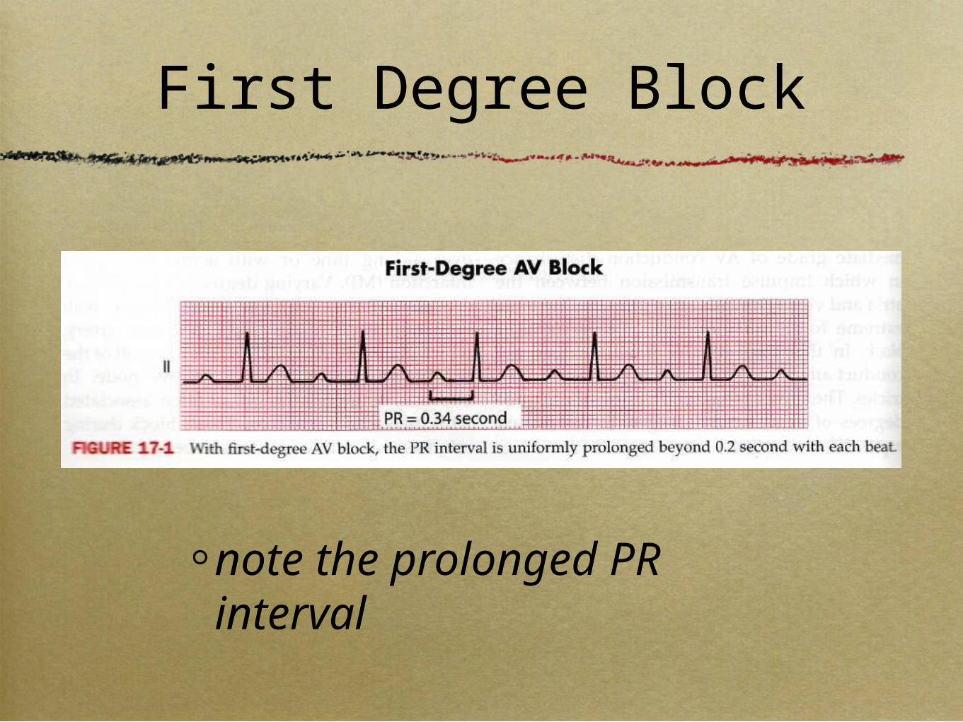
First Degree Block
note the prolonged PR interval

Second Degree AV Block
Mobitz type I or Winckebach
Mobitz type II
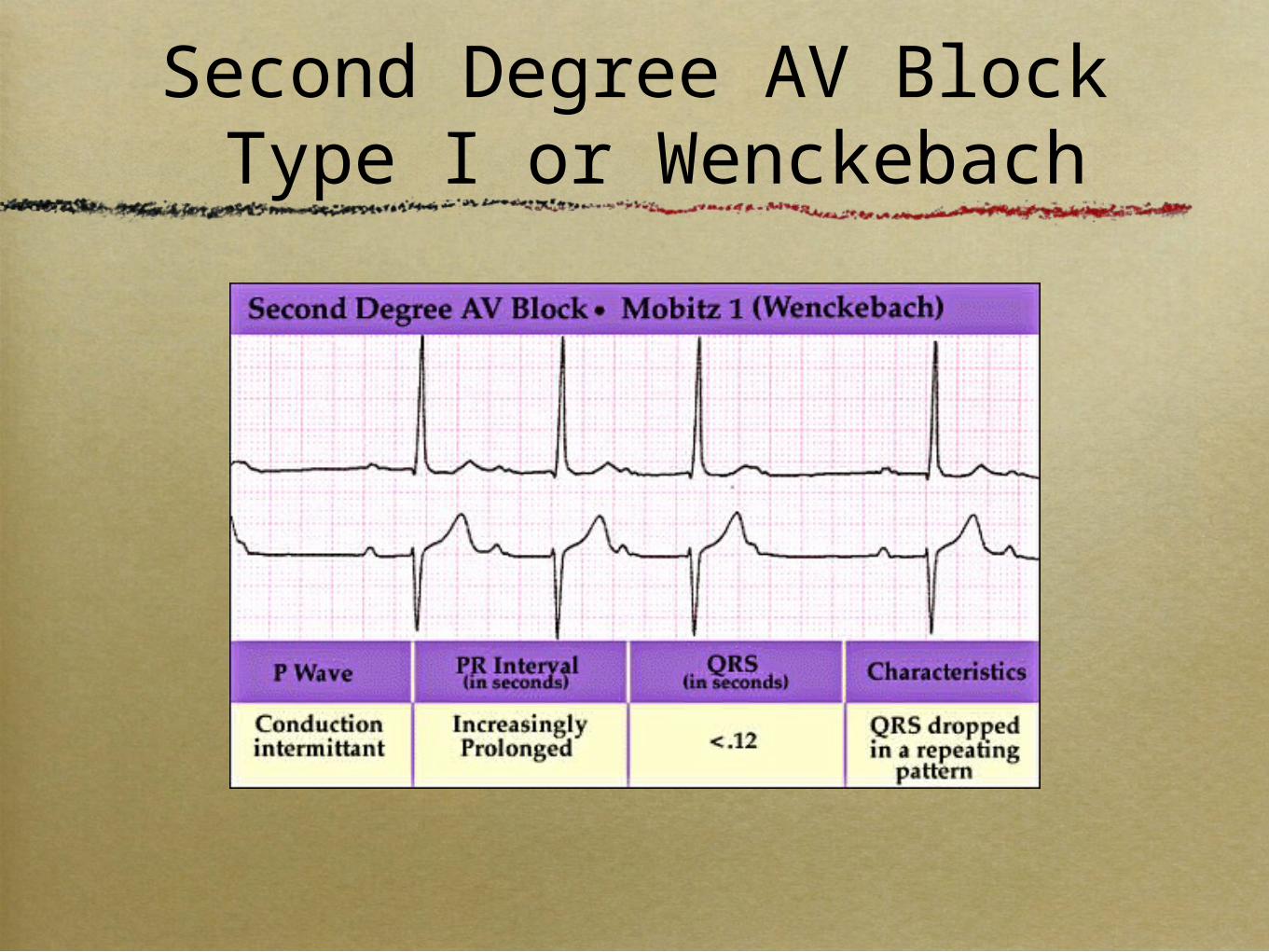
Second Degree AV Block Type I or Wenckebach

Second Degree AV Block Type I or Wenckebach
1.Progressive lengthening of the PR interval from beat to beat until a beat is dropped.
2.The PR interval after the nonconducted P wave is shorter than the PR interval before the nonconducted P wave.
3.May be grouping of QRS complexes


Second Degree AV BlockType II
1. Sudden appearance of a single, non-conducted sinus P wave...
2. ...without...
1. ...the progressive prolongation of the PR intervals…
2. ...and the shortening of the PR interval in the beat after the non-conducted P wave.

Second Degree AV BlockType II

2:1 AV Blocks
• Often are type II blocks • look for slightly prolonged QRS
• But they can be type I blocks• look at long rhythm strip
• Sometimes they are labeled a “second degree block” only
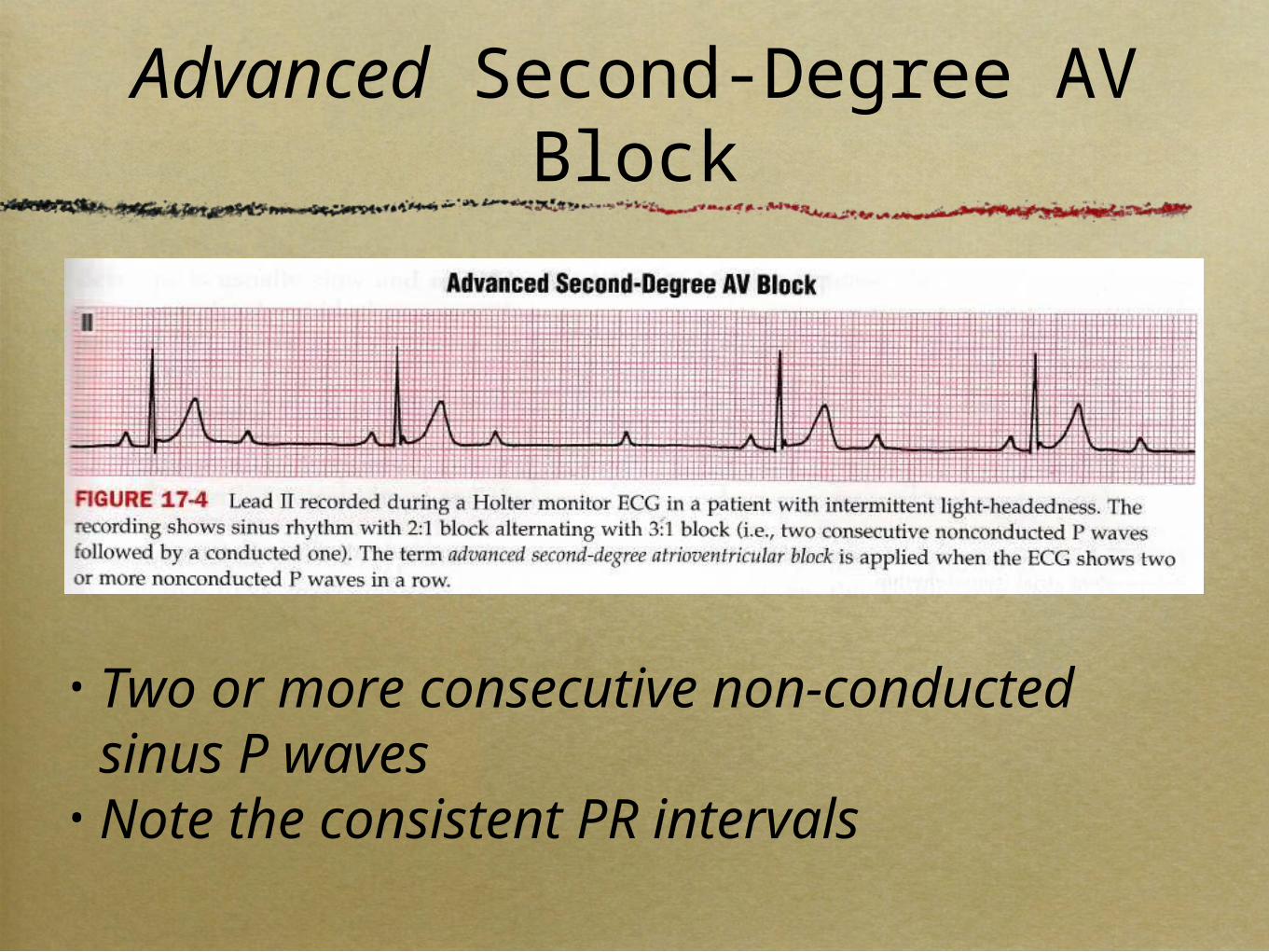
Advanced Second-Degree AV Block
• Two or more consecutive non-conducted sinus P waves
• Note the consistent PR intervals

Third-Degree (Complete) AV Block

Third-Degree (Complete) AV Block
1. P waves are present, with a regular atrial rate faster than the ventricular rate
2. QRS complexes are present, with a slow (usually fixed) ventricular rate
3. The P wave bears no relation to the QRS complexes, and the PR intervals are completely variable
4. (Some properly timed P waves may be conducted)

Third-Degree (Complete) AV Block
• QRS can be normal width or wide

AV Dissociation
• SA is pacing the atria• AV is pacing the ventricles• Ventricular rate is similar to atria rate• No P wave, even if properly timed, will
be conducted.

AV Dissociation

AV Dissociation

Third-Degree (Complete) AV Block

100 b/min42 b/min
NoComplete heart block


