Wnt5a inhibits canonical Wnt signaling in hematopoietic ... · PDF fileWnt5a inhibits...
6
Wnt5a inhibits canonical Wnt signaling in hematopoietic stem cells and enhances repopulation Michael J. Nemeth*, Lilia Topol † , Stacie M. Anderson*, Yingzi Yang † , and David M. Bodine* ‡ *Genetics and Molecular Biology Branch and † Genetic Disease Research Branch, National Human Genome Research Institute, Bethesda, MD 20892-4442 Edited by Hal Broxmeyer, Walther Oncology Center, Indianapolis, IN, and accepted by the Editorial Board August 10, 2007 (received for review May 21, 2007) The mechanisms that regulate hematopoietic stem cell (HSC) fate decisions between proliferation and multilineage differentiation are unclear. Members of the Wnt family of ligands that activate the canonical Wnt signaling pathway, which utilizes -catenin to relay the signal, have been demonstrated to regulate HSC function. In this study, we examined the role of noncanonical Wnt signaling in regulating HSC fate. We observed that noncanonical Wnt5a inhib- ited Wnt3a-mediated canonical Wnt signaling in HSCs and sup- pressed Wnt3a-mediated alterations in gene expression associated with HSC differentiation, such as increased expression of myc. Wnt5a increased short- and long-term HSC repopulation by main- taining HSCs in a quiescent G 0 state. From these data, we propose that Wnt5a regulates hematopoiesis by the antagonism of the canonical Wnt pathway, resulting in a pool of quiescent HSCs. cell cycle hematopoiesis hematopoietic stem cell transplantation H ematopoietic stem cells (HSC) are a rare population of cells that are responsible for life-long generation of all blood cell types (1). To maintain their numbers, HSCs must generate at least one daughter cell that retains the stem cell phenotype. HSCs are capable of rapid proliferation after transplantation and are the only cells capable of both short- (6–8 weeks) and long-term hematopoietic engraftment. The mechanisms that regulate HSC fate between proliferation and differentiation are still unclear, but understanding these mechanisms and develop- ing strategies to manipulate them are imperative for multiple therapeutic goals, such as corrective gene therapy for hemato- logic disorders. The Wnt signaling pathway regulates cell-fate decisions at all stages of development in multiple tissues, including embryonic and adult intestinal and skin stem cells (2–5). There are multiple pathways by which Wnt ligands transduce signals, the best characterized of which is the ‘‘canonical’’ pathway (reviewed in ref. 2). In this pathway, the Wnt ligand binds to its cognate receptor Frizzled and the low-density lipoprotein receptor- related protein (LRP) 5/6 coreceptors at the cell surface, result- ing in the inhibition of glycogen synthase kinase-3 (GSK-3). One of the targets of this kinase is -catenin, the critical factor in relaying canonical Wnt signals. In the absence of Wnt ligand, GSK-3 phosphorylates -catenin, which targets it for ubiquiti- nation and subsequent degradation. However, when Wnt ligand is bound to its receptors, degradation of -catenin is suppressed, and -catenin can translocate to the nucleus where it binds to T cell factor (TCF)/lymphoid enhancer factor (LEF) transcription factors and induces target gene expression. Inappropriate acti- vation of the pathway has been implicated in tumorigenesis; therefore it is necessary that canonical Wnt signaling be precisely controlled [reviewed in (3)]. Reya et al., showed that recombinant Wnt3a could activate canonical Wnt signaling in HSCs and promote in vitro expansion and self-renewal of apoptosis-resistant transgenic HSCs (4). Activation of the canonical Wnt signaling pathway has also been demonstrated to promote self-renewal of leukemia stem cells (5). HSCs deficient in the chromatin-binding protein Hmgb3 exhibit increased canonical Wnt signaling which correlated with a bias toward self-renewal (6). These studies suggest that the canonical Wnt signaling pathway plays a critical role in hema- topoiesis. However, HSCs deficient in -catenin possess the same capacity for hematopoietic repopulation as wild-type HSCs (7). Furthermore, long-term constitutive stabilization of - catenin leads to inhibition of multilineage differentiation and the eventual loss of HSCs (8, 9), indicating that the context in which canonical Wnt signaling is activated may determine the physi- ological effect and that other factors contribute to regulate canonical Wnt signaling to properly maintain stem cell numbers. Some members of the Wnt ligand family (e.g., Wnt5a) can activate signaling pathways other than the canonical pathway, depending on which receptors are present at the cell surface (10–13). It has been shown that noncanonical Wnt ligands can inhibit canonical Wnt signaling in transformed cell lines and in Xenopus and mouse embryos (10, 14, 15). The mechanism of Wnt5a-mediated inhibition of the canonical pathway is unclear. Wnt5a has been shown to induce Siah2, a member of an E3 ubiquitin ligase complex that can target -catenin for degrada- tion, and Wnt5a-deficient mice exhibit increased levels of - catenin in the distal hind limb (15). However, loss of Wnt5a did not affect -catenin levels in fetal liver cells (11). Wnt5a can also inhibit canonical Wnt signaling downstream of -catenin stabi- lization through the calcium-dependent activation of Nemo-like kinase (12) as well as other calcium-independent mecha- nisms (10). The canonical Wnt3a is expressed in bone marrow mononu- clear cells (16). Wnt5a is expressed in both fetal liver and bone marrow cells, especially B220 B cells, and deficiency in Wnt5a leads to a cell-autonomous increase in B cell numbers (11, 16–18). Wnt5a is also expressed in cultured primary bone marrow stromal cells. In theory, both Wnt3a and Wnt5a can regulate hematopoietic cells by autocrine and paracrine mech- anisms. We hypothesized that Wnt5a contributes to the regula- tion of HSC function through suppression of canonical Wnt signaling. To test this hypothesis, we cultured HSCs under serum-free conditions in the presence of recombinant Wnt3a and/or Wnt5a. We observed that Wnt5a either alone or in combination with Wnt3a, enhanced short- and long-term hema- topoietic repopulation of donor HSCs, whereas Wnt3a did not enhance repopulation of HSCs. Wnt5a suppressed changes in gene expression associated with Wnt3a and maintained HSCs in Author contributions: M.J.N. and D.M.B. designed research; M.J.N. and L.T. performed research; S.M.A. and Y.Y. contributed new reagents/analytic tools; M.J.N. and D.M.B. analyzed data; and M.J.N. and D.M.B. wrote the paper. The authors declare no conflict of interest. This article is a PNAS Direct Submission. H.B. is a guest editor invited by the Editorial Board. Freely available online through the PNAS open access option. Abbreviations: HSC, hematopoietic stem cell; LKSI, lineage-negative, c-kit HI , Sca-1 HI , IL-7R . ‡ To whom correspondence should be addressed at: Genetics and Molecular Biology Branch, National Human Genome Research Institute, 49 Convent Drive, Room 4A04, Bethesda, MD 20892-4442. E-mail: [email protected]. This article contains supporting information online at www.pnas.org/cgi/content/full/ 0704747104/DC1. 15436 –15441 PNAS September 25, 2007 vol. 104 no. 39 www.pnas.orgcgidoi10.1073pnas.0704747104
Transcript of Wnt5a inhibits canonical Wnt signaling in hematopoietic ... · PDF fileWnt5a inhibits...
Wnt5a inhibits canonical Wnt signaling inhematopoietic stem cells and enhances repopulationMichael J. Nemeth*, Lilia Topol†, Stacie M. Anderson*, Yingzi Yang†, and David M. Bodine*‡
*Genetics and Molecular Biology Branch and †Genetic Disease Research Branch, National Human Genome Research Institute, Bethesda, MD 20892-4442
Edited by Hal Broxmeyer, Walther Oncology Center, Indianapolis, IN, and accepted by the Editorial Board August 10, 2007 (received for reviewMay 21, 2007)
The mechanisms that regulate hematopoietic stem cell (HSC) fatedecisions between proliferation and multilineage differentiationare unclear. Members of the Wnt family of ligands that activate thecanonical Wnt signaling pathway, which utilizes �-catenin to relaythe signal, have been demonstrated to regulate HSC function. Inthis study, we examined the role of noncanonical Wnt signaling inregulating HSC fate. We observed that noncanonical Wnt5a inhib-ited Wnt3a-mediated canonical Wnt signaling in HSCs and sup-pressed Wnt3a-mediated alterations in gene expression associatedwith HSC differentiation, such as increased expression of myc.Wnt5a increased short- and long-term HSC repopulation by main-taining HSCs in a quiescent G0 state. From these data, we proposethat Wnt5a regulates hematopoiesis by the antagonism of thecanonical Wnt pathway, resulting in a pool of quiescent HSCs.
cell cycle � hematopoiesis � hematopoietic stem cell transplantation
Hematopoietic stem cells (HSC) are a rare population of cellsthat are responsible for life-long generation of all blood cell
types (1). To maintain their numbers, HSCs must generate atleast one daughter cell that retains the stem cell phenotype.HSCs are capable of rapid proliferation after transplantationand are the only cells capable of both short- (6–8 weeks) andlong-term hematopoietic engraftment. The mechanisms thatregulate HSC fate between proliferation and differentiation arestill unclear, but understanding these mechanisms and develop-ing strategies to manipulate them are imperative for multipletherapeutic goals, such as corrective gene therapy for hemato-logic disorders.
The Wnt signaling pathway regulates cell-fate decisions at allstages of development in multiple tissues, including embryonicand adult intestinal and skin stem cells (2–5). There are multiplepathways by which Wnt ligands transduce signals, the bestcharacterized of which is the ‘‘canonical’’ pathway (reviewed inref. 2). In this pathway, the Wnt ligand binds to its cognatereceptor Frizzled and the low-density lipoprotein receptor-related protein (LRP) 5/6 coreceptors at the cell surface, result-ing in the inhibition of glycogen synthase kinase-3� (GSK-3�).One of the targets of this kinase is �-catenin, the critical factorin relaying canonical Wnt signals. In the absence of Wnt ligand,GSK-3� phosphorylates �-catenin, which targets it for ubiquiti-nation and subsequent degradation. However, when Wnt ligandis bound to its receptors, degradation of �-catenin is suppressed,and �-catenin can translocate to the nucleus where it binds to Tcell factor (TCF)/lymphoid enhancer factor (LEF) transcriptionfactors and induces target gene expression. Inappropriate acti-vation of the pathway has been implicated in tumorigenesis;therefore it is necessary that canonical Wnt signaling be preciselycontrolled [reviewed in (3)].
Reya et al., showed that recombinant Wnt3a could activatecanonical Wnt signaling in HSCs and promote in vitro expansionand self-renewal of apoptosis-resistant transgenic HSCs (4).Activation of the canonical Wnt signaling pathway has also beendemonstrated to promote self-renewal of leukemia stem cells(5). HSCs deficient in the chromatin-binding protein Hmgb3exhibit increased canonical Wnt signaling which correlated with
a bias toward self-renewal (6). These studies suggest that thecanonical Wnt signaling pathway plays a critical role in hema-topoiesis. However, HSCs deficient in �-catenin possess thesame capacity for hematopoietic repopulation as wild-type HSCs(7). Furthermore, long-term constitutive stabilization of �-catenin leads to inhibition of multilineage differentiation and theeventual loss of HSCs (8, 9), indicating that the context in whichcanonical Wnt signaling is activated may determine the physi-ological effect and that other factors contribute to regulatecanonical Wnt signaling to properly maintain stem cell numbers.
Some members of the Wnt ligand family (e.g., Wnt5a) canactivate signaling pathways other than the canonical pathway,depending on which receptors are present at the cell surface(10–13). It has been shown that noncanonical Wnt ligands caninhibit canonical Wnt signaling in transformed cell lines and inXenopus and mouse embryos (10, 14, 15). The mechanism ofWnt5a-mediated inhibition of the canonical pathway is unclear.Wnt5a has been shown to induce Siah2, a member of an E3ubiquitin ligase complex that can target �-catenin for degrada-tion, and Wnt5a-deficient mice exhibit increased levels of �-catenin in the distal hind limb (15). However, loss of Wnt5a didnot affect �-catenin levels in fetal liver cells (11). Wnt5a can alsoinhibit canonical Wnt signaling downstream of �-catenin stabi-lization through the calcium-dependent activation of Nemo-likekinase (12) as well as other calcium-independent mecha-nisms (10).
The canonical Wnt3a is expressed in bone marrow mononu-clear cells (16). Wnt5a is expressed in both fetal liver and bonemarrow cells, especially B220� B cells, and deficiency in Wnt5aleads to a cell-autonomous increase in B cell numbers (11,16–18). Wnt5a is also expressed in cultured primary bonemarrow stromal cells. In theory, both Wnt3a and Wnt5a canregulate hematopoietic cells by autocrine and paracrine mech-anisms. We hypothesized that Wnt5a contributes to the regula-tion of HSC function through suppression of canonical Wntsignaling. To test this hypothesis, we cultured HSCs underserum-free conditions in the presence of recombinant Wnt3aand/or Wnt5a. We observed that Wnt5a either alone or incombination with Wnt3a, enhanced short- and long-term hema-topoietic repopulation of donor HSCs, whereas Wnt3a did notenhance repopulation of HSCs. Wnt5a suppressed changes ingene expression associated with Wnt3a and maintained HSCs in
Author contributions: M.J.N. and D.M.B. designed research; M.J.N. and L.T. performedresearch; S.M.A. and Y.Y. contributed new reagents/analytic tools; M.J.N. and D.M.B.analyzed data; and M.J.N. and D.M.B. wrote the paper.
The authors declare no conflict of interest.
This article is a PNAS Direct Submission. H.B. is a guest editor invited by the Editorial Board.
Freely available online through the PNAS open access option.
Abbreviations: HSC, hematopoietic stem cell; LKSI, lineage-negative, c-kitHI, Sca-1HI,IL-7R��.
‡To whom correspondence should be addressed at: Genetics and Molecular Biology Branch,National Human Genome Research Institute, 49 Convent Drive, Room 4A04, Bethesda, MD20892-4442. E-mail: [email protected].
This article contains supporting information online at www.pnas.org/cgi/content/full/0704747104/DC1.
15436–15441 � PNAS � September 25, 2007 � vol. 104 � no. 39 www.pnas.org�cgi�doi�10.1073�pnas.0704747104
a quiescent state. Our data suggest a model in which Wnt5a-mediated regulation of canonical Wnt signaling is critical formaintenance of quiescent, transplantable HSCs.
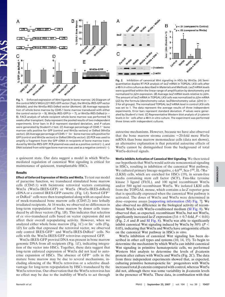
ResultsEffect of Enforced Expression of Wnt3a and Wnt5a. To test our modelof paracrine function, we transduced stimulated bone marrowcells (CD45.1) with bicistronic retroviral vectors containingWnt3a (Wnt3a-IRES-GFP) or Wnt5a (Wnt5a-IRES-dsRed)cDNA or a control IRES-GFP cassette. (Fig. 1A). Sorted GFP�
or DsRed� cells were then transplanted along with an equal doseof mock-transduced bone marrow cells (CD45.2) into lethallyirradiated recipients. At 16 weeks, we observed no differences inlong-term repopulation of bone marrow by donor cells trans-duced by all three vectors (Fig. 1B). This indicates that selectionof ex vivo-transduced cells based on vector expression did notaffect their overall repopulating activity. However, when weanalyzed either whole bone marrow (Fig. 1C) or lin� cells (Fig.1D) for cells that expressed the retroviral vector, we observedonly control IRES-GFP� and Wnt5a-IRES-DsRed� cells. Nocells with the Wnt3a-IRES-GFP retrovirus expressed GFP.
Wnt3a-IRES-GFP proviral DNA was present in bone marrowgenomic DNA from all recipients (Fig. 1E), indicating integra-tion of the vector into HSCs. Together, these data suggest thatlong-term enforced expression of Wnt3a did not lead to auto-crine expansion of HSCs. The absence of GFP� cells in themature bone marrow may be due to several mechanisms, in-cluding silencing of the Wnt3a retrovirus or a selective disad-vantage for long-term repopulation by HSCs transduced by theWnt3a retrovirus. Our observation that the Wnt5a retrovirus hasno effect may be due to the inability of Wnt5a to act through
autocrine mechanisms. However, because we have also observedthat the bone marrow stroma contains �20-fold more Wnt5amRNA than bone marrow mononuclear cells (data not shown),an alternative explanation is that potential autocrine effects ofWnt5a cannot be distinguished from the background of totalWnt5a-derived signals.
Wnt5a Inhibits Activation of Canonical Wnt Signaling. We then testedour hypothesis that Wnt5a would activate noncanonical signalingin HSCs, resulting in inhibition of the canonical Wnt pathway.We cultured primary lineage-negative, c-kitHI, Sca-1HI, IL-7R��
(LKSI) cells, which are enriched for HSCs (19), in serum-freemedia containing stem cell factor (SCF), Fms-like tyrosinekinase 3 ligand (Flt3L), and 100 ng/ml recombinant Wnt3aand/or 500 ng/ml recombinant Wnt5a. We isolated LKSI cellsfrom the TOPGAL mouse, which contains a lacZ reporter genethat is specifically expressed when the canonical Wnt pathway isactivated. The doses of Wnt3a and Wnt5a were determined indose–response assays [supporting information (SI) Fig. 7]. Wealso observed no difference in the biological activity of recom-binant Wnt3a with Wnt3a-conditioned medium (SI Fig. 8). Weobserved that, as expected, recombinant Wnt3a, but not Wnt5a,significantly increased lacZ expression (3.6 � 0.7-fold, P � 0.01)(Fig. 2 A and B and SI Fig. 8). Wnt5a was able to significantlyinhibit canonical Wnt signaling induced by Wnt3a by 50% (P �0.05), indicating that Wnt3a and Wnt5a have antagonistic effectson the canonical Wnt pathway in HSCs in vitro.
Wnt5a inhibition of canonical Wnt signaling has been de-scribed in other cell types and systems (10, 14, 15). To begin todetermine the mechanism by which Wnt5a can inhibit canonicalWnt signaling in primitive hematopoietic cells, we performedWestern blot analysis to determine the levels of �-cateninprotein after culture with Wnt3a and Wnt5a (Fig. 2C). The datafrom three independent experiments showed that, as expected,culturing primitive hematopoietic lin� progenitors with Wnt3aincreased total �-catenin compared with control, whereas Wnt5adid not, although there was some variability in �-catenin levelsin the presence of Wnt5a. These data, in combination with that
Fig. 1. Enforced expression of Wnt ligands in bone marrow. (A) Diagram ofthe control MSCV MGirL22Y IRES-GFP vector (Top), the Wnt3a-IRES-GFP vector(Middle), and the Wnt5a-IRES-DsRed vector (Bottom). (B) Average repopula-tion of whole bone marrow by CD45.1 bone marrow transduced with eitherthe control vector (n � 9), Wnt3a-IRES-GFP (n � 7), or Wnt5a-IRES-DsRed (n �8). FACS analysis of whole recipient whole bone marrow was performed 16weeks after transplant. Data represent the pooled results of two independentexperiments. Error bars in B–D represent standard deviation, and P valueswere generated by Student’s t test. (C) Average percentage of CD45.1� bonemarrow cells positive for GFP (control and Wnt3a vectors) or DsRed (Wnt5avector). (D) Average percentage of CD45.1� lin� bone marrow cells positive forGFP (control and Wnt3a vectors) or DsRed (Wnt5a vector). (E) PCR was used toamplify a fragment from the GFP cDNA in recipients of bone marrow trans-duced by Wnt3a-IRES-GFP. PCR plasmid was used as a positive control (�), andDNA isolated from wild-type bone marrow was used as a negative control (�).
Fig. 2. Inhibition of canonical Wnt signaling in HSCs by Wnt5a. (A) Semi-quantitative duplex RT-PCR analysis of lacZ mRNA in TOPGAL LKSI cells aftera 48-h in vitro culture as described in Materials and Methods. LacZ mRNA levelswere quantified within the linear range of amplification by densitometry andnormalized to �2m expression. (B) Average lacZ mRNA levels relative to �2m.The amount of lacZ mRNA in TOPGAL LKSI cells was normalized to �2m mRNA(�2) by the formula (densitometry value: lacZ/densitometry value: �2m) (n �3 for all groups). The normalized TOPGAL lacZ mRNA level in control LKSI cellswas set to 1. The data represent the average results of three independentexperiments. Error bars represent standard deviation. P values were gener-ated by Student’s t test. (C) Representative Western blot analysis of �-cateninlevels in lin� cells after a 48-h in vitro culture. The experiment was performedthree times with independent cultures.
Nemeth et al. PNAS � September 25, 2007 � vol. 104 � no. 39 � 15437
IMM
UN
OLO
GY
presented in Fig. 2B, indicate that, in hematopoietic progenitors,Wnt5a primarily signals through noncanonical pathways. Fur-thermore, culturing lin� cells with both Wnt3a and Wnt5adecreased �-catenin protein compared with Wnt3a alone, sug-gesting that Wnt5a inhibits canonical Wnt signaling in primitivehematopoietic cells by promoting the destabilization of�-catenin.
Wnt5a Inhibits HSC Expansion in Vitro. We hypothesized thatWnt5a-mediated destabilization of �-catenin would suppressWnt3a-mediated effects on LKSI cell proliferation. To test this,we cultured wild-type LKSI cells in the presence of Wnt3a and/orWnt5a. We observed that under serum-free conditions, thepresence of Wnt3a significantly reduced total cell expansion3-fold (P � 0.05) after 4 days in culture compared with controlconditions (SCF and Flt3L alone) (Fig. 3A). However, Wnt5aeither alone (12-fold, P � 0.01) or in combination with Wnt3a(16-fold, P � 0.01) also significantly reduced cell expansioncompared with control conditions. There was no differenceamong the groups in the percentage of cells that retained theLKSI phenotype (data not shown).
To test whether the Wnt-mediated inhibition of cell prolifer-ation could be due to induction of apoptosis, we cultured LKSIcells with either Wnt3a or Wnt5a (Fig. 3 B and C). Both (Wnt3a:2.4-fold; Wnt5a: 3-fold) significantly increased the percentage oftotal cells actively undergoing apoptosis (P � 0.05) from 1% to3%. Therefore, it is probable that other mechanisms in additionto apoptosis contribute toward inhibition of proliferation byWnt3a and Wnt5a.
To further investigate the role of apoptosis in Wnt-mediatedinhibition of cell proliferation, we cultured LKSI cells that wereisolated from transgenic mice that overexpressed the humanantiapoptotic gene BCL-2 (20). We observed that culturing
BCL-2� LKSI cells with Wnt3a and/or Wnt5a resulted in de-creased total cell expansion compared with control conditions(P � 0.01) (Fig. 3D). Wild type and BCL-2� LKSI cells expandedequally when cultured under either control conditions or withWnt3a. In contrast, BCL-2� LKSI cells proliferated more in thepresence of Wnt5a compared with wild type (P � 0.01). Thesedata suggest that the induction of apoptosis by Wnt3a or Wnt5aoccurs through different mechanisms, with Wnt5a-mediatedinduction reliant on pathways governed by BCL-2.
Effect of Wnt3a and Wnt5a on HSC Repopulating Ability. We exam-ined the effects of activating Wnt signaling pathways on HSCfunction (measured through competitive repopulation assays).Wild-type CD45.1 LKSI cells were cultured for 6 days underserum-free conditions with Wnt3a and Wnt5a before transplan-tation. Six weeks after transplant, we analyzed recipients’ bonemarrow for short-term repopulation (mediated by both short-term and long-term HSCs) by cultured donor LKSI cells (Fig.4A). We defined a recipient having �1% CD45.1� bone marrowcells as positive for hematopoietic chimerism. We did notobserve a significant difference in short-term repopulation bycontrol LKSI cells and LKSI cells cultured with Wnt3a (Fig. 4B).However, LKSI cells cultured with either Wnt5a alone or bothWnt3a and Wnt5a showed significant 3- to 5-fold increases,respectively, in short-term repopulation compared with control
Fig. 4. Analysis of Wnt-mediated effects on hematopoietic repopulation. (A)Raw data of short-term (6 weeks) engraftment of cultured LKSI cells. Recipi-ents were defined as positive for engraftment if they contained �1% hema-topoietic chimerism. (B) Average short-term repopulation of cultured LKSIcells. Data represent the pooled results of two independent experiments. Onlyrecipients with �1% chimerism were included in this analysis. Error barsrepresent standard deviation. P values were generated by Student’s t test. (C)Raw data of long-term (16 weeks) engraftment of cultured LKSI cells. Recip-ients were defined as positive for engraftment if they contained �1% hema-topoietic chimerism. (D) Average long-term repopulation of cultured LKSIcells. Data represent the pooled results of three independent experiments.Only recipients with �1% chimerism were included in this analysis. Error barsrepresent standard deviation. P values were generated by Mann–Whitneynonparametric analysis.
Fig. 3. Analysis of Wnt-mediated HSC expansion and apoptosis. (A) Averagefold expansion of wild-type LKSI cells after 4 days in culture (n � 3 for allconditions). Error bars represent standard deviation. P values were generatedby Student’s t test (*, P � 0.05; **, P � 0.01). (B) Representative FACS analysesof apoptotic wild-type LKSI cells cultured for 4 days under control conditions(Top), with recombinant Wnt3a (Middle), and recombinant Wnt5a (Bottom).Actively apoptotic cells actively were defined as Annexin V� and PI�. (C)Average percentage of apoptotic wild-type LKSI cells after 4 days in culture(n � 4 for all conditions). Error bars represent standard deviation. P valueswere determined by Student’s t test. (D) Average fold expansion of H2K-BCL-2LKSI cells after 4 days in culture (n � 4 for all conditions). Error bars representstandard deviation. P values were generated by Student’s t test
15438 � www.pnas.org�cgi�doi�10.1073�pnas.0704747104 Nemeth et al.
(P � 0.01) (Fig. 4B). We observed no differences in the homingabilities of LKSI cells cultured under control conditions, withWnt3a, or with Wnt5a (SI Fig. 9), indicating that Wnt5a does notpromote hematopoietic repopulation by enhancing the ability ofcultured LKSI cells to home.
To determine the effects of Wnt3a and Wnt5a on primaryhematopoietic repopulation mediated solely by long-term HSCs,we analyzed recipients’ bone marrow 16 weeks after transplant,using the same criteria for engraftment outlined above. As withshort-term repopulation, there was no significant difference inlong-term repopulation by control LKSI cells and LKSI cellscultured with Wnt3a (Fig. 4 C and D). Similarly, we observedsignificant 3.6-fold and 5.7-fold increases in long-term hemato-poietic repopulation by LKSI cells cultured with Wnt5a (P �0.01) and Wnt5a and Wnt3a (P � 0.01), respectively. Combinedwith the data in Fig. 4B, this indicates that Wnt3a does notenhance repopulation by either short- or long-term HSCs.Finally, to determine whether Wnt3a prolonged the period inwhich HSCs could be cultured and still repopulate, we trans-planted LKSI cells that had been cultured with and withoutWnt3a for 2 weeks and found that neither condition yielded anylong-term engraftment (data not shown). These data indicatethat short-term treatment with Wnt5a, but not Wnt3a, canincrease the primary repopulating ability of cultured HSCs.
Our observations that stimulating the canonical Wnt signalingpathway has an inhibitory effect on cell expansion and no effecton the ability of in vitro-cultured LKSI cells to engraft contrastswith previous reports that showed a significant positive effect onHSC function (4, 21). One explanation for the discrepancybetween our data and those from other studies is that othersignals are necessary to enable Wnt3a to expand HSCs in vitro.Because HSC expansion is regulated by apoptosis (20), wetransplanted BCL-2� LKSI cells (CD45.1) that were culturedunder identical conditions as their wild-type counterparts.Wnt3a inhibited long-term engraftment of BCL-2� HSCs be-cause only 1 of 7 recipients showed any long-term engraftment(1.2% CD45.1� bone marrow cells) whereas BCL-2� LKSI cellscultured under control conditions engrafted in 7 of 7 recipients(17.8 � 6.8% CD45.1� cells) (SI Fig. 10). BCL-2� LKSI cellscultured with Wnt5a showed similar levels of engraftmentcompared with BCL-2� LKSI cells cultured under controlconditions (P � 0.40).
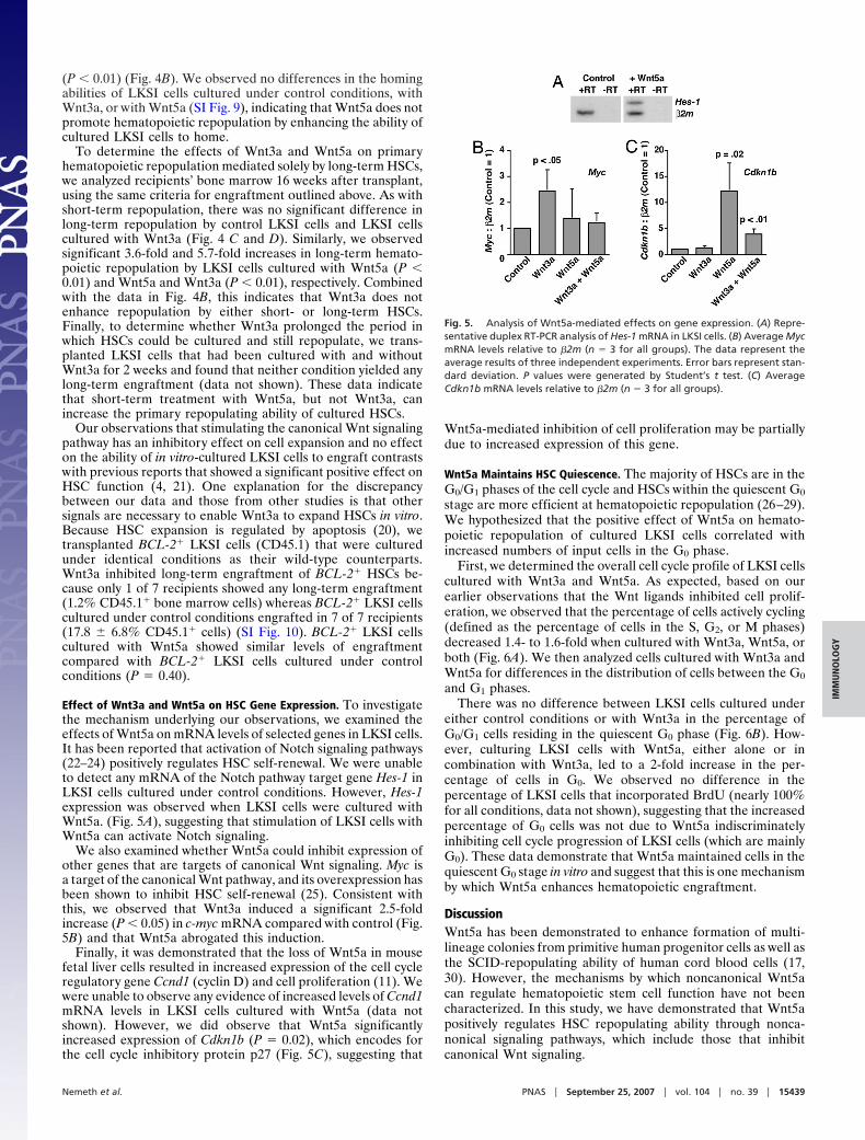
Effect of Wnt3a and Wnt5a on HSC Gene Expression. To investigatethe mechanism underlying our observations, we examined theeffects of Wnt5a on mRNA levels of selected genes in LKSI cells.It has been reported that activation of Notch signaling pathways(22–24) positively regulates HSC self-renewal. We were unableto detect any mRNA of the Notch pathway target gene Hes-1 inLKSI cells cultured under control conditions. However, Hes-1expression was observed when LKSI cells were cultured withWnt5a. (Fig. 5A), suggesting that stimulation of LKSI cells withWnt5a can activate Notch signaling.
We also examined whether Wnt5a could inhibit expression ofother genes that are targets of canonical Wnt signaling. Myc isa target of the canonical Wnt pathway, and its overexpression hasbeen shown to inhibit HSC self-renewal (25). Consistent withthis, we observed that Wnt3a induced a significant 2.5-foldincrease (P � 0.05) in c-myc mRNA compared with control (Fig.5B) and that Wnt5a abrogated this induction.
Finally, it was demonstrated that the loss of Wnt5a in mousefetal liver cells resulted in increased expression of the cell cycleregulatory gene Ccnd1 (cyclin D) and cell proliferation (11). Wewere unable to observe any evidence of increased levels of Ccnd1mRNA levels in LKSI cells cultured with Wnt5a (data notshown). However, we did observe that Wnt5a significantlyincreased expression of Cdkn1b (P � 0.02), which encodes forthe cell cycle inhibitory protein p27 (Fig. 5C), suggesting that
Wnt5a-mediated inhibition of cell proliferation may be partiallydue to increased expression of this gene.
Wnt5a Maintains HSC Quiescence. The majority of HSCs are in theG0/G1 phases of the cell cycle and HSCs within the quiescent G0
stage are more efficient at hematopoietic repopulation (26–29).We hypothesized that the positive effect of Wnt5a on hemato-poietic repopulation of cultured LKSI cells correlated withincreased numbers of input cells in the G0 phase.
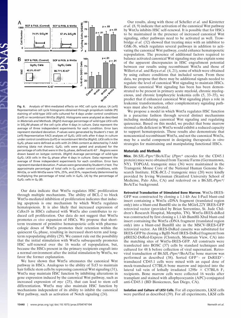
First, we determined the overall cell cycle profile of LKSI cellscultured with Wnt3a and Wnt5a. As expected, based on ourearlier observations that the Wnt ligands inhibited cell prolif-eration, we observed that the percentage of cells actively cycling(defined as the percentage of cells in the S, G2, or M phases)decreased 1.4- to 1.6-fold when cultured with Wnt3a, Wnt5a, orboth (Fig. 6A). We then analyzed cells cultured with Wnt3a andWnt5a for differences in the distribution of cells between the G0
and G1 phases.There was no difference between LKSI cells cultured under
either control conditions or with Wnt3a in the percentage ofG0/G1 cells residing in the quiescent G0 phase (Fig. 6B). How-ever, culturing LKSI cells with Wnt5a, either alone or incombination with Wnt3a, led to a 2-fold increase in the per-centage of cells in G0. We observed no difference in thepercentage of LKSI cells that incorporated BrdU (nearly 100%for all conditions, data not shown), suggesting that the increasedpercentage of G0 cells was not due to Wnt5a indiscriminatelyinhibiting cell cycle progression of LKSI cells (which are mainlyG0). These data demonstrate that Wnt5a maintained cells in thequiescent G0 stage in vitro and suggest that this is one mechanismby which Wnt5a enhances hematopoietic engraftment.
DiscussionWnt5a has been demonstrated to enhance formation of multi-lineage colonies from primitive human progenitor cells as well asthe SCID-repopulating ability of human cord blood cells (17,30). However, the mechanisms by which noncanonical Wnt5acan regulate hematopoietic stem cell function have not beencharacterized. In this study, we have demonstrated that Wnt5apositively regulates HSC repopulating ability through nonca-nonical signaling pathways, which include those that inhibitcanonical Wnt signaling.
Fig. 5. Analysis of Wnt5a-mediated effects on gene expression. (A) Repre-sentative duplex RT-PCR analysis of Hes-1 mRNA in LKSI cells. (B) Average MycmRNA levels relative to �2m (n � 3 for all groups). The data represent theaverage results of three independent experiments. Error bars represent stan-dard deviation. P values were generated by Student’s t test. (C) AverageCdkn1b mRNA levels relative to �2m (n � 3 for all groups).
Nemeth et al. PNAS � September 25, 2007 � vol. 104 � no. 39 � 15439
IMM
UN
OLO
GY
Our data indicate that Wnt5a regulates HSC proliferationthrough multiple mechanisms. The ability of BCL-2 to blockWnt5a-mediated inhibition of proliferation indicates that induc-ing apoptosis is one mechanism by which Wnt5a regulateshematopoiesis. It is also likely that increased expression ofCdkn1b in HSCs cultured with Wnt5a also contributes to re-duced cell proliferation. Our data do not suggest that Wnt5apromotes ex vivo expansion of HSCs. We propose that short-term treatment of primitive hematopoietic cells with pharma-cologic doses of Wnt5a promotes their retention within thequiescent G0 phase, resulting in increased short-term and long-term repopulating ability (29). We cannot rule out the possibilitythat the initial stimulation with Wnt5a subsequently promotesHSC self-renewal over the 16 weeks of repopulation, but,because the HSCs present in the primary recipients engraft intoa normal environment after the initial stimulation by Wnt5a, wefavor the former explanation.
We have shown that Wnt5a attenuates the canonical Wntpathway in HSCs. Analogous to the ability of Tcf3 to maintainhair follicle stem cells by repressing canonical Wnt signaling (31),Wnt5a may maintain HSC function by inhibiting alterations ingene expression induced by the canonical Wnt pathway, such asincreased expression of myc (25), that can lead to stem celldifferentiation. Wnt5a may also maintain HSC function bymechanisms independent of its ability to inhibit the canonicalWnt pathway, such as activation of Notch signaling (24).
Our results, along with those of Scheller et al. and Kirstetteret al. (8, 9) indicate that activation of the canonical Wnt pathwayby Wnt3a inhibits HSC self-renewal. It is possible that for HSCsto be maintained in the presence of increased canonical Wntsignaling, other pathways need to be activated as well. Trow-bridge et al. (32) showed that treating mice with an inhibitor toGSK-3b, which regulates several pathways in addition to acti-vating the canonical Wnt pathway, could enhance hematopoieticrepopulation. The presence of additional factors required tobalance activated canonical Wnt signaling may also explain someof the apparent discrepancies in HSC engraftment potentialbetween our results using recombinant Wnt3a and those ofWillert et al. and Reya et al. (4, 21), some of which were obtainedby using culture conditions that included serum. From thesedata, we propose that there may be additional signals needed toregulate the level of canonical Wnt signaling to maintain HSCs.Because canonical Wnt signaling has been has been demon-strated to be present in primary acute myeloid, chronic myelog-enous, and chronic lymphocytic leukemia cells (5, 33–35), wepredict that if enhanced canonical Wnt signaling plays a role inleukemic transformation, other complementary signaling path-ways must also be activated.
We propose a model in which Wnt5a regulates HSC functionin a paracrine fashion through several distinct mechanismsincluding modulating canonical Wnt signaling and regulatingquiescence. Based on this model, we predict that bone marrowstromal cells deficient in Wnt5a would exhibit a reduced capacityto support hematopoiesis. These results also demonstrate thatnoncanonical recombinant Wnt5a, and not the canonical Wnt3a,may be a useful component in designing therapeutic in vitrostrategies for maintaining and manipulating functional HSCs.
Materials and MethodsMice. B6.SJL-Ptprca/BoAiTac (Ptprca encodes for the CD45.1protein) mice were obtained from Taconic Farms (Germantown,NY). TOPGAL transgenic mice (36) were maintained in theTransgenic Mouse Core of the National Human Genome Re-search Institute. H2K-BCL-2 transgenic mice (20) were kindlyprovided by Irving Weissman (Stanford University School ofMedicine, Palo Alto, CA) and rederived on a B6.SJL-Ptprca/BoAiTac background.
Retroviral Transduction of Stimulated Bone Marrow. Wnt3a-IRES-GFP was constructed by cloning a 1.1-kb Asc I-PacI blunt-endinsert containing a Wnt3a cDNA fragment (translated regiononly) into a blunt-end BamHI site in the MGirL22Y IRES-GFPretroviral vector (provided by Brian Sorrentino, St. Jude Chil-dren’s Research Hospital, Memphis, TN). Wnt5a-IRES-dsRedwas constructed by first cloning a 1.1-kb BamHI-XbaI blunt-endinsert containing the Wnt5a cDNA fragment (translated regiononly) into a blunt-end BamHI site in the MSCV-IRES-GFPretroviral vector. An IRES-DsRed cassette was substituted forIRES-GFP by cloning a BglII-NotI IRES-DsRed fragment frompIRES2-DsRed-Express (Clontech, Mountain View, CA) intothe matching sites of Wnt5a-IRES-GFP. All constructs weretransfected into BOSC (37) cells by standard techniques andcultured for 48 h before collection of viral supernatant. Retro-viral transduction of B6.SJL-Ptprca/BoAiTac bone marrow wasperformed as described (38). Sorted GFP�- or DsRED�-transduced CD45.1 cells were mixed with an equal dose ofmock-transduced C57BL/6 bone marrow and injected into thelateral tail vein of lethally irradiated 129Sv � C57BL/6 F1recipients. Bone marrow cells were collected 16 weeks aftertransplant and stained with allophycocyanin (APC)-conjugatedanti-CD45.1 (BD Biosicences, San Diego, CA).
Isolation and Culture of LKSI Cells. For all experiments, LKSI cellswere purified as described (39). For all experiments, LKSI cells
Fig. 6. Analysis of Wnt-mediated effects on HSC cell cycle status. (A Left)Representative cell cycle histograms obtained through propidium iodide (PI)staining of wild-type LKSI cells cultured for 4 days under control conditions(Left) or recombinant Wnt5a (Right). Histograms were analyzed as describedin Materials and Methods. (Right) Average percentage of wild-type LKSI cellsin S/G2/M phases of the cell cycle after 4 days in culture. Data represent theaverage of three independent experiments for each condition. Error barsrepresent standard deviation. P values were generated by Student’s t test. (BLeft) Representative FACS analyses of G0/G1 LKSI cells after 4 days in cultureunder control conditions (Left) or recombinant Wnt5a (Right). LKSI cells in theG0/G1 phase were defined as cells with 2n DNA content as detected by 7-AADstaining (data not shown). G0/G1 cells were gated and analyzed for thepercentage of cells that were in the G0 phase, defined as Ki-67�. Regions weredrawn based on isotype controls. (Right) Average percentage of wild-typeG0/G1 LKSI cells in the G0 phase after 4 days in culture. Data represent theaverage of three independent experiments for each condition. Error barsrepresent standard deviation. P values were generated by Student’s t test. Theapproximate percentage of total cells in G0 under control conditions, withWnt3a, or with Wnt5a were 10%, 25%, and 35%, respectively (determined bymultiplying the percentage of total cells in G0/G1 (A) by the percentage ofG0/G1 cells in G0 (B).
15440 � www.pnas.org�cgi�doi�10.1073�pnas.0704747104 Nemeth et al.
were cultured in serum-free StemSpan media (Stem Cell Tech-nologies, Vancouver, BC, Canada) supplemented with 30 ng/mlSCF and Flt3L. Recombinant mouse Wnt3a and Wnt5a (R & DSystems, Minneapolis, MN) were added at concentrations of 100and 500 ng/ml. Length of the culture period differed accordingto the experiment.
Western Blot Analysis. Lin� cells were isolated and cultured for48 h. Whole-cell extracts were then isolated from 2 � 106 cells.Cells were washed twice in PBS and lysed in boiling 2�NuPAGE LDS sample buffer (Invitrogen, Carlsbad, CA) beforesonication (three times for 10 seconds each). Western blotanalysis for �-catenin protein was performed as described (15).Anti-�-actin (Sigma, St. Louis, MO) was used as a loadingcontrol.
RT-PCR. Semiquantitative RT-PCR for detecting gene expressionlevels was performed on LKSI cells after in vitro culture for 48 h.This procedure was performed as described previously by usinga duplex-PCR with one primer pair amplifying the test gene andthe second primer pair amplifying �2-microglobulin as an inter-nal control (39). Limiting dilutions of LKSI mRNA were used toensure that amplifications remained within the linear range.Primer sets and their annealing temperatures are described in SITable 1.
In Vitro Analysis of HSC Cell Cycle Status and Apoptosis. To deter-mine the percentage of actively apoptotic cells, LKSI cells werestained after 4 days in culture with APC-conjugated anti-Annexin V (BD Biosicences) and propidium iodide (PI) accord-
ing to the manufacturer’s instructions before analysis by flowcytometry. Cells actively undergoing apoptosis were defined asAnnexin V� and PI�. Cell cycle status was determined asdescribed (39). To determine the percentage of cells within theG0 phase, LKSI cells were stained after 4 days in culture withmouse anti-human f luorescein isothiocyanate (FITC)-conjugated Ki-67 and 7-AAD (BD Biosicences). Cells were fixedfor nuclear staining by using Cytofix/Cytoperm buffer accordingto the manufacturer’s instructions (BD Biosicences). Quiescentcells were defined as Ki-67� and 7-AAD�. P values weregenerated by Student’s t test.
Competitive Repopulation of Cultured HSCs. Independent experi-ments were performed to analyze repopulation at 6 and 16 weeksafter transplant. Isolated LKSI cells (from both wild-type andH2K-BCL2 transgenic mice; CD45.1) were cultured for 6 daysbefore being mixed with primary whole bone marrow cells(CD45.2) at a cell ratio of 1:100. Cells were injected into the tailveins of lethally irradiated (950 cGy) 129Sv � C57BL/6 F1recipients. For analysis at 6 weeks, 1 � 104 cultured HSCs wereinjected. For analysis at 16 weeks, the number of cultured LKSIcells transplanted ranged from 3 to 5 � 103 cells. Hematopoieticrepopulation was measured by staining whole bone marrow cellswith APC-conjugated anti-CD45.1 and phycoerythrin (PE)-conjugated anti-CD45.2 (BD Biosicences). Successful engraft-ment was defined as �1% CD45.1-positive bone marrow cells.Statistical differences in total repopulation by wild-type LKSIcells were calculated by using the Student’s t test (6 weeks) andMann–Whitney test (16 weeks). Statistical differences in totalrepopulation by H2K-BCL2 LKSI cells were calculated by usingStudent’s t test (16 weeks).
1. Kondo M, Wagers AJ, Manz MG, Prohaska SS, Scherer DC, Beilhack GF,Shizuru JA,Weissman IL (2003) Annu Rev Immunol 21:759–806.
2. Logan CY, Nusse R (2004) Annu Rev Cell Dev Biol 20:781–810.3. Reya T, Clevers H (2005) Nature 434:843–850.4. Reya T, Duncan AW, Ailles L, Domen J, Scherer DC, Willert K, Hintz L, Nusse
R, Weissman IL (2003) Nature 423:409–414.5. Jamieson CH, Ailles LE, Dylla SJ, Muijtjens M, Jones C, Zehnder JL, Gotlib
J, Li K, Manz MG, Keating A, et al. (2004) N Engl J Med 351:657–667.6. Nemeth MJ, Kirby MR, Bodine DM (2006) Proc Natl Acad Sci USA 103:13783–
13788.7. Cobas M, Wilson A, Ernst B, Mancini SJ, MacDonald HR, Kemler R, Radtke
F (2004) J Exp Med 199:221–229.8. Scheller M, Huelsken J, Rosenbauer F, Taketo MM, Birchmeier W, Tenen DG,
Leutz A (2006) Nat Immunol 7:1037–1047.9. Kirstetter P, Anderson K, Porse BT, Jacobsen SE, Nerlov C (2006) Nat
Immunol 7:1048–1056.10. Mikels AJ, Nusse R (2006) PLoS Biol 4:e115.11. Liang H, Chen Q, Coles AH, Anderson SJ, Pihan G, Bradley A, Gerstein R,
Jurecic R, Jones SN (2003) Cancer Cell 4:349–360.12. Ishitani T, Kishida S, Hyodo-Miura J, Ueno N, Yasuda J, Waterman M,
Shibuya H, Moon RT, Ninomiya-Tsuji J, Matsumoto K (2003) Mol Cell Biol23:131–139.
13. He X, Saint-Jeannet JP, Wang Y, Nathans J, Dawid I, Varmus H (1997) Science275:1652–1654.
14. Torres MA, Yang-Snyder JA, Purcell SM, DeMarais AA, McGrew LL, MoonRT (1996) J Cell Biol 133:1123–1137.
15. Topol L, Jiang X, Choi H, Garrett-Beal L, Carolan PJ, Yang Y (2003) J CellBiol 162:899–908.
16. Reya T, O’Riordan M, Okamura R, Devaney E, Willert K, Nusse R, GrosschedlR (2000) Immunity 13:15–24.
17. Austin TW, Solar GP, Ziegler FC, Liem L, Matthews W (1997) Blood89:3624–3635.
18. Van Den Berg DJ, Sharma AK, Bruno E, Hoffman R (1998) Blood 92:3189–3202.
19. Kondo M, Weissman IL, Akashi K (1997) Cell 91:661–672.20. Domen J, Gandy KL,Weissman IL (1998) Blood 91:2272–2282.
21. Willert K, Brown JD, Danenberg E, Duncan AW, Weissman IL, Reya T, YatesJR, III, Nusse R (2003) Nature 423:448–452.
22. Varnum-Finney B, Xu L, Brashem-Stein C, Nourigat C, Flowers D, BakkourS, Pear WS, Bernstein ID (2000) Nat Med 6:1278–1281.
23. Stier S, Cheng T, Dombkowski D, Carlesso N, Scadden DT (2002) Blood99:2369–2378.
24. Duncan AW, Rattis FM, DiMascio LN, Congdon KL, Pazianos G, Zhao C,Yoon K, Cook JM, Willert K, Gaiano N, Reya T (2005) Nat Immunol6:314–322.
25. Wilson A, Murphy MJ, Oskarsson T, Kaloulis K, Bettess MD, Oser GM, PascheAC, Knabenhans C, Macdonald HR, Trumpp A (2004) Genes Dev 18:2747–2763.
26. Cheshier SH, Morrison SJ, Liao X, Weissman IL (1999) Proc Natl Acad Sci USA96:3120–3125.
27. Fleming WH, Alpern EJ, Uchida N, Ikuta K, Spangrude GJ, Weissman IL(1993) J Cell Biol 122:897–902.
28. Ramshaw HS, Rao SS, Crittenden RB, Peters SO, Weier HU, Quesenberry PJ(1995) Blood 86:924–929.
29. Gothot A, van der Loo JC, Clapp DW, Srour EF (1998) Blood 92:2641–2649.30. Murdoch B, Chadwick K, Martin M, Shojaei F, Shah KV, Gallacher L, Moon
RT, Bhatia M (2003) Proc Natl Acad Sci USA 100:3422–3427.31. Merrill BJ, Gat U, DasGupta R, Fuchs E (2001) Genes Dev 15:1688–1705.32. Trowbridge JJ, Xenocostas A, Moon RT, Bhatia M (2006) Nat Med 12:89–98.33. Simon M, Grandage VL, Linch DC, Khwaja A (2005) Oncogene 24:2410–2420.34. Ysebaert L, Chicanne G, Demur C, De Toni F, Prade-Houdellier N, Ruidavets
JB, Mansat-De Mas V, Rigal-Huguet F, Laurent G, Payrastre B, et al. (2006)Leukemia 20:1211–1216.
35. Lu D, Zhao Y, Tawatao R, Cottam HB, Sen M, Leoni LM, Kipps TJ, Corr M,Carson DA (2004) Proc Natl Acad Sci USA 101:3118–3123.
36. DasGupta R, Fuchs E (1999) Development (Cambridge, UK) 126:4557–4568.37. Pear WS, Nolan GP, Scott ML, Baltimore D (1993) Proc Natl Acad Sci USA
90:8392–8396.38. Nemeth MJ, Curtis DJ, Kirby MR, Garrett-Beal LJ, Seidel NE, Cline AP,
Bodine DM (2003) Blood 102:1298–1306.39. Nemeth MJ, Cline AP, Anderson SM, Garrett-Beal LJ, Bodine DM (2005)
Blood 105:627–634.
Nemeth et al. PNAS � September 25, 2007 � vol. 104 � no. 39 � 15441
IMM
UN
OLO
GY