WITec alpha300 Series€¦ · WITec alpha300 Series | 11 alpha300 access alphaControl Microscope...
36
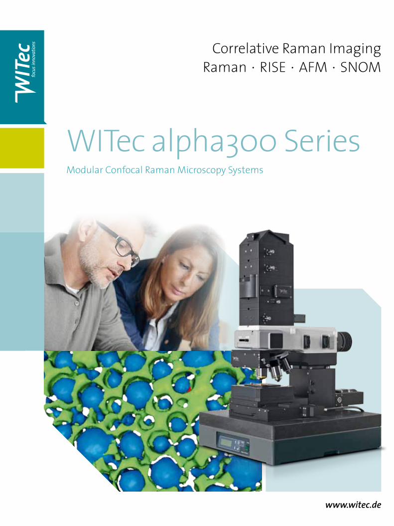
www.witec.de Correlative Raman Imaging Raman • RISE • AFM • SNOM WITec alpha300 Series Modular Confocal Raman Microscopy Systems
Transcript of WITec alpha300 Series€¦ · WITec alpha300 Series | 11 alpha300 access alphaControl Microscope...
www.witec.de
Correlative Raman ImagingRaman • RISE • AFM • SNOM
WITec alpha300 SeriesModular Confocal Raman Microscopy Systems
vegetative cell energy storage metabolite spores
nucleoli
nucleus endoplasmic reticulum
cytoplasm
mitochondriae
nuclearmembrane
10 µm
0.5 µm
polymer substrate drug-eluting polymer coating drug
1 µm30 µm
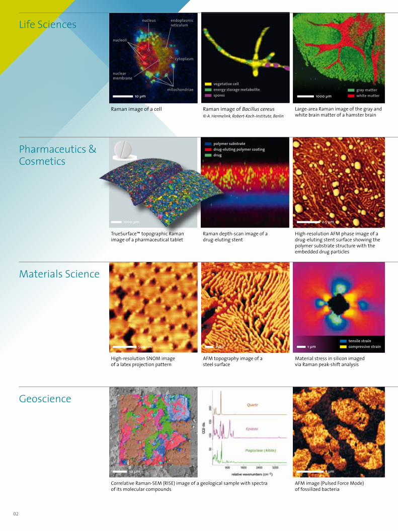
Life Sciences
Geoscience
Raman image of a cell Raman image of Bacillus cereus© A. Hermelink, Robert-Koch-Institute, Berlin
AFM image (Pulsed Force Mode) of fossilized bacteria
Raman depth-scan image of a drug-eluting stent
Correlative Raman-SEM (RISE) image of a geological sample with spectra of its molecular compounds
Large-area Raman image of the gray and white brain matter of a hamster brain
02
tensile strain compressive strain500 nm 2 µm 1 µm
Materials Science
High-resolution SNOM image of a latex projection pattern
AFM topography image of a steel surface
Material stress in silicon imaged via Raman peak-shift analysis
02
High-resolution AFM phase image of a drug-eluting stent surface showing the polymer substrate structure with the embedded drug particles
1000 µm
Pharmaceutics & Cosmetics
TrueSurface™ topographic Raman image of a pharmaceutical tablet
1000 µm
gray matter white matter
1000 µm
PlagioclaseTitanium oxideQuartzFluoriteOrganicsCalcite
2 µm 8 µm
10 µm
2 µm
G-layer S2-layer middle lamella1 µm
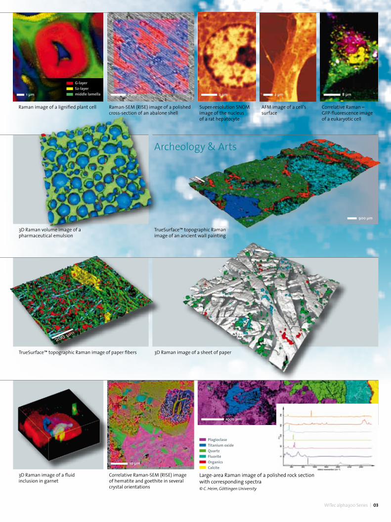
3D Raman image of a fluid inclusion in garnet
Correlative Raman-SEM (RISE) image of hematite and goethite in several crystal orientations
WITec alpha300 Series | 03
900 µm
Super-resolution SNOM image of the nucleus of a rat hepatocyte
AFM image of a cell's surface
Correlative Raman – GFP-fluorescence image of a eukaryotic cell
Raman image of a lignified plant cell
3D Raman image of a sheet of paperTrueSurface™ topographic Raman image of paper fibers
2 µm
Large-area Raman image of a polished rock section with corresponding spectra© C. Heim, Göttingen University
Raman-SEM (RISE) image of a polished cross-section of an abalone shell
Archeology & Arts
TrueSurface™ topographic Raman image of an ancient wall painting
3D Raman volume image of a pharmaceutical emulsion
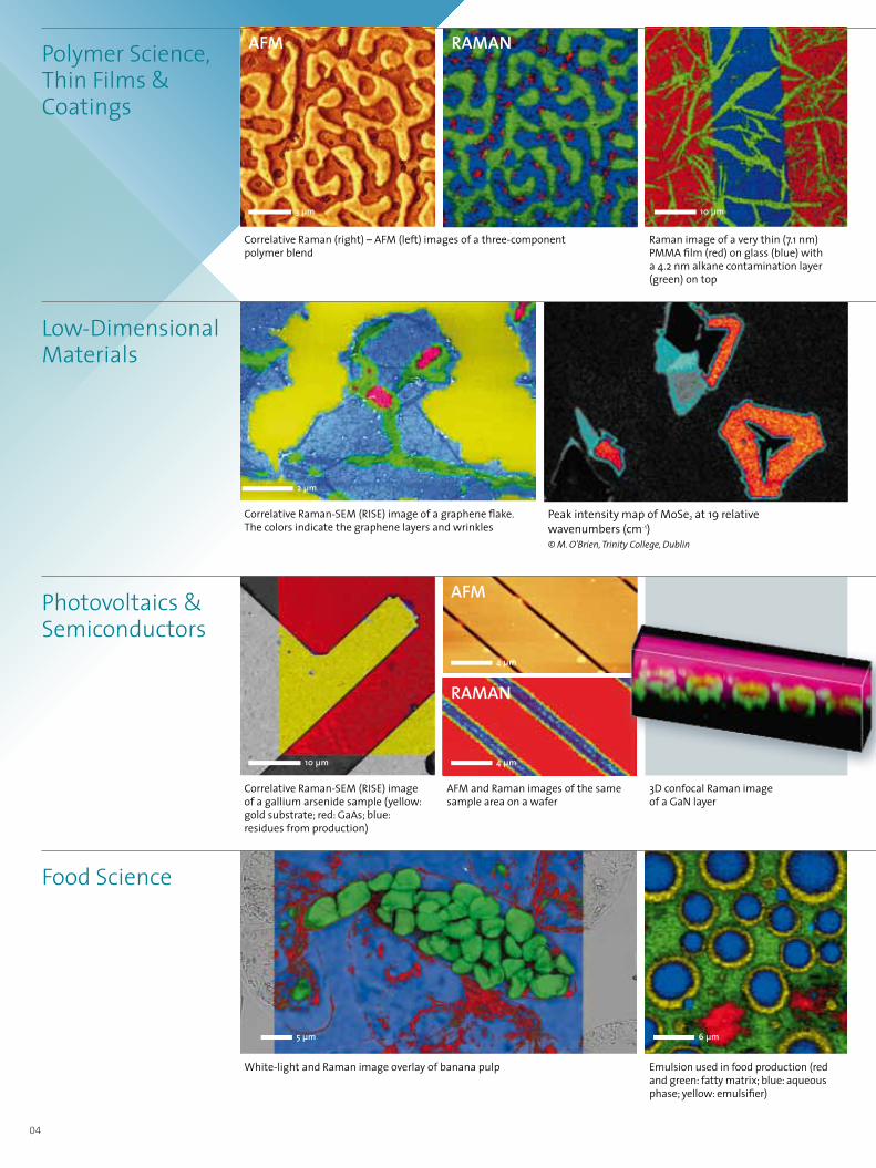
Photovoltaics & Semiconductors
Low-Dimensional Materials
Correlative Raman-SEM (RISE) image of a gallium arsenide sample (yellow: gold substrate; red: GaAs; blue: residues from production)
AFM and Raman images of the same sample area on a wafer
White-light and Raman image overlay of banana pulp
Correlative Raman-SEM (RISE) image of a graphene flake. The colors indicate the graphene layers and wrinkles
Peak intensity map of MoSe2 at 19 relative wavenumbers (cm-1)© M. O'Brien, Trinity College, Dublin
5 µm
Raman image of a very thin (7.1 nm) PMMA film (red) on glass (blue) with a 4.2 nm alkane contamination layer (green) on top
10 µm
4 µm
4 µm
10 µm
Food Science
2 µm
04
AFM
Raman
Polymer Science, Thin Films & Coatings
Raman
3 µm
Correlative Raman (right) – AFM (left) images of a three-component polymer blend
3D confocal Raman image of a GaN layer
6 µm
Emulsion used in food production (red and green: fatty matrix; blue: aqueous phase; yellow: emulsifier)
AFM
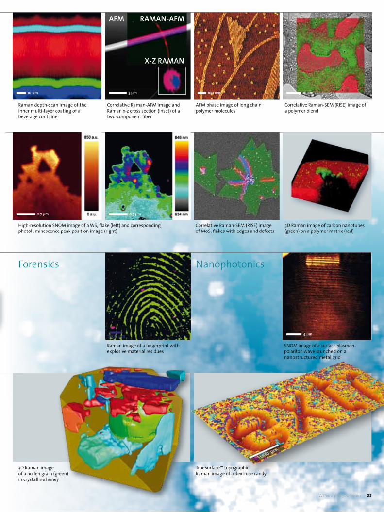
Forensics
Correlative Raman-AFM image and Raman x-z cross section (inset) of a two-component fiber
AFM phase image of long chain polymer molecules
Correlative Raman-SEM (RISE) image of a polymer blend
3 µm 100 nm 10 nm
Raman depth-scan image of the inner multi-layer coating of a beverage container
WITec alpha300 Series | 05
Raman-aFm
x-z Raman
AFM
High-resolution SNOM image of a WS2 flake (left) and corresponding photoluminescence peak position image (right)
Correlative Raman-SEM (RISE) image of MoS2 flakes with edges and defects
3D Raman image of carbon nanotubes (green) on a polymer matrix (red)
3D Raman image of a pollen grain (green) in crystalline honey
Raman image of a fingerprint with explosive material residues
TrueSurface™ topographic Raman image of a dextrose candy
10 µm
0.7 µm 0.7 µm
Nanophotonics
SNOM image of a surface plasmon-polariton wave launched on a nanostructured metal grid
4 µm
06
Raman
SNOM
AFM
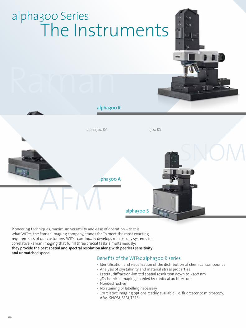
alpha300 Series The Instruments
Pioneering techniques, maximum versatility and ease of operation – that is what WITec, the Raman imaging company, stands for. To meet the most exacting requirements of our customers, WITec continually develops microscopy systems for correlative Raman imaging that fulfill three crucial tasks simultaneously:they provide the best spatial and spectral resolution along with peerless sensitivity and unmatched speed.
Benefits of the WITec alpha300 R series• Identification and visualization of the distribution of chemical compounds• Analysis of crystallinity and material stress properties• Lateral, diffraction-limited spatial resolution down to ~200 nm• 3D chemical imaging enabled by confocal architecture• Nondestructive• No staining or labelling necessary• Correlative imaging options readily available (i.e. fluorescence microscopy, AFM, SNOM, SEM, TERS)
alpha300 RS
alpha300 S
alpha300 A
alpha300 R
alpha300 RA

Design & Components Modular & Upgradable
AFM bEAM-dEFlECTIoN uNITfor cantilever-based topography and near-field optical measurements
ouTpuT CouplEr*primary spectrometer output
lASEr INpuT CouplErS*single & triple wavelength modules combinable and stackable for more wavelengths
VIdEo CAMErA CouplEr*able to house different camera models
KöHlEr WHITE-lIgHT IlluMINATIoN*with adjustable LED intensity
010203
04
05
MICroSCopE TurrET*up to 6 objectives07STEppEr MoTorfor z-movement, focusing, depth profiling, 3D stacks
08poSITIoNINg & SCANNINg STAgEScooperative scanning of piezo-driven and/or motorized stages
09
06
MICroSCopE CoNTrol & dATA EVAluATIoN SoFTWArEWITec Control WITec Project WITec Project PlusWITec TrueMatch
10
WITec alpha300 Series | 07
SNOM
*motorized options available
01
02
07
03
04
05
10
08
06
09
ouTpuT CouplEr*outputs for additional spectrometers

Confocal Raman Microscopy &
08
Additional sample information from Raman spectra:a. Peak intensity: Quantity of a specific compoundb. Peak shift: Identification of stress and strain statesc. Peak width: Degree of crystallinityd. Polarization state: Crystal symmetry and orientation
3D Raman Imaging
The Raman principle• The Raman effect is based on inelastic scattering of excitation
light by the molecules of gaseous, liquid or solid materials. The interaction of a molecule with photons causes vibrations of its chemical bonds, leading to specific energy shifts in the scattered light that can be identified in its Raman spectrum.
• Any given chemical compound can be easily identified by this individual spectral “fingerprint”.
a b c d
Raman imagingWhen Raman spectra are collected at every measurement point using a confocal microscope combined with a spectrometer, a Raman image can be generated that visualizes the distribution of the sample's compounds. Due to the high confocality of WITec Raman systems, volume scans and 3D images can also be generated.

WITec alpha300 Series | 09
Confocal Raman Microscopy & Resolution
Sensitivity
Speed
constantly simultaneously
routinely provable
resolution: Lateral resolution is physically limited to ~200 nm, depending on the wavelength of the incident light. Speed: The more sensitive a system is, the shorter the acquisition time for a single spectrum. WITec's Ultrafast Raman Imaging reduces acquisition times for single Raman spectra down to well below 1 ms. Sensitivity: A high confocality increases the signal-to-noise ratio by reducing the background. With the UHTS series, WITec developed lens-based, wavelength-optimized spectrometers with an ultimate spectral resolution down to 0.1 relative wavenumbers (@633 nm excitation).
3D Raman Imaging
3D Raman image of a honey droplet containing a pollen grain (green) and several crystalline phases (red, blue, cyan), reconstructed from 50 individual 2D Raman images acquired along the z-axis.
No need for compromisesThe Raman effect is extremely weak, so everyRaman photon is important for imaging.Therefore WITec Raman imaging systemscombine an exceptionally sensitive confocalmicroscope with an ultrahigh-throughputspectrometer system (UHTS).
The precise adjustment of all optical and mechanical elements guarantees the highest resolution, outstanding speed and extraordinary sensitivity simultaneously!
This optimization allows the detection of Raman signals of even weak Raman scatterers and extremely low material concentrations or volumes with the lowest excitation energy levels. This is an unrivaled advantage of WITec systems.

10
alpha300 R Raman Microscope The alpha300 R is a confocal Raman microscopy system available in several configurations with different features to fulfill a wide variety of customer requirements.
Whether you want to perform micro-Raman mapping or high-end 3D Raman imaging, as a Raman newcomer or an experienced user, in a scientific setting or an industrial laboratory, WITec offers the perfect Raman imaging solution for you.
Key features and benefits• True confocality, ideally suited to depth profiling and 3D Raman
image generation• Lateral resolution limited only by physical law• Spectral resolution down to 0.1 relative wavenumbers
(@633 nm excitation)• Focus stabilization compensates for thermal and mechanical
variations during long-term measurements• Laser wavelength selectable from UV to NIR• Throughput-optimized UHTS spectrometers with a variety of focal
lengths• Fast Raman Imaging™ and Ultrafast Raman Imaging with
motorized or piezo-driven scanning stages• 3D images and depth profiles with motorized or piezo-driven
scanning stages
WITec alpha300 Series | 11
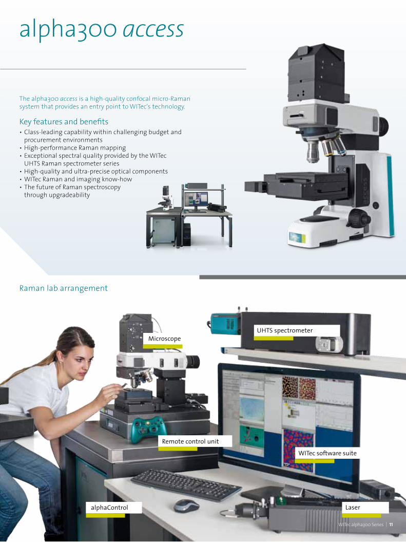
alpha300 access
alphaControl
Microscope
Remote control unit
WITec software suite
UHTS spectrometer
Laser
Raman lab arrangement
The alpha300 access is a high-quality confocal micro-Raman system that provides an entry point to WITec's technology.
Key features and benefits• Class-leading capability within challenging budget and procurement environments• High-performance Raman mapping • Exceptional spectral quality provided by the WITec UHTS Raman spectrometer series• High-quality and ultra-precise optical components • WITec Raman and imaging know-how• The future of Raman spectroscopy through upgradeability
12
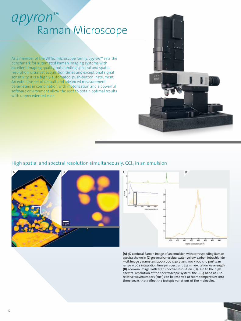
apyron™ Raman Microscope As a member of the WITec microscope family, apyron™ sets the benchmark for automated Raman imaging systems with excellent imaging quality, outstanding spectral and spatial resolution, ultrafast acquisition times and exceptional signal sensitivity. It is a highly-automated, push-button instrument. An extensive set of default and advanced measurement parameters in combination with motorization and a powerful software environment allow the user to obtain optimal results with unprecedented ease.
High spatial and spectral resolution simultaneously: CCl4 in an emulsion
(A) 3D confocal Raman image of an emulsion with corresponding Raman spectra shown in (C) green: alkane; blue: water; yellow: carbon tetrachloride + oil. Image parameters: 200 x 200 x 20 pixels, 100 x 100 x 10 µm3 scan range, 0.06 s integration time per spectrum, 532 nm excitation wavelength. (B) Zoom-in image with high spectral resolution. (D) Due to the high spectral resolution of the spectroscopic system, the CCI4 band at 460 relative wavenumbers (cm-1) can be resolved at room temperature into three peaks that reflect the isotopic variations of the molecules.
B C DA

WITec alpha300 Series | 13
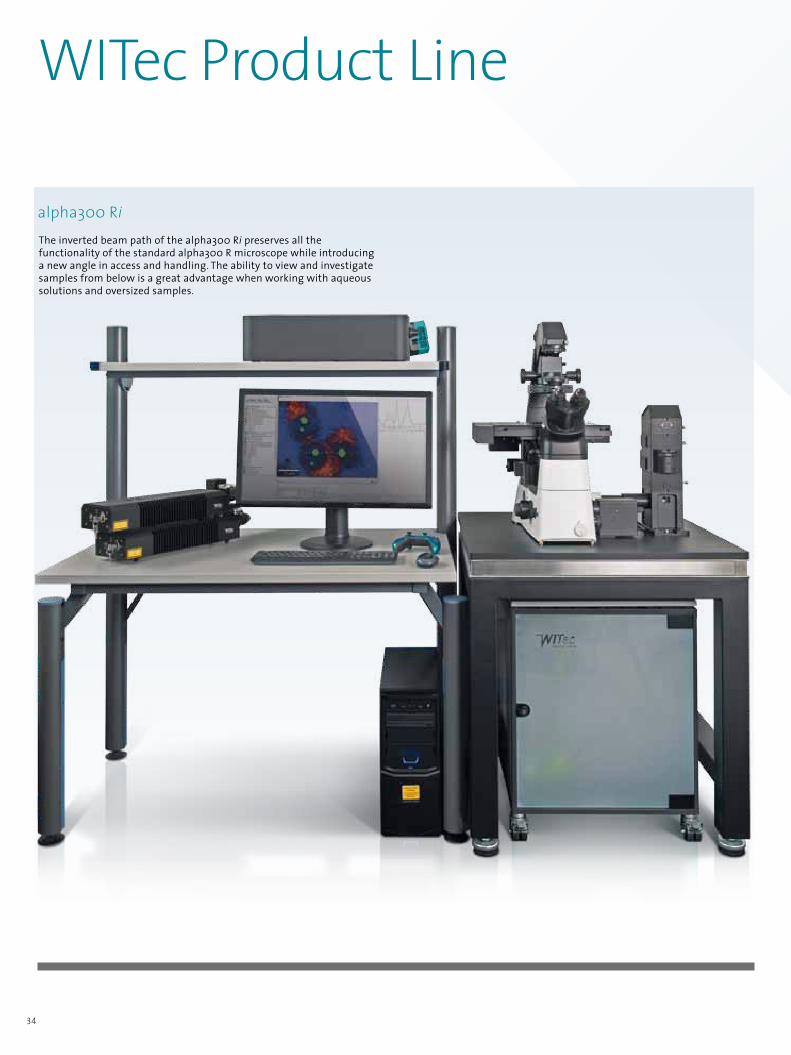
alpha300 Ri Inverted Confocal Raman Microscope
An inverted beam path allows liquid samples to be placed on the fixed plane of the stage for quick and repeatable measurements.
The alpha300 Ri turns chemical imaging upside down. Its inverted beam path preserves all the functionality of WITec's alpha300 R microscopes while introducing a new angle in access and handling. The ability to view and investigate samples from below is a great advantage when working with aqueous solutions or oversized samples. Studies in life sciences, biomedicine, pharmaceutics and geosciences in particular will benefit from the consistency and flexibility provided by the geometry of the alpha300 Ri. Many accessories and upgrade possibilities developed for the alpha300 series are compatible with the Ri version.
Bulky samples that would be challenging to investigate underneath a conventional microscope objective turret can be accommodated by placing them on the stage of the alpha300 Ri. The motorized sample stage
also facilitates the mounting of environmental enclosures and other accessories.

14
219 nm
0 nm
C
The WITec AFM objective allows simultaneous sample and cantilever survey from above with high resolution. It facilitates accurate and convenient AFM tip alignment and positioning for even very small sample structures. (A) Simultaneous cantilever and sample view, (B) overlay of the optical image with the AFM image (AC mode, Scan range: 3 x 3 µm2), (C) 3D representation of the AFM image from (B).
Simultaneous cantilever and sample view for easy determination of the measurement position
alpha300 A Atomic Force Microscope
A B
AFM modes• AC Mode• Contact Mode• Digital Pulsed Force Mode™ (DPFM)• Electrostatic Force Microscopy (EFM)• Kelvin Probe Microscopy• Lateral Force Microscopy (LFM) • Lift Mode™• Magnetic Force Microscopy (MFM)• Nanomanipulation/Lithography
Key features and benefits• Surface characterization on the nanometer scale• Optical and AFM combination• Nondestructive imaging• Minimal, if any, sample preparation• Ease of use in air and liquids• Combinable with confocal Raman imaging and scanning near-field optical microscopy (SNOM)• TrueScan™ controlled piezo-driven scanning stages with capacitive feedback loops: – 30 x 30 x 10 μm3 – 100 x 100 x 20 μm3 – 200 x 200 x 20 μm3
The WITec alpha300 A atomic force microscope integrated with a research-grade optical microscope provides superior optical access, easy cantilever alignment and high-resolution sample survey. Using optical pre-inspection by means of various illumination and detection techniques (e.g. bright field, dark field, polarization, fluorescence, etc.), the user can conveniently determine the area of interest for the AFM measurement. By simply rotating the microscope turret, the user can switch between conventional microscopy and AFM modes quickly and easily. The WITec AFM objective provides a direct view of both sample and cantilever for straightforward and precise cantilever tip positioning. The alpha300 A system includes an extremely linear and precise capacitive feedback-controlled scanning stage featuring TrueScan™ for exceptional accuracy over the entire scan range.

Adhesion Stiffnessx
y
Adhesion Stiffnessx
y
Viscosity Contact Time
Topography
Viscosity Contact Time
Topography
Viscosity Contact Time
Topography
WITec alpha300 Series | 15
(A) The PFM electronics induces a sinusoidal modulation of the z-piezo of the AFM with an amplitude of 10 – 500 nm at a user-selectable frequency of between 100 Hz and 2 kHz, what is far below the resonant frequency of the cantilever. A complete force-distance cycle is carried out at this rate, resulting in the force signal as shown in the figure. (B) An ethyl-hexyl-acrylate/polystyrene blend (EHA/PS) was spin-coated onto a glass substrate. Various surface properties were analyzed and imaged simultaneously using DPFM. Dark areas in the images correspond to low values.
Free Cantilever Oscillation
Baseline
Force Signal
Stiffness
Adhesion Peak
Snap
Time
Forc
e
Fmax
y
x
Digital Pulsed Force Mode (DPFM)Pulsed Force Mode (PFM) is a non-resonant, intermittent contact mode for atomic force microscopy that allows the characterization of material properties such as adhesion, stiffness and viscosity along with the sample topography. Additionally, lateral forces are virtually eliminated. Therefore high-resolution mapping of delicate samples in air and liquids is easily achievable while maintaining a scanning speed comparable to contact mode AFM. In contrast to most other intermittent contact techniques, the perpendicular forces on the sample (introduced by the AFM tip) are controlled by a feedback loop.
BA
Imaging of surface properties with DPFM

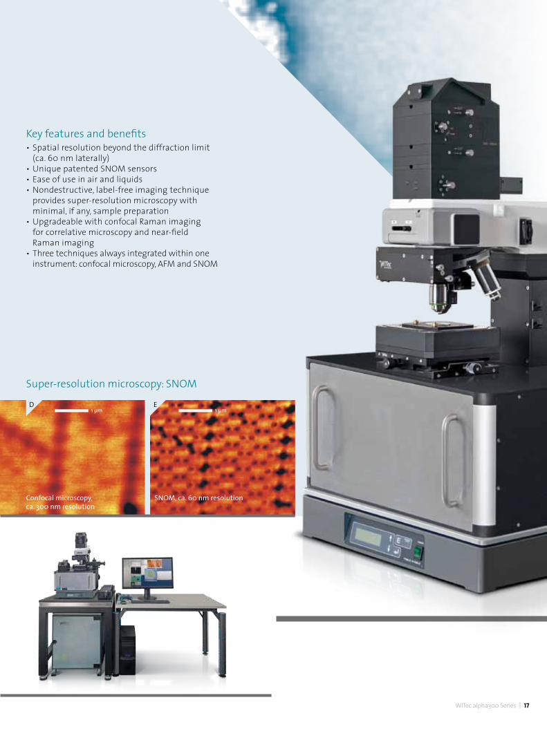
Super-resolution microscopy down to 60 nm can be achieved with the WITec scanning near-field optical microscope (SNOM) alpha300 S. SNOM, as compared to other optical super-resolution techniques such as STED or STORM is not dependent on fluorescent tags or specialized laser excitation sources. The operation of the system is straightforward as various measurement procedures such as a high-speed automatic cantilever approach and adjustment can be controlled through the intuitive software.
The alpha300 S combines the advantages of SNOM, confocal microscopy and AFM in a single instrument. By rotating the objective turret one can easily switch between the different modes. One field that can benefit immensely from this technique is nanotechnology research, in particular nano-photonics and nano-optics. In life sciences and materials research, SNOM permits optical detection of the most miniscule surface structures of transparent as well as opaque samples.
16
alpha300 S Scanning Near-field Optical Microscope
A B C
(A) Video camera top view of a SNOM sensor and sample, (B) side view of a cantilever pyramid, (C) SNOM cantilever wafer.
Comparison of optical resolution between confocal microscopy (D) and scanning near-field optical microscopy (E) on a latex projection pattern.
Cantilever SNOM-sensors The alpha300 S uses unique, patented, high-quality micro-fabricated SNOM sensors consisting of a silicon cantilever with a hollow pyramid tip. The SNOM aperture is at the apex of the pyramid. The laser light used for optical imaging is focused into the back of the tip and then onto the sample. Due to the wide opening angle of the pyramid, the transmission coefficient is much higher than that of fiber probes with the same aperture diameter. An established and proven manufacturing process
produces tips with apertures of varying sizes to meet customers' individual requirements. Cantilever SNOM sensors are, unlike fiber tips, very robust and flexible in the z-direction and allow the beam deflection technique to precisely control the tip-sample distance. All of these innovative characteristics make the handling of probes during near-field measurements very user-friendly for the most reliable optical imaging available beyond the diffraction limit.
WITec alpha300 Series | 17
1 µm 1 µm
Confocal microscopy, ca. 300 nm resolution
SNOM, ca. 60 nm resolution
E
Super-resolution microscopy: SNOM
Key features and benefits• Spatial resolution beyond the diffraction limit (ca. 60 nm laterally)• Unique patented SNOM sensors• Ease of use in air and liquids• Nondestructive, label-free imaging technique provides super-resolution microscopy with minimal, if any, sample preparation• Upgradeable with confocal Raman imaging for correlative microscopy and near-field Raman imaging• Three techniques always integrated within one instrument: confocal microscopy, AFM and SNOM
D

Switching microscopy modes by rotating the objective turretScanning Probe Microscopy (SPM) mode for AFM or SNOM: The SPM cantilever is held at the end of the objective's arm. The cantilever is aligned with an integrated highly-precise inertial drive.
WITec's alpha300 microscope series was designed with inherent flexibility and configurability to address users' individual requirements. This not only involves easy and cost-effective upgrade and extension options, but also the possibility to add other microscopy techniques to the instrument.
As no single imaging approach can reveal every feature of a sample, a more comprehensive understanding can be achieved by using several techniques and correlating the results.
This advantage is particularly pronounced when using methods that deliver images of the morphology of a sample while also determining its molecular composition. WITec's modular product line incorporates nearly all scanning probe and optical microscopy techniques. Each microscope model can always be equipped with the functionality of another variation of the alpha300 family either as a built-in feature or as a later upgrade. The WITec hardware and software environment is used for all new features and upgrades, ensuring the best possible compatibility and ease of use.
Correlative Raman Imaging Made Easy
18
Optical modes: Rotating the turret provides access to the optical modes, allowing high-resolution confocal optical and chemical imaging to be seamlessly linked with AFM or SNOM measurements without transferring the sample.

Correlative, high-resolution AFM-confocal Raman imaging of a polymer mixture (A) The sample's phase image was created using AFM in tapping mode. (B) Raman spectra were recorded at each image pixel of the area and displayed as a false color image. Red: polystyrene, green: ethyl-hexyl-acrylate (EHA), violet: styrene-butadiene-rubber (SBR), blue: mixed spectrum of SBR and EHA. (C) The overlay of the AFM phase and the Raman image identifies and locates the sample's components.
WITec alpha300 Series | 19
A B C
Raman-AFMBy combining the materials analysis capability of confocal Raman imaging with the ultrahigh topographic and lateral resolution of an AFM, the chemical properties of a sample can be easily correlated with its surface characteristics.
The well-established alpha300 RA was the first integrated Raman-AFM system on the market and it continues to set the benchmark for combined instrument configurations.
The alpha300 RA incorporates the features of the alpha300 R for chemical imaging along with the alpha300 A for atomic force microscopy. For TERS options, please see page 20.
(A) Near-field Raman image of the G-Band intensity of graphene. (B) The graph shows the G-band intensity along the red line and reveals the measurable signal variations between the small sample and the substrate. (C) Corresponding Raman spectrum obtained by the Raman-SNOM measurement.
1 µm
A B C
Raman-SNOMThe alpha300 RS combines Raman characterization and imaging with scanning near-field optical microscopy for optical imaging with resolution beyond the diffraction limit. It combines all features of the alpha300 S and alpha300 R instruments.
alpha300 R and alpha300 S

20
Confocal Raman imaging and scanning electron microscopy (SEM) are techniques that are ideally suited to correlating structural and chemical information.
RISE™ – Raman Imaging and Scanning Electron Microscopy
Principle of RISE™ microscopySamples are automatically transferred from one measuring position to the other within the vacuum chamber of the combined Raman-SEM instrument, streamlining the workflow and drastically improving ease of use.
SEM Raman
Sample
h0 h(0±)
XYZ Offset

WITec alpha300 Series | 21
The sample remains inside the vacuum chamber during both the SEM and Raman measurements to ensure seamless correlative microscopy.
Key features and benefitsRISE™ microscopy systems combine all features of a stand-alone SEM and a WITec research-grade confocal Raman imaging microscope within one instrument to provide:• Quick and convenient switching between Raman and SEM modes• Automated sample transfer from one measuring position to the other within the vacuum chamber• An integrated software interface for user-friendly measurement control• Easy correlation of experimental results and image overlay• SEM and Raman imaging capabilities without compromise• A truly confocal optical path• Research-grade optical imaging
A CB
(A) In the SEM image a piece of hematite (Fe2O3) displays some structural characteristics. (B) Hematite and goethite (FeO(OH)) in several crystalorientations were identified from their Raman spectra. Crystal forms of hematite are depicted in red, blue, green, orange and pink and those of goethitein light blue and cyan. From the spectra, a Raman image was generated. (C) Correlation of Raman and SEM data resulted in the RISE™ image.
Application iron mineralogy
1o µm

22
WITec's modular design relies on efficient beam delivery via optical fiber. Long-term investments in research and development have yielded an exceptional understanding of fiber-coupling mechanisms, enabling benefits unattainable with more conventional methods.
• Virtually lossless energy transmission• Fibers provide a diffraction-limited point light source for the highest confocality and spatial
resolution• Pre-configured and pre-aligned fiber coupling units guarantee long-term stability and user-
friendliness without the need for further adjustment • Polarization direction of the light is maintained for the most intricate polarization-dependent
measurements• Lasers and spectrometers can be mounted far away from the microscope, allowing flexible and
compact system footprints that can alleviate thermal or vibrational disturbances
Fiber-Based Beam Delivery Facts and Benefits

Modular Components & Accessories Beam Path Options Excitation and detection optionsThe WITec alpha300 microscope series features several beam path geometries. The upright microscope enables measurements of opaque samples while the inverted microscope (p. 13) is ideal for experiments on transparent samples. Side-illumination options are also available. The modularity and inherent flexibility of WITec's unique alpha300 microscope systems provide optimized instrumentation for many different types of experiments.
WITec TERS module for the alpha300 seriesTip-Enhanced Raman Spectroscopy (TERS) enables the acquisition of chemical information with a lateral resolution far below the diffraction limit. The TERS tip illumination can be performed from either above, below or from the side. The WITec alpha300 microscope series accomodates all excitation approaches and TERS measurements can be carried out alongside Raman and AFM investigations with the same integrated instrument.
TERS working principle
alpha300 RExcitation & detection from above
alpha300 TERS experiment on a carbon nanowire: The enhancement of the spectral intensity in TERS-mode is clearly visible.
alpha300 RiExcitation & detection from below
Diffraction limited focus spot
Laser
AFM cantilever
Ag- or Au-coated AFM tipStrong localized field enhancement
WITec alpha300 Series | 23
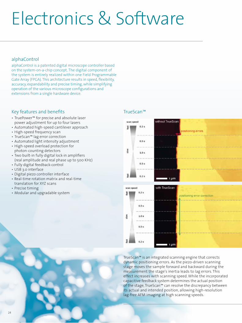
Key features and benefits• TruePower™ for precise and absolute laser power adjustment for up to four lasers• Automated high-speed cantilever approach• High-speed frequency scan• TrueScan™ lag error correction• Automated light intensity adjustment• High-speed overload protection for photon-counting detectors• Two built-in fully digital lock-in amplifiers (real amplitude and real phase up to 500 KHz)• Fully digital feedback-control• USB 3.0 interface • Digital piezo controller interface• Real-time rotation matrix and real-time translation for XYZ scans• Precise timing• Modular and upgradable system
Electronics & Software
24
alphaControl alphaControl is a patented digital microscope controller based on the system-on-a-chip concept. The digital component of the system is entirely realized within one Field Programmable Gate Array (FPGA). This architecture results in speed, flexibility, accuracy, expandability and precise timing, while simplifying operation of the various microscope configurations and extensions from a single hardware device.
TrueScan™ is an integrated scanning engine that corrects dynamic positioning errors. As the piezo-driven scanning stage moves the sample forward and backward during the measurement the stage's inertia leads to lag errors. This effect increases with scanning speed. While the incorporated capacitive feedback system determines the actual position of the stage, TrueScan™ can resolve the discrepancy between its actual and intended position, allowing high-resolution lag-free AFM imaging at high scanning speeds.
TrueScan™
1 µm
1 µm

Experimental setup, instrument control and data analysis are made easy with WITec's software tools. • Sophisticated data acquisition, evaluation, post-processing and image generation for confocal Raman microscopy, AFM and SNOM.• The software architecture and graphical user interface offer an integrated and consolidated functionality incorporating every technique and measurement mode.• Suitable for all experience levels and user requirements through an individually-adjustable user interface.
WITec alpha300 Series | 25
WITec Suite FIVE Powerful Software Environment
WITec Project FIVE+• Advanced offline data and image processing • Chemometric post-processing features and superior data analysis tools: Cluster Analysis, Principal Component Analysis, Spectral Demixing, Advanced Spectral Fitting, Image Correlation and many more • Single user license
WITec microscope family with alphaControl
Control FIVE Project FIVE Project FIVE+
WITec Project FIVE• Offline data and image processing • Licensed for an unlimited number of users
WITec Control FIVE• Centralized experiment and instrument settings • Acquisition of imaging and/or spectroscopy data • On- and offline data analysis and image processing

26
Sample positioning and scanning stagesWITec offers manual, stepper motor-driven and piezo-actuated stages to accommodate a wide variety of samples and applications. • Manual translation stage for accurate sample positioning• Motorized translation stages for large-scale positioning, mapping and imaging• Piezo-driven scanning stages with closed-loop operation for AFM and high-resolution Raman measurements The stages feature several different travel ranges for the highest flexibility.
Spectroscopy solutions: The UHTS seriesThe WITec Ultra-High Throughput Spectrometer (UHTS) series consists of exceptionally flexible and precise devices that meetthe demands of cutting-edge confocal Raman imaging.
Varied excitation sources and scattering experiments requirespecialized optics for optimal throughput. The WITec UHTSdesign approach takes this into account by employing a series of lens-based on-axis spectrometers for a range of excitationwavelengths.
Heating & cooling stagesFor experiments requiring an accurate temperature control, the alpha300 series can be equipped with heating and cooling stages with ranges from -269°C up to 1500°C (4 K - 1773 K) for AFM and high-resolution Raman measurements. Sample mounting stagesA selection of mounting stages is available for securing samples of various shapes and environmental requirements. Microscope slides, Petri dishes, NUNC™ flasks and cylindrical samples such as stents can all be accommodated. The integration of third-party cryostats is available upon request.
Modular Components & Accessories
UV VIS NIR
400 nm 500 nm 600 nm 700 nm 900 nm 1100 nm 1300 nm 1500 nm 1700 nm
UHTS 400 UV - VIS
UHTS 300 VIS
UHTS 400 NIR
BroadBand UHTS 300 VIS - nIr
01
05
03
04
02 UHTS 600 VIS
UHTS 300 IR06
Spectrometers of the UHTS series allow more than 70%total transmission for high-speed and high-resolution Ramanimaging. They feature an optical fiber port and a motorizedtriple-grating turret. Customers can choose from amongseveral focal lengths and gratings to match individualrequirements in terms of spectral range or resolution.

WITec alpha300 Series | 27
TrueSurface™Modular Components & Accessories
TrueSurface™ microscopy WITec's patented TrueSurface™ microscopy option enables confocal Raman imaging guided by surface topography. With TrueSurface™ chemical characterization on rough, inclined or irregularly-shaped samples can be carried out precisely because the sensor actively monitors the sample surface and keeps it in focus. This topographic imaging module also compensates for variations during measurements with long integration times. TrueSurface™ pioneered by WITec uses an advanced optical profilometer integrated within the instrument to provide one-pass simultaneous acquisition of both topographic and molecular information.
Laser power adjustment with TruePower™ TruePower is a new experience in laser power adjustment for confocal Raman imaging measurements. The absolute laser power delivered to the microscope is measured and adjusted with an accuracy of 0.1 mW. A laser shutter shields the sample from the laser light and opens only during Raman analysis with an optimized power level to avoid any sample degradation. TruePower is the only laser power regulation system on the market to provide such accuracy. • Optimal laser power determination for the preservation of delicate samples and reproducibility in measurement conditions• Power series measurements enabled by making power- induced spectral changes quantifiable• Automated documentation of laser settings• Retrofittable for most WITec imaging systems
Laser sourcesExcitation laser sources for Raman, SNOM and photoluminescence imaging can be individually chosen and attached in concert to the microscope via optically-matched fiber coupling. Available laser wavelengths range from the UV to the NIR to fulfill a variety of experimental excitation requirements. Switching between different lasers is a matter of simply rotating a wheel in the coupling unit or by a click in the software.
100
mW0 43 53,5 4,5
1000 µm

Modular Components & Accessories
28
RayShield coupling unit for Raman imaging at low wavenumbersThe RayShield coupler includes a specialized narrow band filter set which is optimally aligned for the detection of Stokes and anti-Stokes Raman lines close to the Rayleigh line (ca. 10 cm-1) while maintaining an ideal Rayleigh shielding. Anti-Stokes extensionA wavelength-optimized filter unit for the detection of anti-Stokes Raman signals is available as an option for advanced Raman imaging experiments. Polarization modulePolarization dependent measurements can be used to analyze molecular orientations and geometries of materials. The module is comprized of an excitation wavelength specific polarization kit for rotating the polarization of the laser light and an analyzer for filtering information of a certain polarization. StrobeLockStrobeLock is a WITec extension that enables time-correlated single photon counting measurements of unprecedented accuracy. The available imaging modes include fluorescence lifetime imaging microscopy (FLIM).
TrueMatch™ Raman spectral database managementTrueMatch™ is a powerful and innovative software module for identifying the components of a sample and for accessing and developing Raman spectral databases. With this technology, existing databases containing representative Raman spectra of common materials or substrates can be searched and referenced. It also allows users to create their own catalog of spectra pertinent to their research. TrueMatch™ is fully integrated with the WITec Suite FIVE software environment and is a perfect complement to its data evaluation and post-processing capabilities. LabVIEW interfaceThe optional LabVIEW interface provides access for alphaControl in order to design and control individual measurement procedures with LabVIEW.
ControlSpectral data acquisition
TrueMatchprojectSpectral data evaluation & post-processing
500
100
200
300
CCD
cts
400
500
1000 1500 2000relative wavenumbers (cm-1)
3000 35002500
500
100
200
300
CCD
cts
400
500
1000 1500 2000relative wavenumbers (cm-1)
3000 35002500500
100
200
300
CCD
cts
400
500
1000 1500 2000relative wavenumbers (cm-1)
3000 35002500
- Simultaneous multi-spectra search (1:1 match)
- Multiple component search: combine up to three database components to describe a measured composite spectrum
- Automated and simultaneous demixing analysis of multiple components

WITec alpha300 Series | 29
Enclosure & rigid support frameFor maximum reduction of environmental interference such as acoustic or vibrational noise, the WITec support frame and enclosure system greatly contributes to achieving exceptional imaging results. The enclosure can be configured as an air-tight option to allow control of the gas-phase during experiments. Equipped with an interlock, safety regulations (e.g. laser class I requirements) can be easily accommodated. ObjectivesA wide range of objectives for various microscopy techniques are available to meet virtually any experimental requirement. Imaging in liquidsImaging in liquids can be easily performed by using a water immersion objective in optical mode or the liquid imaging extension for AFM and SNOM modes. A specialized cantilever holder and inertial drive allows operation in fluids (e.g. in Petri dishes). Customized solutionsIf your application requires other components, WITec looks forward to discussing your individual requirements and helping to get your experiment up and running. The inherent modularity of our product line allows additional high-quality parts and detectors to be easily integrated.
AFM & SNOM cantileversWITec provides several types of high-quality AFM-cantilevers for various AFM imaging modes. WITec is the only company capable of producing highly reliable SNOM cantilever sensors with apertures ranging from 60 nm to 100 nm. Each SNOM cantilever is individually tested before shipment. AC (Tapping™) mode for AFM & SNOMAC mode for WITec microscope systems allows state-of-the- art resonant intermittent-contact AFM and SNOM imaging specifically tailored for soft and delicate samples. Acoustic AC mode enhances the imaging capabilities of contact mode AFM on such samples, eliminating lateral forces and delivering optimized resolution. DaVinci nanolithographyAdvanced nanotechnology often requires accurate and reliable nano-manipulation or nanolithography tools for precise surface structuring. The WITec DaVinci nanolithography package allows these kinds of experiments in AFM as well as in optical modes with an integrated laser shutter control. Featured light microscopy techniquesThe alpha300 series offers a selection of sample survey and data acquisition methods such as: fluorescence contrast imaging, DIC, dark field microscopy, petrography and polarization-dependent measurements. Photoluminescence detectionFor the detection of photoluminescence spectra beyond 1.1 μm, an InGaAs detector can be easily integrated with the spectroscopic setup. Raman experiments can be extended to acquire additional information in the near-infrared photoluminescence regime (or for 1064 nm Raman measurements).
DAPIER
nucleoli
Correlative Raman – fluorescence microscopy image of eukaryotic cells. Nuclei were stained with DAPI (blue). Endoplasmic reticulum (red) and nucleoli (green) were identified by their Raman signals.

2008 • R&D 100 for the alpha500
2011 • PITTCON Editors' Gold Award for TrueSurface™ Microscopy
• R&D 100 for TrueSurface™ Microscopy
• Microscopy Today Innovation Award for TrueSurface™ Microscopy
2012 • Photonics Prism Award Winner (TrueSurface™ Microscopy)
2015 • Photonics Prism Award 2015 Winner (RISE™ Microscopy)
• Achema Innovation Award 2015 (apyron™ automated Raman imaging system)
1997• SNOM system with unique cantilever SNOM sensors• Pulsed Force Mode1999• Confocal Raman Microscope (WITec CRM 200) for fast 3D Raman imaging2003• WITec Mercury 100 Atomic Force Microscope • Digital Pulsed Force Mode• World's first integrated Raman microscope/AFM combination2006• Modular alpha300 series with FPGA-based control unit alphaControl2008• alpha500 series for large-area and automated multi-point measurements2010• TrueSurface™ Microscopy for confocal microscopy along with large-area optical profiling2014• RISE™ Microscopy: First integration of Raman Imaging and Scanning Electron Microscopy for correlative Raman-SEM Imaging2015• Automated Raman imaging system apyron™2016• alpha300 series design evolution • alpha300 access
2018• TrueSurface™ redefined: innovation in one-pass profilometer- guided Raman imaging• Inverted confocal Raman microscope alpha300 Ri• TrueMatch integrated Raman spectral database software
To be continued …
Since its founding in 1997, the Raman imaging company WITec has established itself as a market leader in the fields of Raman microscopy and correlative Raman-AFM, Raman-SNOM and Raman-SEM (RISE) microscopy. WITec's innovative spirit has kept the alpha300 microscope series at the forefront of the Raman imaging market since it initially revolutionized the field and established Fast Raman Imaging™ as a standard technique.
Ongoing development of the first truly confocal Raman imaging system continues to enable the setting of benchmarks in sensitivity, speed and spectral and spatial resolution. As reflected in WITec's maxim “Focus Innovations” our success is based on continually introducing new technologies and a commitment to maintaining customer satisfaction through high-quality, flexible and innovative products.
WITec
Awards WITec milestones
30

WITec North AmericaWITec Instruments Corp.Knoxville, Tennessee, USA
WITec HeadquartersWITec GmbHLise-Meitner-Str. 6 D-89081 Ulm, GermanyPhone +49 (0) 731 140700 Fax +49 (0) 731 [email protected]
WITec Offices EuropeBarcelona, Lyon, London
WITec China WITec Beijing Representative Office Beijing
WITec South East AsiaWITec Pte. Ltd., Singapore
WITec JapanWITec K.K., Kanagawa
WITec alpha300 Series | 31

32
WITec Product Line
The WITec product portfolio includes imaging systems for Raman, AFM and SNOM analyses as single technique solutions as well as correlative imaging configurations (Raman-AFM, Raman-SNOM, Raman-SEM). All WITec microscopes are high-quality modular systems with exceptional optical throughput, unparalleled signal sensitivity and outstanding imaging capabilities.
The common thread throughout is that all systems are based on the same hardware and software architecture. Whenever required it is possible to simply upgrade any system, even the most basic, with additional features and components, allowing our customers to keep pace with future challenges.
alpha300 A alpha300 S
The WITec alpha300 A atomic force microscope provides nanoscale surface characterization with an integrated research-grade optical microscope for easy cantilever alignment and high-resolution sample survey.
The alpha300 S is a super-resolution scanning near-field optical microscope (SNOM) that combines the advantages of SNOM, confocal microscopy and AFM in a single instrument. It uses unique micro-fabricated SNOM cantilever sensors for spatial resolution well beyond the diffraction limit.
alpha300 R
The WITec alpha300 R is a research-grade microscope combined with an ultrahigh-throughput spectrometer (UHTS) for Raman imaging with exceptional performance in speed, sensitivity, confocality and optical and spectral resolution.

WITec alpha300 Series | 33
alpha300 RA alpha300 RS
apyron™The WITec apyron™ is an intuitive motorized microscope system for Raman spectroscopy and chemical imaging. The push-button principle of the system has the advantage of drastically reducing the time required to become familiar with the operation of the instrument, which greatly accelerates the initiation of measurements and increases the rate of sample turnover. With the apyron‘s straightforward interface the user can concentrate solely on their experiment.
The alpha300 RS facilitates confocal Raman imaging in combination with scanning near-field optical microscopy for optical imaging with super-resolution down to 60 nm. This microscope combines all features of SNOM and confocal Raman imaging and many AFM modes.
The alpha300 RA incorporates the features of the alpha300 R along with those of the alpha300 A. This instrument enables correlative Raman-AFM imaging for a more comprehensive understanding of samples.
34
WITec Product Line
alpha300 RiThe inverted beam path of the alpha300 Ri preserves all the functionality of the standard alpha300 R microscope while introducing a new angle in access and handling. The ability to view and investigate samples from below is a great advantage when working with aqueous solutions and oversized samples.
WITec alpha300 Series | 35
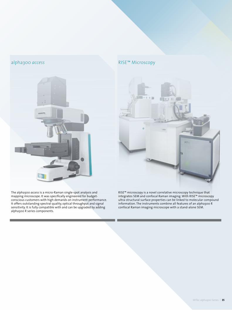
The alpha300 access is a micro-Raman single-spot analysis and mapping microscope. It was specifically engineered for budget-conscious customers with high demands on instrument performance. It offers outstanding spectral quality, optical throughput and signal sensitivity. It is fully compatible with and can be upgraded by adding alpha300 R series components.
RISE™ microscopy is a novel correlative microscopy technique that integrates SEM and confocal Raman imaging. With RISE™ microscopy ultra-structural surface properties can be linked to molecular compound information. The instruments combine all features of an alpha300 R confocal Raman imaging microscope with a stand-alone SEM.
RISE™ Microscopyalpha300 access
We take care
WITec uses environm
entally friendly printed materials. W
hile this policy is only a sm
all contribution to a healthy environment, w
e at WITec believe
that focusing on details can effect positive change in the world.
WITec HeadquartersWITec GmbHLise-Meitner-Str. 6 D-89081 Ulm . GermanyPhone +49 (0) 731 140700 Fax +49 (0) 731 [email protected] www.witec.de
WITec North AmericaWITec Instruments Corp.130G Market Place Blvd. Knoxville . TN 37922 . USAPhone 865 984 4445 Fax 865 984 [email protected] www.witec-instruments.com
WITec South East AsiaWITec Pte. Ltd.25 International Business Park#03-59A German Centre Singapore 609916Phone +65 9026 5667 [email protected]
WITec ChinaWITec Beijing Representative OfficeUnit 507, Landmark Tower 18 North Dongsanhuan Road Beijing, PRC., 100004Phone +86 (0) 10 6590 0577 [email protected] www.witec.de/cn
WITec JapanWITec K.K.Mita 2-3227, Chome, Tama-ku, Kawasaki-shi, Kanagawa-ken 214-0034 JapanPhone +81 44 819 7773 [email protected] www.witec.de/jp
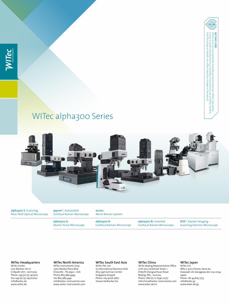
RISE™: Raman Imaging – Scanning Electron Microscope
alpha300 S: Scanning Near-field Optical Microscope
alpha300 Ri: Inverted Confocal Raman Microscope
access: Micro-Raman System
apyron™: Automated Confocal Raman Microscope
alpha300 R: Confocal Raman Microscope
alpha300 A: Atomic Force Microscope
WITec alpha300 Series