Wingfield Review C - cms.wounds-uk.comcms.wounds-uk.com/media/W_Essentials/2009_importance.pdfof...
7
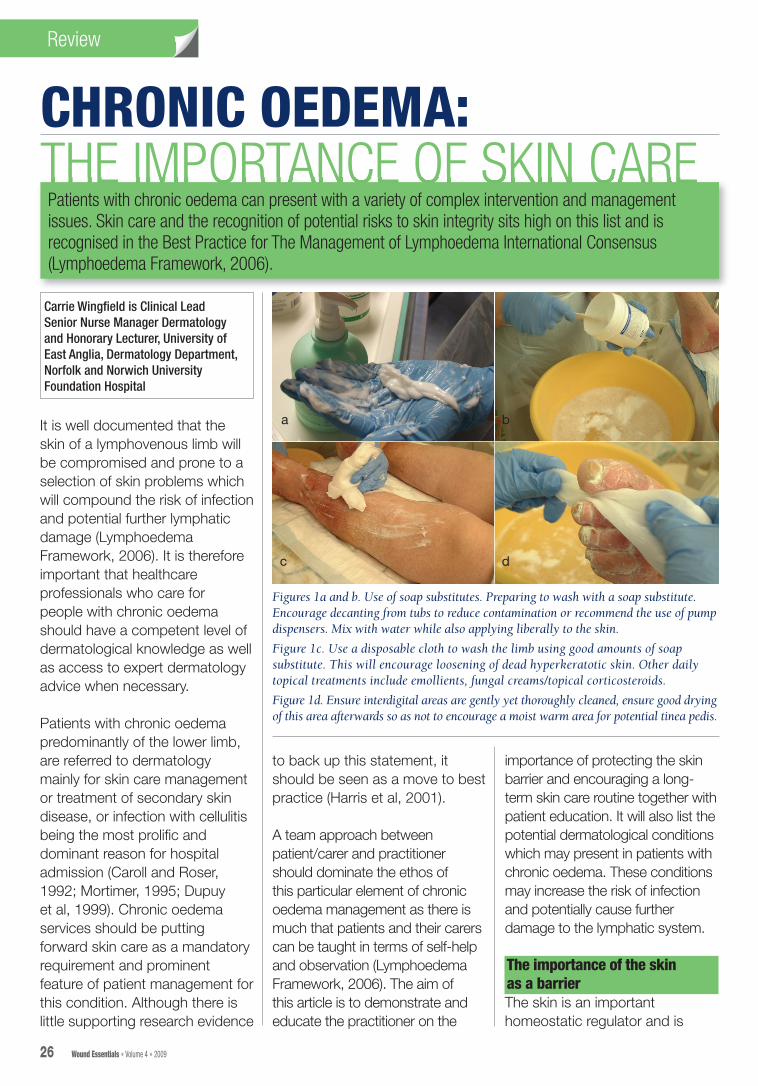
Review CHRONIC OEDEMA: THE IMPORTANCE OF SKIN CARE Carrie Wingfield is Clinical Lead Senior Nurse Manager Dermatology and Honorary Lecturer, University of East Anglia, Dermatology Department, Norfolk and Norwich University Foundation Hospital 26 Wound Essentials • Volume 4 • 2009 Patients with chronic oedema can present with a variety of complex intervention and management issues. Skin care and the recognition of potential risks to skin integrity sits high on this list and is recognised in the Best Practice for The Management of Lymphoedema International Consensus (Lymphoedema Framework, 2006). It is well documented that the skin of a lymphovenous limb will be compromised and prone to a selection of skin problems which will compound the risk of infection and potential further lymphatic damage (Lymphoedema Framework, 2006). It is therefore important that healthcare professionals who care for people with chronic oedema should have a competent level of dermatological knowledge as well as access to expert dermatology advice when necessary. Patients with chronic oedema predominantly of the lower limb, are referred to dermatology mainly for skin care management or treatment of secondary skin disease, or infection with cellulitis being the most prolific and dominant reason for hospital admission (Caroll and Roser, 1992; Mortimer, 1995; Dupuy et al, 1999). Chronic oedema services should be putting forward skin care as a mandatory requirement and prominent feature of patient management for this condition. Although there is little supporting research evidence to back up this statement, it should be seen as a move to best practice (Harris et al, 2001). A team approach between patient/carer and practitioner should dominate the ethos of this particular element of chronic oedema management as there is much that patients and their carers can be taught in terms of self-help and observation (Lymphoedema Framework, 2006). The aim of this article is to demonstrate and educate the practitioner on the importance of protecting the skin barrier and encouraging a long- term skin care routine together with patient education. It will also list the potential dermatological conditions which may present in patients with chronic oedema. These conditions may increase the risk of infection and potentially cause further damage to the lymphatic system. The importance of the skin as a barrier The skin is an important homeostatic regulator and is Figures 1a and b. Use of soap substitutes. Preparing to wash with a soap substitute. Encourage decanting from tubs to reduce contamination or recommend the use of pump dispensers. Mix with water while also applying liberally to the skin. Figure 1c. Use a disposable cloth to wash the limb using good amounts of soap substitute. This will encourage loosening of dead hyperkeratotic skin. Other daily topical treatments include emollients, fungal creams/topical corticosteroids. Figure 1d. Ensure interdigital areas are gently yet thoroughly cleaned, ensure good drying of this area afterwards so as not to encourage a moist warm area for potential tinea pedis. a b c d
Transcript of Wingfield Review C - cms.wounds-uk.comcms.wounds-uk.com/media/W_Essentials/2009_importance.pdfof...
ReviewReview ReviewReview
CHRONIC OEDEMA:THE IMPORTANCE OF SKIN CARE
Carrie Wingfi eld is Clinical Lead Senior Nurse Manager Dermatology and Honorary Lecturer, University of East Anglia, Dermatology Department, Norfolk and Norwich University Foundation Hospital
26 Wound Essentials • Volume 4 • 2009
THE IMPORTANCE OF SKIN CAREPatients with chronic oedema can present with a variety of complex intervention and management issues. Skin care and the recognition of potential risks to skin integrity sits high on this list and is recognised in the Best Practice for The Management of Lymphoedema International Consensus (Lymphoedema Framework, 2006).
It is well documented that the skin of a lymphovenous limb will be compromised and prone to a selection of skin problems which will compound the risk of infection and potential further lymphatic damage (Lymphoedema Framework, 2006). It is therefore important that healthcare professionals who care for people with chronic oedema should have a competent level of dermatological knowledge as well as access to expert dermatology advice when necessary.
Patients with chronic oedema predominantly of the lower limb, are referred to dermatology mainly for skin care management or treatment of secondary skin disease, or infection with cellulitis being the most prolifi c and dominant reason for hospital admission (Caroll and Roser, 1992; Mortimer, 1995; Dupuy et al, 1999). Chronic oedema services should be putting forward skin care as a mandatory requirement and prominent feature of patient management for this condition. Although there is little supporting research evidence
to back up this statement, it should be seen as a move to best practice (Harris et al, 2001).
A team approach between patient/carer and practitioner should dominate the ethos of this particular element of chronic oedema management as there is much that patients and their carers can be taught in terms of self-help and observation (Lymphoedema Framework, 2006). The aim of this article is to demonstrate and educate the practitioner on the
importance of protecting the skin barrier and encouraging a long-term skin care routine together with patient education. It will also list the potential dermatological conditions which may present in patients with chronic oedema. These conditions may increase the risk of infection and potentially cause further damage to the lymphatic system.
The importance of the skin as a barrierThe skin is an important homeostatic regulator and is
Figures 1a and b. Use of soap substitutes. Preparing to wash with a soap substitute. Encourage decanting from tubs to reduce contamination or recommend the use of pump dispensers. Mix with water while also applying liberally to the skin.
Figure 1c. Use a disposable cloth to wash the limb using good amounts of soap substitute. This will encourage loosening of dead hyperkeratotic skin. Other daily topical treatments include emollients, fungal creams/topical corticosteroids.
Figure 1d. Ensure interdigital areas are gently yet thoroughly cleaned, ensure good drying of this area afterwards so as not to encourage a moist warm area for potential tinea pedis.
a b
c d
Wingfield Review C.indd 2 9/6/09 14:12:37

ReviewReview ReviewReview
28 Wound Essentials • Volume 4 • 2009
required to provide a barrier to the external environment while acting as a protector to the internal environment (Ersser et al, 2007). Individuals with an impaired lymphatic system are at high risk of contracting infection via the skin. Any trauma to the affected limb, minor or major, will become a portal for bacteria to penetrate the dormant, protein-rich lymph fluid which provides a welcoming environment for colonisation by opportunistic bacteria.
When caring for the skin, practitioners need to complete a thorough skin assessment (Table 1) and address good hygiene, emollient therapy, effective control of tinea pedis, venous eczema, folliculitis, maceration, ulceration and any other conditions which could present (Mortimer, 1995; Dupuy et al, 1999; Roujeau et al, 2004; Swartz, 2004; Bjornsdottir et al, 2005).
Use of emollient therapyEmollient therapy is documented as having a role to play in skin barrier function. There is no strong evidence as to which is the best product to use (Rawlings et al, 2004). However, emollients’ mode of action is well documented and is identified as either ‘occlusive’ or ‘active’ (Fendler, 2000; Flynn et al, 2001; Rawlings and Harding, 2004; Ersser et al, 2007). Emollient therapy can be listed as having the following benefits:8 Occlusive — trapping water in
the stratum corneum8 Active — moving water from the
dermis to the epidermis 8 Exfoliative8 Anti-inflammatory8 Antimitotic8 Antipruritic
8 Accelerates regeneration of skin barrier.
Skin care for patients withchronic oedemaTeaching patients the importance of protecting their skin against bacteria is paramount in any care plan for patients with chronic oedema. Areas that they should be advised on can be listed as follows:8 Encourage daily skin inspection
(if not impeded by bandaging)
— observe for cracks, cuts, dry skin, non-healing areas, rashes, and signs of infection such as increased temperature and tenderness
8 Encourage particular attention to areas with reduced sensation or where there is natural occlusion caused by skin folds or interdigitally
8 Daily cleansing of skin with appropriate topical treatment. Advise patients to use a
Table 1
Skin assessment (adapted from The Lymphoedema Framework, 2006)
8 Quality of life (psychological): How is the person coping with the way their skin looks, smells and feels?8 Hydration of skin: Dyness is associated with hyperkeratosis — a build up of dead skin (Figures 4a and b).
Topical emollients and gentle manual removal is recommended. Determine: what topical and washing regimen is being implemented? What topical treatments are being used? For example, soap substitutes, emollients, fungal creams, or topical corticosteroids. How often is the skin thoroughly washed and moisturised? What resources are in place to carry out this intervention?
8 Are there any known skin sensitivities to topical treatment or bandages?8 Observe for signs of cellulitis/erysipelas: (Figure 5) The presence of cellulitis/erysipelas is commonly associated
with chronic oedema, particularly of the lower limb. The term cellulitis and erysipelas are often used to depict different levels of infection, with cellulitis affecting subcutaneous tissues and erysipelas the superficial dermal tissues. The two conditions often co-exist. Common pathogens are Streptococci A or G and Staphylococcus aureus (Kilburn et al, 2003). Classic signs are increased temperature of the skin, pain, tenderness, oedema, blisters, and tracking of erythema (redness) up the limb
8 Colour/circulation of the skin: Is there redness, pallor, cyanosis? Is the skin warm or cold?8 Pigmentation/lipodermatosclerosis: Is there brown haemosiderin staining consistent with venous insufficiency
of the lower leg? Thickening/induration of the subcutaneous tissues is commonly seen and associated with venous eczema
8 Fungal infections: (Figures 6a and b: tinea pedis and onychomycosis) Inspect between the toes, and soles of feet, looking for signs of maceration, fungal infections (tinea pedis), fungal toenails (onychomycosis), ingrowing toenails, dry skin, non-healing lesions and ulcers
8 Fragility: Is the skin vulnerable? For example, check for any breaks to the skin, cuts and trauma. Observe for new or known existing rashes; venous, contact, gravitational and discoid eczema; lichen simplex chronicus, lichen planus and psoriasis (Figure 7); thinning of skin (atrophy); scarring; blistering; itching; dry, dead skin build up; hyperkeratosis; maceration and swelling. Examine for decreased sensation (neuropathy), venous/arterial ulcers, potential skin cancer lesions, and leakage of lymph fluid from skin (lymphorrhoea). Check pressure areas, such as the heels
8 Observe for any skin changes directly associated with chronic oedema 8Papillomatosis (elephantiasis nostra verrucosa [ENV]): raised papule lesions of the skin, development of
verrucous, cobblestone, hyperkeratotic plaques, often covered with a loose adherent crust, sometimes weepy and/or foul smelling, commonly seen on shins (Figure 8)
8Lymphangiectasia: blister-like lesions; dilation of blood vessels 8Lymphorrhoea: the weeping or oozing of clear or straw-coloured fluid from the skin 8Deepened skin folds (Figure 9) 8Orange peel skin (peau d’orange) 8Kaposi-stemmer sign (Figure 10) skin on dorsum of second toe cannot be pinched as a fold
by the fingers
Wingfield Review C.indd 4 9/6/09 14:12:38
ReviewReview ReviewReview
Wound Essentials • Volume 4 • 2009 29
soap substitute to encourage avoidance of detergent perfumed products such as soap and shower gels which will do little to hydrate the skin (Figures 1a–d). There are many available soap substitutes (Table 2) and where possible the patient should participate in the decision of which to use. Stress the importance of effective washing and drying of skin, especially interdigitally and in between skin folds.
Choosing the right emollient/soap substitute is not always straightforward and little education is given to the practitioner/prescriber, particularly concerning methods of application (Figure 2). All of the topical
treatments in Table 2 can be used as soap substitutes and most can also double as emollients. Although adverse effects of soap substitutes and emollients are uncommon, practitioners should familiarise themselves with problems that can occur (Table 3) (Marks, 1997). The British National Formulary lists the common preservatives which may in some cases cause sensitivity. These sensitisations can be proven if suspected by referral to a dermatologist for patch testing (Figure 3). There is some evidence to recommend aqueous cream — perhaps the most frequently issued emollient — to be used as a soap substitute and not as a leave-on emollient due to its high water and preservative content. When used as a leave-on emollient, stinging and discomfort has been reported in some patients (Cork et al, 2003). Practitioners should ensure that the use of emollients and soap substitutes are demonstrated to the patient/carer and that repeat prescriptions are readily obtainable to encourage concordance. Soap substitutes should be water soluble and warning should be given to the patient that bathing/showering areas can become slippery when using these products and that caution should be taken. Encouragement to use disposable cloths rather than flannels in the patient’s home is preferable to reduce possible contamination with bacteria. Pump dispensers are preferrable as they are easy to use and they also reduce the
Table 2
Common soap substitutes
8 Aqueous cream 500mg8 Epaderm® (Medlock)/Hydromol® ointment 500g
(Ferndale) 8 Epaderm cream 500mg8 Dermol® 500 lotion/cream 500mg (Dermal)
(antibacterial)8 Cetraben® cream 500mg (Sankyo)8 Zerobase® 500mg (Zeroderma)8 Diprobase® cream 500mg (Schering Plough)8 Oilatum® cream 500g (Stiefel)
Table 3
Possible adverse effects of soap substitutes/ emollients
8 Stinging8 Discomfort8 Irritation8 Allergic reaction such as contact dermatitis8 Patient does not like 8 Folliculitis
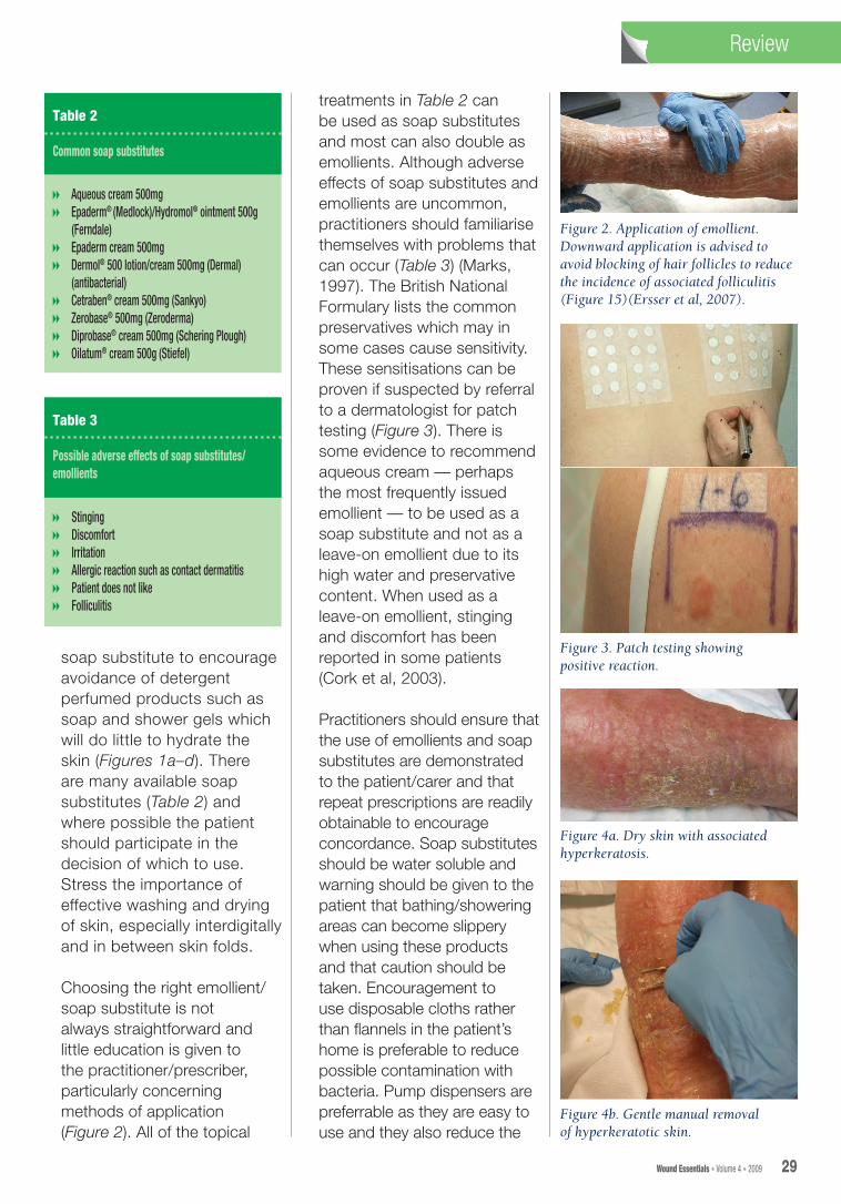
Figure 2. Application of emollient.Downward application is advised to avoid blocking of hair follicles to reduce the incidence of associated folliculitis (Figure 15)(Ersser et al, 2007).
Figure 3. Patch testing showing positive reaction.
Figure 4a. Dry skin with associated hyperkeratosis.
Figure 4b. Gentle manual removal of hyperkeratotic skin.
Wingfield Review C.indd 5 9/6/09 14:12:38

ReviewReview ReviewReview
30 Wound Essentials • Volume 4 • 2009
risk of bacterial contamination. If petroleum products such as liquid paraffin/soft paraffin are used, advice should be given to avoid naked flames as they can be easily ignited when soaked into bandages and clothing (British Medical Association and Royal Pharmaceutical Society of Great Britain, 2007).
Other considerations include:8Advise regular checking of
fingernails/toenails for any signs of infection, cracks, fungus or hangnails. Encourage filing of nails rather than cutting to avoid accidental trauma. The patient should see a podiatrist or chiropodist if nail care is difficult for them to do themselves. Good fitting shoes for the lower limb in patients with chronic oedema are also essential. The use of antifungal foot powders may be advisable, particularly if tinea pedis (athlete’s foot) has been an issue
8Teach patients/carers to recognise signs of infection, which include redness, heat, tenderness, blistering, malaise, raised temperature, or feeling unwell. Small grazes and/or cuts should be cleaned, covered and observed
8It is advisable to avoid injections, blood samples or taking blood pressure on an affected limb
8Patients need to be advised to protect affected limbs from sun burn and to wear appropriate sun protection and protective clothing
8Advise protection of limbs for daily activities such as gardening, e.g. by the wearing of gloves and trousers to minimise risk of accidental trauma.
Skin assessmentSkin assessment is a crucial part of chronic oedema management and should cover the key areas listed in Table 1. The epidermis can be directly affected by chronic oedema. This is particularly demonstrated in lower limbs where verrucous, cobblestone plaques, known as elephantiasis nostra verrucosa (ENV) can occur. Plaques of ENV can be covered with crusting and can become weepy, producing excessive exudate which can in turn make the epidermis soft, soggy and macerated and prone to ulceration and infection. The build up of hyperkeratosis and papillomatosis, especially in skin folds, produces odour from the microbes harboured in these areas (Figure 4a). Salicylic acid ointment can be used to assist removal of scale, together with daily washing and moisturising (Figure 4b).
Poorly controlled weeping of lymph from the skin surface (lymphorrhea) will quickly saturate dressings, clothes and footwear causing further risk of infection through contamination and maceration of skin (Mortimer, 1995). Patients with ‘wet’ or ‘leaky legs’ are often referred to dermatology, resulting in a complete reassessment of the patient’s management.
Quality of life and psychological aspects should be included in any chronic oedema assessment, in particular with reference to the skin. Chronic oedema and associated problems of leakage of lymph fluid, malodorous wounds and skin disease and infections can cause considerable
distress, especially if the patient has not been able to access a comprehensive lymphoedema service (Dealey, 1999).
Recognising and treatingcommon skin diseasesassociated with chronicoedemaTineaLymphoedematous limbs are susceptible to fungal infections (Moffatt et al, 2006). Common sites are skin folds, the interdigital area and sometimes associated with coexisting fungal nails (tinea unguium). Interdigitally it can be moist with a white powdery scale and irritation. Maceration can be seen and the area will be at high risk for subsequent cellulitic infections. Skin scrapings can be taken to confirm mycology if diagnosis is uncertain or when topical treatments have failed and oral medication is being considered (Clinical Knowledge Summary, 2006) (Figures 6a and b; 11a and c). Treatment for fungal skin infections is recommended with one of the following topical treatments:8Imidazoles (2–4 weeks)8Terbinafine (1 week —
adults only)8Undecenoates (2–4 weeks
— adults only).
It is advised to continue treatment for 1–2 weeks once the skin is healed. Oral treatment should be considered if fungal infection is extensive or topical treatment has failed. Refer to the British National Formulary for prescription guidelines or Clinical Knowledge Summary (2006). Fungal nails will require systemic treatment for best results and the duration
Wingfield Review C.indd 6 9/6/09 14:12:39

32 Wound Essentials • Volume 4 • 2009
ReviewReview
Figure 5. Common presentation of cellulitis.
Figure 6a. Tinea pedis (athlete’s foot).
Figure 6b. Tinea pedis with associated onychomycosis (fungal toenails).
Figure 7. Stasis eczema, lipodermatoslerosis.
Figure 8. Papillomatosis.
Figure 9. Deepened skin folds.
Figure 10. Kaposi-Stemmer positive sign.
Figure 11a. Dermapak® for skin scrapings.
Figure 11c. Taking nail clippings for mycology.
Figure 11b. Tinea spread to dorsum, note peripheral scaling. This is a common diagnostic sign.
of treatment will depend on the response. Topical treatments such as Loceryl® (Galderma) can be used, but treatment needs to be for a period of 6–12 months (Docherty, 2001).
Venous stasis eczema (varicose eczema)This common inflammatory condition usually affects the lower
limbs and is often seen to coexist with varicose veins. Clinical signs include inflamed, red, eczematous skin; itching; scaling; pigmentation (haemosiderin deposit); hardened, tight, red/brown skin/tissues (lipodermatosclerosis which is vulnerable to ulceration) and atrophy blanching. Weeping is seen if secondary infection is a factor (Figures 12a and b).
Practitioners should monitor areas of non-healing ulceration as there is an association of repeated episodes of chronic ulceration and healing stimulating proliferation of keratinocytes which may contribute to neoplastic transformation. Referral to the most appropriate specialist is required if the ulcer is not responsive or there is a drop in ankle brachial pressure
Wingfield Review C.indd 8 9/6/09 14:12:40

ReviewReview
Wound Essentials • Volume 4 • 2009 33
index (ABPI). The condition lymphangiosarcoma (Stewart-Treves syndrome) although rare should not be overlooked (Figure 13). This presents as a red/purple macular area of discoloration/bruising, progressing to ulceration, crusting, and ultimately extensive necrosis as it spreads widely. Urgent referral to an oncologist
would be required (Lymphoedema Framework, 2006). Treatment of venous stasis eczema usually consists of topical corticosteroids and emollient therapy. Assessment for potency should not only cover local policy, but also logistics of application, particularly when bandages/stockings are being used for compression. There is limited evidence to the efficacy of compression stockings in venous stasis eczema (Partsch, 2003; Barron et al, 2007; Duffill, 2008). Efficacy of topical corticosteroids is helped by correct diagnosis, an understanding of mode of action and potential side-effects. It can sometimes be more effective
to use a potent steroid for a short amount of time rather than a milder potency for a longer period of time (Davis, 2001). A potent corticosteroid would be acceptable for a period of seven days, then reducing to a milder potency. In the real world this may not be possible depending on the patient’s mobility, comorbidities and age. It needs a common-sense approach, as a poor response will follow if topical corticosteroids are not applied frequently enough or they are at too low a potency. Good use of emollients and the use of paste bandages to aid occlusion and encourage skin barrier repair are relevant therapies (Figure 14). Refer to a dermatologist if the condition is not responding to treatment or if contact dermatitis or a differential diagnosis is considered (Smith, 2006; Barron et al, 2007; Middleton 2007).
FolliculitisFolliculitis is caused by inflammation of the hair follicles causing a rash with pustules (Figure 15). It can occur on any surface of the body that has hair follicles. The pathogen is usually Staphylococcus aureus. Swabs can be taken if exudate is present, in particular if resistant to treatment. Treatments follow the route of reducing S. aureus colonisation using an antiseptic wash/lotion containing chlorhexidine and benzalkonium (e.g. Hibiscrub® [SSL] or Dermol® 500 [Dermal]). Heavy duty petroleum-based topical therapies can introduce or encourage folliculitis (Buxton, 1998). A light, non-greasy emollient is recommended to reduce the risk of further blockage of hair follicles. Referral to a dermatologist is
Figures 12a and b. Venous stasis eczema-associated ulceration.
Figure 13. Lymphangiosarcoma.
Figure 14. Use of paste bandaging.
Figure 15. Folliculitis.
a
b
Wingfield Review C.indd 9 9/6/09 14:12:41

34 Wound Essentials • Volume 4 • 2009
ReviewReview
suggested if there is no response to the treatment after one month.
ConclusionEmollient therapy and topical regimens require involvement of the patient in terms of choice and education, especially if they are going to participate in their own care. A patient’s choice of emollient products is important, particularly for their comfort and subsequent concordance. Correct diagnosis of secondary dermatological disorders and appropriate referral is essential. Incorrect use of topical corticosteroids could lead to unwanted side-effects and disguise the original presentation which may impede diagnosis. Appropriate use of topical corticosteroids in terms of potency and duration, combined with realistic management plans are an important treatment for some inflammatory dermatoses (Davis, 2001).
This brief overview of skin care for patients with lymphoedema touches areas which require more in-depth explanation and understanding. Used as a starting point, it can direct the practitioner to possible areas in their practice where deficits can be recognised and addressed as part of their professional development.
Barron GS, Jacob SE, Kirsner RS (2007) Dermatologic complications of venous disease: medical management and beyond. Ann Vasc Surg 21(5): 652–62
Bjornsdottir S, Gottfredsson M, Thorisdottir AS (2005) Risk factors for acute cellulites of the lower limb. A prospective case-controlled study. Clin Infect Dis 41: 1416–22
BMA and Royal Pharmaceutical Society (2007) British National
Formulary. BMA and Royal Pharmaceutical Society of Great Britain, London
Buxton PK (1998) ABC of Dermatology. BMJ Publishing Group, London: 54–5
Carroll D, Roser K (1992) Treatment leads to significant improvement. Effect of conservative treatment on pain in lymphoedema. Prof Nurse 8(1): 32–6.
Clinical Knowledge Summary (2006) Fungal (dermatophyte) Skin Infections. Available online at: www.cks.library.nhs.uk (accessed 8 March, 2009)
Cork MJ, Timmins J, Holden C, et al (2003) An audit of adverse drug reactions to aqueous cream in children with atopic eczema. Pharma J 271: 747–8
Davis R (2001) Treatment issues relating to dermatology. In: Hughes E, Van Onselen J, eds. Dermatology Nursing. A Practical Guide. Harcourt Health Sciences, Philadelphia
Dealey C (1999) The management of patients with chronic wounds. In: The Care of Wounds. A Guide for Nurses. 2nd edn. Blackwell Science, Oxford
Docherty C (2001) Infections and infestations. In: Hughes E, Van Onselen J, eds. Dermatology Nursing. A Practical Guide. Harcourt Health Sciences, Philadelphia: 206–10
Duffill M (2008) Lipdermatoslerosis. DermNet NZ. Available online at: www.dermnetnz.org (last accessed 8 February, 2009)
Dupuy A, Benchilchi H, Roujeau J, Bernard P, Vaillant L, et al (1999) Risk factors for erysipelas of the leg (cellulites): case control study. Br Med J 318: 1591–4
Ersser S, Maguire S, Nicol N, Penzer R, Peters J (2007) Best Practice Statement on Emollient Therapy. A Statement for Healthcare Professionals. Dermatology UK, Aberdeen
Fendler E (2000) Physico-chemical considerations In: Loden M, Maibach HI, eds. Dry Skin and Moisturisers —
Chemistry and Function. CRC Press, Boca Raton
Flynn T C, Petros J, Clarke R E, Viehman G E (2001) Dry skin and moisturisers. Clin Dermatol 19(4): 387–92
Harris SR, Hugi MR, Olivoto O, et al (2001) Clinical practice guidelines for the care and treatment of breast cancer. 11 Lymphoedema. CMAJ 164(2): 191–9
Kilburn S, Featherstone P, Higgins B, Brindle R, Severs N (2003) Intervention for cellulitis and erysipelas (protocol). Cochrane Database of Sytematic Reviews. 2003; Issue 1, Art No CD004299. DOI: 101–1002/14651858
Lymphoedema Framework (2006) Best Practice for the Management of Lymphoedema. International consensus. MEP Ltd, London
Marks R (1997) How to measure the effects of emollients. J Dermatol Treat 8: s515–8
Middleton H (2007) Exploring the aetiology and management of venous eczema. Br J Community Nurs 12(9): 516–23
Moffatt CJ, Morgan PA, (2006) International consensus on managing lymphoedema. Nurs Times 102(44): 42–4
Mortimer P (1995) Managing lymphedema. Clin Dermatol 13: 499–505
Partsch I (2003) Evidence base compression therapy. VASA 32(Suppl 163): 1–41
Rawlings A V, Harding C R (2004) Moisturisation and skin barrier. Dermatologic Ther 17(1): 43–8
Roujeau J-C, Sigurgeirsson B, Kortinh H-C, Kerl H, Paul C (2004) Risk factors for acute bacterial cellulitis of the leg. Dermatology 209(4): 301–7
Smith PC (2006) The causes of skin damage and leg ulceration in chronic venous disease. Int J Lower Extrem Wounds 5(3): 160–8
Swartz MN (2004) Cellulitis. N Engl J Med 4(350): 912–4
WE
Wingfield Review C.indd 10 9/6/09 14:12:41


![Malassezia Folliculitis versus Truncal Acne Vulgaris ... · 278 Malassezia Folliculitis versus Truncal Acne Vulgaris (Clinical and Histopathological Study) support the diagnosis [5,6,10].](https://static.fdocuments.in/doc/165x107/5cdf712988c99399558c9005/malassezia-folliculitis-versus-truncal-acne-vulgaris-278-malassezia-folliculitis.jpg)