Windsor Veterinary Research ...
11
RESEARCH Open Access Cattle remain immunocompetent during the acute phase of foot-and-mouth disease virus infection Miriam A Windsor 1 , B Veronica Carr 2 , Bartomiej Bankowski 1 , Debi Gibson 1 , Elizabeth Reid 1 , Pip Hamblin 1 , Simon Gubbins 1 , Nicholas Juleff 1 and Bryan Charleston 1,2* Abstract Infection of cattle with foot-and-mouth disease virus (FMDV) results in the development of long-term protective antibody responses. In contrast, inactivated antigen vaccines fail to induce long-term protective immunity. Differences between susceptible species have also been observed during infection with FMDV, with cattle often developing persistent infections whilst pigs develop more severe symptoms and excrete higher levels of virus. This study examined the early immune response to FMDV in naïve cattle after in-contact challenge. Cattle exposed to FMDV were found to be viraemic and produced neutralising antibody, consistent with previous reports. In contrast to previous studies in pigs these cattle did not develop leucopenia, and the proliferative responses of peripheral blood mononuclear cells to either mitogen or third party antigen were not suppressed. Low levels of type 1 interferon and IL-10 were detected in the circulation. Taken together, these results suggest that there was no generalised immunosuppression during the acute phase of FMDV infection in cattle. Introduction Foot-and-mouth disease (FMD) is an extremely conta- gious and economically important disease of livestock. Outbreaks in normally disease-free countries, such as the UK in 2001 [1] and Japan in 2010 [2], have cost billions of dollars in lost revenue. The current vaccines available for use in endemic countries do not confer long-lasting immunity and highly purified vaccine antigen is required to distinguish between vaccinated and infected animals. Understanding the complex relationship between virus and host is vital in designing new vaccines that can be targeted to those areas of the immune system most likely to induce an effective response. The causative agent, foot-and-mouth disease virus (FMDV), spreads rapidly between animals and is quickly disseminated within the host, presumably in order to avoid the adaptive immune response (for an overview see Golde et al. [3]). In cattle, the primary sites of infection in aerosol trans- mission are the nasopharangeal tissues [4], and associated epithelial tissues [5]. Whilst several studies have examined the host response to FMDV in swine [6-10], little is known about the innate or adaptive response to FMDV in cattle. Type 1 (alpha and beta) interferons (IFN) are induced early in the innate immune response and are con- sidered a dominant factor in shaping both innate and adaptive immune responses [11]. Type 1 IFN certainly seems to play a role in FMD pathogenesis in swine, and Chinsamgaram et al. propose that during infection, type 1 IFN production is regulated by the leader protein of FMDV (L pro ) [12]. However, prophylactic administration of IFN by adenovirus vector prior to challenge, rapidly induces a protective state in swine [13]. Two studies in swine used direct inoculation of FMDV challenge methods to identify a period of lymphopenia approximately 2 to 4 days post challenge that coincided with peak viraemia [7,14]. In addition, in both studies the animals showed suppression of T cell proliferation in response to mitogen from day 1 to day 7 [14] and day 2 to day 5 or 8 depending on the virus used [7]. Lymphopenia had also been corre- lated with loss of plasmacytoid dendritic cell (PDC) * Correspondence: [email protected] 1 Pirbright Laboratory, Institute for Animal Health, Ash Road, Woking, Surrey, GU24 0NF, UK Full list of author information is available at the end of the article Windsor et al. Veterinary Research 2011, 42:108 http://www.veterinaryresearch.org/content/42/1/108 VETERINARY RESEARCH © 2011 Windsor et al; licensee BioMed Central Ltd. This is an Open Access article distributed under the terms of the Creative Commons Attribution License (http://creativecommons.org/licenses/by/2.0), which permits unrestricted use, distribution, and reproduction in any medium, provided the original work is properly cited.
Transcript of Windsor Veterinary Research ...
RESEARCH Open Access
Cattle remain immunocompetent during theacute phase of foot-and-mouth disease virusinfectionMiriam A Windsor1, B Veronica Carr2, Bartomiej Bankowski1, Debi Gibson1, Elizabeth Reid1, Pip Hamblin1,Simon Gubbins1, Nicholas Juleff1 and Bryan Charleston1,2*
Abstract
Infection of cattle with foot-and-mouth disease virus (FMDV) results in the development of long-term protectiveantibody responses. In contrast, inactivated antigen vaccines fail to induce long-term protective immunity.Differences between susceptible species have also been observed during infection with FMDV, with cattle oftendeveloping persistent infections whilst pigs develop more severe symptoms and excrete higher levels of virus. Thisstudy examined the early immune response to FMDV in naïve cattle after in-contact challenge. Cattle exposed toFMDV were found to be viraemic and produced neutralising antibody, consistent with previous reports. In contrastto previous studies in pigs these cattle did not develop leucopenia, and the proliferative responses of peripheralblood mononuclear cells to either mitogen or third party antigen were not suppressed. Low levels of type 1interferon and IL-10 were detected in the circulation. Taken together, these results suggest that there was nogeneralised immunosuppression during the acute phase of FMDV infection in cattle.
IntroductionFoot-and-mouth disease (FMD) is an extremely conta-gious and economically important disease of livestock.Outbreaks in normally disease-free countries, such as theUK in 2001 [1] and Japan in 2010 [2], have cost billionsof dollars in lost revenue. The current vaccines availablefor use in endemic countries do not confer long-lastingimmunity and highly purified vaccine antigen is requiredto distinguish between vaccinated and infected animals.Understanding the complex relationship between virusand host is vital in designing new vaccines that can betargeted to those areas of the immune system most likelyto induce an effective response.The causative agent, foot-and-mouth disease virus
(FMDV), spreads rapidly between animals and is quicklydisseminated within the host, presumably in order toavoid the adaptive immune response (for an overviewsee Golde et al. [3]).
In cattle, the primary sites of infection in aerosol trans-mission are the nasopharangeal tissues [4], and associatedepithelial tissues [5]. Whilst several studies have examinedthe host response to FMDV in swine [6-10], little isknown about the innate or adaptive response to FMDV incattle. Type 1 (alpha and beta) interferons (IFN) areinduced early in the innate immune response and are con-sidered a dominant factor in shaping both innate andadaptive immune responses [11]. Type 1 IFN certainlyseems to play a role in FMD pathogenesis in swine, andChinsamgaram et al. propose that during infection, type 1IFN production is regulated by the leader protein ofFMDV (Lpro) [12]. However, prophylactic administrationof IFN by adenovirus vector prior to challenge, rapidlyinduces a protective state in swine [13]. Two studies inswine used direct inoculation of FMDV challenge methodsto identify a period of lymphopenia approximately 2 to 4days post challenge that coincided with peak viraemia[7,14]. In addition, in both studies the animals showedsuppression of T cell proliferation in response to mitogenfrom day 1 to day 7 [14] and day 2 to day 5 or 8 dependingon the virus used [7]. Lymphopenia had also been corre-lated with loss of plasmacytoid dendritic cell (PDC)
* Correspondence: [email protected] Laboratory, Institute for Animal Health, Ash Road, Woking, Surrey,GU24 0NF, UKFull list of author information is available at the end of the article
Windsor et al. Veterinary Research 2011, 42:108http://www.veterinaryresearch.org/content/42/1/108 VETERINARY RESEARCH
© 2011 Windsor et al; licensee BioMed Central Ltd. This is an Open Access article distributed under the terms of the Creative CommonsAttribution License (http://creativecommons.org/licenses/by/2.0), which permits unrestricted use, distribution, and reproduction inany medium, provided the original work is properly cited.
function and inhibition of T cell function [10]. A study incattle and Indian buffalo has provided limited evidence ofa transient lymphopenia immediately after infection [15].In swine this immune suppression has also been linkedwith elevated levels of IL-10 in serum [10]. IL-10 is widelyacknowledged to contribute to the anti-inflammatoryresponse and to the inhibition of cellular responses via avariety of mechanisms (for a review see [16]). There is alsoevidence that natural killer (NK) cells may be functionallydefective during infection [17].In cattle, cytotoxic T lymphocytes (CTL) have been
shown to play a role in the FMDV immune response dur-ing infection and vaccination [18,19] in a cross serotypicmanner [20]. Studies carried out on the proliferativeresponse of cattle peripheral blood lymphocytes followingvaccination showed a heterotypic reaction, indicating asharing of T cell epitopes [21]. When Garcia-Valcarcel etal. inoculated an animal with FMDV, little proliferationwas seen until a subsequent re-challenge, when a crossserotype response was observed [22].The humoral response to FMDV is well documented,
with a rapid IgM response switching to IgG [23,24] whichconfers protective immunity for many years [25]. It hasbeen suggested that this long-lasting antibody response isin part due to the presence of viral particles held by folli-cular dendritic cells in the lymph nodes of cattle, longafter the disease has been resolved [26]. Depletion of Tcell subsets by monoclonal antibodies showed that theearly antibody response to infection is T cell independent[23].The aim of the current study was to define the early
innate and adaptive immune responses of cattle infectedwith O serotype FMDV, after they were held in closecontact with cattle infected by intra-dermolingual chal-lenge. Specifically, we determined whether there was gen-eralised immune-suppression during the acute phase ofFMDV infection in cattle by monitoring the number ofleucocytes in the blood and assaying for inflammatoryand anti-inflammatory cytokines and suppression of theT cell response. We also determined how rapidly FMDV-specific humoral and cell-mediated immune responsesdeveloped.
Materials and methodsInfection with FMDVMale Holstein/Friesian cattle weighing approximately 150kg were used for these studies. Two animals wereexposed to two separate intra-dermolingually challengedcattle (1 × 105.7 TCID50 of cattle-adapted FMDV O UKG34/2001) for 24 h. These inoculates formed no part ofthe subsequent study and only the “naturally” exposedanimals will be referred to from here on. This procedurewas repeated in three sequential studies and the data pre-sented here are an accumulation of these replicates
(replicate one = animal C1, replicate two = animals C2and C3 and replicate three = C4 to C6).
VaccinationFive Holstein/Friesian cattle (C7 to C11) were vaccinatedintramuscularly with 2 mL of O1Manisa vaccine (O1Manisa vaccine from the UK FMDV emergency vaccinebank).
HaematologyBlood samples were collected into anti-coagulant,EDTA, and total leucocyte counts performed on thesame day as collection. Each sample was counted in tri-plicate using a Sysmex haematology analyser (Sysmex F-800 Sysmex corporation Kobe Japan).
Virus detectionNucleic acid extraction and analysis was performed usingquantitative real-time reverse transcription polymerasechain reaction (qRT-PCR) as described previously [27].
Virus-neutralising antibody testSerum samples were tested for anti-FMDV neutralisingantibodies as described in the Office International desEpizooties Manual of Diagnostic Tests and Vaccines forTerrestrial Animals. Sera with titres greater than orequal to 45 were considered to be positive [28].
Liquid-phase blocking ELISAThe liquid-phase blocking ELISA was performed asdescribed by Hamblin et al. [29]. Briefly; virus wasbound to immunosorbent plates by being trapped withrabbit anti-O1 Manisa antibody. Test samples of bovinesera were mixed with known standards of guinea pigsera and the resultant competition determined by mea-suring the amount of guinea pig serum bound to theantigen.
Interferon assayType 1 IFN biological activity was measured in serumsamples using an Mx/chloramphenicol acetyltransferase(Mx/CAT) promoter-reporter gene assay [30].
IL-10 ELISAIL-10 was measured in serum following the method ofKwong et al. [31]. Briefly, ELISA plates were coated withanti-IL-10, cattle sera were applied in duplicate, along withan IL-10 standard series, and detected with biotinylatedanti-bovine IL-10. This assay was repeated 3 times.
Vaccination with commercial bovine herpes virus-1 (BHV)vaccineApproximately one month prior to FMDV challenge,animals C1 to C6 were immunised with Tracherine
Windsor et al. Veterinary Research 2011, 42:108http://www.veterinaryresearch.org/content/42/1/108
Page 2 of 11

(Intervet, NL). Cattle were assayed for proliferativeresponse to BHV antigen immediately prior to challengewith FMDV. However, the BHV-specific T cell prolifera-tive response to the vaccine was variable; as a conse-quence, only animals with T cell proliferative responsesconsistently higher than 30 000 cpm prior to FMDVinfection (C1, C4, C5 and C6) were included in the ana-lysis of whether FMDV infection significantly affectedspecific recall responses.
Proliferation assaysHeparinised blood was diluted with phosphate-bufferedsaline (PBS) (Invitrogen, Paisley, UK) and centrifugedover Histopaque-1077 (Sigma) at 1328 g. Peripheralblood mononuclear cells (PBMC) were collected fromthe interface, the red blood cells were lysed in erythrocytelysis buffer (155 mM ammonium chloride, 0.1 mMEDTA and 10 mM sodium bicarbonate, pH 7.2) andPBMC were washed three times with cold PBS. PBMC (2× 10⁵ per well) in proliferation medium (RPMI 1640 sup-plemented with 5% BVDV-free FCS, 1% non-essentialamino acids (Invitrogen) 1 mM sodium pyruvate (VWR,Leicestershire, UK), 10 μg/mL gentamicin and 50 μM 2-mercaptoethanol) were incubated with a range of anti-gens; control and test antigens were added to each well.Antigens included; medium alone, pokeweed mitogen(2.5 μg/mL Sigma), inactivated FMDV antigen, mockinfected BHK-21 cell lysate and BHV antigen (heat inac-tivated prior to use). FMDV antigen was kindly providedby Merial Animal Health, (total antigen concentrationwas known by the vaccine company but undisclosed).Both FMDV and BHV antigens were previously titratedfor use in proliferation assays using PBMC from appro-priately vaccinated cattle, with the final concentrationsused being 1/1000 for FMDV antigen and 1/100 forBHV. The cultures were incubated for 5 days before0.037 MBq [3H] Thymidine (Amersham, Buckingham-shire, UK) was added to each well. After a further over-night incubation cells were harvested onto filter mats andincorporated radioactivity was measured using a 1450Microbeta counter (Wallac, Finland).
Statistical analysisLinear mixed models were used to investigate the rela-tionship between log10 CPM levels, antigen and time.Model selection proceeded by stepwise deletion of non-significant terms (as judged by the Akaike informationcriterion), starting from a model including antigen, time(as a factor) and an interaction between them; animalwas included as a random effect. A linear mixed modelwas also used to compare IL-10 levels over time, withthe model including time (as a factor) as a fixed effectand animal as a random effect.
ResultsViraemia and neutralising antibody responses afterchallenge with FMDVThe onset and duration of viraemia after cattle havebeen challenged with FMDV has been described pre-viously, and the results obtained in this study are consis-tent with previous reports [27,32]. In the six animalsstudied, viraemia was first detected between 2 and 4days after the animals were placed in contact withFMDV needle challenged animals. Typical clinical signswere observed in all the cattle from 2 to 4 days postinfection (data not shown), and viraemia was resolved inall animals by day 7 (Figure 1a). All animals had sero-converted to FMDV as demonstrated by liquid-phaseblocking ELISA (data not shown). All these animals haddetectable neutralising antibodies seven days after chal-lenge (Figure 1b) and all were considered to have pro-tective titres (> 45) by 14 days after challenge.
Total circulating leucocyte counts during the acute phaseof FMDV infectionTotal circulating leucocyte counts did not significantlyfluctuate between day -2 prior to challenge and day 8after challenge (Figure 2). In addition, the mean circulat-ing leucocyte count of the cattle did not deviate aboveor below the normal physiological range [33].Whole lysed blood from two of the cattle (C2 and C3)
was also examined for any changes in CD4, CD8, WC1,CD21 and NK cell leucocyte populations [7] but thesewere not found to vary during the course of infection(data not shown), which is in keeping with the results ofJuleff et al. [23].
Proliferative response of PBMC to mitogen (PWM) duringthe acute phase of FMDV infectionDetailed analysis on the proliferative response to Poke-weed mitogen (PWM) in all six animals (C1 to C6) wascarried out (Figure 3) (there were minor variations insample days due to unavoidable differences in samplingschedules between replicates). Marked proliferation,(greater than 200 000 cpm), of PBMC to PWM wasdetected at various time-points between day -1 prior toFMDV infection and day 19 after challenge.
Development of FMDV-specific T cell responses duringthe resolution of acute infectionFMDV-specific T cell responses were measured by pro-liferation of PBMC to virus antigen in animals C1 to C6(Figure 3). Although there appears to be a small rise inproliferation to FMDV antigen at day 19 in animals C2to C6, it was not found to be significant when the datafor all animals were exposed to statistical examination.More precisely, there was no significant (P = 0.57)
Windsor et al. Veterinary Research 2011, 42:108http://www.veterinaryresearch.org/content/42/1/108
Page 3 of 11

Figure 1 Cattle infected with FMDV were viraemic during acute infection and developed neutralising antibodies. A. Onset and durationof viraemia of all six FMDV challenged cattle used in this study as obtained by quantitative real-time reverse transcription polymerase chainreaction (qRT-PCR). B. Neutralising antibody titre in serum of infected cattle. Sera with titres greater than or equal to 45 were considered to bepositive (dotted line).
Windsor et al. Veterinary Research 2011, 42:108http://www.veterinaryresearch.org/content/42/1/108
Page 4 of 11
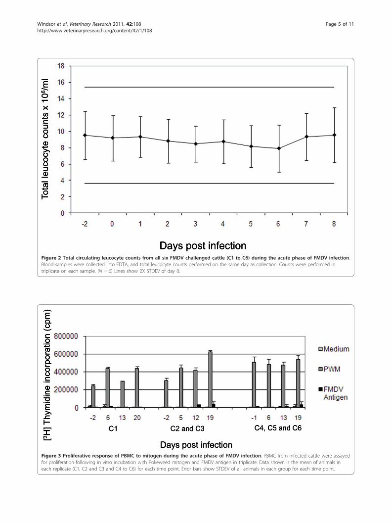
Figure 2 Total circulating leucocyte counts from all six FMDV challenged cattle (C1 to C6) during the acute phase of FMDV infection.Blood samples were collected into EDTA, and total leucocyte counts performed on the same day as collection. Counts were performed intriplicate on each sample. (N = 6) Lines show 2X STDEV of day 0.
Figure 3 Proliferative response of PBMC to mitogen during the acute phase of FMDV infection. PBMC from infected cattle were assayedfor proliferation following in vitro incubation with Pokeweed mitogen and FMDV antigen in triplicate. Data shown is the mean of animals ineach replicate (C1, C2 and C3 and C4 to C6) for each time point. Error bars show STDEV of all animals in each group for each time point.
Windsor et al. Veterinary Research 2011, 42:108http://www.veterinaryresearch.org/content/42/1/108
Page 5 of 11

interaction between time and antigen, and log10 CPMlevels did not change significantly (P = 0.31) over timefor any of the antigens (medium, FMDV or PWM).There was however, a significant (P < 0.001) differencein log10 CPM levels amongst antigens, with levels forPWM being significantly higher than for FMDV or med-ium (which did not differ significantly (P = 0.08) fromone another).
Analysis of established specific memory T cell responsesduring the acute phase of FMDV infectionVaccination with BHV resulted in variable T cellresponses in individual animals (Figure 4), but log10CPM levels were significantly (P < 0.001) higher forBHV compared with medium and mock (log10 CPMlevels did not differ significantly (P = 0.71) betweenmock and medium). However, for BHV, there was no
significant change in log10 CPM levels at any time pointpost FMDV infection.
Development of FMDV-specific T cell responses aftervaccinationFive cattle (C7 to C11) were vaccinated with O1 Manisacommercial vaccine and examined for T cell responses byPBMC proliferation assays (Figure 5). After day 21 animalsC8, C9 and C10 were boosted. An increase in proliferativeresponse was seen in these animals with counts per min-ute rising to approximately 300 000, 400 000 and 500 000cpm respectively. The T cell response to FMDV antigenfollowing vaccination was variable in individual cattle, butby inspection, when a response was seen, it was alwaysearlier and of greater magnitude than after infection (it isnot appropriate to perform statistical analyses between thestudies in Figures 3 and 5).
Figure 4 Proliferative response to third party antigen during the acute phase of FMDV infection. PBMC from infected cattle (previouslyvaccinated with BHV) were assayed for proliferation following in vitro incubation with BHV antigen, in triplicate. Due to the variation inmagnitude of response to BHV, the data are displayed separately for each of the cattle C1, C4, C5 and C6. Error bars show STDEV of the mean ofthree wells for each time point.
Windsor et al. Veterinary Research 2011, 42:108http://www.veterinaryresearch.org/content/42/1/108
Page 6 of 11
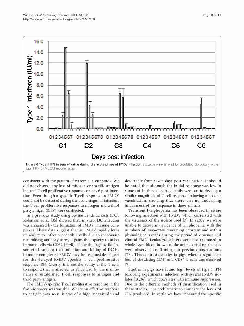
Detection of circulating type 1 IFN during the acutephase of FMDV infectionType 1 IFN was assayed from the sera of six infected cattle(Figure 6). Production of type 1 IFN following infectionwith FMDV was variable, with the peak values varyingbetween 1 and 14 international units/mL (IU/mL).
Detection of circulating IL-10 during the acute phase ofFMDV infectionSix cattle (C1 to C6), were assayed for circulating IL-10before, during, and after the acute phase of FMDV infec-tion (Day -2 through to day 8, Figure 7). Low, but statisti-cally significant compared to pre-challenge (P < 0.03),levels of IL-10 were detected in all animals, rising overtime, reaching a peak on average of 1.35 IU/mL betweenday 3 and 4, after which they declined.
DiscussionEarly studies by Cunliffe et al. [25] established the longduration of immunity following infection with FMDV incattle. In contrast, current commercial vaccination proto-cols require regular re-inoculation to maintain immunity.Analysing the differences in the immune response in thenatural host during these two processes may lead to thedesign of more effective vaccines.
By exposing six cattle to FMDV this study aimed to;investigate whether a generalised suppressive state isinduced, compare early T cell responses to those of vacci-nates, and examine the expression of key immune-modula-tory cytokines.After natural exposure to FMDV, viraemia is typically
detectable within 2 to 3 days followed by clinical signs [27].Production of high titres of neutralising antibodies can bedetected as early as six days following natural infection, bya process shown previously to be T cell independent in cat-tle [23]. These previous observations were confirmed in allof the cattle used in this study. Compared to the rapid pro-duction of antibody, significant FMDV-specific T cellresponses (assayed by proliferation of PBMC) were notobserved following natural infection. These data are sup-ported by other cattle studies, with no FMDV-specific Tcell response detectable up to 32 days post exposure insome cases (M. Windsor unpublished data). We also inves-tigated whether there was any loss of mitogen or specificantigen induced T cell proliferative responses during thecourse of acute infection. Abrogation of proliferation ofPBMC to mitogen in pigs occurs between 2 and 8 days butmost noticeably at day 5 after needle challenge [7]. Follow-ing natural challenge, peak viraemia in cattle occurs atleast two days later than needle challenge [34], which is
Figure 5 Proliferative response to FMDV antigen in vaccinated cattle. PBMC from five cattle vaccinated with FMDV O1 Manisa commercialvaccine were assayed for proliferative response to FMDV antigen. The kinetics of the proliferative response for each animal for the first 21 dayspost vaccination are shown. Error bars show STDEV for each of the five animals at each time point.
Windsor et al. Veterinary Research 2011, 42:108http://www.veterinaryresearch.org/content/42/1/108
Page 7 of 11
consistent with the pattern of viraemia in our study. Wedid not observe any loss of mitogen or specific antigeninduced T cell proliferative responses on day 6 post-infec-tion. Even though a specific T cell response to FMDVcould not be detected during the acute stages of infection,the T cell proliferative responses to mitogen and a thirdparty antigen (BHV) were unaffected.In a previous study using bovine dendritic cells (DC),
Robinson et al. [35] showed that, in vitro, DC infectionwas enhanced by the formation of FMDV immune com-plexes. These data suggest that as FMDV rapidly losesits ability to infect susceptible cells due to increasingneutralising antibody titres, it gains the capacity to infectimmune cells via CD32 (FcgR). These findings by Robin-son et al. suggest that infection and killing of DC byimmune-complexed FMDV may be responsible in partfor the delayed FMDV-specific T cell proliferativeresponse [35]. Clearly, it is not the ability of the T cellsto respond that is affected, as evidenced by the mainte-nance of established T cell responses to mitogen andthird party antigen.The FMDV-specific T cell proliferative response in the
five vaccinates was variable. Where an effective responseto antigen was seen, it was of a high magnitude and
detectable from seven days post vaccination. It shouldbe noted that although the initial response was low insome cattle, they all subsequently went on to develop asimilar magnitude of T cell response following a boostervaccination, showing that there was no underlyingimpairment of the response in these animals.Transient lymphopenia has been observed in swine
following infection with FMDV which correlated withthe virulence of the isolate used [7]. In cattle, we wereunable to detect any evidence of lymphopenia, with thenumbers of leucocytes remaining constant and withinphysiological ranges during the period of viraemia andclinical FMD. Leukocyte subsets were also examined inwhole lysed blood in two of the animals and no changeswere observed, confirming our previous observations[23]. This contrasts studies in pigs, where a significantloss of circulating CD4+ and CD8+ T cells was observed[7].Studies in pigs have found high levels of type 1 IFN
following experimental infection with several FMDV iso-lates [10,36], which correlates with immune suppression.Due to the different methods of quantification used inthese studies, it is problematic to compare the levels ofIFN produced. In cattle we have measured the specific
Figure 6 Type 1 IFN in sera of cattle during the acute phase of FMDV infection. Six cattle were assayed for circulating biologically activetype 1 IFN by Mx CAT reporter assay.
Windsor et al. Veterinary Research 2011, 42:108http://www.veterinaryresearch.org/content/42/1/108
Page 8 of 11

activity of type 1 IFN (in international units), whereastotal circulating type 1 IFN protein was measured in pigserum. However, this IFN is not necessarily biologicallyactive. Several sources, including manufacturers’ datasheets, give type 1 IFN specific activity (cross species) atbetween 1 and 3 × 10⁸ IU/mg of total IFN. Using thiscalculation we can speculatively compare type 1 IFN inthe sera of pigs and cattle during FMDV infection, andpostulate that pigs do indeed produce more IFN thanthe cattle in this study. The most compatible study inpigs was performed by Nfon et al., who used O1Camposchallenge [10]. At least 9 fold more type 1 IFN was pro-duced in pigs compared to our results in cattle. In pigs,IFN clearly has an important role to play in the resolu-tion of infection, as the administration of type 1 IFN ina viral vector protects against subsequent challenge withFMDV [37].Our study implies that cattle produce very little type 1
IFN in comparison to pigs, which is in keeping withprevious studies [38], yet cattle still resolve the diseaseeffectively. Depletion of CD4+ cells in cattle duringinfection further reduces circulating type I IFN, butinfection is still resolved [23], indicating that the pre-sence of type 1 IFN in the circulation may not have anybearing on the resolution of disease. The IFN detected
in the circulation during FMDV infection in cattle isthought to be produced by CD4+ PDCs interacting withimmune-complexed virus [39]. The PDCs found in cat-tle secondary lymphoid tissue are capable of producinglarge amounts of IFN in vitro [39]. It is possible there-fore, that IFN does play a local role at the sites of infec-tion such as lesions, or in the lymph nodes, and that theIFN found in the circulation is derived from these highconcentration sources. Summerfield et al. and Nfon etal. found large numbers of circulating PDCs in pigs[10,40] in comparison to the low numbers detectable incattle. Nfon also found that these PDCs were function-ally impaired during the latter stages of infection withFMDV.Low levels of IL-10, corresponding with the peak of
clinical signs, were found in the serum of the cattle stu-died. Whilst IL-10 does appear to play a role in immunesuppression in pigs during infection with FMDV [8], itis most commonly associated with the maintenance ofchronic infections such as Hepatitis C in humans[41,42] and Mycobacterium Bovis in cattle [43]. Studiesin mice have shown that FMDV infected DC can stimu-late splenic CD9+ B cells to produce T-independentneutralising IgM antibodies, via an IL-10 dependent pro-cess [44]. We propose that the absence of leucopenia
Figure 7 IL-10 in sera of cattle during the acute phase of FMDV infection. Six cattle were assayed for circulating IL-10 by ELISA from day -2to day 8 post infection. Data shown is the mean of all six animals (C1 to C6). Error bars show STDEV of the mean.
Windsor et al. Veterinary Research 2011, 42:108http://www.veterinaryresearch.org/content/42/1/108
Page 9 of 11
and immunosuppression in cattle, during acute FMDVinfection, is associated with the low levels of type 1 IFNand IL-10. These differences in cytokine profile betweenpigs and cattle may also explain why, in general, moresevere clinical signs are seen in pigs infected withFMDV [45].
AcknowledgementsThe authors would like to thank Paul Barnett, Sarah Cox, Luke Fitzpatrick,Colin Randall and Luca Colmari for assistance with animal work andcollection of samples and the BBSRC for funding.
Author details1Pirbright Laboratory, Institute for Animal Health, Ash Road, Woking, Surrey,GU24 0NF, UK. 2Compton laboratory, Institute for Animal Health, Compton,Berkshire, RG20 7NN, UK.
Authors’ contributionsMW carried out the circulating leucocyte counts, proliferation assays ofinfected cattle, IL10 ELISAs, FACS analysis, analysis and interpretation of dataand wrote the manuscript. BVC carried out proliferation assays of vaccinatedcattle. BB conducted in vivo experiments, determined viraemia andcontributed to experimental design. DG conducted in vivo experimentsdetermined viraemia and carried out competition ELISAs. ER carried outInterferon assays. PH carried out VNT assays. SG carried out statisticalanalysis. NJ conducted in vivo experiments, carried out IFN assays,coordinated experimental design, analysis and interpretation of data andassisted with manuscript. BC initiated experimental design, analysis andinterpretation of data and assisted with manuscript. All authors have readand approved the final manuscript.
Authors’ InformationBC is a Jenner investigator and NJ is a Welcome Trust intermediate clinical fellow.
Competing interestsThe authors declare that they have no competing interests.
Received: 17 May 2011 Accepted: 20 October 2011Published: 20 October 2011
References1. Ferguson NM, Donnelly CA, Anderson RM: The foot-and-mouth epidemic
in Great Britain: pattern of spread and impact of interventions. Science2001, 292:1155-1160.
2. Paton DJ, King DP, Knowles NJ, Hammond J: Recent spread of foot-and-mouth disease in the Far East. Vet Rec 2010, 166:569-570.
3. Golde WT, de Los Santos T, Robinson L, Grubman MJ, Sevilla N,Summerfield A, Charleston B: Evidence of activation and suppressionduring the early immune response to foot-and-mouth disease virus.Transbound Emerg Dis 2011, 58:283-290.
4. Arzt J, Pacheco JM, Rodriguez LL: The early pathogenesis of foot-and-mouth disease in cattle after aerosol inoculation: identification of thenasopharynx as the primary site of infection. Vet Pathol 2010,47:1048-1063.
5. Zhang Z, Alexandersen S: Quantitative analysis of foot-and-mouth diseasevirus RNA loads in bovine tissues: implications for the site of viralpersistence. J Gen Virol 2004, 85:2567-2575.
6. Barnett PV, Cox SJ, Aggarwal N, Gerber H, McCullough KC: Further studieson the early protective responses of pigs following immunisation withhigh potency foot and mouth disease vaccine. Vaccine 2002,20:3197-3208.
7. Bautista EM, Ferman GS, Golde WT: Induction of lymphopenia andinhibition of T cell function during acute infection of swine with footand mouth disease virus (FMDV). Vet Immunol Immunopathol 2003,92:61-73.
8. Diaz-San Segundo F, Rodriguez-Calvo T, de Avila A, Sevilla N:Immunosuppression during acute infection with foot-and-mouth diseasevirus in swine is mediated by IL-10. PLoS One 2009, 4:e5659.
9. Golde WT, Nfon CK, Toka FN: Immune evasion during foot-and-mouthdisease virus infection of swine. Immunol Rev 2008, 225:85-95.
10. Nfon CK, Toka FN, Kenney M, Pacheco JM, Golde WT: Loss ofplasmacytoid dendritic cell function coincides with lymphopenia andviremia during foot-and-mouth disease virus infection. Viral Immunol2010, 23:29-41.
11. Theofilopoulos AN, Baccala R, Beutler B, Kono DH: Type I interferons(alpha/beta) in immunity and autoimmunity. Annu Rev Immunol 2005,23:307-336.
12. Chinsangaram J, Koster M, Grubman MJ: Inhibition of L-deleted foot-and-mouth disease virus replication by alpha/beta interferon involvesdouble-stranded RNA-dependent protein kinase. J Virol 2001,75:5498-5503.
13. Dias CC, Moraes MP, Segundo FD, de Los Santos T, Grubman MJ: Porcinetype I interferon rapidly protects swine against challenge with multipleserotypes of foot-and-mouth disease virus. J Interferon Cytokine Res 2010,31:227-236.
14. Diaz-San Segundo F, Salguero FJ, de Avila A, de Marco MM, Sanchez-Martin MA, Sevilla N: Selective lymphocyte depletion during the earlystage of the immune response to foot-and-mouth disease virusinfection in swine. J Virol 2006, 80:2369-2379.
15. Maddur MS, Gajendragad MR, Gopalakrishna S, Singh N: Comparative studyof experimental Foot-and-Mouth Disease in cattle (Bos indicus) andbuffaloes (Bubalis bubalus). Vet Res Commun 2008, 32:481-489.
16. Pestka S, Krause CD, Sarkar D, Walter MR, Shi Y, Fisher PB: Interleukin-10and related cytokines and receptors. Annu Rev Immunol 2004, 22:929-979.
17. Toka FN, Nfon C, Dawson H, Golde WT: Natural killer cell dysfunctionduring acute infection with foot-and-mouth disease virus. Clin VaccineImmunol 2009, 16:1738-1749.
18. Childerstone AJ, Cedillo-Baron L, Foster-Cuevas M, Parkhouse RME:Demonstration of bovine CD8(+) T-cell responses to foot and mouthdisease virus. J Gen Virol 1999, 80:663-669.
19. Guzman E, Taylor G, Charleston B, Skinner MA, Ellis SA: An MHC-restrictedCD8+ T-cell response is induced in cattle by foot-and-mouth diseasevirus (FMDV) infection and also following vaccination with inactivatedFMDV. J Gen Virol 2008, 89:667-675.
20. Guzman E, Taylor G, Charleston B, Ellis SA: Induction of a cross-reactiveCD8(+) T cell response following foot-and-mouth disease virusvaccination. J Virol 2010, 84:12375-12384.
21. van Lierop MJ, van Maanen K, Meloen RH, Rutten VP, de Jong MA,Hensen EJ: Proliferative lymphocyte responses to foot-and-mouthdisease virus and three FMDV peptides after vaccination orimmunization with these peptides in cattle. Immunology 1992,75:406-413.
22. Garcia-Valcarcel M, Doel T, Collen T, Ryan M, Parkhouse RM: Recognition offoot-and-mouth disease virus and its capsid protein VP1 by bovineperipheral T lymphocytes. J Gen Virol 1996, 77:727-735.
23. Juleff N, Windsor M, Lefevre EA, Gubbins S, Hamblin P, Reid E,McLaughlin K, Beverley PC, Morrison IW, Charleston B: Foot-and-mouthdisease virus can induce a specific and rapid CD4+ T-cell-independentneutralizing and isotype class-switched antibody response in naivecattle. J Virol 2009, 83:3626-3636.
24. Salt JS, Mulcahy G, Kitching RP: Isotype-specific antibody responses tofoot-and-mouth disease virus in sera and secretions of “carrier” and“non-carrier” cattle. Epidemiol Infect 1996, 117:349-360.
25. Cunliffe HR: Observations on the duration of immunity in cattle afterexperimental infection with foot-and-mouth disease virus. Cornell Vet1964, 54:501-510.
26. Juleff N, Windsor M, Reid E, Seago J, Zhang Z, Monaghan P, Morrison IW,Charleston B: Foot-and-mouth disease virus persists in the light zone ofgerminal centres. PLoS One 2008, 3:e3434.
27. Charleston B, Bankowski BM, Gubbins S, Chase-Topping ME, Schley D,Howey R, Barnett PV, Gibson D, Juleff ND, Woolhouse ME: Relationshipbetween clinical signs and transmission of an infectious disease and theimplications for control. Science 2011, 332:726-729.
28. Epizooties O.I.d: Manual of diagnostic tests and vaccines for terrestrial animals2004.
29. Hamblin C, Barnett IT, Hedger RS: A new enzyme-linked immunosorbentassay (ELISA) for the detection of antibodies against foot-and-mouthdisease virus. I. Development and method of ELISA. J Immunol Methods1986, 93:115-121.
Windsor et al. Veterinary Research 2011, 42:108http://www.veterinaryresearch.org/content/42/1/108
Page 10 of 11

30. Fray MD, Mann GE, Charleston B: Validation of an Mx/CAT reporter geneassay for the quantification of bovine type-I interferon. J ImmunolMethods 2001, 249:235-244.
31. Kwong LS, Hope JC, Thom ML, Sopp P, Duggan S, Bembridge GP,Howard CJ: Development of an ELISA for bovine IL-10. Vet ImmunolImmunopathol 2002, 85:213-223.
32. Alexandersen S, Zhang Z, Donaldson AI, Garland AJM: The pathogenesisand diagnosis of foot-and-mouth disease. J Comp Pathol 2003, 129:1-36.
33. Reece WO: Functional anatomy and physiology of domestic animalsLippincott Williams and Wilkins; 2005.
34. Pacheco JM, Arzt J, Rodriguez LL: Early events in the pathogenesis offoot-and-mouth disease in cattle after controlled aerosol exposure. Vet J2010, 183:46-53.
35. Robinson L, Windsor M, McLaughlin K, Hope J, Jackson T, Charleston B:Foot-and-mouth disease virus exhibits an altered tropism in thepresence of specific immunoglobulins, enabling productive infectionand killing of dendritic cells. J Virol 2011, 85:2212-2223.
36. Nfon CK, Ferman GS, Toka FN, Gregg DA, Golde WT: Interferon-alphaproduction by swine dendritic cells is inhibited during acute infectionwith foot-and-mouth disease virus. Viral Immunol 2008, 21:68-77.
37. Dias CC, Moraes MP, Segundo FD, de los Santos T, Grubman MJ: Porcinetype I interferon rapidly protects swine against challenge with multipleserotypes of foot-and-mouth disease virus. J Interferon Cytokine Res 2011,31:227-236.
38. Stenfeldt C, Heegaard PM, Stockmarr A, Tjornehoj K, Belsham GJ: Analysisof the acute phase responses of Serum Amyloid A, Haptoglobin andType 1 Interferon in cattle experimentally infected with foot-and-mouthdisease virus serotype O. Vet Res 2011, 42:66.
39. Reid E, Juleff N, Gubbins S, Prentice H, Seago J, Charleston B: Bovineplasmacytoid dendritic cells are the major source of type-1 interferon inresponse to foot-and-mouth disease virus in vitro and in vivo. J Virol2011, 85:4297-4308.
40. Summerfield A, Guzylack-Piriou L, Schaub A, Carrasco CP, Tache V,Charley B, McCullough KC: Porcine peripheral blood dendritic cells andnatural interferon-producing cells. Immunology 2003, 110:440-449.
41. Flynn JK, Dore GJ, Hellard M, Yeung B, Rawlinson WD, White PA, Kaldor JM,Lloyd AR, Ffrench RA: Early IL-10 predominant responses are associatedwith progression to chronic hepatitis C virus infection in injecting drugusers. J Viral Hepat 2011, 18:549-561.
42. Kaplan DE, Ikeda F, Li Y, Nakamoto N, Ganesan S, Valiga ME, Nunes FA,Rajender Reddy K, Chang KM: Peripheral virus-specific T-cell interleukin-10responses develop early in acute hepatitis C infection and becomedominant in chronic hepatitis. J Hepatol 2008, 48:903-913.
43. Witchell J, Maddipatla SV, Wangoo A, Vordermeier M, Goyal M: Timedependent expression of cytokines in Mycobacterium bovis infectedcattle lymph nodes. Vet Immunol Immunopathol 2010, 138:79-84.
44. Ostrowski M, Vermeulen M, Zabal O, Zamorano PI, Sadir AM, Geffner JR,Lopez OJ: The early protective thymus-independent antibody responseto foot-and-mouth disease virus is mediated by splenic CD9+ Blymphocytes. J Virol 2007, 81:9357-9367.
45. Alexandersen S, Quan M, Murphy C, Knight J, Zhang Z: Studies ofquantitative parameters of virus excretion and transmission in pigs andcattle experimentally infected with foot-and-mouth disease virus. JComp Pathol 2003, 129:268-282.
doi:10.1186/1297-9716-42-108Cite this article as: Windsor et al.: Cattle remain immunocompetentduring the acute phase of foot-and-mouth disease virus infection.Veterinary Research 2011 42:108. Submit your next manuscript to BioMed Central
and take full advantage of:
• Convenient online submission
• Thorough peer review
• No space constraints or color figure charges
• Immediate publication on acceptance
• Inclusion in PubMed, CAS, Scopus and Google Scholar
• Research which is freely available for redistribution
Submit your manuscript at www.biomedcentral.com/submit
Windsor et al. Veterinary Research 2011, 42:108http://www.veterinaryresearch.org/content/42/1/108
Page 11 of 11