wiki.isikhnas.comwiki.isikhnas.com/.../T1a_Basic_Course_Resource_V13.1.docx · Web viewThe role of...
127
Basic Field Epidemiology – Course Resource Book Basic Field Epidemiology Course Resource Book Issue Date: 24/6/14 Page 1 Version number: 13.1 T1a_BFE_13.1
Transcript of wiki.isikhnas.comwiki.isikhnas.com/.../T1a_Basic_Course_Resource_V13.1.docx · Web viewThe role of...
Basic Field Epidemiology – Course Resource Book
Basic Field EpidemiologyCourse Resource Book
Issue Date: 24/6/14 Page 1
Version number: 13.1 T1a_BFE_13.1
Basic Field Epidemiology – Course Resource Book
Table of Contents1 Overview of Field Epidemiology..................................................................................4
1.1 The role of para-veterinarians....................................................................................4
1.2 What is field epidemiology.........................................................................................4
1.3 Why epidemiological skills are useful to para-vets.....................................................5
1.4 Using epidemiology and clinical skills together..........................................................7
1.5 Epidemiological skills can help prevent zoonoses......................................................9
2 Signs, syndromes, and making a diagnosis................................................................10
2.1 The effect of disease on animal health and production...........................................10
2.2 Signs of disease........................................................................................................11
2.3 Syndromes................................................................................................................16
2.4 Differential diagnoses...............................................................................................19
2.5 Definitive diagnosis..................................................................................................19
3 Disease investigation................................................................................................20
3.1 The approach to a disease investigation..................................................................20
3.2 Developing a list of differential diagnoses................................................................24
4 Causes of disease......................................................................................................28
5 How disease progresses............................................................................................38
5.1 Progression of disease in an individual animal.........................................................38
5.2 Progression of disease in a population.....................................................................43
6 Transmission and spread of diseases.........................................................................50
7 Using a field epidemiology approach to larger disease investigations.......................55
7.1 Describing cases and non-cases...............................................................................57
8 Collecting data and counting cases............................................................................60
Issue Date: 24/6/14 Page 2
Version number: 13.1 T1a_BFE_13.1
Basic Field Epidemiology – Course Resource Book
9 Making sense of the information you collect.............................................................62
9.1 Describing patterns of disease.................................................................................62
9.2 Arranging and analysing data to look for associations.............................................67
9.3 Developing disease control strategies......................................................................71
10 Application of epidemiological approach to routine cases.........................................74
10.1 Field Epidemiology in day to day work.....................................................................74
10.2 Field Epidemiology in priority disease control programs.........................................82
Issue Date: 24/6/14 Page 3
Version number: 13.1 T1a_BFE_13.1
Basic Field Epidemiology – Course Resource Book
1 Overview of Field Epidemiology
1.1The role of para-veterinariansPara-veterinarians in Indonesia provide services to livestock owners to diagnosis, treat and
prevent disease in animals. These services help improve the health and production of
livestock.
Para-veterinarians are often employed by local government as district animal health officers
to help with activities such as disease investigations, control and vaccination programs,
collecting census data and provision of breeding services.
iSIKHNAS is Indonesia’s animal health information system. The success of iSIKHNAS depends
on para-vets contributing data into the iSIKHNAS system. Using simple SMS messages para-
vets can enter animal health and disease information quickly and efficiently from the field.
The system has been designed to improve the quality and efficiency of data collection and
to make the data available quickly to those who may need it for making good evidence-
based decisions. iSIKHNAS allows all animal health related staff including para-veterinarians
to provide better services to livestock owners and bring better outcomes to their
communities.
This training course is designed to support their work and increase the job satisfaction of
para-vets.
1.2What is field epidemiology
Epidemiology is the study of the patterns and causes of disease in groups of animals or
populations.
Field epidemiology refers to applying epidemiology skills in the field - on farms and in day-
to-day work to address real problems for livestock owners.
Issue Date: 24/6/14 Page 4
Version number: 13.1 T1a_BFE_13.1
Basic Field Epidemiology – Course Resource Book
Field epidemiology skills are as important as veterinary clinical skills when dealing with
disease in either individual animals or groups of animals.
Field epidemiology skills help para-vets to look beyond the individual sick animal and to
think about patterns and causes of disease within the wider population. This broader
approach will help the para-vet to treat individual sick animals more effectively and to
provide better advice to farmers to control the spread of disease, prevent further deaths or
sickness, and reduce the presence of chronic problems in the livestock.
Field epidemiology skills will also allow para-vets to assist farmers understand measures of
livestock production (things like weight gain, milk production or fertility) and to understand
how to use better animal and farm management to improve production.
1.3Why epidemiological skills are useful to para-vets
Field epidemiology training will help para-veterinarians to:
understand causes of disease at the population level to explain why diseases are
occurring, even when you are not sure of the exact cause
provide better advice to farmers on disease treatment and prevention
Para-veterinarians use a combination of clinical veterinary skills and field epidemiology skills
all the time to diagnose, treat and prevent disease either in individual animals or groups of
animals.
Field epidemiology skills will help you to gain a deeper understanding of how and why
disease occurs and this will lead to being better able to advise farmers on how to treat and
prevent disease in animals.
Issue Date: 24/6/14 Page 5
Version number: 13.1 T1a_BFE_13.1
Basic Field Epidemiology – Course Resource Book
Field epidemiology skills will also help you to provide good data to iSIKHNAS and to use
iSIKHNAS information to help monitor, prevent, and treat disease within your area.
Improving your skills in field epidemiology will help your local community and also increase
the importance of your role within that community. The benefits will include;
Better disease prevention and animal management will lead to healthier animals
which are more productive. Farmers will get better outcomes and improve their
overall well-being and financial security.
Improved appreciation for and trust in para-veterinary services will lead to
farmers asking their local para-vets to help more quickly and more often when
they have an animal health problem. Para-veterinarians will have more
opportunity to treat animals and increase their income if farmers tell them
about the sick animals
The village/community will be more productive and healthier (animals and
people) through having healthier animals, satisfied farmers and less zoonotic
disease.
The use of better skills in disease investigation, disease control, and reporting at
local, District and Province levels will improve:
- identification of diseases
- surveillance for local disease needs with some information available to
the national level
- information to government to help share resources to reduce disease (i.e.
vaccination programs) and the effects of disease within a District or
Province
- community economic benefits through having healthier animals
For example field epidemiology skills will help in the following situations:
Better explain why a known disease has occurred at a particular time and place
Q: Why has anthrax occurred at this place and time in these animals?
Issue Date: 24/6/14 Page 6
Version number: 13.1 T1a_BFE_13.1
Basic Field Epidemiology – Course Resource Book
Identify treatments and preventions
Q: Anthrax - How can I stop animals dying and more becoming sick?
Investigate and prevent disease whose cause(s) may be unknown or not well understood.
Q: I don’t know what the disease is, but how can I stop animals dying and more
becoming sick?
Explain how and why disease occurs through understanding the interaction between multiple causes of disease
Q: Why during the rainy season do my cows always get diarrhoea 3 weeks after
my neighbour’s cattle are sick?
1.4Using epidemiology and clinical skills together
Clinical skills and laboratory tests are used to gather information from a single sick animal to
diagnose the cause of the disease.
Epidemiology gathers information from a group of animals (sick and healthy) to describe
patterns which help us determine the possible causes of disease.
At the individual animal level, veterinary clinical skills are used to:
Examine a sick animal
Identify the condition or disease causing the animal to be sick
Apply a treatment to help that animal recover
On Budi’s farm, a single calf develops an abscess at the navel (where the
umbilical cord attached to the calf during pregnancy). The calf gets sick and
stops drinking. Pak Paimin, the para-veterinarian examines the calf, finds the
Issue Date: 24/6/14 Page 7
Version number: 13.1 T1a_BFE_13.1
Basic Field Epidemiology – Course Resource Book
abscess at the navel, cuts it open to drain the pus and treats the calf with
antibiotics. The calf then makes a complete recovery.
Epidemiology applies a structured approach to investigate disease and causes of disease in
groups of animals (populations). The most common approach is to collect information on
animals affected with a disease and similar animals that are not affected (sick animals and
healthy animals). Information on these two groups is then compared looking for differences
that explain why some animals are getting sick and others are not.
On Soleh’s farm, there are many cows and a large number of calves are born in
one season. This year most of the calves develop abscesses on the navel. Soleh
wants his animals treated so they can recover and wants to know why so many
of this animals developed the problem this year. He also wants to know what
he can do to prevent it from happening again next year.
Para-vet Ibu Putri, visited Pak Soleh’s farm. She found that all the calves that
had abscesses on the navel were born in a small yard that was very dirty. All
the calves that were born out in paddocks on the grass were healthy.
She concluded that calves born in the dirty yard were getting sick because the
dirty environment was exposing them to bacterial infection soon after birth.
She advisors Pak Soleh that cleaning the calving yard or calving cows in clean
pasture will help to reduce the risk of abscess in the future.
The reason a disease occurs at a particular time, place, and in only some animals is because
the causes for that disease are present for some animals and are not present for others . If
we can understand the causes then we may be able to change management practices in
order to prevent disease.
Good veterinarians and para-veterinarians need both individual clinical skills and field
epidemiology skills to provide the best services to livestock owners.
Issue Date: 24/6/14 Page 8
Version number: 13.1 T1a_BFE_13.1
Basic Field Epidemiology – Course Resource Book
Even where a particular disease is very well known and easily diagnosed, field epidemiology
skills allow a para-vet to provide better advice to a farmer on why the disease may have
occurred and how the farmer might prevent this disease either in other animals on the farm
now or in future years.
Field epidemiology skills are especially useful when a new or unknown disease occurs or
where the causes of disease are not known.
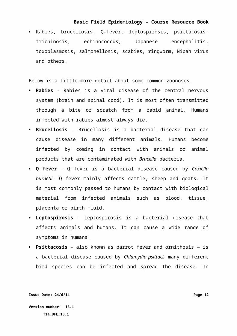
1.5Epidemiological skills can help prevent zoonoses
Zoonoses are animal diseases that are able to infect humans.
Epidemiology skills help you to understand how zoonotic diseases occur and how to prevent
both animals and humans from exposure and infection. There are a number of zoonotic
diseases of animals in Indonesia. Examples include:
Rabies, brucellosis, Q-fever, leptospirosis, psittacosis, trichinosis, echinococcus, Japanese
encephalitis, toxoplasmosis, salmonellosis, scabies, ringworm, Nipah virus and others.
Below is a little more detail about some common zoonoses.
Rabies - Rabies is a viral disease of the central nervous system (brain and spinal cord). It
is most often transmitted through a bite or scratch from a rabid animal. Humans
infected with rabies almost always die.
Brucellosis - Brucellosis is a bacterial disease that can cause disease in many different
animals. Humans become infected by coming in contact with animals or animal products
that are contaminated with Brucella bacteria.
Q fever - Q fever is a bacterial disease caused by Coxiella burnetii. Q fever mainly affects
cattle, sheep and goats. It is most commonly passed to humans by contact with
biological material from infected animals such as blood, tissue, placenta or birth fluid.
Leptospirosis - Leptospirosis is a bacterial disease that affects animals and humans. It
can cause a wide range of symptoms in humans.
Issue Date: 24/6/14 Page 9
Version number: 13.1 T1a_BFE_13.1
Basic Field Epidemiology – Course Resource Book
Psittacosis – also known as parrot fever and ornithosis — is a bacterial disease caused by
Chlamydia psittaci, many different bird species can be infected and spread the disease.
In humans it can cause severe pneumonia and other serious health problems.
Trichinosis - also called trichinellosis is a parasitic disease passed to humans by eating
raw or undercooked meat of animals that are infected with the trichinella worm larvae.
Infection by trichinella occurs commonly in wild carnivorous (meat-eating) animals but
can also occur in pigs.
Issue Date: 24/6/14 Page 10
Version number: 13.1 T1a_BFE_13.1
Basic Field Epidemiology – Course Resource Book
2 Signs, syndromes, and making a diagnosis
2.1The effect of disease on animal health and production
Diseases in animals will often result in reduced health and production and may result in
death.
Livestock are generally raised for production of meat, milk, hides, eggs, manure, and
offspring (calves, chickens, etc.). Healthy livestock are more productive than livestock that
are not healthy.
Some diseases cause animals to change their behaviour and look sick. Some of the sick
animals may die and others may recover. It is easy to pick animals that are very sick with a
disease.
Sometimes animals may be affected by a disease but still look healthy and it can be hard to
pick whether or not these animals are sick at all. They may not show obvious signs of a
problem. That is why it is important for farmers to watch (monitor) their animals
frequently.
Sick animals may stop eating for a while and lose weight. Sometimes animals may look
normal but suffer from reduced production (weight loss, infertility, pregnancy loss, reduced
egg production). Many diseases cause a decrease in animal production.
There are many things other than infectious diseases that cause poor production in animals.
Examples include animals that are offered poor quality feed, not enough feed, or feed and
water that may be spoiled.
Epidemiology and clinical skills need to be used when investigating poor performance in
livestock. A good investigator will be able to tell whether poor production or poor health is
due to an infectious disease or due to some other cause such as poor feed.
Issue Date: 24/6/14 Page 11
Version number: 13.1 T1a_BFE_13.1
Basic Field Epidemiology – Course Resource Book
Yesterday Budi reported to his local Pelsa that one of his cows was lame. The
Pelsa sent a General Signs report (Tanda Umum) to iSIKHNAS.
Pak Paimin (the para-vet) received the notification from iSIKHNAS and talks to
Budi. Budi tells Pak Paimin that there is one cow that is lame and she has been
lame for several weeks now. She is skinny and her calf is weak. There are also 2
cows with diarrhoea.
Pak Paimin listens to Pak Budi describe the lame cow and comes up with a list
of possible causes (differential diagnoses) for this situation. They include:
injury, abscess, infectious diseases (brucellosis, black leg, etc.), or trauma
(broken leg).
The 2 cows that have diarrhoea could be sick because of parasites, grain
overload, bacterial infection, or liver disease from poisons. These diseases are
differential diagnoses for diarrhoea in cows.
Pak Paimin decides to conduct a farm visit to get more information to narrow
down the list.
2.2Signs of disease
Signs are changes in an animal that are caused by disease and that people can detect.
Many diseases make animals feel sick, develop a fever and they might stop eating or
drinking for a period of time. Many signs of disease are easily observed by people. Farmers
that know their animals well will be able to detect small changes in behaviour that indicate
an animal may be getting sick. This will help them to get assistance from their para-vet
early, before the case becomes very serious.
It can be easy to see the signs of some diseases but more difficult to see the signs of other
diseases.
Issue Date: 24/6/14 Page 12
Version number: 13.1 T1a_BFE_13.1
Basic Field Epidemiology – Course Resource Book
Signs of disease that are easy to see include lameness, coughing, diarrhoea, severe
weight loss and death
Signs of disease that may be harder to see include infertility and reduced weight gain
or milk production
Most signs provide an indication of which parts of the body and body systems are being
changed by the disease
For example:
Diarrhoea indicates diseases that affect gut motility or absorption (digestive
system). There are many diseases that can cause diarrhoea.
Coughing and difficulty breathing indicates disease of the lungs or airways
(respiratory system)
Saliva drooling from the mouth indicates inability to swallow either due to
nervous system disease, physical obstruction of the throat, or increased
production of saliva
Lameness or altered gait may indicate disease or injury affecting the leg(s),
spine or brain
Technical staff are trained to conduct clinical examinations of sick animals, to look for signs
that indicate the animal may be sick. These include changes in:
rectal temperature
heart rate
pulse rate
respiratory rate
respiratory sounds
gut sounds
mucous membrane colour
The time it takes for signs of disease to develop can vary depending on the infectious
disease. Therefore, the time it takes you to identify if an animal is sick can also vary.
Issue Date: 24/6/14 Page 13
Version number: 13.1 T1a_BFE_13.1
Basic Field Epidemiology – Course Resource Book
For example:
Anthrax and haemorrhagic septicaemia are diseases that develop very
rapidly and produce severe clinical signs or death.
Papilloma virus (causing warts on the skin of cattle) is a disease that
produces mild signs in many animals with little impact on health or
productivity.
Bovine Johnes disease (BJD) is a disease which develops very slowly, it
causes chronic diarrhoea and wasting in older cattle. It takes a long time for
BJD to these produce serious effects, the cattle are infected when they are
very young but do not show signs till they are older. BJD produces the same
signs as a cow with an intestinal worm infection.
Sometimes animals show signs such as weight loss that may be due to things other than an
infectious disease, such as an injury (broken jaw), problems with teeth or even just poor
feed.
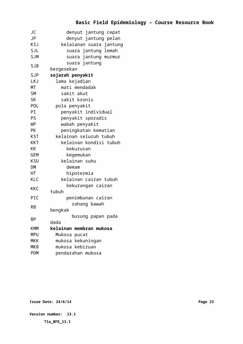
iSIKHNAS has developed lists of common signs and codes to make reporting these signs
easier and more consistent. Village reporters (pelsa), para-vets and veterinarians play a vital
role in the identification, reporting, and treatment of these signs.
Issue Date: 24/6/14 Page 14
Version number: 13.1 T1a_BFE_13.1
Basic Field Epidemiology – Course Resource Book
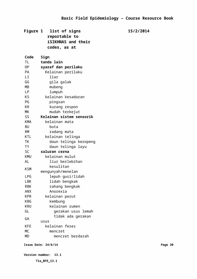
Figure 1 list of signs reportable to iSIKHNAS and their codes,
as at 15/2/2014
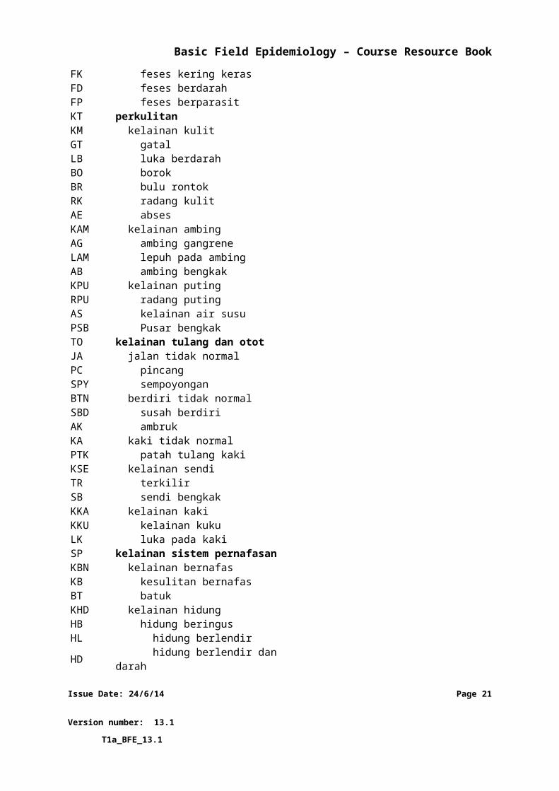
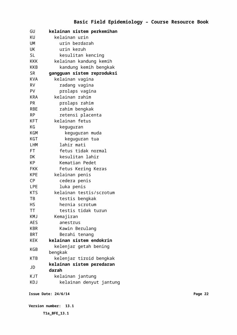
Code SignTL tanda lainOP syaraf dan perilakuPA Kelainan perilakuLI liarGG gila galakMB mubengLP lumpuhKS kelainan kesadaranPG pingsanKR kurang responMK mudah terkejutSS Kelainan sistem sensorikKMA kelainan mataBU butaRM radang mataKTL kelainan telingaTK daun telinga keropengTY daun telinga layuSC saluran cernaKMU kelainan mulutAL liur berlebihanKSM kesulitan mengunyah/menelanLPG lepuh gusi/lidahLBK lidah bengkakRBK rahang bengkakANX AnorexiaKPR kelainan perutKBG kembungKRU kelainan rumenGL gerakan usus lemahGA tidak ada gerakan ususKFE kelainan fesesMC mencretMD mencret berdarahFK feses kering kerasFD feses berdarahFP feses berparasitKT perkulitanKM kelainan kulit
Issue Date: 24/6/14 Page 15
Version number: 13.1 T1a_BFE_13.1
Basic Field Epidemiology – Course Resource Book
GT gatalLB luka berdarahBO borokBR bulu rontokRK radang kulitAE absesKAM kelainan ambingAG ambing gangreneLAM lepuh pada ambingAB ambing bengkakKPU kelainan putingRPU radang putingAS kelainan air susuPSB Pusar bengkakTO kelainan tulang dan ototJA jalan tidak normalPC pincangSPY sempoyonganBTN berdiri tidak normalSBD susah berdiriAK ambrukKA kaki tidak normalPTK patah tulang kakiKSE kelainan sendiTR terkilirSB sendi bengkakKKA kelainan kakiKKU kelainan kukuLK luka pada kakiSP kelainan sistem pernafasanKBN kelainan bernafasKB kesulitan bernafasBT batukKHD kelainan hidungHB hidung beringusHL hidung berlendirHD hidung berlendir dan darahGU kelainan sistem perkemihanKU kelainan urinUM urin berdarahUK urin keruhSL kesulitan kencingKKK kelainan kandung kemihKKB kandung kemih bengkak
Issue Date: 24/6/14 Page 16
Version number: 13.1 T1a_BFE_13.1
Basic Field Epidemiology – Course Resource Book
SR gangguan sistem reproduksiKVA kelainan vaginaRV radang vaginaPV prolaps vaginaKRA kelainan rahimPR prolaps rahimRBE rahim bengkakRP retensi placentaKFT kelainan fetusKG keguguranKGM keguguran mudaKGT keguguran tuaLHM lahir matiFT fetus tidak normalDK kesulitan lahirKP Kematian PedetFKK Fetus Kering KerasKPE kelainan penisCP cedera penisLPE luka penisKTS kelainan testis/scrotumTB testis bengkakHS hernia scrotumTT testis tidak turunKMJ KemajiranAES anestrusKBR Kawin BerulangBRT Berahi tenangKEK kelainan sistem endokrinKGB kelenjar getah bening bengkakKTB kelenjar tiroid bengkakJD kelainan sistem peredaran darahKJT kelainan jantungKDJ kelainan denyut jantungJC denyut jantung cepatJP denyut jantung pelanKSJ kelaianan suara jantungSJL suara jantung lemahSJM suara jantung murmurSJB suara jantung bergesekanSJP sejarah penyakitLKJ lama kejadianMT mati mendadakSM sakit akut
Issue Date: 24/6/14 Page 17
Version number: 13.1 T1a_BFE_13.1
Basic Field Epidemiology – Course Resource Book
SK sakit kronisPOL pola penyakitPI penyakit individualPS penyakit sporadisWP wabah penyakitPK peningkatan kematianKST kelainan seluruh tubuhKKT kelainan kondisi tubuhKK kekurusanGEM kegemukanKSU kelainan suhuDM demamHT hipotermiaKLC kelainan cairan tubuhKKC kekurangan cairan tubuhPIC penimbunan cairanRB rahang bawah bengkakBP busung papan pada dadaKMM kelainan membran mukosaMPU Mukosa pucatMKK mukosa kekuninganMKB mukosa kebiruanPDM pendarahan mukosa
Issue Date: 24/6/14 Page 18
Version number: 13.1 T1a_BFE_13.1
Basic Field Epidemiology – Course Resource Book
2.3Syndromes
Syndrome refers to a particular sign or a group of signs that can be easily recognised and
which may indicate a particular important disease.
For example:
A respiratory syndrome may be defined as including any animal that shows
one or more of the following clinical signs: coughing, difficulty breathing,
nasal discharge, elevated respiratory rate, and so on.
Syndromes are used to identify animals that may be suffering from a specific disease of
importance.
For example:
Rabies is an important disease. It is not possible to diagnose rabies for certain
in a live dog (or any animal). Definitive diagnosis of rabies can only occur when
brain or other tissue is examined by a pathologist after the animal has died or
has been killed.
However, many dogs infected with rabies will show changes in behaviour –
becoming more aggressive, showing drooling of saliva from the mouth and
more likely to attack and bite other animals and people. Some dogs show these
signs even though they are not infected with rabies – just because they are
aggressive dogs or drooling because they have something stuck in their mouth
or throat.
We use the syndrome of changes in behaviour (aggression, biting, salivation,
depression) as a way of identifying dogs that may have rabies. Dogs that show
these signs can be isolated and watched to see if they continue to develop
Issue Date: 24/6/14 Page 19
Version number: 13.1 T1a_BFE_13.1
Basic Field Epidemiology – Course Resource Book
signs that are suggestive of rabies and they may be killed or sent for post
mortem to test for rabies.
Some syndromes are strongly related to one disease so that when an animal shows these
particular signs it is very likely that it has the disease. Examples include rabies (as described
above) or sudden death with blood from the orifices (likely to be anthrax).
Syndromes are often used in disease control programs for important diseases to make sure
that these diseases are identified when they occur. Livestock owners and animal health staff
are encouraged to look for animals with defined syndromes. Animals that show those signs
can then be examined more carefully or sampled for laboratory testing to try and diagnose
the disease. Some animals will be found not to have the disease of interest. If the disease of
interest is confirmed then other disease control activities may need to be done.
iSIKHNAS uses broad syndromes, this is because the diseases of interest are very important
and we don’t want to miss any cases. Village reporters (pelsa) and para-vets should report
every suspected priority syndrome case. The important thing is that we all remain alert to
the threat of these diseases. Most priority reports will probably end in a diagnosis which is
not the priority disease. It is still very important that you report a priority syndrome (using a
P SMS reporting message) and let the vet carry out a more thorough investigation.
iSIKHNAS uses the following priority syndromes for disease reporting:
MMU - Sudden increase in mortality in chickens and other poultry
- this syndrome is trying to identify cases of Avian Influenza
- Other infectious diseases that could produce this syndrome include: Newcastle disease, infectious laryngotracheitis, and duck plague.
- Other non-infectious causes include: acute poisoning.
KGS - Abortion in third trimester or swollen joints in cattle
- this syndrome is trying to identify cases of Brucellosis
Issue Date: 24/6/14 Page 20
Version number: 13.1 T1a_BFE_13.1
Basic Field Epidemiology – Course Resource Book
- Other infectious diseases that could produce this syndrome include: many bacterial and viral infections can cause abortion and swollen joints.
- Other non-infectious causes of this syndrome include: genetic conditions, exposure to poisons, and administration of some drugs.
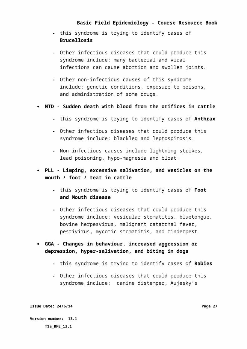
MTD - Sudden death with blood from the orifices in cattle
- this syndrome is trying to identify cases of Anthrax
- Other infectious diseases that could produce this syndrome include: blackleg and leptospirosis.
- Non-infectious causes include lightning strikes, lead poisoning, hypo-magnesia and bloat.
PLL - Limping, excessive salivation, and vesicles on the mouth / foot / teat in cattle
- this syndrome is trying to identify cases of Foot and Mouth disease
- Other infectious diseases that could produce this syndrome include: vesicular stomatitis, bluetongue, bovine herpesvirus, malignant catarrhal fever, pestivirus, mycotic stomatitis, and rinderpest.
GGA - Changes in behaviour, increased aggression or depression, hyper-salivation, and biting in dogs
- this syndrome is trying to identify cases of Rabies
- Other infectious diseases that could produce this syndrome include: canine distemper, Aujesky’s disease, and any infectious disease involving the brain.
- Non-infectious causes include - neoplasia, trauma, oral foreign bodies and poisoning.
DMB - High fever, conjunctivitis, and increased mortality in pigs
- this syndrome is trying to identify cases of Classical Swine Fever
- Other infectious diseases that could produce this syndrome include: African swine fever virus, and many other bacterial and viral infections.
- Non-infectious causes include exposure to some poisons such as anticoagulant.
Issue Date: 24/6/14 Page 21
Version number: 13.1 T1a_BFE_13.1
Basic Field Epidemiology – Course Resource Book
2.4Differential diagnoses
A differential diagnosis is a disease that could cause the clinical signs that have been
observed. Often there is more than one disease that can cause the same signs. When sick
animals are examined it is common to produce a list of multiple differential diagnoses as
possible diseases that could be affecting the animals.
It is common practice to list diseases in order from most likely to least likely.
At Budi’s farm there were 2 cows with diarrhoea. Immediately Pak Paimin can
create a list of common diseases that could cause the clinical signs.
Parasites (worms, coccidia, liver fluke)
Grain overload
Poisoning
Salmonella infection in the gut (bacteria)
Johne’s disease infection (bacteria)
Bovine virus diarrhoea infection (virus)
Very rich, fresh pasture
The disease investigation is then used to try and determine which diseases are unlikely to be
causing the signs and which ones might be more likely. This process leads to a short list and
sometimes a single differential diagnosis.
2.5Definitive diagnosis
A definitive diagnosis is reached when the vet is confident there is one disease that is most
likely to be affecting the sick animal(s). The vet uses all the diagnostic information available
(including history, clinical, environmental, laboratory and epidemiological information).
Issue Date: 24/6/14 Page 22
Version number: 13.1 T1a_BFE_13.1
Basic Field Epidemiology – Course Resource Book
Often the para-vet will need to carry out a disease investigation before a definitive diagnosis
can be reached. This may include colleting some samples from affected (and possibly
unaffected) animals for laboratory testing.
Issue Date: 24/6/14 Page 23
Version number: 13.1 T1a_BFE_13.1
Basic Field Epidemiology – Course Resource Book
3 Disease investigation
3.1 The approach to a disease investigationDisease investigations usually start because a farmer is concerned that one or more animals
are showing abnormal signs or are dead. The first part of a disease investigation involves
four activities. These activities combine field epidemiology and veterinary clinical skills.
1. Taking a good history
To find out what happened in the period before disease was noticed a para-vet will need to
talk carefully to the farmer about his animals and about how he manages his animals. Good
history taking is an art; it requires diplomacy, use of non-technical language, and a good
relationship with the farmer. History-taking involves collecting information about:
The animal: Species, breed, sex, age, identification of the affected animal(s)
The problem: clinical signs, development of signs over time, number of animals
affected
Any treatment(s) given by the farmer
The housing and feeding: where the animals are kept and their access to feed
and water
Other animals on the farm
Any recent events that may be related (animal movements on or off the farm,
floods, chemical treatments of plants or release of chemicals into the
environment)
Pak Paimin asks Budi about the history of his 2 cows that have diarrhoea. Budi
tells us that they are both 2 year old Sapi Bali cows and he has been keeping
them separate from the other cows as these two don’t have calves. Budi
wormed all his cows 3 weeks ago. He doesn’t feed these 2 cows anything else
Issue Date: 24/6/14 Page 24
Version number: 13.1 T1a_BFE_13.1
Basic Field Epidemiology – Course Resource Book
apart from short grass that is in the small paddock near the shed. There are no
other cows on his neighbours’ properties.
2. Clinical examination of the sick animal(s)
A basic examination of a sick animal consists of the following:
General appearance
- Body conditions score of animal
- Swellings/lumps
- Bright, depressed, dull
- Examination of dung or urine already on ground
Respiratory rate and lung sounds
Heart rate
Pulse rate and strength
Temperature of animal
Mucous membrane colour
Capillary refile time (<2 sec)
Gastro intestinal sounds
Palpation – lymph nodes, skin, joints, abdomen,
There may be reason to conduct further examination of problem areas such as:
rectal examination (pregnancy test in cows)
inspect mouth
further investigate any abnormal signs that are noticed
All observations should be recorded. Once the clinical examination is completed the para-
veterinarian should consider the signs of disease shown and interpret them with the history
to decide which diseases may be causing the problem (differential diagnosis list).
Where affected animals have died, a post mortem examination should be performed to look
for changes that may suggest possible diseases to add to the differential diagnosis list.
Pak Paimin examines Budi’s 2 cows that have diarrhoea. He looks at them and
they both look a little weak, their eyes are sunken and they are depressed.
From a distance they have very watery diarrhoea.
Issue Date: 24/6/14 Page 25
Version number: 13.1 T1a_BFE_13.1
Basic Field Epidemiology – Course Resource Book
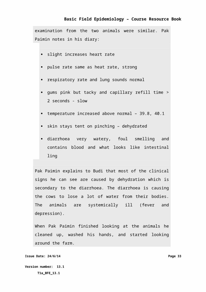
Pak Paimin puts his gloves on and then examines both animals closely. Overall
the results of the clinical examination from the two animals were similar. Pak
Paimin notes in his diary:
slight increases heart rate
pulse rate same as heat rate, strong
respiratory rate and lung sounds normal
gums pink but tacky and capillary refill time > 2 seconds - slow
temperature increased above normal – 39.8, 40.1
skin stays tent on pinching – dehydrated
diarrhoea very watery, foul smelling and contains blood and what looks
like intestinal ling
Pak Paimin explains to Budi that most of the clinical signs he can see are
caused by dehydration which is secondary to the diarrhoea. The diarrhoea is
causing the cows to lose a lot of water from their bodies. The animals are
systemically ill (fever and depression).
When Pak Paimin finished looking at the animals he cleaned up, washed his
hands, and started looking around the farm.
3. Examination of the environment
Examination of the environment where the animals are kept is an important part of the
initial clinical examination. The value of environmental examination for veterinary clinicians
is based on an understanding that there are multiple causes of a disease. Each cause
influences whether or not disease occurs in a particular animal or population.
For animals maintained outdoors it may be useful think about:
Issue Date: 24/6/14 Page 26
Version number: 13.1 T1a_BFE_13.1
Basic Field Epidemiology – Course Resource Book
Their environment - what soils and pasture are available and what is the water
source?
Are there areas of muddy and boggy land (swamp), fast flowing water or steep hills?
What wildlife exists in the area and are they capable of carrying any diseases that
may affect livestock?
Is there sufficient shelter from prevailing winds and rain or sun?
Is there sufficient feed supply for the animals that are present?
Is there any evidence to suggest that animals might be gaining access to poisons or
toxins (garbage dumps, industrial areas, agricultural chemicals)?
Is there any evidence of overcrowding that may lead to stress, contamination of the
environment and increased risk of disease?
If animals are housed indoors then you should think about issues like:
flooring
ventilation
air quality
level of crowding
bedding
general hygiene
It is also important to look specifically at the feed and water supply to make sure animals
are receiving enough feed and water and that there supplied feed and water are of
acceptable quality.
The ability of veterinarians and para-veterinarians to assess and interpret information
gathered during an environmental examination is based on understanding about multiple
causes of diseases.
Pak Paimin walks around Budi’s farm while asking him questions. He finds that
the cows with calves have good pasture and Budi also feed them some extra
food. He finds that the 2 sick cows have been in paddock where the only water
is from a drain. This drain carries all the waste water from the other paddocks
including the pen where calves are held if they are sick. Budi mentions he was
given a young calf that was sick for a long time; it is still alive and is more like a
pet now.
Issue Date: 24/6/14 Page 27
Version number: 13.1 T1a_BFE_13.1
Basic Field Epidemiology – Course Resource Book
4. Collecting samples for laboratory testing
The combination of history, clinical examination and environmental examination will often
result in a diagnosis or list of differential diagnoses.
Sometimes it is helpful to collect samples for laboratory testing. Samples may be collected
for laboratory testing, from the affected animals and sometimes from some healthy animals
as well. Commonly collected samples include:
blood or serum
faeces
milk
urine
If one or more animals have died or are severely sick it may be useful to conduct a post
mortem and collect additional samples including tissues to send to the laboratory for
testing. Laboratories will provide information on how to collect and submit samples for the
best results.
Pak Paimin kept the 2 gloves from the rectal examinations he did on the cows.
They both have enough faeces to send to the laboratory. After his examination
of the environment he decides to collect some blood to send to the lab as well.
3.2Developing a list of differential diagnosesThe findings from the investigation will identify the signs or syndromes displayed by affected
animals and a list of differential diagnoses.
Correct diagnosis of disease in an individual animal often requires clinical skill and may
require laboratory testing and/or pathology examination of post mortem material.
Most of the time you will start to create a list of differential diagnoses when talking to a
farmer about his sick animals. As each of the four steps of the examination are conducted
the differential diagnosis list should be reviewed and changed depending on new
information or findings of any examination.
Issue Date: 24/6/14 Page 28
Version number: 13.1 T1a_BFE_13.1
Basic Field Epidemiology – Course Resource Book
For example:
You find out that the affected animals are all young cows having their first or
second calf, and that these cows are aborting dead calves. Knowing this may
immediately narrow your list of possible diagnoses to one of several diseases
capable of causing abortion in cattle.
Using the findings from the clinical examination to determine what body systems that may
be affected is usually helpful.
For example:
If the affected animal is a lactating cow and she has a fever and a swollen, hot,
painful udder with abnormal milk (foul smelling, watery fluid) present in two
quarters and no other abnormal signs. Then the affected body system is the
udder and the cow is likely to be suffering from mastitis.
As the examination process proceeds and new information is collected will be used to
update our understanding of the body systems affected and the possible causes. Each new
piece of information will contribute to your differential diagnoses list. In most cases new
information will help add a new diseases or remove some diseases from the list.
Laboratory testing needs to be used and interpreted with care. Laboratory testing may take
time, cost money and may or may not make a useful contribution to the diagnosis and
management of disease in animals.
Pak Paimin has been thinking all the time what the list of possible causes could
be. He started with a long list in his head and he even included the possibility
this could be a new disease that no one had seen before.
As his investigation progressed he started to cross things off his list or move
them lower down on the list in terms of likelihood. In this case Pak Paimin
doesn’t think poisoning can be a cause as he has not identified anything in the
Issue Date: 24/6/14 Page 29
Version number: 13.1 T1a_BFE_13.1
Basic Field Epidemiology – Course Resource Book
history or on the farm that indicates possible access to poisonous plants or
chemicals. From talking to Budi he doesn’t think he would have overdosed
these 2 cows on the worm treatment.
There was no evidence to suggest the cows could have eaten large amounts of
grain and the grass they have been eating is not rich and green. This rules out
two more differential diagnoses.
So, Pak Paimin’s differential diagnoses contains;
Bacterial enteritis – Salmonella, E-coli (most likely)
Bovine Viral Diarrhoea Virus
Parasites (least likely)
The two cows with diarrhoea have a fever and the diarrhoea is foul smelling
and contains blood and what looks like bits of intestinal lining. These findings
are strongly suggestive of salmonella infection, a bacterial infection that can
cause diarrhoea and systemic infection.
Before Pak Paimin leaves Budi’s farm he sends a Response, Laboratory and
Treatment reports to iSIKHNAS from his phone.
Pak Paimin is not 100% certain of the diagnosis but he is pretty confident that
the infectious agent is a bacterial infection causing enteritis and making the
cows sick.
Pak Paimin treats the two cows with broad spectrum antibiotic and using field
epidemiology skills, advises Budi about possible reasons for how the disease
occurred and about future control. Pak Paimin knows the disease can be
zoonotic and advises Budi to pay attention to general hygiene (hand washing)
after handling cows. Often cows can develop this disease following some
Issue Date: 24/6/14 Page 30
Version number: 13.1 T1a_BFE_13.1
Basic Field Epidemiology – Course Resource Book
stressful event such as calving or following feeding of feed that has been
contaminated with faeces from other animals. Pak Paimin advises Budi to
check that the animals have clean feed and water and that any animals he buys
in future should be from someone who he trusts to supply him with healthy
animals. All of these strategies involve a broader understanding of how
salmonellosis behaves in a cattle population and will help Budi reduce his risk
of salmonellosis occurring again in the future.
The 2 sick cows should be separated from other healthy animals on the farm.
All healthy cows are to be kept higher up steam along the drain line than the 2
sick cows and the calf.
The example above has been carried through to illustrate how a disease investigation might
start with a report of one or more sick cows, progress through an investigation and end with
a likely diagnosis, treatment of affected animals and with the para-vet also providing advice
to the farmer about preventing spread to other animals, how to prevent the same disease
from occurring in the future and for zoonotic diseases how to avoid humans getting sick.
More information is provided in following sections about the application of epidemiology
knowledge and skills for investigating and managing diseases in animals.
Issue Date: 24/6/14 Page 31
Version number: 13.1 T1a_BFE_13.1
Basic Field Epidemiology – Course Resource Book
4 Causes of diseaseA cause is anything that can influence whether or not a disease occurs in one or more
animals.
When diseases occur in animals there are nearly always some animals that develop disease
and others that do not. There are a very small number of diseases that are so infectious that
when they occur nearly every animal gets infected and develops disease. These are rare.
Causes of disease include many different things. Infectious diseases will have an infectious
agent as one of the causes. For example, rabies virus is the infectious agent that causes
rabies. For diseases that have only one infectious cause, if the infectious agent is not present
we can be confident that the disease will not occur. BUT, exposure of an animal to an
infectious agent does not mean that the disease will occur.
Often many different causes have to occur together before an animal will develop disease.
For example:
When one dog bites another dog there is a chance that the second dog might
develop rabies. The likelihood of rabies occurring is much higher if the first
dog:
is infected with rabies
is shedding rabies virus in its saliva
the bite causes a break in the skin and the rabies virus can get into the tissues of the second dog
If the first dog is not shedding virus in its saliva then the bite may not cause
rabies. If the bite does not break the skin then the virus may not get into the
tissues of the second dog and it may not develop rabies. If the second dog is
vaccinated against rabies then it may not develop rabies even if bitten by a
rabid dog.
Issue Date: 24/6/14 Page 32
Version number: 13.1 T1a_BFE_13.1
Basic Field Epidemiology – Course Resource Book
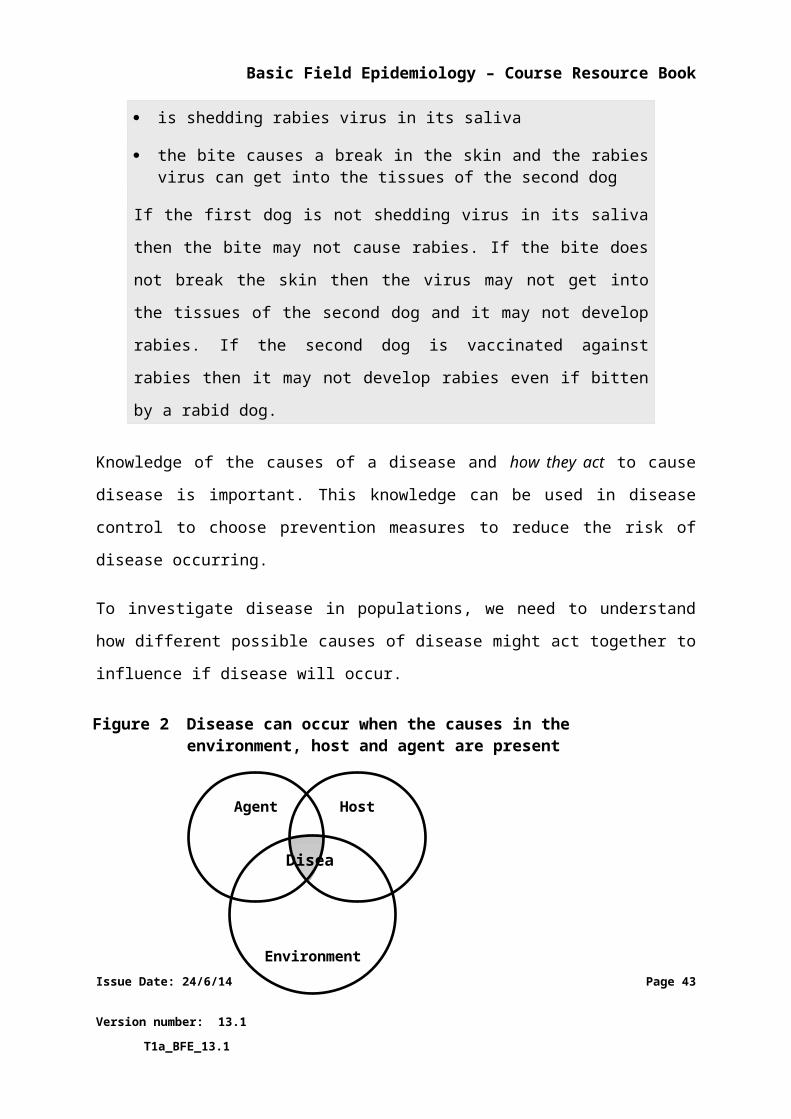
Knowledge of the causes of a disease and how they act to cause disease is important. This
knowledge can be used in disease control to choose prevention measures to reduce the risk
of disease occurring.
To investigate disease in populations, we need to understand how different possible causes
of disease might act together to influence if disease will occur.
Host refers to the animal which can get the disease. The host (animal) has a variety of
characteristics that may influence if disease will occur or not. Some diseases occur only in
young animals while others only in older animals, for example. Pregnancy and abortion can
only occur in females. The vaccination status of animal influences the risk of getting the
disease.
Pinkeye is more common in younger cattle because they generally have not
been exposed to Moraxella bovis, the bacteria that is involved in pinkeye. Lack
of exposure in younger cattle mean they have a lower immunity to this
bacteria and therefore a higher risk than older cattle of getting the disease.
Issue Date: 24/6/14 Page 33
Version number: 13.1 T1a_BFE_13.1
Figure 2 Disease can occur when the causes in the environment, host and agent are present
Environment
HostAgent
Disease
Basic Field Epidemiology – Course Resource Book
Pinkeye will be more common in animals with physical characteristics where
the eyes stick out (protrude) more than in others animals. This characteristic
may be inherited and associated with a certain sire or breed.
Agent refers to the particular infectious agent that is a cause of the disease: virus, bacteria,
fungus, parasite, or other microbe. Different strains of the same bacteria or virus may either
produce no disease or more severe disease. There are many different infectious agents and
different sub-types or strains, these may all have different abilities to cause disease.
Sometimes non-infectious causes are classified as agents for example lead as the agent that
can cause lead poisoning or a plant toxin that can cause a type of plant poisoning.
Some strains of avian influenza do not cause a high mortality rate, whereas
other strains do.
Some strains of Moraxella bovis (the infectious agent causing pinkeye in cattle)
produce a toxin that is more powerful and causes more severe eye disease
than other strains.
Environment refers to external things that affect the host and agent and influence if disease
will occur. Environmental characteristics include: land type, humidity, rainfall, insects that
transmit the agent, crowding, sanitation, etc.
Ultraviolet light damages the eyes (cornea) of cattle which is a cause of
pinkeye. Pinkeye is more common in summer when ultraviolet radiation is
high.
Pinkeye is more common where a lot of cattle are kept very close together and
less common where cattle have a lot of space.
Pinkeye is more common when there are lots of flies.
Pinkeye is more common when cattle are eating long grass which is hard. Hard
grass can cause small injuries to the cornea which allows the bacteria to get in.
Issue Date: 24/6/14 Page 34
Version number: 13.1 T1a_BFE_13.1
Basic Field Epidemiology – Course Resource Book
Simple exposure to the agent does not mean disease will occur. There are many causes and
many combinations of these causes required for disease to occur.
The relationships among causes of disease can be represented in diagrams that show causes
and how they act together to influence the occurrence of disease. These diagrams are often
called causal diagrams or causal webs (because they can resemble a spider web of lines
linking various causes to a disease). Causal diagrams can show in a diagram how causes
related to the environment, the agent can act together to influence risk of disease occurring
in animals. Some causes are defined as being necessary causes – meaning that disease will
not occur if this cause is not present. Other causes may just act to increase or reduce the
risk of disease occurring and these causes may not be necessary causes. Understanding
causes often allows us to identify measures that can be used to reduce disease risk through
activities such as changes to management practices or husbandry.
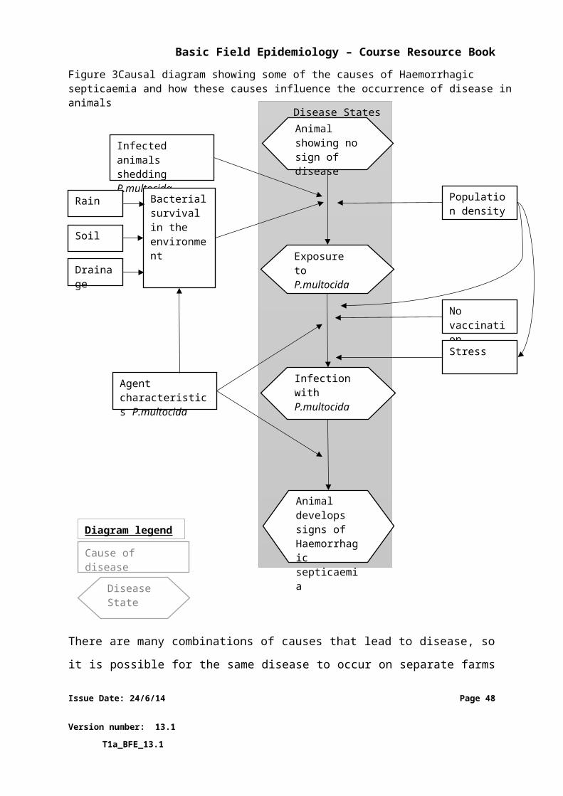
The bacteria Pasturella multocida is the infectious agent involved in Haemorrhagic
septicaemia. The diagram below shows some of the causes of Haemorrhagic septicaemia
and how these causes influence the occurrence of disease in an individual animal. You can
see that this diagram is trying to show a complex interaction between many different
individual causes that can influence whether or not disease occurs in any one animal.
Issue Date: 24/6/14 Page 35
Version number: 13.1 T1a_BFE_13.1
Basic Field Epidemiology – Course Resource Book
There are many combinations of causes that lead to disease, so it is possible for the same
disease to occur on separate farms or at different times due to a different combination of
causes.
Issue Date: 24/6/14 Page 36
Version number: 13.1 T1a_BFE_13.1
Figure 3 Causal diagram showing some of the causes of Haemorrhagic septicaemia and how these causes influence the occurrence of disease in animals
Diagram legend
Cause of disease
Disease State
Animal develops signs of Haemorrhagic septicaemia
Infection with P.multocida
Exposure to P.multocida
Animal showing no sign of disease
Stress
No vaccination
Population density
Agent characteristics P.multocida
Bacterial survival in the environment
Drainage
Soil
Rain
Infected animals shedding P.multocida
Disease States
Basic Field Epidemiology – Course Resource Book
Epidemiological investigations help identify the important causes of disease and
epidemiologic knowledge about the causes of a disease and how they interact with each
other can be used to develop measures that vets, para-vets and farmers can apply to
prevent or control disease.
We may find in a research study that cattle farms feeding dry grass and hay are
at five times the risk of having pinkeye outbreaks in weaner cattle compared
with those using silage or fresh grass.
Using this information, a para-vet or vet might advise a farmer to change from
dry feed to fresh grass or silage when lots or pink eye cases are occurring in the
district. The farmer may even spray dry feed with water to reduce the risk of
dust and small particles getting in to the eyes of cattle as they eat. These
approaches may reduce the risk of having pinkeye in the weaner cattle.
A disease may be prevented by doing something that breaks an important relationship
between different causes.
For example:
using better hygiene measures and managing the property to avoid
environmental contamination with the infectious agent(s)
vaccinating animals to increase host immunity and help prevent infection
with the infectious agent
Understanding the different causes can help find cost effective ways to prevent or reduce
the effects of the disease.
If shade is provided for cattle, the damage caused by UV light to the eyes may
be less and the amount of pinkeye disease reduced.
Issue Date: 24/6/14 Page 37
Version number: 13.1 T1a_BFE_13.1
Basic Field Epidemiology – Course Resource Book
If we vaccinate all the cattle for haemorrhagic septicaemia and avoid crowding
during the wet season we would reduce the number of disease events.
Below are some additional examples of causal diagrams.
CAUSAL WEBS
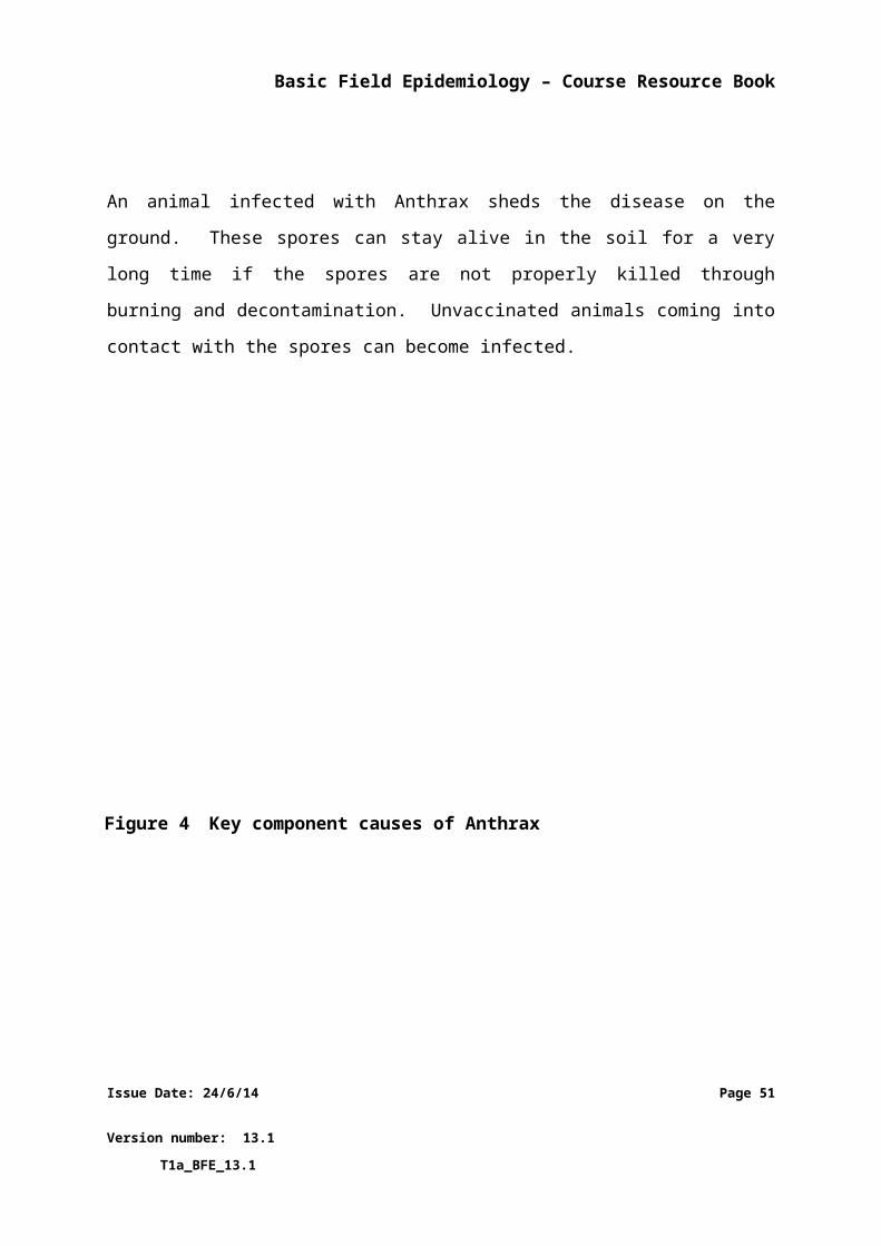
An animal infected with Anthrax sheds the disease on the ground. These spores can stay
alive in the soil for a very long time if the spores are not properly killed through burning and
decontamination. Unvaccinated animals coming into contact with the spores can become
infected.
Issue Date: 24/6/14 Page 38
Version number: 13.1 T1a_BFE_13.1
Figure 4 Causal web for anthrax
Disturbed earth
Animals exposed to
anthrax
Animal not vaccinated
Spores from
infected animal
Poor handling of
carcass
Contaminated soil
Infected animal
New infected animal
Basic Field Epidemiology – Course Resource Book
By understanding the interaction between causes in the environment, the way the agent
behaves and the susceptibility of the host you can do things which will lower the risk of
infection to other animals. Burning any carcases to ash, properly disposing of soil
contaminated with body fluids from the infected animals and vaccinating the entire herd
will lower the risk of ongoing infection from spores in the environment. There are many
other management and husbandry activities which can also be considered.
Issue Date: 24/6/14 Page 39
Version number: 13.1 T1a_BFE_13.1
Disturbed earth
Animals exposed to
anthrax
Animal not vaccinated
Spores from
infected animal
Poor handling of
carcass
Contaminated soil
Infected animal
New infected animal
Figure 5 Key component causes of Anthrax
Necessary cause – if not present the disease will not be present.
Possible to influence this
Basic Field Epidemiology – Course Resource Book
Lantana camara is an invasive species of flowering plant. The leaves on the plant are toxic
to animals and cause liver damage. Animals usually avoid the plant but may eat it when
there is no other food due to reasons such as drought or flooding, or poor management.
Poor herding may allow access to the plant. In addition, animals will always seek access to
green fodder (grass, reeds, leaves etc). This causes an accumulation of chlorophyll in the
skin and when exposed to strong sunlight the pale skin of animals quickly becomes badly
damaged by sunburn.
With a better understanding of the causes in the environment, host and the agent, we are
able to significantly influence the outcome of the disease. Careful herding to avoid areas
with lantana, and ensuring sufficient feed of adequate quality can lower the likelihood of
animals eating lantana. Providing cattle who have had access to lantana (particularly pale
skinned animals) with good access to shade can provide immediate protection against
photosensitisation. Providing them with dry fodder (hay etc) instead of green (chlorophyll
Issue Date: 24/6/14 Page 40
Version number: 13.1 T1a_BFE_13.1
Non-pigmented (pale) skin
Sunburn
Sun
Green grass
Other causes of
liver damage
Drought or flood
Poor management
Liver damage
Animal eats
lantana camara
Lack of other feed
Poor herding
Lantana camara
Chlorophyll in skin
Figure 6 Causal web for Photosensitisation
Basic Field Epidemiology – Course Resource Book
filled) feed will also greatly diminish the sunburn. It is also important to remember that
lantana camara poisoning is not the only cause of liver damage however the combination of
chlorophyll and sun for a pale animal with liver damage without shade will result in similar
signs.
Issue Date: 24/6/14 Page 41
Version number: 13.1 T1a_BFE_13.1
Necessary cause – if not present the disease will not be present
Possible to influence thisFigure 7 Key causal components for Photosensitisation
Non-pigmented (pale) skin
Sunburn
Sun
Green fodder
Other causes of
liver damage
Drought or flood
Poor management
Liver damage
Animal eats
lantana camara
Lack of other feed
Poor herding
Lantana camara
Chlorophyll in skin
Basic Field Epidemiology – Course Resource Book
5 How disease progresses
5.1Progression of disease in an individual animalFirst we need to learn and understand some definitions before we learn about the
progression of disease in individual animals.
Infectious agent: living organisms that are capable of causing disease in susceptible
animals. Infectious agents include: bacteria, viruses, parasites, protozoa, and fungi.
Infectious disease: A disease due to a specific infectious agent that occurs due to
transmission of the agent from an infected host to a new host, either directly or
indirectly through intermediate hosts, vectors, or the environmental.
Contagious disease: A contagious disease is an infectious disease that can spread
directly from animal to animal. All contagious diseases are infectious but not all
infectious diseases are contagious.
- Examples of contagious diseases include foot-and-mouth disease (FMD) and
highly pathogenic avian influenza (HPAI). These diseases can pass from one
animal to another directly.
- Examples of infectious diseases that are not contagious include tetanus,
anthrax, and liver fluke infection. These diseases cannot be passed directly
from one animal directly to another. With these diseases, infected animals
contaminate the environment, and other animals get the disease from this
environmental contamination.
Please note: the terms infectious disease and contagious disease are sometimes
incorrectly used interchangeably and this can create confusion sometimes.
Susceptibility: An animal must be susceptible to the infection in order to develop the
disease. Animals that are not susceptible may be exposed to causes of disease
Issue Date: 24/6/14 Page 42
Version number: 13.1 T1a_BFE_13.1
Basic Field Epidemiology – Course Resource Book
including an infectious agent and they will not develop disease.
- Only the horse family (Equidae) are susceptible to equine infectious anaemia
- Younger cattle are more susceptible to pinkeye because they have a lower
immunity to Moraxella bovis compared to older cattle who have been
exposed previously.
Exposure: The interaction between an animal and an infectious agent. Animals that
are not exposed to an infectious agent will not develop the disease. Not every animal
that is exposed will get infected.
- When an influenza virus affects a crowded flock of chickens, every chicken
will be exposed but not every chicken will become sick.
Incubation period: the period of time from infection until the animal develops
clinical signs of disease.
Within the host (animal) there are a number of steps that determine if the animal develops
disease after being exposed to an infectious agent for that disease.
Figure 8 Progression of an infectious disease in individual animals showing disease states and outcomes in shaded boxes at the top. The diagram is ordered in time from left to right.
Issue Date: 24/6/14 Page 43
Version number: 13.1 T1a_BFE_13.1
Possible outcomes of infectionInfectedExposedNo infection
Recovered - signs go away but develops carrier state
Ongoing (chronic) disease
Start of infection
Recovered - signs go away & infectious agent eliminated
Death
Clinical disease (showing signs of infection)
Incubation period (no signs of infection)
Exposure to infectious agent
Susceptible animal
Basic Field Epidemiology – Course Resource Book
Firstly an animal must be susceptible to a disease to become infected. A susceptible animal
must be exposed to an infectious agent in order for infection to occur. Exposure means that
the infectious agent has entered the body of the animal in some way. Not every exposure
will result in infection. Sometimes after exposure the infectious agent will die or be killed by
the animal’s immune system before it can cause infection. If the infectious agent begins to
grow and replicate within the body then at this point the animal moves from exposure to
infection. In the very early stages of infection the animal will usually show no signs of
disease. This is called the incubation period.
During this period the infectious agent is multiplying; as the amount of agent increases
within the animal the greater the effect on the animal (host). The incubation period starts
with infection of the animal and ends with the onset of clinical signs of disease. In some
cases infected animals may not show any signs of disease.
Each infectious disease has a characteristic incubation period. The length of this period is
dependent on:
the way the infectious agent entered the host
the amount of infectious agent that entered the host
how quickly the infectious agent multiplies within the host
the ability of the agent to cause disease
the immune response of the host
This interplay causes the incubation period to vary among different animals even when
infected by the same agent.
Soleh has a lot of chickens. All his chickens have got sick before with influenza.
He noticed that from the day he put he put a sick bird in with the others it took
between 3 – 5days before all his other chickens became sick.
Issue Date: 24/6/14 Page 44
Version number: 13.1 T1a_BFE_13.1
Basic Field Epidemiology – Course Resource Book
Soleh has measured the incubation time of this influenza virus to be between 3
and 5 days.
Infected animals may develop chronic disease (continue to be infected and show signs of
disease), die from the disease or may recover. Recovered animals may recover completely
and eliminated all infectious agent from their system. Sometimes recovered animals stop
showing any clinical signs of disease but continue to carry the infectious agent (carrier).
In some diseases, infected animals may never develop clinical signs of disease. In other
diseases almost all infected animals may develop signs of disease.
Soleh has some cows as well. One year some of his cows had bad diarrhoea.
Pak Paimin, the para-vet, investigated the problem. As part of a university
study he took faecal samples from all the cows. He found all Soleh’s cows were
infected with high worm numbers yet only a few had diarrhoea. All the cows
recovered and became healthy again once they were wormed.
Two years ago, Soleh had several cows die suddenly as a result of anthrax
infection. Anthrax is an example of a disease where almost all infected animals
will die.
Carrier animals may show no signs of disease but may continue to carry the infectious agent
and may shed the agent into the environment either continually or only at times when the
animal is stressed or has some other disease. Carrier animals pose a risk to other susceptible
animals in the population.
For example:
Bovine viral diarrhoea virus can produce a carrier state where the animal
remains persistently infected, never grows very well and continually sheds the
virus. Animals persistently infected with BVD can spread virus to infect other
animals.
Issue Date: 24/6/14 Page 45
Version number: 13.1 T1a_BFE_13.1
Basic Field Epidemiology – Course Resource Book
Some animals that clinically recover from pinkeye will remain carriers of
Moraxella bovis. The bacteria can live in the eyes, nose, and vagina of these
carrier animals. Flies carry the disease from these animals to other non-carrier
animals, sometimes this causes disease if the other host, agent, and
environmental characteristics are right.
Animals that recover from infection often develop immunity to the infectious agent so that
if exposed again they do not become infected. Immunity may last a lifetime for some
diseases while for other diseases it may be shorter. In these situations as the animals
immunity declines the may become susceptible to infection again.
For example:
Bovine babesiosis is a parasite spread by tick bites that is capable of causing
clinical disease in cattle. Animals that recover from infection have a life long
immunity.
Bovine ephemeral fever (BEF) is a viral disease of cattle spread by mosquito
bites. It causes fever and lameness and weight loss in infected animals.
Recovered animals have a lifelong immunity to infection with the same strain.
Cattle that develop bacterial infection of the udder (mastitis) may recover with
or without treatment but do not have long immunity and may get re-infected
with the same bacteria at some time in the future.
Some vaccines are capable of producing long lasting immunity to a disease
while other vaccines produce short acting immunity and animals must be re-
vaccinated at regular intervals to protect against particular diseases.
Issue Date: 24/6/14 Page 46
Version number: 13.1 T1a_BFE_13.1
Basic Field Epidemiology – Course Resource Book
Soleh noticed that once all his chickens got better from the influenza they
didn’t get sick again. Even the new healthy chickens didn’t get sick when he put
some sick chickens in with them. He didn’t get any influenza in his chickens for
about a year.
He asked Ibu Putri, the para-veterinarian, why this might have happened. She
explained that his chickens had immunity for the most common influenza virus.
It must have taken a year for a different or new influenza virus to come along
or the current population of chickens consists of very few of the original
chickens due to them being sold or dying.
5.2Progression of disease in a populationImagine a population of animals that has never been exposed to a particular disease agent
before. This population is likely to be highly susceptible to an infectious disease.
Introduction of infection to the population is likely to result in rapid spread of disease (an
outbreak or epidemic).
Some diseases spread quickly through a population whereas other disease spread slowly.
For example:
Newcastle disease spreads rapidly between birds housed together whereas
bovine diarrhoea virus (BVDv) spreads slowly within a population of cattle.
For diseases that are already present in a population (endemic diseases), the population will
usually be made up of a mixture of animals that are:
Susceptible
Infected
Diseased
Recovered
Immune
Issue Date: 24/6/14 Page 47
Version number: 13.1 T1a_BFE_13.1
Basic Field Epidemiology – Course Resource Book
The amount of disease in the population will depend on the mixture of these classes of
animals within the population at any point in time.
The proportion of animals that are susceptible to infection will have most influence over the
amount of disease that develops in a population following introduction of a disease agent. If
the population is mostly immune then introduction of disease will have relatively little
effect. If there are many susceptible animals then introduction of the disease into the
population may result in a large outbreak (epidemic).
There are many things that can change the spread of a disease within a population
including:
The area where the disease occurred
Why did Jembrana disease appear in Bali? What was special about Bali for this
disease to occur there?
Jembrana disease is an unusual viral disease of Bali cattle caused by Jembrana
disease virus (JDV). The first outbreak occurred in 1964. We don’t know exactly
how the initial infection occurred but there were several events that occurred
shortly before the first outbreak that may have acted as causes:
A ship containing cattle was reported in the area. Could this ship have
introduced one or more infected animals onto Bali?_
There had been a vaccination program in parts of Indonesia the previous
year to vaccine cattle against FMD. Could the vaccine have been
contaminated with JDV?
Mount Agung volcano erupted in 1964 killing many people and
contaminating pasture with volcanic ash. Could this event have contributed
in some way to the appearance of JDV infection?
Issue Date: 24/6/14 Page 48
Version number: 13.1 T1a_BFE_13.1
Basic Field Epidemiology – Course Resource Book
The time period when things happened
Was the whole population exposed to the infection at the same time or did 1
animal get sick and pass it on to 2 others, which passed it on to 8 others, etc.?
Sometimes occurrence of infectious disease in animals can be traced back
to the introduction on one sick animal several days previously. In this
situation the initial sick animal may infect 1-2 other animals who in turn
infect several more animals. The number of disease cases may be very
small to begin with and then rapidly grow as more infected animals spread
the disease.
Sometimes multiple animals can be exposed at once to an infectious agent
and disease occurs all at once in many animals. An example is anthrax
where several animals may die suddenly and then no more cases may occur
for a period.
Population density
How close do animals need to be for disease to spread from animal to animal
either directly or indirectly?
Some diseases like highly pathogenic avian influenza virus (HPAI) and foot-
and-mouth disease (FMD) are capable of spreading very rapidly from one
animal to another through direct contact (touching) or by airborne spread
of virus on nasal droplets from one group of animals to another nearby
group. The most rapid spread of these diseases occurs in animals that are
hosued in large numbers in sheds.
Bacterial agents that can cause diarrhoea and other infections (salmonella,
coliform bacteria) may be able to live for hours or days in water, wet soil or
feed. Other animals can be exposed when they ingest contaminated water
Issue Date: 24/6/14 Page 49
Version number: 13.1 T1a_BFE_13.1
Basic Field Epidemiology – Course Resource Book
or feed. This sort of disease spread tends to be more likely when animals
are housed close together in relatively larger numbers.
The proportion of susceptible animals within the population
If older animals were the only ones to get sick - as is the case with Johne’s
disease - then we would expect a farmer who had many old animals to be
more affected by the disease than a farmer with mostly young animals.
Herd immunity describes a form of immunity that occurs when a significant portion of a
population of animals is immune and this provides a measure of protection for the
susceptible animals in the population
When a large proportion of the population is immune, the immune animals protect
susceptible animals within the population. As the proportion of immune animals (natural or
vaccinated) rises, there is less opportunity for an infectious animal (animal that is infected
and shedding the agent) to encounter a susceptible animal. Therefore, new cases of disease
become less likely and may stop occurring.
Soleh vaccinates his chickens against newcastle disease virus (NDV). A month
later he introduces some unvaccinated chickens into his flock.
Another farmer in the area has not vaccinated his chickens and when many of
his birds get sick and die the veterinary investigation identifies NDV. Soleh’s
birds did not get sick even though one of the sick birds from his neighbour’s
farm had mixed with Soleh’s chickens.
Having a high proportion of vaccinated birds in his flock almost certainly
produced a herd immunity effect that protected both the vaccinated and
unvaccinated birds in Soleh’s flock from infection with NDV.
Issue Date: 24/6/14 Page 50
Version number: 13.1 T1a_BFE_13.1
Basic Field Epidemiology – Course Resource Book
Figure 9 Progression of an infectious disease in a population showing a single infected animal (black circle), susceptible animals (outline with no shading) and immune animals (gray shading). The infected animal is shedding agent and exposing either susceptible animals (no shading) or immune animals (shading).
If there are fewer immune animals in the population then introduction of one or more
infected animals is more likely to result in spread of infection. If there are more immune
animals in a population then an infected animal may contact only immune animals and not
result in any disease spread.
The threshold of immune animals needed in a population to reduce disease will differ for
different diseases.
The concept of herd immunity can be used to estimate the level of vaccine coverage needed
to reduce disease. Due to the effect of herd immunity, disease control programs do not
have to vaccinate the entire population of animals to reduce disease or prevent epidemic.
Issue Date: 24/6/14 Page 51
Version number: 13.1 T1a_BFE_13.1
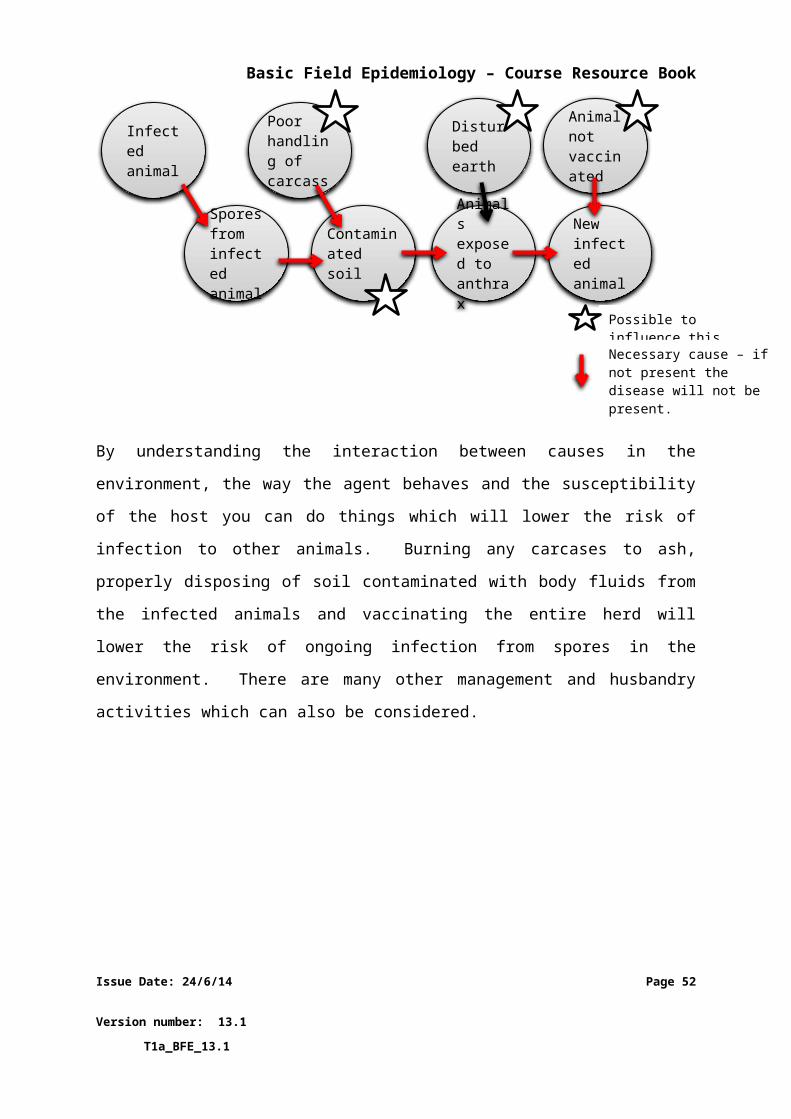
Basic Field Epidemiology – Course Resource Book
For example:
The figure below shows a simulation of a contagious disease being introduced
into a susceptible population. The lines represent the number of animals
that are susceptiblethat are infected and showing clinical signs
that are recovered
Pattern of distribution of susceptible, clinically infected and recovered
animals in a population following introduction of a contagious disease
1 2 3 4 5 6 7 8 9 10 11 12 13 14 150
100
200
300
400
500
600
Susceptible Recovered Clinical
Time (days)
Num
ber o
f ani
mal
s
In this example there are 500 animals in the population at the beginning; all
500 animals are susceptible, there are no clinical infections, and no recovered
Issue Date: 24/6/14 Page 52
Version number: 13.1 T1a_BFE_13.1
Figure 10 Figure showing number of animals in each of three groups (susceptible, infected or clinical, and recovered) following introduction of a contagious disease into a population of 500 animals
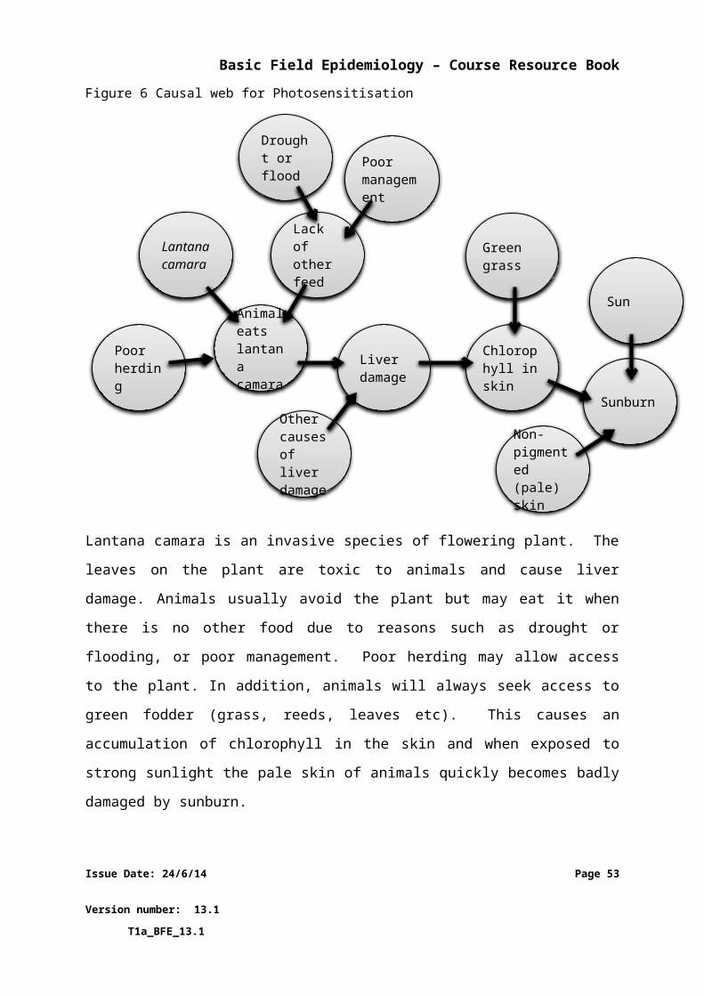
Basic Field Epidemiology – Course Resource Book
animals. This means the disease is not present within the population in the
beginning.
When the contagious disease is introduced, initially there will be a small
number of infected animals. Over time these animals will infect others and the
disease will spread within the population. This is shown by the rise in clinical
animals (infected animals with clinical signs) and the fall in susceptible animals.
After some time the clinical animals will recover from the disease. The final
pattern will depend on how contagious the disease is and how severe it is. If
the disease became endemic there would be a constant mixture of susceptible,
clinical and recovered animals. The number within these categories will move
up and down over time depending on all the causes that influence if disease
will occur.
Issue Date: 24/6/14 Page 53
Version number: 13.1 T1a_BFE_13.1
Basic Field Epidemiology – Course Resource Book
6 Transmission and spread of diseasesIn order for infectious diseases to continue to occur, infection has to be transmitted from an
infected animal to a susceptible animal. If this does not occur, the disease will disappear.
Transmission describes how an infectious agent can move from one animal to another
Maintenance refers to how an infectious agent can survive or continue to occur in a
population over time
Spread describes how an infectious agent moves from one population to another
In order for transmission to occur, an infectious agent has to leave the infected animal and
enter a susceptible animal.
Infectious agents can leave the host in different ways. Examples include:
Body surface – agent in hair, pus or scabs (ringworm)
Nose secretions – (influenza virus)
Mouth – saliva (rabies and foot and mouth viruses, tuberculosis)
Mammary gland – milk (streptococcus bacteria)
Anus – faeces (salmonella bacteria)
Urogenital tract – urine and sperm (leptospirosis, campylobacter)
Eyes – tears (pinkeye)
Blood – (Q fever by tick vectors)
Issue Date: 24/6/14 Page 54
Version number: 13.1 T1a_BFE_13.1
Transmission
Infected animal Susceptible animal
Figure 11 Transmission of an infectious agent from an infected host to a susceptible host
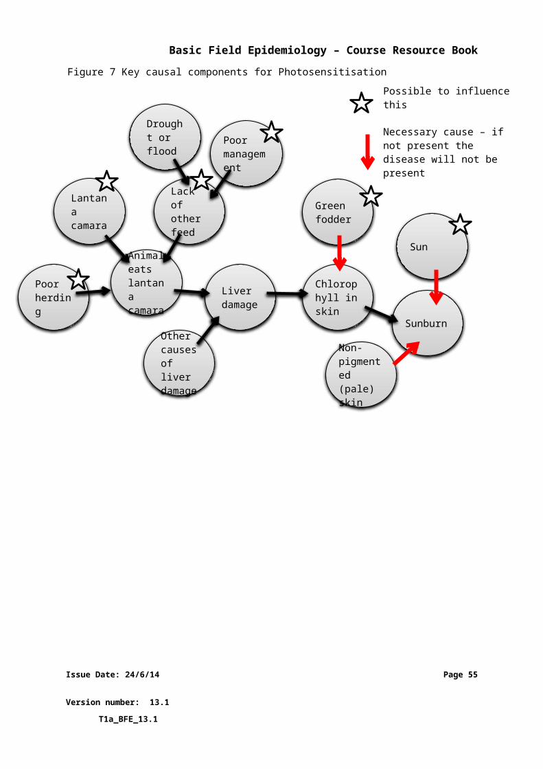
Basic Field Epidemiology – Course Resource Book
Infectious agents can enter the new host by different ways. Possible ways to enter a host
include:
Orally – by being eaten (many parasites/worms survive on grass or feed)
Respiratory – through breathing the agent into the nose, throat and airways,
(avian influenza virus, bacteria causing pneumonia).
Mucous membranes – through contact with the eye, mouth, or nose. (Pinkeye)
Skin - through intact skin (ringworm) or through damaged skin (human anthrax)
or through biting insects (bluetongue) or through animal bites (rabies)
Coitus - transmitted when animals breed (vibriosis in cattle)
Iatrogenic – through some management or veterinary procedure (surgery,
vaccination, dehorning, castration, injections) Bluetongue in livestock and
HIV/AIDS or Hepatitis in humans
Transmission – For disease to get from one animal to another the infectious agent must be
transmitted between the animals. This can occur:
Directly where an infectious agent is passed from one animal to another by:
- animal-to-animal contact
- an animal contacting infected discharge from another animal (nasal
secretions, urine, faeces etc.)
- infection of the embryo in the pregnant uterus (mammals) or of the egg
(birds, reptiles, amphibians, fish and arthropods), or through the milk
from dam to offspring
Indirectly where the infectious agent is passed from one animal to another via
other living and non-living things.
Issue Date: 24/6/14 Page 55
Version number: 13.1 T1a_BFE_13.1
Basic Field Epidemiology – Course Resource Book
- Living things (intermediate host) that can transmit some infectious
diseases include insects such as mosquitoes in spreading Malaria between
humans or ticks in spreading Babesia between cattle and many other
animals or insects.
- Non-living things that can transmit some infectious agents include feed,
equipment, clothing, needles and syringes.
Spread - Common methods for an infectious agent to spread between populations of animals include:
Movement of infected animals into a new population
Movement of contaminated materials
Spread via contaminated equipment such as veterinary, farming, etc.
Movement of vectors such as ticks, birds, wild animals between populations
Airborne spread of bacteria, viruses (foot and mouth disease), or fungi from one population to another
Maintenance - Infectious agents need to survive over time or the infection will disappear.
Infectious agents have developed different ways to survive either in animals or the
environment. These include:
developing a resistant form that can survive for a long time in the environment
(anthrax spores, helminth eggs, toxoplasma cysts)
continual transmission to susceptible hosts through:
- rapid infections that cause little immune response
- or by altering their genetic makeup to avoid any immunity that developed
against previous forms
forming persistent infections in host animals (Johnes disease, tapeworm, maedi-
visna virus, BVD)
Issue Date: 24/6/14 Page 56
Version number: 13.1 T1a_BFE_13.1
Basic Field Epidemiology – Course Resource Book
developing the capacity to infect multiple different species to increase likelihood
of continual transmission (Nipah virus infects bats, pigs, humans, rabies infects all
mammals)
avoidance of any external or environmental forms as a way of avoiding dying in
the external environment (trichinella species)
The following two figures provide examples of transmission and maintenance for some diseases using life cycle diagrams.
Figure 12 The trichostrongylus spp. life cycle in sheep and cattle.
The Trichostrongylus spp. life cycle includes the following steps.
1. Eggs passed in faeces
2. 1st stage larvae hatch from eggs in faeces
3. 1st and 2nd stage larvae feed on bacteria in faecal pallets
4. 3rd stage larvae is protected from drying out by its outer shell
5. 3rd stage larvae move out of faeces and onto grass. The free living stage in the environment of the life cycle can take from 2 -12 weeks to complete
Issue Date: 24/6/14 Page 57
Version number: 13.1 T1a_BFE_13.1
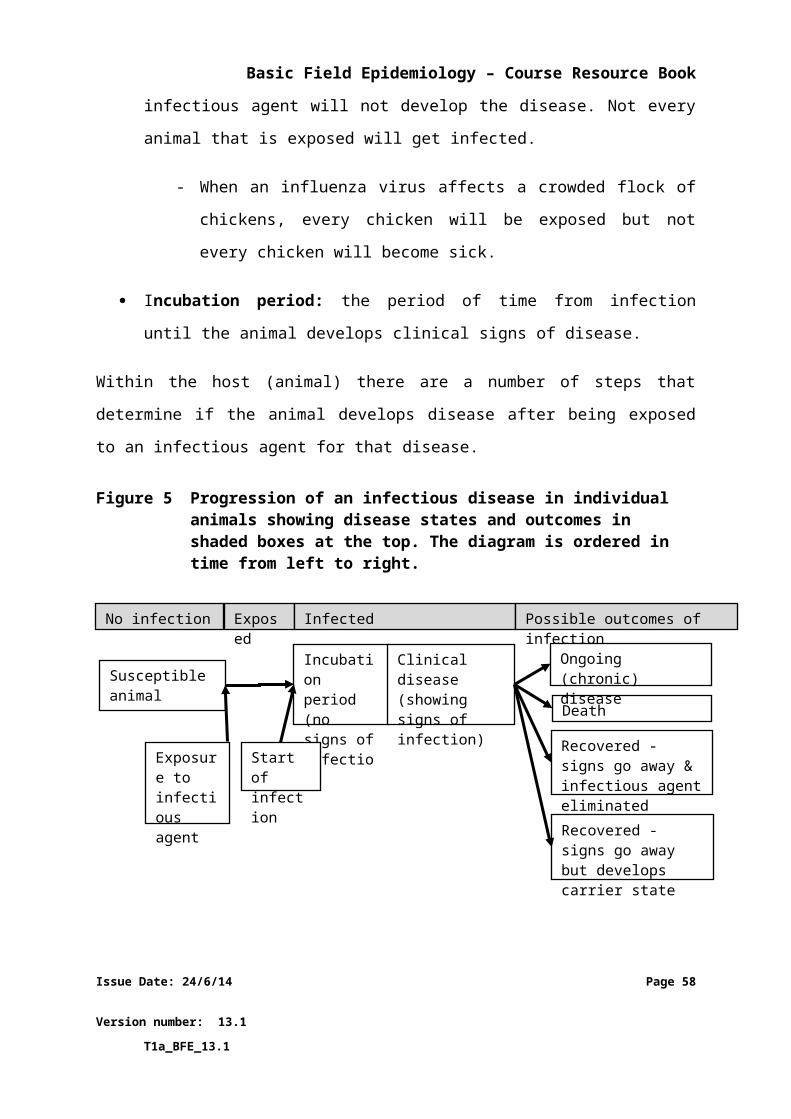
Basic Field Epidemiology – Course Resource Book
6. Sheep eat grass and 3rd stage larvae. These moult in the small intestine to 4th stage larvae which then become adult worms. Adult worm produce eggs which are passed in the faeces. The host stage of the life cycle can take 18-21 days to complete
Figure 13 Anthrax life cycle
The anthrax life cycle includes the following steps:
1. Anthrax spores are ingested, inhaled, or contacted by the host (human or animal)
2. Disease and death occur in the host
3. Vegetative cells are released into the environment following death of the host
4. Anthrax infection can occur from contact with animals that have died from anthrax
5. Vegetative cells in the environment turn into spores and remain infective to hosts for many years
Issue Date: 24/6/14 Page 58
Version number: 13.1 T1a_BFE_13.1
Basic Field Epidemiology – Course Resource Book
7 Using a field epidemiology approach to larger disease investigations
Field epidemiology skills are important in all disease investigations even when only one
animal or one farm is affected.
The epidemiologic approach to disease investigation becomes more and more important
where:
there are larger numbers of animals are affected
the disease is spreading rapidly
the causes of the disease are uncertain (affected animals displaying signs that are
different to commonly seen diseases or more severe)
Examples of these situations include outbreaks of exotic diseases or new diseases or where
a common disease has changed so that it spreads more rapidly or has a higher mortality
rate. In these situations, an epidemiological investigation is likely to provide the best
approach to understand possible causes and identify control measures for the disease.
The advantage of an epidemiological approach is even when you do not know what the
disease is or what the specific infectious agent(s) might be you can often draw conclusions
about:
the likely causes of the disease
identify possible control measures
Larger disease events often involve one of the following situations:
1. Where the causes of the disease are known
There may be interest in trying to understand how or why the disease occurred in
this a particular event so control measures can be put in place. The agent causing the
Issue Date: 24/6/14 Page 59
Version number: 13.1 T1a_BFE_13.1
Basic Field Epidemiology – Course Resource Book
outbreak will be known or identified early in the investigation. In these situations the
investigation is directed at identifying causes that contributed to this particular
occurrence of the disease.
2. Where the causes of the disease are not known
For new disease or exotic disease the investigation is directed at identifying causes
and possible control measures. The diagnosis is important but may not occur until
after initial control measures are implemented.
For example:
The initial investigation of Mad Cow Disease or Bovine Spongiform
Encephalopathy (BSE) in the United Kingdom suggested that the disease was
likely to have been spread through contamination of cattle feed products with
meat and bone meal. Preventative measures involving a ban of feeding
ruminant-derived protein were identified from initial epidemiologic
investigations and put in place well before the agent was identified. Years later
additional research identified the causal agent (a prion) and confirmed that the
initial recommendations were effective in preventing further spread of the
disease.
An epidemiologic disease investigation follows a systematic approach, and includes the
following activities.
1. Develop a case definition and assign animals to ‘cases’ or ‘non-cases’
2. Collect data on cases and non-cases
3. Apply simple analyses to the data collected to describe the patterns of disease and
identify possible causes
4. Describe initial findings and make recommendations
Each of these four steps will be described in more detail in the following sections.
Issue Date: 24/6/14 Page 60
Version number: 13.1 T1a_BFE_13.1
Basic Field Epidemiology – Course Resource Book
NOTE: It is likely that epidemiological investigations will involve a district veterinarian
working with para-veterinarians to plan and conduct the investigation. Para-
veterinarians need to understand the general approach and the types of data and
information that need to be collected.
7.1Describing cases and non-cases
A case definition is a set of standard criteria for deciding whether an individual animal has a
particular disease or other aspect of interest.
A case is an animal with characteristics and clinical signs that meet a case definition for the
disease being investigated
This step involves first completing the initial investigation (Section 3) to describe signs or
symptoms of animals that are affected by the disease of interest. This involves examination
of sick animals as well as animals that are not sick, i.e. unaffected animals. By comparing
affected and unaffected animals we can develop a case definition.
It is often useful to have 3 separate levels of a case definition:
1. confirmed case where all the criteria for the case are met
2. suspect case where some but not all the criteria are met
3. non-case where the animal can confidently be declared as not meeting either the suspect or confirmed criteria
For field investigation of an unusual disease it may be best to start with a broad and non-
specific case definition. This may mean that some non-cases will be classified as cases.
However, it should mean that few true cases are missed and wrongly classified as non-cases.
Once a case definition is developed, all sick animals are compared to the case definition and
assigned to one of the levels (confirmed case, suspect case, or non-case). Case definitions
are used so information collected on cases and non-cases can be compared to look for
associations that identify potential causes.
Issue Date: 24/6/14 Page 61
Version number: 13.1 T1a_BFE_13.1
Basic Field Epidemiology – Course Resource Book
For example:
Case definitions that might be used when investigating mastitis in dairy cows
Study Unit Case definition
Animal A cow with visible clots in the milk
Animal A cow with a milk cell count greater than 200,000
cells/ml
In some cases of mastitis there are few or no visible clots in affected milk. Thus,
the first case definition will result in some false negative results. In any test
system, there is always a trade-off between false positive and false negative
results.
Case definitions are important in epidemiological investigation as a population of animals
may have many different diseases occurring at once. Only some of the sick animals may be
affected with the disease that is being investigated.
Imagine if you are investigating a disease outbreak where there were several
cases of sudden death due to a new disease. But there might also be 1-2 cases
of death due to other common diseases - one animal died of acute mastitis and
another died from a twisted stomach.
It will be important to examine all the dead animals and develop a case
definition for the new disease. This should allow the 2 animals that died from
well-known diseases (mastitis and twisted stomach) to be identified and
removed from the group of animals with the new disease.
Issue Date: 24/6/14 Page 62
Version number: 13.1 T1a_BFE_13.1
Basic Field Epidemiology – Course Resource Book
A case definition will allow you to identify and separate information about different diseases
that may be occurring in the population. It also helps keep the disease investigation focused
on the disease of interest.
Pak Paimin starts to think that this case may need a more epidemiological
approach. He starts to put all the information together that he has found out
so far.
He thinks about the cow that was lame, the 2 cows with diarrhoea and the calf
that Budi was given who was sick. Pak Paimin needs to go over the dates of
when all the animals got sick from his diary and put a time line together. The
only cows to get sick were down stream of the pen where the sick calf would
have been kept.
Pak Paimin decides on a case definition for this stage of the investigation:
Any cow (or calf) that has watery diarrhoea and temperature above 39°C
Issue Date: 24/6/14 Page 63
Version number: 13.1 T1a_BFE_13.1
Basic Field Epidemiology – Course Resource Book
8 Collecting data and counting cases To collect data on cases and non-case you will need to ask the farmer questions about their
animals.
For example:
When did the first case of this disease become apparent?
Is it possible to get the date when each affected animal first showed clinical
signs?
Where were animals when they got sick and where were they in the days
to weeks before they got sick?
Is it possible to collect information on all the animals on the affected farm
(or that mix together in a village population) over the days or weeks before
cases started to occur?
Have there been any animal movements into this group or out of this
group?
Were any treatments given to animals (what was administered and when)?
Were there any other changes (change of feed, chemicals dumped in the
paddock or river, new fence erected, etc)?
Data can also be collected from making direct observations of signs of disease from animals
or from laboratory results.
Pak Paimin thinks about the additional important information he needs. He will
ring Budi and ask him what was happening during the 2 weeks before these
animals became sick and where the calf came from.
Issue Date: 24/6/14 Page 64
Version number: 13.1 T1a_BFE_13.1
Basic Field Epidemiology – Course Resource Book
Budi finally tells Pak Paimin that he was away from the farm for 10 days and
came back 4 days before the animals got sick. His brother in law was looking
after the farm and he was in a bad mood the whole time. Budi is embarrassed
because he is sure that his brother in law caused the cow to get an injury and
go lame, he is also sure that he caused his animals a lot of stress.
The calf was given to him from a friend, Soleh, who is from another village. The
calf had been sick for a long time and that was why he gave it to Budi.
Now Pak Paimin must ring Soleh and find out some information from him. But
first he logs onto iSIKHNAS to see if there are any reports of sickness or
diarrhoea from that village or area.
Issue Date: 24/6/14 Page 65
Version number: 13.1 T1a_BFE_13.1
Basic Field Epidemiology – Course Resource Book
9 Making sense of the information you collect
This step then involves arranging and analysing the data that has been collected to try and
describe the disease and identify possible causes.
If it is not possible to identify a diagnosis, then it is best to try and determine:
the body system(s) that may be affected
if the disease is likely to be infectious or non-infections
if it may be spreading from one animal to another (contagious)
The following types of activities may be completed to help answer the above questions:
1. Describe the patterns of disease in terms of time, place, and animal
2. Arrange and analyse data looking for associations between disease and possible
causes
3. Developing ideas on possible diseases, causes, and control measures
9.1Describing patterns of disease
To describe patterns of disease we need to count cases (animals with disease) and non-cases
(animals without the disease) on the affected farms.
Epidemiology studies patterns of disease occurrence. To find the patterns we need to count
cases and non-cases. These counts can be used to describe disease occurrence in terms of
time, place, and animal. Doing this will help us understand the disease, its impact, and
explain how and why the disease occurred. This will help develop control strategies.
Issue Date: 24/6/14 Page 66
Version number: 13.1 T1a_BFE_13.1
Basic Field Epidemiology – Course Resource Book
Time
Describing disease occurrence in time is usually done using epidemic curves. An epidemic
curve is a plot of the number of new cases of disease over time.
For example:
The epidemic curve below shows time in days on the x-axis (horizontal) and a
count of new cases on the y-axis (vertical).
1 3 5 7 9 11 13 15 17 19 21 23 25 27 290
20
40
60
80
100
120
Time (days)
Num
ber o
f cas
es
This is a very common way of describing the pattern of disease occurrence. Epidemic curves
do not require information on non-cases.
An epidemic curve can be produced from counts of cases and the time of onset of clinical
signs. This is generally the day or date when clinical signs were first detected. Epidemic
curves can provide a lot of information about the disease under investigation.
For example:
Epidemic curves can provide information about the type of disease and or type
of exposure.
Issue Date: 24/6/14 Page 67
Version number: 13.1 T1a_BFE_13.1
Basic Field Epidemiology – Course Resource Book
Highly contagious diseases have an exponential pattern with a steep rise
followed by a progressive decline as susceptible animals are exhausted in
the population.
1 3 5 7 9 11 13 15 17 19 21 23 25 27 290
20
40
60
80
100
120
Time (days)
Num
ber o
f cas
es
A disease outbreak associated with point exposure of animals (single
exposure at a point in time e.g. toxic spill or food poisoning at a picnic) will
show a sharp rise in cases and then a sharp fall.
1 3 5 7 9 11 13 15 17 19 210
20
40
60
80
100
120
Time (days)
Num
ber o
f cas
es
Endemic diseases that are not very contagious will show a fairly level
pattern with some small peaks and troughs but no large rises.
Issue Date: 24/6/14 Page 68
Version number: 13.1 T1a_BFE_13.1
Basic Field Epidemiology – Course Resource Book
1 2 3 4 5 6 7 8 9 1011121314151617181920212223242526272829300
20
40
60
80
100
120
Time (days)
Num
ber o
f cas
es
Place
Describing disease occurrence by place is usually done by mapping. Some highly skilled
epidemiologists may often turn to computer based mapping software. However simple,
hand-drawn maps remain very useful tools to summarise disease occurrence.
Figure 14 Example of a simple map generated by iSIKHNAS to help you monitor where disease is occurring
Issue Date: 24/6/14 Page 69
Version number: 13.1 T1a_BFE_13.1
Basic Field Epidemiology – Course Resource Book
Animal
Describing disease occurrence by animal is usually done by arranging counts of cases by
some host characteristic. Common characteristics used to describe disease occurrence by
animal are:
age group
breed
species
sex
body condition
vaccination status
This may also include descriptions of outcomes such as; the number of animals that were
infected, the number that have died, and the number that have recovered.
Pak Paimin finds there have been lots of reported diarrhoea cases in the village
where the calf came from and also in the next Kabupaten where the drain runs
to from Budi farm. Pak Paimin alerts the DINAS vet and together they create
the below epidemic curve and plan a more detailed epidemiological
investigation.
10/1/2
013
10/3/2
013
10/5/2
013
10/7/2
013
10/9/2
013
10/11/2
013
10/13/2
013
10/15/2
013
10/17/2
013
10/19/2
013
10/21/2
0130
2
4
6
8
10
12
14
16
Date
Num
ber o
f cas
es
Issue Date: 24/6/14 Page 70
Version number: 13.1 T1a_BFE_13.1
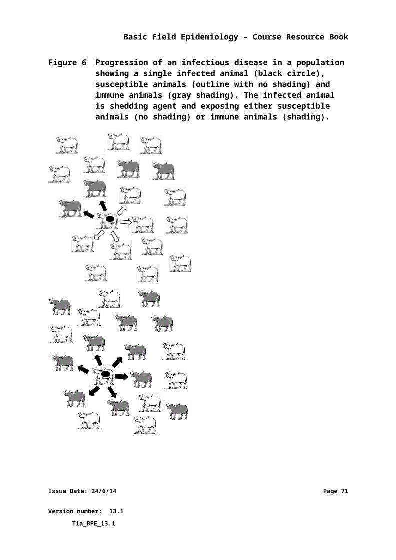
Basic Field Epidemiology – Course Resource Book
9.2Arranging and analysing data to look for associations
Proportions are often used to express disease occurrence. Proportions can be easier to
understand and sometimes more useful than whole numbers. To calculate a proportion you
will need to count two different groups of animals:
Number of cases of disease, either as:
- The number of new cases over a period of time, or
- The number of cases at a point in time (this method possibly includes old
cases as well as new cases)
The number of cases of disease forms the numerator (the number on top) in the
calculation of proportion.
Population-at-risk
- The number of animals that may be considered eligible to get infected with
the disease. The population-at-risk should include all those animals in the
area of interest (farm, village, district, province etc.) that could get the
disease. This may not include every animal.
In an outbreak of Milk Fever (Calcium deficiency) in cattle, the population-at-
risk is all pregnant female cattle on the farm. Male cattle, and non-pregnant
females are not part of the population-at-risk.
The population-at-risk for retained placenta would be all cows and heifers
which calve in the herd; for porcine parvovirus, the population-at-risk would be
serologically negative gilts and sows.
The population-at-risk forms the denominator (the number on the bottom) in the
calculation of a proportion.
Issue Date: 24/6/14 Page 71
Version number: 13.1 T1a_BFE_13.1
Basic Field Epidemiology – Course Resource Book
Proportion= numeratordenominator
= number of casespopulation−at−risk
Describing disease occurrence using proportions may help identify differences in
proportions that may indicate a cause of disease. Looking at data in this way can help
describe patterns of disease in more detail and identify possible causes of disease.
Often it is useful to describe the proportion or percentage of animals that are affected
based on other characteristics associated with the host, agent, or environment.
For example:
Some characteristics that are useful to arrange the data by:
Unit of study Some relevant characteristics to compare
Animal, cage, mob, paddock
species, sex, age, breed, weight, size, soil type, stocking density, stage of production, available feed, water source
Farm location, size, source of stock, source of feed and water, production method, other business
Village location, number of farms, farm types, geography, climate
District location, size, government services, geography, climate
Data on cases and non-cases may show that most cases occurred in one
paddock or in one age-group of animals.
The proportion of animals developing disease can be arranged using other characteristics to
look for patterns that may offer clues about causes of disease. If the proportion of animals
developing disease is much higher in young animals than in adult animals then this may
suggest that the disease is more likely to infect younger animals. This information may allow
the para-vet to identify some possible diseases to add to the differential diagnosis list.
Issue Date: 24/6/14 Page 72
Version number: 13.1 T1a_BFE_13.1
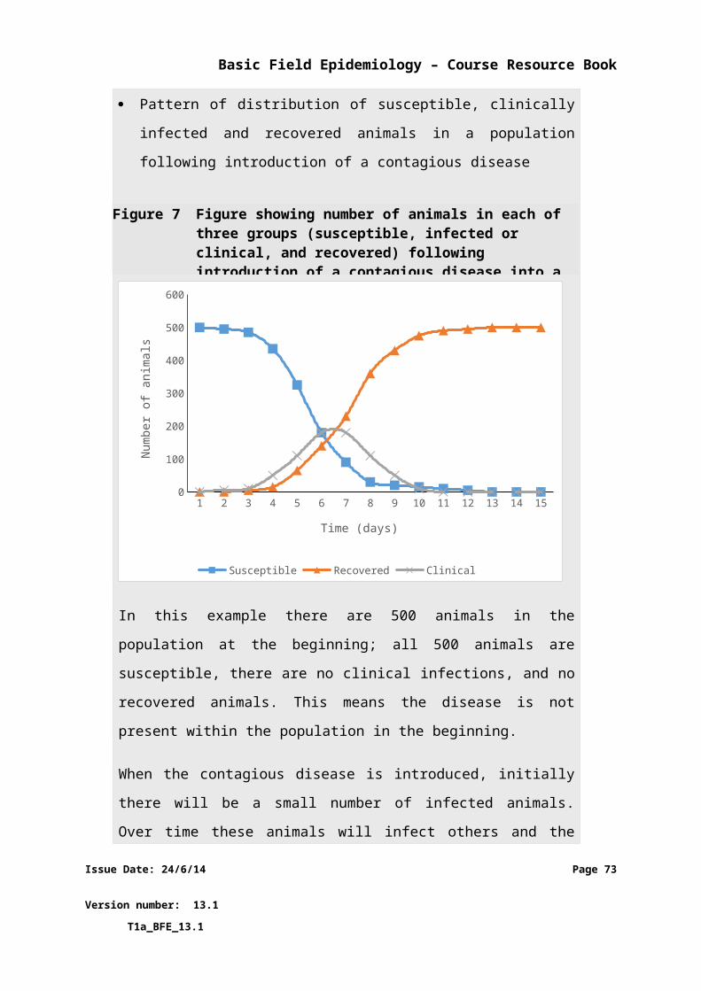
Basic Field Epidemiology – Course Resource Book
Attack rates (AR) can be used to compare proportions between different levels of some
other characteristic. Attack rates require a numerator (number of cases arranged by each
characteristic) and a denominator (total number of animals on the affected farm arranged
by each characteristic).
Pak Paimin and the Drh. Agung DINAS vet find the case and population data
out of iSIKHNAS for the two affected Kabupaten.
They make the following calculations:
For Kabupaten 1, AR1 = Cases of diarrhoea in cattle
Total cattle in Kabupaten 1
For Kabupaten 2, AR2 = Cases of diarrhoea in cattle
Total cattle in Kabupaten 2
Pak Paimin and Drh. Agung think during the time period the disease problem
has been occurring, there are 100 cattle and 10 cases of diarrhoea in
Kabupaten 1, and 300 cattle and 84 cases of diarrhoea in Kabupaten 2.
Issue Date: 24/6/14 Page 73
Version number: 13.1 T1a_BFE_13.1
Basic Field Epidemiology – Course Resource Book
The initial data analysis from the epidemiological investigation that started
with disease on Budi’s farm:
Count of cattle Attack Rate
Kabupaten Cases Total AR
1
2
10
84
100
300
10%
28%
In the above table, attack rates are expressed as percentages.
There are 100 cows in Kabupaten 1 and 10 of these are classified as cases,
leading to an attack rate of 10/100= 0.1 or 10%. There are 300 cows in
Kabupaten 1 and 84 of these are classified as cases, leading to an attack rate of
84/300= 0.28 or 28%.
Looking at attack rates for the different Kabupaten suggests that cattle in
Kabupaten 2 are more likely to get diarrhoea than cattle in Kabupaten 1.
Dividing one AR by another AR produces a measure of the relative risk (RR).
Cattle in Kabupaten 2 have a 2.8-fold higher risk of diarrhoea than cattle in
Kabupaten 1 (RR=28%/10%).
In this example the cattle in Kabupaten 2 are more likely to have diarrhoea.
From our investigation we know that the cases on Budi’s farm were some of
the very first cases of diarrhoea reported over this time. From the mapping of
cases we know that most of the cases are very close to the drain that runs from
Budi’s farm.
Issue Date: 24/6/14 Page 74
Version number: 13.1 T1a_BFE_13.1
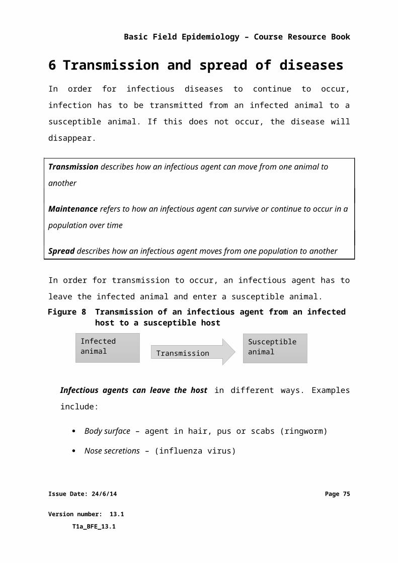
Basic Field Epidemiology – Course Resource Book
This finding could lend support to the idea that cattle down stream of this drain
are more likely to develop diarrhoea and that the drain is somehow involved in
the spread of disease.
More detailed analyses of epidemiologic data are possible and may be important to identify
possible causes. More detailed analyses are beyond the scope of this Basic Field
Epidemiology Resource Book. Additional information is provided in the Advanced Field
Epidemiology Resource Book.
9.3Developing disease control strategiesWhen investigating a disease it is important to develop control strategies quickly to
minimise the number of animals that are affected. This is very important for diseases that
have a high mortality rate or spread rapidly.
It is often important to implement some control measures as the initial investigation is
finishing and before the para-vet (or veterinarian) leaves the farm. Even in situations where
you may have multiple differential diagnoses or when you may not be certain of what the
causes might be, it is usually possible to put into practice some useful control measures
before leaving the farm. Sometimes additional investigations may be done or laboratory
tests performed to try and identify possible causes and work towards a definitive diagnosis
and in that case the control measures may change as you gather additional information.
Examples of possible control measures that might be put in place during the initial
investigation include:
removing susceptible animals to a new area may control the disease if the
investigation suggests the cause of disease is a point source exposure such as a
poisonous plant – Movement of hosts
improving farm management, husbandry, or feeding if the investigation suggests
that contamination, poor feed, or poor husbandry is a cause of disease – Animal
management
Issue Date: 24/6/14 Page 75
Version number: 13.1 T1a_BFE_13.1
Basic Field Epidemiology – Course Resource Book
If the investigation suggests an infectious agent that may be contagious then initial
strategies should include:
treatment of affected animals – Animal treatments
stopping animals and people moving onto and off the affected farm – Control of
animal movements
separating the affected animals from unaffected animals – Isolation
using and promoting good hygiene, disinfection, and other management strategies –
Biosecurity
In many cases it is necessary to put some initial control strategies in place, then conduct
further investigations to identify possible causes. As more information is available then
treatments and prevention can be modified.
It is expected that District Veterinarians will be involved in the planning and interpretation
of detailed epidemiologic investigations.
Some diseases will have pre-planned national control programs that are implemented when
the disease is either suspected or confirmed. This sort of control is usually limited to specific
diseases of importance where there is a policy of control or eradication (priority diseases
and exotic disease of importance). Often a large amount is known about these diseases. This
sort of control program often involves national legislative support and animal health policies
that define what actions should be taken to investigate and control the diseases. Additional
strategies that can be used in these types of control programs include:
separating infected and suspect animals from susceptible animals as well as
preventing animals or humans from moving onto of off affected farms will help
reduce the spread and transmission of disease - Quarantine
killing diseased animals and possibly suspect animals, disposal of the carcasses and
other infectious material to reduce the number of sources of the infectious agent -
Issue Date: 24/6/14 Page 76
Version number: 13.1 T1a_BFE_13.1
Basic Field Epidemiology – Course Resource Book
Slaughter
using vaccination to increase the immunity against the disease to reduce the
number of susceptible animals – Vaccination
reducing the number of intermediate hosts (such as insects and wild animals) that
transmit the disease will help reduce the spread – Control of intermediate host
More detailed information on these matters is provided in the Advanced Field Epidemiology
Resource Manual.
Issue Date: 24/6/14 Page 77
Version number: 13.1 T1a_BFE_13.1
Basic Field Epidemiology – Course Resource Book
10 Application of epidemiological approach to routine cases
10.1 Field Epidemiology in day to day workThinking about the possible causes of disease, the host, the agent, and the environment
when looking at every sick animal will help para-veterinarians provide better services to
farmers for treatment and prevention of disease in animals. Para-vets should be involving
field epidemiology skills in all aspects of their every-day activities. The following examples
are intended to illustrate how field epidemiology skills can be useful in all sorts of activities
ranging from managing an individual sick animal to a larger scale disease outbreak.
Example 1
Case introduction
You visit a farm where a calf is sick and has stopped drinking.
Information collected during investigation
The mother of the calf has had poor milk The calves were all born in clean grassy paddocks
Para-veterinarian’s actions
You examine the calf, find an abscess on the navel, and you treat the calf.
You advise the farmer that causes for these abscesses include: dirty calving
environment, damp/wet conditions, exposure to bacteria, and lack of milk
(colostrum) in the first 6 hours.
You advise the farmer how to prevent further cases maintaining a clean
environment for calving and to help calves drink from their mothers in the first
6 hours of life.
Epidemiology context
Issue Date: 24/6/14 Page 78
Version number: 13.1 T1a_BFE_13.1
Basic Field Epidemiology – Course Resource Book
Epidemiology skills help you to think about how this case might have occurred –
multiple causes of disease (host, agent, and environment).
Knowing the common causes for this disease (the epidemiology of the disease)
allows you to provide the best advice on how to treat the individual sick calf and
even more importantly how the farmer can improve management to prevent further
cases of this diseases from occurring in the future.
Example 2
Case introduction
You visit a farm where there is a number of calves with abscesses on the navel
and a number of calves without abscesses.
Information collected during investigation
Some new cattle arrived 9 weeks ago The cows moved paddocks 9 weeks ago All the sick calves are between 6 and 8 weeks of age All the healthy calves are older than 10 weeks of age All the sick calves were born in a small yard that was very dirty All the healthy calves were born in clean grass paddocks
Para-veterinarian’s actions
You examine the calves and treat them.
You advise the farmer that causes for these abscesses include: dirty calving
environment, damp/wet conditions, exposure to bacteria, and lack of milk
(colostrum) in the first 6 hours.
You advise the farmer how to prevent further cases mainly by trying to have a
clean environment for calving. The small yard is a cause that can easily be
changed to prevent further cases. It is possible the new cows may have
brought a new bacteria with them because the older calves do not have the
disease. However, because the cows also changed paddocks it is hard to
determine if this is true.
Issue Date: 24/6/14 Page 79
Version number: 13.1 T1a_BFE_13.1
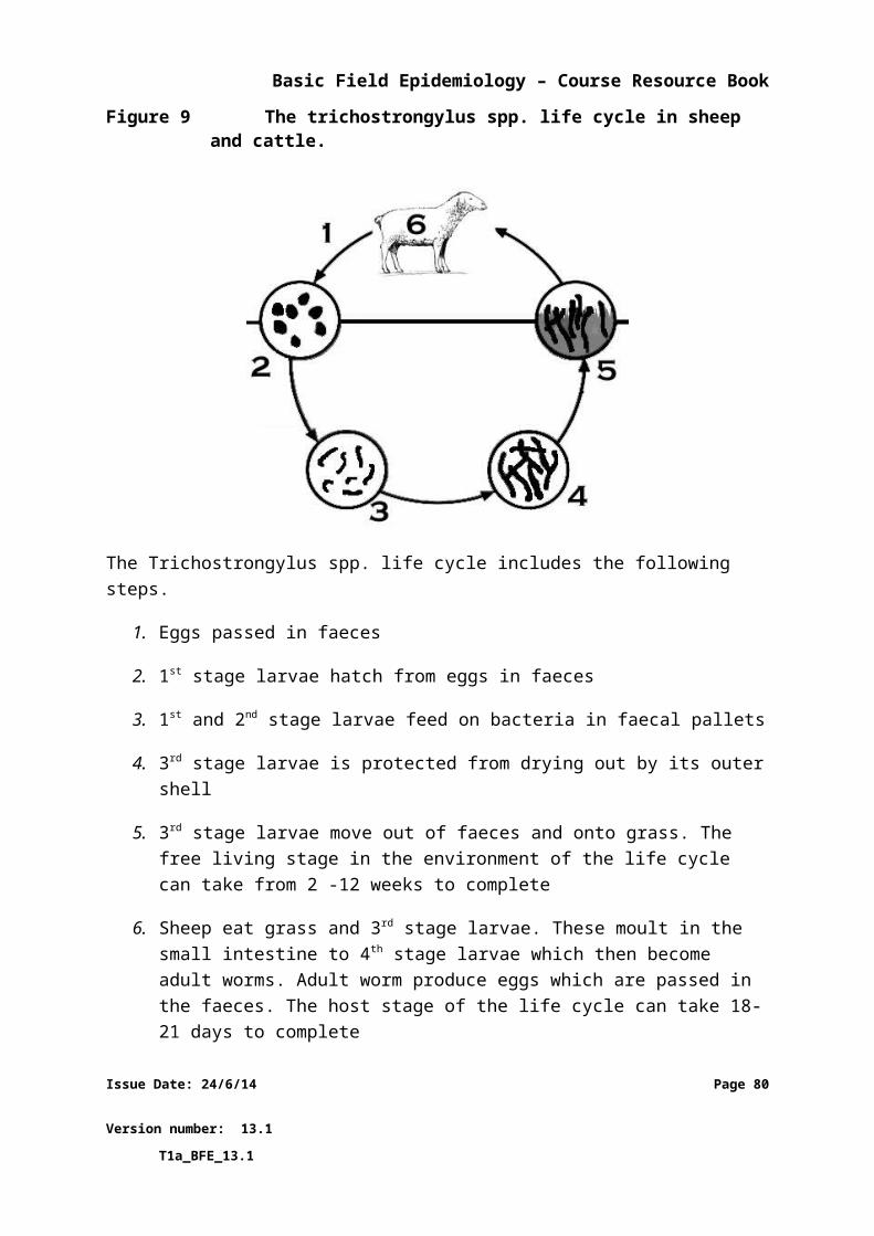
Basic Field Epidemiology – Course Resource Book
Epidemiology context
Epidemiology skills help us to think about the patterns of disease in the two groups
of calves (sick and healthy) and how these differences can be related to the likely
causes of disease.
We can use this information to provide the best treatment for affected calves and
advise the farmer about what he can do to reduce the amount of disease in the
future, providing long term benefit to the farmer that will improve animal health and
productivity and reduce costs and losses into the future.
Example 3
Case introduction
You visit a farm where there are 2 cows with diarrhoea.
Information collected during investigation
The 2 cows are both 2 year old Bali cows They have been kept separate from the other cows because they
recently calved All the cows on the farm were wormed 3 weeks ago. All they have to eat is short grass in the small paddock near the shed. There are no cows on the neighbouring properties. Both cows look weak, their eyes are sunken and they are depressed. They have very foul smelling watery diarrhoea that contains fresh blood They are dehydrated Their temperatures are increased above normal: 39.8° and 40.1°
Para-veterinarian’s actions
You examine the cows, take samples for laboratory investigation, and treat
them.
You advise the farmer that the infectious agents that may cause diarrhoea
(differential diagnoses) include: Bacterial infection of the intestines
(Salmonella, E-coli), Bovine Viral Diarrhoea Virus, Parasites. Due to the fever
Issue Date: 24/6/14 Page 80
Version number: 13.1 T1a_BFE_13.1
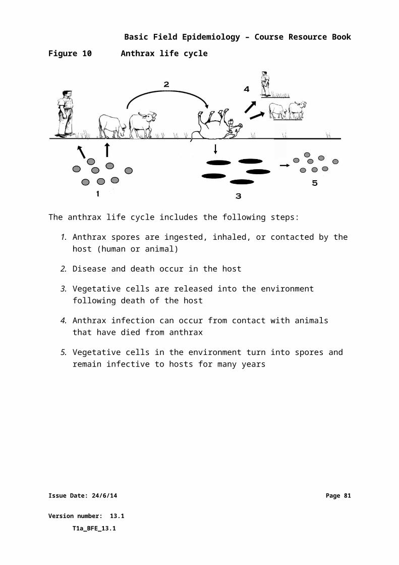
Basic Field Epidemiology – Course Resource Book
and severe illness and foul smelling diarrhoea a bacterial infection is most
likely.
You advise the farmer this may be a zoonotic disease. Everyone must be
careful and be sure to wash their hands before they eat or prepare food,
especially after coming in contact with the cows.
You advise the farmer how to prevent further cases:
Keep the 2 cows and the calf separate from the other cattle (isolate) Healthy cows are to be kept higher up stream along the drain line from
the 2 sick cows and the calf
Epidemiology context
Epidemiology skills help para-veterinarian to understand likely causes of disease and
use this information to provide the best advice for treatment of the two sick cows
and also for prevention of transmission of the disease to other animals on the farm.
Epidemiology skills also allows you to recognise when a zoonotic disease may be
present and to provide the best advice to people to help prevent humans getting the
disease.
Example 4
Case introduction
You visit a feedlot and cattle trader where there are cows and calves with
pinkeye.
Information collected during investigation
There are many cattle with pinkeye in one or both eyes The feedlot feeds very stalky grass in a large feed trough off the ground It is very dry, there has been no rain for some time There are big piles of dung close to the feedlot and there are many flies There is little shade in the feedlot There are always cattle coming and going but there is always pinkeye.
At the moment the feedlot is very full
Issue Date: 24/6/14 Page 81
Version number: 13.1 T1a_BFE_13.1
Basic Field Epidemiology – Course Resource Book
Para-veterinarian’s actions
You examine the animals and treat them.
You advise the farmer that causes of pinkeye include:
Young cattle Flies Dust Physical characteristics of the animal, eye conformation (bulging eyes),
lack of coloured (pigmented) skin around the eyes High sunlight (UV) exposure Crowding (high population density) Feeding long stalky grass
You advise the farmer that the infectious agent of pinkeye is Moraxella bovis.
Some strains cause more severe eye disease than other strains.
You provide treatment for the affected animals.
You advise the farmer how to prevent further cases:
Separate animals with pinkeye from the healthy animals (isolate), monitor the cattle and remove any newly affected animals as soon as they are noticed.
Move the piles of dung further away from the feedlot and try and reduce the number of flies.
If possible source another type of feed that is not long and stalky, consider modifying the feeding troughs so the feed is not standing upwards, and consider spraying dry feed to reduce dust.
Try and do cattle work in the mornings when there is less dust and sunlight. Consider watering the yards before working the cattle
Recognise that having more cattle in the feedlot will increase the occurrence of disease, particularly when other causes are present (dry, hot, dusty conditions). When multiple causes are present, preventive strategies should be implemented even if there are no cases in order to try and prevent new cases from occurring.
If vaccination is available and economically viable consider vaccination to reduce disease.
Do not bring new cattle that have pinkeye as you will introduce the infection to the animals in the feedlot. Try to avoid moving infected animals from the feedlot to other groups of cattle for the same reason.
Epidemiology context
Issue Date: 24/6/14 Page 82
Version number: 13.1 T1a_BFE_13.1
Basic Field Epidemiology – Course Resource Book
Epidemiology skills also help us think about the causes of disease and the methods
of spread and transmission the infectious agent uses to infect animals.
Knowing about the epidemiology of this disease will help you to provide the best
treatment for affected animals but even more importantly, field epidemiology skills
allow you to provide advice for preventing the disease from occurring.
Prevention is important when there are cases already occurring in order to minimise
the number of animals that get affected and require treatment. These are control
measures that are implemented in the face of an outbreak.
Finally, the trader can also act on your advice to put preventive strategies into place
when there is no pink eye occurring but when known causes of the disease are
present. This allows the trader to reduce the risk of disease occurring, which will
improve animal growth and profitability and reduce his costs into the future.
Example 5
Case introduction
You visit a chicken farm where there are some chickens that have drooping
heads, closed eyes, sneezing, and greenish diarrhoea and there has been
chicken deaths.
Information collected during investigation
this is mainly a problem in the young chickens the chickens are not eating and have little energy there were a few new birds that were sick when they arrived at the
farm 8 days ago there were a few sick chickens 3 or 4 days ago and now there are many there are many chickens in a small area and it is a bit dirty there are wild birds nesting in the roof
Para-veterinarian’s actions
You conduct some post-mortems on dead chickens and take samples from
these and from sick chickens for laboratory testing.
Issue Date: 24/6/14 Page 83
Version number: 13.1 T1a_BFE_13.1
Basic Field Epidemiology – Course Resource Book
You advise the farmer that causes of this disease could include:
Introduction of the disease into the farm by newly bought chickens Wild birds No vaccination Young age of chickens Crowding (high population density) Visitors with dirty clothes (contaminated with the infectious agent) Contaminated food or water
You advise the farmer that the infectious agent (differential diagnoses) could
be Newcastle disease, avian influenza (Highly pathogenic HPAI), duck plague,
acute poisoning, fowl cholera, and mycoplasmosis. The incubation period could
between 3 and 5 days. For your area the most likely infectious agent is
Newcastle disease.
You advise the owner that the chickens have clinical signs fit a priority
syndrome (MMU or sudden increase in mortality in chickens and other
poultry). This has to be reported to iSIKHNAS as MMU. Further actions
including government response activities and control strategies, may be
required if a priority disease is confirmed once laboratory results are obtained.
You advise the farmer this may be a zoonotic disease. Everyone must be really
careful to avoid very close contact with sick or dying birds, not to drink or wash
with water from the chicken shed and be sure to wash carefully after handling
birds and before eating or preparing food.
You advise the farmer how to prevent cases in general:
Don’t keep chicken too many chickens in a small area and keep the area clean
Keep chickens in good health – worm, vaccinate, provide good feed and water
Dispose of dead birds properly (bury or burn them) Separate sick birds from healthy birds Keep wild birds from coming into contact with your chickens Develop strategies to reduce the likelihood of introducing the disease
into your birds from new birds or from visitors – only bring healthy
Issue Date: 24/6/14 Page 84
Version number: 13.1 T1a_BFE_13.1
Basic Field Epidemiology – Course Resource Book
chickens onto your farm, separate new chickens for 10 days if possible to see if any signs of disease develop, and ensure that any visitors are clean and have not come straight from other poultry farms to your farm.
Epidemiology context
Epidemiology skills help para-veterinarian to understand likely causes of disease and
use this information to provide the best advice for treatment and prevention of
disease in other animals on the farm.
Field epidemiology and your knowledge of priority diseases and priority syndromes
and your training in iSIKHNAS allow you to report this case as a priority syndrome
and ensure that appropriate disease investigations are conducted to diagnose the
cause. If a priority disease (such as HPAI) is confirmed this will then lead to the
correct response actions as outlined in national policy.
Where the investigation identifies some other disease agent (such as Newcastle
disease) as the cause and rules out a priority disease, then routine disease control
measures will apply based on treatment and prevention measures for that disease.
This is an example of successful disease surveillance and indicates that the iSIKHNAS
system and the para-vet service is doing exactly what they should be doing –
investigating disease occurrence to identify the causes and act accordingly.
Epidemiology skills allow you to provide useful advice on future management of the
farm to prevent new infections being introduced.
Epidemiology skills also help you to think about possible zoonoses and provide
appropriate advice to the farmer and his family so they can minimise the chance of
getting sick themselves.
Issue Date: 24/6/14 Page 85
Version number: 13.1 T1a_BFE_13.1
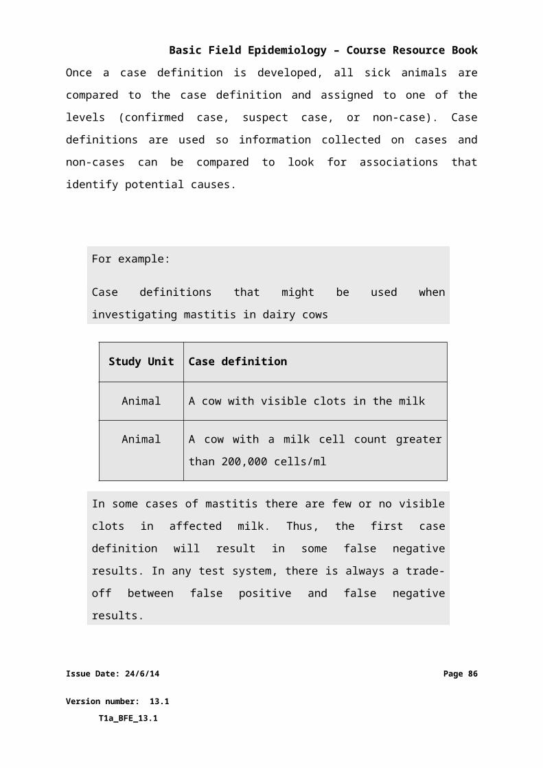
Basic Field Epidemiology – Course Resource Book
10.2 Field Epidemiology in priority disease control programs
Field epidemiology skills are very important for para-vets involved in priority disease control
programs. The understanding of causes and the effect of disease at a population level is very
helpful when thinking about why different strategies are used to control specific diseases,
and also in explaining processes to farmers.
Example 6
Case introduction
You visit a farm where there are dead cow with un-clotted blood oozing from
their orifices.
Information
There have been cases of anthrax on the same farm years ago There are 4 dead cows and another 15 live cows on the farm
Para-veterinarian’s actions
You examine the dead animals and take samples.
You advise the farmer that this is a priority syndrome (MTD - Sudden death
with blood from the orifices in cattle), and that you need to report this to
iSIKHNAS. There will most likely be a formal government investigation and
response. This is also a dangerous zoonotic disease.
You advise the farmer of initial control strategies that they can start to put into
place today to prevent further cases:
remove any live animals from the immediate area where the dead cattle are or from potential sources of anthrax - Animal management
consider treating live animals that have been in contact with the dead animals with penicillin to treat or prevent anthrax – Animal treatments
stop any new animals entering the farm or sick animals leaving - Control of animal movements
proper disposal of the carcasses of dead animals is very important, and all people should pay attention to hygiene when handling the dead
Issue Date: 24/6/14 Page 86
Version number: 13.1 T1a_BFE_13.1
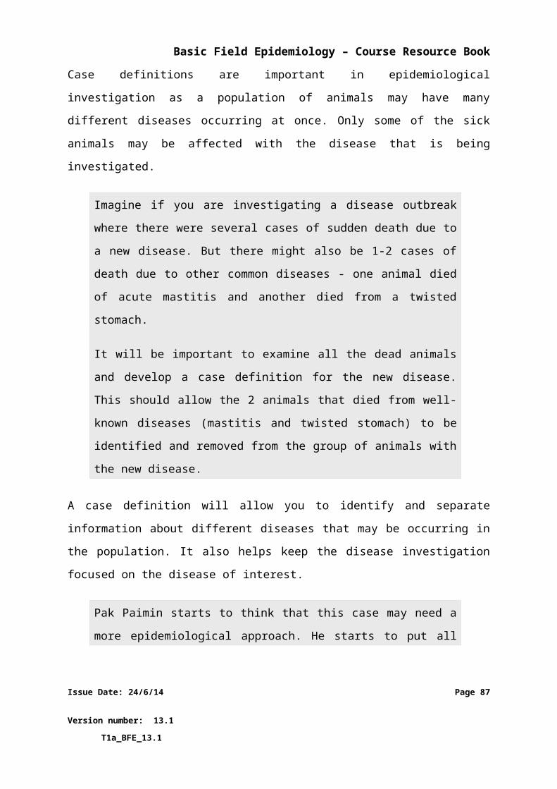
Basic Field Epidemiology – Course Resource Book
animals and the dead animals are not suitable for eating - Biosecurity
You advise the farmer that it is really important to control this disease because
it can last so long in the environment and can also kill humans. Additional
control strategies may be put into place once anthrax is confirmed including:
Separating animals and humans from potential sources of infection and preventing animals and sometime humans from going onto or off the farm - Quarantine
Conducting a vaccination program for live animals on the farm and possible surrounding farms or district - Vaccination
Epidemiology context
Epidemiology skills help us to understand causes of disease and think about control
strategies to prevent further cases.
Effective use of iSIKHNAS will help in rapid reporting of a priority syndrome and in
triggering an investigation with appropriate laboratory testing to confirm the
diagnosis of anthrax. This ensures that effective control measures can be put into
place as soon as possible and minimise the risk of additional animal deaths or
infection in humans.
With anthrax this is important as it can cause disease and death in humans. Some
control strategies can be implemented immediately to help prevent further cases.
Other control strategies need formal government direction such as quarantine or
more in depth planning and funding such as vaccination programs.
Example 7
Case introduction
A cow is reported to be drooling saliva on a nearby farm.
Information available
The cow has been examined and some lesions observed on the mouth and tongue that resemble ulcers and fluid filled blisters or vesicles.
Issue Date: 24/6/14 Page 87
Version number: 13.1 T1a_BFE_13.1
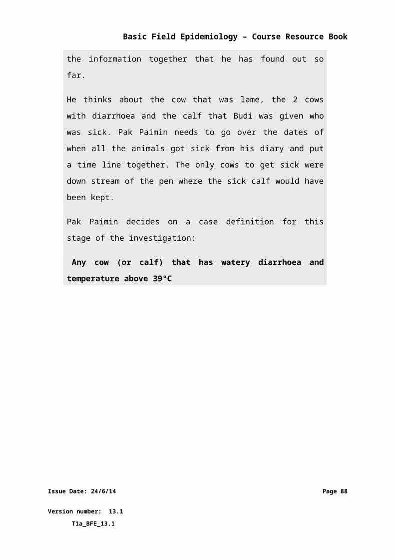
Basic Field Epidemiology – Course Resource Book
The case has been identified as a priority syndrome (PLL – limping, excessive salivation and vesicles on the mouth / foot / teat in cattle).
Samples were sent to the laboratory The case has been reported to iSIKHNAS and there has been a formal
government response involving DINAS veterinarians. The following control strategies have been implemented:
o Quarantine - locallyo Biosecurity
The IndovetPlan for foot-and-mouth disease outlines further control strategies if the laboratory result is positive. These may include
o Quarantine for larger areaso Slaughter programso Vaccination programs
The laboratory result and other information rule out foot-and-mouth disease for this particular case
Para-veterinarian’s actions
You ensure the community, especially local farmers, are aware of the serious
nature of FMD to Indonesia and that they understand why control measures
are being used before a confirmed case exists.
You need to communicate and celebrate with the community and the affected
farmer that FMD has been ruled out because of the testing that was done. This
will include having to explain why it was important to have initial control
measures in place before the test results were released, just as a precaution in
case the cow did have FMD. As soon as the negative test result is released the
control measures were removed.
Epidemiology context
Epidemiology skills help to understand signs, syndromes, differential diagnoses,
how a definitive diagnosis is reached, and the approach to disease investigation.
This combined with an understanding of causes, transmission, and spread of
diseases and knowledge of the need to recognise, investigate and control priority
diseases will allow you to explain why it is important to start control strategies
before a diagnosis is confirmed.
Issue Date: 24/6/14 Page 88
Version number: 13.1 T1a_BFE_13.1
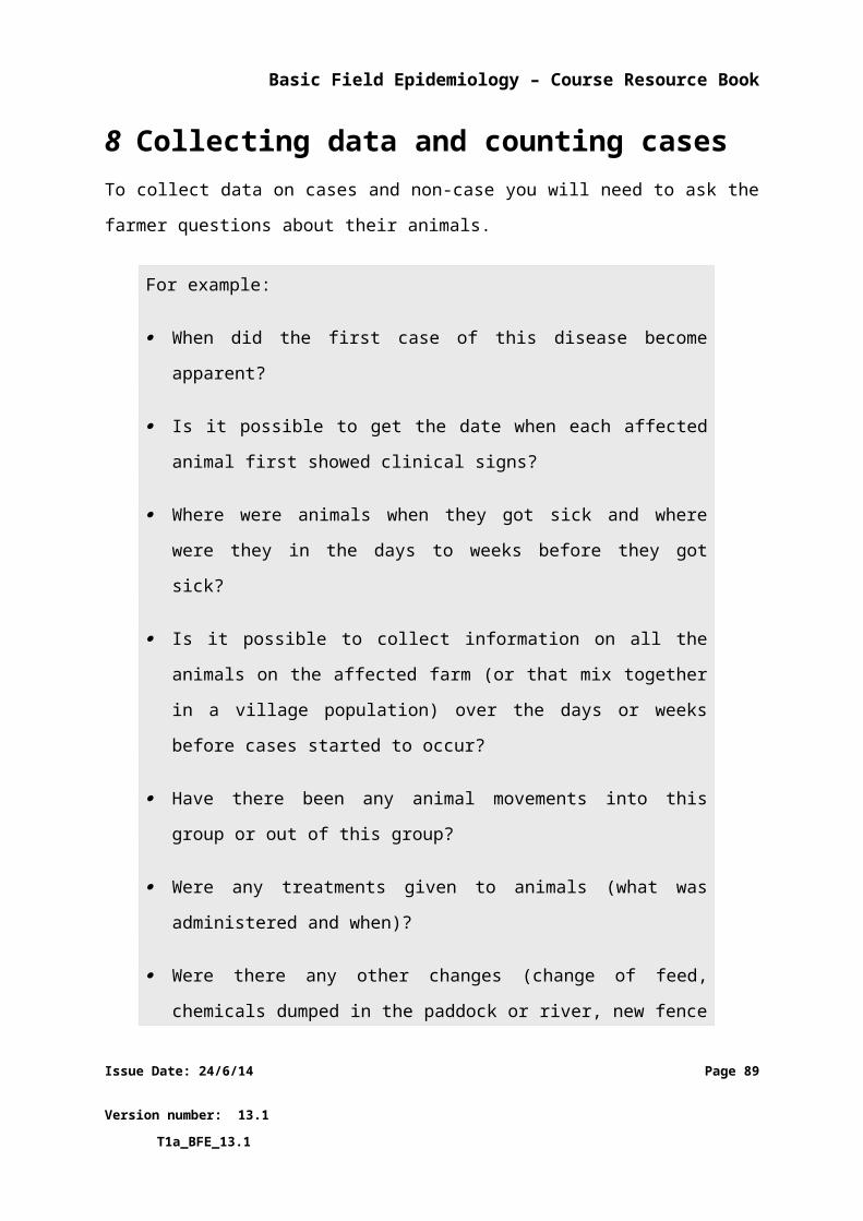
Basic Field Epidemiology – Course Resource Book
Effective use of iSIKHNAS ensures that the initial priority syndrome is reported
rapidly and that appropriate measures including laboratory testing and initial control
measures are carried out quickly to prevent any possible spread of disease just in
case it had been a priority disease such as FMD.
This is an example of successful disease surveillance and indicates that the iSIKHNAS
system and the para-vet service is doing exactly what they should be doing –
investigating disease occurrence to identify the causes and act accordingly.
Example 8
Case introduction
There has been a rabies control program running on this area for the last 2
years
Information available
In the last 2 years there have been 86 confirmed rabies cases in dogs and 3 cases in humans
The current vaccination program has been running for 18 months The current estimate of dogs in the area is 2000 The current estimate of vaccinated dogs is 1200 (60%) The program’s goal is to vaccinate 80% of dogs
Para-veterinarian’s actions
You ensure the community is educated about the risk of rabies, the control
program, and the importance to avoid being bitten by dogs.
You work with the dog catching and vaccination team. You need to ensure that
as many dogs are vaccinated as possible, they are given booster vaccination if
required, the vaccine is always stored at the right temperature, results of each
days vaccinations are entered into iSIKHNAS.
Epidemiology context
Issue Date: 24/6/14 Page 89
Version number: 13.1 T1a_BFE_13.1
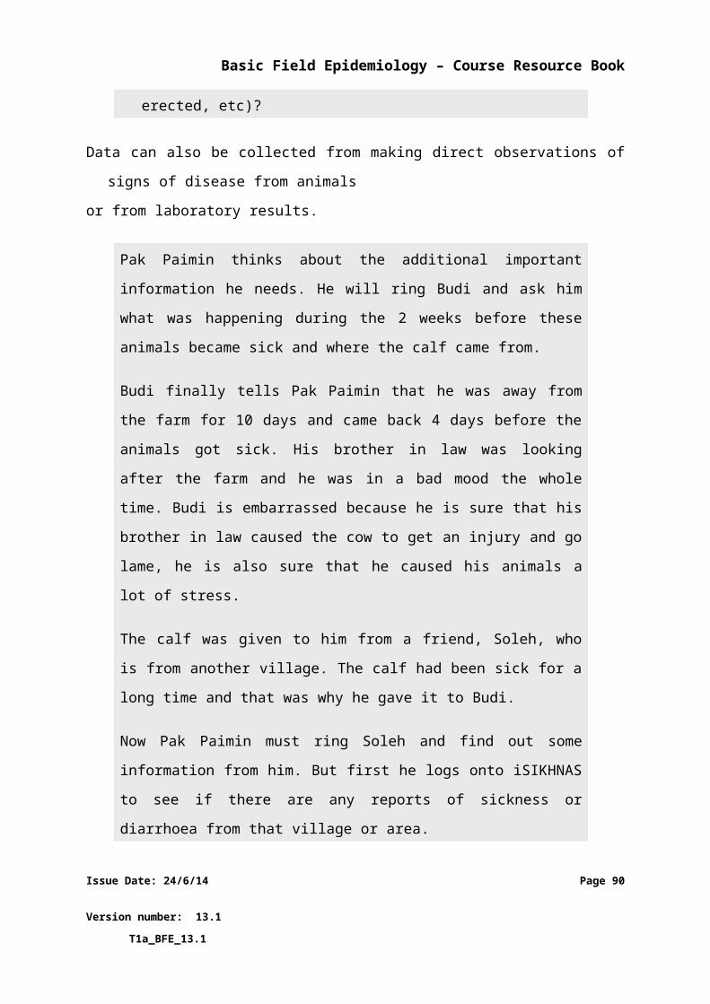
Basic Field Epidemiology – Course Resource Book
Epidemiology skills help us to understand disease progression in individuals and
populations and how vaccination can be used to reducing the susceptible
population. The vaccination program aims to reduce the number of susceptible
animals by increasing immunity and reducing the transmission and spread of
disease. Epidemiology also helps us to understand how the concept of herd
immunity is helpful in disease control since it means that provided a high proportion
of the dog population is vaccinated (more than 70-80%), the herd immunity effect
will mean that the vaccination program should result in a progressive reduction in
the number of new cases of rabies in dogs.
Knowledge of zoonotic diseases helps us to communicate effective strategies for
humans to prevent exposure (avoid being bitten) and to go to a doctor for post-
exposure treatment (PET) if they have been bitten by a dog that may have rabies.
Example 9
Case introduction
There has been HPAI confirmed in a chicken farm in your area
Information available
The investigations identified many chickens were sick or dead Laboratory samples were taken and the laboratory confirmed HPAI
virus The laboratory test result and the clinical signs mean that HPAI has
been confirmed as the definitive diagnosis in this case There has been a formal government response including the following
control strategies:o Slaughtero Quarantineo Biosecurityo Vaccination programs for surrounding areas
Para-veterinarian’s actions
You ensure the community is educated about the risk of HPAI, the control
program, and the importance getting rid of this disease. You explain to the
Issue Date: 24/6/14 Page 90
Version number: 13.1 T1a_BFE_13.1
Basic Field Epidemiology – Course Resource Book
community why all the chickens need to be slaughtered and how the disease
can cause deaths in people. You explain why biosecurity is very important to
prevent humans spreading the disease to new farms
You work within the different parts of the control program.
Epidemiology context
Epidemiology skills help us to understand causes of disease and think about control
strategies to prevent further cases. HPAI is important as it can cause disease and
death in humans. A number of control strategies need to be used together to best
prevent spread of the disease.
Issue Date: 24/6/14 Page 91
Version number: 13.1 T1a_BFE_13.1