Widespread cortical PET abnormalities in an adolescent ... · and dissociative-state regional...
9
1 Scandinavian Journal of Child and Adolescent Psychiatry and Psychology DOI 10.21307/sjcapp-2018-011 Case Report Open Access Widespread cortical PET abnormalities in an adolescent related to a PNES dissociative state, PTSD, ADHD, and domestic violence exposure Dejan Stevanovic 1* , Leposava Brajkovic 2 , Madhur Kumar Srivastava 3 , Ivan Krgovic 1 , Jasna Jancic 1,4 1 Clinic of Neurology and Psychiatry for Children and Youth, Belgrade, Serbia; 2 University of Belgrade Center for Nuclear Medicine, Clinical Center of Serbia, Belgrade, Serbia 3 Department of Nuclear Medicine, Nizam’s Institute of Medical Sciences, Hyderabad, India 4 Faculty of Medicine, University of Belgrade, Belgrade, Serbia * Corresponding author: [email protected] Abstract Background: Accumulated evidence indicates that exposure to trauma is associated with the development of cognitive impairments and psychiatric symptoms in children and adolescents. Objective: In this case study of a female adolescent of 17 years, we aimed to evaluate how cortical positron emission tomography (PET) abnormalities relate to psychogenic non-epileptic seizure (PNES) dissociative state, post-traumatic stress disorder (PTSD), attention-deficit/hyperactivity disorder (ADHD), and domestic violence exposure. Methods: Detailed psychiatric and neuropsychological assessment was performed initially, followed by a PET study. The PET imaging was carried out in the resting-state and in the dissociative-state. Results: The adolescent was suffering from multiple episodes of unconsciousness, all found to be psychogenic; thus, PNES was diagnosed. However, at the psychopathology symptom level, the adolescent had heightened impulsivity, hyperactivity, hyperarousal, anxiety, somatic, and dissociative/ functional neurological symptoms present separately or concurrently at some point during her life; thus, the criteria for PTSD and ADHD were also fulfilled. In the resting state, significant hypometabolism was observed in the occipital, occipitotemporal, polar, and mesial parts of the temporal regions bilaterally, fronto-parietal medial and lateral pericental regions, and fronto-temporal and insular region on the left. The most intense metabolism was observed in the posterior cingulate gyrus and the medial parts of the posterior parietal lobe. In the dissociative state, there was a slight increase in the metabolism of the brain globally compared with the resting state, but with identical distribution of the regional changes observed. Conclusions: Widespread cortical PET abnormalities were found, possibly indicating alterations in large-scale brain networks, in a patient with PNES and a dissociative state, PTSD, and ADHD, who was exposed to chronic domestic violence Keywords: dissociation; default mode network; inhibitory control; trauma Introduction Various impairments in attachment development, emotional processing, cognitive functioning, and academic achievements are linked to exposure to traumatic events in childhood (1). Exposure to traumatic events has also been linked to psychiatric disorders (2, 3), such as anxiety and depression, post- traumatic stress disorder (PTSD), conversion disorder, and attention-deficit/hyperactivity disorder (ADHD). Numerous neurobiological changes have been found in the brain structure and function due to trauma exposure in childhood (3), but the most consistently reported ones are in ventro-medial, orbito-frontal-limbic, lateral fronto-striatal, and parieto-temporal regions (1). It was implicated that alterations in the cortical network organization in children exposed to trauma may result in a diminished capacity to regulate impulses and emotions, to accurately attribute thoughts and intentions to others, and to be mindful of oneself in a social context (3). Specific alterations in the cortical network organization are particularly evident in two disorders related to trauma: PTSD and psychogenic
-
Upload
hoangthien -
Category
Documents
-
view
215 -
download
0
Transcript of Widespread cortical PET abnormalities in an adolescent ... · and dissociative-state regional...

1
Scandinavian Journal of Child and Adolescent Psychiatry and Psychology DOI 10.21307/sjcapp-2018-011
Case Report Open Access
Widespread cortical PET abnormalities in an adolescent related to a PNES dissociative state, PTSD, ADHD, and domestic
violence exposure
Dejan Stevanovic1*, Leposava Brajkovic2, Madhur Kumar Srivastava3, Ivan Krgovic1, Jasna Jancic1,4
1Clinic of Neurology and Psychiatry for Children and Youth, Belgrade, Serbia;
2University of Belgrade Center for Nuclear Medicine, Clinical Center of Serbia, Belgrade, Serbia 3Department of Nuclear Medicine, Nizam’s Institute of Medical Sciences, Hyderabad, India
4Faculty of Medicine, University of Belgrade, Belgrade, Serbia *Corresponding author: [email protected]
Abstract
Background: Accumulated evidence indicates that exposure to trauma is associated with the development of cognitive impairments and psychiatric symptoms in children and adolescents. Objective: In this case study of a female adolescent of 17 years, we aimed to evaluate how cortical positron emission tomography (PET) abnormalities relate to psychogenic non-epileptic seizure (PNES) dissociative state, post-traumatic stress disorder (PTSD), attention-deficit/hyperactivity disorder (ADHD), and domestic violence exposure. Methods: Detailed psychiatric and neuropsychological assessment was performed initially, followed by a PET study. The PET imaging was carried out in the resting-state and in the dissociative-state. Results: The adolescent was suffering from multiple episodes of unconsciousness, all found to be psychogenic; thus, PNES was diagnosed. However, at the psychopathology symptom level, the adolescent had heightened impulsivity, hyperactivity, hyperarousal, anxiety, somatic, and dissociative/ functional neurological symptoms present separately or concurrently at some point during her life; thus, the criteria for PTSD and ADHD were also fulfilled. In the resting state, significant hypometabolism was observed in the occipital, occipitotemporal, polar, and mesial parts of the temporal regions bilaterally, fronto-parietal medial and lateral pericental regions, and fronto-temporal and insular region on the left. The most intense metabolism was observed in the posterior cingulate gyrus and the medial parts of the posterior parietal lobe. In the dissociative state, there was a slight increase in the metabolism of the brain globally compared with the resting state, but with identical distribution of the regional changes observed. Conclusions: Widespread cortical PET abnormalities were found, possibly indicating alterations in large-scale brain networks, in a patient with PNES and a dissociative state, PTSD, and ADHD, who was exposed to chronic domestic violence
Keywords: dissociation; default mode network; inhibitory control; trauma
Introduction Various impairments in attachment development, emotional processing, cognitive functioning, and academic achievements are linked to exposure to traumatic events in childhood (1). Exposure to traumatic events has also been linked to psychiatric disorders (2, 3), such as anxiety and depression, post-traumatic stress disorder (PTSD), conversion disorder, and attention-deficit/hyperactivity disorder (ADHD).
Numerous neurobiological changes have been found in the brain structure and function due to
trauma exposure in childhood (3), but the most consistently reported ones are in ventro-medial, orbito-frontal-limbic, lateral fronto-striatal, and parieto-temporal regions (1). It was implicated that alterations in the cortical network organization in children exposed to trauma may result in a diminished capacity to regulate impulses and emotions, to accurately attribute thoughts and intentions to others, and to be mindful of oneself in a social context (3). Specific alterations in the cortical network organization are particularly evident in two disorders related to trauma: PTSD and psychogenic

Dissociative state, cortical PET abnormalities, and violence exposure
2
non-epileptic seizure (PNES; 4, 5). A recent quantitative meta-analysis showed hyperactivation of the mid/dorsal anterior cingulate cortex, supplementary motor area (SMA), and superior temporal gyrus in PTSD (6). These regions have been previously shown to be parts of the Salience Network (6, 7), which is a controller or a network switcher monitoring information from within and from the external world. In addition, PNES and PTSD are also conceptualized as disorders characterized by altered functional connectivity in the Default Mode Network (DMN; 5, 8-10), which is mainly responsible for self-reflection, self-awareness, and autobiographic memories. It has been only recently shown that pediatric PTSD might be characterized by heightened within-DMN connectivity, which may contribute to re-experiencing symptoms and alterations in autobiographical memory (11).
In this case study of a female adolescent, we aimed to evaluate the relationships between resting-state and dissociative-state regional cortical positron emission tomography (PET) abnormalities, multiple episodes of PNES with a dissociative state, PTSD, ADHD, and domestic violence exposure.
Case presentation M.B. was a 17-year-old adolescent girl brought to our department after being found unconscious in public. The adolescent appeared asleep, unarousable, and unresponsive to any external stimuli. She was lying motionless; speech and spontaneous and purposeful movement were completely absent. As there was no prior history available, usual medical procedures were undertaken. Physical and neurological examination was normal, including all laboratory and toxicological findings of the blood and urine. The data of the cerebrospinal fluid analysis was in the reference range. Electroencephalography showed non-specific changes, while multi-slice computed tomography with angiography of the brain and magnetic resonance imaging were described as expected for age and gender. The N-methyl-D-Aspartate-type glutamate receptor autoantibody test was negative.
Over the past four years, the adolescent experienced repeated episodes of sudden “sleeping” lasting up to several hours or short episodes with hyperventilation and repeated shakings of extremities with/without unresponsiveness to external stimuli. During the age of 12 to 16 years, there were repeated abdominal pains, asthmatic attacks, persistent headaches, and/or increased blood pressure, all diagnosed as somatic symptoms of psychogenic origin. At that time, the adolescent was referred to a psychologist, who reported unexpectedly high tolerance to stress with the presence of possible trauma-related symptoms, such as numbing, self-detachment, and heightened anxiety.
The observed episode of complete unresponsiveness at our department lasted for 59 hours. During this time, few short periods of hyperventilation and abnormal, rhythmic movements of abdominal musculature were observed, resembling forced diaphragmatic breathing. As lorazepam was not available, diazepam 10 mg was administered, but the episode terminated several hours later. After returning to her regular functioning, there was no overt consciousness, perceptual, memory, or affective disturbances, except mild to moderate impulsivity and an inability to recall anything shortly before and during the episode. She complained of being sleepy throughout the day over the past year. A psychiatric examination was requested to which the adolescent and her parents refused. The adolescent was dismissed with a tentative diagnosis of Conversion Disorder (Functional Neurological Symptom Disorder), as PNES, according to the Diagnostic and Statistical Manual of Mental Disorders Fifth Edition (DSM-5; 12). However, the condition was specified as PNES status (13) or PNES dissociative state, resembling dissociative stupor (14) or the most recently described “psychogenic or functional coma” (15). No medications were given.
The adolescent was readmitted after 10 days. The main presenting sign was again complete unresponsiveness, with physical and neurological examination being normal. This episode occurred shortly after an argument in the family. Over the next 20 hours, the adolescent hyperventilated periodically, and there were short episodes of apnea and increases in blood pleasure, with occasional myoclonic twitches/jerks, rhythmic movements of abdominal musculature, generalized trembling, twitching of extremities, fumbling, and hypersalivation. There were no specific electroencephalography changes. Diazepam 10 mg was given twice in an hour on the admission, but the episode was not terminated. Midazolam 15 mg was given twice on the second day, and the episode terminated few hours later. Over the next five days, several short episodes were observed with generalized trembling or uncoordinated movements involving the whole body with hyperventilation, closed eyes, and unresponsiveness, all lasting up to 15 minutes, but also two episodes resembling a generalized tonic-clonic epileptic seizure. This time the parents and adolescent agreed to participate in a psychiatric assessment.
Personal and family history The adolescent was born at term and had no history of neurological disorders or head injuries. She presented with normal language, social, and motor development, with early right-handed dominance. The adolescent was attending regular school. Slightly

Dissociative state, cortical PET abnormalities, and violence exposure
3
below average intellectual functioning at age seven was demonstrated when she entered school. There were no reported deteriorations in academic performances. It was reported that she showed mild behavioral difficulties since early education, such as being overactive or impulsive. There was no history of neurological or psychiatric disorders in the family. However, the family was under social welfare system surveillance since the adolescent’s birth, mainly due
to poverty and domestic violence. A local welfare center reported that the father was physically, emotionally, and/or sexually abusive towards the mother and one older son, which was what the adolescent mostly witnessed. There were several placements of the mother and children in protective custody, while the father was eventually sentenced for domestic violence when the adolescent was 15.
TABLE 1. Results of the scales and neuropsychological tests used
Scale/Test Score obtained Cut-off for clinical score/ Mean (SD) reference value/Severity
Revised Children’s Anxiety and Depression Scale (RCADS) mother-report self-report
Anxiety T – score 61; Depression T – score 63 Anxiety T – score 43; Depression T – score < 42
T – score ≥ 70
ADHD Rating Scale-IV – mother report Total score 28 (97 percentile); Hyperactivity/impulsivity score 22 (≥ 99 percentile); Inattention score 6 (75 percentile)
Scores ≥ 95 percentile
Barratt Impulsiveness Scale Version 11 (BIS11) – self-report 77
63.32 (10.16)
Adolescent Dissociative Experiences Scale (A-DES) – self-report 1.1
4.8 (1.12)
UCLA PTSD Reaction Index DSM-5 Version – self-report 46 ≥ 38
Children’s Negative Cognitive Error Questionnaire-Revised (CNCEQ-R) – self-report
27 36.92 (12.01)
Positive Affect, Negative Affect, and Physiological Hyperarousal (PANAS-PH-C) – self-report
Positive affect score 44 Negative affect score 21 Physiological Hyperarousal 27
44.47 (7.93) 19.80 (6.16) 28.88 (9.17)
DSM-5, LEVEL 2 – Somatic Symptom Child Age 11–17 – self-report Prorated Score 10 Moderate severity
Comprehensive Executive Function Inventory (CEFI) – mother-report Emotion Regulation (ER) Inhibitory Control (IC) Self-Monitoring (SM) Planning (PL): 11 items Working Memory (WM) Attention (AT): 12 items Organization (OG) Flexibility (FX) Initiation (IT) Full score
13, 2 percentile 16, 2 percentile 18, 4 percentile 21, 6 percentile 27, 14 percentile 26, 18 percentile 26, 34 percentile 21, 55 percentile 32, 58 percentile 200, 12 percentile
Executive Skills Questionnaire – self-report

Dissociative state, cortical PET abnormalities, and violence exposure
4
Response inhibition Organization Metacognition Time management Task initiation Emotional control Flexibility Goal-directed persistence Working memory Sustained attention Planning/prioritizing
6 12 13 14 14 15 17 17 18 21 21
Minimal score 3 Maximum score 21, a higher score indicates a more developed skill
Wechsler Adult Intelligence Scale (WAIS) Verbal IQ Performance IQ Full Scale IQ
83 86 84
Low Average
Wisconsin card sorting test (WCST)* % Errors % Perseverate errors
50, > 90 percentile 32.81, > 90 percentile
NA
Tower of London* Extra moves Time
35, > 90 percentile 163.87 sec, 75 percentile
NA
GO-No-Go Task* Total Correct Total Errors Mean Accuracy Mean Error Response time “Go” Response time “No-go”
268 52 0.83 0.16 428.81 sec 356.25 sec
NA
Note. *Computer administered PEBL, the Psychology Experiment Building Language; NA, not available
Neuropsychological and psychiatric findings Several scales were used to assess levels of psychopathological symptoms, as well as affectivity and thinking styles (Table 1). Elevated scores were evident for hyperactivity, impulsivity, somatic, and trauma-related symptoms.
The adolescent was referred to neuropsychological testing, but she was unwilling to participate and only the Wechsler Adult Intelligence Scale, Wisconsin card sorting test, Tower of London, Go-No-Go Task, and behavioral scales of executive functions were completed. The cognitive ability testing indicated on low average intellectual functioning, with specific deficits in executive functioning skills, predominantly emotional regulation and inhibitory control and to a moderate degree in self-monitoring, planning/problem solving, and cognitive flexibility. The Schedule for Affective Disorders and Schizophrenia for School-Age Children Present and Lifetime Version (16) and the Dissociative Disorders Interview Schedule (17) – DSM-5 Version were used to guide the psychiatric interview. According to the DSM-5 (12), Conversion disorder (PNES) was diagnosed as the primary diagnosis, with PTSD with dissociative symptoms due to domestic violence and ADHD predominantly hyperactive/impulsive
presentation of moderate severity as the secondary diagnoses. The following DSM-5 diagnoses were likely present at some point since early childhood: Separation anxiety disorder (SAD), ADHD predominantly hyperactive/impulsive, Somatic Symptom Disorder with predominant pain and/or breathing difficulties, PTSD due to domestic violence, and PNES with a dissociative state. She did not meet the criteria for a personality disorder.
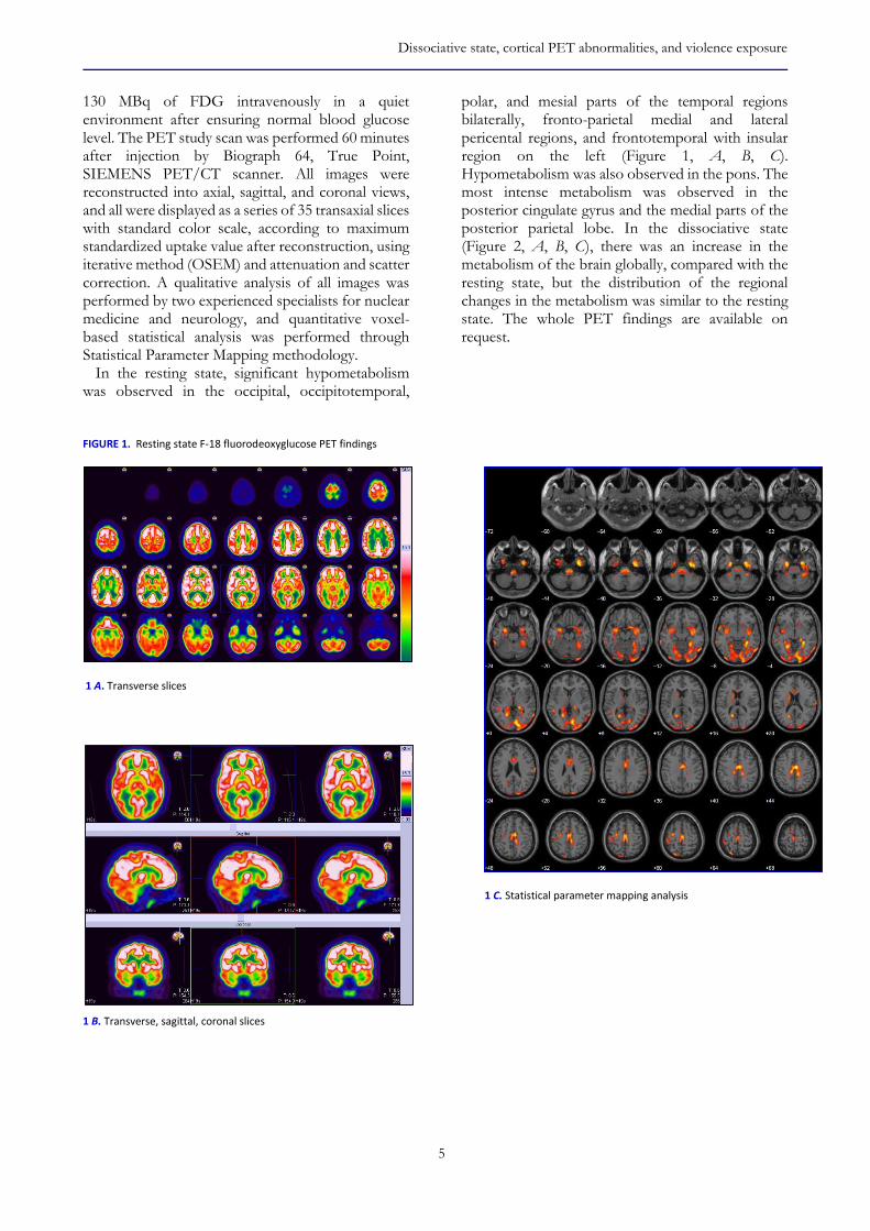
F-18 fluorodeoxyglucose PET findings An F-18 fluorodeoxyglucose PET study was performed in order to detect possible changes in the brain metabolic activity, which is proportional to functional (synaptic) activities in brain regions and networks (18). The PET imaging was performed in the resting state and in the dissociative state, when the adolescent was admitted for the second time in an episode of unconsciousness. In order to ensure that the episode lasted fully during the PET imaging, the adolescent was reexamined on the start, several times called to the name, and occasionally was applied a mild painful stimulus to which there was no verbal or motor response. The adolescent was unresponsive over the next seven hours after the imaging. On both occasions, the patient was injected
Dissociative state, cortical PET abnormalities, and violence exposure
5
130 MBq of FDG intravenously in a quiet environment after ensuring normal blood glucose level. The PET study scan was performed 60 minutes after injection by Biograph 64, True Point, SIEMENS PET/CT scanner. All images were reconstructed into axial, sagittal, and coronal views, and all were displayed as a series of 35 transaxial slices with standard color scale, according to maximum standardized uptake value after reconstruction, using iterative method (OSEM) and attenuation and scatter correction. A qualitative analysis of all images was performed by two experienced specialists for nuclear medicine and neurology, and quantitative voxel-based statistical analysis was performed through Statistical Parameter Mapping methodology.
In the resting state, significant hypometabolism was observed in the occipital, occipitotemporal,
polar, and mesial parts of the temporal regions bilaterally, fronto-parietal medial and lateral pericental regions, and frontotemporal with insular region on the left (Figure 1, A, B, C). Hypometabolism was also observed in the pons. The most intense metabolism was observed in the posterior cingulate gyrus and the medial parts of the posterior parietal lobe. In the dissociative state (Figure 2, A, B, C), there was an increase in the metabolism of the brain globally, compared with the resting state, but the distribution of the regional changes in the metabolism was similar to the resting state. The whole PET findings are available on request.
FIGURE 1. Resting state F-18 fluorodeoxyglucose PET findings
1 A. Transverse slices
1 C. Statistical parameter mapping analysis
1 B. Transverse, sagittal, coronal slices

Dissociative state, cortical PET abnormalities, and violence exposure
6
FIGURE 2. Dissociative state F-18 fluorodeoxyglucose PET findings
2 A. Transverse slices
Treatment 2 B. Transverse, sagittal, coronal slices 2 C. Statistical parameter mapping analysis
Treatment Trauma-focused psychotherapy was planned, and OROS methylphenidate (i.e., Concerta) 18 mg was prescribed for ADHD symptoms and fluoxetine 20 mg for PTSD symptoms. The adolescent did not come regularly to scheduled visits, and there was poor treatment adherence over a 7-month follow-up. The adolescent did not participate in any psychotherapy sessions, and she stopped taking fluoxetine after several days for no reason. She only continued to take regularly Concerta due to some beneficial effects observed shortly after, such as being less fidgeting and more patient. Over the next six months, there was only one short episode of complete unresponsiveness. However, it was not possible to judge the treatment response completely.
Discussion On the basis of accumulated evidence, exposure to trauma in childhood, especially interpersonal trauma such as physical abuse or family violence, is associated with multiple psychiatric symptoms (2, 19). In our case, who was exposed to domestic
violence for years, the main presenting psychiatric diagnosis was functional neurological disorder, PNES, but, at the psychopathology symptom-level, heightened impulsivity, hyperactivity, hyperarousal, anxiety, somatic, and dissociative/conversion symptoms were present separately or concurrently at some point of time over the past 5 to 6 years. All these symptoms have been also previously reported among adolescents with PNES and PTSD (20, 21), except for ADHD symptoms, which are relatively infrequent, especially among girls with PNES (22). Previous studies evaluating childhood trauma also reported that traumatic experiences pose significant deteriorating effects on neuropsychological development including intelligence (23), especially on response inhibition and emotional regulation (1, 2). In our adolescent, slightly below average intellectual functioning was present, which could pre-exist, with emotional dysregulation and poor inhibitory control, as the most evident deficits in executive functions.
The PET findings showed widespread cortical abnormalities, but of particular importance here are the precuneus, posterior cingulate gyrus, and to a

Dissociative state, cortical PET abnormalities, and violence exposure
7
lesser extent the hippocampus, amygdales, parietal cortex, and insular regions. All of these regions were recognized as parts of large-scale brain networks implicated in psychopathology (6, 24). The precuneus and posterior cingulate gyrus, with an increase in the brain metabolism in our adolescent, together with the medial prefrontal cortex and inferior parietal lobules, are parts of the DMN (7). As in PNES, it was recently demonstrated that alterations in the DMN are evident in disorders of consciousness (25), in particular in the cases with alterations in consciousness (5, 10). Considering the glucose metabolism observed in our adolescent and clinical presentation, it appeared that the dysfunctions of the DMN might be linked with the observed psychopathology, especially with the dissociation, which likely originates due to the dysfunctions in functional connectivity of this network (16). Some previous studies evaluating dissociative identity disorder also reported changes in these regions during dissociative states (26, 27). However, there are no previous reports of PET abnormalities or any other imaging data for complete unconsciousness as a dissociative episode within adolescents or adults to compare with our findings. Overall, it could be inferred that the dissociative state was caused by excessive activation in affected brain regions, which led to imbalance in functional networks and dissociative state of consciousness.
As related to observable PET findings, the co-existence of severe dissociation, as a form of PTSD, and ADHD symptoms is of particular importance, because they both might originate due to the deficits in inhibitory control (2). ADHD is considered a neurodevelopmental disorder, and, as such, it might be present irrespective of trauma; contrary to PTSD, which is a trauma/stressor-related disorder (12). However, accumulated evidence indicates that inhibitory control accounts for hyperactivity-impulsivity symptoms (28), as well as symptoms of functional neurological disorders (29) and PTSD (30). The brain networks comprising the inferior frontal cortex and SMA, both found to be of a hypometabolic activity here, with subcortical and brain stem nuclei regions are mainly included in inhibitory control (31).
In particular to ADHD, it is evidenced that its symptoms arise as result of abnormal anatomical functioning and connectivity throughout fronto-striatal, fronto-temporal, fronto-parietal, and/or fronto-striato-parieto-cerebellar circuits (32). Besides that, many of these studies supported hypofunctioning or compromised white matter integrity within these networks; the results have been inconsistent, and some studies have found increased rather than decreased regional brain activation, especially throughout the DMN. Cingulate-
precuneus interactions were implicated in ADHD, as well (33). In our case, abnormalities were evident in fronto-temporal, fronto-parietal, precuneus, and cingulate gyrus. Although the treatment response was not possible to judge completely, it might be possible that methylphenidate given for ADHD symptoms was helpful for both hyperactivity/impulsivity and dissociation, which again could be linked to the DMN and changes in the affected regions. There are reports that methylphenidate influences brain connectivity within the DMN (34), which possibly resulted in adolescents over the next 6 months having only one episode of dissociative state.
Furthermore, the only study evaluating adults with PNES, including cases with unresponsiveness, in resting states showed significant areas of hypometabolism in the right inferior parietal/central region and the bilateral anterior cingulate cortex (35). Moreover, increased metabolic correlation was found between these regions and the cerebellum and the left parahippocampal gyrus, with no zones of increased metabolism. In addition, PNES had increased coactivation of several regions in the resting-state networks associated with fronto-parietal activation, executive control, sensorimotor functioning, and the DMN (10). This is contrary to our observation, considering that, generally speaking, changes were more in the posterior and lateral regions and only partially overlapping with the DMN. Finally, the greatest parts of the frontal lobe, especially the dorsolateral and ventromedial prefrontal parts did not show altered glucose metabolism in our case, although these were reported to be involved brain regions in psychopathologies such as ADHD, PTSD, and PNES (9, 10, 32). However, there was hypometabolism in the occipital regions, suggesting the involvements of the visual system in the overt psychopathology or neurocognitive functioning in our case, as previously reported (36). Conclusions We found widespread resting-state and dissociative-state cortical PET abnormalities possibly indicating alterations in large-scale brain networks in a patient exposed to chronic domestic violence with PNES, PTSD, and ADHD. In particular, we were able to report functional imaging data for a prolonged, dissociative state, with clear alterations in the precuneus, posterior cingulate gyrus, and SMA, and occurring symptoms of PNES, PTSD, and ADHD. There are several clinical and research implications from this case. First, the PET brain uptake correlated well with the involvement of the various cortical regions, suggesting that it can be a useful tool to discriminate between “real” complete

Dissociative state, cortical PET abnormalities, and violence exposure
8
unconsciousness and the dissociative forms. Second, although we were not able to clearly demonstrate the contribution of early and repeated exposures to domestic violence, it is indicative that there could be specific changes in large brain networks and severe dissociation in the presence of traumatic events. Of particular relevance thus would be to study dissociation in PTSD, considering different age groups, neuroplasticity, and cortical abnormalities. Third, considering deficits in inhibitory control and possibly large brain network dysfunctions, especially the DMN, future research should focus on these aspects, as related to domestic violence and relations to PNES. Disclosure The patient provided informed written consent for the publication, and it was assured that the identity of the patient will remain unknown. The institutional ethics of the Clinic of Neurology and Psychiatry for Children and Youth Belgrade approved the case presentation. This is a
non-funded study.
Conflicts of interest There is no conflict of interest to report by the authors.
References 1. Hart H, Rubia K. Neuroimaging of child abuse: a critical review.
Front Hum Neurosci 2012;6:1-24.
2. Dvir Y, Ford JD, Hill M, Frazier JA. Childhood maltreatment, emotional dysregulation, and psychiatric comorbidities. Har Rev Psychiatry 2014;22:149-61.
3. Teicher MH, Samson JA. Annual research review: enduring
neurobiological effects of childhood abuse and neglect. J Child Psychol Psychiatry 2016;57:241-66.
4. Nicholson AA, Densmore M, Frewen PA, Théberge J, Neufeld RW,
McKinnon MC, et al. The dissociative subtype of posttraumatic stress disorder: unique resting-state functional connectivity of basolateral and centromedial amygdala complexes. Neuropsychopharmacology 2015;40:2317-26.
5. Ding J, An D, Liao W, Wu G, Xu Q, Zhou D, et al. Abnormal
functional connectivity density in psychogenic non-epileptic seizures. Epilepsy Res 2014;108:1184-94.
6. Hayes JP, Hayes SM, Mikedis AM. Quantitative meta-analysis of
neural activity in posttraumatic stress disorder. Biol Mood Anxiety Disord 2012;2:9.
7. Menon V. Large-scale brain networks and psychopathology: a
unifying triple network model. Trends Cogn Sci 2011;15:483-506.
8. Daniels JK, Bluhm RL, Lanius RA. Intrinsic network abnormalities in posttraumatic stress disorder: research directions for the next decade. Psychological Trauma 2013;5:142-8.
9. Peterson A, Thome J, Frewen P, Lanius RA. Resting-state
neuroimaging studies: a new way of identifying differences and similarities among the anxiety disorders? Can J Psychiatry 2014;59:294-300.
10. van der Kruijs SJ, Jagannathan SR, Bodde NM, Besseling RM,
Lazeron RH, Vonck KE, et al. Resting-state networks and
dissociation in psychogenic non-epileptic seizures. J Psychiatr Res 2014;54:126-33.
11. Patriat R, Birn RM, Keding TJ, Herringa RJ. Default-mode network
abnormalities in pediatric posttraumatic stress disorder. J Am Acad Child Adolesc Psychiatry 2016;55:319-27.
12. American Psychiatric Association. Diagnostic and statistical manual
of mental disorders (DSM-5®). Washington: American Psychiatric Publishing; 2013.
13. Rechlin T, Loew TH, Joraschky P. Pseudoseizure “status”. J
Psychosom Res 1997;42:495-8.
14. World Health Organization. The ICD-10 classification of mental and behavioural disorders: clinical descriptions and diagnostic guidelines. World Health Organization; 1992.
15. Ludwig L, McWhirter L, Williams S, Derry C, Stone J. Functional
coma. Handb Clin Neurol 2017;139:313-27.
16. Kaufman J, Birmaher B, Brent D, Rao UM, Flynn C, Moreci P, Williamson D, Ryan N. Schedule for affective disorders and schizophrenia for school-age children-present and lifetime version (K-SADS-PL): initial reliability and validity data. J Am Acad Child Adolesc Psychiatry 1997;36:980-8.
17. Ross CA, Heber S, Norton GR, Anderson D, Anderson G, Barchet
P. The dissociative disorders interview schedule: a structured interview. Dissociation 1989;2:169-89.
18. Passow S, Specht K, Adamsen TC, Biermann M, Brekke N, Craven
AR, et al. Default‐mode network functional connectivity is closely related to metabolic activity. Hum Brain Mapp 2015; 36:2027-38.
19. Beghi M, Cornaggia I, Magaudda A, Perin C, Peroni F, Cornaggia
CM. Childhood trauma and psychogenic nonepileptic seizures: a review of findings with speculations on the underlying mechanisms. Epilepsy Beh 2015;52:169-73.
20. Say GN, Taşdemir HA, İnce H. Semiological and psychiatric
characteristics of children with psychogenic nonepileptic seizures: gender-related differences. Seizure 2015;31:144-8.
21. Reilly C, Menlove L, Fenton V, Das KB. Psychogenic nonepileptic
seizures in children: a review. Epilepsia 2013;54:1715-24.
22. Plioplys S, Doss J, Siddarth P, Bursch B, Falcone T, Forgey M, et al. Risk factors for comorbid psychopathology in youth with psychogenic nonepileptic seizures. Seizure 2016;38:32-7.
23. De Bellis MD, Zisk A. The biological effects of childhood trauma.
Child Adolesc Psychiatr Clin N Am 2014;23:185-222.
24. Smith SM, Fox PT, Miller KL, Glahn DC, Fox PM, Mackay CE, et al. Correspondence of the brain's functional architecture during activation and rest. Proc Natl Acad Sci U S A 2009;106:13040-5.
25. Di Perri C, Bahri MA, Amico E, Thibaut A, Heine L, Antonopoulos
G, et al. Neural correlates of consciousness in patients who have emerged from a minimally conscious state: a cross-sectional multimodal imaging study. Lancet Neurology 2016;15:830-42.
26. Reinders AS, Nijenhuis ER, Quak J, Korf J, Haaksma J, Paans AM,
et al. Psychobiological characteristics of dissociative identity disorder: a symptom provocation study. Biol Psychiatry 2006;60:730-40.
27. Reinders AS, Willemsen AT, Vos HP, den Boer JA, Nijenhuis ER.
Fact or factitious? A psychobiological study of authentic and simulated dissociative identity states. PLoS One 2012;7:e39279.
28. Hart H, Radua J, Nakao T, Mataix-Cols D, Rubia K. Meta-analysis
of functional magnetic resonance imaging studies of inhibition and attention in attention-deficit/hyperactivity disorder: exploring task-specific, stimulant medication, and age effects. JAMA Psychiatry 2013;70:185-98.

Dissociative state, cortical PET abnormalities, and violence exposure
9
29. Ejareh dar M, Kanaan RA. Uncovering the etiology of conversion disorder: insights from functional neuroimaging. Neuropsychiatr Dis Treat 2016; 12:143-53.
30. Falconer E, Bryant R, Felmingham KL, Kemp AH, Gordon E,
Peduto A, et al. The neural networks of inhibitory control in posttraumatic stress disorder. J Psychiatry Neurosci 2008;33:413-22.
31. Bari A, Robbins TW. Inhibition and impulsivity: behavioral and
neural basis of response control. Prog Neurobiol 2013;108:44-79.
32. Weyandt L, Swentosky A, Gudmundsdottir BG. Neuroimaging and ADHD: fMRI, PET, DTI findings, and methodological limitations. Dev Neuropsychol 2013;38:211-25.
33. Castellanos FX, Margulies DS, Kelly C, Uddin LQ, Ghaffari M,
Kirsch A, et al. Cingulate-precuneus interactions: a new locus of dysfunction in adult attention-deficit/hyperactivity disorder. Biol Psychiatry 2008;63:332-7.
34. Battel L, Kieling RR, Kieling C, Anés M, Aurich NK, da Costa JC,
et al. Intrinsic brain connectivity following long-term treatment with methylphenidate in children with attention-deficit/hyperactivity disorder. J ChildAdolesc Psychopharmacol 2016;26:555-61.
35. Arthuis M, Micoulaud-Franchi JA, Bartolomei F, McGonigal A,
Guedj E. Resting cortical PET metabolic changes in psychogenic non-epileptic seizures (PNES). J Neurol Neurosurg Psychiatry 2014;86:106-1112.
36. Rinne-Albers MA, van der Wee NJ, Lamers-Winkelman F,
Vermeiren RR. Neuroimaging in children, adolescents and young adults with psychological trauma. Eur Child Adolesc Psychiatry 2013;22:745-55.