Chilomastix mesnili Enteromonas hominis Trichomonas tenax ...

E-Mail [email protected]
Techniques
Acta Cytologica 2014;58:388–397 DOI: 10.1159/000365046
Whole Slide Imaging of Pap Cell Block Preparations versus Liquid-Based Thin-Layer Cervical Cytology: A Comparative Study Evaluating the Detection of Organisms and Nonneoplastic Findings
Ossama Tawfik a Marilyn Davis a Susan Dillon a Laila Tawfik a
Francisco J. Diaz b Fang Fan a
Departments of a Pathology and Laboratory Medicine and b Biostatistics, Kansas University Medical Center, Kansas City, Kans. , USA
tified by WSI. Conclusions: The concept of WSI from Pap CB preparations is potentially feasible for adoption. Digital re-mote web-based technology eliminates the need for an in-dividual on site, saving time and resources.
© 2014 S. Karger AG, Basel
Introduction
In recent years, digital slide imaging has been gaining acceptance in the pathology community. Applications for remote frozen section, remote immunohistochemistry, remote consultation, education, quality assurance and image analysis are becoming more frequent in pathology laboratories all over the world [1–7] . Digital imaging of cytopathology slides is currently being used for similar activities such as consultation, automated screening of Pap smears, training and education as well as proficiency testing [8–15] . However, the practical application of dig-ital pathology in routine day-to-day pathology remains
Key Words
Whole slide imaging · Pap staining · Cell block preparations · Organisms · Nonneoplastic changes
Abstract
Objective: Cervical cancer is one of the most common ma-lignancies worldwide, yet it is preventable by population screening. In a previous study, we confirmed the feasibility of utilizing whole slide imaging (WSI) of cell block (CB) prep-arations to overcome the limitations of digitizing cytologic samples. In this study, we evaluated the accuracy of WSI in identifying various organisms and nonneoplastic findings. Study Design: A total of 335 WS images from Pap CB prepa-rations were analyzed using the Aperio system. The test per-formance characteristics of ThinPrep (TP) and WSI samples were compared for adequacy, for the presence of bacterial vaginosis (BV), fungi, Trichomonas vaginalis (TV) and herpes simplex virus (HSV) and for nonneoplastic findings. Results: The WSI samples contained optimal material from all prepa-rations. BV was diagnosed in 33 WSI versus 36 TP samples. Budding yeasts and/or pseudohyphal forms were noted in 18 WSI versus 19 TP samples. TV organisms (10 of 11 sam-ples) and 1 HSV case were accurately identified in the WSI and TP samples. Squamous metaplasia, keratosis and reac-tive/reparative and inflammatory changes were easily iden-
Received: May 14, 2014 Accepted after revision: June 2, 2014 Published online: July 16, 2014
Correspondence to: Prof. Ossama Tawfik Department of Pathology and Laboratory Medicine University of Kansas Medical Center 3901 Rainbow Boulevard, Kansas City, KS 66160 (USA) E-Mail otawfik @ kumc.edu
© 2014 S. Karger AG, Basel0001–5547/14/0584–0388$39.50/0
www.karger.com/acy
Some data from this manuscript were previously shown in a post-er presentation at the annual meeting of the United States and Canadian Academy of Pathology, Washington, D.C., March 20–26, 2010, and at the annual meeting of the American Society of Cytopa-thology, Boston, Mass., November 12–16, 2010.
Dow
nloa
ded
by:
Arc
hie
R. D
ykes
Lib
rary
at T
he U
nive
rsity
of K
ansa
s M
edic
al C
ente
r19
8.14
3.32
.65
- 9/
25/2
015
12:5
2:08
AM

WSI of Pap CB Preparations Acta Cytologica 2014;58:388–397DOI: 10.1159/000365046
389
uncertain, especially in the field of cytopathology. One of the main concerns is the scarcity of studies that address the issue of validation of digital imaging for clinical use. The College of American Pathologists has recently started a dialogue among experts in the field for coming up with guidelines for the validation of digital slide imaging in clinical use.
In a recent study, we investigated the feasibility of uti-lizing whole slide imaging (WSI) of Pap smear cell block (CB) preparations. CB processing has been proven to provide a randomized even distribution of cells on the slide with a better representative transfer of cells from the specimen, mitigating sampling errors [16–23] . It has also been shown to significantly improve specimen quality compared with conventional and liquid-based samples, with the advantage of maintenance of cellular architec-ture [16–23] . We demonstrated that WSI is as sensitive as liquid-based methods in detecting epithelial squamous and glandular abnormalities and appears to be highly spe-cific for the detection of low- and high-grade squamous intraepithelial lesions (LSIL and HSIL) [23] . We were also able to demonstrate the feasibility of accurately diagnos-ing human papillomavirus-infected cells by in situ hy-bridization studies and accurately performing p16 immu-nostaining on CB-prepared material [24, 25] .
This study was undertaken to further validate the WSI of CB preparations as a potentially useful screening meth-od. We report our findings describing the ‘negative for intraepithelial lesion or malignancy’ category of the 2001 Bethesda Diagnostic Terminology for Pap samples. An accurate identification of common cytologically detect-able infectious agents along with their cytopathic changes and an accurate description of a variety of nonneoplastic findings, such as squamous metaplasia, reactive cellular changes associated with inflammation, repair, atrophy and others, are provided.
Materials and Methods
Samples This retrospective study was approved by the Institutional Re-
view Committee at the Kansas University Medical Center. The study included a total of 335 samples that were collected from 3 different institutions (1 academic institution, 1 private laboratory and 1 large reference laboratory). The mean age of all patients was 52 years, with a range of 21–62 years. The samples were selected based on the avail-ability of discarded Pap test material following liquid-based cyto-logic evaluation. The samples were selected to include representative classic findings routinely reported in cytology laboratories.
Some of the included samples were analyzed following triaging aliquots for other ancillary tests such as Human Papillomavirus
Hybrid Capture and Cervista testing, Chlamydia trachomatis and Neisseria gonorrhoeae testing and occasional herpes simplex virus (HSV) type 1 and 2 testing. The entire cohort comprised 233 nor-mal and/or negative samples for intraepithelial lesions or malig-nancy, 44 atypical squamous cells of undetermined significance (ASCUS), 44 LSIL and 14 HSIL samples. The samples with infec-tious agents included 36 with bacterial vaginosis (BV), 19 with fun-gal organisms, 11 with Trichomonas vaginalis (TV) and 1 with HSV. Many samples with nonneoplastic findings were studied, in-cluding those with squamous metaplasia, keratosis and reactive cellular changes associated with inflammation, radiation, repair, glandular cells status after hysterectomy and atrophy.
Procedure CB were prepared from the residual cellular sediments of Thin-
Prep (TP) samples, using the Cellient Automated CB System from Hologic (Marlborough, Mass., USA). Histologic sections were pre-pared from the CB in similar fashion to routinely handled surgical specimens and nongynecologic cytologic specimens. Five-mi-crometer sections were cut from each CB, placed on glass slides and stained with hematoxylin and eosin (H&E). These slides were then scanned with the Aperio digital imaging system (Aperio ScanScope XT scanner; Aperio Technologies, Vista, Calif., USA) at 40× magnification. No Z-stacking was included in the scanning any of the images. Reviewers used their workstations to evaluate the images. The monitors’ resolutions varied from 1,680 × 1,050 to 1,920 × 1,200. All digital slides were archived on a secure server at our institution.
Histologic/Cytologic Review of the Samples and Statistical Analysis Four board-certified cytopathologists and 2 cytotechnologists
evaluated all samples. All reviewers analyzed the WS images from all the samples on the Aperio system and recorded their findings blinded to the liquid-based cytology results. Any disagreement was resolved by re-review of the slides until a consensus was reached between the reviewers. The standardized 2001 Bethesda Reporting System and terminology were followed in reporting the diagnoses from the WSI slides by all reviewers. Each sample was evaluated for adequacy and cellularity. The accuracy of the WSI diagnoses was assessed by computing sensitivities and their 95% exact confi-dence intervals (CI), using the originally reported cytologic diag-noses as the reference standard. Exact CI were computed with Sta-ta (StataCorp LP, College Station, Tex., USA).
Results
Organisms and Infections Shift in Flora Suggestive of BV BV was correctly diagnosed in 33 WS images from Pap
CB preparations as compared with 36 TP samples. The WSI method sensitivity for detecting BV was high at 92% [CI (78, 98)]. The presence of a mixed bacterial infiltrate of small cocci, coccobacilli and small curved rods was noted. The WSI samples with BV had a cloudy back-ground of coccobacilli in an identical fashion as conven-
Dow
nloa
ded
by:
Arc
hie
R. D
ykes
Lib
rary
at T
he U
nive
rsity
of K
ansa
s M
edic
al C
ente
r19
8.14
3.32
.65
- 9/
25/2
015
12:5
2:08
AM

Tawfik/Davis/Dillon/Tawfik/Diaz/Fan Acta Cytologica 2014;58:388–397DOI: 10.1159/000365046
390
tional and liquid-based samples ( fig. 1 ). Squamous cells were noted to be covered with coccobacilli, most fre-quently Gardnerella vaginalis , due to the shift in vaginal flora ( fig. 1 c, d). The so-called clue cells with a purple col-or ( fig. 1 c, d) were easily identified.
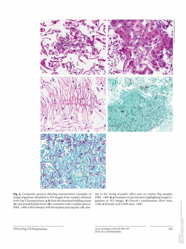
Fungal Organisms Morphologically Consistent with Candida Species Budding yeasts and/or pseudohyphal forms were cor-
rectly noted in 18 WS images from CB samples compared with 19 TP samples. Thus, the sensitivity of WSI for de-tecting fungal organisms was very high at 95% [CI (74, 99)]. The WSI samples showed pseudohyphae and bud-ding yeast forms (3–7 μm in diameter) of the Candida organisms. Similarly noted is the ‘spearing’ of squamous cells, also known as the ‘shish kebab’ effect or ‘string of
pearls’, with a surrounding acute inflammatory infiltrate ( fig. 2 a–c). Figure 2 d and e shows confirmatory fungal stains including Grocott’s methenamine silver and peri-odic acid-Schiff stains, respectively, which became avail-able for the cytologic specimens by using the CB prepara-tions.
Trichomonas vaginalis TV organisms were accurately diagnosed in 10 WS im-
ages from CB samples as compared with 11 TP samples, showing that the WSI sensitivity for detecting TV was very high at 91% [CI (59, 99)]. Multiple TV organisms with their classic pale, grayish, pear-shaped and ovoid or-ganisms are noted in the background of several represen-tative samples. Classic eccentric nuclei and granular eo-sinophilic cytoplasm are noted at higher magnification.
Colo
r ver
sion
ava
ilabl
e on
line
Fig. 1. Examples of low- and high-power views of BV in WS im-ages from samples obtained from Pap CB preparations. Abundant bacteria are arranged either coating the squamous cells or present
in large numbers in the background of the slide. Note the ‘clue cells’ present at the center and top of the image. H&E. a ×100. b–d ×400.
a b
c d
Dow
nloa
ded
by:
Arc
hie
R. D
ykes
Lib
rary
at T
he U
nive
rsity
of K
ansa
s M
edic
al C
ente
r19
8.14
3.32
.65
- 9/
25/2
015
12:5
2:08
AM
WSI of Pap CB Preparations Acta Cytologica 2014;58:388–397DOI: 10.1159/000365046
391
Fig. 2. Composite pictures showing representative examples of fungal organisms identified in WS images from samples obtained from Pap CB preparations. a , b Note the abundant budding yeasts ( a ) and pseudohyphal forms ( b ), consistent with Candida species. H&E. ×400. c Microbiopsy with the hyphae piercing the cells, sim-
ilar to the ‘string of pearls’ effect seen on routine Pap samples. H&E. ×400. d , e Examples of special stains highlighting fungal or-ganisms in WS images. d Grocott’s methenamine silver stain. ×100. e Periodic acid-Schiff stain. ×400.
Colo
r ver
sion
ava
ilabl
e on
line
a b
c d
e
Dow
nloa
ded
by:
Arc
hie
R. D
ykes
Lib
rary
at T
he U
nive
rsity
of K
ansa
s M
edic
al C
ente
r19
8.14
3.32
.65
- 9/
25/2
015
12:5
2:08
AM
Tawfik/Davis/Dillon/Tawfik/Diaz/Fan Acta Cytologica 2014;58:388–397DOI: 10.1159/000365046
392
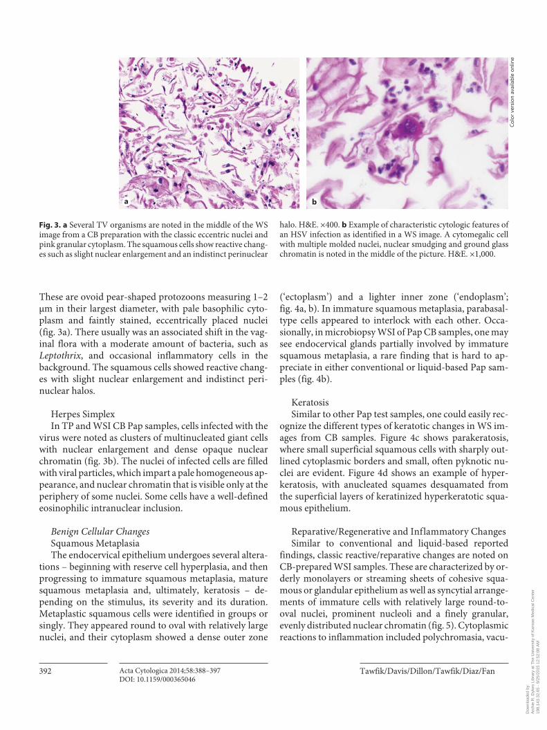
These are ovoid pear-shaped protozoons measuring 1–2 μm in their largest diameter, with pale basophilic cyto-plasm and faintly stained, eccentrically placed nuclei ( fig. 3 a). There usually was an associated shift in the vag-inal flora with a moderate amount of bacteria, such as Leptothrix , and occasional inflammatory cells in the background. The squamous cells showed reactive chang-es with slight nuclear enlargement and indistinct peri-nuclear halos.
Herpes Simplex In TP and WSI CB Pap samples, cells infected with the
virus were noted as clusters of multinucleated giant cells with nuclear enlargement and dense opaque nuclear chromatin ( fig. 3 b). The nuclei of infected cells are filled with viral particles, which impart a pale homogeneous ap-pearance, and nuclear chromatin that is visible only at the periphery of some nuclei. Some cells have a well-defined eosinophilic intranuclear inclusion.
Benign Cellular Changes Squamous Metaplasia The endocervical epithelium undergoes several altera-
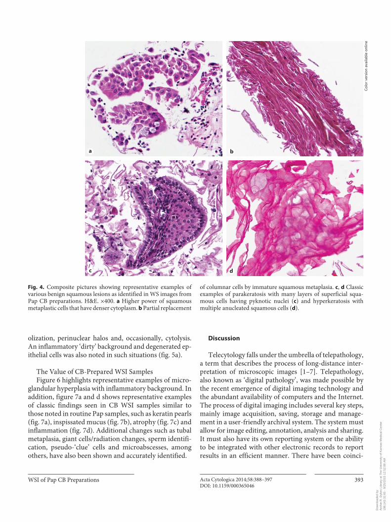
tions – beginning with reserve cell hyperplasia, and then progressing to immature squamous metaplasia, mature squamous metaplasia and, ultimately, keratosis – de-pending on the stimulus, its severity and its duration. Metaplastic squamous cells were identified in groups or singly. They appeared round to oval with relatively large nuclei, and their cytoplasm showed a dense outer zone
(‘ectoplasm’) and a lighter inner zone (‘endoplasm’; fig. 4 a, b). In immature squamous metaplasia, parabasal-type cells appeared to interlock with each other. Occa-sionally, in microbiopsy WSI of Pap CB samples, one may see endocervical glands partially involved by immature squamous metaplasia, a rare finding that is hard to ap-preciate in either conventional or liquid-based Pap sam-ples ( fig. 4 b).
Keratosis Similar to other Pap test samples, one could easily rec-
ognize the different types of keratotic changes in WS im-ages from CB samples. Figure 4 c shows parakeratosis, where small superficial squamous cells with sharply out-lined cytoplasmic borders and small, often pyknotic nu-clei are evident. Figure 4 d shows an example of hyper-keratosis, with anucleated squames desquamated from the superficial layers of keratinized hyperkeratotic squa-mous epithelium.
Reparative/Regenerative and Inflammatory Changes Similar to conventional and liquid-based reported
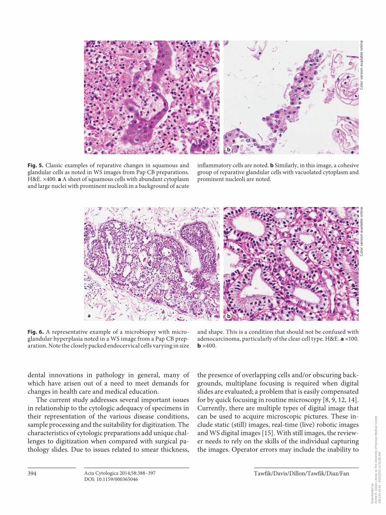
findings, classic reactive/reparative changes are noted on CB-prepared WSI samples. These are characterized by or-derly monolayers or streaming sheets of cohesive squa-mous or glandular epithelium as well as syncytial arrange-ments of immature cells with relatively large round-to-oval nuclei, prominent nucleoli and a finely granular, evenly distributed nuclear chromatin ( fig. 5 ). Cytoplasmic reactions to inflammation included polychromasia, vacu-
Fig. 3. a Several TV organisms are noted in the middle of the WS image from a CB preparation with the classic eccentric nuclei and pink granular cytoplasm. The squamous cells show reactive chang-es such as slight nuclear enlargement and an indistinct perinuclear
halo. H&E. ×400. b Example of characteristic cytologic features of an HSV infection as identified in a WS image. A cytomegalic cell with multiple molded nuclei, nuclear smudging and ground glass chromatin is noted in the middle of the picture. H&E. ×1,000.
Colo
r ver
sion
ava
ilabl
e on
line
a b
Dow
nloa
ded
by:
Arc
hie
R. D
ykes
Lib
rary
at T
he U
nive
rsity
of K
ansa
s M
edic
al C
ente
r19
8.14
3.32
.65
- 9/
25/2
015
12:5
2:08
AM
WSI of Pap CB Preparations Acta Cytologica 2014;58:388–397DOI: 10.1159/000365046
393
olization, perinuclear halos and, occasionally, cytolysis. An inflammatory ‘dirty’ background and degenerated ep-ithelial cells was also noted in such situations ( fig. 5 a).
The Value of CB-Prepared WSI Samples Figure 6 highlights representative examples of micro-
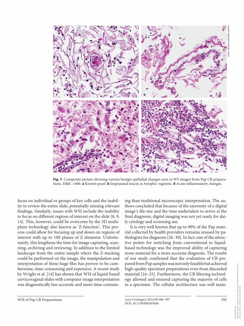
glandular hyperplasia with inflammatory background. In addition, figure 7 a and d shows representative examples of classic findings seen in CB WSI samples similar to those noted in routine Pap samples, such as keratin pearls ( fig. 7 a), inspissated mucus ( fig. 7 b), atrophy ( fig. 7 c) and inflammation ( fig. 7 d). Additional changes such as tubal metaplasia, giant cells/radiation changes, sperm identifi-cation, pseudo-‘clue’ cells and microabscesses, among others, have also been shown and accurately identified.
Discussion
Telecytology falls under the umbrella of telepathology, a term that describes the process of long-distance inter-pretation of microscopic images [1–7] . Telepathology, also known as ‘digital pathology’, was made possible by the recent emergence of digital imaging technology and the abundant availability of computers and the Internet. The process of digital imaging includes several key steps, mainly image acquisition, saving, storage and manage-ment in a user-friendly archival system. The system must allow for image editing, annotation, analysis and sharing. It must also have its own reporting system or the ability to be integrated with other electronic records to report results in an efficient manner. There have been coinci-
Fig. 4. Composite pictures showing representative examples of various benign squamous lesions as identified in WS images from Pap CB preparations. H&E. ×400. a Higher power of squamous metaplastic cells that have denser cytoplasm. b Partial replacement
of columnar cells by immature squamous metaplasia. c , d Classic examples of parakeratosis with many layers of superficial squa-mous cells having pyknotic nuclei ( c ) and hyperkeratosis with multiple anucleated squamous cells ( d ).
Colo
r ver
sion
ava
ilabl
e on
line
a b
c d
Dow
nloa
ded
by:
Arc
hie
R. D
ykes
Lib
rary
at T
he U
nive
rsity
of K
ansa
s M
edic
al C
ente
r19
8.14
3.32
.65
- 9/
25/2
015
12:5
2:08
AM
Tawfik/Davis/Dillon/Tawfik/Diaz/Fan Acta Cytologica 2014;58:388–397DOI: 10.1159/000365046
394
dental innovations in pathology in general, many of which have arisen out of a need to meet demands for changes in health care and medical education.
The current study addresses several important issues in relationship to the cytologic adequacy of specimens in their representation of the various disease conditions, sample processing and the suitability for digitization. The characteristics of cytologic preparations add unique chal-lenges to digitization when compared with surgical pa-thology slides. Due to issues related to smear thickness,
the presence of overlapping cells and/or obscuring back-grounds, multiplane focusing is required when digital slides are evaluated; a problem that is easily compensated for by quick focusing in routine microscopy [8, 9, 12, 14] . Currently, there are multiple types of digital image that can be used to acquire microscopic pictures. These in-clude static (still) images, real-time (live) robotic images and WS digital images [15] . With still images, the review-er needs to rely on the skills of the individual capturing the images. Operator errors may include the inability to
Fig. 5. Classic examples of reparative changes in squamous and glandular cells as noted in WS images from Pap CB preparations. H&E. ×400. a A sheet of squamous cells with abundant cytoplasm and large nuclei with prominent nucleoli in a background of acute
inflammatory cells are noted. b Similarly, in this image, a cohesive group of reparative glandular cells with vacuolated cytoplasm and prominent nucleoli are noted.
Fig. 6. A representative example of a microbiopsy with micro-glandular hyperplasia noted in a WS image from a Pap CB prep-aration. Note the closely packed endocervical cells varying in size
and shape. This is a condition that should not be confused with adenocarcinoma, particularly of the clear cell type. H&E. a ×100. b ×400.
Colo
r ver
sion
ava
ilabl
e on
line
Colo
r ver
sion
ava
ilabl
e on
line
a b
a b
Dow
nloa
ded
by:
Arc
hie
R. D
ykes
Lib
rary
at T
he U
nive
rsity
of K
ansa
s M
edic
al C
ente
r19
8.14
3.32
.65
- 9/
25/2
015
12:5
2:08
AM
WSI of Pap CB Preparations Acta Cytologica 2014;58:388–397DOI: 10.1159/000365046
395
focus on individual or groups of key cells and the inabil-ity to review the entire slide, potentially missing relevant findings. Similarly, issues with WSI include the inability to focus on different regions of interest on the slide [8, 9, 14] . This, however, could be overcome by the 3D multi-plane technology also known as ‘Z-function’. This pro-cess could allow for focusing up and down on regions of interest with up to 100 planes of Z elements. Unfortu-nately, this lengthens the time for image capturing, scan-ning, archiving and reviewing. In addition to the limited landscape from the entire sample where the Z-stacking could be performed on the image, the manipulation and interpretation of these huge files has proven to be cum-bersome, time-consuming and expensive. A recent study by Wright et al. [14] has shown that WSI of liquid-based cervicovaginal slides with computer image interpretation was diagnostically less accurate and more time-consum-
ing than traditional microscopic interpretation. The au-thors concluded that because of the enormity of a digital image’s file size and the time undertaken to arrive at the final diagnosis, digital imaging was not yet ready for dai-ly cytology and screening use.
It is very well known that up to 90% of the Pap mate-rial collected by health providers remains unused by pa-thologists for diagnosis [26–30] . In fact, one of the attrac-tive points for switching from conventional to liquid-based technology was the improved ability of capturing more material for a more accurate diagnosis. The results of our study confirmed that the evaluation of CB pre-pared from Pap samples was not only feasible but achieved high-quality specimen preparations even from discarded material [16–25] . Furthermore, the CB filtering technol-ogy allowed and ensured capturing the majority of cells in a specimen. The cellular architecture was well main-
Fig. 7. Composite picture showing various benign epithelial changes seen in WS images from Pap CB prepara-tions. H&E. ×400. a Keratin pearl. b Inspissated mucin. c Atrophic vaginitis. d Acute inflammatory changes.
Colo
r ver
sion
ava
ilabl
e on
line
a b
c d
Dow
nloa
ded
by:
Arc
hie
R. D
ykes
Lib
rary
at T
he U
nive
rsity
of K
ansa
s M
edic
al C
ente
r19
8.14
3.32
.65
- 9/
25/2
015
12:5
2:08
AM
Tawfik/Davis/Dillon/Tawfik/Diaz/Fan Acta Cytologica 2014;58:388–397DOI: 10.1159/000365046
396
tained with greater consistency in the preparations. Our studies suggest that marrying the samples obtained from cytologic CB preparations with digital pathology technol-ogy would be of great potential value in overcoming many hurdles in the adoption of WSI technology in the cytol-ogy world. We were able to overcome issues related to the length of time required for digital slide reviews without compromising their quality. Scanning slides from CB preparations eliminated the need for multiple Z planes, cutting down on costs, time and the frustration of review-ers. Not only was our WSI technology as sensitive as the current conventional and liquid-based Pap methods for detecting various neoplastic and preneoplastic epithelial lesions as previously shown [23] , but it was also as effi-cient and sensitive in accurately identifying a variety of commonly seen organisms and other nonneoplastic be-nign findings that are routinely noted in a Pap sample.
In our previous study, we demonstrated that the aver-age of overall sensitivities for detecting any lesion from 5 examiners was 82.1%, and that of overall specificities was 86.2%. Also, the average sensitivity and specificity of the 5 examiners using the WSI method were 58.3 and 85.1% for ASCUS lesions, 54.1 and 93.9% for LSIL and 51.8 and 98.8% for HSIL, respectively. The averages of agreement (kappa) measures between individual WSI reviewers and liquid-based cytology were 0.39 for ASCUS, 0.51 for LSIL, 0.52 for HSIL and 0.68 for negative findings. The mea-sures of agreement between the 5 WSI reviewers were substantial: 0.56 for ASCUS [95% CI (0.52, 0.61)], 0.69 (0.65, 0.73) for LSIL, 0.67 (0.61, 0.73) for HSIL and 0.74 (0.70, 0.78) for negative assessments.
In the current study, we expanded our findings and evaluated the various cytologic results described in the ‘negative for intraepithelial lesion or malignancy’ category of the 2001 Bethesda Diagnostic Terminology for Pap test-ing. We were able to accurately identify many infectious agents and characterize their associated cytologic changes. Classic cytologic findings with shifts in flora suggestive of BV including the identification of the characteristic or-ganisms, the cloudy background and ‘clue cells’ were eas-ily noted in WS images from CB preparations. Similarly, budding yeasts and/or pseudohyphal forms as well as TV organisms were correctly identified along with the ‘spear-ing’ of squamous cells and the surrounding acute inflam-matory infiltrate in fungal infection and the reactive squa-mous cells, shifts in vaginal flora and inflammatory chang-es in TV infection. CB preparations provided the opportunity for adding on demand confirmatory tests such Grocott’s methenamine silver and periodic acid-Schiff stains to identify fungal organisms or immunos-
tains for viral or other infections, among others [21, 22, 24, 25] . In WS images of CB preparations, we were able to correctly identify many benign nonneoplastic and non-dysplastic changes. Cytologic changes (such as squamous metaplasia, keratosis and reactive cellular changes associ-ated with inflammation, radiation, repair and atrophy, among others) were correctly identified with ease and confidence. This was very important to further underline the added value of an extra step of digitizing CB prepara-tions. Moreover, we showed that in digitized CB prepara-tions, one is able to find, identify and differentiate glandu-lar endocervical and endometrial cells with ease. We were able to capture more microbiopsies, allowing a more con-fident diagnosis of confusing histologic findings such as microglandular hyperplasia, a diagnosis that is hardly made on cytologic specimens with confidence.
New technologies are dramatically changing the mod-ern practice of pathology and assist us in these endeavors. This paper was written, in part, to illustrate the potential practical utility of implementing widely available tech-nologies in mainstream pathology practice. Pathologists are now capable of contributing their expertise in diagno-sis even when far away from the hospital. As a secondary benefit, image processing technologies also endow the pathologist with more flexibility at the workplace. This capability is obviously conducive to the timely delivery of patient care. The technology to do this already exists; we only need to implement it to reap its benefits. Well-stud-ied and well-characterized digitized images from a Pap CB preparation are not just an extra step with costs added to a system that is already in existence, such as ours in the USA. On the contrary, in places and countries where there is a shortage of pathologists and cytotechnologists, one could imagine the value of implementing such a tech-nology to establish a successful practical screening pro-gram at reasonable costs.
In conclusion, in this pilot study, we introduced the concept of WSI of samples obtained from Pap CB prepa-rations. The technology is potentially feasible for adop-tion. We were able to obtain high-quality specimen prep-arations in a consistent, reliable and timely manner that could potentially reduce the biopsy load. In addition, the CB preparations have a beneficial value for ancillary his-tochemical, immunohistochemical, in situ hybridization and other molecular studies without the need for addi-tional samples. The digital remote, automated, web-based technology eliminates the need for an on-site pathologist/cytotechnologist and provides a time-saving platform for improved collaboration and exchange of ideas. Further studies are needed to develop and optimize high-through-
Dow
nloa
ded
by:
Arc
hie
R. D
ykes
Lib
rary
at T
he U
nive
rsity
of K
ansa
s M
edic
al C
ente
r19
8.14
3.32
.65
- 9/
25/2
015
12:5
2:08
AM

WSI of Pap CB Preparations Acta Cytologica 2014;58:388–397DOI: 10.1159/000365046
397
put systems for preparing CB at a faster and more auto-mated rate and at economically acceptable costs. The new technology has the potential for becoming integrated with electronic medical records, providing timely reports at a reduced cost affordable to all patients.
Disclosure Statement
None of the authors have a conflict of interest either financial-ly or of a personal nature.
References
1 Dee FR: Virtual microscopy in pathology ed-ucation. Hum Pathol 2009; 40: 1112–1121.
2 Wilbur DC, Madi K, Colvin RB, Duncan LM, Faquin WC, Ferry JA, Frosch MP, Houser SL, Kradin RL, Lauwers GY, Louis DN, Mark EJ, Mino-Kenudson M, Misdraji J, Nielsen GP, Pitman MB, Rosenberg AE, Smith RN, Sohani AR, Stone JR, Tambouret RH, Wu CL, Young RH, Zembowicz A, Klietmann W: Whole-slide imaging digital pathology as a platform for teleconsultation: a pilot study using paired subspecialist correlations. Arch Pathol Lab Med 2009; 133: 1949–1953.
3 Hedvat CV: Digital microscopy past, present, and future. Arch Pathol Lab Med 2010; 134: 1666–1670.
4 Jara-Lazaro AR, Thamboo TP, Teh M: Digital pathology: exploring its applications in diag-nostic surgical practice. Pathol 2010; 42: 512–518.
5 Pantanowitz L: Digital images and the future of digital pathology. J Pathol Inform 2010; 1: 15.
6 Bauer TW, Schoenfield L, Slaw RJ, Yerian L, Sun Z, Henricks WH: Validation of whole slide imaging for primary diagnosis in surgi-cal pathology. Arch Pathol Lab Med 2013; 137: 518–524.
7 Campbell WS, Foster KW, Hinrichs SH: Ap-plication of whole slide image markup and annotation for pathologist knowledge cap-ture. J Pathol Inform 2013; 4: 2–8.
8 Pantanowitz L, Hornish M, Goulart RA: The impact of digital imaging in the field of cyto-pathology. Cytojournal 2009; 6: 6.
9 Kalbuss WE, Pantanowitz L, Parwani AV: Digital imaging in cytopathology. Pathol Res Inst 2011; 2011: 264683.
10 O’Brien MJ, Takahashi M, Brugal G, Christen H, Gahm T, Goodell RM, Karakitsos P, Knesel EA Jr, Kobler T, Kyrkou KA, Labbe S, Long EL, Mango LJ, McGoogan E, Oberholzer M, Reith A, Winkler C: Digital imagery/telecy-tology: International Academy of Cytology Task Force summary. Diagnostic Cytology towards the 21st Century: an international ex-pert conference and tutorial. Acta Cytol 1998; 42: 148–164.
11 Alli PM, Ollayos CW, Thompson LD, Kapa-dia I, Butler DR, Williams BH, Rosenthal DL, O’Leary TJ: Telecytology: intraobserver and interobserver reproducibility in the diagnosis of cervical-vaginal smears. Hum Pathol 2001; 32: 1318–1322.
12 Lee ES, Kim IS, Choi JS, Yeom BW, Kim HK, Han JH, Lee MS, Leong AS: Accuracy and re-producibility of telecytology diagnosis of cer-vical smears: a tool for quality assurance pro-grams. Am J Clin Pathol 2003; 199: 356–360.
13 Eichhorn JH, Brauns TA, Gelfand JA, Croth-ers BA, Wilbur DC: A novel automated screening and interpretation process for cer-vical cytology using the Internet transmission of low-resolution images: a feasibility study. Cancer 2005; 105: 199–206.
14 Wright AM, Smith D, Dhurandhar B, Fairley T, Scheiber-Pacht M, Chakraborty S, Gorman BK, Mody D, Coffey DM: Digital slide imag-ing in cervicovaginal cytology: a pilot study. Arch Pathol Lab Med 2013; 137: 615–624.
15 Prayaga AK, Loya AC, Rao IS: Telecytology: are we ready? J Telemed Telecare 2006; 12: 319–320.
16 Yeoh GP, Chan KW: Cell block preparation on residual ThinPrep sample. Diagn Cytopa-thol 1999; 21: 427–431.
17 Richard K, Dziura B, Hornish A: Cell block preparation as a diagnostic technique com-plementary to fluid-based monolayer cervi-covaginal specimens. Acta Cytol 1999; 43: 69–73.
18 Diaz-Rosario LA, Kabawat SE: Cell block preparation by inverted filter sedimentation is useful in the differential diagnosis of atypi-cal glandular cells of undetermined signifi-cance in ThinPrep specimens. Cancer 2000; 90: 265–272.
19 Keyhani-Rofagha S, Vesey-Shecket M: Diag-nostic value, feasibility, and validity of pre-paring cell blocks from fluid-based gyneco-logic cytology specimens. Cancer 2002; 96: 204–209.
20 Gupta S, Hadler K, Khan VA, Sodhani P: Cell block as an adjunct to conventional Papanico-laou smear for diagnosis of cervical cancer in resource-limited settings. Cytopathology 2007; 18: 309–315.
21 Fetsch PA, Simir A, Brosky K, Abati A: Com-parison of the 3 commonly used cytologic preparations in effusion immunocytochemis-try. Diagn Cytopathol 2001; 26: 61–66.
22 Afify A, Yu C, Hejazi N, Howell L: The diag-nostic utility of cell blocks prepared from re-sidual SurePath Pap material for detection of human papillomavirus. Appl Immunohisto-chem Mol Morphol 2009; 17: 108–114.
23 Amin K, Hernandez-Rios P, Davis M, Fan F, Wilson J, Tawfik O: The potential of telecytol-ogy of cell block from Pap smear samples ‘Tele-PAPology’. Mod Pathol 2010; 23(suppl 1):86A.
24 Andraws N, Davis M, Dillon S, Fan F, Tawfik O: TelePAPology versus liquid-based Thin-Prep cervical cytology: a comparative study evaluating human papillomavirus testing by Hybrid Capture-2 and in-situ hybridization. Cytojournal 2011; 8:16.
25 Pham T, Winters C, Davis M, Fan F, Tawfik O: TelePAPology vs. liquid-based cervical cy-tology: A comparative study evaluating p16 immunohistochemistry and HPV testing by Hybrid Cpature-2 and in-situ hybridization. Mod Pathol 2013; 26(suppl 1):100A.
26 Hutchinson ML, Isenstein LM, Goodman A, Hurley AA, Douglass KL, Mui KK, Patten FW, Zahniser DJ: Homogeneous sampling accounts for the increased diagnostic accura-cy using the ThinPrep Processor. Am J Clin Pathol 1994; 101: 215–219.
27 Lee KR, Ashfaq R, Birdson GG, Corkill ME, McIntosh KM, Inhorn SL: Comparison of conventional Papanicolaou smears and a flu-id-based, thin layer system for cervical screen-ing. Obstet Gynecol 1997; 90: 278–284.
28 Linder J, Zahniser D: ThinPrep Papanicolaou testing to reduce false-negative cervical cytol-ogy. Arch Pathol Lab Med 1998; 122: 139–144.
29 Limaye A, Connor AJ, Huang X, Luff R: Com-parative analysis of conventional Papanico-laou tests and a fluid-based thin-layer meth-od. Arch Pathol Lab Med 2003; 127: 200–204.
30 Davey E, Barratt A, Irwig L, et al: Effect of study design and quality on unsatisfactory rates, cy-tology classifications, and accuracy in liquid-based versus conventional cervical cytology: a systematic review. Lancet 2006; 367: 122–132.
Dow
nloa
ded
by:
Arc
hie
R. D
ykes
Lib
rary
at T
he U
nive
rsity
of K
ansa
s M
edic
al C
ente
r19
8.14
3.32
.65
- 9/
25/2
015
12:5
2:08
AM