Whole exome sequencing identifies genetic variants in...
41
Whole exome sequencing identifies genetic variants in inherited thrombocytopenia with secondary qualitative function defects Johnson, Ben; Morgan, Neil; Lowe, Gillian; Futterer, Jane; Lordkipanidze, Marie; MacDonald, David; Drake, Sian; Bem, Danai; Fletcher, Sarah; Dawood, Ban; Watson, Steve; UK GAPP Study Group; Harrison, Paul DOI: 10.3324/haematol.2016.146316 License: Creative Commons: Attribution (CC BY) Document Version Publisher's PDF, also known as Version of record Citation for published version (Harvard): UK GAPP Study Group & Harrison, P 2016, 'Whole exome sequencing identifies genetic variants in inherited thrombocytopenia with secondary qualitative function defects' Haematologica, vol. 101, pp. 1170-1179. DOI: 10.3324/haematol.2016.146316 Link to publication on Research at Birmingham portal Publisher Rights Statement: Published as detailed above and available online at: http://dx.doi.org/10.3324/haematol.2016.146316 General rights Unless a licence is specified above, all rights (including copyright and moral rights) in this document are retained by the authors and/or the copyright holders. The express permission of the copyright holder must be obtained for any use of this material other than for purposes permitted by law. • Users may freely distribute the URL that is used to identify this publication. • Users may download and/or print one copy of the publication from the University of Birmingham research portal for the purpose of private study or non-commercial research. • User may use extracts from the document in line with the concept of ‘fair dealing’ under the Copyright, Designs and Patents Act 1988 (?) • Users may not further distribute the material nor use it for the purposes of commercial gain. Where a licence is displayed above, please note the terms and conditions of the licence govern your use of this document. When citing, please reference the published version. Take down policy While the University of Birmingham exercises care and attention in making items available there are rare occasions when an item has been uploaded in error or has been deemed to be commercially or otherwise sensitive. If you believe that this is the case for this document, please contact [email protected] providing details and we will remove access to the work immediately and investigate. Download date: 27. Sep. 2018
Transcript of Whole exome sequencing identifies genetic variants in...
Whole exome sequencing identifies genetic variantsin inherited thrombocytopenia with secondaryqualitative function defectsJohnson, Ben; Morgan, Neil; Lowe, Gillian; Futterer, Jane; Lordkipanidze, Marie; MacDonald,David; Drake, Sian; Bem, Danai; Fletcher, Sarah; Dawood, Ban; Watson, Steve; UK GAPPStudy Group; Harrison, PaulDOI:10.3324/haematol.2016.146316
License:Creative Commons: Attribution (CC BY)
Document VersionPublisher's PDF, also known as Version of record
Citation for published version (Harvard):UK GAPP Study Group & Harrison, P 2016, 'Whole exome sequencing identifies genetic variants in inheritedthrombocytopenia with secondary qualitative function defects' Haematologica, vol. 101, pp. 1170-1179. DOI:10.3324/haematol.2016.146316
Link to publication on Research at Birmingham portal
Publisher Rights Statement:Published as detailed above and available online at: http://dx.doi.org/10.3324/haematol.2016.146316
General rightsUnless a licence is specified above, all rights (including copyright and moral rights) in this document are retained by the authors and/or thecopyright holders. The express permission of the copyright holder must be obtained for any use of this material other than for purposespermitted by law.
•Users may freely distribute the URL that is used to identify this publication.•Users may download and/or print one copy of the publication from the University of Birmingham research portal for the purpose of privatestudy or non-commercial research.•User may use extracts from the document in line with the concept of ‘fair dealing’ under the Copyright, Designs and Patents Act 1988 (?)•Users may not further distribute the material nor use it for the purposes of commercial gain.
Where a licence is displayed above, please note the terms and conditions of the licence govern your use of this document.
When citing, please reference the published version.
Take down policyWhile the University of Birmingham exercises care and attention in making items available there are rare occasions when an item has beenuploaded in error or has been deemed to be commercially or otherwise sensitive.
If you believe that this is the case for this document, please contact [email protected] providing details and we will remove access tothe work immediately and investigate.
Download date: 27. Sep. 2018
Whole exome sequencing identifies genetic variants in inheritedthrombocytopenia with secondary qualitative function defects
by Ben Johnson, Gillian C. Lowe, Jane Futterer, Marie Lordkipanidze', David MacDonald, Michael A. Simpson, Isabel Sánchez Guiu', Sian Drake, Danai Bem, Vincenzo Leo, Sarah J. Fletcher, Ban Dawood, Jose' Rivera, David Allsup, Tina Biss, Paula H.B. Bolton-Maggs,Peter Collins, Nicola Curry, Charlotte Grimley, Beki James, Mike Makris, Jayashree Motwani,Sue Pavord, Katherine Talks, Jecko Thachil, Jonathan Wilde, Mike Williams, Paul Harrison,Paul Gissen, Stuart Mundell, Andrew Mumford, Martina E. Daly, Steve P. Watson, and Neil V. Morgan
Haematologica 2016 [Epub ahead of print]
Citation: Johnson B, Lowe GC, Futterer J, Lordkipanidze' M, MacDonald D, Simpson MA, Guiu' IS,Drake S, Bem D, Leo V, Fletcher SJ, Dawood B, Rivera J, Allsup D, Biss T, Bolton-Maggs PH, Collins P,Curry N, Grimley C, James B, Makris M, Motwani J, Pavord S, Talks K, Thachil J, Wilde J, Williams M,Harrison P, Gissen P, Mundell S, Mumford A, Daly ME, Watson SP, and Morgan NV. Whole exome sequencing identifies genetic variants in inherited thrombocytopenia with secondary qualitative function defects.Haematologica. 2016; 101:xxxdoi:10.3324/haematol.2016.146316
Publisher's Disclaimer.E-publishing ahead of print is increasingly important for the rapid dissemination of science.Haematologica is, therefore, E-publishing PDF files of an early version of manuscripts thathave completed a regular peer review and have been accepted for publication. E-publishingof this PDF file has been approved by the authors. After having E-published Ahead of Print,manuscripts will then undergo technical and English editing, typesetting, proof correction andbe presented for the authors' final approval; the final version of the manuscript will thenappear in print on a regular issue of the journal. All legal disclaimers that apply to thejournal also pertain to this production process.
Copyright 2016 Ferrata Storti Foundation.Published Ahead of Print on June 16, 2016, as doi:10.3324/haematol.2016.146316.
Whole exome sequencing identifies genetic variants in inherited
thrombocytopenia with secondary qualitative function defects
Ben Johnson,1 Gillian C. Lowe,1 Jane Futterer,1 Marie Lordkipanidzé,1
David MacDonald,1 Michael A. Simpson,2 Isabel Sanchez-Guiú,3 Sian Drake,1
Danai Bem,1 Vincenzo Leo,4 Sarah J. Fletcher,1 Ban Dawood,1 José Rivera,3 David
Allsup,5 Tina Biss,6 Paula HB Bolton-Maggs,7 Peter Collins,8 Nicola Curry,9
Charlotte Grimley,10 Beki James,11 Mike Makris,4 Jayashree Motwani,12 Sue
Pavord,13 Katherine Talks,6 Jecko Thachil,7 Jonathan Wilde,14 Mike Williams,12 Paul
Harrison,15 Paul Gissen,16 Stuart Mundell,17 Andrew Mumford,18 Martina E. Daly,4
Steve P. Watson,1 Neil V. Morgan1 on behalf of the UK GAPP Study Group
1Institute for Cardiovascular Sciences, College of Medical and Dental Sciences,
University of Birmingham, UK
2Division of Genetics and Molecular Medicine, King's College, London, UK
3Centro Regional de Hemodonación, Universidad de Murcia, IMIB-Arrixaca, Murcia,
Spain
4Department of Infection, Immunity and Cardiovascular Disease, University of
Sheffield Medical School, University of Sheffield, UK
5Hull Haemophilia Treatment Centre, Hull and East Yorkshire Hospitals NHS trust,
Castle Hill Hospital, Hull
6Department of Haematology, Royal Victoria Infirmary, Newcastle Upon Tyne, UK
7Department of Haematology, Manchester Royal Infirmary, Manchester, UK
8Arthur Bloom Haemophilia Centre, School of Medicine, Cardiff University, Cardiff,
UK
9Oxford Haemophilia & Thrombosis Centre, Churchill Hospital, Oxford, UK

10Nottingham Haemophilia Centre, Nottingham University Hospital, Nottingham, UK
11Regional Centre for Paediatric Haematology, Leeds Children's Hospital, Leeds, UK
12Department of Haematology, Birmingham Children's Hospital, Birmingham, UK
13HaemostasisThrombosis Unit, Leicester Royal Infirmary, Leicester, UK
14Adult Haemophilia Centre, Queen Elizabeth Hospital, Birmingham, UK
15School of Immunity and Infection, College of Medical and Dental Sciences,
University of Birmingham, UK
16Medical Research Council, Laboratory for Molecular Cell Biology, University
College London, London, UK
17School of Physiology, Pharmacology and Neuroscience, University of Bristol,
Bristol, UK
18School of Cellular and Molecular Medicine, University of Bristol, Bristol, UK
Address correspondence to:
Dr Neil V. Morgan, Institute for Cardiovascular Sciences, University of Birmingham,
Institute of Biomedical Research, Edgbaston, Birmingham, B15 2TT, United
Kingdom.
Tel: (+44) 121 414 6820 Fax: (+44) 121 415 8817
e-mail: [email protected]

Key points
1. “Pathogenic” or “likely pathogenic” classified variants in known genes were
discovered in 46% of index cases with an inherited thrombocytopenia of unknown
aetiology.
2. Whole exome sequencing combined with platelet phenotyping is a valuable
research tool for discovering potentially pathogenic variants in known and novel
genes for further research.
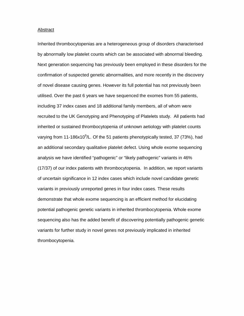
Abstract
Inherited thrombocytopenias are a heterogeneous group of disorders characterised
by abnormally low platelet counts which can be associated with abnormal bleeding.
Next generation sequencing has previously been employed in these disorders for the
confirmation of suspected genetic abnormalities, and more recently in the discovery
of novel disease causing genes. However its full potential has not previously been
utilised. Over the past 6 years we have sequenced the exomes from 55 patients,
including 37 index cases and 18 additional family members, all of whom were
recruited to the UK Genotyping and Phenotyping of Platelets study. All patients had
inherited or sustained thrombocytopenia of unknown aetiology with platelet counts
varying from 11-186x109/L. Of the 51 patients phenotypically tested, 37 (73%), had
an additional secondary qualitative platelet defect. Using whole exome sequencing
analysis we have identified “pathogenic” or “likely pathogenic” variants in 46%
(17/37) of our index patients with thrombocytopenia. In addition, we report variants
of uncertain significance in 12 index cases which include novel candidate genetic
variants in previously unreported genes in four index cases. These results
demonstrate that whole exome sequencing is an efficient method for elucidating
potential pathogenic genetic variants in inherited thrombocytopenia. Whole exome
sequencing also has the added benefit of discovering potentially pathogenic genetic
variants for further study in novel genes not previously implicated in inherited
thrombocytopenia.
Introduction
Inherited thrombocytopenias (IT) are a heterogeneous group of disorders
characterised by platelet counts of less than 150x109/L in whole blood. Platelet
counts are considered normal when maintained at levels between 150-450x109/L.
This is achieved by homeostatic processes controlling platelet production
(thrombopoiesis), platelet senescence and platelet consumption/destruction.
Pathogenic mutations can result in a disruption of these balanced processes causing
inherited thrombocytopenia. However, the clinical manifestation of bleeding is often
dependent on both a decreased platelet count and a qualitative or acquired platelet
defect. Clinical complications can vary dramatically from severe and potentially life
threatening bleeding to being asymptomatic. This variation is noted amongst
individuals shown to have the same underlying genetic causes of disease,
suggesting that bleeding risk and phenotype is a complex trait (1).
The average incidence of IT is estimated to be approximately 270 cases per 1 million
live births (2). To date there are 27 individual IT disorders with known causative
mutations registered within OMIM, although 33 disease causing genes have been
described (3).
Genetic studies have played a major role in the diagnosis and progressive
understanding of IT. The genes implicated in the disease encode proteins that vary
widely in function and include transcription factors (ETV6, FLI1, GATA1, GFI1B and
RUNX1) and proteins involved in cytoskeleton rearrangement and organisation
(ACTN1, FLNA, GP1BA, GP1BB, GP9, TUBB1 and WAS). However, some protein
functions currently remain unknown (SLFN14 and GNE) (4-9). Although our
knowledge of the causes of IT continues to grow, presently a genetic diagnosis is
only reported in approximately 50% of individuals (10-12).
Previously, genetic investigation into IT has focused on candidate gene sequencing
and individual cases of whole exome sequencing (WES) when a causative gene is
not obvious (9). With 50% of patients currently undiagnosed, a change in the way we
approach genetic diagnosis is necessary. Here we present the first large scale WES
only approach to patients with suspected IT. We demonstrate its application in
determining possible genetic origins of IT including identification of variants in novel
candidate causative genes. We combine this with an approach implemented by the
Genotyping and Phenotyping of Platelets (GAPP) study, which combines WES
analysis with extensive platelet phenotyping to create a complete method of
diagnosis and gene discovery in this subset of patients.
Methods
Study approval
The UK-GAPP study was approved by the National Research Ethics Service
Committee of West Midlands–Edgbaston (REC reference: 06/MRE07/36) and
participants gave written informed consent in accordance with the Declaration of
Helsinki. This study was registered at www.isrctn.org as #ISRCTN 77951167. The
GAPP study is included in the National Institute of Health Research Non-Malignant
Haematology study portfolio (ID9858).
Platelet counts, morphology and white blood cell counts
Patient samples were compared to the range of healthy volunteers for the specific
method of morphology used. Platelet counts for light transmission aggregometry
(LTA) and flow cytometry analysis as well as mean platelet volume (MPV) in platelet
rich plasma (PRP) were originally measured using the Beckman coulter counter
(n=44). Subsequently, platelet counts, morphology and white blood cell counts in
whole blood were measured using the Sysmex XN-1000 (n=11). The PLT-F channel
was used to determine platelet counts in whole blood and the immature platelet
fraction (IPF). MPV was determined from the impedance PLT-I channel. White blood
cell counts were determined using the Sysmex XN-DIFF channel. All samples were
tested against a normal range which was established by measuring the counts for 40
healthy individuals using the Sysmex XN-1000.
Platelet preparation and platelet function testing
Platelet function was assessed by light transmission aggregometry, including
lumiaggregometry, for samples having platelet counts in PRP of >1x10⁸/mL (n=13).
An in-house flow-cytometry assay was developed to assess platelet function in
patients having platelet counts in PRP <1x10⁸/mL (n=22). Platelets from individuals
with borderline platelet counts in PRP between 1.0 and 1.5x10⁸/mL were assessed
using both assays (n=16).
Aggregometry was performed as previously described (13, 14). For flow cytometry,
resting surface levels of CD42b, CD41 and GPVI were assessed. PRP was then
stimulated with ADP (3 and 30 μM), CRP (0.3 and 3μg/ml) and PAR-1 peptide (10
and 100 μM). Membrane expression of P-selectin (FITC-conjugated mouse anti-
human CD62P antibody, BD Pharmingen), a marker of platelet alpha granule
release, as well as fluorescent fibrinogen binding (marker of integrin activation) was
assessed by flow cytometry on an Accuri C6 flow cytometer. Incubation took place at
37ºC for 2 min and was terminated by adding a fivefold excess of ice cold PBS.
Whole exome sequencing
WES and bioinformatics analysis was performed as described previously (8, 15, 16)
(Figure 1).
Pathogenicity of variants was determined and called using the consensus guidelines
as set out by the American College of Medical Genetics and Genomics and the
Association for Molecular Pathology (ACMG guidelines) (17). Segregation was
determined by Sanger sequencing of candidate variants in both affected and
unaffected family members, when available, and classification was adapted
appropriately for the specific study and small sample size.
Sanger sequencing

To verify candidate mutations and examine their segregation among family members
Sanger sequencing was performed using standard methods on an ABI 3730
automated sequencer as described previously (8).
Results
Patient recruitment
To date, 55 patients with a suspected IT or sustained reduced platelet counts have
been enrolled from 25 UK Haemophilia Care Centres and investigated as part of the
GAPP study. Before enrolment in the study, all patients underwent clinical and
genetic work-up to exclude known platelet disorders (including Bernard-Soulier
Syndrome and MYH9-related disorders, analysed initially by blood film), idiopathic
thrombocytopenic purpura (ITP) and other non-platelet disorders including von
Willebrand disease and inherited coagulation factor deficiencies. Patient bleeding
phenotypes are displayed in Table 1. WES was performed on genomic DNA from all
patients, including 37 index cases, all of whom met the study’s entry criteria. All
patients, excluding F35.I and F35.II, were of white British or mixed British ethnicity.
All results following platelet function testing and WES were reported back to the
referring haematological consultants to aid in genetic counselling and disease
management.
Platelet counts, morphology and function testing
Patients were recruited with a platelet count in whole blood, at the time of enrolment,
of less than 150x109/L. Patients having platelet counts in the range of 150-200x109/L
remained enrolled in the study if they showed a similar phenotype to related affected
family members and a platelet count below 150x109/L had been observed prior to
enrolment (patients F4.II, F11.III, F13.I and F30.II). Platelet counts, MPVs and IPFs
are displayed in Table 1. Of the 55 recruited patients, 12 were deemed to have a
macrothrombocytopenia and three a microthrombocytopenia (Table 1). White cell
counts were within the normal range (normal range to 2SD 3.78 - 10.11x10⁹/L, n=40)
across all patients (n=11) analysed.
Platelet function studies revealed the presence of a secondary qualitative defect in
addition to the reduction in platelet count in 37/51 (73%) of the 55 patients whose
DNA underwent WES and who were also available for platelet function testing (PFT)
(Table 1). Of the 37 patients with a secondary qualitative defect, 89% (33/37)
displayed defects in both alpha and dense granule secretion. Five of these patients
with an observed granule secretion defect were also suspected to have an additional
Gi defect due to a reduction in response to all concentrations of ADP. The remaining
four patients without an observable granule secretion defect showed abnormalities in
alternative pathways (Integrin activation, cyclooxygenase pathway and GPVI surface
levels) in addition to the reduction in platelet count (Table 1).
Whole exome sequencing
WES was performed on genomic DNA from all 55 patients, comprising of 37 index
cases, following PFT. Average fold coverage of 111 was observed across all DNA
samples analysed by WES with an average of 91% of target sequences having >20x
coverage. Areas of poor coverage were analysed manually when occurring in
previously IT associated genes.
WES revealed between 24,000 and 25,000 variants (SNVs, small scale
insertions/deletions, and splice site variations) in the DNA from each patient, with an
average of 197 novel variants per exome. On average, per individual, 2401 variants
with a MAF of <0.01 were observed excluding synonymous variants. Over 99%
sensitivity and an approximate 3% false discovery rate was found by evaluating the
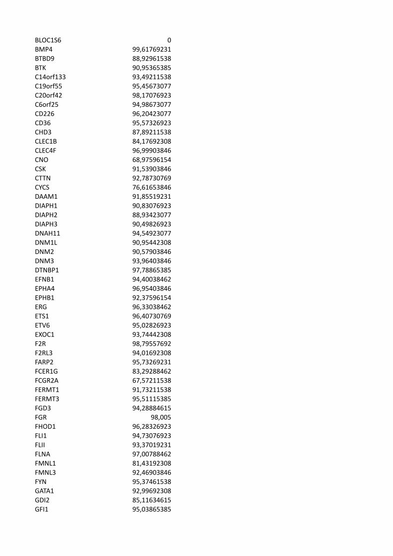
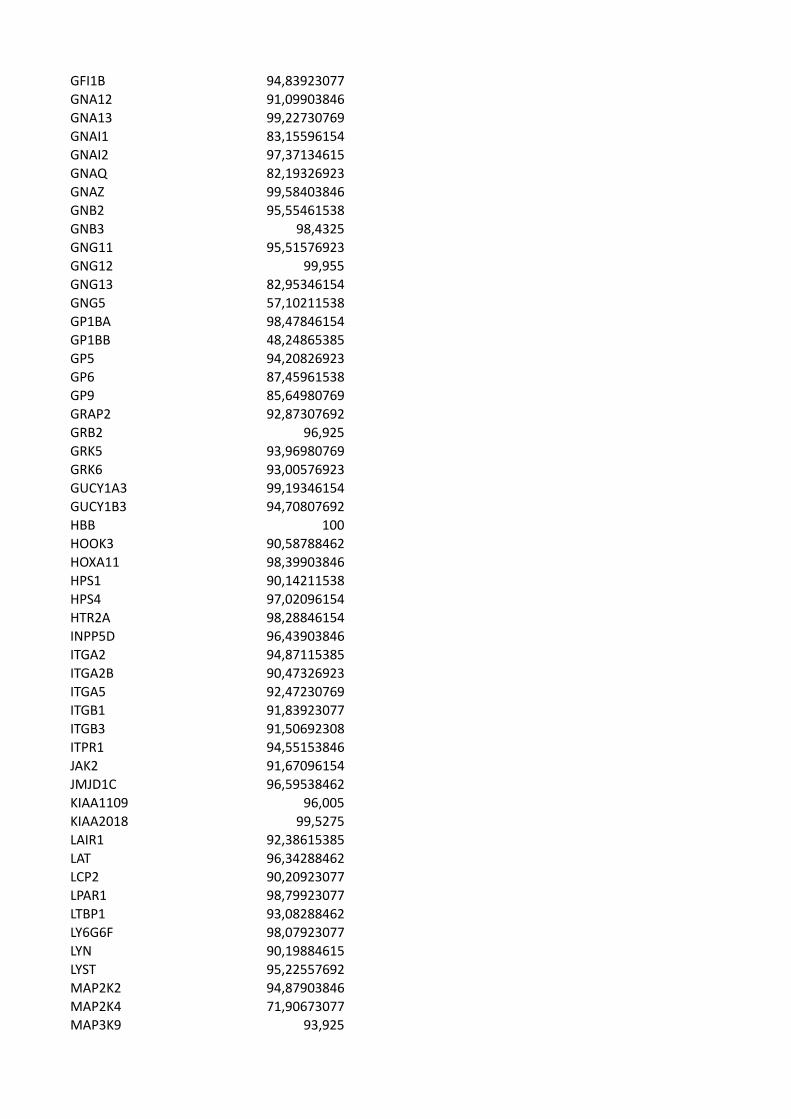
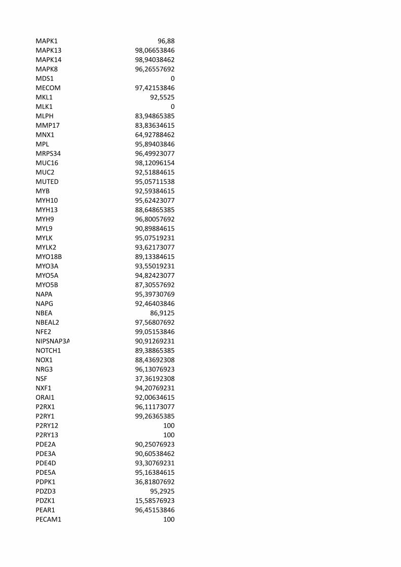
specificity of the pipeline in calling small variations. Percentage of the gene with

≤20x coverage for panel of 358 platelet-related genes is included within
Supplementary Table 1.
CNVs were detected using ExomeDepth (18) and analysis revealed an average of
137 CNVs per exome (n=32). No CNVs were deemed potential candidates either
due to a high allele frequency or a lack of expression or functional role of the gene
within the megakaryocyte/platelet lineage.
Variants in known thrombocytopenia causing genes
WES and downstream analysis identified variants in 25 index cases (68%) within the
33 known IT causing genes. All variants exceeded 30x sequence coverage at the
point of variation and have been confirmed by Sanger sequencing. Variants were
selected from positive hits to genes within the panel of 358 IT associated genes
(Supplementary Table 1). On average, 37 variants per individual (range 11-52) were
noted in genes from the panel of 358 IT associated genes, of which on average four
(range 0-7) variants were significant per exome analysed.
In total 28 variants were noted in 14 genes previously known to cause IT (Table 2).
Twenty-one index cases possessed a single variant in a gene previously known to
cause IT. Four index cases possessed two variants in genes previously known to
cause IT. One variant, RUNX1; c.270+1G>T, was noted in two index cases (F13.I
and F14.I). Candidate variations were present within; ACTN1, the 5’-UTR of
ANKRD26, CYCS, FLI1, GFI1B, ITGB3, GP1BA (heterozygous), MYH9, NBEAL2,
RUNX1, SLFN14, STIM1, TPM4 and TUBB1. All but six variants were novel and not
present within the variant databases previously mentioned. Three variants,
ANKRD26; c.-126T>G in F2.I, MYH9; c.3493C>T (rs80338829) in F9.I and RUNX1;
c.530G>A in F19.I and F19.II have been previously associated with IT (1, 19, 20).
The remaining three variants that have been previously observed occurred at
frequencies in available databases < 0.005 (0.05%). One of the databases
scrutinised was that of the ExAC consortium (http://exac.broadinstitute.org) which
may include data from individuals with low platelet counts who were either
undiagnosed or recruited through an unrelated study (Table 2). Seven variants have
previously been published as part of two separate publications from the UK-GAPP
study group (8, 15).
Classification of the 28 variants occurring within the known IT-related genes,
following the interpretation guidelines as set out by Richards et al. (17), revealed
four variants to be “pathogenic”, 13 to be “likely pathogenic” and 11 to be of
“uncertain significance”. Variants classified as “pathogenic” were either already
known to be a genetic cause of IT; ANKRD26; c.-126T>G in F2.I and MYH9;
p.Arg1165Cys in F9.I, or were predicted loss of function variants in genes where a
loss of function is known to cause disease; FLI1; p.Asn331Thr fs*4, in F4.I and F4.II
and RUNX1; pTrp79* in P12.I.
On average, less than one novel variant was expected to be observed in the known
IT causing genes in which variants were observed. The number of variants occurring
also exceeds the expected number when extending the analysis to cover variants
with a MAF of <0.01.
Of the 37 index patients, four presented with two candidate variations in known
disease linked genes, which in one case were present in the same gene. These
were as follows: F6.I (GFI1B; c.676+1G>A and STIM1; p.Ala610Thr), F10.I
(NBEAL2; (p.Leu459Arg fs*13 and p.Asn2298Ser), F12.I (RUNX1; p.Trp79* and
ITGB3 p.Arg117Trp) and F23.I (TPM4; p.Ala183Val and TUBB1; p.Phe242Leu).
Of the 25 index cases having variants in known disease causing genes, nine were
observed to have variants within the RUNT1-related transcription factor gene;
RUNX1. One variant, RUNX1; p.Arg177Gln, observed in F19.I and F19.II has been
previously reported as causative germline mutation of familial platelet disorder in two
individuals from the same pedigree (20). The variations consisted of five missense
variants, two splice-site variants and one nonsense variant. One splice-site variation,
c.270+1G>T, was present within three affected individuals from two separate
families (F13 and F14). All variants, with the exception of a missense substitution
(p.D6N), lie within the genetic region encoding the RUNT homology domain (RHD)
which mediates DNA binding and heterodimerization with CBFβ (Figure 2) (21).
Platelets from the majority of these patients (10/13) demonstrated a reduction in ATP
secretion and, in keeping with previous reports, several of these patients displayed
additional clinical features. Variations in RUNX1 are associated with a propensity to
myelodysplastic syndrome and acute myeloid leukaemia (AML). To date,
haematological malignancies have not been reported in any patients; however, the
brother of F16.1 did have a history of AML but was unavailable for testing.
Potentially damaging variants in novel candidate genes
After scrutinising individuals for variants within the panel of 358 platelet associated
genes (Supplementary table 1), individuals without a variant in a previously IT
associated gene were analysed for variants in novel genes. WES analysis revealed
potentially damaging candidate variants occurring within three families with currently
unknown genetic aetiology (Table 3). All candidate variants are novel (excluding a
previously annotated variant in MKL1; p.Val575Met, which occurs at a frequency of
0.007718 within the ExAC consortium), segregate with the disease status and have
been confirmed by Sanger sequencing.
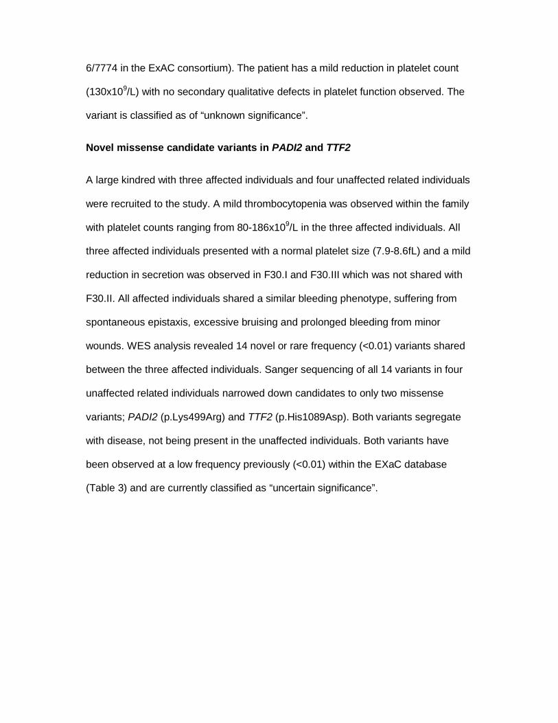
Variants within ANKRD18A, GNE and FRMPD1 in two related individuals from
consanguineous relationships
WES analysis of two related patients (F35.I and F35.II) of South Asian ethnicity was
approached differently to other patients in this study. Both patients displayed a
similarly severe clinical phenotype with a significant reduction in circulating platelets
(15x109/L). PFT revealed a reduction in P-selectin (CD62P) expression upon
stimulation and variable fluorescent fibrinogen binding which was consistent across
both affected individuals. The patients were cousins born from consanguineous
relationships within a single consanguineous kindred so analysis was focused on
identification of a shared homozygous variant due to the recessive segregation of
disease. Three variants occurring within ANKRD18A; p.Glu799del, GNE;
p.Gly447Arg and FRMPD1; p.Ala509Val were present within both affected
individuals and within a tightly linked region of homozygosity on chromosome 9p.
The variations within ANKRD18A and GNE were novel within the databases
previously mentioned whereas the variant in FRMPD1 has been observed at a
frequency of 0.0003708 including 39 times within the South Asian population
(rs571037699). There is no ClinVar entry for this variant and all three variants are
classified as variants of “uncertain significance”.
One missense variant in the recently proposed IT linked gene; MKL1
One individual was shown to harbour a rare frequency (<0.01) missense variant
within the Megakaryoblastic Leukaemia (translocation) 1 gene; MKL1. The variant
was the only variant occurring within a gene of haemostatic relevance within 109
significant novel variants. The variant; MKL1; c.1723G>A, p.Val575Met present in
patient F37.I has been noted previously at a frequency of 0.0007718 (allele count of
6/7774 in the ExAC consortium). The patient has a mild reduction in platelet count
(130x109/L) with no secondary qualitative defects in platelet function observed. The
variant is classified as of “unknown significance”.
Novel missense candidate variants in PADI2 and TTF2
A large kindred with three affected individuals and four unaffected related individuals
were recruited to the study. A mild thrombocytopenia was observed within the family
with platelet counts ranging from 80-186x109/L in the three affected individuals. All
three affected individuals presented with a normal platelet size (7.9-8.6fL) and a mild
reduction in secretion was observed in F30.I and F30.III which was not shared with
F30.II. All affected individuals shared a similar bleeding phenotype, suffering from
spontaneous epistaxis, excessive bruising and prolonged bleeding from minor
wounds. WES analysis revealed 14 novel or rare frequency (<0.01) variants shared
between the three affected individuals. Sanger sequencing of all 14 variants in four
unaffected related individuals narrowed down candidates to only two missense
variants; PADI2 (p.Lys499Arg) and TTF2 (p.His1089Asp). Both variants segregate
with disease, not being present in the unaffected individuals. Both variants have
been observed at a low frequency previously (<0.01) within the EXaC database
(Table 3) and are currently classified as “uncertain significance”.
Discussion
Here we present the first large scale application of WES analysis to patients with
inherited bleeding diatheses presenting with thrombocytopenia of unknown
aetiology.
Platelet counts and phenotypic presentation vary among our patients considerably
which is consistent with the variability observed in the spectrum of IT. However, the
majority of patients (73%) were noted to have a secondary qualitative defect in
platelet function which may explain the disproportionate bleeding when compared to
the patient’s platelet counts. A lack of consistency was noted in families 13 and 30
where affected individuals are observed both with and without defects in platelet
function. Clinical complications are shared among the affected family members so
this most likely represents limitations in the sensitivity of platelet function testing or
intra-familial variability.
Overall, when considering pathogenicity WES analysis revealed 46% of index cases
(17/37) to have a positive prediction of pathogenicity (classified “pathogenic” or
“likely pathogenic” in a gene consistent with the patients phenotype and zygosity
consistent with expected inheritance). Twenty-two percent of the index cases (8/37)
were of uncertain/possible pathogenicity (results classified of “uncertain significance”
in known IT causing genes). The remaining 32% of index cases (12/37) had a
negative prediction of pathogenicity (no convincing variants identified in known
causing genes). WES is not without its limitations and like with any genetic analysis
all variants must be functionally confirmed as deleterious to the coded protein.
However, our positive variant discovery rate is comparable to or exceeds previous
large scale WES clinical multicentre studies of Mendelian disorders (22, 23).
Focusing our genetic analysis on patients with unknown aetiology of disease with
minor prior genetic testing has produced a spectrum of variants different from
previous large scale targeted genetic studies of IT. Patients were recruited to the
study with clinically diagnosed bleeding disorders of unknown aetiology. One caveat
of this approach results in possible exclusion of individuals with known BSS and
MYH9-related disorders as these two forms of IT are routinely tested for in many
haematological centres within the UK. However, 3 index cases have been noted with
variants in either GP1BA or MYH9 in our analysis representing cases with atypical
presentation of BSS or MYH9-related disorder and therefore potentially falsely-
negatively reported cases. The individuals with variants within GP1BA and MYH9
showed a slight increase in MPV; however this was not at the magnitude of giant
platelets normally attributed to this group of disorders and only patient F9.I showed
any secondary syndromic symptoms with the individual suffering from congenital
cataracts.
One attribute of excluding patients with known variants in GP1BA, GP1BB, MYH9
and potentially GP9 is the discovery of a relatively large percentage of individuals
analysed (24% of index cases) with variants in RUNX1 as a primary likely cause of
disease. With the exception of one predicted loss of function variant, the variants
present within RUNX1 are currently classified as either “likely pathogenic” or of
“uncertain significance” and need functional confirmation to be disease causing.
However, the presence of these variants in a large number of individuals with an
often shared secondary functional defect in secretion does suggest the prevalence of
RUNX1 variants may be higher than previously thought. This raises the question
whether it should be considered as clinically significant as BSS and MYH9-related
disorders and be primarily screened for genetically upon initial diagnosis with IT.
An advantage of using WES is the lack of limitations allowing the possibility of finding
candidate variations in novel genes in cases that did not possess variants in known
IT genes. To determine whether these variants are pathogenic relies on functional
confirmation of the deleterious effect of the variant. However, WES analysis,
especially with combined segregation analysis by Sanger sequencing in extensive
kindreds, can provide indications as to which may be of scientific and clinical
relevance. This strategy has recently been utilised in the discovery of novel
candidate variations in SLFN14 initially as part of the GAPP study (15, 24).
Family 35 is an interesting case of two affected related individuals born from
consanguineous relationships. The molecular function of ANKRD18A is currently
unknown and FRMPD1 functions to regulate the subcellular localisation of activator
of G-protein signalling 3 (AGS3) (25). Both genes show weak expression in cells of
the haematopoietic lineage, however, GNE; an enzyme in the sialic acid biosynthetic
pathway, is expressed within all cells of the haematopoietic lineage. There are
currently 88 registered mutations in GNE within the Human Genome Mutation
Database (www.hgmd.cf.ac.uk). Mutations are known to be the genetic cause of
sialuria (OMIM269921) and Hereditary Inclusion Body Myopathy (HIBM;
OMIM600737) (26, 27). Recently, two separate groups have reported patients with
compound heterozygous variations in GNE, causing GNE related myopathy with
congenital thrombocytopenia (28, 29). Platelet counts within the four reported
affected individuals were below 45x109/L; however no MPV measurements were
recorded. None of the patients displayed signs of myopathy until mid-
adolescence/early adulthood; F35.I and F35.II are currently aged 10 and 6
respectively. Without functional characterisation of the effects of each variation, we
cannot definitively conclude the genetic aetiology of these two individuals’ severe
thrombocytopenia. However, WES analysis has allowed us to focus our efforts on
three potentially pathogenic variants in novel genes.
MKL1 was initially included in our panel of 358 genes for post WES analysis due to
its role in megakaryocyte maturation elucidated via its binding partner Serum
response factor (SRF) (30-32). Recently, the first case of a homozygous mutation in
MKL1 in a patient with a severe immunodeficiency and no haematological
malignancies was reported (33). One interesting phenotypic presentation within the
affected individual was an intermittent mild thrombocytopenia with a reduced platelet
count in whole blood of 50-150x109/L. Here we present one novel variant within
MKL1, at a highly conserved genetic site. The missense variant observed in F37.I
represents the only variant to occur in a gene with previous haematological
implications. One further variant in MKL1 was observed in addition to a “likely
pathogenic” frameshift causing insertion within TUBB1 in patient F25.I. Due to the
predicted loss of function of the frameshift causing TUBB1 variant it is unlikely that
the variant with MKL1 is additive to patient F25.I’s phenotype. However, the
presence of the variant of uncertain significance in patient F37.I is an interesting
candidate to take forward for functional studies.
WES and segregation determination using Sanger sequencing revealed candidate
variants in PADI2 and TTF2 that segregate with disease in F30.I, .II and .III.
Phenotypic presentation does vary between the patients but clinical presentation
remains consistent which may reflect limitations in the sensitivity of platelet function
testing. Neither gene has previously been implicated in haematological abnormalities
with mutations in PADI2 causing schizophrenia, breast cancer and rheumatoid
arthritis, and mutations in TTF2 associated with Thyroid dysgenesis (34-37). WES
analysis has therefore provided us with the first steps in determining the impact of
these two variants of uncertain significance and whether they have the propensity to
be disease causing.
In summary, we show WES can be applied to identify the underlying genetic cause
in known IT causing genes for patients with thrombocytopenia and unclear
aetiologies of disease. We show similar positive detection rates when compared to
prior targeted studies and with the addition of complementary functional studies
show an improved detection rate when compared to WES analysis of other
developmental disorders. We also suggest the applicability of WES in providing
preliminary insight into novel genes and their potential mechanism of action through
candidate variations of unknown significance. This approach provides a foundation
to enhance our current knowledge on megakaryopoiesis, platelet function and
platelet senescence/death upon subsequent functional studies.
Acknowledgements
We thank the families for providing samples and our clinical and laboratory colleagues for their
help. This work was supported by the British Heart Foundation (RG/PG/13/36/30275; RG/09/007),
an MRC Doctoral Training Partnership grant (BJ), a Wellcome Trust Combined Training
Programme Fellowship (093994) (GCL), the Healing Foundation (PH) and the Platelet Charity. We
thank the NIHR Haematology Specialty Group for their help in recruiting to the study, and all our
clinical investigators and collaborators. The authors also acknowledge support from the
Department of Health via the National Institute for Health Research (NIHR) comprehensive
Biomedical Research Centre award to Guy's & St Thomas' NHS Foundation Trust in partnership
with King's College London and King's College Hospital NHS Foundation Trust. We thank the
Queen Elizabeth Hospital Charity for funding the Sysmex XN-1000.
Authorship Contributions
BJ, GCL, ML, SPW and NVM designed the research. BJ, GCL, JF, ML, DM, MAS, ISG, SD, DB,
VL, SJF, BD, JR, PH and NVM performed the research and analysed data. GCL, DA, TB, PHB B-
M, PC, NC, CG, BJ, MM, JM, SP, KT, JT, JW and MW provided patient samples and clinical data.
GCL, ML, SPW and NVM undertook the research governance of the study. BJ and NVM wrote the
paper and all authors critically reviewed and edited the paper. GL, PH, PG, SM, AM, MD, SPW
and NVM coordinated the GAPP study.
Disclosure of Conflicts of Interest
PH was a previous consultant for Sysmex UK.
References
1. Noris P, Perrotta S, Seri M, et al. Mutations in ANKRD26 are responsible for a frequent form of inherited
thrombocytopenia: analysis of 78 patients from 21 families. Blood. 2011;117(24):6673-66780.
2. Balduini CL. Diagnosis and management of inherited thrombocytopenias. European Human Genetics
Conference 2014; 2014; Milan, Italy.
3. Johnson B, Fletcher SF, Morgan NV. Inherited thrombocytopenia: Novel insights into megakaryocyte
maturation, proplatelet formation and platelet lifespan. Platelets. 2016:1-7. Epub ahead of print.
4. Canales ML, Mauer AM. Sex-linked hereditary thrombocytopenia as a variant of Wiskott-Aldrich syndrome. N
Engl J Med. 1967;277(17):899-901.
5. Kunishima S, Kobayashi R, Itoh TJ, Hamaguchi M, Saito H. Mutation of the beta1-tubulin gene associated
with congenital macrothrombocytopenia affecting microtubule assembly. Blood. 2009;113(2):458-461.
6. Kunishima S, Okuno Y, Yoshida K, et al. ACTN1 mutations cause congenital macrothrombocytopenia. Am J
Hum Genet. 2013;92(3):431-438.
7. Nichols KE, Crispino JD, Poncz M, et al. Familial dyserythropoietic anaemia and thrombocytopenia due to an
inherited mutation in GATA1. Nat Genet. 2000;24(3):266-270.
8. Stockley J, Morgan NV, Bem D, et al. Enrichment of FLI1 and RUNX1 mutations in families with excessive
bleeding and platelet dense granule secretion defects. Blood. 2013;122(25):4090-4093.
9. Zhang MY, Churpek JE, Keel SB, et al. Germline ETV6 mutations in familial thrombocytopenia and
hematologic malignancy. Nat Genet. 2015;47(2):180-185.
10. Balduini CL, Pecci A, Noris P. Inherited thrombocytopenias: the evolving spectrum. Hamostaseologie.
2012;32(4):259-270.
11. Balduini CL, Savoia A. Genetics of familial forms of thrombocytopenia. Hum Genet. 2012;131(12):1821-1832.
12. Savoia A. Molecular basis of inherited thrombocytopenias. Clin Genet. 2016;89(2):154-162.
13. Dawood BB, Wilde J, Watson SP. Reference curves for aggregation and ATP secretion to aid diagnose of
platelet-based bleeding disorders: effect of inhibition of ADP and thromboxane A(2) pathways. Platelets.
2007;18(5):329-345.
14. Dawood BB, Lowe GC, Lordkipanidze M, et al. Evaluation of participants with suspected heritable platelet
function disorders including recommendation and validation of a streamlined agonist panel. Blood.
2012;120(25):5041-5049.
15. Fletcher SJ, Johnson B, Lowe GC, et al. SLFN14 mutations underlie thrombocytopenia with excessive bleeding
and platelet secretion defects. J Clin Invest. 2015;125(9):3600-3605.
16. Leo VC, Morgan NV, Bem D, et al. Use of next-generation sequencing and candidate gene analysis to identify
underlying defects in patients with inherited platelet function disorders. J Thromb Haemost. 2015;13(4):643-650.
17. Richards S, Aziz N, Bale S, et al. Standards and guidelines for the interpretation of sequence variants: a joint
consensus recommendation of the American College of Medical Genetics and Genomics and the Association for
Molecular Pathology. Genet Med. 2015;17(5):405-424.
18. Plagnol V, Curtis J, Epstein M, et al. A robust model for read count data in exome sequencing experiments
and implications for copy number variant calling. Bioinformatics. 2012;28(21):2747-2754.
19. Seri M, Cusano R, Gangarossa S, et al. Mutations in MYH9 result in the May-Hegglin anomaly, and Fechtner
and Sebastian syndromes. The May-Heggllin/Fechtner Syndrome Consortium. Nature genetics. 2000;26(1):103-105.
20. Preudhomme C, Renneville A, Bourdon V, et al. High frequency of RUNX1 biallelic alteration in acute myeloid
leukemia secondary to familial platelet disorder. Blood. 2009;113(22):5583-5587.
21. Kamachi Y, Ogawa E, Asano M, et al. Purification of a mouse nuclear factor that binds to both the A and B
cores of the polyomavirus enhancer. J Virol. 1990;64(10):4808-4819.
22. Chong JX, Buckingham KJ, Jhangiani SN, et al. The Genetic Basis of Mendelian Phenotypes: Discoveries,
Challenges, and Opportunities. Am J Hum Genet. 2015;97(2):199-215.
23. Yang Y, Muzny DM, Reid JG, et al. Clinical whole-exome sequencing for the diagnosis of mendelian disorders.
N Engl J Med. 2013;369(16):1502-1511.
24. Marconi C, Di Buduo CA, Barozzi S, et al. SLFN14-related thrombocytopenia: identification within a large
series of patients with inherited thrombocytopenia. Thromb Haemost. 2016;115(5):1076-1079.
25. An N, Blumer JB, Bernard ML, Lanier SM. The PDZ and band 4.1 containing protein Frmpd1 regulates the
subcellular location of activator of G-protein signaling 3 and its interaction with G-proteins. J Biol Chem.
2008;283(36):24718-24728.
26. Seppala R, Lehto VP, Gahl WA. Mutations in the human UDP-N-acetylglucosamine 2-epimerase gene define
the disease sialuria and the allosteric site of the enzyme. Am J Hum Genet. 1999;64(6):1563-1569.
27. Eisenberg I, Avidan N, Potikha T, et al. The UDP-N-acetylglucosamine 2-epimerase/N-acetylmannosamine
kinase gene is mutated in recessive hereditary inclusion body myopathy. Nat Genet. 2001;29(1):83-87.
28. Izumi R, Niihori T, Suzuki N, et al. GNE myopathy associated with congenital thrombocytopenia: a report of
two siblings. Neuromuscul Disord. 2014;24(12):1068-1072.
29. Zhen C, Guo F, Fang X, Liu Y, Wang X. A family with distal myopathy with rimmed vacuoles associated with
thrombocytopenia. Neurol Sci. 2014;35(9):1479-1481.
30. Cheng EC, Luo Q, Bruscia EM, et al. Role for MKL1 in megakaryocytic maturation. Blood. 2009;113(12):2826-
2834.
31. Smith EC, Thon JN, Devine MT, et al. MKL1 and MKL2 play redundant and crucial roles in megakaryocyte
maturation and platelet formation. Blood. 2012;120(11):2317-2329.
32. Halene S, Gao Y, Hahn K, et al. Serum response factor is an essential transcription factor in megakaryocytic
maturation. Blood. 2010;116(11):1942-1950.
33. Record J, Malinova D, Zenner HL, et al. Immunodeficiency and severe susceptibility to bacterial infection
associated with a loss-of-function homozygous mutation of MKL1. Blood. 2015;126(13):1527-1535.
34. Watanabe Y, Nunokawa A, Kaneko N, et al. A two-stage case-control association study of PADI2 with
schizophrenia. J Hum Genet. 2009;54(7):430-432.
35. McElwee JL, Mohanan S, Griffith OL, et al. Identification of PADI2 as a potential breast cancer biomarker and
therapeutic target. BMC Cancer. 2012;12:500.
36. Chang X, Xia Y, Pan J, Meng Q, Zhao Y, Yan X. PADI2 is significantly associated with rheumatoid arthritis. PLoS
One. 2013;8(12):e81259.
37. Castanet M, Polak M. Spectrum of Human Foxe1/TTF2 Mutations. Horm Res Paediatr. 2010;73(6):423-429.
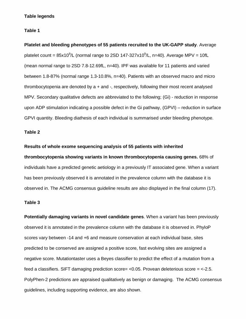
Table legends
Table 1
Platelet and bleeding phenotypes of 55 patients recruited to the UK-GAPP study. Average
platelet count = 85x109/L (normal range to 2SD 147-327x109/L, n=40). Average MPV = 10fL
(mean normal range to 2SD 7.8-12.69fL, n=40). IPF was available for 11 patients and varied
between 1.8-87% (normal range 1.3-10.8%, n=40). Patients with an observed macro and micro
thrombocytopenia are denoted by a + and -, respectively, following their most recent analysed
MPV. Secondary qualitative defects are abbreviated to the following; (Gi) - reduction in response
upon ADP stimulation indicating a possible defect in the Gi pathway, (GPVI) – reduction in surface
GPVI quantity. Bleeding diathesis of each individual is summarised under bleeding phenotype.
Table 2
Results of whole exome sequencing analysis of 55 patients with inherited
thrombocytopenia showing variants in known thrombocytopenia causing genes. 68% of
individuals have a predicted genetic aetiology in a previously IT associated gene. When a variant
has been previously observed it is annotated in the prevalence column with the database it is
observed in. The ACMG consensus guideline results are also displayed in the final column (17).
Table 3
Potentially damaging variants in novel candidate genes. When a variant has been previously
observed it is annotated in the prevalence column with the database it is observed in. PhyloP
scores vary between -14 and +6 and measure conservation at each individual base, sites
predicted to be conserved are assigned a positive score, fast evolving sites are assigned a
negative score. Mutationtaster uses a Beyes classifier to predict the effect of a mutation from a
feed a classifiers. SIFT damaging prediction score= <0.05. Provean deleterious score = <-2.5.
PolyPhen-2 predictions are appraised qualitatively as benign or damaging. The ACMG consensus
guidelines, including supporting evidence, are also shown.

Table 1
Family Patient Platelet count (x109/l) MPV (fL) IPF (%) Secondary defect Bleeding Phenotype
1 I 73 9.6 Yes (Fibrinogen) Cutaneous bruising/bleeding, menorrhagia 2 I 50 8.6 No Cutaneous bruising 3 I 80 10.3 Yes (Cyclooxygenase) Cutaneous bruising, menorrhagia II 50 12 Yes (Cyclooxygenase) Cutaneous bruising, epistaxis, purpura III 98 10.5 3.2 N/A Cutaneous bruising, epistaxis, purpura 4 I 142 11.8+ Yes (Secretion and Gi) Oral cavity bleeding, epistaxis, menorrhagia II 157 11.4+ Yes (Secretion) Cutaneous bruising, oral cavity bleeding 5 I 92 8.8 Yes (Secretion) Cutaneous bruising, epistaxis, bleeding into joints II 100 8.6 Yes (Secretion) Cutaneous bruising, life threatening bleeding following surgery 6 I 110 Yes (Secretion) Cutaneous bruising, excessive bleeding following surgery II 100 8.9 Yes (Secretion) Cutaneous bruising, epistaxis 7 I 50 10.4 Yes (Secretion) Cutaneous bruising, menorrhagia 8 I 70 10.7+ No Cutaneous bruising, menorrhagia, post-partum haemorrhage
II 70 10.1 No Cutaneous bruising, epistaxis, haematuria, menorrhagia, post-partum haemorrhage
9 I 35 11.4+ No Epistaxis, cutaneous bruising. 10 I 55 N/A Cutaneous bruising, epistaxis 11 I 62 Yes (Secretion) Cutaneous bruising
II N/A Yes (Secretion) Cutaneous bruising, epistaxis III 146 Yes (Secretion) Cutaneous bruising, epistaxis, menorrhagia
12 I 100 8 Yes (Secretion) Excessive cutaneous bleeding 13 I 163 9.1 No Cutaneous bruising, epistaxis, haematoma
II 45 11.9+ Yes (GPVI) Cutaneous bruising, epistaxis 14 I 139 8 Yes (Secretion) Epistaxis, haematoma 15 I 90 7.6- Yes (Secretion) Cutaneous bruising, petechiae 16 I 130 7.1- Yes (Secretion and Gi) Cutaneous bruising, epistaxis, oral cavity bleeding
II 70 7.5- Yes (Secretion and Gi) Cutaneous bruising, epistaxis, oral cavity bleeding 17 I N/A N/A N/A Excessive bruising/bleeding 18 I 110 8.1 Yes (Secretion) Cutaneous bruising/bleeding, petechiae, haematoma 19 I 100 9 No Cutaneous bruising/bleeding
II 100 9.2 No Cutaneous bruising/bleeding 20 I 89 13+ 17.5 Yes (Secretion and Gi) Cutaneous bruising 21 I 63 11.9 19.1 Yes (Secretion) Cutaneous bruising, epistaxis, haematoma
II 83 11.9 24.3 Yes (Secretion) Cutaneous bruising, epistaxis, haematoma 22 I 74 11.2 Yes (Secretion and Gi) Cutaneous bruising/bleeding, haematuria
II 62 12.7+ 20.8 Yes (Secretion and Gi) Cutaneous bruising/bleeding, menorrhagia, post-partum haemorrhage, haematoma
III 109 11 Yes (Secretion) Cutaneous bruising, haematoma, menorrhagia 23 I 119 11.1 Yes (Secretion) Cutaneous bruising/bleeding 24 I 104 9.6 No Menorrhagia, post-partum haemorrhage
II 133 8.6 No Epistaxis 25 I 11 13.4+ Yes (Secretion and Gi) Cutaneous bruising 26 I 43 14+ No Cutaneous bruising, menorrhagia, oral cavity bleeding 27 I 100 10.3 Yes (Secretion) Cutaneous bruising/bleeding, epistaxis, oral cavity bleeding 28 I 25 8.5 Yes (Secretion) Cutaneous bruising 29 I 15 9.4 Yes (Secretion) Haematomas 30 I 137 7.9 Yes (Secretion) Cutaneous bruising, epistaxis, menorrhagia
II 186 8.6 No Cutaneous bruising, menorrhagia, haematuria III 80 8.2 Yes (Secretion) Cutaneous bruising
31 I 20 9.7 N/A Cutaneous bruising, epistaxis, oral cavity bleeding 32 I 15 9.5 20.2 No Cutaneous bruising 33 I 66 9.9 1.8 Yes (Secretion) Cutaneous bleeding 34 I 93 14.4+ 20.5 Yes (Secretion) Cutaneous bleeding, epistaxis
35 I 15 10.4 87 Yes (Secretion and other) Cutaneous bruising, epistaxis, haematomas
II 14 15+ 83 Yes (Secretion and other) Cutaneous bleeding
36 I 104 13.3+ 17 No Menorrhagia 37 I 130 9.7 No Cutaneous bruising, epistaxis, menorrhagia
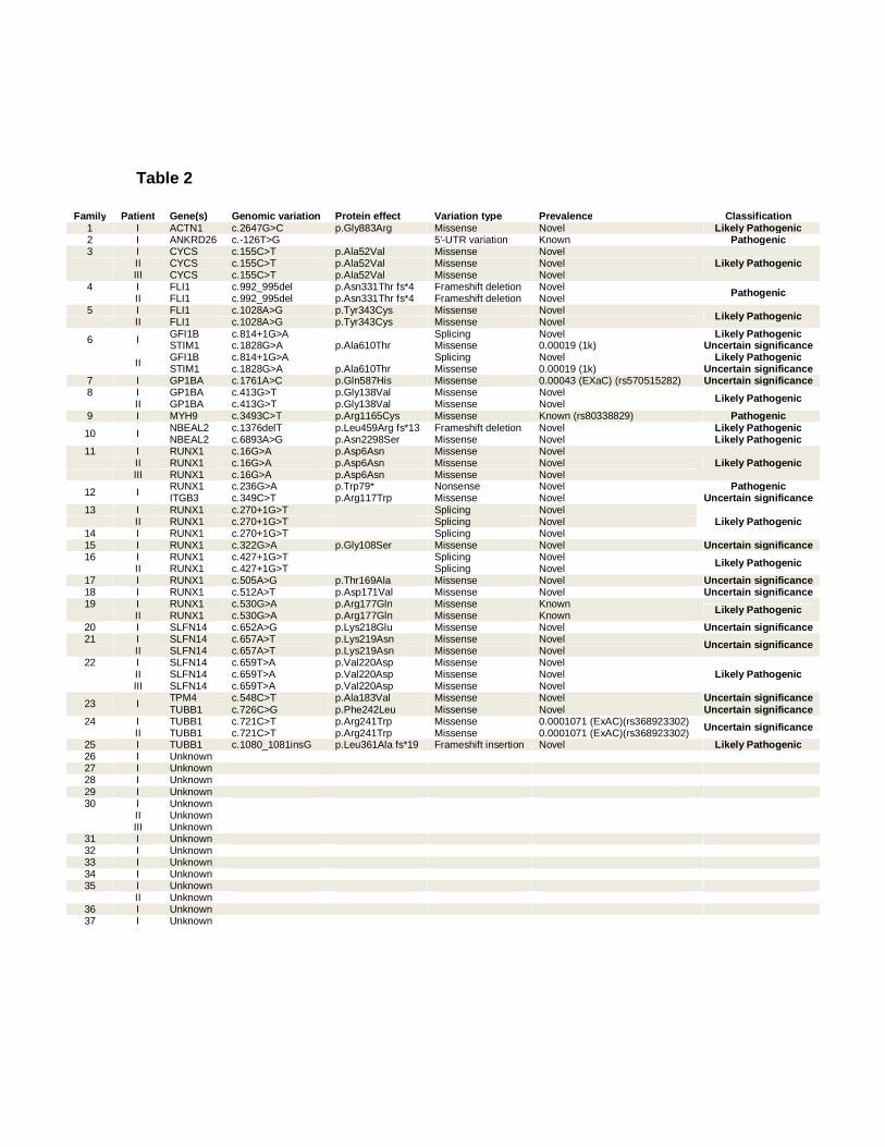
Table 2
Family Patient Gene(s) Genomic variation Protein effect Variation type Prevalence Classification 1 I ACTN1 c.2647G>C p.Gly883Arg Missense Novel Likely Pathogenic 2 I ANKRD26 c.-126T>G 5'-UTR variation Known Pathogenic 3 I CYCS c.155C>T p.Ala52Val Missense Novel
Likely Pathogenic II CYCS c.155C>T p.Ala52Val Missense Novel III CYCS c.155C>T p.Ala52Val Missense Novel
4 I FLI1 c.992_995del p.Asn331Thr fs*4 Frameshift deletion Novel Pathogenic II FLI1 c.992_995del p.Asn331Thr fs*4 Frameshift deletion Novel 5 I FLI1 c.1028A>G p.Tyr343Cys Missense Novel Likely Pathogenic II FLI1 c.1028A>G p.Tyr343Cys Missense Novel
6 I GFI1B c.814+1G>A Splicing Novel Likely Pathogenic STIM1 c.1828G>A p.Ala610Thr Missense 0.00019 (1k) Uncertain significance
II GFI1B c.814+1G>A Splicing Novel Likely Pathogenic
STIM1 c.1828G>A p.Ala610Thr Missense 0.00019 (1k) Uncertain significance 7 I GP1BA c.1761A>C p.Gln587His Missense 0.00043 (EXaC) (rs570515282) Uncertain significance 8 I GP1BA c.413G>T p.Gly138Val Missense Novel Likely Pathogenic II GP1BA c.413G>T p.Gly138Val Missense Novel 9 I MYH9 c.3493C>T p.Arg1165Cys Missense Known (rs80338829) Pathogenic
10 I NBEAL2 c.1376delT p.Leu459Arg fs*13 Frameshift deletion Novel Likely Pathogenic NBEAL2 c.6893A>G p.Asn2298Ser Missense Novel Likely Pathogenic
11 I RUNX1 c.16G>A p.Asp6Asn Missense Novel Likely Pathogenic II RUNX1 c.16G>A p.Asp6Asn Missense Novel
III RUNX1 c.16G>A p.Asp6Asn Missense Novel
12 I RUNX1 c.236G>A p.Trp79* Nonsense Novel Pathogenic ITGB3 c.349C>T p.Arg117Trp Missense Novel Uncertain significance
13 I RUNX1 c.270+1G>T Splicing Novel Likely Pathogenic II RUNX1 c.270+1G>T Splicing Novel
14 I RUNX1 c.270+1G>T Splicing Novel 15 I RUNX1 c.322G>A p.Gly108Ser Missense Novel Uncertain significance 16 I RUNX1 c.427+1G>T Splicing Novel Likely Pathogenic II RUNX1 c.427+1G>T Splicing Novel 17 I RUNX1 c.505A>G p.Thr169Ala Missense Novel Uncertain significance 18 I RUNX1 c.512A>T p.Asp171Val Missense Novel Uncertain significance 19 I RUNX1 c.530G>A p.Arg177Gln Missense Known Likely Pathogenic II RUNX1 c.530G>A p.Arg177Gln Missense Known 20 I SLFN14 c.652A>G p.Lys218Glu Missense Novel Uncertain significance 21 I SLFN14 c.657A>T p.Lys219Asn Missense Novel Uncertain significance II SLFN14 c.657A>T p.Lys219Asn Missense Novel
22 I SLFN14 c.659T>A p.Val220Asp Missense Novel Likely Pathogenic II SLFN14 c.659T>A p.Val220Asp Missense Novel
III SLFN14 c.659T>A p.Val220Asp Missense Novel
23 I TPM4 c.548C>T p.Ala183Val Missense Novel Uncertain significance TUBB1 c.726C>G p.Phe242Leu Missense Novel Uncertain significance
24 I TUBB1 c.721C>T p.Arg241Trp Missense 0.0001071 (ExAC)(rs368923302) Uncertain significance II TUBB1 c.721C>T p.Arg241Trp Missense 0.0001071 (ExAC)(rs368923302)
25 I TUBB1 c.1080_1081insG p.Leu361Ala fs*19 Frameshift insertion Novel Likely Pathogenic 26 I Unknown 27 I Unknown 28 I Unknown 29 I Unknown 30 I Unknown II Unknown III Unknown
31 I Unknown 32 I Unknown 33 I Unknown 34 I Unknown 35 I Unknown II Unknown
36 I Unknown 37 I Unknown
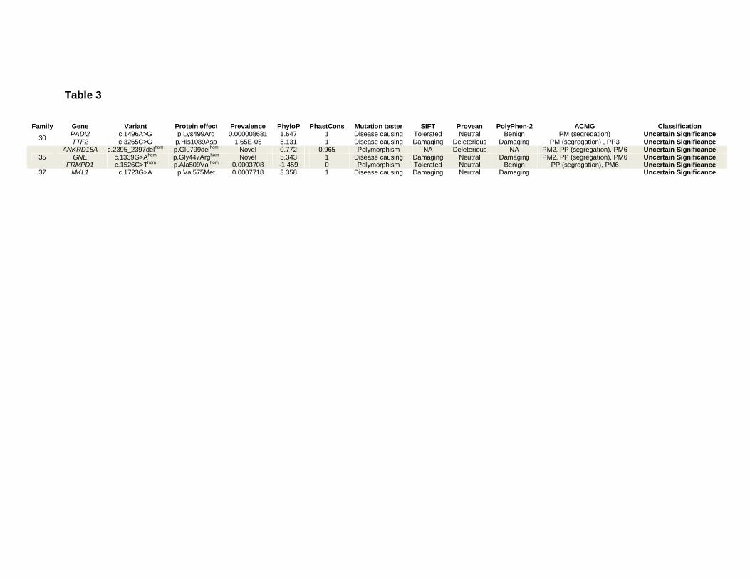
Table 3
Family Gene Variant Protein effect Prevalence PhyloP PhastCons Mutation taster SIFT Provean PolyPhen-2 ACMG Classification
30 PADI2 c.1496A>G p.Lys499Arg 0.000008681 1.647 1 Disease causing Tolerated Neutral Benign PM (segregation) Uncertain Significance TTF2 c.3265C>G p.His1089Asp 1.65E-05 5.131 1 Disease causing Damaging Deleterious Damaging PM (segregation) , PP3 Uncertain Significance
35 ANKRD18A c.2395_2397delhom p.Glu799delhom Novel 0.772 0.965 Polymorphism NA Deleterious NA PM2, PP (segregation), PM6 Uncertain Significance
GNE c.1339G>Ahom p.Gly447Arghom Novel 5.343 1 Disease causing Damaging Neutral Damaging PM2, PP (segregation), PM6 Uncertain Significance FRMPD1 c.1526C>Thom p.Ala509Valhom 0.0003708 -1.459 0 Polymorphism Tolerated Neutral Benign PP (segregation), PM6 Uncertain Significance
37 MKL1 c.1723G>A p.Val575Met 0.0007718 3.358 1 Disease causing Damaging Neutral Damaging Uncertain Significance
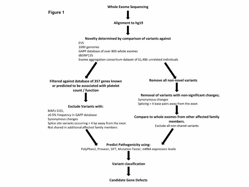
Figure 1
Bioinformatics pipeline analysis of whole exome sequencing data. Initial WES analysis
focused upon comparison to a panel of 358 genes (supplementary table 1). After which screening
of exomes variants focused upon novel variants. Variant classification was performed utilising the
ACMG consensus guidelines.
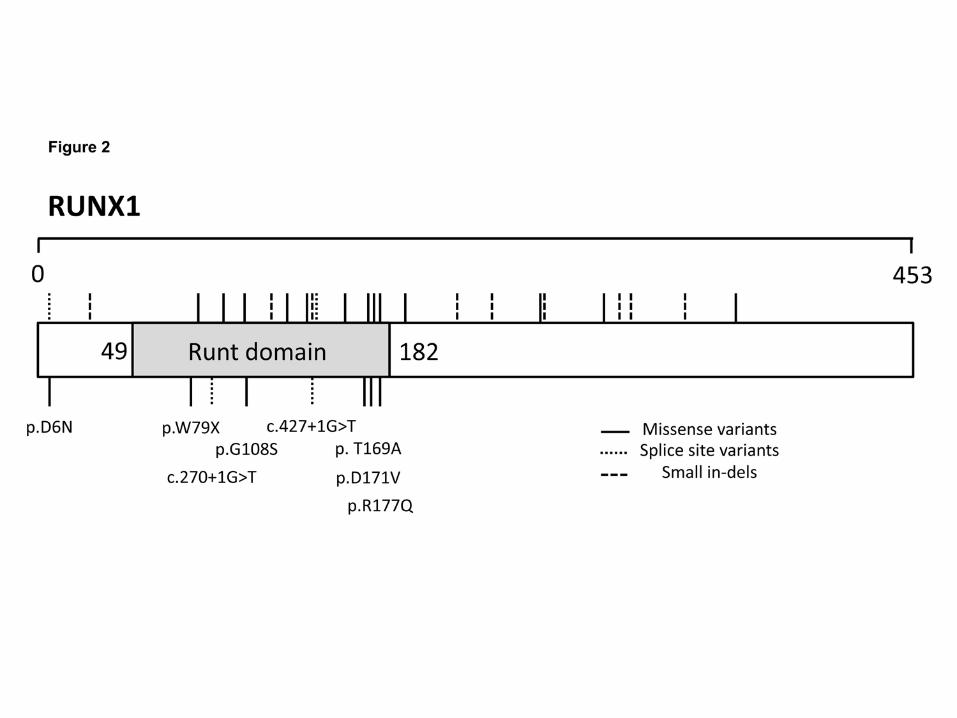
Figure 2
Spatial amino acid locations of all thrombocytopenia causing variants present within RUNT
transcription factor 1 (RUNX1) (RefSeq NP_001001890). Previously disease causing variants
found within the HGMD (www.hgmd.cf.ac.uk) and ClinVar (www.ncbi.nlm.nih.gov/clinvar/)
databases are denoted above. The eight variants found within RUNX1 in the GAPP cohort of 54
patients who have undergone whole exome sequencing are denoted below and the effect on the
protein or predicted splice-site shown.
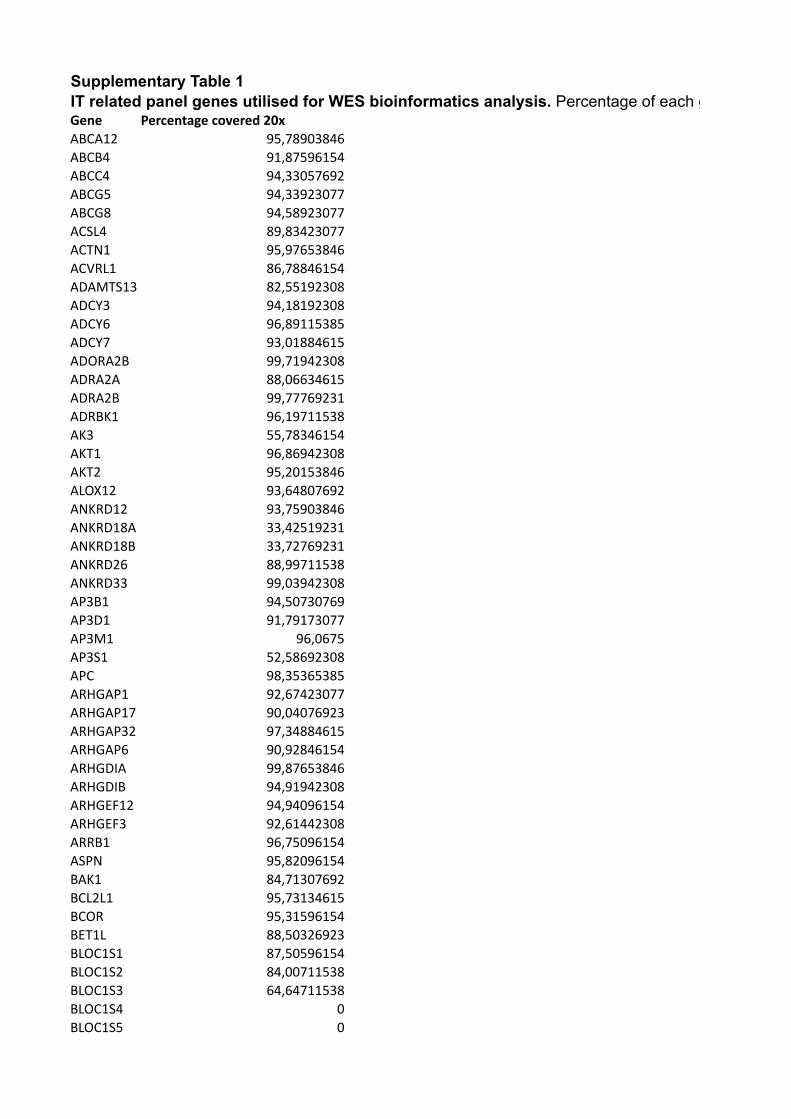
Supplementary Table 1IT related panel genes utilised for WES bioinformatics analysis. Percentage of each gene covered ≤20x is also shown.Gene Percentage covered 20xABCA12 95,78903846ABCB4 91,87596154ABCC4 94,33057692ABCG5 94,33923077ABCG8 94,58923077ACSL4 89,83423077ACTN1 95,97653846ACVRL1 86,78846154ADAMTS13 82,55192308ADCY3 94,18192308ADCY6 96,89115385ADCY7 93,01884615ADORA2B 99,71942308ADRA2A 88,06634615ADRA2B 99,77769231ADRBK1 96,19711538AK3 55,78346154AKT1 96,86942308AKT2 95,20153846ALOX12 93,64807692ANKRD12 93,75903846ANKRD18A 33,42519231ANKRD18B 33,72769231ANKRD26 88,99711538ANKRD33 99,03942308AP3B1 94,50730769AP3D1 91,79173077AP3M1 96,0675AP3S1 52,58692308APC 98,35365385ARHGAP1 92,67423077ARHGAP17 90,04076923ARHGAP32 97,34884615ARHGAP6 90,92846154ARHGDIA 99,87653846ARHGDIB 94,91942308ARHGEF12 94,94096154ARHGEF3 92,61442308ARRB1 96,75096154ASPN 95,82096154BAK1 84,71307692BCL2L1 95,73134615BCOR 95,31596154BET1L 88,50326923BLOC1S1 87,50596154BLOC1S2 84,00711538BLOC1S3 64,64711538BLOC1S4 0BLOC1S5 0
BLOC1S6 0BMP4 99,61769231BTBD9 88,92961538BTK 90,95365385C14orf133 93,49211538C19orf55 95,45673077C20orf42 98,17076923C6orf25 94,98673077CD226 96,20423077CD36 95,57326923CHD3 87,89211538CLEC1B 84,17692308CLEC4F 96,99903846CNO 68,97596154CSK 91,53903846CTTN 92,78730769CYCS 76,61653846DAAM1 91,85519231DIAPH1 90,83076923DIAPH2 88,93423077DIAPH3 90,49826923DNAH11 94,54923077DNM1L 90,95442308DNM2 90,57903846DNM3 93,96403846DTNBP1 97,78865385EFNB1 94,40038462EPHA4 96,95403846EPHB1 92,37596154ERG 96,33038462ETS1 96,40730769ETV6 95,02826923EXOC1 93,74442308F2R 98,79557692F2RL3 94,01692308FARP2 95,73269231FCER1G 83,29288462FCGR2A 67,57211538FERMT1 91,73211538FERMT3 95,51115385FGD3 94,28884615FGR 98,005FHOD1 96,28326923FLI1 94,73076923FLII 93,37019231FLNA 97,00788462FMNL1 81,43192308FMNL3 92,46903846FYN 95,37461538GATA1 92,99692308GDI2 85,11634615GFI1 95,03865385
GFI1B 94,83923077GNA12 91,09903846GNA13 99,22730769GNAI1 83,15596154GNAI2 97,37134615GNAQ 82,19326923GNAZ 99,58403846GNB2 95,55461538GNB3 98,4325GNG11 95,51576923GNG12 99,955GNG13 82,95346154GNG5 57,10211538GP1BA 98,47846154GP1BB 48,24865385GP5 94,20826923GP6 87,45961538GP9 85,64980769GRAP2 92,87307692GRB2 96,925GRK5 93,96980769GRK6 93,00576923GUCY1A3 99,19346154GUCY1B3 94,70807692HBB 100HOOK3 90,58788462HOXA11 98,39903846HPS1 90,14211538HPS4 97,02096154HTR2A 98,28846154INPP5D 96,43903846ITGA2 94,87115385ITGA2B 90,47326923ITGA5 92,47230769ITGB1 91,83923077ITGB3 91,50692308ITPR1 94,55153846JAK2 91,67096154JMJD1C 96,59538462KIAA1109 96,005KIAA2018 99,5275LAIR1 92,38615385LAT 96,34288462LCP2 90,20923077LPAR1 98,79923077LTBP1 93,08288462LY6G6F 98,07923077LYN 90,19884615LYST 95,22557692MAP2K2 94,87903846MAP2K4 71,90673077MAP3K9 93,925
MAPK1 96,88MAPK13 98,06653846MAPK14 98,94038462MAPK8 96,26557692MDS1 0MECOM 97,42153846MKL1 92,5525MLK1 0MLPH 83,94865385MMP17 83,83634615MNX1 64,92788462MPL 95,89403846MRPS34 96,49923077MUC16 98,12096154MUC2 92,51884615MUTED 95,05711538MYB 92,59384615MYH10 95,62423077MYH13 88,64865385MYH9 96,80057692MYL9 90,89884615MYLK 95,07519231MYLK2 93,62173077MYO18B 89,13384615MYO3A 93,55019231MYO5A 94,82423077MYO5B 87,30557692NAPA 95,39730769NAPG 92,46403846NBEA 86,9125NBEAL2 97,56807692NFE2 99,05153846NIPSNAP3A 90,91269231NOTCH1 89,38865385NOX1 88,43692308NRG3 96,13076923NSF 37,36192308NXF1 94,20769231ORAI1 92,00634615P2RX1 96,11173077P2RY1 99,26365385P2RY12 100P2RY13 100PDE2A 90,25076923PDE3A 90,60538462PDE4D 93,30769231PDE5A 95,16384615PDPK1 36,81807692PDZD3 95,2925PDZK1 15,58576923PEAR1 96,45153846PECAM1 100
PGM3 94,78634615PHOX2A 54,54903846PIK3CA 90,36384615PIK3CB 94,71692308PIK3CD 87,63288462PIK3CG 98,25461538PIK3R1 95,99730769PIK3R3 96,83307692PIK3R5 96,96980769PLA2G4A 96,91615385PLA2G4C 93,49153846PLCB2 97,0875PLCB3 92,18576923PLCG2 94,48692308PLDN 75,15980769PPP1CA 99,00038462PPP1CB 97,86730769PPP1CC 98,79423077PPP1R12A 91,18615385PPP1R12C 78,43576923PPP1R14A 85,47346154PPP1R2 45,66384615PRKACA 96,39538462PRKACB 92,21788462PRKACG 100PRKAR1A 77,49673077PRKAR2A 94,83557692PRKCA 92,09307692PRKCB 94,39903846PRKCD 96,76076923PRKCQ 91,7475PRKD1 98,66307692PRKG1 91,3625PRKG2 95,72442308PTEN 83,83673077PTGIR 92,86269231PTGS1 99,03711538PTK2 93,98653846PTPN1 93,99519231PTPN11 55,94115385PTPN12 95,05057692PTPN18 91,53596154PTPN2 70,88884615PTPN6 94,96384615PTPN7 88,78PTPN9 92,46019231PTPRA 97,68PTPRC 91,14269231PTPRJ 92,07307692RAB27A 99,31019231RAB27B 97,62057692RAB38 99,27326923
RAB4A 90,98846154RABGGTA 90,39365385RAC1 56,83538462RAF1 93,3425RAI1 98,35096154RAP1B 58,62884615RAP1GAP 92,23673077RAP1GAP2 94,07826923RAP1GDS1 85,94403846RASGRP2 90,82RBM8A 85,60326923RGS10 90,32192308RGS18 94,02134615RGS19 96,77903846RGS20 91,42634615RGS9 92,94538462RHOA 95,28730769RHOC 96,63519231RHOF 92,28615385ROCK1 86,35980769ROCK2 94,94538462RUNX1 89,41538462SCAMP2 87,93230769SCAMP5 85,06346154SCFD1 94,55307692SELP 94,06769231SERPINE2 93,29230769SH2B3 87,26384615SIRPA 74,93711538SLC35D3 90,26326923SLC9A3R1 94,555SLC9A3R2 75,83384615SLFN14 95,47711538SMAD1 97,22134615SMAD6 79,03769231SNAP23 89,95788462SNAP25 89,25403846SNAP29 99,19SNAPIN 93,63538462SNX1 88,6075SRA1 94,33307692SRC 88,58730769SRF 91,81730769STIM1 90,21519231STOM 91,51615385STX11 99,55711538STX12 96,40942308STX2 90,69057692STX4 93,61519231STX6 96,60596154STX7 98,15153846STXBP1 95,39442308
STXBP2 96,60769231STXBP3 93,66923077STXBP4 93,48961538STXBP5L 95,54057692STXBP6 88,68615385SUZ12 70,34346154SYK 97,63346154SYTL3 91,77326923SYTL4 93,77846154TAL1 76,37923077TAOK1 93,57307692TBXA2R 82,61076923TEC 91,73115385TGFBR3 94,92346154THPO 90,60980769TLN1 97,19653846TLR2 98,30980769TMCC2 90,78711538TPM1 96,63076923TPM4 77,58057692TRAF4 95,36480769TREML1 98,26788462TRPM7 94,52211538TTC37 97,67211538TUBA3C 76,80365385TUBB1 99,98019231UNC13A 94,45807692UNC13B 97,44692308VAMP2 99,25480769VAMP3 98,30519231VAMP7 16,34615385VAMP8 84,83596154VAV1 91,29730769VAV2 95,41961538VAV3 94,33903846VPS11 98,45057692VPS16 94,97403846VPS18 98,01461538VPS33A 95,89807692VPS33B 95,43365385VPS39 94,17423077VPS41 91,65442308VPS4B 97,05673077VPS52 92,45634615VPS8 90,68557692VWF 75,24192308WAS 82,84884615WDR66 92,87173077ZFPMI 66,54634615

IT related panel genes utilised for WES bioinformatics analysis. Percentage of each gene covered ≤20x is also shown.