WHO/BS/2015.2269 ENGLISH ONLY - WHO | World … · WHO/BS/2015.2269 Page 3 Platelet Transfusion...
64
WHO/BS/2015.2269 ENGLISH ONLY EXPERT COMMITTEE ON BIOLOGICAL STANDARDIZATION Geneva, 12 to 16 October 2015 Collaborative Study to Enlarge the First WHO Repository of Platelet Transfusion-Relevant Bacterial Reference Strains Eva Spindler-Raffel, Kay-Martin Hanschmann, Thomas Montag-Lessing†* and the Collaborative Study Group** *Division of EU cooperation / Microbiology, Head Dr Isabelle Bekeredjian-Ding Paul-Ehrlich-Institut, Paul-Ehrlich-Strasse 51-59, D-63225 Langen, Germany **ISBT WP Transfusion Transmitted Infectious Diseases Subgroup on Bacteria Chairs: Carl P McDonald and Richard J Benjamin See Appendix 1 NOTE: This document has been prepared for the purpose of inviting comments and suggestions on the proposals contained therein, which will then be considered by the Expert Committee on Biological Standardization (ECBS). Comments MUST be received by 14 September 2015 and should be addressed to the World Health Organization, 1211 Geneva 27, Switzerland, attention: Technologies, Standards and Norms (TSN). Comments may also be submitted electronically to the Responsible Officer: Dr M. Nübling at email: [email protected] © World Health Organization 2015 All rights reserved. Publications of the World Health Organization are available on the WHO web site (www.who.int) or can be purchased from WHO Press, World Health Organization, 20 Avenue Appia, 1211 Geneva 27, Switzerland (tel.: +41 22 791 3264; fax: +41 22 791 4857; e-mail: [email protected]). Requests for permission to reproduce or translate WHO publications – whether for sale or for noncommercial distribution – should be addressed to WHO Press through the WHO web site: (http://www.who.int/about/licensing/copyright_form/en/index.html). The designations employed and the presentation of the material in this publication do not imply the expression of any opinion whatsoever on the part of the World Health Organization concerning the legal status of any country, territory, city or area or of its authorities, or concerning the delimitation of its frontiers or boundaries. Dotted lines on maps represent approximate border lines for which there may not yet be full agreement. The mention of specific companies or of certain manufacturers’ products does not imply that they are endorsed or recommended by the World Health Organization in preference to others of a similar nature that are not mentioned. Errors and omissions excepted, the names of proprietary products are distinguished by initial capital letters. All reasonable precautions have been taken by the World Health Organization to verify the information contained in this publication. However, the published material is being distributed without warranty of any kind, either expressed or implied. The responsibility for the interpretation and use of the material lies with the reader. In no event shall the World Health Organization be liable for damages arising from its use. The named authors alone are responsible for the views expressed in this publication.
Transcript of WHO/BS/2015.2269 ENGLISH ONLY - WHO | World … · WHO/BS/2015.2269 Page 3 Platelet Transfusion...

WHO/BS/2015.2269
ENGLISH ONLY
EXPERT COMMITTEE ON BIOLOGICAL STANDARDIZATION
Geneva, 12 to 16 October 2015
Collaborative Study to Enlarge the First WHO Repository of Platelet
Transfusion-Relevant Bacterial Reference Strains
Eva Spindler-Raffel, Kay-Martin Hanschmann, Thomas Montag-Lessing†*
and the Collaborative Study Group**
*Division of EU cooperation / Microbiology, Head Dr Isabelle Bekeredjian-Ding
Paul-Ehrlich-Institut, Paul-Ehrlich-Strasse 51-59, D-63225 Langen, Germany
**ISBT WP Transfusion Transmitted Infectious Diseases Subgroup on Bacteria
Chairs: Carl P McDonald and Richard J Benjamin
See Appendix 1
NOTE:
This document has been prepared for the purpose of inviting comments and suggestions on the
proposals contained therein, which will then be considered by the Expert Committee on
Biological Standardization (ECBS). Comments MUST be received by 14 September 2015 and
should be addressed to the World Health Organization, 1211 Geneva 27, Switzerland, attention:
Technologies, Standards and Norms (TSN). Comments may also be submitted electronically to
the Responsible Officer: Dr M. Nübling at email: [email protected]
© World Health Organization 2015
All rights reserved. Publications of the World Health Organization are available on the WHO web site (www.who.int) or can be
purchased from WHO Press, World Health Organization, 20 Avenue Appia, 1211 Geneva 27, Switzerland (tel.: +41 22 791
3264; fax: +41 22 791 4857; e-mail: [email protected]).
Requests for permission to reproduce or translate WHO publications – whether for sale or for noncommercial distribution –
should be addressed to WHO Press through the WHO web site:
(http://www.who.int/about/licensing/copyright_form/en/index.html).
The designations employed and the presentation of the material in this publication do not imply the expression of any opinion
whatsoever on the part of the World Health Organization concerning the legal status of any country, territory, city or area or of its
authorities, or concerning the delimitation of its frontiers or boundaries. Dotted lines on maps represent approximate border lines
for which there may not yet be full agreement.
The mention of specific companies or of certain manufacturers’ products does not imply that they are endorsed or recommended
by the World Health Organization in preference to others of a similar nature that are not mentioned. Errors and omissions
excepted, the names of proprietary products are distinguished by initial capital letters.
All reasonable precautions have been taken by the World Health Organization to verify the information contained in this
publication. However, the published material is being distributed without warranty of any kind, either expressed or implied. The
responsibility for the interpretation and use of the material lies with the reader. In no event shall the World Health Organization
be liable for damages arising from its use. The named authors alone are responsible for the views expressed in this publication.

WHO/BS/2015.2269
Page 2
Summary
Bacterial contamination of platelet concentrates (PCs) remains a persistent problem in
transfusion [1, 2, 3, 4 and 8]. For method validation and for assessment of blood safety measures
it is crucial to use bacterial strains that are able to proliferate in blood components, e.g. in PCs
under usual storage conditions [5, 6].
Four bacterial strains were adopted by ECBS in 2010 as the 1st WHO International Reference
Repository of Platelet Transfusion Relevant Bacterial Reference Strains: Staphylococcus
epidermidis PEI-B-P-06, Streptococcus pyogenes PEI-B-P-20, Escherichia coli PEI-B-P-19 and
Klebsiella pneumoniae PEI-B-P-08. [7] The committee requested detailed instructions for use,
which were provided by PEI in February 2011. The four bacterial strains have been cultivated
and distributed by the Paul-Ehrlich-Institut since 2014 (PEI code number 8483/13).
A proposal for future expansion of the repository was endorsed by WHO ECBS in 2010. To
characterize new candidate strains, a second international collaborative study was performed.
This study was coordinated by the Paul Ehrlich Institut (PEI) in cooperation with the ISBT
Working Party Transfusion Transmitted Infectious Diseases (WP-TTID), Subgroup on Bacteria.
Eleven further bacterial candidate strains were evaluated together with the already established
strains in an international study under routine conditions, which means the simulation of
bacterial contamination during blood donation by low bacterial cell count spiking (< 1 Colony
Forming Unit per milliliter) directly into PC-bags and determination of their ability to proliferate
in PC from multiple donors. Bacterial counts were performed at days 2, 4 and 7 after inoculation
to assess the bacterial growth kinetics.
The candidate strains included the 2 spore forming bacterial strains (Bacillus cereus PEI-B-P-07-
S and Bacillus thuringiensis PEI-B-P-57-S), Gram-negative species (Enterobacter cloacae PEI-
B-P-43, Morganella morganii PEI-B-P-74, Proteus mirabilis PEI-B-P-55, Pseudomonas
fluorescens PEI-B-P-77, Salmonella choleraesuis PEI-B-P-78, Serratia marcescens PEI-B-P-
56), and Gram-positive species (Staphylococcus aureus PEI-B-P-63, Streptococcus dysgalactiae
PEI-B-P-71, and Streptococcus bovis PEI-B-P-61). The study was performed in fourteen centres
(ten different countries) that tested each bacterial strain in triplicate. With the exception of the
Morganella morganii strain, all bacterial strains showed moderate to excellent growth at day 7
after inoculation. The individual growth curves showed variation from slow to fast growth.
Bacillus cereus, Bacillus thuringiensis, Escherichia coli, Klebsiella pneumoniae, Pseudomonas
fluorescens, Serratia marescens, Staphylococcus aureus and Streptococcus dysgalactiae showed
growth of significantly more than 2 log10 CFU/mL up to 8 log10 CFU/mL by day 2 of storage.
For Enterobacter cloacae, Proteus mirabilis, Staphylococcus epidermidis, Streptococcus bovis
and Streptococcus pyogenes, this growth level was reached at day 4. Growth for Salmonella
choleraesuis was lower than for the other strains and showed a high variability among the results
of the different participants. In addition, the study provided information regarding the growth
behaviour and kinetics of different bacterial species in PC. Stability testing was performed at PEI
to ensure the growth ability at a defined content of living cells in deep frozen suspensions.

WHO/BS/2015.2269
Page 3
Platelet Transfusion Relevant Bacteria Reference Strains which are provided as ready to use
deep frozen suspensions in a defined cell count are a feasible tool for validation and assessment
of various microbiological methods for improving blood safety. Neither resuspension nor
cultivation is needed prior to use. In the collaborative study referenced, 11 candidate strains were
characterized. Those that demonstrate growth independent of donor effects under “real life”
conditions, will be recommended for inclusion in the WHO International Reference Repository
of Platelet Transfusion Relevant Bacterial Reference Strains.
Introduction
Essential instruments suitable for preventing bacterial contamination of blood components
include careful donor selection, selection of the punction site, effective skin disinfection,
separation of the initial volume from the blood donation (pre-donation sampling, also called
diversion). Nevertheless bacterial contamination is considered as one of the most common
transfusion-associated causes of death [1, 2, 3, 4, 11].
The fundamental difference between contamination by viruses in comparison to bacteria is that
the latter can replicate strongly in a PC during its shelf life [5, 6]. Under the usual storage
conditions at 22.5°C with agitation, microorganisms contaminating a PC can grow up to 10 log10
Colony Forming Units (CFU) per bag. In addition to bacterial cells themselves, pyrogenic
substances (i.e. endotoxins and/or exotoxins) may accumulate in the PC bags, depending on the
bacterial species and strain. Even relatively apathogenic bacterial strains can cause life-
threatening infections in the recipient after transfusion [10].
The International Society of Blood Transfusion (ISBT) Working Party Transfusion-Transmitted
Infectious Diseases, Subgroup on Bacteria (former chair: Dr Thomas Montag-Lessing) had
organized an international validation study on Platelet Transfusion-Relevant Bacteria Reference
Strains (PTRBRS) to be used as a tool for development, validation and comparison of the
respective methods. Four blinded bacterial strains had been sent in replicates to participating
laboratories worldwide for bacterial count calculation, strain identification and evaluation of
growth properties in PCs. The results were submitted to the Expert Committee on Biological
Standardization (ECBS) and were established as the WHO Repository of Platelet Transfusion-
Relevant Bacteria Reference Strains [7].
They are available at PEI as the First WHO Platelet Transfusion Relevant Bacterial Reference
Strain Repository.
In the first collaborative study (WHO BS/10.2154, 2010) the four strains were sent to the
participating laboratories. The study partners had to identify, enumerate and spike the bacterial
strains into the PCs in two different concentrations (10 CFU and 100 CFU per bag). Sampling
and enumeration was performed on day 4. In contrast, in the enlargement study, the candidate
bacterial strains were not blinded with respect to the strain identity and only one concentration
was spiked into PCs (10 to 25 CFU per bag). To get more information of the growth kinetics of
the candidate strains sampling and enumeration was carried out on day 2, 4, and 7 after
inoculation. To ensure the stability of the bacterial strains (number of living cells in the deep

WHO/BS/2015.2269
Page 4
frozen suspension) during the study period and even beyond, cell counting had been performed
routinely at PEI.
Those 9 strains that demonstrated donor independent growth properties under “real life”
conditions and showed stability during the storage time will be recommended for inclusion in the
WHO International Reference Repository of Platelet Transfusion Relevant Bacterial Reference
Strains. As the tested Morganella morganii PEI-B-P-74 strain showed no growth, a second strain
was tested in 8 laboratories in accordance with the study protocol. The new strain showed growth
in all tested PCs. The data are very consistent as the statistical evaluation shows. The growth
potential as well as the match of inoculum is comparable to the already existing WHO bacteria
repository. It is recommended to add this strain to the bacteria extension list.
Materials and Methods
Participants and Study Design
Sixteen laboratories world-wide were asked, fourteen which participated in the enlargement
study and received the samples. All fourteen study partners finished the tests and sent the results.
The participants were from Germany (3), Austria (1), The Netherlands (1), England (1), Canada
(1), USA (3), Mexico (1), Pakistan (1), Japan (1), and South Africa (1). Details on participants
and laboratories are given in Appendix 1: Design of the collaborative study
The study protocol (scheme shown in Fig.1) was discussed and confirmed by the TTID WP
subgroup on bacteria and presented at the meeting in Amsterdam 2013. The results were
presented to the TTID WP in several meetings (i.e. ISBT Congress Seoul 2014, extraordinary
meeting TTID WP subgroup on bacteria Philadelphia 2014, ISBT Regional Congress London
2015).
WHO/BS/2015.2269
Page 5
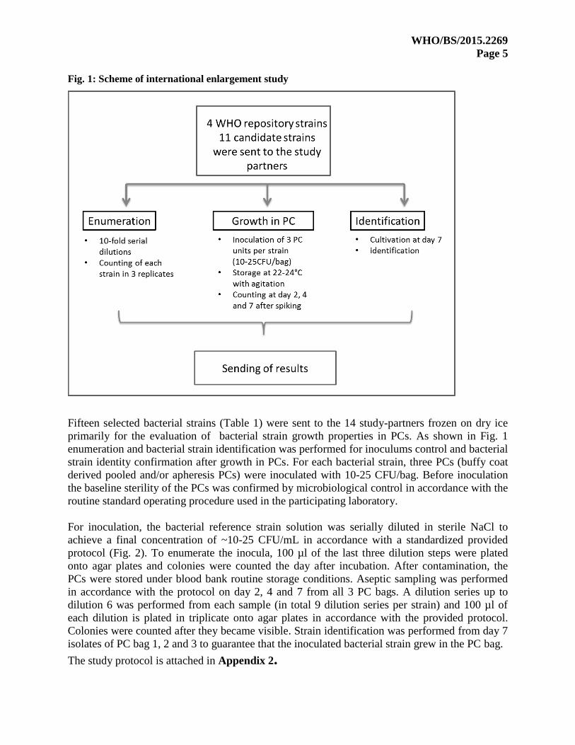
Fig. 1: Scheme of international enlargement study
Fifteen selected bacterial strains (Table 1) were sent to the 14 study-partners frozen on dry ice
primarily for the evaluation of bacterial strain growth properties in PCs. As shown in Fig. 1
enumeration and bacterial strain identification was performed for inoculums control and bacterial
strain identity confirmation after growth in PCs. For each bacterial strain, three PCs (buffy coat
derived pooled and/or apheresis PCs) were inoculated with 10-25 CFU/bag. Before inoculation
the baseline sterility of the PCs was confirmed by microbiological control in accordance with the
routine standard operating procedure used in the participating laboratory.
For inoculation, the bacterial reference strain solution was serially diluted in sterile NaCl to
achieve a final concentration of ~10-25 CFU/mL in accordance with a standardized provided
protocol (Fig. 2). To enumerate the inocula, 100 µl of the last three dilution steps were plated
onto agar plates and colonies were counted the day after incubation. After contamination, the
PCs were stored under blood bank routine storage conditions. Aseptic sampling was performed
in accordance with the protocol on day 2, 4 and 7 from all 3 PC bags. A dilution series up to
dilution 6 was performed from each sample (in total 9 dilution series per strain) and 100 µl of
each dilution is plated in triplicate onto agar plates in accordance with the provided protocol.
Colonies were counted after they became visible. Strain identification was performed from day 7
isolates of PC bag 1, 2 and 3 to guarantee that the inoculated bacterial strain grew in the PC bag.
The study protocol is attached in Appendix 2.

WHO/BS/2015.2269
Page 6
Fig.2: Inoculation and sampling procedure

WHO/BS/2015.2269
Page 7
Selection and characterization of bacterial candidate strains
In total, 15 bacteria strains (4 established repository strains, 11 new candidate strains, 4 vials
each strain) were sent to the participants in purpose-built containers on dry ice. Each vial was
labelled with the name of the bacterial strain and PEI-identification/lot number (PEI-B-P-XX).
The candidate strains are bacterial strains which were selected for their ability to replicate in PCs
under routine storage conditions used in transfusion medicine. The strains are prepared using a
specifically developed procedure which guarantees defined bacterial suspensions (deep frozen,
ready to use, stable, shippable, defined in count of living cells) [7].
The enlargement strains are either isolates from blood products (blood bag and/or recipient) or
tested regarding their growth ability in PCs at PEI. The bacterial strains were characterized for
their ability to grow in PCs under current routine blood bank conditions (original bag volume,
storage under agitation, temperature controlled).
Enumeration and stability testing was performed at PEI before and after production/deep
freezing and, additionally, during the operating time of the study. For the stability testing six
vials of each bacterial strain solution were defrosted and two dilution series of each vial were
produced. Samples were transferred directly from deep freezer to a dry incubator at 37ºC for 10
minutes. If ice crystals were still evident, the vial was warmed in the hand until the content had
melted. The stock suspensions were used immediately after thawing. Plating assays were carried
out (n = 6) for one defined dilution of both dilution series. Thereafter, mean values were
calculated.
The identity of the bacteria strains was tested by a combination of classical and molecular
microbiological procedures. Classical characteristics used are growth properties, colony
morphology, Gram-staining, and biochemical parameters like metabolism of certain sugars (API-
System). Additionally, part of the 16s ribosomal RNA gene was sequenced.
WHO/BS/2015.2269
Page 8
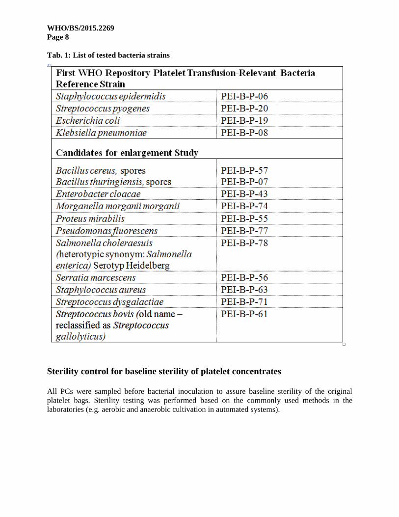
Tab. 1: List of tested bacteria strains
Sterility control for baseline sterility of platelet concentrates
All PCs were sampled before bacterial inoculation to assure baseline sterility of the original
platelet bags. Sterility testing was performed based on the commonly used methods in the
laboratories (e.g. aerobic and anaerobic cultivation in automated systems).

WHO/BS/2015.2269
Page 9
Dilution procedure and artificial contamination
For low count spiking the bacteria reference strain solutions were diluted. The test strains were
defrosted and vortexed for 15 seconds. Serial dilution of each vial was performed in sterile
saline, as described in the dilution procedure in the protocol.
The first dilution was termed the D1 (10-1
) dilution, the following was termed D2, D3 etc. to the
final dilution step containing ~10-25 CFU/mL. Inoculation of each PC bag was perfomed in
accordance with the study protocol.
Sampling, enumeration and documentation
Sampling was performed on day 2, 4 and 7 (48, 96, 168 hours) after inoculation during PC
storage (storage at 20 - 24°C under agitation). Sample withdrawal was performed following the
study protocol (Fig.2).
Stability testing of the bacterial strains
The stability of all strains was tested at PEI continuously during the storage time and the results
were statistically evaluated. For this purpose, 6 vials per lot (start, mid, end of production) were
diluted and the colony forming units (CFU) were determined by plate counting method on 6
agar-plates in parallel (Fig.3).
Fig. 3: Inoculation and sampling procedure performed for stability testing
Statistical methods
Statistical analysis was performed at PEI based on the raw data sent by the participants.
Evaluation was based on log10 CFU/mL; zero CFU/mL concentrations were set to 0.1 before log-
transformation. Taking into account an inoculation of 10-25 CFU/bag and bag sizes of 100 to
300 mL, inoculation per milliliter ranged from 0.033 to 0.250 CFU/mL (i.e. -1.48 to -0.60 log10
CFU/mL).
2 Dilution series per vial 6 Agarplates: plate counting method Vial with bacterial suspension (Test of 6 vials per lot)

WHO/BS/2015.2269
Page 10
Growth data were analyzed per strain and day. Overall mean for each strain was estimated by
means of a mixed linear model with log10 CFU as dependent variable and random factors
bacterial strain and participant.
Analysis of stability of inoculum data at PEI was performed for up to five determinations per test
strain by means of a linear regression model with dependent variable log10 CFU and date of
determination as explanatory variable.
The statistical analysis was performed with SAS®/STAT software, version 9.3, SAS System for
Windows. Results for bacterial growth were presented in Box- and-Whisker plots (Fig. 4.)
Fig. 4: Box-and-Whisker plots for growth
Results
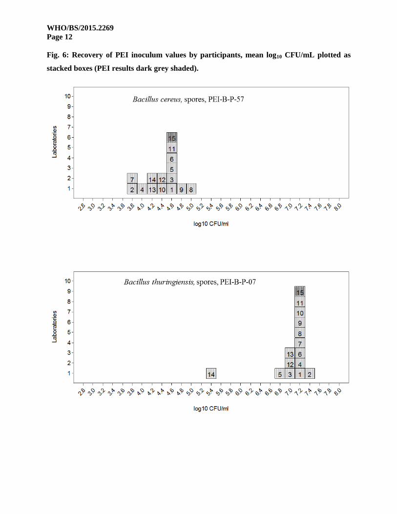
Recovery of inoculum
The bacteria reference strain solutions were diluted for low count spiking. The cell count of each
bacterial strain was provided in the study protocol and displayed in colony forming units per mL
(CFU/mL). The match of inoculum by the participating labs was statistically evaluated and
results are shown in Fig. 5 and 6.
WHO/BS/2015.2269
Page 11
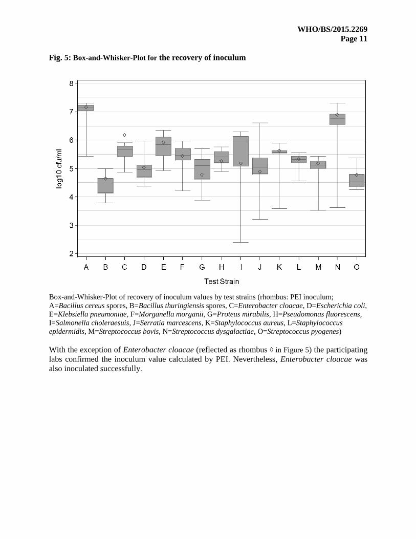
Fig. 5: Box-and-Whisker-Plot for the recovery of inoculum
Box-and-Whisker-Plot of recovery of inoculum values by test strains (rhombus: PEI inoculum;
A=Bacillus cereus spores, B=Bacillus thuringiensis spores, C=Enterobacter cloacae, D=Escherichia coli,
E=Klebsiella pneumoniae, F=Morganella morganii, G=Proteus mirabilis, H=Pseudomonas fluorescens,
I=Salmonella choleraesuis, J=Serratia marcescens, K=Staphylococcus aureus, L=Staphylococcus
epidermidis, M=Streptococcus bovis, N=Streptococcus dysgalactiae, O=Streptococcus pyogenes)
With the exception of Enterobacter cloacae (reflected as rhombus ◊ in Figure 5) the participating
labs confirmed the inoculum value calculated by PEI. Nevertheless, Enterobacter cloacae was
also inoculated successfully.
WHO/BS/2015.2269
Page 12
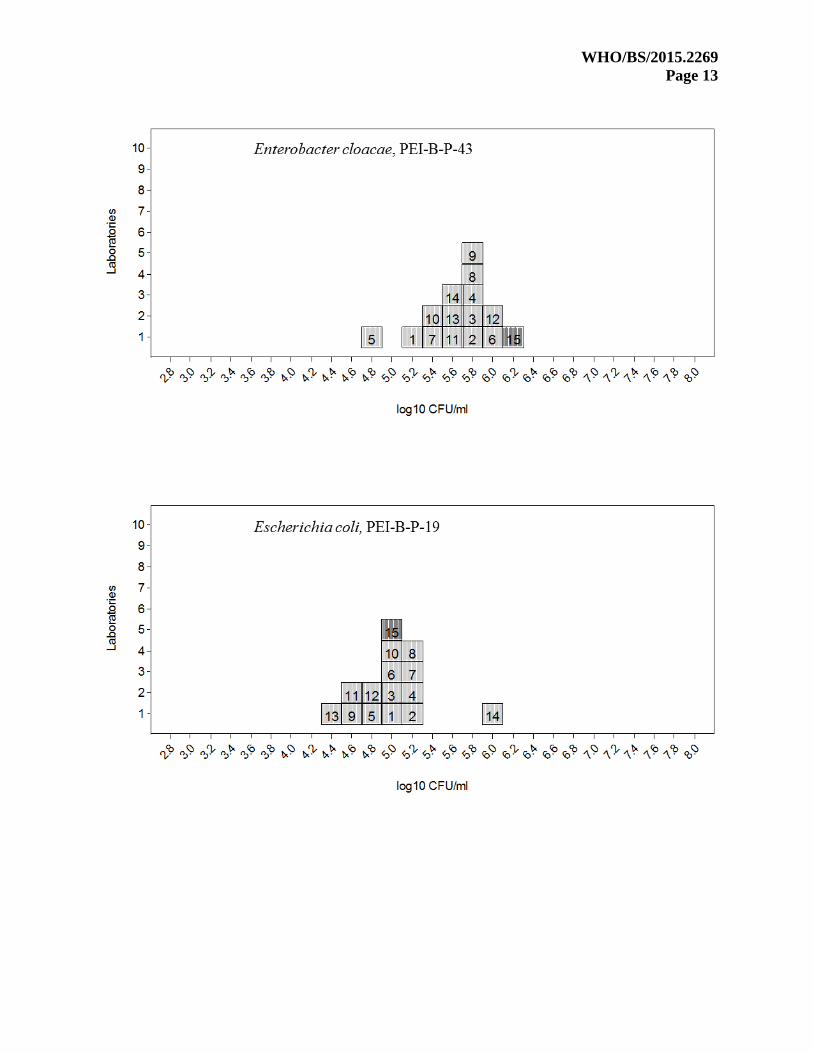
Fig. 6: Recovery of PEI inoculum values by participants, mean log10 CFU/mL plotted as
stacked boxes (PEI results dark grey shaded).
WHO/BS/2015.2269
Page 13

WHO/BS/2015.2269
Page 14

WHO/BS/2015.2269
Page 15

WHO/BS/2015.2269
Page 16

WHO/BS/2015.2269
Page 17

WHO/BS/2015.2269
Page 18

WHO/BS/2015.2269
Page 19

WHO/BS/2015.2269
Page 20
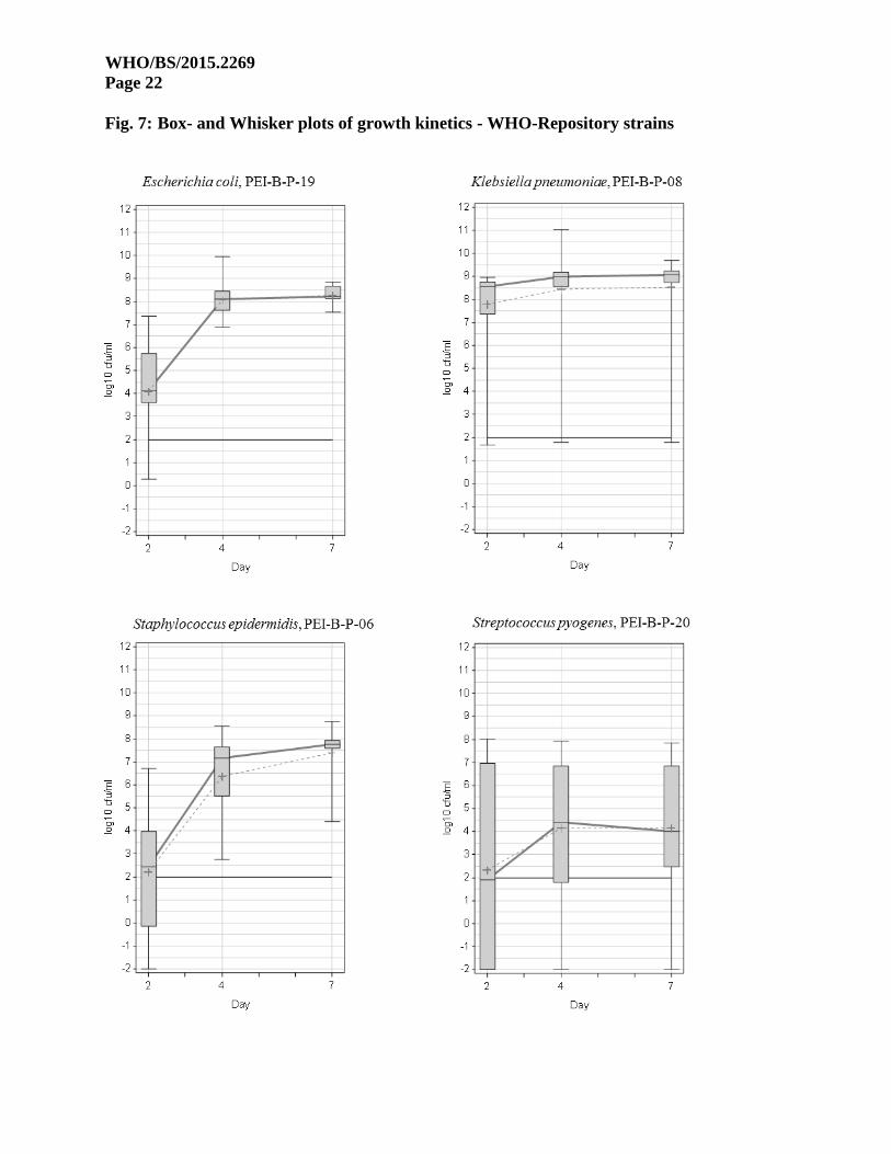
Bacterial Growth in PCs
To gain more information about the growth behaviour of each bacterial strain, bacterial cell
counting was performed at day 2, 4 and 7 after spiking. Cell counts are summarized in Table 2
and results are presented as Box- and Whisker plots in Fig. 7 and 8.
Tab. 2: Statistical evaluation of growth ability for each bacterial strain and day post
inoculation

WHO/BS/2015.2269
Page 21
WHO/BS/2015.2269
Page 22
Fig. 7: Box- and Whisker plots of growth kinetics - WHO-Repository strains

WHO/BS/2015.2269
Page 23
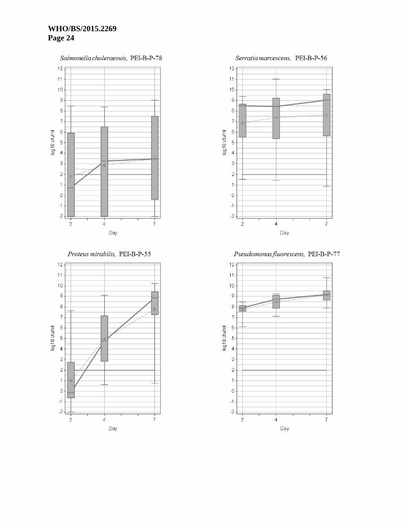
Fig. 8: Box- and Whisker plots of growth kinetics - Candidate strains
log
10
cfu
/ml
-2
-1
0
1
2
3
4
5
6
7
8
9
10
11
12
Day
2 4 7
Morganella morganii
log
10
cfu
/ml
-2
-1
0
1
2
3
4
5
6
7
8
9
10
11
12
Day
2 4 7
Enterobacter cloacae
log
10
cfu
/ml
-2
-1
0
1
2
3
4
5
6
7
8
9
10
11
12
Day
2 4 7
Bacillus thuringiensis spores
log
10
cfu
/ml
-2
-1
0
1
2
3
4
5
6
7
8
9
10
11
12
Day
2 4 7
Bacillus cereus sporesBacillus cereus, spores, PEI-B-P-57
Bacillus thuringiensis, spores, PEI-B-P-07
Enterobacter cloacae, PEI-B-P-43
Morganella morganii, PEI-B-P-74
WHO/BS/2015.2269
Page 24

WHO/BS/2015.2269
Page 25
WHO/BS/2015.2269
Page 26
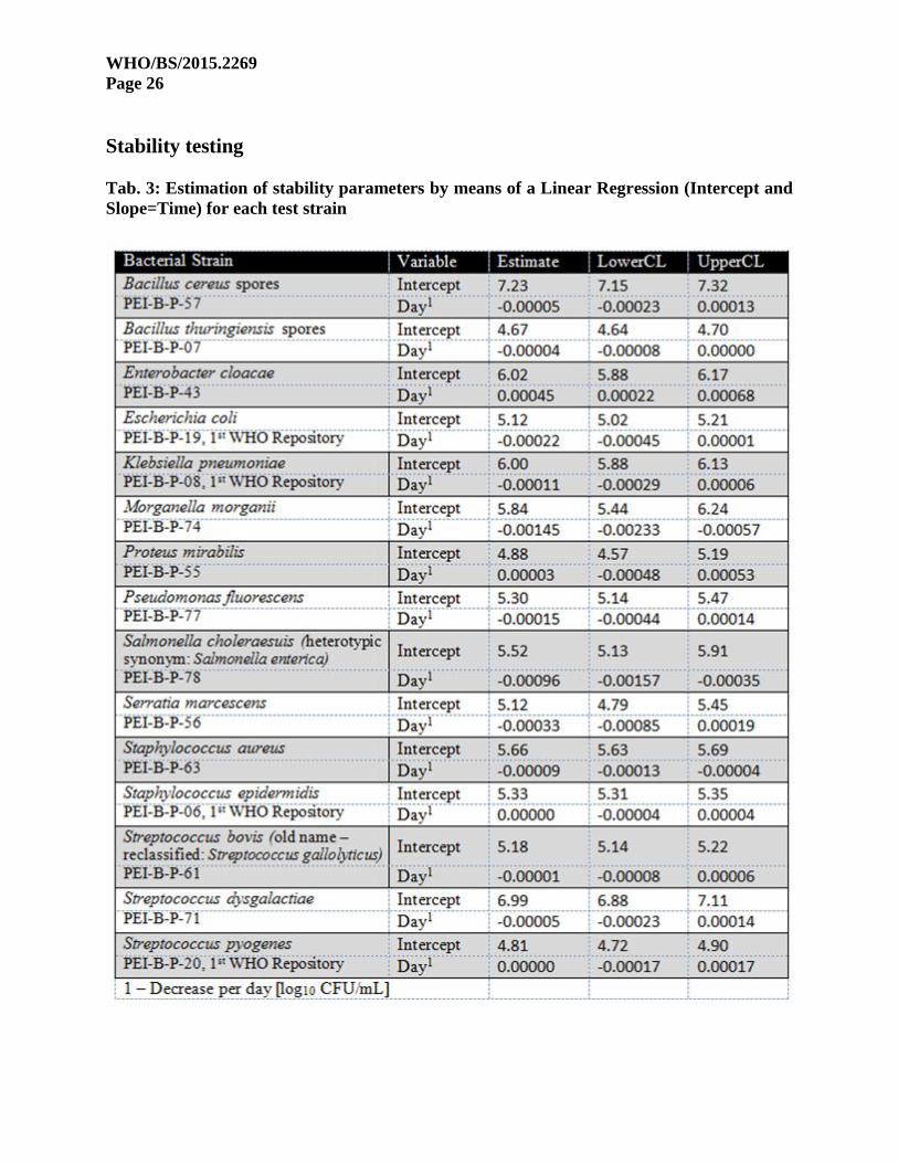
Stability testing
Tab. 3: Estimation of stability parameters by means of a Linear Regression (Intercept and
Slope=Time) for each test strain

WHO/BS/2015.2269
Page 27
Fig. 9: Stability WHO-Repository Strains (mean values) performed by PEI
Fig. 10: Stability Candidate Strains (mean values) performed by PEI
CFU/m
L
CFU/m
L

WHO/BS/2015.2269
Page 28
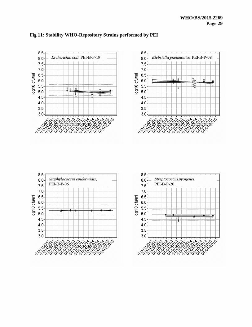
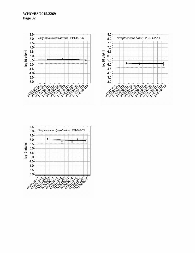
Stability testing – Plots
Specification was set by data from first determination at PEI as mean (bold base line) ± 0.5 log10
CFU/mL (dotted lines). A strain can be regarded as stable until the lower 95% confidence
interval for an individual prediction (thin line; including variance of the error as well as variance
of the parameter estimates) for the linear trend (thick line) intersects the lower specification limit
(dotted line).
WHO/BS/2015.2269
Page 29
Fig 11: Stability WHO-Repository Strains performed by PEI

WHO/BS/2015.2269
Page 30
Fig 12: Stability Candidate strains

WHO/BS/2015.2269
Page 31
WHO/BS/2015.2269
Page 32

WHO/BS/2015.2269
Page 33
Bacillus thuringiensis spores, Staphylococcus aureus, Staphylococcus epidermidis and
Streptococcus bovis showed stability with good precision. Another six strains, Bacillus cereus
spores, Escherichia coli, Klebsiella pneumoniae, Pseudomonas fluorescens, Streptococcus
dysgalactiae, and Streptococcus pyogenes, are stable over the observed period with variability of
data. For Pseudomonas fluorescens and Streptococcus pyogenes this could be due to outliers at
the second (third) timepoint. Enterobacter cloacae was stable until April 2014 (upper prediction
limit intersects the dotted specification line). Here a sampling error at the first stability testing
might be responsible for the “increase” in bacteria cells during storage. Serratia marcescens
showed stability until July 2014 (for 1.5 years). This could be due to outliers at the third
timepoint. The two remaining strains (Morganella morganii and Salmonella choleraesuis) are
less stable than the other strains.
Bacterial Identification After the testing of the growth kinetics of the bacterial strains the participants identified the
strains following their routine protocols as used in the respective microbiological lab (Tab. 4).
The results of identification corresponded with the results provided by PEI. According to
classification issues Enterobacter cloacae is a gene complex containing six genovars. One of
them is Enterobacter hormaechei steigerwaltii. Salmonella choleraesuis (hemolytic synonym
and serotype of S.enterica) identified as Salmonella enterica. Streptococcus bovis (biotype I,
taxonomy of the group) is currently named Streptococcus gallolyticus pasteurianus due to
phylogenetic results.

WHO/BS/2015.2269
Page 34
Tab. 4: Identification of the strains by the participating labs L
ab
1L
ab
2L
ab
3L
ab
4L
ab
5L
ab
6L
ab
7L
ab
8L
ab
9L
ab
10
Lab
11
Lab
12
Lab
13
Lab
14
AccuG
ENX-ID
MA
LDI-TO
F IDA
ccuPRO
-IDG
ram stain/A
PIG
ram stain/A
PIM
aldi-TOF M
S
/Vitek M
S
BD Phoenix
Identification
Vite ID
/ BBL
chystal gram
positive 4.0
Mass
spectrometry
Gram
stain/Colony
morphology
Maldi-TO
FG
ram stain /
Microscan/ A
PI
MA
LDI m
ass
spectrometry
system / Biolog
ELx808
MA
LDI-TO
F16rR
NA
gene
sequence16S rR
NA
Gram
stain/
Colony
morphology
WH
O R
ep
osito
ry
Sta
phylo
coccu
s
epid
erm
idis
PE
I-B-
P-0
6a
aa
aa
aG
ram positive
coccusa
aa
a100%
98%G
ram positive
coccus
Stre
pto
coccu
s
pyo
genes
PE
I-B-
P-2
0a
aa
aa
yes
(Streptococcus
type A)
Gram
positive
coccusa
aa
a99%
98%G
ram positive
coccus
Esch
erich
ia co
liP
EI-B
-
P-1
9a
aa
aa
aG
ram negative
rodsa
a
Escherichia coli /
(Stenotropomona
s maltophilia in
bag 1)
a99%
98%G
ram negative
rods
Kle
bsie
lla
pneum
onia
e
PE
I-B-
P-0
8a
aa
aa
aG
ram negative
rodsa
aa
a99%
99%G
ram negative
rods
Bacillu
s cere
us
spore
s
PE
I-B-
P-5
7Bacillus spp
Bacillus genusBacillus cereus
groupa
aa
Gram
positive
rodsa
aBacillus cereus/
thuringiensisa
Bacillus cereus /
Bacillus
thuringiensis (2
species had equal
scores ) 100 '%
99%G
ram positive
rods
Bacillu
s
thurin
gie
nsis
spore
s
PE
I-B-
P-0
7a
Possibility of
Bacillus
thuringingiensis -
low discrim
ination
Bacillus cereus
groupa
Bacillus cereusa
Gram
positive
rodsa
aBacillus cereus/
thuringiensisa
Bacillus cereus,
Bacillus
thuringiensis,
Bacillus
toyonensis (3
species had equal
scores) 99 %
98%G
ram positive
rods
Ente
robacte
r
cloaca
e
PE
I-B-
P-4
3
En
tero
bacte
r
horm
aech
ei
steig
erw
altii
aa
aa
aa
Gram
negative
rodsa
aa
a99%
99%G
ram negative
rods
Morg
anella
morg
anii
PE
I-B-
P-7
4
Morg
an
ella
morg
an
ii sub
sp.
morg
an
ii
Morg
an
ella
morg
an
ii
morg
an
ii
Morg
an
ella
morg
an
iino grow
thno grow
thno grow
thno grow
tha
no growth
(identifiation of
control)
Gram
negative
rodsa
ano grow
thno grow
th99%
99%G
ram negative
rods
Pro
teus m
irabilis
PE
I-B-
P-5
5a
aa
aa
aG
ram negative
rodsa
Yes - Gram
negative rods
/API
aa
100%99%
Gram
negative
rods
Pse
udom
onas
fluore
scens
PE
I-B-
P-7
7P
seu
dom
on
as sp
ano grow
tha
aa
aG
ram negative
rodsa
no /
Pseudomonas
putidaa
a99%
98%G
ram negative
rods
Salm
onella
chole
rasu
is
(hete
rotyp
ic
synon
ym)
PE
I-B-
P-7
8
Salm
on
ella
en
terica
en
terica
NC
TC
6017
Con
trol
Salmonella
spp/Salmonella
choleraeSalm
onella spp
Salmonella spp
(Serotyp:
heidelberg)
Salmonella spp.
yesSalm
onella spp.G
ram negative
rodsa
Antisera typing:
Salmonella
choleraesuis
Salomenella
species (unable
to speciate
organism)
Maldi-TO
F/
Kaufman-w
hite
schema:
Salmonella
choleraesuis
no growth
98%G
ram negative
rods
Serra
tia
marce
scens
PE
I-B-
P-5
6
Serra
tia
marce
scen
s
marce
scen
s /
nem
ato
dip
hila
aa
aa
aa
Gram
negative
rodsa
aSerratia spp.
a99%
100%G
ram negative
rods
Sta
phylo
coccu
s
aure
us
PE
I-B-
P-6
3
Sta
ph
yloco
ccus
au
reu
s /
Sta
ph
yloco
ccus
au
reu
s AT
CC
6538 C
on
trol
aa
aa
aa
Gram
positive
coccusa
aStaphylococcus
aureus ss aureusa
100%99%
Gram
positive
coccus
Stre
pto
coccu
s
dysg
ala
ctiae
PE
I-B-
P-7
1
Stre
pto
coccu
s
dysg
ala
ctiae
eq
uisim
ilisa
no growth
aa
aStreptococcus
dysgalactiae ssp.
equisimilis
Gram
positive
coccusa
aStreptococcus
dysgalactiae ssp.
equisimilis
a100%
99%G
ram positive
coccus
Stre
pto
coccu
s
bovis (o
ld n
am
e -
recla
ssified
)
PE
I-B-
P-6
1
Stre
pto
coccu
s
gallo
lyticus
paste
uria
nu
sa
aa
aa
aG
ram positive
coccusa
aStreptococcus
gallolyticus
Streptococcus
gallolyticus
Streptococcus
bovis /
Streptococcus
pasteurianis
(equal scores)
100 %
99%no grow
th
Meth
od
of Id
en
tificatio
n
Salm
on
ella
en
terica
Sero
typin
g: S
alm
on
ella
Heid
elb
erg
Serra
tia m
arce
scen
s
Sta
ph
yloco
ccus a
ure
us
Stre
pto
coccu
s
dysg
ala
ctiae
Stre
pto
coccu
s gallo
lyticus
Can
did
ate
s fo
r En
larg
em
en
t stu
dy
Bacillu
s cere
us
Bacillu
s thu
ring
ien
sis
En
tero
bacte
r horm
aech
ei
Sero
typ
ing
: En
tero
bacte
r
cloaca
e
Pro
teu
s mira
bilis
Pse
ud
om
on
as flu
ore
scen
s
PE
I
Sta
ph
yloco
ccus e
pid
erm
idis
Stre
pto
coccu
s pyo
gen
es
Esch
erich
ia co
li Sero
typ
ing
: Esch
erich
ia
coli, S
ero
var O
6:H
16
Kle
bsie
lla p
neu
mon
iae

WHO/BS/2015.2269
Page 35
Discussion
All participants received the deep frozen bacteria strains in good condition without complaint. As
in the first study, deep frozen, pathogenic bacteria strains could be shipped worldwide without
any difficulties. The tested inocula proliferated well and were successfully used for spiking. The
bacterial identification performed by the study partners complied with the ID of PEI. The results
of bacteria counting of all participants are homogenous since the measured divergence factors
represent an acceptable value in the estimation of high bacteria cell counts.
The results of the four strains of the existing WHO Repository are equivalent to the first study.
With an initial bacterial count of approx. 0.03 CFU/mL (spiking of 10 to 25 CFU per PC unit)
Escherichia coli and Staphylococcus epidermidis grew up until day 7 in 100%, Klebsiella
pneumoniae in 95% of the tested PC units. These three strains showed consistent growth at all
test sites. Streptococcus pyogenes proliferated in 73% of the tested PC units. This strain showed
no growth at all at only one site. In the previous collaborative study, this strain did not grow at 3
sites at an inoculum of 10 CFU per bag. The most likely interpretation of these failures are
specific or unspecific inhibitory mechanisms directed at the bacterial strain in the donor
population [7].
The actual results confirmed the suitability of the strains as 1st WHO Repository.
Nine of the eleven candidate strains showed good (70%) to excellent growth (100%).
Enterobacter cloacae and Streptococcus bovis failed to proliferate at two sites. The tested
Morganella morganii strain grew only at two sites. Salmonella choleraesuis showed no growth
at five sites. These results might be explained by antibodies against these strains in the blood
donor population. These two strains also exhibited lower stability [Fig.12].
The statistical evaluation of the growth ability showed for Bacillus cereus spores, Bacillus
thuringiensis spores, Escherichia coli, Klebsiella pneumoniae, Pseudomonas fluorescens,
Serratia marcescens, Staphylococcus aureus and Streptococcus dysgalactiae a growth to
significantly more than 2 log10 CFU/mL by day 2.
For Enterobacter cloacae, Proteus mirabilis, Staphylococcus epidermidis, Streptococcus bovis
and Streptococcus pyogenes this growth milestone was reached at day 4.
Growth for Salmonella choleraesuis was lower than for other strains and showed a high
variability among participants which might be explained by the low storage temperature or pre-
existing antibodies.
Morganella morganii failed to grow beyond that amount of bacteria in the initial inoculation.
As Morganella morganii caused transfusion incidents in several countries it was decided to
qualify another strain of this species. In an additional study 8 partners tested a second strain in
accordance with the study protocol as a proposal for replacing the Morganella morganii strain
from the main study. The results are documented in Appendix 3 in Fig. 12 and 13.

WHO/BS/2015.2269
Page 36
Conclusion and Proposals
Platelet Transfusion-Relevant Bacteria Reference Strains are a practical tool for the objective
validation and assessment of methods for screening and pathogen reduction in blood
components. The collaborative study confirmed the growth ability, stability and consistency of
the already approved four WHO strains. Nine of the candidate strains fulfilled the predefined
requirements for addition to the repository. After spiking with low bacteria counts they grew up
to high counts in platelet concentrates under routine conditions independently from individual
donors properties. As the candidates cover very fast growing bacteria (high bacterial counts
already after day 2) and intermediate growers (to high bacterial counts after day 4 to day 7) the
bacterial panel well reflects the different needs both of validation of pathogen inactivation
technologies and of bacterial detection systems. It is proposed to add these bacterial strains to the
existing bacteria repository as WHO Platelet Transfusion-Relevant Bacteria Reference Strains.
As the tested Morganella morganii PEI-B-P-74 strain showed no growth, a second strain was
tested in 8 laboratories in accordance with the study protocol. The new strain showed growth in
all tested PCs. The data are very consistent. It is recommended to add this strain to the WHO
bacteria list as well.

WHO/BS/2015.2269
Page 37
Acknowledgements
Christian Gabriel, Susanne Süssner, Claudia Renke, Ingrid Lindlbauer, Austrian Red Cross,
Blutzentrale Linz
Dana Devine, Sandra Ramirez-Arcos, Heather Perkins, Yuntong Kou, Adriana Zapata, Canadian
Blood Service, Ottawa Blood Service, Ottawa
Axel Seltsam, Bernd Lambrecht, Birgit Jarck, German Red Cross Blood Service NSTOB,
Springe, Germany
Birgit Gathof, Melanie Störmer, Transfusion Medicine, University Hospital Cologne, Germany
Erhard Seifried, Kai Hourfar, Simone Schwientek, German Red Cross, Frankfurt, Germany
Carl McDonald, Kate Aplin, Anjana Roy, NHS Blood and Transplant, London, England
Dirk de Korte, Willy Karssing, Herbert Korsten, Sanquin Blood Supply Foundation, Jan
Marcelis, Jaap van Meeteren, Eveline Thijssen, Elisabeth Hospital, Tilburg, The Netherlands
Richard J. Benjamin, Stephen J. Wagner, Anne Hapip, American Red Cross, Blood Component
Dep, Rockville, USA
Roslyn Yomtovian†, Michael R. Jacobs, Caryn Good, Case Western Reserve University,
Cleveland, USA
Truscha Niekerk, Katlego Moagi, Xoliswa Mpumlwana, Nokuthula Chilwane, Nolwazi
Nkambule, South African National Blood Service, Weltevreden Park, South Africa
Susanne Marschner, Shawn D. Keil, Denise Gilmour, Meghan Hudziec, Therumo, BCT,
Lakewood, USA
Julieta Rojo, Gabriela Ibañez- Cervantes, Juan Manuel Bello-López, Centro Nacional de la
Transfusión Sanguínea, Mexico
Masahiro Satake, Hideto Nagumo, Mami Matsumoto, Kumiko Shinozaki, Kumi Kinno, Moe
Kaneko, Japanese Red Cross, Tokyo, Japan
Zainab Mukhtar, Shaheen Sharafat, Institute of Biological, Biochemical and Pharmaceutical
Sciences, Dow Medical College, DUHS Karachi, Pakistan
Julia Brachert, Rekia Beshir, Anna-Maria Scheder, Annemarie Mück, Anja Schneider, Sigrid
Hanitsch, Utta Schurig, Ute Sicker, Jan-Oliver Karo, Ingo Spreitzer, Paul-Ehrlich-Institut,
Langen, Germany
WHO/BS/2015.2269
Page 38
References
1 Brecher ME, Blajchman MA, Yomotovian R, et al: Adressing the risk of bacterial
contamination of platelets within the United States: A history to help illuminate the futuere.
Transfusion 2013; 53:221-231
2 Lohmann A, Halbauer j, Witzenhausen C, et al: Hemovigilance Report oft he Paul-Ehrlich-
Institut, 2012
3 Funk M, Günay S, Lohmann A: Hämovigilanzbericht des Paul-Ehrlich-Instituts, 2014
4 Fatalities Reported to FDA Following Blood Collection and Transfusion – Annual Summary
for Fiscal Year 2013
5 Montag T: Perspectives and limitations in the bacterial screening of platelet concentrate. J Lab
Med 2006; 30: 60-65
6 Palaveino EL, Yomotovian RA, Jacobs MR: Bacterial contamination of platelets. Transfusion
Apheres Sci 2010; 42:71-82
7 Störmer M, Arroyo A, Brachert J, Montag T. et al: Establishment of the first international
repository for transfusion-relevant bacteria reference strains: ISBT working party transfusion-
transmitted infectious diseases (WP-TTID), subgroup on bacteria. Vox Sang. 2012; 102:22-31.
8 Müller B, Walter-Wenke G, Kalus M, Alt T, Bux j, Zeiler T, Schottstedt V: Routine bacterial
screening of platelet concentrates by flow cytometry and is impact on product safety and supply.
Vox Sang. 2015, 108 209-218.
9 Schrezemeier H, Walter-Wenke G, Müller TH. Et al: Bacterial contamination of platelet
concentrates: results of a prospective multicenter study comparing pooled whole blood-derived
platelets and apheresis platelets. Transfusion 2007; 47:644-652
10 Montag T: Strategies of bacteria screening in cellular blood components. Clin Chem Lab
Med. 2008; 46:926-32.
11 Störmer M, Vollmer T. Diagnostic Methods for Platelet Bacteria Screening: Current Status
and Developments. Transfus Med Hemother 2014; 41:19–27.

WHO/BS/2015.2269
Page 39
Appendix 1
Study partners

WHO/BS/2015.2269
Page 40
Appendix 2
WHO/BS/2015.2269
Page 41
Project Team:
Co-Chair ISBT WP-TTID, Subgroup on Bacteria Dr. Carl P. McDonald Head of Bacteriology National Bacteriology Laboratory NHS Blood and Transplant Charcot Avenue Colindale London NW9 5BG ENGLAND Co-Chair ISBT WP-TTID, Subgroup on Bacteria Dr. Richard J. Benjamin MD PhD FRCPath Chief Medical Officer American Red Cross Holland Laboratories 15601 Crabbs Branch Way Rockville, MD 20855-2743 USA Dr. Melanie Störmer Transfusion Medicine University Hospital Cologne Kerpener Str. 62 50937 Cologne Germany Dr. Eva Spindler-Raffel (Corresponding address) FG Bakteriologische Sicherheit / Microbial Safety Paul-Ehrlich-Institut Paul-Ehrlich-Str. 51-59 63225 Langen Germany
WHO/BS/2015.2269
Page 42
CONTENTS
1. Background ..........................................................................................................43
2. Study Design ........................................................................................................45
3. Shipping and Storage ..........................................................................................46 3.1. Labelling of Bacterial Reference Strains .................................................................. 46
3.2. Storage of Bacterial Reference Strains ................................................................. 47
4.1. Materials ................................................................................................................... 47
4.2. Sterility control for baseline sterility of platelet concentrates .................................. 47
4.3. Thawing / Defrosting Bacterial Reference Strains ................................................... 47
4.4. Dilution Procedure .................................................................................................. 48
4.5. Artificial contamination of platelets /platelet spiking ............................................... 49
4.6. Control (Enumeration) of inoculum ...................................................................... 50
4.7. Sampling, Enumeration and Documentation ....................................................... 50
5.1. Enumeration .............................................................................................................. 53
5.1.1. Enumeration 1: after 2 days ...................................................................................... 53
5.1.2. Enumeration 2: after 4 days ...................................................................................... 54
5.1.3. Enumeration 3: after 7 days ...................................................................................... 55
5.2. Identification of grown microorganism .................................................................... 56
6. Lab Protocol ........................................................................................................57
7. Questionnaire .....................................................................................................61

WHO/BS/2015.2269
Page 43
1. Background
Bacterial contamination of platelet concentrates (PCs) still remains a persistent problem in
transfusion [1]. To mitigate the risk of bacterial contamination of blood components, blood
centres have implemented donor screening along with bacterial detection systems or pathogen
reduction technologies (PRT). In order to validate and to compare these methods, it is crucial to
use bacterial strains which are able to proliferate in blood components, e.g. in PCs during
storage [2].
The International Society of Blood Transfusion (ISBT) Working Party Transfusion-Transmitted
Infectious Diseases, Subgroup on Bacteria (former chair: Dr Thomas Montag-Lessing) had
organized an international validation study on Platelet Transfusion-Relevant Bacteria Reference
Strains (PTRBRS) to be used as a tool for development, validation and comparison of the
respective methods. Four blinded bacterial strains were sent in replicate to participating
laboratories worldwide for bacterial count calculation, strain identification and evaluation of
growth properties in PCs. The results were submitted to the Expert Committee on Biological
Standardization (ECBS) and were established as the WHO Repository of Platelet Transfusion-
Relevant Bacteria Reference Strains [3].
Platelet Bacteria Reference strains (PBRS) are a feasible tool for objective validation and
assessment of methods for screening and pathogen reduction in blood components. For this
purpose, four PBRS were tested and approved. They are available as the WHO Platelet
Transfusion Relevant Bacterial Reference Strains Repository.
The next step will be to enlarge this bacterial panel to approve more strains as PBRS.
In contrast to the previous validation study where both the bacterial count and the growth
properties of several replicates in PCs was evaluated, only the ability to proliferate in PCs under
“real life”/routine conditions after low spiking (< 1CFU/mL) requires evaluation in this WHO –
ISBT enlargement study. For this reason 11 selected bacterial strains that were discussed
during the meetings of ISBT WP-TTID Subgroup on Bacteria will be sent to the participating
laboratories to evaluate their ability to proliferate in PCs in different regions of the world.

WHO/BS/2015.2269
Page 44
In addition to these 11 new strains, the 4 already approved repository strains will be tested
again as a reference.
For statistical reasons each bacteria strain has to be tested in 3 platelet bags for 7 days.
Enumeration will be performed on day 2, 4 and 7. Whether tested in parallel or on 3 different
days is up to the participating lab, but must be documented.
References
1. Palavecino EL, Yomtovian RA, Jacobs MR. Bacterial contamination of platelets. Transfus
Apher Sci. 2010; 42:71-82.
2. Montag T, Strategies of bacteria screening in cellular blood components. Clin Chem Lab
Med. 2008; 46:926-32.
3. Störmer M, Arroyo A, Brachert J, Carrero H, Devine D, Epstein JS, Gabriel C, Gelber C,
Goodrich R, Hanschmann KM, Heath DG, Jacobs MR, Keil S, de Korte D, Lambrecht B, Lee CK,
Marcelis J, Marschner S, McDonald C, McGuane S, McKee M, Müller TH, Muthivhi T, Pettersson
A, Radziwon P, Ramirez-Arcos S, Reesink HW, Rojo J, Rood I, Schmidt M, Schneider CK, Seifried
E, Sicker U, Wendel S, Wood EM, Yomtovian RA, Montag T. Establishment of the first
international repository for transfusion-relevant bacteria reference strains: ISBT working party
transfusion-transmitted infectious diseases (WP-TTID), subgroup on bacteria. Vox Sang. 2012;
102:22-31.

WHO/BS/2015.2269
Page 45
2. Study Design
15 different bacterial reference strains will be sent to the partners. For each bacterial strain,
three PCs (pooled and/or apheresis PCs) have to be artificially contaminated with 10-25
CFU/bag. Before contamination the baseline sterility of the PCs needs to be proven by
microbiological control in accordance with the routine standard operating procedure used in the
participating laboratory.
For contamination, the reference strain solution needs to be diluted in sterile NaCl to achieve a
final concentration of 10-25 CFU/mL in the tube. To enumerate the inocula, 100 µl of the last
three dilution steps are plated onto agar plates and colonies are counted the following day after
incubation. After contamination the PCs are stored under routine conditions. Sampling is
performed according to the protocol on days 2, 4 and 7 from all 3 PC bags. A dilution series up
to dilution 6 is performed from each sample (in total 9 dilution series per strain) and 100 µl of
each dilution is plated in triplicate onto agar plates. Colonies are counted. Strain identification
has to be performed from day 7 isolates of PC bag 1, 2 and 3 to guarantee that the inoculated
bacterial strain grew in the PC bag.

WHO/BS/2015.2269
Page 46
3. Shipping and Storage
The 15 bacterial strains will be sent in purpose-built containers with dry ice. Please check the
containers immediately after receiving. To assure the stability of the bacterial load of the
Bacteria Strain, the cold chain must not be interrupted and the strains must be used
immediately after thawing.
Note: Check the vials immediately after arrival. If the samples show any
signs of thawing, they must be discarded!
In this case please inform the study coordinating team immediately.
3.1. Labelling of Bacterial Reference Strains
15 different bacteria strains are contained in vials in 4-replicates of 4.
Each vial is labelled with the name of the bacterial strain and PEI-identification/lot number (PEI-
B-P-XX).
List of Blood Bacteria Reference Strains
WHO Repository
Staphylococcus epidermidis PEI-B-P-06
Streptococcus pyogenes PEI-B-P-20
Escherichia coli PEI-B-P-19
Klebsiella pneumoniae PEI-B-P-08
Candidates for Enlargement Study
Bacillus cereus, spores Bacillus thuringiensis, spores
PEI-B-P-57
PEI-B-P-07
Enterobacter cloacae PEI-B-P-43
Morganella morganii PEI-B-P-74
Proteus mirabilis PEI-B-P-55
Pseudomonas fluorescens PEI-B-P-77
Salmonella cholerae-suis PEI-B-P-78
Serratia marcescens PEI-B-P-56
Staphylococcus aureus PEI-B-P-63
Streptococcus dysgalactiae PEI-B-P-71
Streptococcus bovis PEI-B-P-61

WHO/BS/2015.2269
Page 47
3.2. Storage of Bacterial Reference Strains
Store the vials immediately after arrival in a deep freezer at -80°C without secondary
packaging. Test of growth of selected transfusion-relevant bacterial reference strains in platelet
concentrates (PCs)
4.1. Materials
1 vial of each bacteria test strain.
(The below mentioned materials are calculated for one strain and dilution series for 3 PC bags
in parallel. If the spiking is done on different dates you will need more NaCl aqueous solution.)
3 platelet units for each strain (apheresis or whole blood derived PCs.
Preferably use fresh PCs, if not available use outdated ones)
Usual platelet storage device (agitation, temperature controlled, 22± 2°C)
Dry incubator, 37°C and 30°C
Sterile welding equipment (e.g. sterile connecting device)
Sterile NaCl aqueous solution (0.85 %) in sterile tubes with caps
Trypticase Soy Agar plates (alternatively Columbia Blood Agar)
Sterile applicators (spattles / spreaders)
Sterile syringes
Luer Lock connection device/ sterile Coupler spike
Before starting the experiments please ensure sufficient supplies are
available i.e. agar plates and NaCl.
4.2. Sterility control for baseline sterility of platelet concentrates
All PCs have to be sampled before bacterial inoculation to assure baseline sterility of the original platelet bags.
Perform sterility testing, commonly used in your laboratory (e.g. aerobic and anaerobic cultivation in automated systems) and describe the procedure in the result section.
4.3. Thawing / Defrosting Bacterial Reference Strains
Transfer the vial directly from deep freezer to a dry incubator and defrost the vial at 37ºC
for 10 minutes.
If ice crystals are still evident, warm the vial in the hand until the content has melted
completely.

WHO/BS/2015.2269
Page 48
Note: The bacterial strains (stock suspensions) must be used immediately
after thawing.
4.4. Dilution Procedure
For low count spiking the bacteria reference strain solutions need to be diluted. For this reason
the cell count of each bacterial strain is provided by the organizing committee and displayed in
colony forming units per mL (CFU/mL). For each strain the numbers of dilution steps are
provided in the appendices.
For all test strains, defrost the vials as described above (Section 4.3.) and vortex for 15 seconds
at the highest speed. Perform a series of dilutions of each vial in sterile saline, as described in
the dilution procedure (see Section 7 Appendices for each bacterial strain).
The undiluted (stock) suspension is termed the D0 (100) dilution. Unless otherwise specified in
the dilution procedure, prepare 1:10 serial dilutions using 9 mL of sterile saline (NaCl) each and
1 mL of the stock or 1 mL dilution from previous dilution step. Consequently, each dilution is
1/10th the concentration of the previous dilution. The first 10-fold dilution is termed the D1 (10-
1) dilution, the following is termed D2, D3 etc. to the final dilution step (containing 10-25
CfU/mL) that is needed.
Dilute each bacterial strain down to approximately 10 to 25 CFU/mL in sterile NaCl.
Note: The final dilution step is dependent on the bacterial count stated in the
strain dilution specification (see Section 7 Appendices).
D1
D2
D3
D4
D5
D6
Dilution series
D0
1mL 1mL 1mL 1mL 1mL 1mL

WHO/BS/2015.2269
Page 49
Make sure…
… that the dilution series of the stock tubes is prepared immediately after thawing
the stock suspension.
… that the stock suspension as well as each dilution is intensively vortexed
(highest speed) for 15 seconds.
… that tips are changed after each step!
4.5. Artificial contamination of platelets /platelet spiking
If possible: All work mentioned below should be done in a Laminar Flow & Biosafety Cabinet to
avoid contaminations.
Connect each platelet bag with a luer-lock connection device (e.g. a short tube using Sterile
Connecting Device), or insert a sterile Coupler spike (with luer-lock Safesite valve) through a
port into the pack.
Luer-Lock
If using the luer-lock connection device - draw 5 mL out of the platelet bag using a sterile
syringe but do not discard it (see below). Ensure aseptic technique is followed!
Using a second sterile syringe, inoculate 1 mL of the final dilution through the same port
into the platelet bag. Afterwards the final bacterial load will be 10 – 25 CFU per bag.
Add the previously removed 5 mL PC sample back into the bag to flush the tube segment
of the bag if using the luer-lock connection device.
Close the luer-lock port.
Coupler-Spike
If using the sterile Coupler-Spike - inoculate 1 mL of the final dilution
(~ 10 CFU/mL to 25 CFU/mL) through the port into the platelet bag.
Rinse the syringe 3 times with PC.
Incubate the contaminated PC units at 22-24°C under agitation for 7 days (168 hours after
inoculation).

WHO/BS/2015.2269
Page 50
Note:
Close the tube by clamp in case of any opening of the luer-lock device
(e.g. before connection with a syringe, change of syringes etc.). The
procedure described is used to overcome the “dead-volume” of the tube, i.e.
to bring the inoculum directly into PCs main bag.
Additionally, bacteria attached to the inner surface of the tube will be
detached.
Avoid any entry of air into the platelet unit during the inoculation process!
4.6. Control (Enumeration) of inoculum
Plate 100 µl of the last three dilution steps from abstract 4.4 in triplicate onto agar
plates and incubate strain specific at 30°C or 37°C for 24-48h.
Count the colonies and document the results in the lab protocol
(Section 6, “dilution of stock”).
4.7. Sampling, Enumeration and Documentation
Following inoculation with approximately 10 to 25 CFU per bag, growth kinetics of the test
strains during usual PC storage conditions (storage at 20 - 24°C), are monitored as described
below:
If possible, all work mentioned below should be done in a Laminar Flow & Biosafety cabinet to
avoid contaminations.
Sampling will be performed on days 2, 4 and 7 (48, 96, 168 hours).
Sample drawing shall be performed following the principles described in Section 4.5.
(Artificial contamination of platelets /platelet spiking).
If using the Luer-Lock connection device - remove the first 5 mL of the PC using a sterile
syringe but do not discard it, use a second sterile syringe to take a sample (1 mL) of each
platelet bag and then add back the previously removed 5 mL PC sample in order to enable a
repetition if necessary.
WHO/BS/2015.2269
Page 51
If using the sterile Coupling-Spike device – rinse the syringe with PC from the pack 3 times
to ensure the sample is from the bag, remove a 1 mL sample of each platelet bag and close
the luer-lock port.
Enumerate the bacterial count by diluting the 1 mL sample up to 10-6 (D6) and plating out
(see Section 4.8).
Complete documentation in Section 5.

WHO/BS/2015.2269
Page 52

WHO/BS/2015.2269
Page 53
Documentation 5.1. Enumeration
Please copy for each bacterial strain! 5.1.1. Enumeration 1: after 2 days
Bacterial strain: _____________________________________________ Pool or apheresis PC:_________________________________________ Inocula:____________________________________________________
PC Unit Dilution
100µl of…
Plate 1 Plate 2 Plate 3 Mean value
Strain
PC bag 1
date
Dilution 1
Dilution 2
Dilution 3
Dilution 4
Dilution 5
Dilution 6
PC Unit Dilution
100µl of…
Plate 1 Plate 2 Plate 3 Mean value
Strain
PC bag 2
Date
Dilution 1
Dilution 2
Dilution 3
Dilution 4
Dilution 5
Dilution 6
PC Unit Dilution
100µl of…
Plate 1 Plate 2 Plate 3 Mean value
Strain
PC bag 3
Date
Dilution 1
Dilution 2
Dilution 3
Dilution 4
Dilution 5
Dilution 6

WHO/BS/2015.2269
Page 54
Please copy for each bacterial strain! 5.1.2. Enumeration 2: after 4 days
Bacterial strain: _____________________________________________ Pool or apheresis PC:_________________________________________ Inocula:____________________________________________________
PC Unit Dilution
100µl of…
Plate 1 Plate 2 Plate 3 Mean value
Strain
PC bag 1
Date
Dilution 1
Dilution 2
Dilution 3
Dilution 4
Dilution 5
Dilution 6
PC Unit Dilution
100µl of…
Plate 1 Plate 2 Plate 3 Mean value
Strain
PC bag 2
Date
Dilution 1
Dilution 2
Dilution 3
Dilution 4
Dilution 5
Dilution 6
PC Unit Dilution
100µl of…
Plate 1 Plate 2 Plate 3 Mean value
Strain
PC bag 3
Date
Dilution 1
Dilution 2
Dilution 3
Dilution 4
Dilution 5
Dilution 6

WHO/BS/2015.2269
Page 55
Please copy for each bacterial strain! 5.1.3. Enumeration 3: after 7 days
Bacterial strain: _____________________________________________ Pool or apheresis PC:_________________________________________ Inocula:____________________________________________________
PC Unit Dilution
100µl of…
Plate 1 Plate 2 Plate 3 Mean value
Strain
PC bag 1
Date
Dilution 1
Dilution 2
Dilution 3
Dilution 4
Dilution 5
Dilution 6
PC Unit Dilution
100µl of…
Plate 1 Plate 2 Plate 3 Mean value
Strain
PC bag 2
Date
Dilution 1
Dilution 2
Dilution 3
Dilution 4
Dilution 5
Dilution 6
PC Unit Dilution
100µl of…
Plate 1 Plate 2 Plate 3 Mean value
Strain xy
PC bag 3
Date
Dilution 1
Dilution 2
Dilution 3
Dilution 4
Dilution 5
Dilution 6

WHO/BS/2015.2269
Page 56
Please copy for each bacterial strain! 5.2. Identification of grown microorganism
Strain: Identification (number) of sample: Growth after day: Macroscopic view Colony morphology: Microscopic view: (shape: rod, coccus) Result of Gram-staining: Gram-negative
Gram-positive Description of identification method (down to species level, i.e. API) : (Identification panel)

WHO/BS/2015.2269
Page 57
6. Lab Protocol
Please copy for each bacterial strain! Test strain_________________________________ Platelet Concentrates : apheresis-PC pool-PC (donors n= ) Volume PC: Shelf life data: Control inoculum (dilution of stock):__________________CFU/mL (mean value) (CFU plate 1: __________ CFU plate 2:____________ CFU plate 3: ______________ ) Result of enumeration of inoculum control: _________________CFU/ml Storage conditions: Bacterial proliferation:
Bacterial growth after storage
Sampling after Yes (growth) no 2 days (48 h) 4 days (96 h) 7 days (168 h)
Results of Identification: Method (please add details like reactions) Microorganism identified:
Match of inoculated strain (name) ________________ yes: no:
Notes Laboratory: Responsibility:

WHO/BS/2015.2269
Page 58
Appendices To obtain comparable results from the participants the strains are put together in groups.
Please perform the working steps specified below and send the results of every group to the
corresponding address after finishing the tests of each group.
Group 1: Staphylococcus epidermidis PEI-B-P-06 Bacillus cereus spores PEI-B-P-57 Enterobacter cloacae PEI-B-P-43 Morganella morganii PEI-B-P-74 Group 2: Streptococcus pyogenes PEI-B-P-20 Bacillus thuringingiensis spores PEI-B-P-57 Proteus mirabilis PEI-B-P-55 Pseudomonas fluorescens PEI-B-P-77 Group 3: Escherichia coli PEI-B-P-19 Serratia marcescens PEI-B-P-56 Staphylococcus aureus PEI-B-P-63 Streptococcus dysgalactiae PEI-B-P-71 Group 4: Klebsiella pneumoniae PEI-B-P-08 Streptococcus bovis PEI-B-P-61 Salmonella cholerae-suis PEI-B-P-78

WHO/BS/2015.2269
Page 59
Dilution steps WHO Repository

WHO/BS/2015.2269
Page 60
Dilution steps, enlargement strains
WHO/BS/2015.2269
Page 61
7. Questionnaire
Please complete this questionnaire and return with the first set of completed results to allow
accurate assessment.
Study partner: Contact name: Contact details: (Postal address, fax, phone, email) Were you a participant of the first WHO-ISBT International Validation Study on Blood Bacteria Standards? Yes / No Lab equipment used: Microbiological Safety Cabinet (Class II) / Laminar flow hood: Yes / No If yes, please give details: (Make, model) If no, please give other details (e.g. performed on bench, with bunsen burner): 37°C incubator: Yes / No If no, please give details of temperature used: 30°C incubator: Yes / No If no, please give details of temperature used: 22-24°C incubator: Yes / No If no, please give details of temperature used: Blood culture system (automated system for sterility testing): (Mark, model) Deep freezer (-80°C): Yes / No If no, please give details of alternative used:

WHO/BS/2015.2269
Page 62
Deviation to the protocol: If any deviations to the protocol have been used please describe: (e.g. different method of inoculation / sampling) Microbiological procedures: Established identification system: (e.g. grams staining, biochemical methods, automated identification systems) Established cultivation methods: (TSA / Columbia Blood Agar / other) Established enumeration methods: (Manual counting / automated plate counter)
Many thanks for taking the time to complete this questionnaire.
WHO/BS/2015.2269
Page 63
Appendix 3
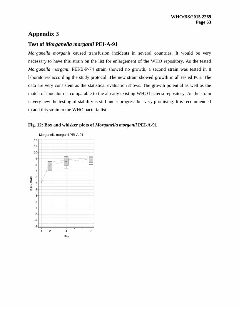
Test of Morganella morganii PEI-A-91
Morganella morganii caused transfusion incidents in several countries. It would be very
necessary to have this strain on the list for enlargement of the WHO repository. As the tested
Morganella morganii PEI-B-P-74 strain showed no growth, a second strain was tested in 8
laboratories according the study protocol. The new strain showed growth in all tested PCs. The
data are very consistent as the statistical evaluation shows. The growth potential as well as the
match of inoculum is comparable to the already existing WHO bacteria repository. As the strain
is very new the testing of stability is still under progress but very promising. It is recommended
to add this strain to the WHO bacteria list.
Fig. 12: Box and whisker plots of Morganella morganii PEI-A-91
log
10
cfu
/ml
-2
-1
0
1
2
3
4
5
6
7
8
9
10
11
12
Day
1 2 4 7
Morganella morganii PEI-A-91
WHO/BS/2015.2269
Page 64
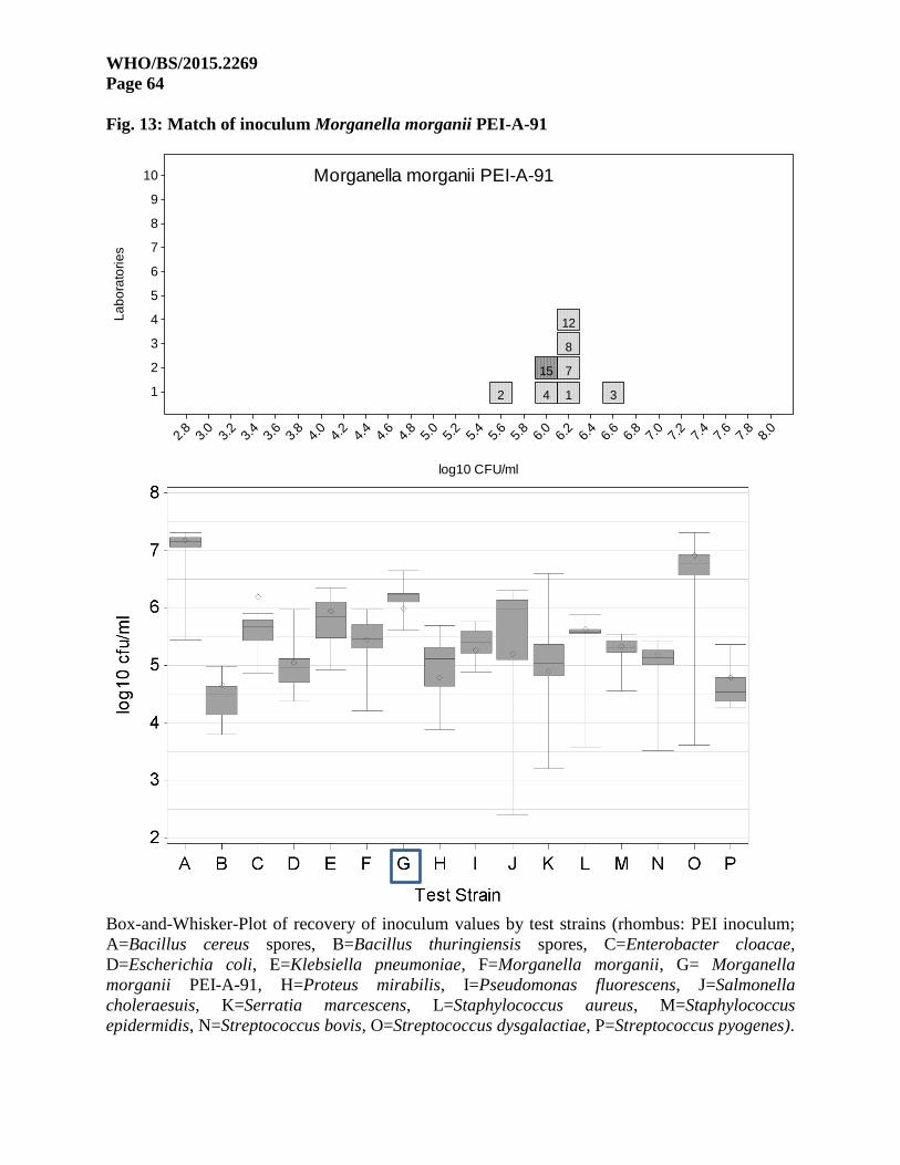
Fig. 13: Match of inoculum Morganella morganii PEI-A-91
Box-and-Whisker-Plot of recovery of inoculum values by test strains (rhombus: PEI inoculum;
A=Bacillus cereus spores, B=Bacillus thuringiensis spores, C=Enterobacter cloacae,
D=Escherichia coli, E=Klebsiella pneumoniae, F=Morganella morganii, G= Morganella
morganii PEI-A-91, H=Proteus mirabilis, I=Pseudomonas fluorescens, J=Salmonella
choleraesuis, K=Serratia marcescens, L=Staphylococcus aureus, M=Staphylococcus
epidermidis, N=Streptococcus bovis, O=Streptococcus dysgalactiae, P=Streptococcus pyogenes).
2 4
15
1
7
8
12
3
Morganella morganii PEI-A-91
La
bo
rato
rie
s
1
2
3
4
5
6
7
8
9
10
log10 CFU/ml
2.8
3.0
3.2
3.4
3.6
3.8
4.0
4.2
4.4
4.6
4.8
5.0
5.2
5.4
5.6
5.8
6.0
6.2
6.4
6.6
6.8
7.0
7.2
7.4
7.6
7.8
8.0