WHO Drug Information · WHO Drug Information Quality Assurance Highlights ... 5.3 Disintegration...
76
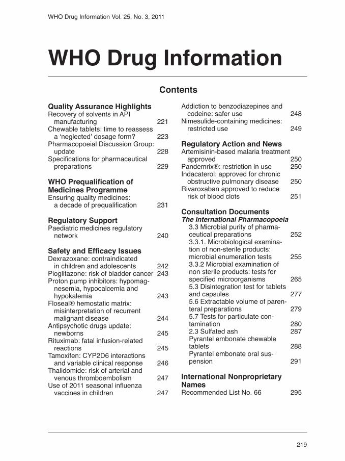
219 WHO Drug Information Vol. 25, No. 3, 2011 WHO Drug Information Quality Assurance Highlights Recovery of solvents in API manufacturing 221 Chewable tablets: time to reassess a ‘neglected’ dosage form? 223 Pharmacopoeial Discussion Group: update 228 Specifications for pharmaceutical preparations 229 WHO Prequalification of Medicines Programme Ensuring quality medicines: a decade of prequalification 231 Regulatory Support Paediatric medicines regulatory network 240 Safety and Efficacy Issues Dexrazoxane: contraindicated in children and adolescents 242 Pioglitazone: risk of bladder cancer 243 Proton pump inhibitors: hypomag- nesemia, hypocalcemia and hypokalemia 243 Floseal® hemostatic matrix: misinterpretation of recurrent malignant disease 244 Antipsychotic drugs update: newborns 245 Rituximab: fatal infusion-related reactions 245 Tamoxifen: CYP2D6 interactions and variable clinical response 246 Thalidomide: risk of arterial and venous thromboembolism 247 Use of 2011 seasonal influenza vaccines in children 247 Addiction to benzodiazepines and codeine: safer use 248 Nimesulide-containing medicines: restricted use 249 Regulatory Action and News Artemisinin-based malaria treatment approved 250 Pandemrix®: restriction in use 250 Indacaterol: approved for chronic obstructive pulmonary disease 250 Rivaroxaban approved to reduce risk of blood clots 251 Consultation Documents The International Pharmacopoeia 3.3 Microbial purity of pharma- ceutical preparations 252 3.3.1. Microbiological examina- tion of non-sterile products: microbial enumeration tests 255 3.3.2 Microbial examination of non sterile products: tests for specified microorganisms 265 5.3 Disintegration test for tablets and capsules 277 5.6 Extractable volume of paren- teral preparations 279 5.7 Tests for particulate con- tamination 280 2.3 Sulfated ash 287 Pyrantel embonate chewable tablets 288 Pyrantel embonate oral sus- pension 291 International Nonproprietary Names Recommended List No. 66 295 Contents
-
Upload
truongthuan -
Category
Documents
-
view
220 -
download
1
Transcript of WHO Drug Information · WHO Drug Information Quality Assurance Highlights ... 5.3 Disintegration...
219
WHO Drug Information Vol. 25, No. 3, 2011
WHO Drug Information
Quality Assurance Highlights Recovery of solvents in API manufacturing 221Chewable tablets: time to reassess a ‘neglected’ dosage form? 223Pharmacopoeial Discussion Group: update 228Specifications for pharmaceutical preparations 229
WHO Prequalification of Medicines ProgrammeEnsuring quality medicines: a decade of prequalification 231
Regulatory SupportPaediatric medicines regulatory network 240
Safety and Efficacy IssuesDexrazoxane: contraindicated in children and adolescents 242Pioglitazone: risk of bladder cancer 243Proton pump inhibitors: hypomag- nesemia, hypocalcemia and hypokalemia 243Floseal® hemostatic matrix: misinterpretation of recurrent malignant disease 244Antipsychotic drugs update: newborns 245Rituximab: fatal infusion-related reactions 245Tamoxifen: CYP2D6 interactions and variable clinical response 246Thalidomide: risk of arterial and venous thromboembolism 247Use of 2011 seasonal influenza vaccines in children 247
Addiction to benzodiazepines and codeine: safer use 248Nimesulide-containing medicines: restricted use 249
Regulatory Action and NewsArtemisinin-based malaria treatment approved 250Pandemrix®: restriction in use 250Indacaterol: approved for chronic obstructive pulmonary disease 250Rivaroxaban approved to reduce risk of blood clots 251
Consultation DocumentsThe International Pharmacopoeia
3.3 Microbial purity of pharma-ceutical preparations 2523.3.1. Microbiological examina-tion of non-sterile products:microbial enumeration tests 2553.3.2 Microbial examination of non sterile products: tests for specified microorganisms 2655.3 Disintegration test for tablets and capsules 2775.6 Extractable volume of paren-teral preparations 2795.7 Tests for particulate con-tamination 2802.3 Sulfated ash 287Pyrantel embonate chewable tablets 288Pyrantel embonate oral sus-pension 291
International Nonproprietary NamesRecommended List No. 66 295
Contents

220
WHO Drug Information Vol. 25, No. 3, 2011
WHO Drug Information &
digital libraryare available online at:
http://www.who.int/druginformation

221
WHO Drug Information Vol. 25, No. 3, 2011
Solvent recovery is a routine practice in the pharmaceutical industry when it is technically and economically viable for the particular waste stream. Organic solvents are ubiquitous in the reaction and separation steps of pharmaceutical processes. The replacement of organic solvents appears to be difficult owing to the strong influence on the outcome of the reaction and /or quality of the separa-tion (e.g., crystallization). The use of mul-tiple solvents and reagents for different purposes within a process frequently leads to the formation of solvent mixtures.
It is hard and often impossible to recover solvent in pure form from such a waste stream consisting of multiple solvents and reagents used in the reaction and separation process. Therefore, the use of recovered solvents and the pooling of solvents must be appropriately qualified to assure product quality and avoid cross contamination during active pharmaceuti-cal ingredient (API) production.
The use of recovered solvents can gene-rate problems from a product purity standpoint. For example, a recovered solvent can be of an azeotropic composition that
may become the solvent during a reac-tion. This change of solvent may cause changes in the spectrum of impurities present in the final product. On the other hand, intermediate and product isolation via crystallization can be affected by the composition and even the impurity profile of the solvent.
The following section drawn from the WHO good manufacturing practices for active pharmaceutical ingredients, which is equivalent to ICHQ7, discusses specific issues associated with solvent recovery in a pharmaceutical manufacturing setting (1). Recommendations have been made for inspection of API manufacturing facil-ties when dealing with solvent recovery. The relevant provisions to recovery of solvents in WHO good manufacturing practices for active pharmaceutical ingre-dients are:
14.40 Recovery (e.g., from mother liquor or filtrates) of reactants, intermediates, or the API is considered acceptable provided that approved procedures exist for the recovery and that the recovered materials meet specifications suitable for their intended use.
Recovery of solvents in API manufacturing
Quality Assurance Highlights
The practice of recovering and using solvents in the pharmaceutical industry has implications for the quality of active pharmaceutical ingredients (APIs) in finished pharmaceutical products. Various recovery processes and installations are cur-rently being utilized. This article attempts to present the latest developments and collective provisions related to the topic and has been compiled based on expe-rience gained as part of activities related to the WHO Prequalification of Medicines Programme. Importantly, it presents typical questions that should be addressed when an API manufacturing site is being inspected. Of particular relevance is the reproducibility of the given recovery process and, by implication, the quality of the recovered solvent. Such considerations are crucial to the quality of the final product. Comments on this article are welcome and should be forwarded to [email protected].

222
WHO Drug Information Vol. 25, No. 3, 2011
Solvents, as stated in the current guide-line, can be recovered and re-used within the same process or in different processes, provided that recovery procedures are strictly controlled and monitored to ensure that solvents meet appropriate standards (critical process parameters) before re-use or co-mingling with other approved materials. It is highly recommended that recovered solvents be used in the same process or in the same step of the aforementioned process to minimize potential cross-contamination.
Fresh and recovered solvents can be combined if adequate testing has shown their suitability for all manufacturing pro-cesses. The ratio of fresh vs recovered solvents should be defined in each step. The use of recovered solvents should be adequately documented in the process including the date of recovery (sto-rage time) and history of the recovered solvents. The recovered solvents should be avoided as media for the final purifica-tion of the API. However, use is permitted if the recovered solvent meets predefined specifications in composition and purity and there is no additional risk of impact on the purity of the API and its crystal form. Clarification or amendment may be requested in the next Expert Committee discussion for revision of the WHO gui-deline regarding impurity originating from other/different processes.
In evaluating solvents recovered during the API manufacturing process, ICH Q9 risk assessment may be employed. The specifications of recovered solvents, including the ratio of fresh vs recovered solvent, should be established for each individual API production and controlled before use. Risk assessment of the use of recovered solvents for final purification of APIs should be conducted and docu-mented. Use of the recovered solvent should be avoided when assessment indicates that the risk is relatively high.
When a recovered solvent with high unknown impurities is used, the manufac-
14.41 Solvents can be recovered and re-used in the same processes or in different processes, provided that the recovery procedures are controlled and monitored to ensure that solvents meet appropriate standards before re-use or co-mingling with other approved materials.
14.42 Fresh and recovered solvents and reagents can be combined if adequate testing has shown their suitability for all manufacturing processes in which they may be used.
14.43 The use of recovered solvents, mother liquors and other recovered mate-rials should be adequately documented.
According to the provisions, a recovery process is allowed only when an esta-blished procedure for a specific operation and an appropriate approval for such a process is in place. The procedure should detail the designated recovery system (e.g., type of distillation apparatus, reco-very process flowsheets). Specifications for a given recovered solvent should be established and be met for the release.
For the purpose of comparison, the specifications of both fresh and recovered solvents should include tests and para-meters to take account of accumulation of impurities. Impurities and composition of the recovered solvents for re-use should be determined and documented in the form of a certificate of analysis (COA) for recovered solvents. Analysis of the impurity profile of the recovered solvents is of utmost importance to avoid cross-contamination and accumulation of impurities in the process. Knowledge of the composition of the solvents recovered from a multi-component solvent process is crucial for strengthening reliability of the process to ensure the quality of the product (yield, crystal forms, etc.). Both impurity profile and composition of the recovered solvents intended for re-use in the process should br defined as critical process parameters.
Quality Assurance Highlights

223
WHO Drug Information Vol. 25, No. 3, 2011
turer needs to show suitability in re-using recovered solvent and demonstrate how the risk of “carry over” will be avoided. It is highly recommended to characterize all impurities before use for the purpose of risk mitigation. The impurities from the recovered solvents, and their implications for stability of the API, should be moni-tored as well.
The following typical questions should be addressed when an API manufacturing site is being inspected:
• Are there any recovered solvents in-volved in API manufacturing?
• If no recovered solvents are being used in the process, a statement to this effect should be made by the applicant.
• Is the use of recovered solvents in the process declared?
• What are the recovery methods (distilla-tion, column fractionation, etc.) and are batches pooled?
• Is there an approved procedure/stan-dard operating procedure (SOP) for recovered solvents in place?
• Is there a recovered process flowchart including source and destination?
• Is the facility and apparatus for recov-ered solvents qualified?
• Are specifications (composition and im-purity profile, specification sheets and
standard operating procedures) estab-lished and do they meet the COA when recovered solvents are used?
• Is there any process in place to improve quality of the recovered solvents?
• Do the recovered solvents come from the current API process or from a differ-ent API process? In the event that they come from different processes, what are the tests and acceptance criteria proposed by the API manufacturer to control the carry over of impurities from these other processes?
• In what step is the recovered solvent used?
• Is the use of recovered solvents docu-mented in the batch manufacturing record?
• Is the yield and purity profile of the product affected and whether it is proved by validation?
• Is there any additional impurity gener-ated by the use of recovered solvents and verified by batch analysis?
References
1. WHO good manufacturing practices for active pharmaceutical ingredients at http://www.who.int/medicines 2. International Conference on Harmoniza-tion. ICH Guidelines: Q3 Impurities, Q7 Good Manufacturing Practice, Q9 Quality Risk Management at http://www.ich.org/
A survey recently conducted in six WHO Member States was aimed at identifying possible quality problems in medicines for neglected tropical diseases (NTD). The medicines concerned were solid oral forms of albendazole, azithromycin, mebendazole, diethylcarbamazine, iver-mectin, and praziquantel. The survey was
coordinated by the WHO/NTD Secretariat and financially supported by USAID, in collaboration with the medicines regula-tory authorities of countries where these products are especially relevant — three in south-east Asia and three in west Africa. Testing was conducted at the National Institute for Drug Quality Control
Quality Assurance Highlights
Chewable tablets: time to reassess a ‘neglected’ dosage form?

224
WHO Drug Information Vol. 25, No. 3, 2011
To our knowledge, helminthic infections in humans have not been shown to deve-lop resistance. However, it is known that resistance to anthelminthics has deve-loped in animals (2, 3) and it has been suggested that known widespread resis-tance in veterinary practice “threatens the success of treatment in humans” (4–6). In that event, low potency and/or poor availability would be aggravating factors.
Current pharmacopoeial references to chewable tablets are listed in Table 1.
No explicit mention of chewable tablets was found in the text for disintegration time or dissolution rate in the Chinese (Ch.Ph), Indian (Ph.Ind) or Interna-tional Pharmacopoeia (Ph.Int). The British Pharmacopoeia (BP) 2008 expli-citly states that a dissolution rate test is “inappropriate“ for chewable tablets; this statement was not found in the 2011 BP but no alternative test was found. The USP 2010, and both the 2008 and 2010 editions of the BP, exclude chewable tablets from the requirement to comply with a disintegration test. However, the USP requires compliance with a dissolu-tion test whereas the BP does not. The assumption appears to be that all patients (including children and the elderly) will chew a tablet if it is labelled as chewable, which is improbable. It should be assu-med that some patients will swallow tablets that are labelled ‘chewable’ with-out chewing them or after chewing them incompletely. The aforementioned survey of NTD medi-cines found that some albendazole and mebendazole tablets were described as chewable but prescribing information was inconsistent as to whether patients were instructed to chew before swallowing. For example the same manufacturer used the words chew before swallowing or can be chewed in different countries for the same product. This raises a number of questions:
(NIDQC), Hanoi, Viet Nam and the Labo-ratoire National de Contrôle des Médica-ments, Rabat, Morocco. Both testing sites have been prequalified by WHO.
Testing results show that 41 samples out of 72 in the South-East Asian part of the survey did not conform to the requirements of the 32nd release of the United States Pharmacopoea (USP32). In all the 41 cases of non-conformity, the samples failed the USP32 dissolution test. In one case (albendazole 400 mg), the sample also failed content of active ingredient and uniformity of dosage units. Particularly striking is the case of meben-dazole: among 22 samples collected in two countries, not a single sample conformed with monograph requirements. In the west-African part of the survey, 10 samples out of 56 failed testing and all of these failed the USP32 dissolution test. In four cases the samples also failed to conform with the disintegration test. In one case, impurities exceeded the mono-graph limit.
In the majority of the cases of non-confor-mity, the product was in chewable tablet form.
The fact that such a high proportion of samples did not meet dissolution rate requirements is a cause of concern and raises questions about the efficacy of NTD medicines marketed in the countries that have participated in the survey. Non-conformity occurred most frequently in samples of albendazole and mebenda-zole. It has been shown that albendazole preparations that fail dissolution testing achieve lower egg reduction rates than preparations that meet dissolution requi-rements (1). In addition, in the absence of any alternative, it is difficult to challenge dissolution testing as the best indica-tor we have for availability of the active drug for absorption by patient or parasite tissue or as an indicator of batch to batch consistency.
Quality Assurance Highlights

225
WHO Drug Information Vol. 25, No. 3, 2011
Pharmacopiea DefinitionBritish Phar-macopoeia (BP) / Euro-pean Pharma-copoiea (Ph.EU)
BP2008: From the general monograph for tablets: “Chewable tablets are prepared to ensure that they are easily crushed by chewing”.“Chewable tablets are not required to comply with the test [for disintegration]”. Supplementary chapter SC I E. Dissolution Testing of Solid Oral Dosage Forms makes it clear that a dissolution test is considered inappropriate for chewable tablets (“…where the nature or intended use of the prepa-ration renders a dissolution test inappropriate (for example, liquid-containing capsules, dispersible, effervescent, chewable or soluble tablets”). BP 2011: All the above statements are no longer present in the BP 2011.
Chinese Phar-macopoeia (Ch.Ph)(2005)
From the general monograph for tablets: “Chewable tablets are intended to be chewed or sucked to disintegrate and then swallowed to effect in gastrointestinal tract, or be absorbed by the gastrointestinal tract for systemic action”. “The hardness of chewable tablets should be suitable”. No explicit mention of chewable tablets was found in the text for disintegration time or dissolution rate.
India Pharma-copoeia (Ph.Ind)(2007)
From the general monograph for tablets: “Tablets for use in the mouth are usually uncoated tablets formulated to be chewed or to effect a slow release and local action of the active ingredient (lozenges) or the release and absorption of the active ingredient under the tongue (sublingual tablets)”. No explicit mention of chewable tablets was found in the text for disintegration time or dissolution rate.
InternationalPharma-copoeiaPh.Int(2008)
From the general monograph for tablets: “Tablets for use in the mouth and chewable tablets are usually uncoated. They are formulated to effect a slow release and local action of the active ingredient(s) (for example, compressed lozenges) or the release and absorption of the active ingredient(s) under the tongue (sublingual tablets) or in other parts of the mouth (buccal) for systemic action”. The same monograph exempts chewable tablets from compliance with a disintegration test. The Ph.Int requires a dis-solution test only when a requirement is specified in the individual monograph. No explicit mention of chewable tablets was found in the sections of the Ph.Int entitled Disintegration test for tablets and capsules and Dissolution test for solid oral dosage forms.
United States Pharmaco-poea, USP 32 (2010)
From the general chapter on pharmaceutical dosage forms: “Chewable tablets are formulated and manufactured so that they may be chewed, producing a pleasant tasting residue in the oral cavity that is easily swallowed and does not leave a bitter or unpleasant aftertaste. These tablets have been used in tablet formulations for children, especially multivitamin formulations, and for the administration of antacids and selected antibiotics. Chewable tablets are prepared by compression, usually utilizing mannitol, sorbitol, or sucrose as binders and fillers, and containing colors and flavors to enhance their appearance and taste”.“Disintegration is an essential attribute of tablets intended for administration by mouth, except for those intended to be chewed before being swallowed and for some types of extended-release tablets. A disintegration test is provided [see cross reference] and limits on the times in which disintegration is to take place, appropriate for the types of tablets concerned, are given in the individual monographs”. From <1088> In vitro and in vivo evaluation of dosage forms: “The state of science is such that conduct of in vivo testing is necessary in the development and evaluation of dosage forms. Also, no product, including suspensions and chewable tablets, should be developed without dissolution or drug release characterization where a solid phase exists. This chapter sets forth, for products intended for human use, guidelines for characterizing a drug that include: (1) developing in vitro test methods for immediate-release and modified-release dosage forms, (2) designing in vivo protocols, and (3) demonstrating and assessing in vitro-in vivo correlations for modified-release dosage forms”.
Table 1. Pharmacopoeial references to chewable tablets
Quality Assurance Highlights

226
WHO Drug Information Vol. 25, No. 3, 2011
phase exists”. It goes on to outline the development of in vitro test methods and design of in vivo study protocols. Consistent with the USP, the Food and Drug Administration (FDA) Bioavailability and Bioequivalence Studies for Orally Administered Drug Products — Gene-ral Considerations (2003), page 17 (8), states: “We recommend that rapidly dissolving drug products, such as buccal and sublingual dosage forms (and chew-able tablets), be tested for in vitro dissolu-tion and in vivo BA and/or BE. We recom-mend that chewable tablets (as a whole) be subject to in vitro dissolution testing because they might be swallowed by a patient without proper chewing. In gen-eral, we recommend that in vitro dissolu-tion test conditions for chewable tablets be the same as for non-chewable tablets of the same active ingredient or moiety. Infrequently, different test conditions or acceptance criteria can be indicated for chewable and non-chewable tablets, but we recommend these differences, if they exist, be resolved with the appropriate review division.”
A joint position paper (9) by the Internatio-nal Pharmaceutical Federation ( FIP) and American Association of Pharmaceutical Scientists (AAPS) has reviewed some of these questions. Their paper includes the statement “….In principle, the test procedure employed for chewable tablets should be the same as that used for regu-lar tablets. This concept is based on the possibility that a patient might swallow the dosage form without proper chewing, in which case the drug would still need to be released to ensure the desired pharma-cological action. Where applicable, test conditions would preferably be the same as used for conventional tablets of the same active pharmaceutical ingredient, but because of the non-disintegrating na-ture of the dosage form, it may be neces-sary to alter test conditions (e.g., increase the agitation rate) and specifications (e.g., increase the test duration). The reciproca-ting cylinder (USP apparatus 3) with the addition of glass beads may also provide
1. What is meant by the word chewable? Patients are generally unaware of defini-tions included in compendia. Chambers English dictionary (7) defines the suffix -able to mean “capable of being”. Thus, the simple English meaning of chewable is capable of being chewed and not must be chewed. The USP definition is consistent with this interpretation, using the words “may be chewed”. By way of comparison, if a liquid is labelled inflam-mable that is not to suggest that the user should set it on fire. The BP, Ch.Ph, Ph.Ind and Ph.Int do not state explicitly whether chewable tablets may alternati-vely be swallowed whole, but exemption from compliance with a disintegration test, as in the BP and Ph.Int, implies an assumption that chewable tablets will always be chewed.
2. Is it necessary to test whether the same tablet is bioequivalent when it is chewed or when it is swallowed whole?
3. Can tablets that are described as chewable (meaning “may be chewed”) be considered bioequivalent to tablets for which the recommendation is only to swallow whole?
4. Should chewable tablets be required to comply with tests for dissolution rate? If chewable tablets may be swallowed whole, the same dissolution rate test and limit should apply as if the tablet were intended to be swallowed whole.
5. Should chewable tablets be required to comply with a test for disintegration time? It is established practice to exempt tablets from compliance with a test for disinte-gration time if there is also a specification for dissolution rate. Consequently, this question arises only if there is no dissolu-tion rate test.
As noted in the above table, the USP states: “... no product, including suspen-sions and chewable tablets, should be developed without dissolution or drug release characterization where a solid
Quality Assurance Highlights

227
WHO Drug Information Vol. 25, No. 3, 2011
• The term “chewable tablet“ should be defined as “a conventional tablet that can also be chewed”.
• Because in practice chewable tablets may be swallowed without chew-ing (even if the label states “must be chewed”), they should be tested for the release of the API(s) even when swal-lowed whole. Tablets labelled as “chew-able” should be bioequivalent when chewed or swallowed whole.
• Tablets labelled as “chewable” should be bioequivalent to any other chewable or non-chewable tablets on the same market that contain the same APIs in the same dose.
• In the absence of suitable requirements for testing release of the API(s) even if swallowed whole, chewable tablets should not be used for potent medi-cines and especially not for those hav-ing the potential for variable bioavail-ability such as mebendazole. For the paediatric population, a better option would be dispersible solid oral dosage forms that must be dispersed before swallowing. It is probably reasonable to assume that carers will be more reliable than patients.
• Medicines regulatory authorities should ensure that manufacturers justify and demonstrate the biopharmaceutical characteristics of the chewable dosage form in each case.
Comments or feedback on the above dis-cussion paper would be welcome and can be sent to the authors: Valerio Reggi at [email protected] and Susan Walters at [email protected]
References
1. Albonico M, Mathema P, Montresor A et al. Comparative study of the quality and efficacy of originator and generic albendazole for mass treatment of soil-transmitted nematodeinfections in Nepal. Tr R Soc Trop Med & Hyg 2007;101:454–460
more “intensive” agitation for in vitro dissolution testing of chewable tablets. As another option, mechanical breaking of chewable tablets prior to exposing the specimen to dissolution testing could be considered. While this option would more closely reflect the administration of the product and the corresponding formulation and manufacturing features, no approach for validating such a method has been reported in the literature or pre-sented during the workshops….”
Our comments are not intended to sug-gest a final conclusion on the issue of chewable tablets but rather to raise ideas for consideration. The current absence of clear guidance on dissolution rate requi-rements has led to a situation in which there are no consistent and suitable qua-lity requirements with which the manufac-turers of chewable tablets must conform. Many regulatory authorities in NTD ende-mic countries may be unable to insist on requiring conformity to dissolution rate testing for chewable tablets if internatio-nal references such as The International Pharmacopoeia do not require it.
In addition, the current definition of chew-able tablet as it appears in The Internatio-nal Pharmacopoeia, 4th edition, 2008 is as follows:
Tablets for use in the mouth (sublin-gual, buccal) and chewable tablets
DefinitionTablets for use in the mouth and chew-able tablets are usually uncoated. They are formulated to effect a slow release and local action of the active ingredient(s) (for example, compressed lozenges) or the release and absorption of the active ingredient(s) under the tongue (sublingual tablets) or in other parts of the mouth (buccal) for systemic action.
This does not address the problems iden-tified in the above mentioned survey.
Against this background, we make the following propositions for discussion:
Quality Assurance Highlights

228
WHO Drug Information Vol. 25, No. 3, 2011
2. Prichard RK et al. Towards markers for anthelminthic resistance in helminths of im-portance in animal and human health. Parasit 2009;134:1073–1076
3. Gaba S, Silvestre A. Mathematical models for the management of helminth parasites: from biological processes to the evolution of anthelminthic resistance. Antiinfec Agents in Med Chem 2011;9:3,139-147
4. James C et al. Drug Resistance mecha-nisms in helminths: is it survival of the fittest? Trends in Paras;25:7,328–335.
5. Albonico M. Potential drug resistance in helminth control programmes. 6th Interna-tional Conference on Tropical Medicine and
worldwide by avoiding duplication during the preparation of dossiers and studies, thus reducing the time required for inno-vative medicines to become available. This conference takes place twice a year with the location of meetings rotating between Europe, Japan and the United States of America.
Work of the Pharmacopoeial Discussion Group The Pharmacopoeial Discussion Group (PDG) comprises representatives from the Ph.EU, the JP and the USP. It consi-ders proposals made by national associa-tions of manufacturers of pharmaceutical products and excipients in order to select general methods of analysis and exci-pient monographs for addition to its work programme. To promote exchange and synergy, the PDG has organized, since 2001, hearings for representatives of the pharmaceutical and excipient industries.
Each pharmacopoeia is responsible for a programme of international harmoniza-tion. Each text drafted by the three coor-dinating pharmacopoeia’s is published for public comment at stage four in each of the respective forums.
The need for international harmonizationGlobalization and expansion in interna-tional trade present a growing need to develop global quality standards for medi-cines. As standards are a vital instrument for registration, market surveillance and free movement and trade of medicines among as many countries as possible, harmonization among the world’s three major pharmacopoeias— the European Pharmacopoeia (Ph.EU), the Japanese Pharmacopoeia (JP) and the United States Pharmacopoeia (USP) — is an important and challenging task. Within the harmonization process, the European Directorate for the Quality of Medicines and Healthcare (EDQM) represents the Ph.EU. All the relevant groups of experts of the European Pharmacopoeia are involved.
International Conference on Harmonization In 1990, a trilateral programme, the Inter-national Conference on Harmonization (ICH), for the harmonization of testing of medicines among the European Union, the United States and Japan was set up. This programme aims to reduce the overall cost of pharmaceutical research
Quality Assurance Highlights
Pharmacopoeial Discussion Group: update
International Health at http://www.festmih.eu/document/1791.
6. Harhay MO et al. Epidemiology and Control of human gastrointestinal parasites in children. Exp Rev of Antiinfect Ther 2010;8:3,219–234.
7. Chambers 20th Century Dictionary. Ed. Kirkpatrick EM. W & R Chambers Ltd, 1983; Edinburgh.
8. US Food and Drug Administration. htt://www.fda.gov/downloads/Drugs/Guidance-ComplianceRegulatoryInformation/Guidances
9. American Association of Pharmaceutical Scientists http://www.aapspharmscitech.org/articles/pt0401/pt040107/pt040107.pdf

229
WHO Drug Information Vol. 25, No. 3, 2011
Excipients CouncilA meeting with the International Phar-maceutical Excipients Council (IPEC) Federation was held on June 14, 2011. Topics, among others, included cellulo-sics, viscosity of cellulose derivatives, polyethylene glycol, glycerin, povidone, copovidone, the silicon dioxide monogra-phs, and metal impurities.
Future PDG activitiesThe three Pharmacopoeias emphasized their commitment to further strengthen international harmonization. The PDG will utilize its monthly teleconferences for discussion of technical topics in addition to monitoring status updates. The next face-to-face PDG meeting will be hostedby EDQM on 8–9 November 2011 in Strasbourg, France.
Reference: European Directorate for the Qua-lity of Medicines and Healthcare at http://www.edqm.eu/en/International-Harmonisation-614.html
Latests developmentsThe PDG met from 14–15 June 2011 in Cincinnati, Ohio, USA. The Group reported on work achieved and progress made. To date, 28 of the 35 general chap-ters and 41 of the 62 excipient monogra-phs of the current work programme have been harmonized, including the chapter on microcalorimetry. Revised general chapters include bacterial endotoxins and bulk and tapped density. Excipient sign-offs include revisions to benzyl alcohol, potato starch, wheat starch, calcium phosphate dibasic, and calcium phos-phate dibasic anhydrous monographs. The latter four revisions are the outcome of PDG’s review of previously harmonized excipient monographs.
Representatives from the three pharma-copoeias discussed other topics, inclu-ding microbiological limits, additives in excipients, and metal impurities. Also, the PDG decided to add the isomalt mono-graph to its work programme.
Quality Assurance Highlights
The WHO Medicines Quality Assurance Programme is pleased to announce that the 45th report of the Expert Committee on Specifications for Pharmaceutical Pre-parations is now available at http://www.who.int/medicines/publications/pharm-prep/en/index.html and http://whqlibdoc.who.int/trs/WHO_TRS_961_eng.pdf The Expert Committee on Specifications for Pharmaceutical Preparations provides recommendations and tools to assure the quality of medicines from their develop-ment phase to their final distribution to the patients. The activities discussed during the Expert Committee meetings serve to develop specific additional guidance and specifications as needed for the various medicines recommended by WHO Pro-grammes.
The WHO Prequalification of Medicines Programme functions are based on the guidelines, standards and specifications
adopted by the Expert Committee after passage through its rigorous, interna-tional and wide consultative process.
From a wider perspective, the inter-national guidelines, specifications and nomenclature developed under the aegis of the Expert Committee serve all Mem-ber States, international organizations, United Nations agencies, regional and interregional harmonization efforts, and underpin important initiatives, including the prequalification of medicines, the Roll Back Malaria Programme, Stop TB, essential medicines and medicines for children. The advice and recommenda-tions provided by the Expert Committee are intended to help national and regional authorities and procurement agencies, as well as major international bodies and institutions, such as the Global Fund to Fight AIDS, Tuberculosis and Malaria, and international organizations such as UNICEF – to combat circulation of subs-
Specifications for pharmaceutical preparations
230
WHO Drug Information Vol. 25, No. 3, 2011
• GMP for sterile pharmaceutical products (Annex 6).
• Guiding principles on transfer of technology in pharmaceutical manufac-turing. (Annex 7).
• Good Pharmacy Practice: standards for quality of pharmacy services (joint FIP/WHO, Annex 8).
• Model guidance for the storage and transport of time- and temperature- sensitive pharmaceutical products (Annex 9).
• Procedure for prequalification of phar-maceutical products (Annex 10).
• Guidance on submission of documen-tation for prequalification of innova-tor finished pharmaceutical products (FPPs) approved by stringent regula-tory authorities (Annex 11).
• Procedure for prequalification of laboratories (Annex 12).
• WHO guidelines for preparing a labora-tory information file (Annex 13).
• Guidelines for preparing a Site Master File (Annex 14).
• Guideline for submission of documenta-tion for a multisource (generic) finished product (Annex 15).
Reference: World Health Organization 45th Expert Committee on Specifications for Phar-maceutical Preparations. Information available at http://www.who.int/medicines/areas/quality_safety/quality_assurance/en/index.html
tandard medicines and to work towards access to quality medicines.
In conclusion, the Expert Committee on Specifications for Pharmaceutical Pre-parations gives recommendations and provides independent international stan-dards and guidelines in the area of quality assurance for implementation by WHO Member States, international organiza-tions, United Nations agencies, regional and interregional harmonization efforts, as well as WHO’s medicines related pro-grammes and initiatives.
The following new recommendations were adopted at the 45th meeting of the Expert Committee on Specifications for Pharmaceutical Preparations.
• Monographs for inclusion in the Interna-tional Pharmacopoeia.
• International reference standards:
• New procedure for the release of ICRS (see also Annex 1).
• General policy regarding interna-tional standards for human recom-binant insulin.
• Procedure for adoption of Interna-tional Chemical Reference Substances (ICRS) (Annex 1).
• Good Practices for Pharmaceutical Microbiology Laboratories (Annex 2).
• GMP: main principles (Annex 3).
• GMP for blood establishments (jointly with ECBS) (Annex 4).
• Supplementary GMP for HVAC (Annex 5)
Quality Assurance Highlights

231
WHO Drug Information Vol. 25, No. 3, 2011
In March 2001, United Nations partners initiated a project, managed by the World Health Organization, to facilitate access to quality medicines used in the treat-ment of HIV/AIDS. Partnering with WHO were UNICEF, UNAIDS, and UNFPA. The World Bank also supported this project. The first manager for the programme was appointed by WHO on a six-month con-tract to establish, implement and manage the pilot project. The project was prin-cipally funded by donations and grants from Member States.
Objectives of the WHO Prequalification of Medicines Programme (PQP) were to:
1. Propose a list of prequalified products manufactured in sites that meet WHO norms and standards.
2. Follow-up on products and manufactur-ing facilities for quality issues.
3. Ensure that prequalification and update of the original approved list is carried out periodically and that variations and changes are correctly controlled.
4. Assist national drug regulatory autho-rities to build capacity in assessment, inspection and control of medicines for priority diseases.
In designing the project, a quality sys-tem was established consisting of a Procedure for Prequalification that was adopted by the WHO Expert Committee on Specifications for Pharmaceutical Preparations (ECSPP), various guide-lines, norms and standards, and standard
operating procedures (SOPs). Prequalifi-cation (PQ) was based on existing WHO norms and standards approved by the ECSPP. In those cases where WHO did not have guidelines, relevant guidelines from ICH were used. A web site was also established to disseminate PQ vision, mission, procedures, guidelines, training material, results and information (1).
The initial focus was to prequalify medi-cines used in the treatment of HIV/AIDS. It was estimated that the number of people needing antiretroviral (ARV) therapy in 2003 was in the range of 100 000 with an ARV therapy coverage of only around 2%. It was further estimated that less than 10% of people in most African countries had access to ARV treatment (2). Figure 1 shows the number of deaths per 100 000 in the US population in the period 1982–1993 (3).
Later on, due to the pressing need for quality medicines in other disease areas, the project was expanded to include products used in the treatment of tubercu-losis (TB) and malaria.
Prequalifying medicines is achieved through an extensive evaluation pro-cedure that consists of assessment of product data and information that are voluntarily submitted by interested appli-cants and manufacturers expressing their interest to participate in the project. This is followed by inspection of manufactur-ing and testing sites. No fees have been charged by WHO since the beginning of prequalification but this practice is under
Ensuring quality medicines: a decade of prequalificationReflections from A. J. van Zyl, First Programme Manager for the WHO Prequalification of Medicines Programme
WHO Prequalification of Medicines Programme

232
WHO Drug Information Vol. 25, No. 3, 2011
review. Sites are inspected to verify data submitted in the product dossiers and to assess compliance with WHO good manufacturing practices (GMP), good clinical practices (GCP) and other appro-priate guidelines.
Reasons for initiating the PQ pilot project was concern about low quality products circulating in the international market, the prevalence of spurious products and also as a result of the recommendation in a report from a group of independ-ent experts. The report found that many procurement organizations had no, or very limited, quality assurance systems in place to ensure that good quality products were procured.
Human resourcesIn terms of staff appointments, the PQP team slowly grew from one manager in 2001 to a manager plus one coordina-tor for assessments and one assistant during the first two years. One inspector was seconded from the French medi-cines regulatory agency in 2003, and in subsequent years the team expanded to include one manager, a coordinator for assessments and three inspectors.
The organization chart in 2011 comprises:
• one PQP manager• one head of inspections with five in-
spectors• one head of assessments with seven
assessors• eight support staff • one person each for liaison, capacity
building and training, and sampling and monitoring (which includes prequalifica-tion of quality control laboratories).
Expansion of the programme after 2006 was possible due to the financial support from the Bill & Melinda Gates Foundation. Today, the programme is largely financed by UNITAID but is seeking a broader donor base.
External assessor groupFrom the initial one staff member with six external assessors present at the assess-ment meeting in June 2001, the group has grown over the past ten years to include on average seven internal as-sessors and more than 20 external assessors at a group session.
WHO Prequalification of Medicines Programme
Figure 1: USA deaths per 100 000 population in the period 1982– 1993
HIV InfectionUnintentionalInjuriesCancer
HeartDisease
SuicideHomicide
Liver DiseaseStrokeDiabetes
National Vital Statistics * Provisional Data82 83 84 85 86 87 88 89 90 91 92 93*
0
5
10
15
20
25
30
35
40

233
WHO Drug Information Vol. 25, No. 3, 2011
Complaints on prequalified products that are received in PQP are logged and investigated by the inspectors. Inspec-tors and assessors must comply with the confidentiality and conflict of interest rules of WHO.
Inspectors publish a quarterly newsletter available on the PQP web site as well as submitting articles to the WHO Phar-maceuticals Newsletter and WHO Drug Information (see “Further reading” on page 239).
MonitoringField sampling and testing projects have been carried out by PQP in order to moni-tor the quality of medicines (both WHO-prequalified and non-WHO prequalified) procured by UN agencies (8). Through cooperation with medicines regulatory authorities (MRAs), these projects also contribute to national quality control of medicines, to strengthening of health systems and capacity building. Samples are collected by MRA staff and tested at WHO-prequalified laboratories and results are published. Several reports and publications in scientific journals have be-come available over the ten-year period.
Brief overviewAfter the initial establishment of the project in 2001, the first list of prequalified products was published in March 2002. The project expanded to include prequali-fication of quality control laboratories and tuberculosis and malaria medicines in 2003–2004. Due to inspection findings of non-compliance with GCP, some products were withdrawn from the list in 2004. In order to improve patient compliance and ease of dosing, fixed dose combinations were developed. PQP was instrumental in providing the corresponding guideline. As the applicant of a prequalified medicinal product invariably makes changes to a supplied product during the product’s life cycle, a variation guideline was also de-veloped to ensure appropriate oversight of such changes.
AssessmentsData and specifications are submitted and assessed by teams of assessors from national medicines regulatory autho-rities and WHO staff. Data and specifi-cations include but are not limited to the active pharmaceutical ingredient (API), formulae, manufacturing process, stability (appropriate packaging and suitable for the intended market) and bio-equivalence data (for generic products).
Group assessment sessions are held every two months at the UNICEF offices in Copenhagen. Requirements for product data and information have also intensified over the years. In 2011, the recommendation is that manufacturers should submit a dossier in the common technical document (CTD) format (4–6). To build capacity in developing countries, a unique three-month rotational post was established in the area of dossier assess-ment in 2006. Since then, 14 developing country regulators from nine countries have benefited from the arrangement.
During assessment, multisource (generic) drug products are expected to satisfy the same quality standards as those appli-cable to the originator/reference product. In addition, assurance has to be provided that they are clinically interchangeable with equivalent originator products (4).
InspectionsThe inspection unit operates in accor-dance with an established quality system consisting of documented SOPs, formats for reports and letters, a training pro-gramme and related aspects as recom-mended in guidelines (7). Inspections are performed at the facilities of finished product manufacturers, API manufac-turers, quality control laboratories and clinical sites including contract research organizations (CROs). Feedback on the implementation of norms and standards is given to the relative unit in WHO and recommendations are made for the development of new GMP guidelines (or revision of existing ones) as appropriate.
WHO Prequalification of Medicines Programme
234
WHO Drug Information Vol. 25, No. 3, 2011
As the PQP become successful, it was extended to include HIV/AIDS, TB and malaria, reproductive health products, zinc sulphate for the treatment of diar-rhoea in children, products used in treat-ment of influenza and diethylcarbamazine (DEC).
Mutual confidentiality agreements were signed in 2005 between the US Food and
Drug Administration (FDA), PQP and the Quality Assurance and Safety: Medicines Unit of WHO and in 2011 between the European Directorate for the Quality of Medicines (EDQM) and WHO.
Within the biopharmaceutical classifica-tion system, PQP assisted in the devel-opment of a guideline on comparative dissolution for biowaiver applications.
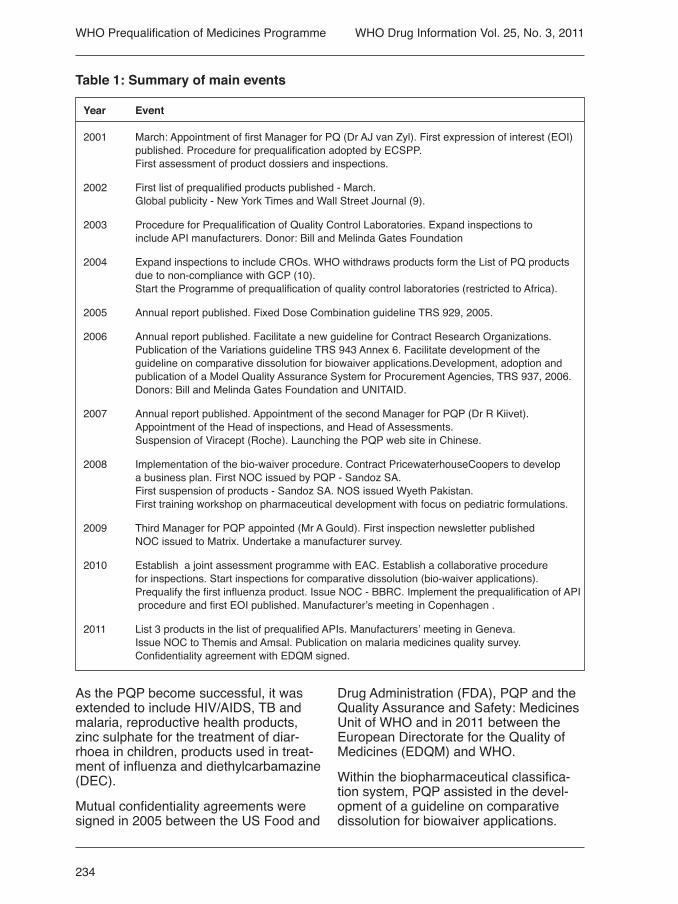
Year Event
2001 March: Appointment of first Manager for PQ (Dr AJ van Zyl). First expression of interest (EOI) published. Procedure for prequalification adopted by ECSPP. First assessment of product dossiers and inspections.
2002 First list of prequalified products published - March. Global publicity - New York Times and Wall Street Journal (9).
2003 Procedure for Prequalification of Quality Control Laboratories. Expand inspections to include API manufacturers. Donor: Bill and Melinda Gates Foundation
2004 Expand inspections to include CROs. WHO withdraws products form the List of PQ products due to non-compliance with GCP (10). Start the Programme of prequalification of quality control laboratories (restricted to Africa).
2005 Annual report published. Fixed Dose Combination guideline TRS 929, 2005.
2006 Annual report published. Facilitate a new guideline for Contract Research Organizations. Publication of the Variations guideline TRS 943 Annex 6. Facilitate development of the guideline on comparative dissolution for biowaiver applications.Development, adoption and publication of a Model Quality Assurance System for Procurement Agencies, TRS 937, 2006. Donors: Bill and Melinda Gates Foundation and UNITAID.
2007 Annual report published. Appointment of the second Manager for PQP (Dr R Kiivet). Appointment of the Head of inspections, and Head of Assessments. Suspension of Viracept (Roche). Launching the PQP web site in Chinese.
2008 Implementation of the bio-waiver procedure. Contract PricewaterhouseCoopers to develop a business plan. First NOC issued by PQP - Sandoz SA. First suspension of products - Sandoz SA. NOS issued Wyeth Pakistan. First training workshop on pharmaceutical development with focus on pediatric formulations.
2009 Third Manager for PQP appointed (Mr A Gould). First inspection newsletter published NOC issued to Matrix. Undertake a manufacturer survey.
2010 Establish a joint assessment programme with EAC. Establish a collaborative procedure for inspections. Start inspections for comparative dissolution (bio-waiver applications). Prequalify the first influenza product. Issue NOC - BBRC. Implement the prequalification of API procedure and first EOI published. Manufacturer’s meeting in Copenhagen .
2011 List 3 products in the list of prequalified APIs. Manufacturers’ meeting in Geneva. Issue NOC to Themis and Amsal. Publication on malaria medicines quality survey. Confidentiality agreement with EDQM signed.
Table 1: Summary of main events
WHO Prequalification of Medicines Programme

235
WHO Drug Information Vol. 25, No. 3, 2011
Due to cases of non-compliance identi-fied during inspections at CROs, it was decided to facilitate the development of an additional guideline for CROs to assist in better understanding the application of GCP for bioequivalence studies.
Due to reporting of low quality repro-ductive health products and problems in procuring good quality products, PQP expanded its scope and included repro-ductive health products within the PQP in cooperation with UNFPA.
In September 2008, the USA issued an import alert against Ranbaxy, a phar-maceutical company based in India. As there were several of their products listed in PQP, a joint inspection with Canada, Australia and the United Kingdom was undertaken at Ranbaxy to investigate impact. At that time, to respond to World Health Assembly Resolution 57.14 and the request by Member States and inter-national procurement organizations to enhance transparency, PQP published a first Notice of Concern (NOC) for manu-facturing sites. Provision was also made for issuing Notices of Suspension (NOS) for products. Resolution 57.14 requested WHO, among other actions to “ensure that the prequalification review process and the results of inspection and assess-ment reports of the listed products, aside from proprietary and confidential informa-tion, are made publicly available”.
As a consequence, publication of WHO Public Inspection Reports (positive outcomes of site inspections) and WHO Public Assessment Reports (positive outcomes of dossier assessment) and the list of prequalified products provides the public and regulators with extensive infor-mation on the PQ evaluation of products and sites.
The structure of PQP changed in 2007 with the appointment of a new Pro-gramme Manager, appointment of a Head of Inspections, and a Head of Assess-ments. In the same year, the launching of
the PQP web site in Chinese followed, as well as implementation of the biowaiver procedure. All NOC and NOS were also published. In keeping interested parties informed of the activities of PQP, an in-spection newsletter was regularly pub-lished as well as articles in publications. To further ensure transparency and better serve clients, PQP undertook a manufac-turer’s survey in 2009.
In an effort to expedite registration of prequalified products, prevent duplication, and promote harmonization, PQP estab-lished and implemented a joint assess-ment programme with the East African Community (EAC) for product dossiers and a collaborative procedure for inspec-tions (joint inspections and recognition of inspection reports among regulators). Both initiatives deserve more in depth clarification. It is anticipated that activities will be described in more detail in future publications.
With an increasing number of product dossiers containing comparative disso-lution data, the inspection unit began inspections at sites to verify reliability of dissolution data and GMP compliance (biowaiver applications).
A major step forward in assisting MRAs to obtain information on the quality of APIs and API manufacturing sites, was imple-mentation of the procedure for prequalifi-cation of APIs in 2010. This procedure is based on the assessment of API Master Files (also known as a Drug Master Files) and inspection of the sites.
In further attempting to ensure the quality of products purchased, a model qual-ity assurance system for procurement agencies was developed. This guideline was adopted by the ECSPP and the Interagency Pharmaceutical Coordina-tion group (IPC) and is used by different organizations including the World Bank.Following publication of the first Expres-sion of Interest for HIV/AIDS products, more than 90 product dossiers were
WHO Prequalification of Medicines Programme
236
WHO Drug Information Vol. 25, No. 3, 2011
received for assessment in the first group assessment session in Copenhagen. The number of product dossiers submitted for assessment has varied from year to year, and between disease groups.
Since 2001, more than 60 training workshops have been organized or co-organized in countries including Austria, Belgium, Brazil, China, Estonia, India, Kenya, Pakistan and Tanzania. Twenty quality control laboratories (QCLs) have been prequalified and four sampling and testing projects have been undertaken.
In 2008, PricewaterhouseCoopers was appointed to assist in the development of a business plan. Recommendations for improvement were made and it was calculated that the return on investment in PQ was 170.1 in the period 2009–2013.
Outcomes of the manufacturer survey carried out in 2009 (11) were presented to a manufacturers’ meeting in Copenha-gen in April 2010 and at the PQP Annual Stakeholders meeting in 2011. The report concluded that both PQP assessors and inspectors are meeting or exceeding manufacturer expectations for service delivery. The structure of PQP generally delivers levels of service at, or above, those expected by manufacturers.
However, the service process is falling short of manufacturer expectations with respect to review/reply time for product dossiers; opportunities for in-person com-munication during the assessment pro-
cess; question/problem resolution during assessment; consistency of membership in the team of assessors throughout the process, and local/national representation in on-site inspection teams. Most manu-facturers view PQP GMP requirements as more stringent than those of the US FDA or European Medicines Agency. The findings from this survey indicate that pharmaceutical manufacturers consider PQP to be a well-designed, well-executed programme. PQP assessors and inspec-tors are meeting or exceeding manufactu-rer expectations for service delivery in all processes.
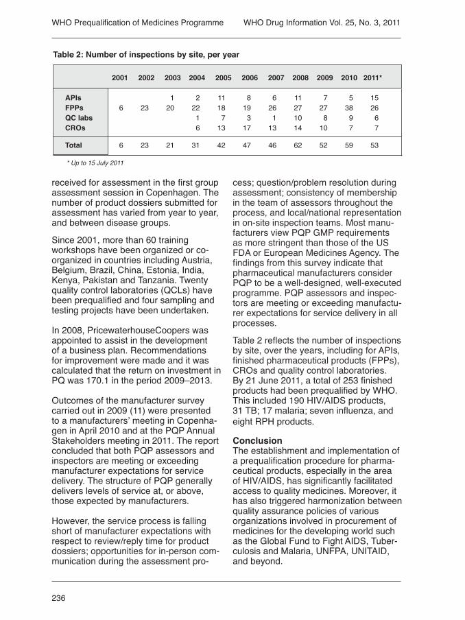
Table 2 reflects the number of inspections by site, over the years, including for APIs, finished pharmaceutical products (FPPs), CROs and quality control laboratories.By 21 June 2011, a total of 253 finished products had been prequalified by WHO. This included 190 HIV/AIDS products, 31 TB; 17 malaria; seven influenza, and eight RPH products.
ConclusionThe establishment and implementation of a prequalification procedure for pharma-ceutical products, especially in the area of HIV/AIDS, has significantly facilitated access to quality medicines. Moreover, it has also triggered harmonization between quality assurance policies of various organizations involved in procurement of medicines for the developing world such as the Global Fund to Fight AIDS, Tuber-culosis and Malaria, UNFPA, UNITAID, and beyond.
WHO Prequalification of Medicines Programme
2001 2002 2003 2004 2005 2006 2007 2008 2009 2010 2011*
APIs 1 2 11 8 6 11 7 5 15 FPPs 6 23 20 22 18 19 26 27 27 38 26 QC labs 1 7 3 1 10 8 9 6 CROs 6 13 17 13 14 10 7 7
Total 6 23 21 31 42 47 46 62 52 59 53
* Up to 15 July 2011
Table 2: Number of inspections by site, per year

237
WHO Drug Information Vol. 25, No. 3, 2011
lable. Where only 13% of patients were able to afford therapy in 1996, the num-ber increased to 44% in 2003. The most common ARV regimen was 3FDC (lami-vudine, stavudine, nervirapine) which was administered to 56% of patients receiving HAART (13). This is supported by the Global Fund’s quality assurance policy (supporting procurement of prequalified products) and Global Fund reports on procurement in countries (14).
Several publications reflect the increase in number of patients on antiretroviral treatment over the last decade, as well as the reduction in price of these medicines (see figure 2 below) (12). For example, in 2005 it was reported that the cost of high-ly active antiretroviral therapy (HAART) decreased from US$ 778 per month in 1996 to US$ 100 per month in 2000, and further to US$ 33 per month in 2003 after the first generic ARVs were made avai-
2001 2002 2003 2004 2005 2006 2007 2008 2009 2010
HIV/AIDS 0 61 13 13 17 33 13 29 24 24 TB 0 0 6 2 0 0 5 5 7 5 Malaria 0 0 0 2 1 2 3 6 3 1 RPH 0 3 5 Influenza 0 7 1 Zn sulphate 0 0 DEC 0 0
Total 0 61 19 17 18 35 21 40 44 36
QC labs 0 3 0 0 6 3 6
Table 3: Number of products and quality control laboratories prequalified 2001–2010
WHO Prequalification of Medicines Programme
Figure 2: Number of people receiving ARV therapy
North Africa and Middle East
Europe and Central Asia
East, South and South-East Asia
Latin America and the Caribbean Sub-Saharan Africa
End 2002 End 2003 End 2004 End 2005 End 2006 End 2007 End 20080
05
1.0
1.5
2.0
2.5
3.0
3.5
4.0
4.5
Peop
le re
ceiv
ing
antir
etro
vira
l the
rapy
(in
mill
ions
)

238
WHO Drug Information Vol. 25, No. 3, 2011
According to a report from Médecins sans Frontières (MSF), the price of FDC lamivudine + nevirapine + stavudine per patient per year dropped significantly from 2000–2001 and from 2008–2009. The originator cost was US$ 10439 in 2000, US$ 727 in 2001, US$ 331 in 2008 and US$ 531 in 2009. The corresponding generic product cost was US$ 2767, US$ 295, US$ 87 and US$ 80 in the corres-ponding years. The price dropped by 99% from 2001 to 2010 (15, 16).
The majority of products on the list of prequalified medicines are multisource/generic products. Generic manufacturers are the main suppliers of essential medi-cines in developing countries: 67% of medicines produced in India are exported to developing countries. Also, according to PEPFAR – 73% of ARVs delivered in focus countries are generic medicines (17).
AcknowledgementsThe prequalification programme has fa-cilitated access to quality medicines and created a mechanism for better medi-cines at better prices. This was made possible by partners, donors, participat-ing industry and the kind assistance of assessors and inspectors from many national medicines regulatory authorities. WHO PQP would like to express a sin-cere thank you to all involved as well as supporting staff in PQP and within WHO. The writer further wishes to thank all staff members in WHO and in particular PQP for their cooperation in the programme and their contribution to the writing of this article.
References
1. World Health Organization. Prequalification of Medicines Programme at http://www.who.int/prequal
2. Antiretroviral therapy coverage in sub-Saharan Africa at http://www.who.int/hiv/data/art_coverage/en/index.html
3. US deaths per 100 000 population in the period 1982–1993 at http://commons.wikime-dia.org/wiki/File:US_cause_of_death.png
4. World Health Organization. Marketing authorization of pharmaceutical products with special reference to multisource products. WHO/DMP/RGS/98.5 9 (1989).
5. World Health Organization. Report of the Expert Committee on Specifications for Phar-maceutical Preparations. Guidelines on sub-mission of documentation for prequalification of innovator finished pharmaceutical products approved by stringent regulatory authorities. Annex 11. Technical Report Series, No. 961, 2011.
6. World Health Organization. Report of the Expert Committee on Specifications for Pharmaceutical Preparations. Guidelines on submission of documentation for a multisource (generic) finished product: general format: preparation of product dossiers in common technical document format. Annex 15. Techni-cal Report Series, No. 961, 2011.
7. World Health Organization. Report of the Expert Committee on Specifications for Pharmaceutical Preparations. Quality systems requirements for national good manufactur-ing practice inspectorates. Annex 8. Technical Report Series, No. 902, 2002.
8. World Health Organization. Expert Commit-tee on Specifications for Pharmaceutical Pre-parations. Guidelines for sampling of pharma-ceutical products and related materials. Annex 15. Technical Report Series, No. 961, 2011.
9. McNeill, DG. New List of Safe AIDS Drugs, Despite Industry Lobby. The New York Times. 20 March 2002 at http://apps.who.int/prequal/Press&Media/ Prequalification Update
10. Alyman LK, McNeill, DG. UN Agency Drops Two Drugs for AIDS Care Worldwide.The New York Times, 16 June 2004, at http://apps.who.int/prequal/Press&Media/ Prequalifi-cation Update.
11. Interclarity Research and Consulting Inc. at http://apps.who.int/prequal/trainingre-sources/pq_pres/stakeholders_2011/presen-tations/Day_1/PQPSurvey.pdf and http://apps.who.int/prequql/info_general/document/ARV_survey.pdf and http://apps.who.int/prequql/
WHO Prequalification of Medicines Programme

239
WHO Drug Information Vol. 25, No. 3, 2011
3. Survey of service quality provided to manu-facturers. Volume 24, No. 4, p. 293 (2010).
4. WHO initiates pilot prequalification of activepharmaceutical ingredients. Volume 24, No. 4, p. 297 (2010).
5. Collaborative participation of nationalinspectors in WHO prequalification. Volume 24, No. 3, p. 201 (2010).
6. Inspection news report. Volume 24, No. 3, p. 205 (2010).
7. Inspection of finished pharmaceutical pro-duct manufacturers. Volume 24, No. 2, p. 87. (2010).
8. Facts and figures for 2009. Volume 24, No. 1, p. 3 (2010).
9. Prequalification of quality control laborato-ries. Volume 23, No. 4, p. 300 (2009).
10. WHO medicines prequalification: progress in 2008. Volume 23, No. 1, p. 201 (2009).
11. WHO Prequalification: GMP deviationsand suspension. Volume 22, No. 3, p. 203 (2008).
12. WHO prequalification: progress in 2007. Volume 22, No. 2, p. 79.
WHO Pharmaceuticals Newsletter
1. Inspection of manufacturing sites for active pharmaceutica ingredients within the WHO Prequalification of Medicines Programme. Number 1, p. 12 (2011).
2. Collaborative participation of inspectors from medicines regulatory authorities (MRAs) in inspections coordinated by theWHO Prequalification of Medicines Pro-gramme. Number 3, p. 12 (2010).
3. WHO Prequalification of Medicines Pro-gramme: Inspection of finished pharmaceuti-cal product manufacturers to increase quality of medicines. Number 2, p. 16 (2010).
4. Prequalification of quality control laborato-ries. Number 1, p. 22 (2010).
5. Inspections of bio-equivalence studies. Number 4, p. 11 (2009).
info_applicants/qclabs/monitoring_documents/WHO_QAMS
12. WHO, UNICEF, UNAIDS. Towards uni-versal access: scaling up priority HIV/AIDS interventions in the health sector. Progress report 2009, p.55 at http://apps.who.int/pre-quql/info_general/document/
13. Kumarasamy N et al. The changing natu-ral history of HIV disease: before and after the introduction of generic antiretroviral therapy in southern India. Clinical Infectious Diseases, 2005;41:1525-8.
14. The Global Fund to Fight AIDS, Tubercu-losis and Malaria at http://www.theglobalfund.org/en/
15. Medecins sans Fontières (MSF). Untan-gling the web at http://www.msfaccess.org/main/hiv-aids/untangling-the-web/
16. Medecins sans Fontières (MSF). Examples of the importance of India as the “Pharmacy of the developing World” (2007) at http://www.msfaccess.org/main
17. The US President’s Emergency Plan for AIDS Relief (PEPFAR) at http://www.pepfar.gov.press/fourth_annual_report/ (2008).
Further reading
1. Dekker TG, van Zyl AJ, Gross O et al. Ongoing monitoring of antiretroviral products as part of WHO’s Prequalification Project. J Generic Med 2006;3:96-105
2. Caudron JM et al. Substandard medicines in resource-poor settings: a problem that can no longer be ignored. Tropical Health and International Health Journal 2008;13(8):1062.
3. Strauch S, Jantratid E, Stahl M et al. The biowaiver procedure: its application to antituberculosis products in the WHO Prequa-lification Programme. J Pharm Sci 2010:DOI 10.1002/jps.22349;Wiley Online Library at wileyonlinelibrary.com
WHO Drug Information
1. Facts and figures for 2010. Volume 25, No. 2, p. 101 (2011).
2. Inspection of API manufacturing sites. Volume 25, No. 1, p. 24 (2011).
WHO Prequalification of Medicines Programme
240
WHO Drug Information Vol. 25, No. 3, 2011
Paediatric medicines regulatory network The Paediatric medicines Regulators’ Network (PmRN) is a network of repre-sentatives from medicines regulatory authorities (MRAs) established by WHO in 2010. Its overall objective is to sup-port the availability of safe, effective and affordable medicines for children through facilitation of communication, collabora-tion and regulatory harmonization among regulators on aspects related to the manufacturing, licensing of medicines (including vaccines and biologicals) and evaluation of clinical trials in children.
Why the network was establishedCurrently, many medicines have not been studied and do not exist in appropriate formulations or dosage forms to allow accurate and safe dosing of medicines for children. Lack of appropriate data on safety, efficacy and dosing in children has left healthcare professionals with no other options than to use unauthorized or off-label medicines in this population. The lack of development of paediatric spe-cific medicines, paired with inconsistent regulatory frameworks, poses significant health risks to a particularly vulnerable patient population.
A few years ago, initiatives were under-taken to overcome this unsatisfactory situation, first in the United States then in Europe, by introducing legislation on paediatric medicines. In 2007, WHO launched the global campaign ‘make me-dicines child size’ to raise awareness and accelerate action to address the need for improved availability and access to safe child-specific medicines for children. The establishment of the PmRN is part of this initiative and follows recommendations
made at the 13th International Confe-rence of Drug Regulatory Authorities (ICDRA) held in 2008.
What the network doesThe PmRN is a forum for regulators to discuss and exchange information in rela-tion to paediatric medicines. The aim of the PmRN is to promote the availability of safe, effective and affordable medicines for children, by enhancing information sharing between MRAs, improving the transparency of the decision-making pro-cess, promoting appropriate ethical and clinical research standards for children, strengthening paediatric pharmacovigi-lance and contributing to capacity building for the licensing of paediatric medicines.
How PmRN worksThe activities of the network focus on key steps in paediatric medicines regu-lation, including the review of clinical trial applications or dossiers for application for marketing authorization, the development of appropriate formulations and dosage forms of paediatric medicines, and the safety aspects of paediatric medicines. The work of the PmRN is coordinated by a Steering Committee under the chair-manship of Agnes Saint Raymond, from the European Medicines Agency (EMA). The Steering Committee comprises mem-bers representing authorities from the EMA, Singapore, South Africa, the United Republic of Tanzania, and the US Food and Drug Administration (FDA). The Stee-ring Committee convenes on a monthly basis via teleconference.
A PmRN public web site has been esta-blished to facilitate communication and information-sharing and is available at http://www.who.int/childmedicines/paedia-tric_regulators/en/
Regulatory Support

241
WHO Drug Information Vol. 25, No. 3, 2011
Members of the PmRN also have access to a restricted web site that can be used to post questions and requests for help and advice from other network mem-bers. A bi-annual network newsletter is prepared and circulated to members of the PmRN. The newsletter can also be accessed via the PmRN web site. It is anticipated that meetings of the network members will take place every 12 months. The 2nd meeting of the PmRN will take place in Dar es Salaam, Tanzania, 3–5 October 2011.
Why a medicines regulatory authority should join the PmRNThe lack of availability of paediatric spe-cific medicines is a global issue. Interna-tionally, there is a growing focus on the need for research and development of medicines specifically for children.
Notwithstanding differences in the needs and challenges faced by MRAs in res-ponding to their domestic and regional requirements, there is a pressing need to support the global availability of safe, effective and affordable medicines for
children. All MRAs have a role to play in the development, registration and post-marketing surveillance of paediatric medicines.
By becoming a member of the PmRN, regulators can benefit from access to the latest information related to the regula-tion of paediatric medicines. Members can also request help from other PmRN members on issues related to paediatric medicine regulation. It is hoped that the exchange of information between regu-latory authorities and the following of a common approach on identified topics will help to strengthen regulatory capacity globally and lead to improvements in the availability of and access to paediatric medicines.
To date, 27 MRAs have become mem-bers of the PmRN and it is expected that more authorities will join.
How to become a memberAll regulators are free to join the PmRN and contribute to discussions. For more information about becoming a member please contact: [email protected].
Regulatory Support

242
WHO Drug Information Vol. 25, No. 3, 2011
Safety and Efficacy IssuesDexrazoxane: contraindicated in children and adolescentsUnited Kingdom — Dexrazoxane (Car-dioxane®) is now contraindicated for use in children and adolescents up to age 18 years due to evidence of serious harm in this age-group. Use is restricted to adults with advanced or metastatic breast cancer.
Dexrazoxane (Cardioxane®) is indicated for the prevention of chronic cumulative cardiotoxicity caused by doxorubicin or epirubicin in patients with advanced or metastatic cancer after previous anthracycline-containing treatment. An analogue of ethylene diamine-tetraace-tic acid (EDTA), it is thought to reduce anthracycline-induced cardiotoxicity by chelation of free iron-containing cations. The drug is also an inhibitor of topoiso-merase II and has cytotoxic properties. Most controlled clinical studies of dexra-zoxane have been done in patients with advanced breast cancer.
Evidence of harm in childrenTwo randomized open studies reported a three-fold increase in the incidence of second primary malignancies (particularly acute myeloid leukaemia [AML] and mye-lodysplastic syndrome) in dexrazoxane-treated children compared with controls (1–2). A significantly increased risk of other toxicities compared with controls, including severe myelosuppression and severe infection, was also reported in one study (3).
Use in adultsFour postmarketing case reports of AML have been reported from France in adults with breast cancer. There is also evi-dence of increased myelosuppression in
patients treated with dexrazoxane. Some studies have observed a higher incidence of death in groups treated with dexra-zoxane plus chemotherapy compared with those given chemotherapy alone. The possibility that dexrazoxane was a contributing factor to this imbalance can-not be ruled out.
Furthermore, a significant decrease in tumour response rate has been repor-ted in a study of patients with advanced breast cancer treated with doxorubicin and dexrazoxane compared with those treated with doxorubicin and placebo (4).Since both dexrazoxane and doxorubicin are topoisomerase inhibitors, it is pos-sible that dexrazoxane may interfere with the antitumour efficacy of doxorubicin.
Advice for healthcare professionals:
• Dexrazoxane is contraindicated for use in children and adolescents up to age 18 years.
• Use is restricted to adults with ad-vanced or metastatic breast cancer.
• Use of dexrazoxane in combination with adjuvant breast cancer therapy or chemotherapy intended as curative is not recommended.
• Patients should be counselled about the risk of leukaemia.
• Patients with breast cancer should have received a cumulative dose of at least 300 mg/m2 doxorubicin or 540 mg/m2 epirubicin before starting dexra-zoxane.
• The dose ratio is now 10:1 for dexrazoxane:doxorubicin and for dexrazoxane:epirubicin.

243
WHO Drug Information Vol. 25, No. 3, 2011
Reference: Medicines Healthcare Regula-tory Agency, Drug Safety Update, Volume 4, Issue 12, July 2011 at http://www.mhra.gov.uk/Safetyinformation
Pioglitazone: risk of bladder cancerEuropean Union — Following its review on pioglitazone-containing antidiabetic medicines and the occurrence of bladder cancer, the European Medicines Agency’s Committee for Medicinal Products for Human Use (CHMP) confirmed that these medicines remain a valid treatment option for certain patients with type 2 diabetes but that there is a small increased risk of bladder cancer. However, the CHMP also concluded that the small increased risk could be reduced by appropriate patient selection and exclusion, including a requi-rement for periodic review of the efficacy and safety of the individual patient’s treatment.
Prescribers are advised not to use these medicines in patients with current or a history of bladder cancer or in patients with uninvestigated macroscopic hae-maturia. Risk factors for bladder cancer should be assessed before initiating treatment. In light of age-related risks, the balance of benefits and risks should be considered both before initiating and du-ring treatment in the elderly. Prescribers should review the treatment of patients on pioglitazone after three to six months (and regularly afterwards) to ensure that only patients who are deriving sufficient benefit continue to take it.
Reference: EMA Press Release, EMA/CHMP/568262/2011, 21 July 2011 at http://www.ema.europa.eu
Proton pump inhibitors: hypomagnesemia, hypo- calcemia and hypokalemiaCanada — The potential association between proton pump inhibitor (PPI) treatment and hypomagnesemia has
been suggested in the literature and com-municated by other regulatory authorities (1–8). Recent studies have suggested that hypomagnesemia can be induced by several if not all PPIs (1,2,4,6).
The mechanism by which PPIs induce hypomagnesemia is unclear. It may involve defects in magnesium absorption in the small intestine by affecting the function of the transient receptor poten-tial melastin 6 (TRPM6) channel (1,2,6). Effects on magnesium absorption have not been reported with short-term use of PPIs. Published case reports suggest that PPI-induced hypomagnesemia occurs after prolonged use (≥ 1 year) (1–6). Magnesium is involved in bone metabo-lism. Its deficiency may induce parathy-roid dysfunction and hypoparathyroidism, thereby affecting the regulation of calcium levels (9–11). Hypomagnesemia may also trigger hypokalemia via activation of the potassium channel of the thick ascending limb of the loop of Henle, resulting in urinary potassium wasting (4,12).
The effects of PPIs on serum magne-sium levels seem to be reversible (1–6). In all published cases, electrolyte levels returned to normal following cessation of PPI treatment (positive dechallenge). Recurrence of hypomagnesemia fol-lowing reintroduction of the PPI (positive rechallenge) was documented in three cases (1,3,6). In most cases, secondary hypokalemia or hypocalcemia, or both, accompanied hypomagnesemia, with some patients presenting with symptoms of potentially life-threatening cardiac ar-rhythmias and neurologic manifestations (e.g., seizures, loss of consciousness and tetany).
As of 31 January 2011, Health Canada had received five reports of hypomagne-semia. One case was life threatening and four patients required hospital care. Secondary hypokalemia was reported in three of the cases.
Health professionals are reminded that, in some patients, hypomagnesemia may
Safety and Eficacy Issues

244
WHO Drug Information Vol. 25, No. 3, 2011
11. Anast CS, Mohs JM, Kaplan SL, et al. Evidence for parathyroid failure in magnesium deficiency. Science 1972;177(49):606-8.
12. Agus ZS. Hypomagnesemia. J Am Soc Nephrol 1999;10(7):1616-22.
Floseal® hemostatic matrix: misinterpretation of recurrent malignant diseaseCanada — Floseal® is a granular hemos-tatic agent that consists of a bovine-de-rived gelatin matrix component and a hu-man-derived thrombin component. Before application, these two components are combined to allow the mixing and recons-titution of the thrombin into the gelatin matrix. Floseal® is indicated in surgical procedures (other than ophthalmic) as an adjunct to hemostasis when control of bleeding by ligature or conventional methods is ineffective or impractical. Flo-seal® is expected to resorb in the tissues within 6 to 8 weeks (1). In Canada, the product is regulated as a class IV medical device (highest risk class).
In 2010, Health Canada received two reports of adverse incidents in which Floseal® was suspected of persisting at surgical sites following partial nephrec-tomy for cancer. In both cases, follow-up radiographic imaging several months after surgery revealed an asymptomatic mass malignant disease. The physician later reinterpreted the mass as a possible persistence of Floseal®. In both cases, the report suggested that the mass could have been related to excess use of Flo-seal® without adequate irrigation. Other cases have been reported in the medical literature in which Floseal® persisted in the tissues after tumour resection and was misinterpreted as recurrent mali-gnant disease during follow-up (2,3).
Extracted from the Canadian Adverse Reactions Newsletter, Volume 21, Issue 3, July 2011.
occur after prolonged treatment with PPIs, and it may be accompanied by hypocalcemia and hypokalemia. This adverse reaction may be underdiagnosed and underreported because of the low frequency of magnesium measurement in routine clinical practice (1,6).
Extracted from the Canadian Adverse Reactions Newsletter, Volume 21, Issue 3, July 2011.
References
1. Broeren MA, Geerdink EA, Vader HL, et al. Hypomagnesemia induced by seve-ral proton-pump inhibitors. Ann Intern Med 2009;151(10):755-6.
2. Cundy T, Dissanayake A. Severe hypo-magnesaemia in long-term users of pro-ton-pump inhibitors. Clin Endocrinol (Oxf) 2008;69(2):338-41.
3. Epstein M, McGrath S, Law F. Proton-pump inhibitors and hypomagnesemic hypoparathy-roidism. N Engl J Med 2006;355(17):1834-6.
4. Hoorn EJ, van der Hoek J, de Man RA, et al. A case series of proton pump inhibitor-induced hypomagnesemia. Am J Kidney Dis 2010;56(1):112-6. 5. Shabajee N, Lamb EJ, Sturgess I, et al. Omeprazole and refractory hypomagnesae-mia. BMJ 2008;337:425.
6. Regolisti G, Cabassi A, Parenti E, et al. Severe hypomagnesemia during long-term treatment with a proton pump inhibitor. Am J Kidney Dis 2010;56(1):168-74.
7. Proton pump inhibitor drugs (PPIs): Drug Safety Communication -- low magnesium levels can be associated with long-term use. Rockville (MD): US Food and Drug Adminis-tration; 2011.
8. Omeprazole and risk of hypomagnesaemia. Prescriber Update 2010;31(2):13-4.
9. Rude RK, Singer FR, Gruber HE. Skeletal and hormonal effects of magnesium deficien-cy. J Am Coll Nutr 2009;28(2):131-41.
10. Paunier L, Radde IC, Kooh SW, et al. Primary hypomagnesemia with secon-dary hypocalcemia in an infant. Pediatrics 1968;41(2):385-402.
Safety and Eficacy Issues

245
WHO Drug Information Vol. 25, No. 3, 2011
References
1. Floseal Hemostatic Matrix [Canadian ins-tructions for use]. Zurich (Switzerland): Baxter Healthcare SA; 2010.
2. Henkel A, Cooper RA, Ward KA, et al. Malignant-appearing microcalcifications at the lumpectomy site with the use of FloSeal hemostatic sealant. AJR Am J Roentgenol 2008;191(5):1371-3.
3. Shashoua AR, Gill D, Barajas R, et al. Caseating granulomata caused by hemostatic agent posing as metastatic leiomyosarcoma. JSLS 2009;13(2):226-8.
Antipsychotic drugs update: newbornsCanada — Health Canada is informing healthcare professionals and consumers that the prescribing information for the entire class of antipsychotic drugs is being updated. The updated labelling will contain safety information on the potential risk of abnormal muscle movements and withdrawal symptoms in newborns whose mothers were treated with these drugs during the third trimester of pregnancy. Antipsychotic drugs are used to treat symptoms of psychiatric disorders such as schizophrenia and bipolar disorder. Health Canada has notified the Canadian manufacturers of antipsychotic drugs (typical and atypical) to update the Pro-duct Monographs to include this safety information.
Women taking an antipsychotic and who are pregnant or thinking of becoming pregnant should talk to their doctor about their treatment. Patients should not stop taking their medication without first speaking to a healthcare practitioner, as abruptly stopping an antipsychotic drug can cause serious adverse events.
Abnormal muscle movements and with-drawal symptoms in newborns include agitation, abnormally increased or decreased muscle tone, tremor, slee-piness, severe difficulty breathing, and
difficulty in feeding. These symptoms can vary in seriousness. In some new-borns, the symptoms may go away within hours or days and not require specific treatment, while in others the symptoms may be more severe and require medical attention.
Reference: Health Canada Media Advisory 2011-78, 15 June 2011 at http://www.hc-sc.gc.ca
Rituximab: fatal infusion-related reactions Canada — The manufacturer of rituximab (Rituxan®) has released important new safety information regarding fatal infusion related reactions following use of rituxi-mab in rheumatoid arthritis patients.
Rituximab is a chimeric mouse/human monoclonal antibody that binds specifical-ly to the transmembrane antigen CD20. It is authorized to reduce the signs and symptoms in adult patients with modera-tely to severely active rheumatoid arthritis who have had an inadequate response or intolerance to one or more tumour necro-sis factor (TNF) inhibitor therapies.
An analgesic/antipyretic (e.g., aceta-minophen) and an antihistaminic drug (e.g., diphenhydramine) should always be administered before each infusion of rituximab. Patients should receive 100 mg IV methylprednisolone 30 minutes prior to each infusion.
Healthcare professionals must be vigilant for signs of hypersensitivity or anaphy-laxis in all patients experiencing an infu-sion reaction during or following rituximab administration.
• If anaphylaxis or other serious hyper-sensitivity/infusion reaction occurs, administration of rituximab should be stopped immediately, and appropriate medical management should be initi-ated.
Safety and Eficacy Issues

246
WHO Drug Information Vol. 25, No. 3, 2011
dose response relationship was apparent, with relative increases in death related to breast cancer associated with increased time of overlapping tamoxifen and paroxetine treatment.
An association did not extend to other SSRIs in this study such as citalopram, escitalopram, sertraline, mirtazapine and venlafaxine.
Advice for prescribers:
• Avoid concomitant use of potent CYP2D6 inhibitors in women taking tamoxifen for breast cancer (e.g., par-oxetine).
• If antidepressant treatment is required, preference should be given to those that show little or no inhibition of CYP2D6.
Further information is available in two recently published reviews (3, 4).
References
1. Medco Health Solutions Inc., and the Indiana University School of Medicine (2009) Increased risk of breast cancer recurrence in women initiating tamoxifen with CYP2D6 inhibitors. Presentation at the 2009 American Society of Clinical Oncology (ASCO) Annual Meeting.
2. Kelly CM, Juurlink DN, Gomes T, et al. Selective serotonin reuptake inhibitors and breast cancer mortality in women receiving tamoxifen: a population based cohort study. BMJ 2010;340:693.
3. Desmarais, JE, and Looper KJ. Interac-tions between tamoxifen and antidepressants via cytochrome P450 2D6. J Clin Psych 2009;70(12):1688–1697
4. Henry NL, Stearns V, Flockhart DA, et al. Drug interactions and pharmacogenomics in the treatment of breast cancer and depres-sion. Am J Psych 2008;165(10):1251–1255.
5. Tamoxifen: CYP2D6 interactions and variable clinical response. Prescriber Update, 32(2) 2011 at http://www.medsafe.govt.nz/profs/PUarticles.asp
• Infusions should not be administered unless they are in a setting where resuscitation equipment is easily and immediately available.
• Patients with pre-existing cardiac condi-tions and those who experienced prior cardiopulmonary adverse reactions need to be monitored closely following the rituximab infusion.
Reference: Health Canada Advisory, Commu-nication from Hoffmann-La Roche Limited , 2 June 2011 at http://www. hc-sc.gc.ca
Tamoxifen: CYP2D6 interactions and variable clinical responseNew Zealand — Recent evidence sug-gests there is a potential risk for higher rates of disease recurrence and death related to breast cancer in women taking tamoxifen concomitantly with CYP2D6 inihibitors. It is noted in the literature that CYP2D6 inhibitors such as selective serotonin reuptake inhibitors (SSRIs) are commonly used concomitantly with tamoxifen.
The interaction centers on endoxifen. Endoxifen is an important active meta-bolite that contributes significantly to the efficacy of tamoxifen and is produced by the metabolism of tamoxifen via CYP2D6. Drugs that inhibit CYP2D6 can therefore lead to reduced plasma concentrations of endoxifen and reduced action.
A study (1) involving over 1200 women found that the two-year breast cancer recurrence rate was 1.9 times higher in patients receiving both tamoxifen and a CYP2D6 inhibitor, compared to those receiving tamoxifen only (13.9% vs 7.5%). In addition the breast cancer recurrence rate was 2.2 times higher in women receiving a moderate to potent CYP2D6 inhibitor.
A more recent population based cohort study (2) found an increased risk of death related to breast cancer in women taking tamoxifen and concomitant paroxetine. A
Safety and Eficacy Issues

247
WHO Drug Information Vol. 25, No. 3, 2011
Thalidomide: risk of arterial and venous thromboembolismUnited Kingdom — Patients treated with thalidomide have an increased risk of arterial thromboembolism, including myocardial infarction and cerebrovascular events, in addition to the established risk of venous thromboembolism. Healthcare professionals should consider venous and arterial thrombotic risk and adminis-ter antithrombotic prophylaxis for at least the first five months in patients commen-cing thalidomide.
Thalidomide (Thalidomide Celgene®) is licensed throughout the European Union for use in combination with melphalan and prednisone as first-line treatment for patients with untreated multiple myeloma who are age 65 years or older, or those who are ineligible for high-dose chemo-therapy. Thalidomide is an immunomo-dulatory agent, which has antineoplastic, antiangiogenic, and antierythropoietic properties.
Information has been published to highlight the key risk of teratogenicity and risk-minimization measures to prevent exposure of pregnant women to thalido-mide. At that time, serious, or potentially serious, side effects were identified as venous thromboembolism, neutropenia, thrombocytopenia, peripheral neuropa-thy (which may be permanent), syncope and bradycardia, serious skin reactions including Stevens-Johnson syndrome, and somnolence and dizziness. A recent review of global postmarketing data has shown that approximately one third of all thromboembolic reactions reported in association with thalidomide were arterial, most of which were myocardial infarction and cerebrovascular events.
Myeloma is clearly a risk factor for throm-bosis. However, the pathophysiology of arterial thrombosis in patients treated with thalidomide is not fully understood. It seems to be an effect associated with this drug class.
Evidence from postmarketing case reports suggests that the risk of arterial thrombotic and thromboembolic reactions is greatest during the first five months of therapy. Antithrombotic prophylaxis should therefore be administered for at least the first five months of treatment, especially in patients with thrombotic risk factors in addition to multiple myeloma.
Antithrombotic prophylactic measures should be prescribed after careful as-sessment of the individual’s underlying risk factors. A history of thromboembolic events, or concomitant use of erythro-poietic agents or other agents such as hormone-replacement therapy, may increase the risk of thromboembolic events.
Advice for healthcare professionals:
• Action should be taken to minimize all modifiable risk factors for thromboem-bolic events (eg, smoking, hyperten-sion, and hyperlipidaemia).
• Healthcare professionals should con-sider venous and arterial thrombotic risk and administer antithrombotic pro-phylaxis for at least the first five months in patients commencing thalidomide.
Reference: Medicines and Healthcare Pro-ducts Regulatory Agency. Drug Safety Update, Volume 4, Issue 12, July 2011 at http://www.mhra.gov.uk/Safetyinformation
Use of 2011 seasonal influenza vaccines in childrenAustralia — The 2011 seasonal influenza vaccines vary in their approved indica-tions and recommendations for use in children. These variations relate to the availability of Australian safety informa-tion for the vaccines and the ability of sponsors to meet requirements for active surveillance of children. The Therapeutic Goods Administration (TGA) requests that consumers and healthcare professio-nals report all adverse events associated
Safety and Eficacy Issues

248
WHO Drug Information Vol. 25, No. 3, 2011
with influenza vaccination in patients of any age and any instances of inadvertent administration to a child of a vaccine not currently recommended for use in child-ren, regardless of whether the child has a reaction.
During the 2010 influenza season an excess number of cases of febrile reac-tions and febrile convulsions was obser-ved in paediatric populations following immunization with one of the registered seasonal trivalent influenza vaccine (1). Consequently, the TGA imposed a condi-tion on the registration of all 2011 seaso-nal influenza vaccines with a paediatric indication which were not supplied in Aus-tralia in 2010. Sponsors were required to undertake active surveillance of children from six months to nine years of age, to ensure effective monitoring of paediatric populations in Australia previously unex-posed to these vaccines.
Two sponsors were unable to meet this condition of registration. Although the safety of Agrippal® and Fluarix® has been demonstrated in the Northern Hemisphere 2010–11 influenza season, the TGA does not have any safety data on the use of these vaccines in Australian children. Hence, the TGA recommends that these vaccines are not used in any child under the age of nine years.
For children under the age of nine years it is recommended that they be vaccinated with either Influvac® or Vaxigrip®. These two vaccines were not associated with increased rates of fever or febrile reac-tions in 2010.
Fluvax® is not approved for use in child-ren under the age of five years for the 2011 influenza season. Although CSL has an active surveillance system in place to actively monitor children aged 5–18 years, the Australian Technical Advisory Group on Immunization (ATAGI) has ad-vised that there is a strong preference for the use of either Vaxigrip® or Influvac® in children aged five years to less than 10
years. ATAGI advises that Fluvax® may be used in children aged five years to less than 10 years when no timely alter-native vaccine is available (2).
References
1.Therapeutic Goods Administration. Investi-gation into febrile reactions in young children following 2010 seasonal trivalent influenza vaccination. Status report as at 2 July 2010, updated 2010 Sep 24. Canberra: TGA; 2010.
2.Australian Technical Advisory Group on Immunization. Clinical advice for immunisation providers regarding the administration of 2011 trivalent seasonal influenza vaccines. Can-berra: ATAGI; 2011.
3. Therapeutic Goods Administration (TGA). Medicines Safety Update, at http://www.tga.gov.au
Addiction to benzodiazepines and codeine: safer use
United Kingdom — Since the 1980s, there have been concerns about the risk of dependence and withdrawal reactions after long term use of benzodiazepines. For more than 20 years, the duration of use of these products has been limited to 2–4 weeks.
Two reports commissioned by the De-partment of Health have recently been published on addiction to prescribed and over-the-counter medicines. Data from reports of the National Treatment Agency and National Addiction Centre showed that the overall level of prescribing of ben-zodiazepines decreased between 1991 and 2009. This fall was mainly in the use of benzodiazepines as hypnotics. Use of anxiolytic benzodiazepines increased during this period. The data also showed a gradual increase in sales of over-the-counter codeine-containing medicines since these were placed on the market in 2006.
Given the risks associated with the use of benzodiazepines, patients should be prescribed the lowest effective dose for
Safety and Eficacy Issues

249
WHO Drug Information Vol. 25, No. 3, 2011
The CHMP reviewed results of epidemio-logical studies conducted by the marke-ting authorization holder at the request of the Committee in 2007, all available reports on adverse drug reactions and data from the published literature.
The Committee noted that, in treatment of acute pain, nimesulide is as effective as other NSAID pain killers, such as diclofe-nac, ibuprofen and naproxen.
In terms of safety, the CHMP noted that nimesulide has the same risk of gas-trointestinal toxicity as other NSAIDs. The CHMP concluded that nimesulide was associated with an increased risk of liver toxicity compared with other anti-inflammatory treatments and had pre-viously imposed several restrictions on the use of systemic nimesulide in order to reduce risks of liver injury. Having reviewed all available data, the CHMP is now recommending, as a further restric-tion, that systemic nimesulide should no longer be used for the treatment of painful osteoarthritis. The Committee considered that the use of systemic nimesulide for the treatment of this chronic condition, would increase the risk of the medicines being used for long-term treatment, with a consequent increase in the risk of liver injury.
Reference: European Medicines Agency, Press Release, EMA/CHMP/486983/2011, 23 June 2011 at http://www.ema.europa.eu/ema/index.
the shortest time possible. Maximum duration of treatment should be 4 weeks, including the dose-tapering phase.
Over-the-counter codeine-containing me-dicines should be used for the short-term (three days) treatment of acute, moderate pain which is not relieved by paracetamol, ibuprofen, or aspirin alone.
Reference: Medicines and Healthcare Pro-ducts Regulatory Agency, Drug Safety Update, Volume 4, Issue 12, July 2011 at http://www.mhra.gov.uk/Safetyinformation
Nimesulide-containing medicines: restricted useEuropean Union — The European Medi-cines Agency’s Committee for Medicinal Products for Human Use (CHMP) has concluded that the benefits of systemic nimesulide-containing medicines continue to outweigh their risks in the treatment of patients with acute pain and primary dys-menorrhoea. However, these medicines should no longer be used for the sympto-matic treatment of osteoarthritis.
Nimesulide is a non-steroidal anti-inflam-matory drug (NSAID) that has been used to treat acute pain, painful osteoarthritis and primary dysmenorrhoea.
The Committee started a full assessment of the benefits and risks of nimesulide-containing medicines for systemic use at the request of the European Commission, because of ongoing concerns over their gastrointestinal and hepatic safety.
Safety and Eficacy Issues
Spontaneous monitoring systems are useful in detecting signals of relatively rare, serious or unexpected adverse drug reactions. A signal is defined as “reported information on a possible causal relationship between an adverse event and a drug, the relationship being unknown or incompletely documented pre-viously. Usually, more than a single report is required to generate a signal, depending upon the seriousness of the event and the quality of the information”. All signals must be vaidated before any regulatory decision can be made.

250
WHO Drug Information Vol. 25, No. 3, 2011
Regulatory Action and NewsArtemisinin-based malaria treatment approvedEuropean Union — The European Medi-cines Agency (EMA) has recommended approval of Eurartesim®, a fixed combi-nation product consisting of dihydroar-temisinin and piperaquine phosphate. Eurartesim® is intended for the treatment of uncomplicated Plasmodium falciparum malaria in adults, children and infants aged 6 months or over and weighing 5 kg or more.
The World Health Organization (WHO) estimates that in 2009, malaria caused nearly 800 000 deaths, mostly among African children. The disease is present in over 100 countries and threatens half of the world’s population. Plasmodium falciparum, the parasite causing the most lethal type of human malaria has become resistant to many conventional treatments in most parts of the world. WHO’s 2010 Malaria Treatment Guidelines recom-mend artemisinin combination therapies (ACTs) as the most effective treatment for malaria.
In the European Union (EU), the medi-cine is recommended for approval as an orphan medicine due to the limited num-ber of patients affected in this region. The framework for orphan medicines provides incentives to encourage the development of medicines for neglected diseases that would not be developed under normal market conditions. While malaria affects hundreds of millions of people world-wide, it is considered a rare disease in the EU, affecting approximately one in 33 000 people. The orphan status will be reviewed at the next meeting of the Com-mittee for Orphan Medicinal Products.
Reference: European Medicines Agency, Press Release, CHMP/496931/2011, 24 June 2011 at http://www.ema.europa.eu/ema/index.
Pandemrix®: restriction in useEuropean Union — Following its review of Pandemrix® and narcolepsy the European Medicines Agency Committee for Medicinal Products for Human Use (CHMP) has recommended that in per-sons under 20 years of age Pandemrix® may only be used if the recommended seasonal trivalent influenza vaccine is not available and if immunization against H1N1 is still needed (e.g., in persons at risk of complications of infection). The CHMP confirmed that the benefit-risk balance of Pandemrix® remains positive.
The review of Pandemrix® was initiated to investigate a possible link between Pandemrix vaccination and narcolepsy, following an increased number of repor-ted cases of narcolepsy among children and adolescents in Finland and Swe-den. The reported cases of narcolepsy occurred following the H1N1 pandemic vaccination campaign in late 2009 and early 2010. The current review has been conducted in the context of seasonal use.
Reference: EMA Press Release, CHMP/ 496931/2011, 24 June 2011 at http://www.ema.europa.eu
Indacaterol: approved for chronic obstructive pulmonary diseaseUnited States of America — The Food and Drug Administration (FDA) has approved indacaterol inhalation powder (Arcapta Neohaler®) for long term, once-daily maintenance bronchodilator treat-

251
WHO Drug Information Vol. 25, No. 3, 2011 Regulatory Action and News
Ticagrelor: approved to treat acute coronary syndromesUnited States of America — The Food and Drug Administration (FDA) has approved the blood-thinning drug ticagre-lor (Brilinta®) to reduce cardiovascular death and heart attack in patients with acute coronary syndromes (ACS).
ACS includes a group of symptoms for any condition, such as unstable angina or heart attack, that could result from reduced blood flow to the heart. Ticagre-lor works by preventing the formation of new blood clots, thus maintaining blood flow in the body to help reduce the risk of another cardiovascular event.
Ticagrelor has been studied in combina-tion with aspirin. A boxed warning to heal-thcare professionals and patients warns that aspirin doses above 100 milligrams per day decrease the effectiveness of the medication.
The boxed warning also says that, like other blood-thinning agents, ticagre-lor increases the rate of bleeding and can cause significant, sometimes fatal, bleeding. The most common adverse reactions reported in clinical trials were bleeding and dyspnea. Ticagrelor was approved with a Risk Evaluation and Miti-gation Strategy. In addition, ticagrelor will be dispensed with a Medication Guide.
Reference: FDA News Release, 20 July 2011 at http://fda.www.gov/NewsEvents/Newsroom/PressAnnouncements/ucm261649.htm
ment of airflow obstruction in people with chronic obstructive pulmonary disease (COPD) including chronic bronchitis and/or emphysema. Arcapta Neohaler® is a new molecular entity in the beta2 adrener-gic agonist class
Arcapta Neohaler® carries a boxed warning that long-acting beta2 adrener-gic agonists (LABA) increase the risk of asthma-related death. LABA, including Arcapta Neohaler®, should not be used in patients with asthma, unless used with a long-term asthma control medication.
Reference: FDA News Release, 1 July 2011 at http://fda.www.gov/NewsEvents/Newsroom/PressAnnouncements/ucm261649.htm
Rivaroxaban approved to reduce risk of blood clots United States of America — The Food and Drug Administration (FDA) has ap-proved rivaroxaban (Xarelto®) to reduce the risk of blood clots, deep vein throm-bosis, and pulmonary embolism following knee or hip replacement surgery.
The safety and effectiveness of rivaroxa-ban was evaluated in patients undergoing hip replacement surgery and patients undergoing knee replacement surgery. The most common side effect observed in patients treated with rivaroxaban was bleeding.
Reference: FDA News Release, July 5, 2011 at http://fda.www.gov/NewsEvents/Newsroom/PressAnnouncements/ucm261649.htm

252
WHO Drug Information Vol. 25, No. 3, 2011
3.3 Microbial purity of pharmaceutical preparations
Draft proposal for revision of a General Method in the 4th Edition of the International Pharmacopoeia (June 2011). Please addess any comments to Quality Assurance and Safety: Medicines, World Health Organization, 1211 Geneva 27, Switzerland. Fax: +41 22 791 4730 or e-mail [email protected]. Working documents are available for comment at http://who.int/medicines.
[Note from Secretariat. During its meeting in October 2010, the Expert Committee on Specifications for Pharmaceutical Preparations recommended that the current method described in The International Pharmacopoeia (Ph.Int.) for the microbial quality of pharmaceutical preparations should be replaced by the internationally-harmonized general test available. To this effect the following revision of method 3.3 Microbial purity of pharmaceutical preparations is proposed.
Microbiological examination of non-sterile products has been a subject for Interna-tional Pharmacopoeial harmonization, which has resulted in three texts: (i) Microbial enumeration tests; (ii) Tests for specified microorganisms, and (iii) Acceptance criteria for pharmaceutical preparations and substances for pharmaceutical use. The text on acceptance criteria is published in the European Pharmacopoeia, Japanese Pharma-copoeia and United States Pharmacopeia as a non-mandatory information chapter.
The International Pharmacopoeia currently has no method text on microbial enume-ration tests and tests for specified microorganisms. The text on 3.3 Microbial purity of pharmaceutical preparations is provided to give information and guidance, and is not regarded as an analytical requirement.
It is proposed to introduce the package of the three internationally-harmonized texts in the Ph.Int. The texts on microbial enumeration tests and tests for specified microorga-nisms are new (3.3.1 and 3.3.2) in the Methods of Analysis section. Furthermore, it is proposed to replace the current 3.3 text on microbial purity by the internationally-har-monized text on acceptance criteria for non-sterile preparations and substances. The text will be provided for information and will, therefore, be moved to the Supplemen-tary Information section. The European Pharmacopoeia contains acceptance criteria for oral dosage forms, other than herbal medicinal products, containing raw materials of natural origin (ani-mal, vegetable, mineral) for which antimicrobial treatment is not feasible and for which the competent authority accepts TAMC exceeding 103CFU per gram or millilitre.
Should similar acceptance criteria be considered for inclusion in the Ph.Int.?]
Consultation Documents
The International Pharmacopoeia

253
WHO Drug Information Vol. 25, No. 3, 2011
Proposed text for supplementary information
Microbiological examination of non-sterile products: acceptance criteria for pharmaceutical preparations and substances for pharmaceutical use
The text is provided to give information and guidance, and is not regarded as an ana-lytical requirement. The acceptance criteria do not apply to herbal drugs and herbal drug preparations. For such preparations reference should be made to “Quality control methods for medicinal plant materials: Determination of microorganisms (WHO 1998)”. The presence of certain microorganisms in non-sterile preparations may have the potential to reduce or even inactivate the therapeutic activity of the product and has a potential to adversely affect the health of the patient. Manufacturers have, therefore, to ensure a low bioburden of finished dosage forms by implementing current guidelines on good manufacturing practice during the manufacture, storage and distri-bution of pharmaceutical preparations.
Microbial examination of non-sterile products is performed according to the methods given in the texts on “3.3.1 Microbial enumeration tests” and “3.3.2 Tests for specified microorganisms”. Acceptance criteria for non-sterile pharmaceutical products based upon the total aerobic microbial count (TAMC) and the total combined yeasts/moulds count (TYMC) are given in Tables 1 and 2. Acceptance criteria are based on individual results or on the average of replicate counts when replicate counts are performed (e.g. direct plating methods).
Table 1 includes a list of specified microorganisms for which acceptance criteria are set. The list is not necessarily exhaustive and for a given preparation it may be neces-sary to test for other microorganisms depending on the nature of the starting materials and the manufacturing process.
When an acceptance criterion for microbiological quality is prescribed it is interpreted as follows:
— 101 CFU: maximum acceptable count = 20 — 102 CFU: maximum acceptable count = 200 — 103 CFU: maximum acceptable count = 2000, and so forth.
If it has been shown that none of the prescribed tests will allow valid enumeration of microorganisms at the level prescribed, a validated method with a limit of detection as close as possible to the indicated acceptance criterion is used.
In addition to the microorganisms listed in Table 1, the significance of other microorga-nisms recovered should be evaluated in terms of:
• use of the product: hazard varies according to the route of administration (eye, nose, respiratory tract);
• nature of the product: does the product support growth, does it have adequate anti-microbial preservation;
• method of application;
• intended recipient: risk may differ for neonates, infants, the debilitated;
Consultation Documents

254
WHO Drug Information Vol. 25, No. 3, 2011
Table 1. Acceptance criteria for microbiological quality of non-sterile dosage forms
Route of adminis-tration
Total aerobicmicrobial count (CFU/g or CFU/ ml)
Total combined yeasts/moulds count(CFU/g or CFU/ ml)
Specified microor-ganism
Non-aqueous prepa-rations for oral use
103
102
Absence of Esch-erichia coli (1 g or 1 ml)
Aqueous prepara-tions for oral use
102 101 Absence of Esch-erichia coli (1 g or 1 ml)
Rectal use 103 10
2
Oromucosal useGingival useCutaneous useNasal useAuricular use
102 101 Absence of Staphylo-coccus aureus (1 g or 1 ml)Absence of Pseudo-monas aeruginosa (1 g or 1 ml)
Vaginal use 102 101 Absence of Pseudo-monas aeruginosa (1 g or 1 ml)Absence of Staphylo-coccus aureus (1 g or 1 ml)Absence of Candida albicans (1 g or 1 ml)
Transdermal patches (limits for one patch including adhesive layer and backing)
102 101 Absence of Staphylo-coccus aureus (1 patch)Absence of Pseudo-monas aeruginosa (1 patch)
Inhalation use (spe-cial requirements apply to liquid prepa-rations for nebuliza-tion)
102 101 Absence of Staphylo-coccus aureus (1 g or 1 ml)Absence of Pseudo-monas aeruginosa (1 g or 1 ml)Absence of bile-tole-rant Gram-negative bacteria (1g or 1 ml)
Consultation Documents

255
WHO Drug Information Vol. 25, No. 3, 2011
Table 2. Acceptance criteria for microbiological quality of non-sterile substances for pharmaceutical use
Total aerobic microbial count (CFU/g or CFU/ ml)
Total combined yeasts/moulds count(CFU/g or CFU/ ml)
Substances for pharmaceutical use
103 102
• use of immunosuppressive agents, corticosteroids, and
• presence of disease, wounds, organ damage.
Where warranted, a risk-based assessment of the relevant factors is conducted by personnel with specialized training in microbiology and the interpretation of microbiolo-gical data. For raw materials the assessment takes account of processing to which the product is subjected, the current technology of testing and the availability of materials of the desired quality.presence of disease, wounds, organ damage.
3.3.1. Microbiological examination of non-sterile products:microbial enumeration tests
Draft proposal for revision of a General Method in the 4th Edition of the International Pharmacopoeia (June 2011). Please addess any comments to Quality Assurance and Safety: Medicines, World Health Organization, 1211 Geneva 27, Switzerland. Fax: +41 22 791 4730 or e-mail [email protected]. Working documents are available for comment at http://who.int/medicines.
[Note from Secretariat. During its meeting in October 2010 the Expert Committee on Specifications for Pharmaceutical Preparations recommended that requirements for microbial enumeration tests be added in The International Pharmacopoeia (Ph.Int.) us-ing as a basis the internationally-harmonized general tests available on microbiologi-cal examination of non-sterile products.
Discussions for the international harmonization of requirements for the microbiological examination of non-sterile products has led to the elaboration of three general texts: (i) Microbial enumeration tests; (ii) Tests for specified microorganisms, and (iii) Accep-tance criteria for pharmaceutical preparations and substances for pharmaceutical use, the latter text on acceptance criteria being published as non-mandatory information.
At present, The International Pharmacopoeia (Ph.Int) has no method text on micro-bial enumeration tests and tests for specified microorganisms. The current text (3.3 Microbial purity of pharmaceutical preparations) is provided to give information and guidance and is not regarded as an analytical requirement.
Consultation Documents
256
WHO Drug Information Vol. 25, No. 3, 2011
It is proposed to introduce these three internationally harmonized general texts in the Ph.Int. The texts on microbial enumeration tests and tests for specified microorga-nisms are new (3.3.1 and 3.3.2) in the Methods of Analysis section. Furthermore, it is proposed to replace the current text 3.3 on microbial purity by the internationally-har-monized one on acceptance criteria for non-sterile preparations and substances. This text will be provided for information and will, therefore, be moved to the Supplemen-tary Information section. If the proposed 3.3.1 and 3.3.2 texts are adopted for inclusion in the Ph.Int. a review will be carried out on the application of these methods within the existing Ph.Int. texts. Such a review would include excipients and consider in which Ph.Int. monographs the methods would be invoked and would propose limits.]
The tests described hereafter will allow quantitative enumeration of mesophilic bacte-ria and fungi which may grow under aerobic conditions.
The tests are designed primarily to determine whether a substance or preparation complies with an established specification for microbiological quality. (When used for such purposes follow the instructions given below, including the number of samples to be taken and interpret the results as stated below.)
The methods are not applicable to products containing viable microorganisms as active ingredients.
Alternative microbiological procedures, including automated methods, may be used, provided that their equivalence to the pharmacopoeial method has been demonstra-ted.
The recommended test solutions and media are described in “3.3.2 Tests for specified microorganisms”.
General proceduresCarry out the determination under conditions designed to avoid extrinsic microbial contamination of the product to be examined. The precautions taken to avoid conta-mination must be such that they do not affect any microorganisms which are to be revealed in the test.
If the product to be examined has antimicrobial activity, this is insofar as possible removed or neutralized. If inactivators are used for this purpose their efficacy and their absence of toxicity for microorganisms must be demonstrated.
If surface-active substances are used for sample preparation, their absence of toxicity for microorganisms and their compatibility with inactivators used must be demonstra-ted.
Enumeration methodsUse the membrane filtration method or the plate-count methods, as prescribed. The most probable number (MPN) method is generally the least accurate method for microbial counts; however, for certain product groups with very low bioburden, it may be the most appropriate method.
Consultation Documents

257
WHO Drug Information Vol. 25, No. 3, 2011
The choice of a method is based on factors such as the nature of the product and the required limit of microorganisms. The method chosen must allow testing of a sufficient sample size to judge compliance with the specification. The suitability of the chosen method must be established.
Growth promotion test, suitability of the counting method and negative controls
General considerationsThe ability of the test to detect microorganisms in the presence of the product to be tested must be established.
Suitability must be confirmed if a change in testing performance, or the product, which may affect the outcome of the test is introduced.
Preparation of test strainsUse standardized stable suspensions of test strains or prepare as stated below. Seed-lot culture maintenance techniques (seed-lot systems) are used so that the viable microorganisms used for inoculation are not more than 5 passages removed from the original master seed-lot. Grow each of the bacterial and fungal test strains separately as described in Table 1.
Use buffered sodium chloride-peptone solution, sterile, pH 7.0, TS or phosphate buf-fer, sterile, pH 7.2, TS to make test suspensions; to suspend A. niger spores, 0.05% of polysorbate 80 may be added to the buffer. Use the suspensions within 2 h or within 24 h if stored at 2–8 °C. As an alternative to preparing and then diluting a fresh suspension of vegetative cells of A. niger or B. subtilis, a stable spore suspension is prepared and then an appropriate volume of the spore suspension is used for test inoculation. The stable spore suspension may be maintained at 2–8 °C for a validated period of time.
Negative controlTo verify testing conditions a negative control is performed using the chosen diluent in place of the test preparation. There must be no growth of microorganisms. A negative control is also performed when testing the products as described under 5. A failed negative control requires an investigation.
Growth promotion of the mediaTest each batch of ready-prepared medium and each batch of medium, prepared either from dehydrated medium or from the ingredients described.
Inoculate portions/plates of casein soya bean digest broth and casein soya bean digest agar with a small number (not more than 100 CFU) of the microorganisms indicated in Table 1, using a separate portion/plate of medium for each. Inoculate plates of Sabouraud-dextrose agar with a small number (not more than 100 CFU) of the microorganisms indicated in Table 1, using a separate plate of medium for each. Incubate in the conditions described in Table 1.
For solid media, growth obtained must not differ by a factor greater than 2 from the calculated value for a standardized inoculum. For a freshly prepared inoculum, growth of the microorganisms comparable to that previously obtained with a previously tested
Consultation Documents

258
WHO Drug Information Vol. 25, No. 3, 2011
Table 1. Preparation and use of test microorganisms
Microorganism
Preparation of test strain
Growth promotionSuitability of counting
method in the presence of the product
Total aerobic microbial
count
Total yeasts and moulds
count
Total aerobic microbial
count
Total yeasts and moulds
countStaphylococ-cus aureussuch as ATCC 6538, NCIMB 9518, CIP 4.83 or NBRC 13276
Casein soya bean digest agar or casein soya bean digest broth30–35 °C18–24 h
Casein soya bean digest agar and casein soya bean digest broth≤ 100 CFU/30–35 °C≤ 3 days
Casein soya bean digest agar/ MPN casein soya bean digest broth≤ 100 CFU/30–35 °C≤ 3 days
Pseudomo-nas aerugi-nosa such as ATCC 9027, NCIMB 8626, CIP 82.118 or NBRC 13275
Casein soya bean digest agar or casein soya bean digest broth30–35 °C18–24 h
Casein soya bean digest agar and casein soya bean digest broth≤ 100 CFU/30–35 °C≤ 3 days
Casein soya bean digest agar/MPN casein soya bean digest broth ≤ 100 CFU/30–35 °C≤ 3 days
Bacillus subtilis such as ATCC 6633, NCIMB 8054, CIP 52.62 or NBRC 3134
Casein soya bean digest agar or casein soya bean digest broth30–35 °C18–24 h
Casein soya bean digest agar and casein soya bean digest broth≤ 100 CFU30–35 °C≤ 3 days
Casein soya bean digest agar/MPN casein soya bean digest broth ≤ 100 CFU30–35 °C≤ 3 days
Candida albicans such as ATCC 10231, NCPF 3179, IP 48.72 or NBRC 1594
Sabouraud-dextrose agar or Sabou-raud-dextrose broth20–25 °C2–3 days
Casein soya bean digest agar≤ 100 CFU30–35 °C≤ 5 days
Sabouraud-dextrose agar≤ 100 CFU20–25 °C≤ 5 days
Casein soya bean digest agar≤ 100 CFU 30–35 °C≤ 5 daysMPN: not applicable
Sabouraud-dextrose agar≤ 100 CFU20–25 °C≤ 5 days
Aspergillus niger such as ATCC 16404, IMI 149007, IP 1431.83 or NBRC 9455
Sabouraud-dextrose agar or potato-dextrose agar20–25 °C5–7 days, or until good sporulation is achieved
Casein soya bean digest agar≤ 100 CFU30–35 °C≤ 5 days
Sabouraud-dextrose agar≤ 100 CFU20–25 °C≤ 5 days
Casein soya bean digest agar≤ 100 CFU30–35 °C≤ 5 daysMPN: not applicable
Sabouraud-dextrose agar≤ 100 CFU20–25 °C≤ 5 days
Consultation Documents

259
WHO Drug Information Vol. 25, No. 3, 2011 Consultation Documents
and approved batch of medium occurs. Liquid media are suitable if clearly visible growth of the microorganisms comparable to that previously obtained with a previously tested and approved batch of medium occurs.
Suitability of the counting method in the presence of product
Preparation of the sample. The method for sample preparation depends on the physical characteristics of the product to be tested. If none of the procedures descri-bed below can be demonstrated to be satisfactory, an alternative procedure must be developed.
Water-soluble products. Dissolve or dilute (usually a 1 in 10 dilution is prepared) the product to be examined in buffered sodium chloride-peptone solution, sterile, pH 7.0, TS, phosphate buffer sterile, pH 7.2, TS or casein soya bean digest broth. If neces-sary adjust to pH 6–8. Further dilutions where necessary are prepared with the same diluent.
Non-fatty products insoluble in water. Suspend the product to be examined (usually a 1 in 10 dilution is prepared) in buffered sodium chloride-peptone solution, sterile, pH 7.0 TS, phosphate buffer , sterile, pH 7.2, TS or casein soya bean digest broth. A surface-active agent such as 1 g/l of polysorbate 80 may be added to assist the sus-pension of poorly wettable substances. If necessary adjust to pH 6–8. Further dilutions where necessary are prepared with the same diluent.
Fatty products. Dissolve in isopropyl myristate R, (new reagent.) sterilized by fil-tration, or mix the product to be examined with the minimum necessary quantity of sterile polysorbate 80 or another non-inhibitory sterile surface-active reagent, heated if necessary to not more than 40 °C, or in exceptional cases to not more than 45 °C. Mix carefully and if necessary maintain the temperature in a water-bath. Add sufficient of the prewarmed chosen diluent to make a 1 in 10 dilution of the original product. Mix carefully whilst maintaining the temperature for the shortest time necessary for the formation of an emulsion. Further serial ten-fold dilutions may be prepared using the chosen diluent containing a suitable concentration of sterile polysorbate 80 or another non-inhibitory sterile surface-active reagent.
Fluids or solids in aerosol form. Aseptically transfer the product into a membrane filter apparatus or a sterile container for further sampling. Use either the total contents or a defined number of metered doses from each of the containers tested.
Transdermal patches. Remove the protective cover sheets (“release liner”) of the transdermal patches and place them, adhesive side upwards, on sterile glass or plas-tic trays. Cover the adhesive surface with sterile porous material, for example, sterile gauze, to prevent the patches from sticking together, and transfer the patches to a suitable volume of the chosen diluent containing inactivators such as polysorbate 80 and/or lecithin. Shake the preparation vigorously for at least 30 min.
Inoculation and dilution. Add to the sample prepared as described above under “Preparation of the sample” and to a control (with no test material included) a sufficient volume of the microbial suspension to obtain an inoculum of not more than 100 CFU. The volume of the suspension of the inoculum should not exceed 1% of the volume of diluted product.

260
WHO Drug Information Vol. 25, No. 3, 2011Consultation Documents
To demonstrate acceptable microbial recovery from the product, the lowest possible dilution factor of the prepared sample must be used for the test. Where this is not possible due to antimicrobial activity or poor solubility, further appropriate protocols must be developed. If inhibition of growth by the sample cannot otherwise be avoided, the aliquot of the microbial suspension may be added after neutralization, dilution or filtration.
Neutralization/removal of antimicrobial activity. The number of microorganisms recovered from the prepared sample diluted as described above under Inoculation and dilution and incubated following the procedure described below under Recovery of microorganism, is compared to the number of microorganisms recovered from the control preparation.
If growth is inhibited (reduction by a factor greater than 2), then modify the procedure for the particular enumeration test to ensure the validity of the results. Modification of the procedure may include, for example (1) an increase in the volume of the diluent or culture medium, (2) incorporation of a specific or general neutralizing agents into the diluent, (3) membrane filtration, or (4) a combination of the above measures.
Neutralizing agents. Neutralizing agents may be used to neutralize the activity of an-timicrobial agents (Table 2). They may be added to the chosen diluent or the medium preferably before sterilization. If used, their efficacy and their absence of toxicity for microorganisms must be demonstrated by carrying out a blank with neutralizer and without product.
If no suitable neutralizing method can be found, it can be assumed that the failure to isolate the inoculated organism is attributable to the microbial activity of the product. This information serves to indicate that the article is not likely to be contaminated with the given species of the microorganism. However, it is possible that the product only inhibits some of the microorganisms specified herein, but does not inhibit others not included amongst the test strains or for which the latter are not representative.Then, perform the test with the highest dilution factor compatible with microbial growth and the specific acceptance criterion.
Recovery of microorganism in the presence of product. For each of the microor-ganisms listed, separate tests are performed. Only microorganisms of the added test strain are counted.
Table 2. Common neutralizing agents for interfering substances
Interfering substance Potential neutralizing method
Glutaraldehyde, Mercurials Sodium hydrogensulfite (Sodium bisulfite)Phenolics, Alcohol, Aldehydes, Sorbate DilutionAldehydes GlycineQuaternary Ammonium Compounds (QACs) LecithinParahydroxybenzoates (Parabens), Bis-biguanides QAC, Iodine, Parabens PolysorbateMercurials ThioglycollateMercurials, Halogens, Aldehydes ThiosulfateEDTA (edetate) Mg or Ca ions

261
WHO Drug Information Vol. 25, No. 3, 2011 Consultation Documents
Membrane filtrationUse membrane filters having a nominal pore size not greater than 0.45 µm. The type of filter material is chosen in such a way that the bacteria-retaining efficiency is not affected by the components of the sample to be investigated. For each of the microor-ganisms listed, one membrane filter is used.
Transfer a suitable amount of the sample prepared as described above under “Suitabi-lity of the counting method in the presence of product” (preferably representing 1 g of the product, or less if large numbers of CFU are expected) to the membrane filter, filter immediately and rinse the membrane filter with an appropriate volume of diluent.
For the determination of total aerobic microbial count (TAMC), transfer the mem-brane filter to the surface of casein soya bean digest agar. For the determination of total combined yeasts/moulds count (TYMC) transfer the membrane to the surface of Sabouraud-dextrose agar. Incubate the plates as indicated in Table 1. Perform the counting.
Plate-count methodsPerform plate-count methods at least in duplicate for each medium and use the mean count of the result.
Pour-plate method For Petri dishes 9 cm in diameter add to the dish 1 ml of the sample prepared as des-cribed under “Suitability of the counting method in the presence of product” and 15–20 ml of casein soya bean digest agar or Sabouraud-dextrose agar, both media being at not more than 45 °C. If larger Petri dishes are used, the amount of agar medium is increased accordingly. For each of the microorganisms listed in Table 1, at least 2 Petri dishes are used.
Incubate the plates as indicated in Table 1. Take the arithmetic mean of the counts per medium and calculate the number of CFU in the original inoculum.
Surface-spread method For Petri dishes 9 cm in diameter, add 15–20 ml of casein soya bean digest agar or Sabouraud-dextrose agar at about 45 °C to each Petri dish and allow to solidify. If larger Petri dishes are used, the volume of the agar is increased accordingly. Dry the plates, for example, in a laminar airflow cabinet or in an incubator. For each of the mi-croorganisms listed in Table 1, at least two Petri dishes are used. Spread a measured volume of not less than 0.1 ml of the sample prepared as described under “Suitability of the counting method in the presence of product” over the surface of the medium. Incubate and count as prescribed under “Pour-plate method”.
Most-probable-number (MPN) methodThe precision and accuracy of the MPN method is less than that of the membrane filtration method or the plate-count method. Unreliable results are obtained particularly for the enumeration of moulds. For these reasons the MPN method is reserved for the enumeration of TAMC in situations where no other method is available. If the use of the method is justified, proceed as follows.
Prepare a series of at least three serial ten-fold dilutions of the product as described under “Suitability of the counting method in the presence of product”. From each level of dilution, 3 aliquots of 1 g or 1 ml are used to inoculate 3 tubes with 9–10 ml of

262
WHO Drug Information Vol. 25, No. 3, 2011Consultation Documents
casein soya bean digest broth. If necessary a surface-active agent such as polysor-bate 80, or an inactivator of antimicrobial agents may be added to the medium. Thus, if three levels of dilution are prepared nine tubes are inoculated.
Incubate all tubes at 30–35 °C for not more than 3 days. If reading of the results is dif-ficult or uncertain owing to the nature of the product to be examined, subculture in the same broth, or casein soya bean digest agar, for 1–2 days at the same temperature and use these results. Determine the most probable number of microorganisms per gram or millilitre of the product to be examined from Table 3.
Results and interpretationWhen verifying the suitability of the membrane filtration method or the plate-count method, a mean count of any of the test organisms not differing by a factor greater than 2 from the value of the control defined above under Inoculation and dilution in the absence of the product must be obtained. When verifying the suitability of the MPN method the calculated value from the inoculum must be within 95% confidence limits of the results obtained with the control.
If the above criteria cannot be met for one or more of the organisms tested with any of the described methods, the method and test conditions that come closest to the criteria are used to test the product.
Testing of products
Amount used for the test Unless otherwise prescribed, use 10 g or 10 ml of the product to be examined taken with the precautions referred to above. For fluids or solids in aerosol form, sample 10 containers. For transdermal patches, sample 10 patches.
The amount to be tested may be reduced for active substances that will be formulated in the following conditions: the amount per dosage unit (e.g., tablet, capsule, injection) is less than or equal to 1 mg or the amount per gram or millilitre (for preparations not presented in dose units) is less than 1 mg. In these cases, the amount of sample to be tested is not less than the amount present in 10 dosage units or 10 g or 10 ml of the product.
For materials used as active substances where sample quantity is limited or batch size is extremely small (i.e. less than 1000 ml or 1000 g), the amount tested shall be 1% of the batch unless a lesser amount is prescribed or justified and authorized.
For products where the total number of entities in a batch is less than 200 (e.g. samples used in clinical trials), the sample size may be reduced to 2 units, or 1 unit if the size is less than 100.
Select the sample(s) at random from the bulk material or from the available containers of the preparation. To obtain the required quantity, mix the contents of a sufficient number of containers to provide the sample.
Examination of the productMembrane filtrationUse a filtration apparatus designed to allow the transfer of the filter to the medium. Prepare the sample using a method that has been shown suitable as described in sec-

263
WHO Drug Information Vol. 25, No. 3, 2011
tion 4 and transfer the appropriate amount to each of two membrane filters and filter immediately. Wash each filter following the procedure shown to be suitable.
For the determination of TAMC, transfer one of the membrane filters to the surface of casein soya bean digest agar. For the determination of TYMC, transfer the other membrane to the surface of Sabouraud-dextrose agar. Incubate the plate of casein soya bean digest agar at 30–35 °C for 3–5 days and the plate of Sabouraud-dextrose agar at 20–25 °C for 5–7 days. Calculate the number of CFU per gram or per millilitre of product.
When examining transdermal patches, filter 10% of the volume of the preparation described under 4.5.1 separately through each of 2 sterile filter membranes. Transfer one membrane to casein soya bean digest agar for TAMC and the other membrane to Sabouraud-dextrose agar for TYMC.
Plate-count methods
Pour-plate method. Prepare the sample using a method that has been shown to be suitable as described in section 4. Prepare for each medium at least two Petri dishes for each level of dilution. Incubate the plates of casein soya bean digest agar at 30–35 °C for 3–5 days and the plates of Sabouraud-dextrose agar at 20–25 °C for 5–7 days. Select the plates corresponding to a given dilution and showing the highest number of colonies less than 250 for TAMC and 50 for TYMC. Take the arithmetic mean per culture medium of the counts and calculate the number of CFU per gram or per milli-litre of product.
Surface-spread method. Prepare the sample using a method that has been shown to be suitable as described in section 4. Prepare at least 2 Petri dishes for each medium and each level of dilution. For incubation and calculation of the number of CFU pro-ceed as described for the pour-plate method.
Most-probable-number method. Prepare and dilute the sample using a method that has been shown to be suitable as described in section 4. Incubate all tubes for 3–5 days at 30–35 °C. Subculture if necessary, using the procedure shown to be suitable.Record for each level of dilution the number of tubes showing microbial growth. Deter-mine the most probable number of microorganisms per gram or millilitre of the product to be examined from Table 3.
Interpretation of the results
The total aerobic microbial count (TAMC) is considered to be equal to the number of CFU found using casein soya bean digest agar; if colonies of fungi are detected on this medium, they are counted as part of TAMC. The total combined yeasts/mould count (TYMC) is considered to be equal to the number of CFU found using Sabouraud-dextrose agar; if colonies of bacteria are detected on this medium, they are counted as part of TYMC. When the TYMC is expected to exceed the acceptance criterion due to the bacterial growth, Sabouraud-dextrose agar containing antibiotics may be used. If the count is carried out by the MPN method the calculated value is the TAMC.
Consultation Documents
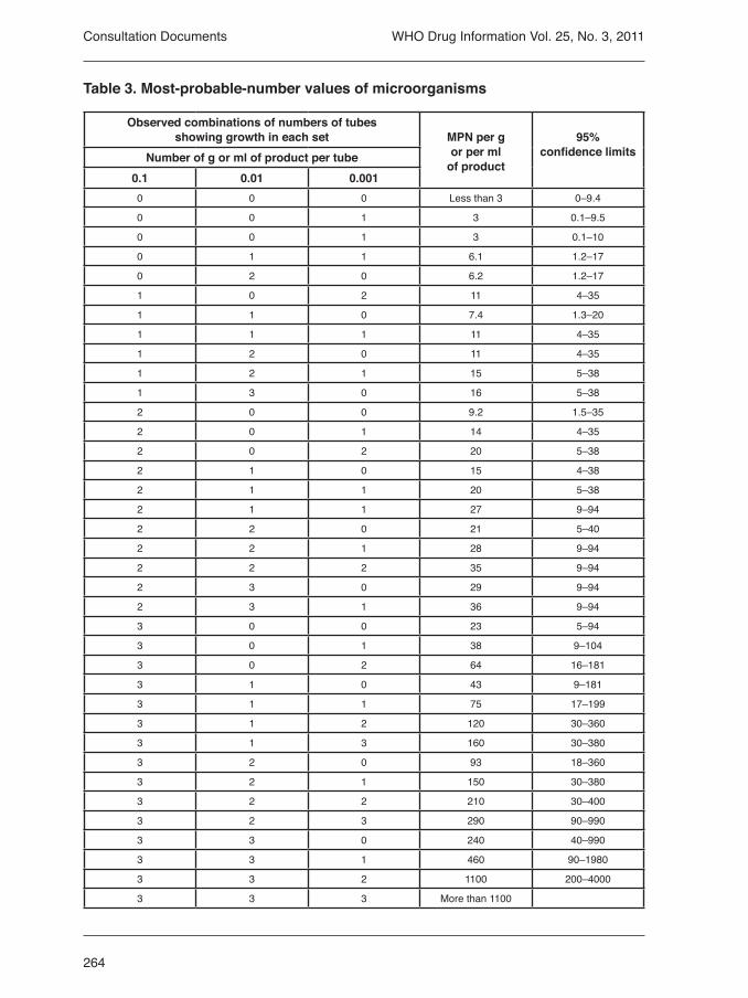
264
WHO Drug Information Vol. 25, No. 3, 2011
Table 3. Most-probable-number values of microorganisms
Observed combinations of numbers of tubes showing growth in each set MPN per g
or per ml of product
95%confidence limitsNumber of g or ml of product per tube
0.1 0.01 0.0010 0 0 Less than 3 0–9.4
0 0 1 3 0.1–9.5
0 0 1 3 0.1–10
0 1 1 6.1 1.2–17
0 2 0 6.2 1.2–17
1 0 2 11 4–35
1 1 0 7.4 1.3–20
1 1 1 11 4–35
1 2 0 11 4–35
1 2 1 15 5–38
1 3 0 16 5–38
2 0 0 9.2 1.5–35
2 0 1 14 4–35
2 0 2 20 5–38
2 1 0 15 4–38
2 1 1 20 5–38
2 1 1 27 9–94
2 2 0 21 5–40
2 2 1 28 9–94
2 2 2 35 9–94
2 3 0 29 9–94
2 3 1 36 9–94
3 0 0 23 5–94
3 0 1 38 9–104
3 0 2 64 16–181
3 1 0 43 9–181
3 1 1 75 17–199
3 1 2 120 30–360
3 1 3 160 30–380
3 2 0 93 18–360
3 2 1 150 30–380
3 2 2 210 30–400
3 2 3 290 90–990
3 3 0 240 40–990
3 3 1 460 90–1980
3 3 2 1100 200–4000
3 3 3 More than 1100
Consultation Documents

265
WHO Drug Information Vol. 25, No. 3, 2011
When an acceptance criterion for microbiological quality is prescribed it is interpreted as follows:
— 101 microorganisms: maximum acceptable count = 20— 102 microorganisms: maximum acceptable count = 200— 103 microorganisms: maximum acceptable count = 2000, and so forth.
New reagent to be added to Ph.Int.
Isopropyl myristate R. Propan-2-yl tetradecanoate. C17H34O2.Description: A clear, colourless, oily liquid. Miscibility: Immiscible with water, miscible with ethanol, fatty oils, liquid paraffin. Relative density: About 0.853
3.3.2 Microbial examination of non sterile products: tests for specified microorganisms
Draft proposal for revision of a General Method in the 4th Edition of the International Pharmacopoeia (June 2011). Please addess any comments to Quality Assurance and Safety: Medicines, World Health Organization, 1211 Geneva 27, Switzerland. Fax: +41 22 791 4730 or e-mail [email protected]. Working documents are available for comment at http://who.int/medicines.
[Note from Secretariat. During its meeting in October 2010 the Expert Committee on Specifications for Pharmaceutical Preparations recommended that requirements for tests for specified microorganisms be added in The International Pharmacopoeia (Ph.Int.) using as a basis the internationally-harmonized general tests available on micro-biological examination of non-sterile products. Discussions for the international harmonization of requirements for the microbiological examination of non-sterile products has led to the elaboration of three general texts: (i) Microbial enumeration tests; (ii) Tests for specified microorganisms, and (iii) Accep-tance criteria for pharmaceutical preparations and substances for pharmaceutical use, the latter text on acceptance criteria being published as non-mandatory information. At present, The International Pharmacopoeia has no method text on microbial enu-meration tests and tests for specified microorganisms. The text 3.3 Microbial purity of pharmaceutical preparations is provided to give information and guidance and is not regarded as an analytical requirement. It is proposed to introduce these three internationally-harmonized general texts in the Ph.Int. The texts on microbial enumeration tests and tests for specified microor-ganisms are new (3.3.1 and 3.3.2) in the Methods of Analysis section. Furthermore, it is proposed to replace the current text 3.3 on microbial purity by the internationally harmonized one on acceptance criteria for non-sterile preparations and substances. This text will be provided for information and will, therefore, be moved to the Supple-mentary Information section.
If the proposed 3.3.1 and 3.3.2 texts are adopted for inclusion in the Ph.Int a review will be carried out on the application of these methods within the existing Ph.Int texts.
Consultation Documents

266
WHO Drug Information Vol. 25, No. 3, 2011
Such a review would include excipients and consider in which Ph.Int. monographs the methods would be invoked and would propose limits.]
The tests described hereafter will allow determination of the absence of, or limited occurrence of specified microorganisms which may be detected under the conditions described.
The tests are designed primarily to determine whether a substance or preparation complies with an established specification for microbiological quality. When used for such purposes follow the instructions given below, including the number of samples to be taken and interpret the results as stated below.
Alternative microbiological procedures, including automated methods may be used, provided that their equivalence to the pharmacopoeial method has been demonstra-ted.
General ProceduresThe preparation of samples is carried out as described in 3.3.1 Microbial enumeration tests.
If the product to be examined has antimicrobial activity, this is insofar as possible removed or neutralized as described in 3.3.1 Microbial enumeration tests.
If surface-active substances are used for sample preparation, their absence of toxicity for microorganisms and their compatibility with inactivators used must be demonstra-ted as described in Microbial enumeration tests.
Growth promoting and inhibitory properties of the media, suitability of the test and negative controlsThe ability of the test to detect microorganisms in the presence of the product to be tested must be established. Suitability must be confirmed if a change in testing perfor-mance, or the product, which may affect the outcome of the test is introduced.
Preparation of test strainsUse standardized stable suspensions of test strains or prepare as stated below. Seed-lot culture maintenance techniques (seed-lot systems) are used so that the viable microorganisms used for inoculation are not more than 5 passages removed from the original master seed-lot.
Aerobic microorganisms Grow each of the bacterial test strains separately in containers containing casein soya bean digest broth or on casein soya bean digest agar at 30–35 °C for 18–24 h. Grow the test strain for Candida albicans separately on Sabouraud-dextrose agar or in Sabouraud-dextrose broth at 20–25 °C for 2–3 days.
Staphylococcus aureus such as ATCC 6538, NCIMB 9518, CIP 4.83 or NBRC 13276,Pseudomonas aeruginosa such as ATCC 9027, NCIMB 8626, CIP 82.118 or NBRC 13275
Escherichia coli such as ATCC 8739, NCIMB 8545, CIP 53.126 or NBRC 3972,Salmonella enterica subsp. enterica serovar Typhimurium such as ATCC 14028 or,
Consultation Documents
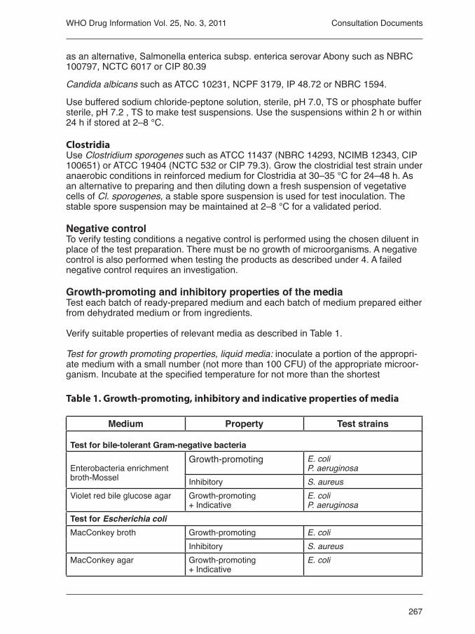
267
WHO Drug Information Vol. 25, No. 3, 2011
as an alternative, Salmonella enterica subsp. enterica serovar Abony such as NBRC 100797, NCTC 6017 or CIP 80.39
Candida albicans such as ATCC 10231, NCPF 3179, IP 48.72 or NBRC 1594.
Use buffered sodium chloride-peptone solution, sterile, pH 7.0, TS or phosphate buffer sterile, pH 7.2 , TS to make test suspensions. Use the suspensions within 2 h or within 24 h if stored at 2–8 °C.
ClostridiaUse Clostridium sporogenes such as ATCC 11437 (NBRC 14293, NCIMB 12343, CIP 100651) or ATCC 19404 (NCTC 532 or CIP 79.3). Grow the clostridial test strain under anaerobic conditions in reinforced medium for Clostridia at 30–35 °C for 24–48 h. As an alternative to preparing and then diluting down a fresh suspension of vegetative cells of Cl. sporogenes, a stable spore suspension is used for test inoculation. The stable spore suspension may be maintained at 2–8 °C for a validated period.
Negative controlTo verify testing conditions a negative control is performed using the chosen diluent in place of the test preparation. There must be no growth of microorganisms. A negative control is also performed when testing the products as described under 4. A failed negative control requires an investigation.
Growth-promoting and inhibitory properties of the mediaTest each batch of ready-prepared medium and each batch of medium prepared either from dehydrated medium or from ingredients. Verify suitable properties of relevant media as described in Table 1.
Test for growth promoting properties, liquid media: inoculate a portion of the appropri-ate medium with a small number (not more than 100 CFU) of the appropriate microor-ganism. Incubate at the specified temperature for not more than the shortest
Table 1. Growth-promoting, inhibitory and indicative properties of media
Medium Property Test strains
Test for bile-tolerant Gram-negative bacteria
Enterobacteria enrichment broth-Mossel
Growth-promoting E. coliP. aeruginosa
Inhibitory S. aureusViolet red bile glucose agar Growth-promoting
+ IndicativeE. coliP. aeruginosa
Test for Escherichia coliMacConkey broth Growth-promoting E. coli
Inhibitory S. aureusMacConkey agar Growth-promoting
+ IndicativeE. coli
Consultation Documents
268
WHO Drug Information Vol. 25, No. 3, 2011
Test for Salmonella
Rappaport VassiliadisSalmonella enrichment broth
Growth-promoting Salmonella enterica subsp. enterica serovar Typhimurium orSalmonella enterica subsp. enterica serovar Abony
Inhibitory S. aureus
Xylose, lysine, deoxycholate agar
Growth-promoting + Indicative
Salmonella enterica subsp. enterica serovar Typhimurium orSalmonella enterica subsp. enterica serovar Abony
Test for Pseudomonas aeruginosaCetrimide agar Growth-promoting P. aeruginosa
Inhibitory E. coli
Test for Staphylococcus aureusMannitol salt agar Growth-promoting
+ IndicativeS. aureus
Inhibitory E. coliTest for ClostridiaReinforced medium for Clostridia
Growth-promoting Cl. sporogenes
Columbia agar Growth-promoting Cl. sporogenesTest for Candida albicansSabouraud dextrose broth Growth-promoting C. albicansSabouraud dextrose agar Growth-promoting
+ IndicativeC. albicans
period of time specified in the test. Clearly visible growth of the microorganism com-parable to that previously obtained with a previously tested and approved batch of medium occurs.
Test for growth promoting properties, solid media: perform surface-spread method, in-oculating each plate with a small number (not more than 100 CFU) of the appropriate microorganism. Incubate at the specified temperature for not more than the shortest period of time specified in the test. Growth of the microorganism comparable to that previously obtained with a previously tested and approved batch of medium occurs.
Test for inhibitory properties, liquid or solid media: inoculate the appropriate medium with at least 100 CFU of the appropriate microorganism. Incubate at the specified temperature for not less than the longest period of time specified in the test. No growth of the test microorganism occurs.
Test for indicative properties: perform surface-spread method, inoculating each plate with a small number (not more than 100 CFU) of the appropriate microorganism. Incu-
Consultation Documents
269
WHO Drug Information Vol. 25, No. 3, 2011
bate at the specified temperature for a period of time within the range specified in the test. Colonies are comparable in appearance and indication reactions to those previ-ously obtained with a previously tested and approved batch of medium.
Suitability of the test methodFor each product to be tested perform sample preparation as described in the relevant paragraph in the section below on “Testing of products”. Add each test strain at the time of mixing, in the prescribed growth medium. Inoculate the test strains individually. Use a number of microorganisms equivalent to not more than 100 CFU in the inocula-ted test preparation.
Perform the test as described in the relevant paragraph in section 4 using the shortest incubation period prescribed.
The specified microorganisms must be detected with the indication reactions as des-cribed below under “Testing of products”. Any antimicrobial activity of the product necessitates a modification of the test proce-dure (see “Neutralization/removal of antimicrobial activity” in “3.3.1 Microbial Enume-ration Tests”).
If for a given product the antimicrobial activity with respect to a microorganism for which testing is prescribed cannot be neutralized, then it is to be assumed that the inhibited microorganism will not be present in the product.
Testing of products
Bile-tolerant Gram-negative bacteria
Sample preparation and pre-incubation Prepare a sample using a 1 in 10 dilution of not less than 1 g of the product to be exa-mined as described in “3.3.1 Microbial enumeration tests”, but using casein soya bean digest broth as the chosen diluent, mix and incubate at 20–25 °C for a time sufficient to resuscitate the bacteria but not sufficient to encourage multiplication of the orga-nisms (usually 2 h. but not more than 5 h.).
Test for absence Unless otherwise prescribed use the volume corresponding to 1g of the product, as prepared in Sample preparation and pre-incubation to inoculate enterobacteria enrich-ment broth-Mossel. Incubate at 30–35 °C for 24–48 h. Subculture on plates of violet red bile glucose agar. Incubate at 30–35 °C for 18–24 h.
The product complies with the test if there is no growth of colonies.
Quantitative testSelection and subculture. Inoculate suitable quantities of enterobacteria enrichment broth-Mossel with the preparation as described under Sample preparation and pre-incubation and/or dilutions of it containing, respectively 0.1 g, 0.01 g and 0.001 g (or 0.1 ml, 0.01 ml and 0.001 ml) of the product to be examined. Incubate at 30–35 °C for 24–48 h. Subculture each of the cultures on a plate of violet red bile glucose agar. Incubate at 30–35 °C for 18–24 h.
Consultation Documents
270
WHO Drug Information Vol. 25, No. 3, 2011
InterpretationGrowth of colonies constitutes a positive result. Note the smallest quantity of the product that gives a positive result and the largest quantity that gives a negative result. Determine from Table 2 the probable number of bacteria.
Table 2. Interpretation of results
Results for each quantity of productProbable number of bacteria per gram or ml of product
0.1 g or 0.1 ml
0.01 g or 0.01 ml
0.001 g or0.001 ml
+ + + more than 103
+ + - less than 103 and more than 102
+ - - less than 102 and more than 10
- - - less than 10 Escherichia coli
Sample preparation and pre-incubationPrepare a sample using a 1 in 10 dilution of not less than 1 g of the product to be exa-mined as described in 3.3.1 Microbial enumeration tests and use 10 ml or the quantity corresponding to 1 g or1mL to inoculate a suitable amount (determined as described under Suitability of the test method) of casein soya bean digest broth, mix and incu-bate at 30–35 °C for 18–24 h.
Selection and subcultureShake the container, transfer 1 mL of casein soya bean digest broth to 100 mL of MacConkey broth and incubate at 42–44 °C for 24–48 h. Subculture on a plate of MacConkey agar at 30–35 °C for 18–72 h.
InterpretationGrowth of colonies indicates the possible presence of E. coli. This is confirmed by identification tests.
The product complies with the test if no colonies are present or if the identification tests are negative.
Salmonella
Sample preparation and pre-incubationPrepare the product to be examined as described in 3.3.1 Microbial enumeration tests and use the quantity corresponding to not less than 10 g or 10 ml to inoculate a sui-table amount (determined as described under Suitability of the test method) of casein soya bean digest broth, mix and incubate at 30–35 °C for 18–24 h.
Selection and subcultureTransfer 0.1 ml of casein soya bean digest broth to 10 ml of Rappaport vassiliadis Sal-monella enrichment broth and incubate at 30–35 °C for 18–24 h. Subculture on plates of xylose, lysine, deoxycholate agar. Incubate at 30–35 °C for 18–48 h.
Consultation Documents
271
WHO Drug Information Vol. 25, No. 3, 2011
InterpretationThe possible presence of Salmonella is indicated by the growth of well-developed, red colonies, with or without black centres. This is confirmed by identification tests. The product complies with the test if colonies of the types described are not present or if the confirmatory identification tests are negative.
Pseudomonas aeruginosa
Sample preparation and pre-incubationPrepare a sample using a 1 in 10 dilution of not less than 1 g of the product to be examined as described in “3.3.1 Microbial enumeration tests” and use 10 ml or the quantity corresponding to 1 g or 1 ml to inoculate a suitable amount (determined as described under Suitability of the test method) of casein soya bean digest broth and mix. When testing transdermal patches, filter the volume of sample corresponding to 1 patch of the preparation described under Preparation of the sample in “3.3.1 Micro-bial enumeration tests” through a sterile filter membrane and place in 100 ml of casein soya bean digest broth. Incubate at 30–35 °C for 18–24 h.
Selection and subcultureSubculture on a plate of cetrimide agar and incubate at 30–35 °C for 18–72 h.
InterpretationGrowth of colonies indicates the possible presence of P. aeruginosa. This is confirmed by identification tests.
The product complies with the test if colonies are not present or if the confirmatory identification tests are negative.
Staphylococcus aureus
Sample preparation and pre-incubationPrepare a sample using a 1 in 10 dilution of not less than 1 g of the product to be examined as described in “3.3.1 Microbial enumeration tests” and use 10 ml or the quantity corresponding to 1 g or 1 ml to inoculate a suitable amount (determined as described under “Suitability of the test method”) of casein soya bean digest broth and homogenize. When testing transdermal patches, filter the volume of sample corres-ponding to one patch of the preparation described under “Preparation of the sample” in “3.3.1 Microbial enumeration tests” through a sterile filter membrane and place in 100 ml of casein soya bean digest broth. Incubate at 30–35 °C for 18–24 h.
Selection and subcultureSubculture on a plate of mannitol salt agar and incubate at 30–35 °C for 18–72 h.
InterpretationThe possible presence of S. aureus is indicated by the growth of yellow/white colonies surrounded by a yellow zone. This is confirmed by identification tests.
The product complies with the test if colonies of the types described are not present or if the confirmatory identification tests are negative.
Consultation Documents

272
WHO Drug Information Vol. 25, No. 3, 2011
Clostridia
Sample preparation and heat treatmentPrepare a sample using a 1 in 10 dilution (with a minimum total volume of 20 ml) of not less than 2 g or 2 ml of the product to be examined as described in “3.3.1 Micro-bial enumeration tests”. Divide the sample into two portions of at least 10 ml. Heat one portion at 80 °C for 10 min and cool rapidly. Do not heat the other portion.
Selection and subcultureUse 10 ml or the quantity corresponding to 1 g or 1 ml of the product to be examined of both portions to inoculate suitable amounts (determined as described under “Suita-bility of the test method”) of Reinforced clostridium medium. Incubate under anaerobic conditions at 30–35 °C for 48 h. After incubation, make subcultures from each contai-ner on Columbia agar and incubate under anaerobic conditions at 30–35 °C for 48 h–72h.
InterpretationThe occurrence of anaerobic growth of rods (with or without endospores) giving a negative catalase reaction indicates the presence of Clostridia. This is confirmed by identification tests.
The product complies with the test if colonies of the types described are not present or if the confirmatory identification tests are negative. Candida albicans
Sample preparation and pre-incubationPrepare the product to be examined as described in “3.3.1 Microbial enumeration tests” and use 10 ml or the quantity corresponding to not less than 1 g or 1 ml to inoculate 100 ml of Sabouraud-dextrose broth and mix. Incubate at 30–35 °C for 3–5 days.
Selection and subcultureSubculture on a plate of Sabouraud-dextrose agar and incubate at 30–35 °C for 24–48h.
InterpretationGrowth of white colonies may indicate the presence of C. albicans. This is confirmed by identification tests.
The product complies with the test if such colonies are not present or if the confirma-tory identification tests are negative.
Recommended test solutions and culture mediaThe following test solutions and culture media have been found satisfactory for the purposes for which they are prescribed in the test for microbial contamination in the pharmacopoeia. Other media may be used provided that their suitability can be demonstrated.
• Stock buffer solution. Transfer 34 g of potassium dihydrogen phosphate to a 1000 ml volumetric flask, dissolve in 500 ml of purified water, adjust to pH 7.2 ± 0.2 with sodium hydroxide, add purified water to volume and mix. Dispense in containers and sterilize. Store at a temperature of 2–8 °C.
Consultation Documents

273
WHO Drug Information Vol. 25, No. 3, 2011
• Phosphate buffer, sterile, pH 7.2, TS. Prepare a mixture of purified water and stock buffer solution (800:1 V/V) and sterilize.
• Buffered sodium chloride-peptone solution pH 7.0
Potassium dihydrogen phosphate 3.6 g Disodium hydrogen phosphate dihydrate 7.2 g equivalent to 0.067 M phosphate Sodium chloride 4.3 g Peptone (meat or casein) 1.0 g Purified water 1000 ml
Sterilize in an autoclave using a validated cycle.
• Casein soya bean digest broth
Pancreatic digest of casein 17.0 g Papaic digest of soya bean 3.0 g Sodium chloride 5.0 g Dipotassium hydrogen phosphate 2.5 g Glucose monohydrate 2.5 g Purified water 1000 ml
Adjust the pH so that after sterilization it is 7.3 ± 0.2 at 25 °C. Sterilize in an autoclave using a validated cycle.
• Casein soya bean digest agar
Pancreatic digest of casein 15.0 g Papaic digest of soya bean 5.0 g Sodium chloride 5.0 g Agar 15.0 g Purified water 1000 ml
Adjust the pH so that after sterilization it is 7.3 ± 0.2 at 25 °C. Sterilize in an autoclave using a validated cycle.
• Sabouraud-dextrose agar
Dextrose 40.0 g Mixture of peptic digest of animal tissue and pancreatic digest of casein (1:1) 10.0 g Agar 15.0 g Purified water 1000 ml
Adjust the pH so that after sterilization it is 5.6 ± 0.2 at 25 °C. Sterilize in an autoclave using a validated cycle.
• Potato dextrose agar
Infusion from potatoes 200 g Dextrose 20.0 g Agar 15.0 g Purified water 1000 ml
Consultation Documents

274
WHO Drug Information Vol. 25, No. 3, 2011
Adjust the pH so that after sterilization it is 5.6 ± 0.2 at 25 °C. Sterilize in an autoclave using a validated cycle.
• Sabouraud-dextrose broth
Dextrose 20.0 g Mixture of peptic digest of animal tissue and pancreatic digest of casein (1:1) 10.0 g Purified water 1000 ml
Adjust the pH so that after sterilization it is 5.6 ± 0.2 at 25 °C. Sterilize in an autoclave using a validated cycle.
• Enterobacteria enrichment broth-Mossel
Pancreatic digest of gelatin 10.0 g Glucose monohydrate 5.0 g Dehydrated ox bile 20.0 g Potassium dihydrogen phosphate 2.0 g Disodium hydrogen phosphate dihydrate 8.0 g Brilliant green 15 mg Purified water 1000 ml
Adjust the pH so that after heating it is 7.2 ± 0.2 at 25 °C. Heat at 100 °C for 30 min and cool immediately.
• Violet red bile glucose agar
Yeast extract 3.0 g Pancreatic digest of gelatin 7.0 g Bile salts 1.5 g Sodium chloride 5.0 g Glucose monohydrate 10.0 gl Agar 15.0 g Neutral red 30 mg Crystal violet 2 mg Purified water 1000 ml
Adjust the pH so that after heating it is 7.4 ± 0.2 at 25 °C. Heat to boiling; do not heat in an autoclave.
• MacConkey broth
Pancreatic digest of gelatin 20.0 g Lactose monohydrate 10.0 g Dehydrated ox bile 5.0 g Bromocresol purple 10 mg Purified water 1000 ml
Adjust the pH so that after sterilization it is 7.3 ± 0.2 at 25 °C. Sterilize in an autoclave using a validated cycle.
Consultation Documents

275
WHO Drug Information Vol. 25, No. 3, 2011
• MacConkey agar
Pancreatic digest of gelatin 17.0 g Peptones (meat and casein) 3.0 g Lactose monohydrate 10.0 g Sodium chloride 5.0 g Bile salts 1.5 g Agar 13.5 g Neutral red 30.0 mg Crystal violet 1 mg Purified water 1000 ml
Adjust the pH so that after sterilization it is 7.1 ± 0.2 at 25 °C. Boil for 1 min with constant shaking then sterilize in an autoclave using a validated cycle.
• Rappaport Vassiliadis Salmonella Enrichment Broth
Soya peptone 4.5 g Magnesium chloride hexahydrate 29.0 g Sodium chloride 8.0 g Dipotassium phosphate 0.4 g Potassium dihydrogen phosphate 0.6 g Malachite green 0.036 g Purified water 1000 ml
Dissolve, warming slightly. Sterilize in an autoclave using a validated cycle, at a temperature not exceeding 115 °C. The pH is to be 5.2 ± 0.2 at 25 °C after heating and autoclaving.
• Xylose, lysine, deoxycholate agar
Xylose 3.5 g L-Lysine 5.0 g Lactose monohydrate 7.5 g Sucrose 7.5 g Sodium chloride 5.0 g Yeast extract 3.0 g Phenol red 80 mg Agar 13.5 g Sodium deoxycholate 2.5 g Sodium thiosulfite 6.8 g Ferric ammonium citrate 0.8 g Purified water 1000 ml
Adjust the pH so that after heating it is 7.4 ± 0.2 at 25 °C. Heat to boiling, cool to 50 °C and pour into Petri dishes. Do not heat in an autoclave.
• Cetrimide agar
Pancreatic digest of gelatin 20.0 g Magnesium chloride 1.4 g Dipotassium sulfite 10.0 g Cetrimide 0.3 g
Consultation Documents

276
WHO Drug Information Vol. 25, No. 3, 2011
Agar 13.6 g Purified water 1 000 ml Glycerol 10.0 ml
Heat to boiling for 1 min with shaking. Adjust the pH so that after sterilization it is 7.2 ± 0.2 at 25 °C. Sterilize in an autoclave using a validated cycle.
• Mannitol salt agar
Pancreatic digest of casein 5.0 g Peptic digest of animal tissue 5.0 g Beef extract 1.0 g D-Mannitol 10.0 g Sodium chloride 75.0 g Agar 15.0 g Phenol red 0.025 g Purified water 1000 mL
Heat to boiling for 1 min with shaking. Adjust the pH so that after sterilization it is 7.4 ± 0.2 at 25 °C. Sterilize in an autoclave using a validated cycle.
• Reinforced medium for Clostridia
Beef extract 10.0 g Peptone 10.0 g Yeast extract 3.0 g Soluble starch 1.0 g Glucose monohydrate 5.0 g Cysteine hydrochloride 0.5 g Sodium chloride 5.0 g Sodium acetate 3.0 g Agar 0.5 g Purified water 1000 ml
Hydrate the agar, dissolve by heating to boiling with continuous stirring. If necessary, adjust the pH so that after sterilization it is about 6.8 ± 0.2 at 25 °C. Sterilize in an autoclave using a validated cycle.
• Columbia agar
Pancreatic digest of casein 10.0 g Meat peptic digest 5.0 g Heart pancreatic digest 3.0 g Yeast extract 5.0 g Maize starch 1.0 g Sodium chloride 5.0 g Agar, according to gelling power 10.0 g to 15.0 g Purified water 1000 ml
Hydrate the agar, dissolve by heating to boiling with continuous stirring. If necessary, adjust the pH so that after sterilization it is 7.3 ± 0.2 at 25 °C. Sterilize in an autoclave using a validated cycle. Allow to cool to 45–50 °C; add, where necessary, gentamicin sulfite corresponding to 20 mg of gentamicin base and pour into Petri dishes.
Consultation Documents

277
WHO Drug Information Vol. 25, No. 3, 2011
5.3 Disintegration test for tablets and capsulesDraft proposal for revision of a General Method in the 4th Edition of the International Pharmacopoeia (April 2011). Please addess any comments to Quality Assurance and Safety: Medicines, World Health Organization, 1211 Geneva 27, Switzerland. Fax: +41 22 791 4730 or e-mail [email protected]. Working documents are available for comment at http://who.int/medicines.
[Note from Secretariat. During its meeting in October 2010, the Expert Committee on Specifications for Pharmaceutical Preparations recommended that the current method described in The International Pharmacopoeia for the disintegration test for tablets and capsules should be replaced by the internationally harmonized general test available. To this effect the following revision of method “5.3 Disintegration test for tablets and capsules” is proposed.
The revision implies both changes of and additions to dimensions and tolerances in the description of the disintegration apparatus. The possibility for retesting when one or two units fail in the first step of the procedure is introduced, as is the possibility to use automatic detection employing modified discs in cases where the use of discs is prescribed.]
This test is provided to determine whether tablets or capsules disintegrate within the prescribed time when placed in a liquid medium under the experimental conditions presented below.
For the purposes of this test, disintegration does not imply complete dissolution of the unit or even of its active constituent. Complete disintegration is defined as that state in which any residue of the unit, except fragments of insoluble coating or capsule shell, remaining on the screen of the test apparatus or adhering to the lower surface of the discs, if used, is a soft mass having no palpably firm core.
ApparatusThe apparatus consists of a basket-rack assembly, a 1000-ml, low-form beaker, 138 to 160 mm in height and having an inside diameter of 97 to 115 mm for the immer-sion fluid, a thermostatic arrangement for heating the fluid between 35 °C and 39 °C, and a device for raising and lowering the basket in the immersion fluid at a constant frequency rate between 29 and 32 cycles per minute, through a distance of not less than 53 mm and not more than 57 mm. The volume of the fluid in the vessel is such that at the highest point of the upward stroke the wire mesh remains at least 15 mm below the surface of the fluid, and descends to not less than 25 mm from the bottom of the vessel on the downward stroke. At no time should the top of the basket-rack assembly become submerged. The time required for the upward stroke is equal to the time required for the downward stroke, and the change in stroke direction is a smooth transition, rather than an abrupt reversal of motion. The basket-rack assembly moves vertically along its axis. There is no appreciable horizontal motion or movement of the axis from the vertical.
Basket-rack assembly. The basket-rack assembly consists of 6 open-ended trans-parent tubes, each 75.0 mm to 80 mm long and having an inside diameter of 20.7 to 23 mm and a wall 1.0 to 2.8 mm thick; the tubes are held in a vertical position by two plates, each 88 to 92 mm in diameter and 5 to 8.5 mm in thickness, with 6 holes, each 22 to 26 mm in diameter, equidistant from the centre of the plate and equally
Consultation Documents

278
WHO Drug Information Vol. 25, No. 3, 2011
spaced from one another. Attached to the under surface of the lower plate is a woven stainless steel wire cloth, which has a plain square weave with 1.8 to 2.2-mm aper-tures and with a wire diameter of 0.57 to 0.66 mm. The parts of the apparatus are assembled and rigidly held by means of three bolts passing through the two plates. A suitable means is provided to suspend the basket-rack assembly from the raising and lowering device using a point on its axis.
The design of the basket-rack assembly may be varied somewhat provided the spe-cifications for the glass tubes and the screen mesh size are maintained. The basket-rack assembly conforms to the dimensions shown in Figure 1.
Discs. The use of discs is permitted only where specified or allowed. Each tube is pro-vided with a cylindrical disc 9.35 mm to 9.65 mm thick and 20.55 mm to 20.85 mm in diameter. The disc is made of a suitable, transparent plastic material having a specific gravity of 1.18 to 1.20. Five parallel 1.9 mm to 2.1 mm holes extend between the ends of the cylinder. One of the holes is centered on the cylindrical axis. The other holes are centered 5.8 mm to 6.2 mm from the axis on imaginary lines perpendicular to the axis and parallel to each other. Four identical trapezoidal-shaped planes are cut into the wall of the cylinder, nearly perpendicular to the ends of the cylinder. The trapezoidal shape is symmetrical; its parallel sides coincide with the ends of the cylinder and are parallel to an imaginary line connecting the centres of two adjacent holes 6 mm from the cylindrical axis. The parallel side of the trapezoid on the bottom of the cylinder has a length of 1.5 mm to 1.7 mm and its bottom edges lie at a depth of 1.5 mm to 1.8 mm from the cylinder’s circumference. The parallel side of the trapezoid on the top of the
22
4
Bottom View
SideView
TopView
DiskBasket-RackAssembly
9.4 ± 0.2
2 ±
0.1
6 ± 0.2
2.6 ± 0.1
90 ± 2
20.7 ± 0.15
1.6
± 0.11.65 ± 0.15
9.5
± 0
.15
6.7
5 ±
1.7
5
6.7
5 ±
1.7
5
77
.5 ±
2.5
1.9± 0.9
21.85 ± 1.15
Figure 1. Diagram for disintegration apparatus
Consultation Documents

279
WHO Drug Information Vol. 25, No. 3, 2011
cylinder has a length of 9.2 mm to 9.6 mm and its centre lies at a depth of 2.5 mm to 2.7 mm from the cylinder’s circumference. All surfaces of the disc are smooth. If the use of discs is specified, add a disc to each tube, and operate the apparatus as direc-ted under procedure. The discs conform to the dimensions found in Figure 1.
The use of automatic detection employing modified discs is permitted where the use of discs is specified or allowed. Such discs must comply with the requirements of density and dimension given in this chapter.
ProcedurePlace 1 dosage unit in each of the six tubes of the basket, and if specified add a disc. Operate the apparatus using water as the immersion fluid unless another liquid is spe-cified and maintain its temperature at 35 °C to 39 °C. At the end of the specified time, lift the basket from the fluid and observe the dosage units: all of the dosage units have disintegrated completely. If 1 or 2 dosage units fail to disintegrate, repeat the test on 12 additional dosage units. The requirements of the test are met if not less than 16 of the 18 dosage units tested are disintegrated.
5.6 Extractable volume of parenteral preparationsDraft proposal for revision of a General Method in the 4th Edition of the International Pharmacopoeia (April 2011). Please addess any comments to Quality Assurance and Safety: Medicines, World Health Organization, 1211 Geneva 27, Switzerland. Fax: +41 22 791 4730 or e-mail [email protected]. Working documents are available at http://who.int/medicines.
[Note from Secretariat. During its meeting in October 2010, the Expert Committee on Specifications for Pharmaceutical Preparations recommended that the current method described in The International Pharmacopoeia for the test of extractable volume of parenteral preparations should be replaced by the internationally harmonized general test available. To this effect the following revision of method “5.6 Extractable volume of parenteral preparations” is proposed.
During the revision of the general monograph for “Parenteral preparations”, consider-ation will be given to requiring compliance with this test; currently the general mono-graph does not refer to method 5.6.]
Suspensions and emulsions must be shaken before withdrawal of the contents and before the determination of the density. Oily and viscous preparations may be warmed according to the instructions on the label, if necessary, and thoroughly shaken imme-diately before removing the contents. The contents are then cooled to 20 °C – 25 °C before measuring the volume.
Single-dose containersSelect one container if the volume is 10 ml or more, three containers if the nominal volume is more than 3 ml and less than 10 ml, or five containers if the nominal volume is 3 ml or less. Take up individually the total contents of each container selected into a dry syringe of a capacity not exceeding three times the volume to be measured, and fitted with a 21-gauge needle not less than 2.5 cm in length. Expel any air bubbles from the syringe and needle, then discharge the contents of the syringe without emp-tying the needle into a standardized dry cylinder (graduated to contain rather than to deliver the designated volumes) of such size that the volume to be measured occupies
Consultation Documents

280
WHO Drug Information Vol. 25, No. 3, 2011
at least 40 per cent of its graduated volume. Alternatively, the volume of the contents in millilitres may be calculated as the mass in grams divided by the density.
For containers with a nominal volume of 2 ml or less the contents of a sufficient number of containers may be pooled to obtain the volume required for the measure-ment provided that a separate, dry syringe assembly is used for each container. The contents of containers holding 10 ml or more may be determined by opening them and emptying the contents directly into the graduated cylinder or tared beaker.
The volume is not less than the nominal volume in case of containers examined indivi-dually, or, in case of containers with a nominal volume of 2 ml or less, is not less than the sum of the nominal volumes of the containers taken collectively.
Multidose containersFor injections in multidose containers labelled to yield a specific number of doses of a stated volume, select one container and proceed as directed for single-dose contai-ners using the same number of separate syringe assemblies as the number of doses specified. The volume is such that each syringe delivers not less than the stated dose.
Cartridges and prefilled syringesSelect one container if the volume is 10 ml or more, three containers if the nominal volume is more than 3 ml and less than 10 ml, or 5 containers if the nominal volume is 3 ml or less. If necessary, fit the containers with the accessories required for their use (needle, piston, syringe) and transfer the entire contents of each container without emptying the needle into a dry tared beaker by slowly and constantly depressing the piston. Determine the volume in millilitres calculated as the mass in grams divided by the density.
The volume measured for each of the containers is not less than the nominal volume.
Parenteral infusionsSelect one container. Transfer the contents into a dry measuring cylinder of such a capacity that the volume to be determined occupies at least 40 per cent of the nominal volume of the cylinder. Measure the volume transferred. The volume is not less than the nominal volume.
5.7 Tests for particulate contaminationDraft proposal for revision of a General Method in the 4th Edition of the International Pharmacopoeia (May 2011). Please addess any comments to Quality Assurance and Safety: Medicines, World Health Organization, 1211 Geneva 27, Switzerland. Fax: +41 22 791 4730 or e-mail [email protected]. Working documents are available for comment at http://who.int/medicines.
[Note from Secretariat. During its meeting in October 2010 the Expert Committee on Specifications for Pharmaceutical Preparations recommended that the current method described in The International Pharmacopoeia for the tests for particulate contamina-tion should be replaced by the internationally harmonized general test available. To this effect the following revision of method “5.7 Tests for particulate contamination” is proposed. Section 5.7 will be entitled “Tests for particulate contamination”, the PDG text will be introduced as “5.7.1 Subvisible particles” and the existing text of 5.7 re-tained as “5.7.2 Visible particles”.
Consultation Documents
281
WHO Drug Information Vol. 25, No. 3, 2011
The current general monograph for “Parenteral preparations” does not contain analyti-cal requirements on particulate contamination. Consideration will be given to requiring compliance with these tests during revision of the general monograph. The proposed test makes a distinction between small volume parenterals and large volume parenterals with a limit at 100 ml. The 100 ml preparation is exempted from the pharmacopoeial harmonization. It is proposed to include 100 ml preparation among small volume parenterals.]
5.7.1 Subvisible particles Particulate contamination of injections and parenteral infusions consists of extraneous, mobile undissolved particles, other than gas bubbles, unintentionally present in the solutions.
For the determination of particulate contamination two procedures, Method 1 (“Light Obscuration Particle Count Test”) and Method 2 (“Microscopic Particle Count Test”), are specified hereinafter. When examining injections and parenteral infusions for sub-visible particles Method 1 is preferably applied. However, it may be necessary to test some preparations by the light obscuration particle count test followed by the micros-copic particle count test to reach a conclusion on conformance to the requirements.Not all parenteral preparations can be examined for sub-visible particles by one or both of these methods. When Method 1 is not applicable, e.g., in case of preparations having reduced clarity or increased viscosity, the test should be carried out according to Method 2. Emulsions, colloids, and liposomal preparations are examples. Similarly, products that produce air or gas bubbles when drawn into the sensor may also require microscopic particle count testing. If the viscosity of the preparation to be tested is sufficiently high so as to preclude its examination by either test method, a quantitative dilution with an appropriate diluent may be made to decrease viscosity, as necessary, to allow the analysis to be performed.
The results obtained in examining a discrete unit or group of units for particulate contamination cannot be extrapolated with certainty to other units that remain untested. Thus, statistically sound sampling plans must be developed if valid infe-rences are to be drawn from observed data to characterize the level of particulate contamination in a large group of units.
Method A. Light obscuration particle count testUse a suitable apparatus based on the principle of light blockage which allows an automatic determination of the size of particles and the number of particles according to size.
The apparatus is calibrated using dispersions of spherical particles of known sizes between 10 µm and 25 µm. These standard particles are dispersed in particle-free water TS. Care must be taken to avoid aggregation of particles during dispersion.
General precautionsThe test is carried out under conditions limiting particulate contamination, preferably in a laminar-flow cabinet.
Very carefully wash the glassware and filtration equipment used, except for the mem-brane filters, with a warm detergent solution and rinse with abundant amounts of water
Consultation Documents

282
WHO Drug Information Vol. 25, No. 3, 2011
to remove all traces of detergent. Immediately before use, rinse the equipment from top to bottom, outside and then inside, with particle-free water TS.
Take care not to introduce air bubbles into the preparation to be examined, especially when fractions of the preparation are being transferred to the container in which the determination is to be carried out.
In order to check that the environment is suitable for the test, that the glassware is properly cleaned and that the water to be used is particle-free, the following test is carried out: determine the particulate contamination of 5 samples of particle-free water TS, each of 5 ml, according to the method described below. If the number of particles of 10 µm or greater size exceeds 25 for the combined 25 ml, the precautions taken for the test are not sufficient. The preparatory steps must be repeated until the environ-ment, glassware and water are suitable for the test.
MethodMix the contents of the sample by slowly inverting the container 20 times successi-vely. If necessary, cautiously remove the sealing closure. Clean the outer surfaces of the container opening using a jet of particle-free water TS and remove the closure, avoiding any contamination of the contents. Eliminate gas bubbles by appropriate measures such as allowing to stand for 2 min. or sonicating.
For large-volume parenterals, single units are tested. For small-volume parenterals less than 25 ml in volume, the contents of 10 or more units are combined in a cleaned container to obtain a volume of not less than 25 ml; where justified and authorized, the test solution may be prepared by mixing the contents of a suitable number of vials and diluting to 25 ml with particle-free water TS or with an appropriate solvent without contamination of particles when particle-free water TS is not suitable. Small-volume parenterals having a volume of 25 ml or more may be tested individually.
Powders for parenteral use are reconstituted with particle-free water TS or with an appropriate solvent without contamination of particles when particle-free water TS is not suitable.
The number of test specimens must be adequate to provide a statistically sound assessment. For large-volume parenterals or for small-volume parenterals having a volume of 25 ml or more, fewer than 10 units may be tested, based on an appropriate sampling plan.
Remove four portions, each of not less than 5 ml, and count the number of particles equal to or greater than 10 µm and 25 µm. Disregard the result obtained for the first portion, and calculate the mean number of particles for the preparation to be exami-ned.
EvaluationFor preparations supplied in containers with a nominal volume of more than 100 ml, apply the criteria of test A.1.
For preparations supplied in containers with a nominal volume of 100 ml or less, apply the criteria of test 1.B.
Consultation Documents

283
WHO Drug Information Vol. 25, No. 3, 2011
If the average number of particles exceeds the limits, test the preparation by the “Microscopic Particle Count Test”.
Test 1.A — Solutions for parenteral infusion or solutions for injection supplied in containers with a nominal content of more than 100 mlThe preparation complies with the test if the average number of particles present in the units tested does not exceed 25 per milliliter equal to or greater than 10 µm and does not exceed 3 per milliliter equal to or greater than 25 µm.
Test 1.B — Solutions for parenteral infusion or solutions for injection supplied in containers with a nominal content of 100 ml or lessThe preparation complies with the test if the average number of particles present in the units tested does not exceed 6000 per container equal to or greater than 10 µm and does not exceed 600 per container equal to or greater than 25 µm.
Method B. Microscopic particle count testUse a suitable binocular microscope, filter assembly for retaining particulate contami-nation and membrane filter for examination.
The microscope is equipped with an ocular micrometer calibrated with an objective micrometer, a mechanical stage capable of holding and traversing the entire filtration area of the membrane filter, two suitable illuminators to provide episcopic illumination in addition to oblique illumination, and is adjusted to 100 ± 10 magnifications.
The ocular micrometer is a circular diameter graticule (see Figure 1) and consists of a large circle divided by crosshairs into quadrants, transparent and black reference circles 10 µm and 25 µm in diameter at 100 magnifications, and a linear scale gra-duated in 10 µm increments. It is calibrated using a stage micrometer that is certified by either a domestic or international standard institution. A relative error of the linear scale of the graticule within ± 2 per cent is acceptable. The large circle is designated the graticule field of view (GFOV).
Two illuminators are required. One is an episcopic bright-field illuminator internal to the microscope, the other is an external, focusable auxiliary illuminator adjustable to give reflected oblique illumination at an angle of 10° to 20°.
The filter assembly for retaining particulate contamination consists of a filter holder made of glass or other suitable material, and is equipped with a vacuum source and a suitable membrane filter.
The membrane filter is of suitable size, black or dark grey in colour, non-gridded or gridded, and 1.0 µm or finer in nominal pore size.
General precautionsThe test is carried out under conditions limiting particulate contamination, preferably in a laminar-flow cabinet.
Very carefully wash the glassware and filter assembly used, except for the mem-brane filter, with a warm detergent solution and rinse with abundant amounts of water to remove all traces of detergent. Immediately before use, rinse both sides of the membrane filter and the equipment from top to bottom, outside and then inside, with particle-free water TS.
Consultation Documents

284
WHO Drug Information Vol. 25, No. 3, 2011
In order to check that the environment is suitable for the test, that the glassware and the membrane filter are properly cleaned and that the water to be used is particle-free, the following test is carried out: determine the particulate contamination of a 50 ml volume of particle-free water TS according to the method described below. If more than 20 particles 10 µm or larger in size or if more than 5 particles 25 µm or larger in size are present within the filtration area, the precautions taken for the test are not sufficient. The preparatory steps must be repeated until the environment, glassware, membrane filter and water are suitable for the test.
MethodMix the contents of the samples by slowly inverting the container 20 times successi-vely. If necessary, cautiously remove the sealing closure. Clean the outer surfaces of the container opening using a jet of particle-free water and remove the closure, avoi-ding any contamination of the contents.
For large-volume parenterals, single units are tested. For small-volume parenterals less than 25 ml in volume, the contents of 10 or more units is combined in a cleaned container; where justified and authorized, the test solution may be prepared by mixing the contents of a suitable number of vials and diluting to 25 ml with particle-free water or with an appropriate solvent without contamination of particles when particle-free water is not suitable. Small-volume parenterals having a volume of 25 ml or more may be tested individually.
Powders for parenteral use are constituted with particle-free water TS or with an appropriate solvent without contamination of particles when particle-free water TS is not suitable.
GFOV Cicle
Reference Circle
Cross Hairs
Linear Scale
Consultation Documents
Figure 1. Circular diameter graticule

285
WHO Drug Information Vol. 25, No. 3, 2011
The number of test specimens must be adequate to provide a statistically sound assessment. For large-volume parenterals or for small-volume parenterals having a volume of 25 ml or more, fewer than 10 units may be tested, based on an appropriate sampling plan.
Wet the inside of the filter holder fitted with the membrane filter with several millilitre of particle-free water TS. Transfer to the filtration funnel the total volume of a solution pool or of a single unit, and apply vacuum. If needed add stepwise a portion of the so-lution until the entire volume is filtered. After the last addition of solution, begin rinsing the inner walls of the filter holder by using a jet of particle-free water TS. Maintain the vacuum until the surface of the membrane filter is free from liquid. Place the filter in a Petri dish and allow the filter to air-dry with the cover slightly ajar. After the filter has been dried, place the Petri dish on the stage of the microscope, scan the entire mem-brane filter under the reflected light from the illuminating device, and count the number of particles that are equal to or greater than 10 µm and the number of particles that are equal to or greater than 25 µm. Alternatively, partial filter count and determination of the total filter count by calculation is allowed. Calculate the mean number of par-ticles for the preparation to be examined.
The particle sizing process with the use of the circular diameter graticule is carried out by transforming mentally the image of each particle into a circle and then comparing it to the 10 µm and 25 µm graticule reference circles. Thereby the particles are not moved from their initial locations within the graticule field of view and are not superim-posed on the reference circles for comparison. The inner diameter of the transparent graticule reference circles is used to size white and transparent particles, while dark particles are sized by using the outer diameter of the black opaque graticule reference circles.
In performing the microscopic particle count test do not attempt to size or enumerate amorphous, semi-liquid, or otherwise morphologically indistinct materials that have the appearance of a stain or discoloration on the membrane filter. These materials show little or no surface relief and present a gelatinous or film-like appearance. In such cases the interpretation of enumeration may be aided by testing a sample of the solution by the light obscuration particle count test.
EvaluationFor preparations supplied in containers with a nominal volume of more than 100 ml, apply the criteria of test B.1.
For preparations supplied in containers with a nominal volume of 100 ml or less, apply the criteria of test B.2.
Test B.1— Solutions for parenteral infusion or solutions for injection supplied in containers with a nominal content of more than 100 ml
The preparation complies with the test if the average number of particles pres-ent in the units tested does not exceed 12 per millilitre equal to or greater than 10 µm and does not exceed 2 per millilitre equal to or greater than 25 µm.
Consultation Documents

286
WHO Drug Information Vol. 25, No. 3, 2011
Test B.2 — Solutions for parenteral infusion or solutions for injection supplied in containers with a nominal content of 100 ml or less
The preparation complies with the test if the average number of particles pres-ent in the units tested does not exceed 3000 per container equal to or greater than 10 µm and does not exceed 300 per container equal to or greater than 25 µm.
5.7.2 Visible particlesParticulate contamination of injections and parenteral infusions consists of extraneous, mobile, undissolved particles unintentionally present in the solutions.
Disregard any gas bubbles.
The types of preparation for which compliance with this test is required are stated in the individual monograph.
This test provides a simple method for the detection of visible particles. It is performed in accordance with the provisions of good manufacturing practices. The test is not intended for use by a manufacturer for batch release purposes. To ensure that a pro-duct will meet pharmacopoeial specifications with respect to visible particulate matter, if and when tested, manufacturers should carry out a 100% inspection and rejection of unsatisfactory items prior to release or use other appropriate means.
Subvisible particles and the nature of the particles are not identified by this method.
Figure 2. Apparatus for visible particles*
Consultation Documents
WHO 98430
Adjustable lampholder
Non-glarewhitepanel
Non-glare white panel
Mattblackpanel
*Reproduced with the permission of the European Pharmacopoeia Commission, European Directorate for the Quality of Medicines, Council of Europe.

287
WHO Drug Information Vol. 25, No. 3, 2011
ApparatusThis method was developed by WHO in collaboration with Group 12 of the European Pharmacopoeia Commission.
The apparatus (Figure 2) consists of a viewing station comprising:
• a matt black panel of appropriate size held in a vertical position;
• a non-glare white panel of appropriate size held in a vertical position next to the black panel;
• an adjustable lamp holder fitted with a shaded, white-light source and with a light diffuser (a viewing illuminator containing two 13-W fluorescent tubes, each 525 mm in length is suitable). The intensity of illumination at the viewing point is maintained between 2000 lux and 3750 lux for clear glass ampoules. Higher values are prefer-able for coloured glass and plastic containers.
Recommended procedureGently swirl or invert each individual container, making sure that no air bubbles are introduced, and observe for about 5 seconds in front of the white panel. Repeat the procedure in front of the black panel.
Record the presence of any particles. Repeat the procedure for a further 19 contai-ners.
The preparation fails the test if one or more particles are found in more than one container.
When the test is applied to reconstituted solutions from powder for injections, the test fails if particles are found in more than two containers.
2.3 Sulfated ashDraft proposal for revision of a General Method in the 4th Edition of the International Pharmacopoeia (April 2011). Please addess any comments to Quality Assurance and Safety: Medicines, World Health Organization, 1211 Geneva 27, Switzerland. Fax: +41 22 791 4730 or e-mail [email protected]. Working documents are available for comment at http://who.int/medicines.
[Note from Secretariat. During its meeting in October 2010, the Expert Committee on Specifications for Pharmaceutical Preparations recommended that the current method described in The International Pharmacopoeia for the test of sulfated ash should be replaced progressively by the internationally harmonized general test available on residue on ignition/sulphated ash test. To this effect the following revision of method “2.3 Sulfated Ash” is proposed. Both methods will be included in The International Pharmacopoeia for an interim period. The internationally harmonized test will be specified in new monographs while, for existing monographs, the current test will be specified until it is replaced during the revision of the monographs in question.]
Consultation Documents
288
WHO Drug Information Vol. 25, No. 3, 2011
The sulfated ash test utilizes a procedure to measure the amount of residual subs-tance not volatilized from a sample when the sample is ignited in the presence of sulfuric acid. The test is usually used for determining the content of inorganic impuri-ties in an organic substance. Unless otherwise indicated in the individual monograph, Method A is used.
Method A Accurately weigh about 1 g of the substance, or the quantity specified in the mono-graph, into a suitable dish (usually platinium) and moisten with sulfuric acid (~1760 g/l) TS. Heat gently to remove the excess of acid and ignite at about 800 °C until all the black particles have disappeared; again moisten with sulfuric acid (~1760 g/l) TS and reignite. Add a small amount of ammonium carbonate R and ignite to constant weight.
Method BProcedure. Ignite a suitable crucible (for example silica, platinium, quartz or porcelain) at 550 °C to 650 °C for 30 minutes, cool the crucible in a desiccator (silica gel or other suitable desiccant) and weigh it accurately. Take the amount of test sample specified in the individual monograph in the crucible and weigh the crucible accurately. Moisten the sample with a small amount (usually 1 ml) of sulfuric acid (~1760 g/l) TS, heat gently at a temperature as low as practicable until the sample is thoroughly charred. After cooling, moisten the residue with a small amount (usually 1 ml) of sulfuric acid (~1760 g/l) TS, heat gently until white fumes are no longer evolved, and ignite at 550 °C to 650 °C until the residue is completely incinerated. Ensure that flames are not produced at any time during the procedure. Cool the crucible in a desiccator (silica gel or other suitable desiccant), weigh accurately, and calculate the percentage of residue.
Unless otherwise specified, if the amount of residue so obtained exceeds the limit specified in the individual monograph, repeat the moistening with sulfuric acid, hea-ting and ignition as before, using a 30-minute ignition period, until two consecutive weighings of the residue do not differ by more than 0.5 mg or until the percentage of residue complies with the limit in the individual monograph.
Pyrantel embonate chewable tabletsDraft proposal for the International Pharmacopoeia (February 2011). Please addess any comments to Quality Assurance and Safety: Medicines, World Health Organization, 1211 Geneva 27, Switzerland; fax: (+41 22 791 4730 or e-mail to [email protected]. Working documents are available for comment at http://www.who.int/medicines.
Category. Anthelminthic.
Storage. Pyrantel embonate chewable tablets should be kept in a tight, light-resis-tant container.
Labelling. The designation on the container of Pyrantel embonate chewable tablets should state that the active ingredient is in the embonate form, and the quantity should be indicated in terms of equivalent amount of pyrantel and should state that the tablets may be chewed or swallowed whole.
Consultation Documents

289
WHO Drug Information Vol. 25, No. 3, 2011
Additional information. Strength in the current WHO Model List of Essential Medi-cines: 250 mg of pyrantel.
Requirements
Comply with the monograph for “Tablets”.
Definition. Pyrantel embonate chewable tablets contain not less than 90.0% and not more than 110.0% of the amount of C11H14N2S stated on the label.Identity tests
Either tests A and E, or tests B, C and D, or tests C and E may be applied.
To a quantity of the powdered tablets equivalent to about 17 mg of Pyrantel add a mix-ture of 10 ml of dichloromethane R, 10 ml of methanol R, and about 1 ml of ammonia (~260 g/l) TS, shake and filter. Evaporate the filtrate to dryness on a water-bath, dis-solve in a small volume of methanol R (about 3 ml), and allow to recrystallize. Sepa-rate the crystals, dry at 80 °C for 2 hours, and use the dried crystals for the “Identity tests A, B, C and D “ and “Related substances A”.
A. Carry out the examination with the dried crystals as described under 1.7 Spectro-photometry in the infrared region. The infrared absorption spectrum is concordant with the spectrum obtained from pyrantel embonate RS or with the reference spectrum of pyrantel embonate.
B. See the test described under “Related substances A”. The principal spots obtained with solution A corresponds in position, appearance, and intensity with that obtained with solution B.
C. Dissolve about 5 mg of the dried crystals in 1 ml of hydrochloric acid (~70 g/l) TS and add 1 ml of formaldehyde/sulfuric acid TS; a violet-red colour is produced.
D. The absorption spectrum of a 13 μg/ml solution of dried crystals in methanol R, when observed between 230 nm and 360 nm, exhibits 2 maxima at about 288 nm and 300 nm. The ratio of the absorbance at 288 nm to that at 300 nm is about 1.0.
E. See the test described under “Assay B”. Retention times of the principal peaks in the chromatogram obtained from solution A are similar to that obtained from solution B.
Related substancesEither method A or B may be applied.
A. TLCCarry out the test as described under “1.14.1 Thin-layer chromatography”, using silica gel R6 as the coating substance and a mixture of 3 volumes of ethyl acetate R, 1 volume of water R, and 1 volume of glacial acetic acid R as the mobile phase. Apply separately to the plate 5 μl of each of 4 solutions in dimethylformamide R containing (A) 10 mg of the dried crystals per ml, (B) 10 mg of the pyrantel embonate RS (equi-valent to about 3.5 mg of pyrantel) per ml, (C) 0.10 mg of the dried crystals per ml, (D) a quantity of solution B being exposed under 2000 lx illumination for 24 hours. After removing the plate from the chromatographic chamber, allow it to dry in a current of air for 10 minutes, and examine the chromatogram in ultraviolet light (254 nm).
Consultation Documents

290
WHO Drug Information Vol. 25, No. 3, 2011
The chromatograms obtained from solution A and solution C exhibit spots for pyrantel and pamoic acid at relative positions corresponding to those obtained from the chro-matogram of solution B. Any spot obtained with solution A, other than the principal spots, is not more intense than the pyrantel spot obtained with solution C. The test is not valid unless solution D exhibits three well separated spots (the Rf values of pyran-tel, pamoic acid and impurity A are about 0.3, 0.9 and 0.2, respectively).
B. HPLC Carry out the test as described under “1.14.4 High-performance liquid chromatogra-phy”, using the conditions given under “Assay B”.
The operations described below must be carried out in subdued light and without any prolonged interruptions, preferably using low-actinic glassware.
Prepare the following solutions. For solution (A) transfer a quantity of the powdered tablets containing the equivalent of about 28 mg of pyrantel, accurately weighed, into a 100 ml volumetric flask. Add 7 ml of a mixture composed of 5 volumes of glacial acetic R, 5 volumes of water R and 2 volumes of diethylamine R. Shake and dilute to volume with acetonitrile R, mix and filter. For solution (B), dilute 1.0 ml of the solution (A) to 100 ml with mobile phase.
Inject alternately 20 μl each of solution (A) and (B) and record the chromatograms for 4 times the retention time of pyrantel.
In the chromatograph obtained with solution (A): the sum of the areas of all peaks, other than the principal peaks and the solvent peak, is not greater than the area of the pyrantel peak obtained with solution (B) (1.0%). Disregard any peak with an area less than 0.05 times the area of the principal peak obtained with solution (B) (0.05%).
AssayEither method A or B may be applied.
A. UVThe operations described below must be carried out in subdued light and without any prolonged interruptions, preferably using low-actinic glassware.
Weigh and powder 20 tablets. Transfer a quantity of the powdered chewable tablets containing the equivalent of about 35 mg pyrantel, accurately weighed, into a 100 ml volumetric flask, dissolve in a mixture of 10 ml of dioxan R and 10 ml of ammo-nia (~100 g/l) TS. Shake for 10 minutes and dilute to volume with perchloric acid (~140 g/l) TS. Filter, discard the first 10 ml of the filtrate, and transfer 5 ml of the subsequent filtrate to a 50 ml volumetric flask. Dilute to volume with perchloric acid (~140 g/l) TS and mix. Transfer 25.0 ml to a 250 ml separatory funnel, and extract with two quantities, each of 100 ml of dichloromethane R. Combine the dichloromethane extracts into the same separatory funnel, and extract with three quantities, each of 50 ml of hydrochloric acid (0.05 mol/l) VS. Combine the aqueous phases in a 200 ml volumetric flask, rinse the separatory funnel draining into the volumetric flask, and dilute to volume with hydrochloric acid (0.05 mol/l) VS. Measure the absorbance of a 1 cm layer at the maximum at about 311 nm against a solvent cell containing hydrochlo-ric acid (0.05mol/l) VS.
Calculate the percentage content of C11H14N2S by comparison with pyrantel embonate RS, similarly and concurrently examined.
Consultation Documents

291
WHO Drug Information Vol. 25, No. 3, 2011
B. HPLCCarry out the test as described under “1.14.4 High-performance liquid chromatogra-phy”, using a stainless steel column (25 cm×4.6 mm) packed with high purity base particles of silica gel for chromatography R (5 μm). Shim-pack HRS-SIL column (25 cm×4.6 mm, 5 μm) has been found suitable.
As the mobile phase, use a mixture of 92.8 volumes of acetonitrile R and 7.2 volumes of a solvent mixture composed of 5 volumes of glacial acetic R, 5 volumes of water R and 2 volumes of diethylamine R. Filter the mobile phase through a 0.45 μm mem-brane and make adjustment if necessary.
The operations described below must be carried out in subdued light and without any prolonged interruptions, preferably using low-actinic glassware.
Prepare the following solutions. For solution (A), weigh and powder 20 tablets. Trans-fer a quantity of the chewable tablets containing the equivalent of about 6.9 mg of pyrantel, accurately weighed, into a 50 ml volumetric flask. Add about 30 ml of mobile phase, dissolve by shaking and dilute with mobile phase to volume, mix and filter. Transfer 2.0 ml of the clear filtrate to 10 ml volumetric flask, dilute with mobile phase to volume and mix. For solution (B), prepare a solution of 0.40 mg of pyrantel embonate RS (equivalent to about 0.14 mg of pyrantel) per ml mobile phase. Transfer 2.0 ml of this solution to a 10 ml volumetric flask, dilute with mobile phase to volume, and mix to obtain a standard preparation having a known concentration of 80 μg of pyrantel embonate RS (equivalent to about 28 μg of pyrantel) per ml. For solution (C), expose 10 ml of solution (A) under 2000 lx illumination for 24 hours.
Operate with a flow rate of 1.0 ml per minute. As a detector, use an ultraviolet spectro-photometer set at a wavelength of about 288 nm.
Inject 20 μl of the solution (C). With reference to pyrantel (retention time of pyrantel is about 14 minutes), the relative retention time for impurity A is about 1.3, and the rela-tive retention time for pamoic acid is about 0.5. The test is not valid unless the resolu-tion factor between the pyrantel peak and the impurity A peak is not less than 4.0. Inject alternately 20 μl each of solution (A) and (B) and record the chromatogramsMeasure the areas of the peak responses obtained in the chromatograms from solution (A) and solution (B), and calculate the content of pyrantel (C11H14N2S) in the chewable tables.
Impurity A is 1-methyl-2-[(Z)-2-(thiophen-2-yl) ethenyl]-1,4,5, 6-tetrahydropyrimidine.
Pyrantel embonate oral suspensionDraft proposal for the International Pharmacopoeia (February 2011). Please addess any comments to Quality Assurance and Safety: Medicines, World Health Organization, 1211 Geneva 27, Switzerland; fax: +41 22 791 4730 or e-mail to [email protected]. Working documents are available for comment at http://www.who.int/medicines.
Category. Anthelminthic.
Consultation Documents
292
WHO Drug Information Vol. 25, No. 3, 2011
Storage. Pyrantel embonate oral suspension should be kept in a tight, light-resistant container.
Labelling. The designation on the container of Pyrantel embonate oral suspension should state that the active ingredient is in the embonate form, and the quantity should be indicated in terms of the equivalent amount of pyrantel.
Additional information. Strength in the current WHO Model List of Essential Medi-cines: 50 mg of Pyrantel/ml.
Requirements
Complies with the monograph for “Liquid preparations for oral use”.
Definition. Pyrantel embonate oral suspension contains not less than 90.0% and not more than 110.0% of the amount of C11H14N2S stated on the label.
Identity testsEither tests A and E, or tests B, C and D, or tests C and E may be applied.
To a quantity of the oral suspension equivalent to about 17 mg of pyrantel add a mix-ture of 10 ml of dichloromethane R, 10 ml of methanol R, and about 1 ml of ammonia (~260 g/l) TS, shake, and filter. Evaporate the filtrate to dryness on a water-bath, dis-solve in a small volume of methanol R (about 3 ml), and allow to recrystallize. Sepa-rate the crystals, dry at 80 °C for 2 hours, and use the dried crystals for the “Identity tests A, B, C and D” and “Related substances A”.
A. Carry out the examination with the dried crystals as described under 1.7 Spectro-photometry in the infrared region. The infrared absorption spectrum is concordant with the spectrum obtained from pyrantel embonate RS or with the reference spectrum of pyrantel embonate.
B. See the test described under “Related substances A”. The principal spots obtained with solution A correspond in position, appearance, and intensity with that obtained with solution B.
C. Dissolve about 5 mg of the dried crystals in 1 ml of hydrochloric acid (~70 g/l) TS and add 1 ml of formaldehyde/sulfuric acid TS; a violet-red colour is produced.
D. The absorption spectrum of a 13 μg/ml solution of dried crystals in methanol R, when observed between 230 nm and 360 nm, exhibits 2 maxima at about 288 nm and 300 nm. The ratio of the absorbance at 288 nm to that at 300 nm is about 1.0.
E. See the test described under “Assay B”. The retention times of the principal peaks in the chromatogram obtained from solution A are similar to that obtained from solution B.
pH. 4.5 to 6.0.
Related substancesEither method A or B may be applied.
Consultation Documents

293
WHO Drug Information Vol. 25, No. 3, 2011
A. TLCCarry out the test as described under 1.14.1 Thin-layer chromatography, using silica gel R6 as the coating substance and a mixture of 3 volumes of ethyl acetate R, 1 volume of water R, and 1 volume of glacial acetic acid R as the mobile phase. Apply separately to the plate 5 μl of each of 4 solutions in dimethylformamide R containing (A) 10 mg of the dried crystals per ml, (B) 10 mg of the pyrantel embonate RS (equi-valent to about 3.5 mg of Pyrantel) per ml, (C) 0.10 mg of the dried crystals per ml, (D) a quantity of solution B being exposed under 2000 lx illumination for 24 hours. After removing the plate from the chromatographic chamber, allow it to dry in a current of air for 10 minutes, and examine the chromatogram in ultraviolet light (254 nm).
The chromatograms obtained from solution A and solution C exhibit spots for pyrantel and pamoic acid at relative positions corresponding to those obtained from the chro-matogram of solution B. Any spot obtained with solution A, other than the two principal spots, is not more intense than the pyrantel spot obtained with solution C. The test is not valid unless solution D exhibits three well separated spots (the Rf values of pyran-tel, pamoic acid and impurity A are about 0.3, 0.9 and 0.2, respectively).
B. HPLCCarry out the test as described under 1.14.4 High-performance liquid chromatography, using the conditions given under “Assay B”.
The operations described below must be carried out in subdued light and without any prolonged interruptions, preferably using low-actinic glassware.
Prepare the following solutions. For solution (A) transfer a quantity of the oral suspen-sion containing the equivalent of about 28 mg of pyrantel, accurately weighed, into a 100 ml volumetric flask. Add 7 ml of a mixture composed of 5 volumes of glacial acetic R, 5 volumes of water R and 2 volumes of diethylamine R. Shake and dilute to volume with acetonitrile R, mix and filter. For solution (B), dilute 1.0 ml of the solution (A) to 100 ml with mobile phase.
Inject alternately 20 μl each of solution (A) and (B) and record the chromatograms for 4 times the retention time of pyrantel.
In the chromatograph obtained with solution (A): the sum of the areas of all peaks, other than the principal peaks and the solvent peak, is not greater than the area of the pyrantel peak obtained with solution (B) (1.0%). Disregard any peak with an area less than 0.05 times the area of the principal peak obtained with solution (B) (0.05%).
AssayEither method A or B may be applied.
A. UVThe operations described below must be carried out in subdued light and without any prolonged interruptions, preferably using low-actinic glassware.
Transfer a quantity of the oral suspension containing the equivalent of about 35 mg pyrantel, accurately weighed, into a 100 ml volumetric flask, dissolve in a mixture of 10 ml of dioxan R and 10 ml of ammonia (~100 g/l) TS. Shake for 10 minutes and dilute to volume with perchloric acid (~140 g/l) TS. Filter, discard the first 10 ml of the filtrate, and transfer 5 ml of the subsequent filtrate to a 50 ml volumetric flask. Dilute
Consultation Documents

294
WHO Drug Information Vol. 25, No. 3, 2011
to volume with perchloric acid (~140 g/l) TS and mix. Transfer 25.0 ml to a 250 ml separatory funnel, and extract with two quantities, each of 100 ml of dichloromethane R. Combine the dichloromethane extracts into the same separatory funnel, and extract with three quantities, each of 50 ml of hydrochloric acid (0.05 mol/l) VS. Combine the aqueous phases in a 200 ml volumetric flask, rinse the separatory funnel draining into the volumetric flask, and dilute to volume with hydrochloric acid (0.05 mol/l) VS. Measure the absorbance of a 1 cm layer at the maximum at about 311 nm against a solvent cell containing hydrochloric acid (0.05 mol/l) VS.
Calculate the percentage content of C11H14N2S by comparison with pyrantel embonate RS, similarly and concurrently examined.
B. HPLCCarry out the test as described under 1.14.4 High-performance liquid chromatography, using a stainless steel column (25 cm×4.6 mm) packed with high purity base particles of silica gel for chromatography R (5 μm ). (Shim-pack HRS-SIL column (25 cm×4.6 mm, 5 μm) has been found suitable).
As the mobile phase, use a mixture of 92.8 volumes of acetonitrile R and 7.2 volumes of a solvent mixture composed of 5 volumes of glacial acetic R, 5 volumes of water R and 2 volumes of diethylamine R. Filter the mobile phase through a 0.45 μm mem-brane and make adjustment if necessary.
The operations described below must be carried out in subdued light and without any prolonged interruptions, preferably using low-actinic glassware.
Prepare the following solutions. For solution (A), transfer a quantity of the oral sus-pension equivalent of about 6.9 mg of pyrantel, accurately weighed, into a 50 ml volumetric flask. Add about 30 ml of mobile phase, dissolve by shaking and dilute with mobile phase to volume, mix and filter. Transfer 2.0 ml of the clear filtrate to a 10 ml volumetric flask, dilute with mobile phase to volume and mix. For solution (B), prepare a solution of 0.40 mg of pyrantel embonate RS (equivalent to about 0.14 mg of pyran-tel) per ml mobile phase. Transfer 2.0 ml of this solution to a 10 ml volumetric flask, dilute with mobile phase to volume, and mix to obtain a standard preparation having a known concentration of 80 μg of pyrantel embonate RS (equivalent to about 28 μg of pyrantel) per ml. For solution (C), expose 10 ml of solution (A) under 2000 lx illumina-tion for 24 hours.
Operate with a flow rate of 1.0 ml per minute. As a detector, use an ultraviolet spectro-photometer set at a wavelength of about 288 nm.
Inject 20 μl of solution (C). With reference to pyrantel (retention time of pyrantel is about 14 minutes), the relative retention time for impurity A is about 1.3, and the rela-tive retention time for pamoic acid is about 0.5. The test is not valid unless the resolu-tion factor between the pyrantel peak and the impurity A peak is not less than 4.0. Inject alternately 20 μl each of solution (A) and (B) and record the chromatograms.Measure the areas of the peak responses obtained in the chromatograms from solu-tion (A) and solution (B), and calculate the content of pyrantel (C11H14N2S) in the oral suspension.
Impurity A is 1-methyl-2-[(Z)-2-(thiophen-2-yl)ethenyl]-1,4,5, 6-tetrahydropyrimidine.
Consultation Documents