WHO Chapter 4: AML, Therapy-Related -...
28
Therapy-Related AML
Transcript of WHO Chapter 4: AML, Therapy-Related -...

Therapy-Related AML

Therapy-Related AML
After cytotoxic chemotherapy and/or
radiation therapy
Two types:
1. Alkylating agent and radiation therapy related
2. Topoisomerase II inhibitor related

Alkylating Agent/Radiation
Therapy Related AML (MDS)
Occur 5-6 years after initiation of treatment
Range: 10-192 months
Risk related to age and cumulative dosage
Mutagenic effects of ionizing radiation and
alkylating agents

Alkylating Agent/Radiation
Therapy Related AML (MDS)
Two-thirds of cases that present as MDS satisfy
the criteria for RCMD
MDS phase can evolve to higher grade MDS or
AML
A minority of cases present as overt AML

Alkylating Agent/Radiation
Therapy Related AML (MDS)
All myeloid cell lines affected
Dyserythropoiesis
Ringed-sideroblasts in 60% of cases (one-third
in excess of 15% ringed-sideroblasts)
Hypogranulation and nuclear hypolobation in
granulocytes

Alkylating Agent/Radiation
Therapy Related AML (MDS)
Dysplastic megakaryocytes increased in 25%
Basophils increased in 25%
Occasional Auer rods

Alkylating Agent/Radiation
Therapy Related AML (MDS)
Bone marrow biopsy
Hypercellular in 50%
Normocellular in 25%
Hypocellular in 25%
Fibrosis in 15%

Alkylating Agent/Radiation
Therapy Related AML (MDS)
Dysplastic normoblasts Ringed-sideroblasts

Alkylating Agent/Radiation
Therapy Related AML (MDS)
Dysplastic normoblasts

Alkylating Agent/Radiation
Therapy Related AML (MDS)
Pseudo Pelger-Huet cells

Alkylating Agent/Radiation
Therapy Related AML (MDS)
Features
AML with maturation
Myelomonocytic
Monocytic
Erythroleukemic
Megakaryoblastic

Alkylating Agent/Radiation Therapy
Related AML (MDS)
Immunophenotype
Blasts often CD34+
CD33+, CD13+
Occasionally CD56+ and CD7+
MDR-1 (multidrug resistance glycoprotein)
expression

Alkylating Agent/Radiation Therapy
Related AML (MDS): Genetics
Increased cytogenetic abnormalities
Similar to de novo MDS, RCMD, RAEB
Unbalanced translocations
Deletions of chromosomes 5 and 7 (long arms)
Other chromosomes abnormalities: 1, 4, 12, 14, 18
Complex chromosomal abnormalities

Alkylating Agent/Radiation Therapy
Related AML (MDS)
Prognosis Poor response to therapy
Poor survival

Topoisomerase II Inhibitor
Related AML (MDS)
Epipodophyllotoxins and related compounds
that target DNA-Topoisomerase II (gyrase)
E.g., Etoposide and teniposide
Also anthracyclines, such as doxorubicin and 4-
epi-doxorubicin

Topoisomerase II Inhibitor
Related AML (MDS)
All ages
Shorter latency 12-130 months (median: 33-34
months)
Latency can be less than 6 months

Topoisomerase II Inhibitor
Related AML (MDS) Usually presents as overt AML without a
previous MDS phase
Significant monocytic component
Most are acute monoblastic or myelomonocytic
(Acute promyelocytic leukemia in some as well as acute megakaryoblastic leukemia)
Bone marrow usually hypercellular

Topoisomerase II Inhibitor
Related AML (MDS)
Acute lymphoblastic leukemia also possible
Usually associated with t(4;11)(q21;q23)
chromosome abnormality

Topoisomerase II Inhibitor
Related AML (MDS)
Genetics
Usually balanced translocation involving 11q23
(MLL gene) and chromosomes 6, 9, and 19
t(8;21), t(3;21), t(6;9)
t(4;11)(q21;q23) (associated with ALL)
t(15;17)(q22;p21) (APL)

Topoisomerase II Inhibitor
Related AML (MDS)
Prognosis
Good initial response to therapy, but
relapses are frequent (especially with 11q23)
Survival variable (but poor in 11q23)
Insufficient data of long-term follow-up

AML with multilineage dysplasia

Definition
AML plus dysplasia
Dysplasia: >50% of cells of 2 or
more myeloid lines in a pre-
treatment specimen.
May occur de novo or following
MDS or MDS/MPD.

Epidemiology and clinical features
Mainly in elderly and rare in
children
Often severe pancytopenia
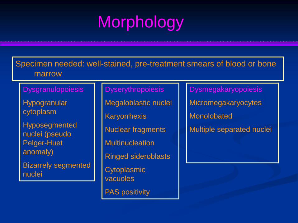
Morphology
Specimen needed: well-stained, pre-treatment smears of blood or bone
marrow
Dysgranulopoiesis
Hypogranular
cytoplasm
Hyposegmented
nuclei (pseudo
Pelger-Huet
anomaly)
Bizarrely segmented
nuclei
Dyserythropoiesis
Megaloblastic nuclei
Karyorrhexis
Nuclear fragments
Multinucleation
Ringed sideroblasts
Cytoplasmic
vacuoles
PAS positivity
Dysmegakaryopoiesis
Micromegakaryocytes
Monolobated
Multiple separated nuclei

Differential diagnosis
M2
M6

Immunophenotype
Generally: CD34 and pan-
myeloid markers (CD13 and
CD33)
Frequently: aberrant exp of
CD56 and CD7
Increased incidence: MDR-1
Genetics
Similar to MDS
Often: -7/del(7q), -5/del(5q), +8, +9, +11, del(11q),
del(12p), -18, +19, del(20q), +21
Less often: t(2;11), t(1;7), 3q21, and 3q26

Prognosis
Multilineage dysplasia: adverse
effect on achieving remission