Wheat Cycle
61
Germination The mature grain contains the embryo wheat plant and enough stored reserves, in the endosperm, to get it growing. The shoot apex of the embryo has three or four leaf primordia and a tiller bud protected by the coleoptile. The root pole has a structure called the coleorhiza, which protects the root until it has broken through the seed coat. After sowing, the germination process starts with the absorption of water (imbibition) and the embryo sends out hormonal signals which induce the synthesis of hydrolytic enzymes in the aleurone. These enzymes degrade the cells walls, starch and storage proteins of the endosperm. The uniformity of this hydration process and the extent of the cell wall degradation are important aspects of malting quality. If the grain starts to germinate in the wheat ear before harvest (pre-harvest sprouting), as it can if the season is cool and damp, flour quality is poor reducing the value and end-use of the crop. Within a few days of imbibition roots start to grow and shortly after that the coleoptile emerges. The coleoptile will grow until it finds the soil surface, after which the leaves will emerge and a seedling will become established. Mobilisation of endosperm storage reserves following germination. After sowing, cereal grains imbibe water and begin a series of events leading to germination of the embryo and subsequent mobilisation of the stored reserves in the endosperm. A chemical stimulus, produced in the embryo, diffuses to the aleurone layer surrounding the starchy endosperm and initiates the synthesis of a range of enzymes that digest cell walls, storage proteins and starch present in the dead cells of the endosperm. Lipids, nucleic acids and mineral reserves are also degraded. The simple sugars and peptides released by the activities of these hydrolytic enzymes are absorbed by the scutellum and are used by the growing embryo before the leaves emerge and photosynthesis begins. We know that a stimulus is derived from the embryo because nothing happens to the endosperm if the embryo is removed before planting. We also know that the plant hormone, gibberellin (GA), is produced by the embryo and that synthetic gibberellic acid (GA3) applied directly to endosperm can substitute for the presence of the embryo. However, this simple model of an embryo stimulus diffusing to the aleurone and leading to the production of hydrolytic enzymes and mobilisation of the endosperm storage reserves is in fact more complex. The initial sites of production of hydrolytic enzymes during cereal germination are the epithelial cells of the scutellum, adjacent to the endosperm. Because of the close proximity to the site of GA production in the scutellum, we do not know whether or not this is a GA-dependent process. Later in development the source of hydrolytic enzyme activity is the aleurone layer. This reflects the pattern of dissolution of the contents of endosperm cells which begins next to the scutellum and moves as a wave advancing fastest beneath the aleurone layer on the dorsal side and flanks towards the brush end of the grain. The pattern of digestion of the contents of these cells is reminiscent of the events that led to the production of the endosperm 'crushed cell layer' during embryo growth in the developing grain. The brewing and distilling industries have benefited from using this information during the production of good quality malt. In this instance, wheat or barley grains are steeped in large tanks containing water, to increase the moisture content, with
description
For those interested in the life cycle of wheat grains.Wheat is one of the oldest and widely used food crops in the world. Products derived from wheat flour includes bread, pastas, noodles, biscuits, crackers, pastries, cookies etc...
Transcript of Wheat Cycle
Germination
The mature grain contains the embryo wheat plant and enough stored reserves, in the
endosperm, to get it growing. The shoot apex of the embryo has three or four leaf
primordia and a tiller bud protected by the coleoptile. The root pole has a structure
called the coleorhiza, which protects the root until it has broken through the seed coat.
After sowing, the germination process starts with the absorption of water (imbibition)
and the embryo sends out hormonal signals which induce the synthesis of hydrolytic
enzymes in the aleurone. These enzymes degrade the cells walls, starch and storage
proteins of the endosperm. The uniformity of this hydration process and the extent of
the cell wall degradation are important aspects of malting quality. If the grain starts to
germinate in the wheat ear before harvest (pre-harvest sprouting), as it can if the
season is cool and damp, flour quality is poor reducing the value and end-use of the
crop.
Within a few days of imbibition roots start to grow and shortly after that the coleoptile
emerges. The coleoptile will grow until it finds the soil surface, after which the leaves
will emerge and a seedling will become established.
Mobilisation of endosperm storage reserves following germination.
After sowing, cereal grains imbibe water and begin a series of events leading to
germination of the embryo and subsequent mobilisation of the stored reserves in the
endosperm. A chemical stimulus, produced in the embryo, diffuses to the aleurone
layer surrounding the starchy endosperm and initiates the synthesis of a range of
enzymes that digest cell walls, storage proteins and starch present in the dead cells of
the endosperm. Lipids, nucleic acids and mineral reserves are also degraded. The
simple sugars and peptides released by the activities of these hydrolytic enzymes are
absorbed by the scutellum and are used by the growing embryo before the leaves
emerge and photosynthesis begins. We know that a stimulus is derived from the
embryo because nothing happens to the endosperm if the embryo is removed before
planting. We also know that the plant hormone, gibberellin (GA), is produced by the
embryo and that synthetic gibberellic acid (GA3) applied directly to endosperm can
substitute for the presence of the embryo.
However, this simple model of an embryo stimulus diffusing to the aleurone and
leading to the production of hydrolytic enzymes and mobilisation of the endosperm
storage reserves is in fact more complex. The initial sites of production of hydrolytic
enzymes during cereal germination are the epithelial cells of the scutellum, adjacent
to the endosperm. Because of the close proximity to the site of GA production in the
scutellum, we do not know whether or not this is a GA-dependent process. Later in
development the source of hydrolytic enzyme activity is the aleurone layer. This
reflects the pattern of dissolution of the contents of endosperm cells which begins
next to the scutellum and moves as a wave advancing fastest beneath the aleurone
layer on the dorsal side and flanks towards the brush end of the grain. The pattern of
digestion of the contents of these cells is reminiscent of the events that led to the
production of the endosperm 'crushed cell layer' during embryo growth in the
developing grain.
The brewing and distilling industries have benefited from using this information
during the production of good quality malt. In this instance, wheat or barley grains are
steeped in large tanks containing water, to increase the moisture content, with
intermittent draining and air rests. The grain is then germinated under controlled
conditions of moisture and temperature allowing the production of hydrolytic
enzymes to digest the contents of the endosperm. In some cases GA3 is added to
increase uniformity of germination and hasten the malting process. In order to restrict
embryo growth, germination is halted by the controlled application of heat (kilning).
The temperature has to be sufficiently high to arrest the activity of the hydrolytic
enzymes but not to destroy them in the finished malt. Kilning temperature also affects
malt colour and flavour, important components of beer quality.
Germination: Diagrams and pictures
Simple diagram of the dry grain showing its principle
parts; the endosperm, aleurone and seed coat, the
scutellum and the embryo with its primordial shoot and
root. During grain milling these tissues are crudely
separated: the endosperm becomes the white flour, the
embryo is the germ and the aleurone layer and the seed
coat are the bran. The seed coat is a complex structure.
The Cell Layers inside the Grain are described in detail
in a separate section.
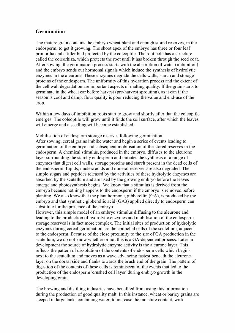
Fully imbibed grain just before the embryo emerges.
After planting the grain quickly takes up water and the
chemical processes of germination start.
The imbibed grain, split open along its long axis, shows
the embryo in close contact, via the scutellum, with the
stored reserves of the endosperm. The internal processes
of germination are well under way. Several hydrolytic
enzymes are activated to perform specific tasks. The cell
walls of the endosperm are broken down, the starch and
storage proteins they contain are degraded and released,
and the aleurone and embryo are activated ready for
growth.
The grain with the embryo dissected away from the
endosperm. The structure between the endosperm and the
embryo is the scutellum; it transfers nutrients from the
endosperm to the embryo during germination. When
millers separate the embryo from the endosperm they call
it the wheat 'germ'. The embryo contains fats and
proteins which may limit the keeping quality of the flour.
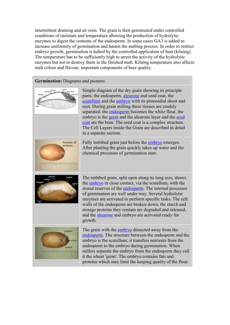
Two days after imbibition and the embryonic axis has
broken through the testa. The first structures to be seen
are the coleoptile, which protects the shoot, and the root
(radicle). The root has broken through its protective
sheath, the coleorhiza.
Germinating wheat grains at two, three and four days
after imbibition. The coleoptile is adapted to thrust its
way through the soil to find the surface. The seedling
roots quickly anchor the grain in the soil. The fine root
hairs absorb water and nutrients from the soil
supplementing the stored reserves from the endosperm
Early growth and tillering
This section covers the development of the leaves and shoots of the wheat plant. The
leaves capture light energy from the sun, and transform it into photosynthates, which
support the growth of the shoot and will, eventually, be transported to and stored in
the harvested seed.
In the temperate zones of the world, where summer days are long and winter days are
short, two distinct types of wheat have been developed. 'Spring' wheat is similar to
wild wheat in that it grows quickly, moving through all the leafy development stages
without check. It is sown in the spring and is induced to flower by increasing
daylength only, so that mature grains can be harvested within 20-25 weeks. In
contrast, 'winter' wheat has been selected to take advantage of a longer leafy stage
when plant development is checked by the cooler temperatures and shorter day length
of winter. During this time the extra leaf area can build up greater reserves which
support higher grain yields when the crop matures. Winter wheat is sown in the
autumn, grows slowly through the winter, is induced to flower by a period of cold and
then flowers in the increasing daylength of spring, maturing the following summer.
The long vegetative phase produces a plant with more side shoots, called tillers. The
tillers form at the base, in the axil, of the first formed leaves of the mainstem and of
the coleoptile. In fact a coleoptile tiller bud is present in the dormant seed but the tiller
emerges only if the seed is sown deeply. Tillers in the axils of Leaf 1, Leaf 2 and Leaf
3 emerge first and are usually strong enough to grow to full canopy height and to set
grain. These tillers produce fewer leaves than the mainstem which has the effect of
synchronising ear emergence, pollen release, grain growth and ripening for all the
shoots in the canopy. Tillers in the axil of Leaf 4 and above and any secondary tillers,
which form in the axils of the leaves of the tillers themselves, will die back during the
phase of rapid shoot growth in the spring. These late tillers contribute to the stored
reserves of the plant.
The following photographs are of winter wheat cv Mercia grown in the field in
southern England. They illustrate the stages of vegetative (leafy) growth above
ground and include some micrographs of the growing point (apex) of the plant, which
is buried beneath the soil surface during the winter.
There are more photographs of the growing point in the section 'Development of the
Wheat Apex'.
Compare the field grown plants with the plants we grew in a Controlled Environment,
which had an artificial winter and spring, in the section 'How We Grew the Plants'.
Early growth and tillering: Diagrams and pictures
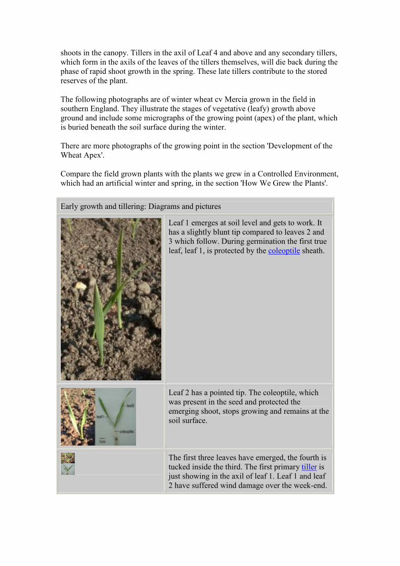
Leaf 1 emerges at soil level and gets to work. It
has a slightly blunt tip compared to leaves 2 and
3 which follow. During germination the first true
leaf, leaf 1, is protected by the coleoptile sheath.
Leaf 2 has a pointed tip. The coleoptile, which
was present in the seed and protected the
emerging shoot, stops growing and remains at the
soil surface.
The first three leaves have emerged, the fourth is
tucked inside the third. The first primary tiller is
just showing in the axil of leaf 1. Leaf 1 and leaf
2 have suffered wind damage over the week-end.
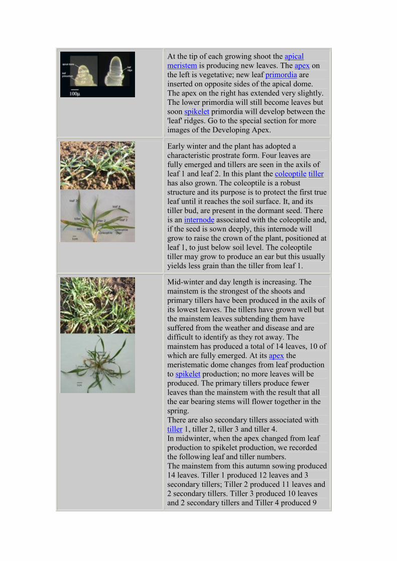
At the tip of each growing shoot the apical
meristem is producing new leaves. The apex on
the left is vegetative; new leaf primordia are
inserted on opposite sides of the apical dome.
The apex on the right has extended very slightly.
The lower primordia will still become leaves but
soon spikelet primordia will develop between the
'leaf' ridges. Go to the special section for more
images of the Developing Apex.
Early winter and the plant has adopted a
characteristic prostrate form. Four leaves are
fully emerged and tillers are seen in the axils of
leaf 1 and leaf 2. In this plant the coleoptile tiller
has also grown. The coleoptile is a robust
structure and its purpose is to protect the first true
leaf until it reaches the soil surface. It, and its
tiller bud, are present in the dormant seed. There
is an internode associated with the coleoptile and,
if the seed is sown deeply, this internode will
grow to raise the crown of the plant, positioned at
leaf 1, to just below soil level. The coleoptile
tiller may grow to produce an ear but this usually
yields less grain than the tiller from leaf 1.
Mid-winter and day length is increasing. The
mainstem is the strongest of the shoots and
primary tillers have been produced in the axils of
its lowest leaves. The tillers have grown well but
the mainstem leaves subtending them have
suffered from the weather and disease and are
difficult to identify as they rot away. The
mainstem has produced a total of 14 leaves, 10 of
which are fully emerged. At its apex the
meristematic dome changes from leaf production
to spikelet production; no more leaves will be
produced. The primary tillers produce fewer
leaves than the mainstem with the result that all
the ear bearing stems will flower together in the
spring.
There are also secondary tillers associated with
tiller 1, tiller 2, tiller 3 and tiller 4.
In midwinter, when the apex changed from leaf
production to spikelet production, we recorded
the following leaf and tiller numbers.
The mainstem from this autumn sowing produced
14 leaves. Tiller 1 produced 12 leaves and 3
secondary tillers; Tiller 2 produced 11 leaves and
2 secondary tillers. Tiller 3 produced 10 leaves
and 2 secondary tillers and Tiller 4 produced 9
leaves and 1 secondary tiller.
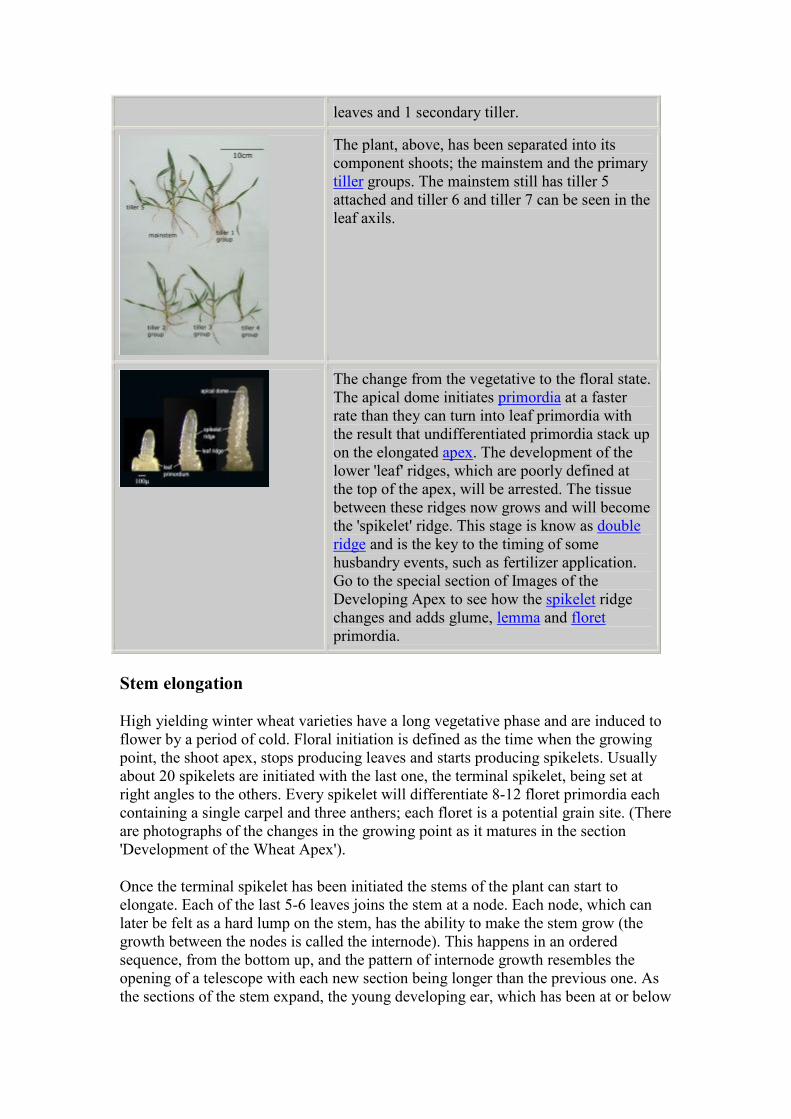
The plant, above, has been separated into its
component shoots; the mainstem and the primary
tiller groups. The mainstem still has tiller 5
attached and tiller 6 and tiller 7 can be seen in the
leaf axils.
The change from the vegetative to the floral state.
The apical dome initiates primordia at a faster
rate than they can turn into leaf primordia with
the result that undifferentiated primordia stack up
on the elongated apex. The development of the
lower 'leaf' ridges, which are poorly defined at
the top of the apex, will be arrested. The tissue
between these ridges now grows and will become
the 'spikelet' ridge. This stage is know as double
ridge and is the key to the timing of some
husbandry events, such as fertilizer application.
Go to the special section of Images of the
Developing Apex to see how the spikelet ridge
changes and adds glume, lemma and floret
primordia.
Stem elongation
High yielding winter wheat varieties have a long vegetative phase and are induced to
flower by a period of cold. Floral initiation is defined as the time when the growing
point, the shoot apex, stops producing leaves and starts producing spikelets. Usually
about 20 spikelets are initiated with the last one, the terminal spikelet, being set at
right angles to the others. Every spikelet will differentiate 8-12 floret primordia each
containing a single carpel and three anthers; each floret is a potential grain site. (There
are photographs of the changes in the growing point as it matures in the section
'Development of the Wheat Apex').
Once the terminal spikelet has been initiated the stems of the plant can start to
elongate. Each of the last 5-6 leaves joins the stem at a node. Each node, which can
later be felt as a hard lump on the stem, has the ability to make the stem grow (the
growth between the nodes is called the internode). This happens in an ordered
sequence, from the bottom up, and the pattern of internode growth resembles the
opening of a telescope with each new section being longer than the previous one. As
the sections of the stem expand, the young developing ear, which has been at or below
soil level all winter, is pushed up through the surrounding leaves. The ear itself grows
rapidly as the last three stem internodes expand. With the expansion of the final stem
internode (the peduncle), the fully developed ear emerges through the ligule of the
last-formed (flag) leaf and soon afterwards anthesis (shedding of pollen) and
fertilisation occurs.
A consequence of all this rapid growth is that there is intense competition for
resources and this causes die-back in some parts of the plant. First, as the stem starts
to grow, the last formed tillers die back and second, as the ear is pushed above the
canopy, the last formed florets within each spikelet are aborted.
Stem elongation: Diagrams and pictures
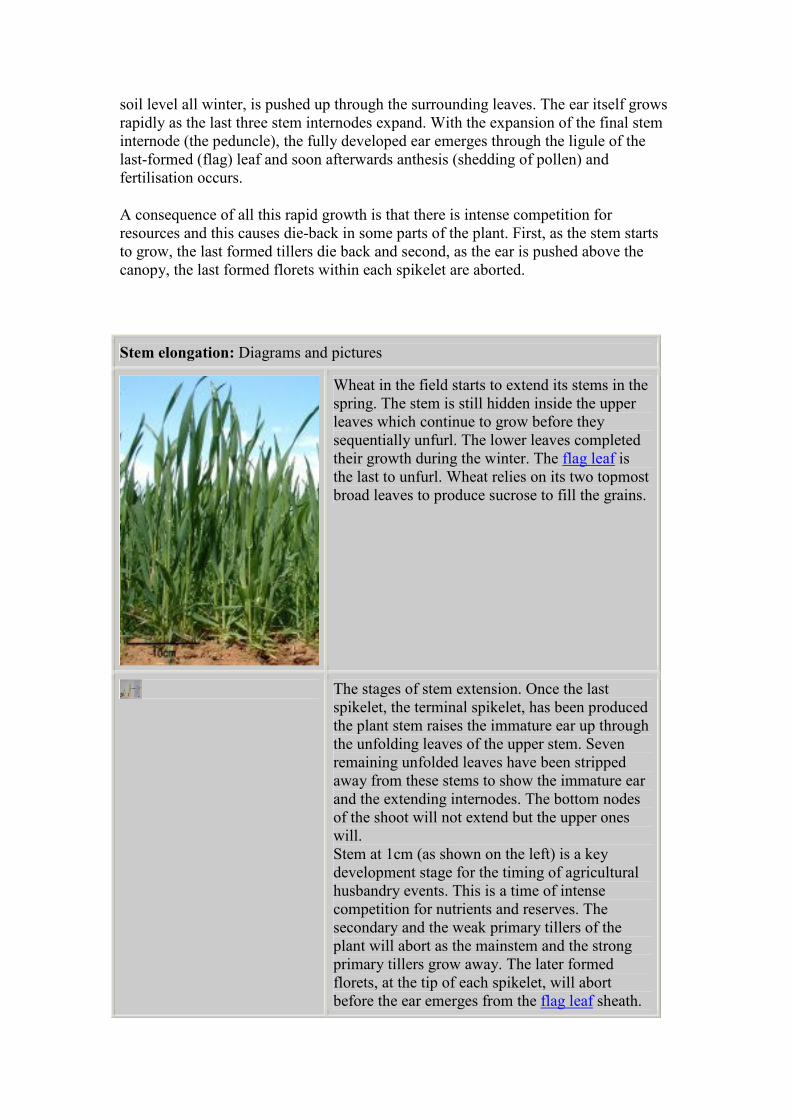
Wheat in the field starts to extend its stems in the
spring. The stem is still hidden inside the upper
leaves which continue to grow before they
sequentially unfurl. The lower leaves completed
their growth during the winter. The flag leaf is
the last to unfurl. Wheat relies on its two topmost
broad leaves to produce sucrose to fill the grains.
The stages of stem extension. Once the last
spikelet, the terminal spikelet, has been produced
the plant stem raises the immature ear up through
the unfolding leaves of the upper stem. Seven
remaining unfolded leaves have been stripped
away from these stems to show the immature ear
and the extending internodes. The bottom nodes
of the shoot will not extend but the upper ones
will.
Stem at 1cm (as shown on the left) is a key
development stage for the timing of agricultural
husbandry events. This is a time of intense
competition for nutrients and reserves. The
secondary and the weak primary tillers of the
plant will abort as the mainstem and the strong
primary tillers grow away. The later formed
florets, at the tip of each spikelet, will abort
before the ear emerges from the flag leaf sheath.
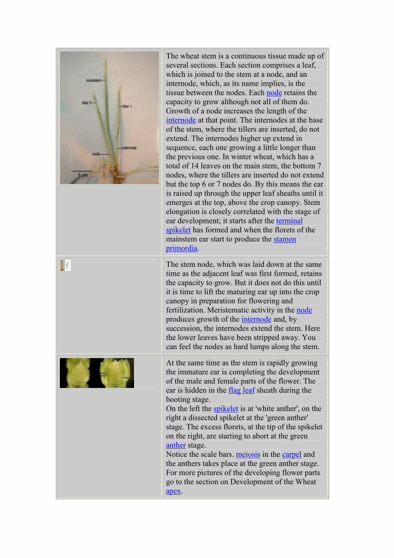
The wheat stem is a continuous tissue made up of
several sections. Each section comprises a leaf,
which is joined to the stem at a node, and an
internode, which, as its name implies, is the
tissue between the nodes. Each node retains the
capacity to grow although not all of them do.
Growth of a node increases the length of the
internode at that point. The internodes at the base
of the stem, where the tillers are inserted, do not
extend. The internodes higher up extend in
sequence, each one growing a little longer than
the previous one. In winter wheat, which has a
total of 14 leaves on the main stem, the bottom 7
nodes, where the tillers are inserted do not extend
but the top 6 or 7 nodes do. By this means the ear
is raised up through the upper leaf sheaths until it
emerges at the top, above the crop canopy. Stem
elongation is closely correlated with the stage of
ear development; it starts after the terminal
spikelet has formed and when the florets of the
mainstem ear start to produce the stamen
primordia.
The stem node, which was laid down at the same
time as the adjacent leaf was first formed, retains
the capacity to grow. But it does not do this until
it is time to lift the maturing ear up into the crop
canopy in preparation for flowering and
fertilization. Meristematic activity in the node
produces growth of the internode and, by
succession, the internodes extend the stem. Here
the lower leaves have been stripped away. You
can feel the nodes as hard lumps along the stem.
At the same time as the stem is rapidly growing
the immature ear is completing the development
of the male and female parts of the flower. The
ear is hidden in the flag leaf sheath during the
booting stage.
On the left the spikelet is at 'white anther', on the
right a dissected spikelet at the 'green anther'
stage. The excess florets, at the tip of the spikelet
on the right, are starting to abort at the green
anther stage.
Notice the scale bars. meiosis in the carpel and
the anthers takes place at the green anther stage.
For more pictures of the developing flower parts
go to the section on Development of the Wheat
apex.
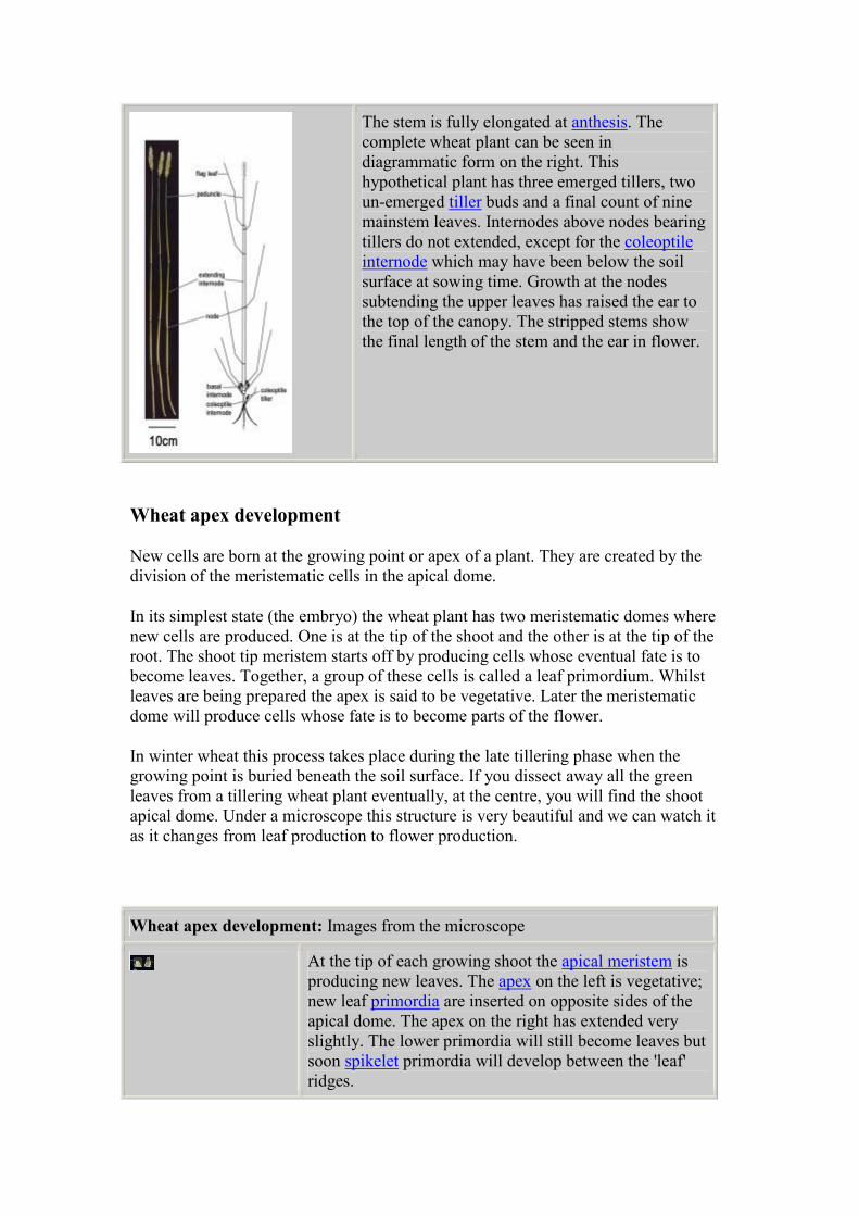
The stem is fully elongated at anthesis. The
complete wheat plant can be seen in
diagrammatic form on the right. This
hypothetical plant has three emerged tillers, two
un-emerged tiller buds and a final count of nine
mainstem leaves. Internodes above nodes bearing
tillers do not extended, except for the coleoptile
internode which may have been below the soil
surface at sowing time. Growth at the nodes
subtending the upper leaves has raised the ear to
the top of the canopy. The stripped stems show
the final length of the stem and the ear in flower.
Wheat apex development
New cells are born at the growing point or apex of a plant. They are created by the
division of the meristematic cells in the apical dome.
In its simplest state (the embryo) the wheat plant has two meristematic domes where
new cells are produced. One is at the tip of the shoot and the other is at the tip of the
root. The shoot tip meristem starts off by producing cells whose eventual fate is to
become leaves. Together, a group of these cells is called a leaf primordium. Whilst
leaves are being prepared the apex is said to be vegetative. Later the meristematic
dome will produce cells whose fate is to become parts of the flower.
In winter wheat this process takes place during the late tillering phase when the
growing point is buried beneath the soil surface. If you dissect away all the green
leaves from a tillering wheat plant eventually, at the centre, you will find the shoot
apical dome. Under a microscope this structure is very beautiful and we can watch it
as it changes from leaf production to flower production.
Wheat apex development: Images from the microscope
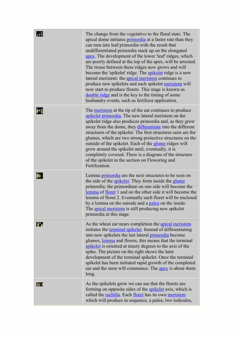
At the tip of each growing shoot the apical meristem is
producing new leaves. The apex on the left is vegetative;
new leaf primordia are inserted on opposite sides of the
apical dome. The apex on the right has extended very
slightly. The lower primordia will still become leaves but
soon spikelet primordia will develop between the 'leaf'
ridges.
The change from the vegetative to the floral state. The
apical dome initiates primordia at a faster rate than they
can turn into leaf primordia with the result that
undifferentiated primordia stack up on the elongated
apex. The development of the lower 'leaf' ridges, which
are poorly defined at the top of the apex, will be arrested.
The tissue between these ridges now grows and will
become the 'spikelet' ridge. The spikelet ridge is a new
lateral meristem: the apical meristem continues to
produce new spikelets and each spikelet meristem will
now start to produce florets. This stage is known as
double ridge and is the key to the timing of some
husbandry events, such as fertilizer application.
The meristem at the tip of the ear continues to produce
spikelet primordia. The new lateral meristem on the
spikelet ridge also produces primordia and, as they grow
away from the dome, they differentiate into the different
structures of the spikelet. The first structures seen are the
glumes, which are two strong protective structures on the
outside of the spikelet. Each of the glume ridges will
grow around the spikelet until, eventually, it is
completely covered. There is a diagram of the structure
of the spikelet in the section on Flowering and
Fertilization.
Lemma primordia are the next structures to be seen on
the side of the spikelet. They form inside the glume
primordia; the primordium on one side will become the
lemma of floret 1 and on the other side it will become the
lemma of floret 2. Eventually each floret will be enclosed
by a lemma on the outside and a palea on the inside.
The apical meristem is still producing new spikelet
primordia at this stage.
As the wheat ear nears completion the apical meristem
initiates the terminal spikelet. Instead of differentiating
into new spikelets the last lateral primordia become
glumes, lemma and florets, this means that the terminal
spikelet is oriented at ninety degrees to the axis of the
spike. The picture on the right shows the later
development of the terminal spikelet. Once the terminal
spikelet has been initiated rapid growth of the completed
ear and the stem will commence. The apex is about 4mm
long.
As the spikelets grow we can see that the florets are
forming on opposite sides of the spikelet axis, which is
called the rachilla. Each floret has its own meristem
which will produce in sequence, a palea, two lodicules,
three stamens and a carpel. There is a diagram of the
spikelet with an expanded floret in the section on
Flowering and Fertilization.
Stem at 1cm is a key development stage for the timing of
agricultural husbandry events. The base of the plant stem
starts to grow quickly, raising the immature ear up
through the unfolding leaves of the upper stem. The
lateral spikelet meristems continue to produce florets and
the floret meristems start to initiate the different parts of
the flower. Go to the section on Stem Extension to find
out how the stem grows so fast.
The apical meristem has produced the terminal spikelet
and the action now moves to the lateral spikelet and
floret meristems. Each spikelet meristem will initiate
between eight and twelve florets. While this continues in
the top of the spikelet, the first formed florets, at the
bottom of the spikelet, will start to differentiate the
different parts of the flower. The primordia of the three
stamens are prominent; the carpel is still hidden.
In the right hand picture, the older spikelet is at the White
anther stage. Each floret has a protective lemma and
palea, the lemmas have small awns, and the whole
spikelet is rapidly becoming enclosed by the two glumes.
Inside this spikelet the anthers were still white and the
carpel was very small.
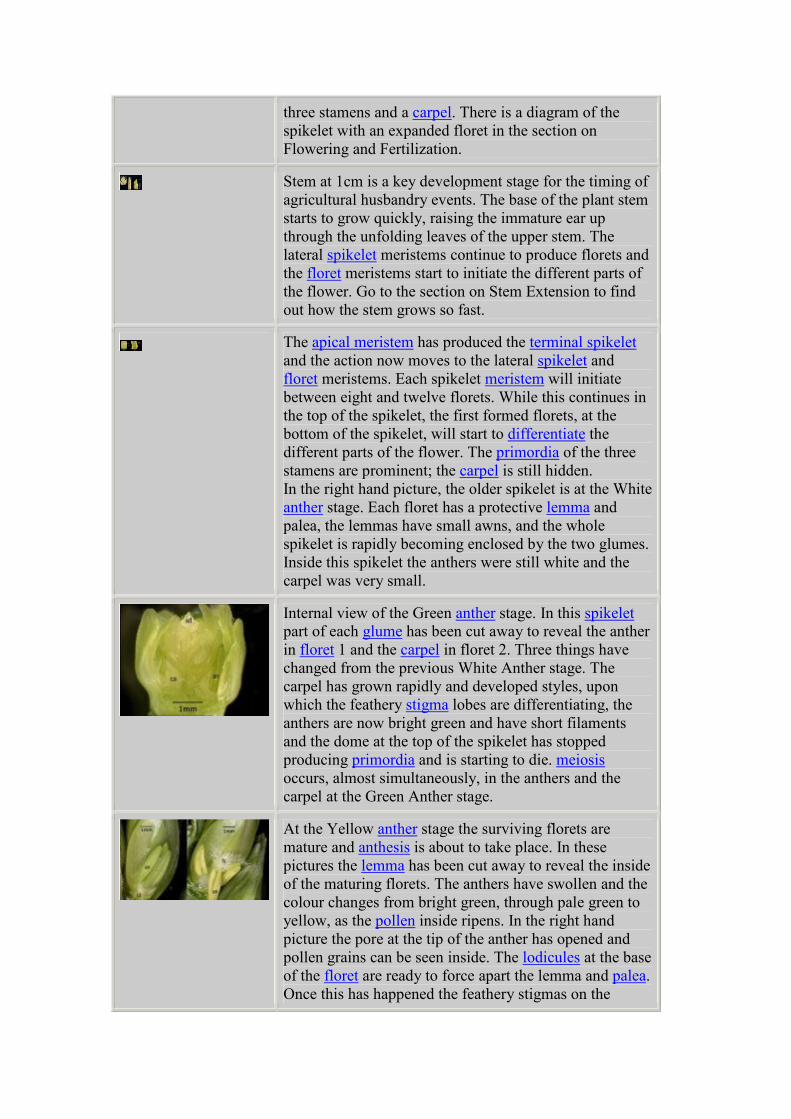
Internal view of the Green anther stage. In this spikelet
part of each glume has been cut away to reveal the anther
in floret 1 and the carpel in floret 2. Three things have
changed from the previous White Anther stage. The
carpel has grown rapidly and developed styles, upon
which the feathery stigma lobes are differentiating, the
anthers are now bright green and have short filaments
and the dome at the top of the spikelet has stopped
producing primordia and is starting to die. meiosis
occurs, almost simultaneously, in the anthers and the
carpel at the Green Anther stage.
At the Yellow anther stage the surviving florets are
mature and anthesis is about to take place. In these
pictures the lemma has been cut away to reveal the inside
of the maturing florets. The anthers have swollen and the
colour changes from bright green, through pale green to
yellow, as the pollen inside ripens. In the right hand
picture the pore at the tip of the anther has opened and
pollen grains can be seen inside. The lodicules at the base
of the floret are ready to force apart the lemma and palea.
Once this has happened the feathery stigmas on the
carpel open and prepare to receive the pollen. The
youngest florets, toward the top of the spikelet, abort at
this stage and the spikelet meristem is completely dead.
For an explanation of Anthesis and more pictures of the
events go to the special section on Pollen Release.
Flowering and fertilisation
While the ear remains within the protection of the leaf sheath, the stage known as
'booting', the flower parts are steadily maturing. Each spikelet produces 8 -12 florets,
many more than it can support, most of them will abort before flowering starts.
(There are photographs of the changes in the growing point as it matures in the
section 'Development of the Wheat Apex').
Flowering or Anthesis takes place a few days after the ear emerges from the leaf
sheath. The flower stalk or peduncle has lifted the ear up into the top of the crop
canopy a few centimetres above the last or flag leaf of each tiller. In open flowering
wheat types a number of co-ordinated events take place within the space of a few
minutes. The stamen filaments grow rapidly to six or eight times their original length.
At the tip of each anther two pores appear through which the pollen will be shed. At
the base of each floret the lodicules swell rapidly forcing apart the lemma and palea.
This allows the stamens to emerge and dangle freely from the floret. The pollen from
the anthers falls onto the receptive feathery stigmas of the carpel which have unfolded
to receive it. (There are more photographs of pollen shed in the section, Photographs
of Pollen Release).
Inside the carpel a double fertilisation event takes place within the embryo sac. One
pollen nucleus fuses with the egg cell which will subsequently divide to form the
diploid embryo. A second pollen nucleus fuses with the two polar nuclei in the
embryo sac (called the second polar event). These nuclei will subsequently divide and
cellularise to form the triploid endosperm. The embryo sac already contains antipodal
cells which will fuel the next stages of grain development.
Flowering and fertilisation: Wheat ear pictures
All the events around anthesis must be well co-ordinated
for the successful release of pollen and fertilization of the
ovule. The ear is quickly raised above the crop canopy by
the growth of the last stem internode or peduncle. It
remains protected inside the sheath of the flag leaf until
the anthers are almost mature.
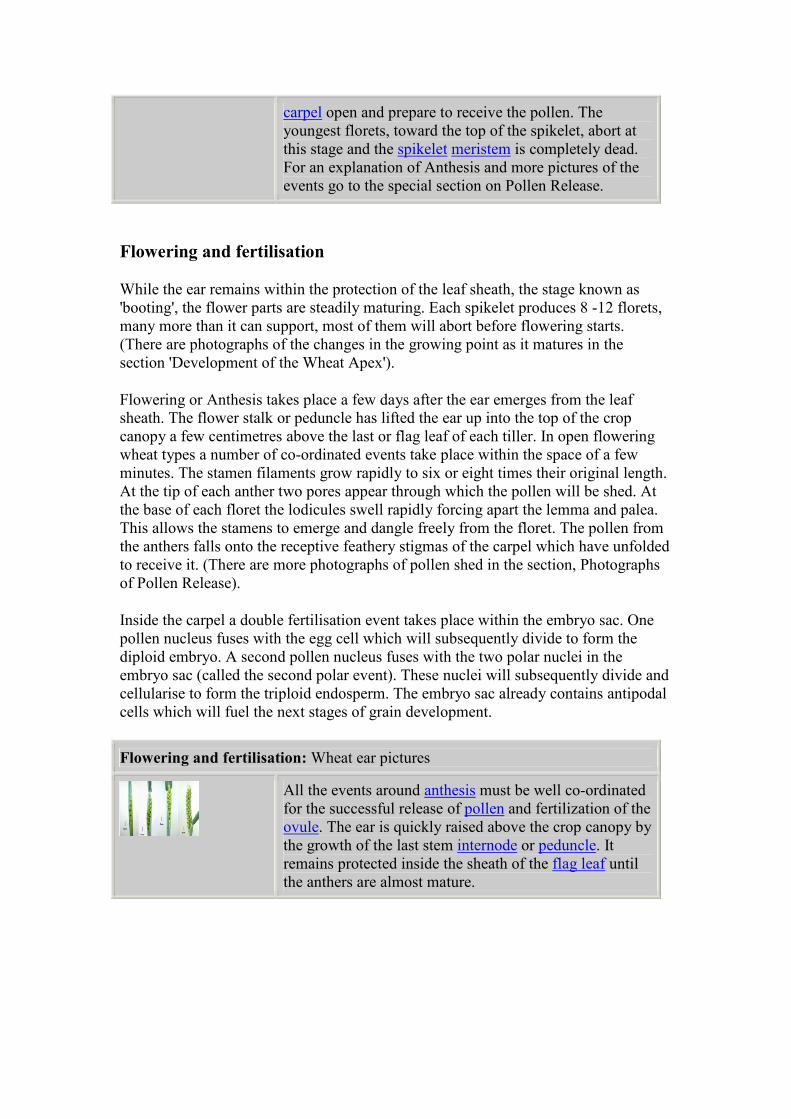
Spikelets are arranged on alternate sides of the rachis.
The collar is a rudimentary spikelet which only rarely
sets grain. The last-formed, terminal spikelet is set at
ninety degrees to the lateral spikelets, making the wheat
ear a determinate structure. More spikelets are found on
the mainstem than on the primary tillers. The final
number is genetically limited; in cv Mercia it is, on
average, 22 plus the terminal spikelet. Cultural
conditions will determine how many florets within each
spikelet remain viable at anthesis.
Each spikelet initiates between eight and twelve florets of
which only four or five will be potentially fertile at
flowering. The outer glumes are barren, their function is
to protect. Similarly the lemma and palea of each floret
protect the delicate
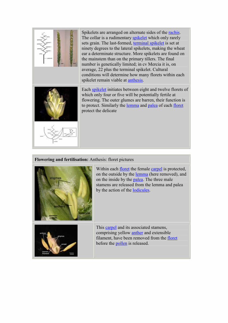
Flowering and fertilisation: Anthesis: floret pictures
Within each floret the female carpel is protected,
on the outside by the lemma (here removed), and
on the inside by the palea. The three male
stamens are released from the lemma and palea
by the action of the lodicules.
This carpel and its associated stamens,
comprising yellow anther and extensible
filament, have been removed from the floret
before the pollen is released.
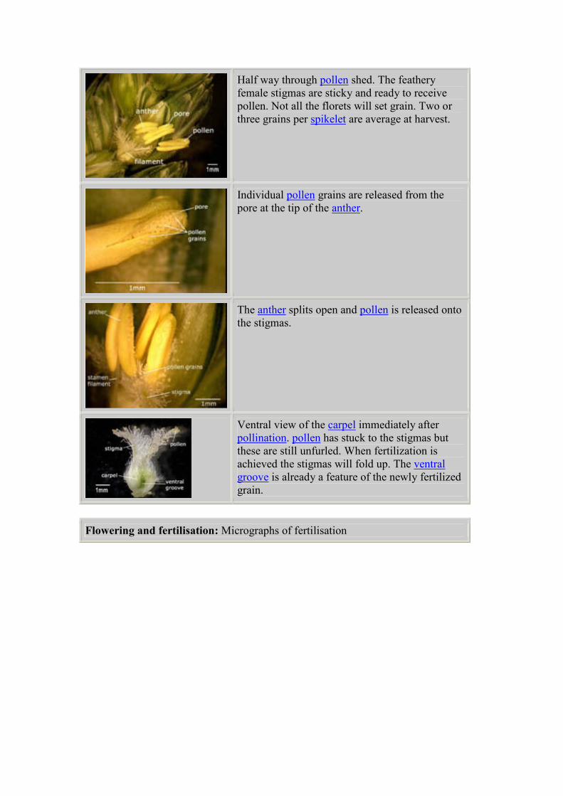
Half way through pollen shed. The feathery
female stigmas are sticky and ready to receive
pollen. Not all the florets will set grain. Two or
three grains per spikelet are average at harvest.
Individual pollen grains are released from the
pore at the tip of the anther.
The anther splits open and pollen is released onto
the stigmas.
Ventral view of the carpel immediately after
pollination. pollen has stuck to the stigmas but
these are still unfurled. When fertilization is
achieved the stigmas will fold up. The ventral
groove is already a feature of the newly fertilized
grain.
Flowering and fertilisation: Micrographs of fertilisation
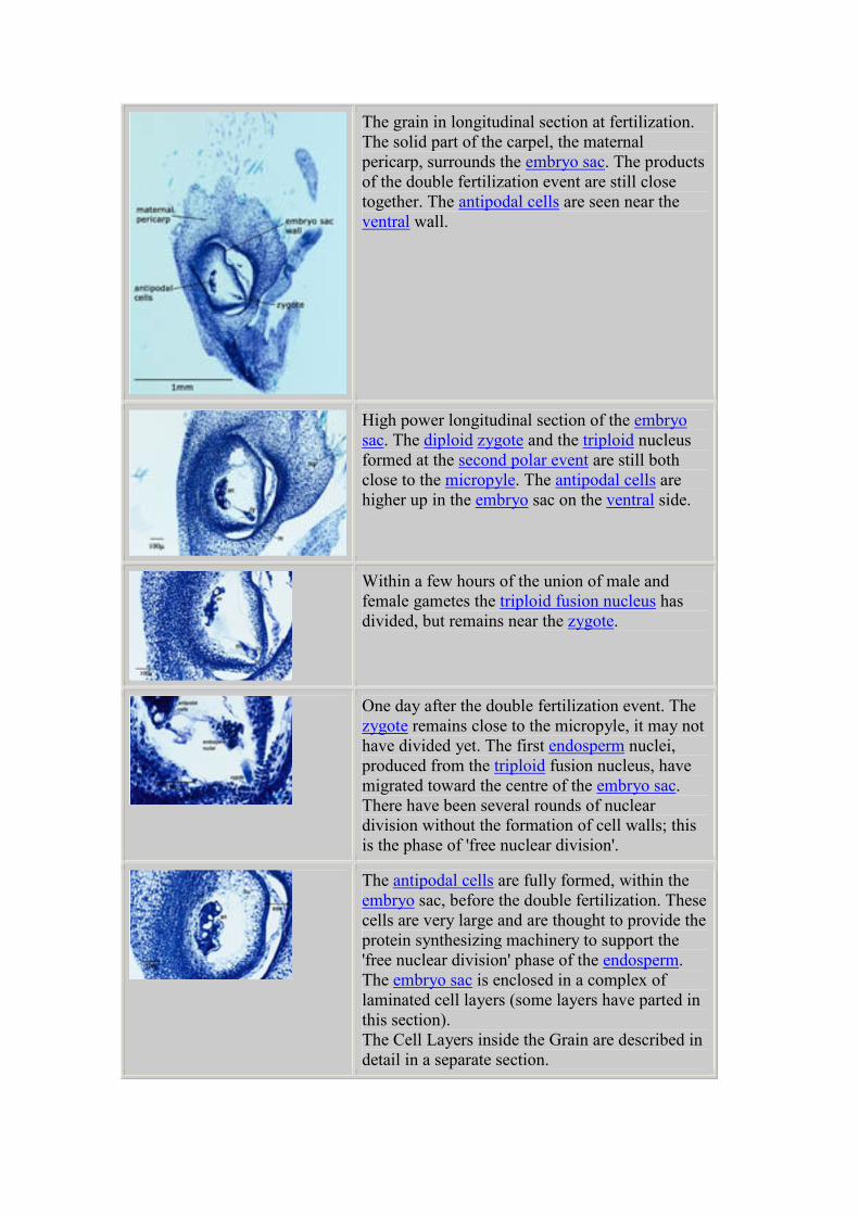
The grain in longitudinal section at fertilization.
The solid part of the carpel, the maternal
pericarp, surrounds the embryo sac. The products
of the double fertilization event are still close
together. The antipodal cells are seen near the
ventral wall.
High power longitudinal section of the embryo
sac. The diploid zygote and the triploid nucleus
formed at the second polar event are still both
close to the micropyle. The antipodal cells are
higher up in the embryo sac on the ventral side.
Within a few hours of the union of male and
female gametes the triploid fusion nucleus has
divided, but remains near the zygote.
One day after the double fertilization event. The
zygote remains close to the micropyle, it may not
have divided yet. The first endosperm nuclei,
produced from the triploid fusion nucleus, have
migrated toward the centre of the embryo sac.
There have been several rounds of nuclear
division without the formation of cell walls; this
is the phase of 'free nuclear division'.
The antipodal cells are fully formed, within the
embryo sac, before the double fertilization. These
cells are very large and are thought to provide the
protein synthesizing machinery to support the
'free nuclear division' phase of the endosperm.
The embryo sac is enclosed in a complex of
laminated cell layers (some layers have parted in
this section).
The Cell Layers inside the Grain are described in
detail in a separate section.
Pollen release
Here is a series of photographs capturing the events of anthesis. The photographs
were taken under a low power dissecting microscope, over a period of 5 minutes.
Normally these events are hidden inside the floret; the first visible sign of anthesis is
the appearance of spent anthers dangling outside the spikelet.
In these pictures the outer glume of the spikelet and the lemma of the first floret have
been removed so that the events that normally happen inside the floret can be seen.
Grain growth 1 to 4 days
The grain grows very rapidly during the first ten days after fertilisation. For
convenience, this has been subdivided into two periods, 1-4 and 4-10 days after
flowering. We describe the external changes in the whole grain and the internal
changes, which can be studied in detail in microscope sections.
The seed increases in size about three fold in the four days after fertilisation. There is
a rapid swelling of the tissues of the carpel, both the pericarp and the embryo sac;
these tissues surround the fertilised embryo. This growth is achieved by expansion of
the cells, rather than cell multiplication, in the cell layers that enclose the embryo sac.
These complex changes are explained in greater detail in a separate page, 'Cell layers
surrounding the Embryo Sac'.
After fertilisation, nuclei from the second polar event divide synchronously inside the
embryo sac but at this stage cell walls are not formed. The nuclei remain in a state of
'free nuclear division' for 3-4 days and the duration of this phase will, in part,
determine the final number of cells in the endosperm. The number of these cells is
closely related to the final weight of the grain and control of this process is an
important target for cereal biologists. The first divisions of the endosperm nucleus
produce the so called 'coenocytic endosperm'. The 'free nuclear division' stage is
fuelled by very large secretory cells at the top of the embryo sac called the antipodal
cells.
The zygote formed at fertilisation has divided only once or twice. It is positioned at
the base of the embryo sac where it is nourished by the densely cytoplasmic 'cellular
endosperm' cells.
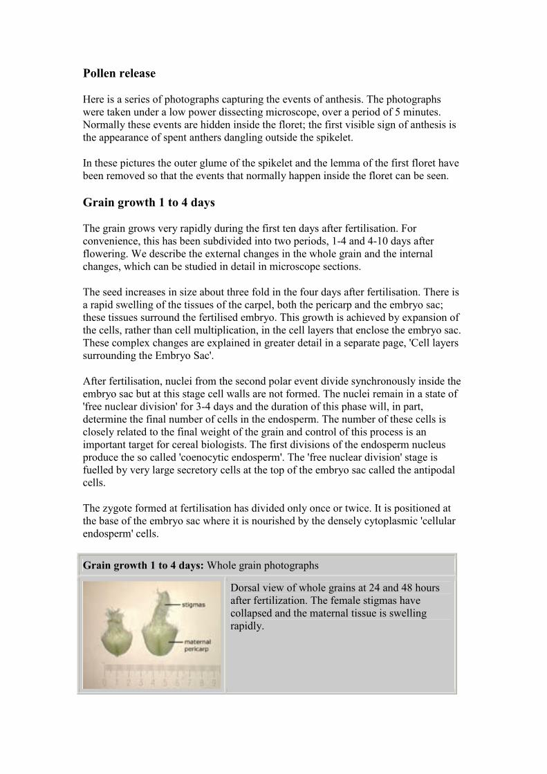
Grain growth 1 to 4 days: Whole grain photographs
Dorsal view of whole grains at 24 and 48 hours
after fertilization. The female stigmas have
collapsed and the maternal tissue is swelling
rapidly.
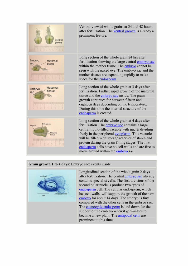
Ventral view of whole grains at 24 and 48 hours
after fertilization. The ventral groove is already a
prominent feature.
Long section of the whole grain 24 hrs after
fertilization showing the large central embryo sac
within the mother tissue. The embryo cannot be
seen with the naked eye. The embryo sac and the
mother tissues are expanding rapidly to make
space for the endosperm.
Long section of the whole grain at 3 days after
fertilization. Further rapid growth of the maternal
tissue and the embryo sac inside. The grain
growth continues for between fifteen and
eighteen days depending on the temperature.
During this time the internal structure of the
endosperm is created.
Long section of the whole grain at 4 days after
fertilization. The embryo sac contains a large
central liquid-filled vacuole with nuclei dividing
freely in the peripheral cytoplasm. This vacuole
will be filled with storage reserves of starch and
protein during the grain filling stages. The first
endosperm cells have no cell walls and are free to
move around within the embryo sac.
Grain growth 1 to 4 days: Embryo sac: events inside
Longitudinal section of the whole grain 2 days
after fertilization. The central embryo sac already
contains specialist cells. The first divisions of the
second polar nucleus produce two types of
endosperm cell. The cellular endosperm, which
has cell walls, will support the growth of the new
embryo for about 14 days. The embryo is tiny
compared with the other cells in the embryo sac.
The coenocytic endosperm is laid down for the
support of the embryo when it germinates to
become a new plant. The antipodal cells are
prominent at this time.
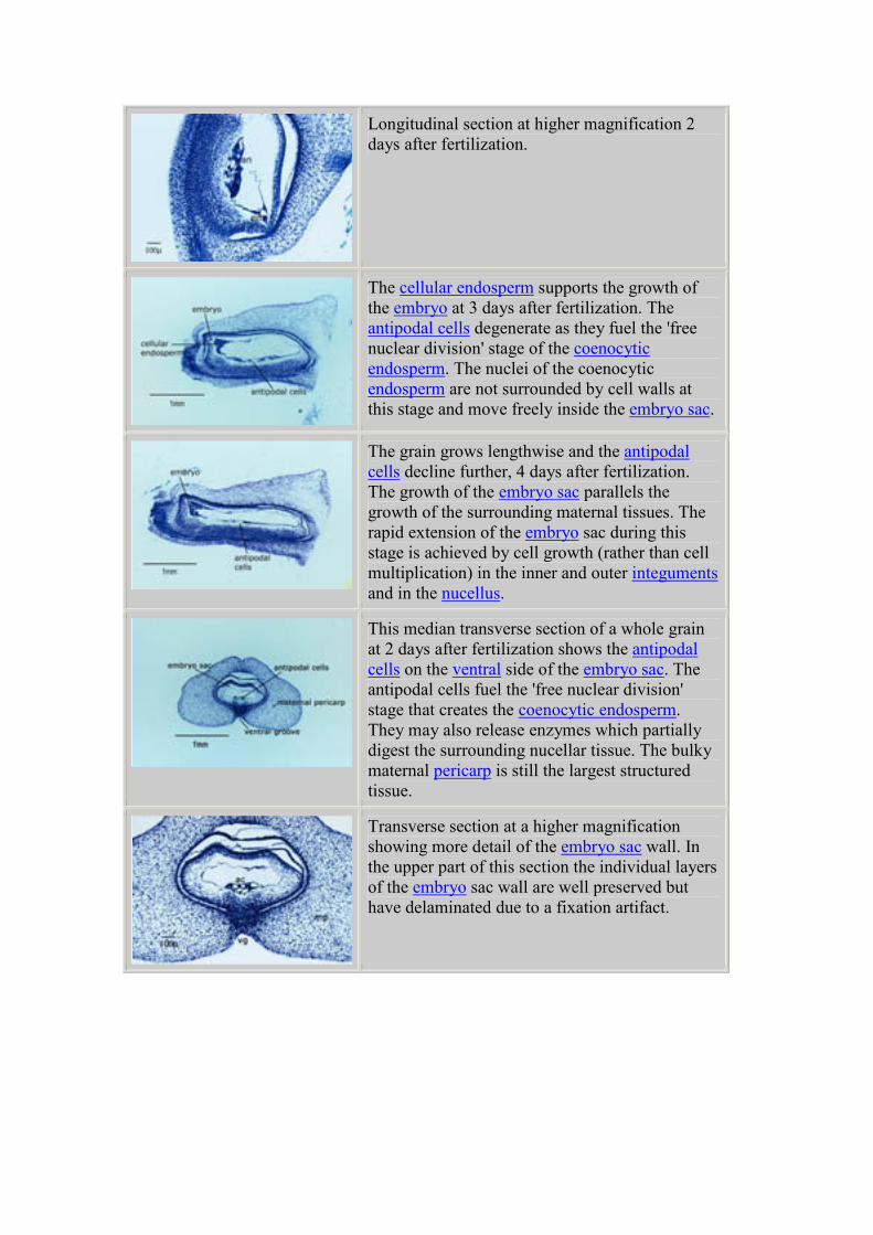
Longitudinal section at higher magnification 2
days after fertilization.
The cellular endosperm supports the growth of
the embryo at 3 days after fertilization. The
antipodal cells degenerate as they fuel the 'free
nuclear division' stage of the coenocytic
endosperm. The nuclei of the coenocytic
endosperm are not surrounded by cell walls at
this stage and move freely inside the embryo sac.
The grain grows lengthwise and the antipodal
cells decline further, 4 days after fertilization.
The growth of the embryo sac parallels the
growth of the surrounding maternal tissues. The
rapid extension of the embryo sac during this
stage is achieved by cell growth (rather than cell
multiplication) in the inner and outer integuments
and in the nucellus.
This median transverse section of a whole grain
at 2 days after fertilization shows the antipodal
cells on the ventral side of the embryo sac. The
antipodal cells fuel the 'free nuclear division'
stage that creates the coenocytic endosperm.
They may also release enzymes which partially
digest the surrounding nucellar tissue. The bulky
maternal pericarp is still the largest structured
tissue.
Transverse section at a higher magnification
showing more detail of the embryo sac wall. In
the upper part of this section the individual layers
of the embryo sac wall are well preserved but
have delaminated due to a fixation artifact.
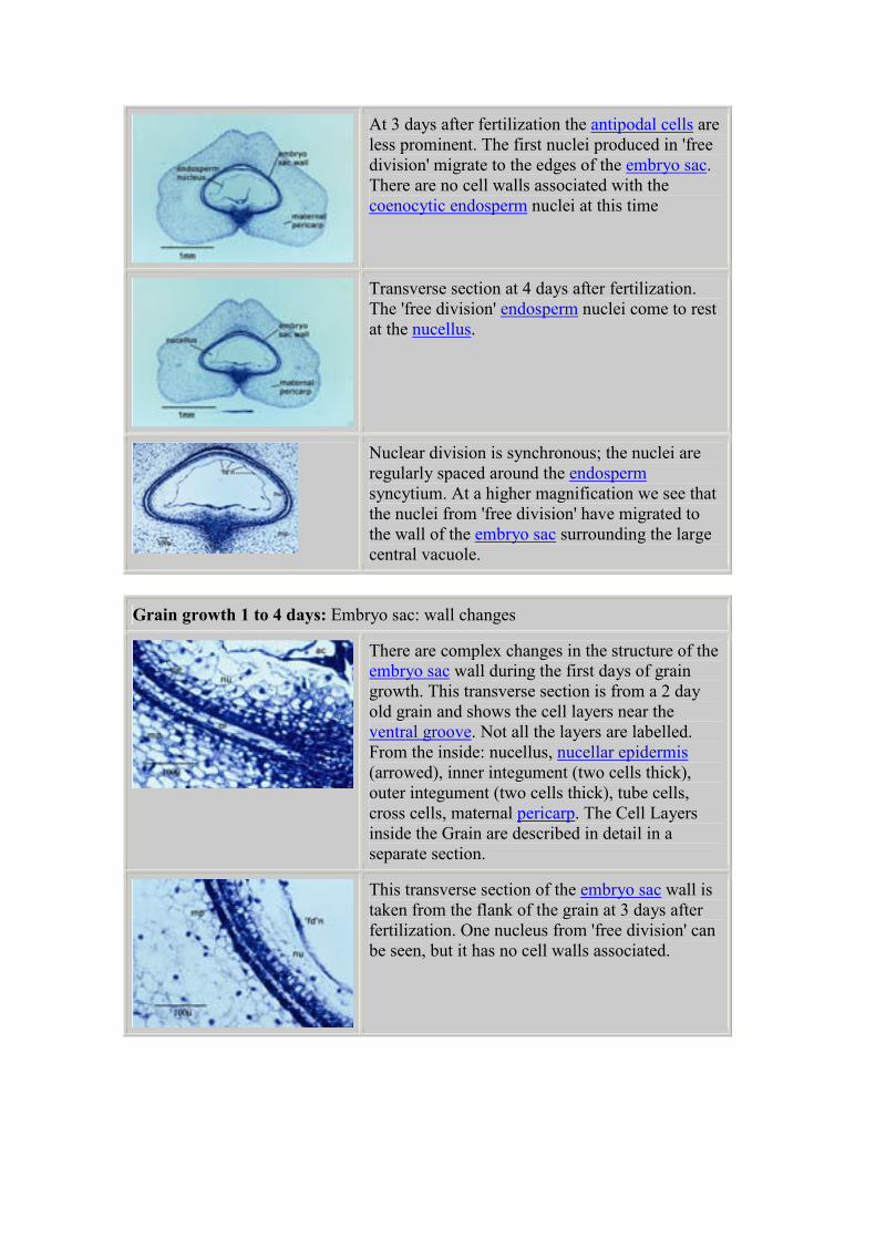
At 3 days after fertilization the antipodal cells are
less prominent. The first nuclei produced in 'free
division' migrate to the edges of the embryo sac.
There are no cell walls associated with the
coenocytic endosperm nuclei at this time
Transverse section at 4 days after fertilization.
The 'free division' endosperm nuclei come to rest
at the nucellus.
Nuclear division is synchronous; the nuclei are
regularly spaced around the endosperm
syncytium. At a higher magnification we see that
the nuclei from 'free division' have migrated to
the wall of the embryo sac surrounding the large
central vacuole.
Grain growth 1 to 4 days: Embryo sac: wall changes
There are complex changes in the structure of the
embryo sac wall during the first days of grain
growth. This transverse section is from a 2 day
old grain and shows the cell layers near the
ventral groove. Not all the layers are labelled.
From the inside: nucellus, nucellar epidermis
(arrowed), inner integument (two cells thick),
outer integument (two cells thick), tube cells,
cross cells, maternal pericarp. The Cell Layers
inside the Grain are described in detail in a
separate section.
This transverse section of the embryo sac wall is
taken from the flank of the grain at 3 days after
fertilization. One nucleus from 'free division' can
be seen, but it has no cell walls associated.
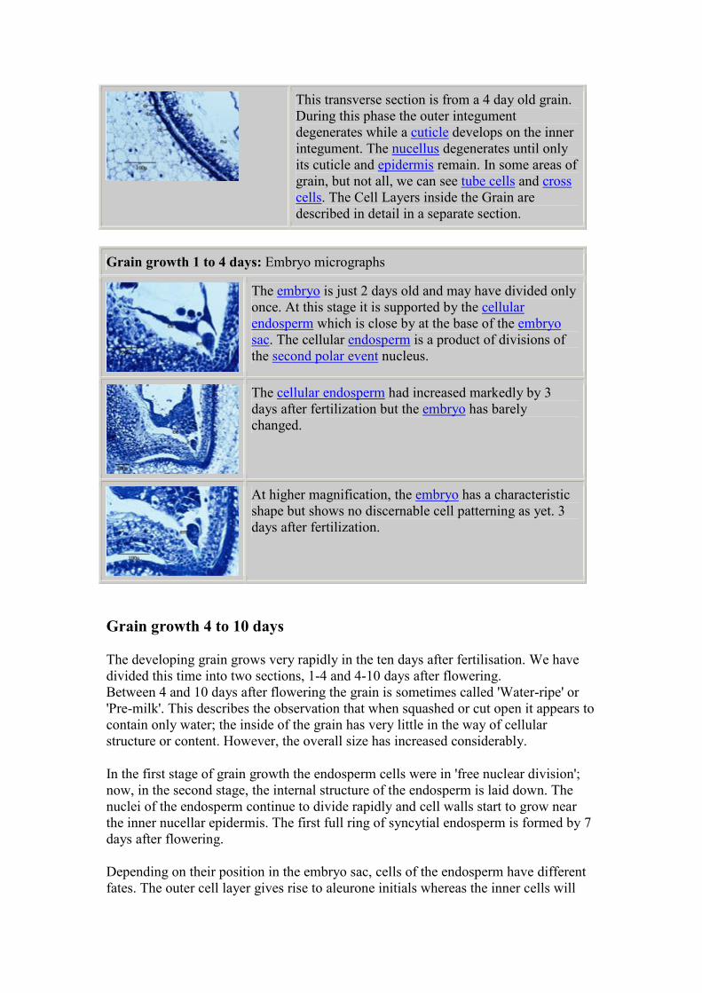
This transverse section is from a 4 day old grain.
During this phase the outer integument
degenerates while a cuticle develops on the inner
integument. The nucellus degenerates until only
its cuticle and epidermis remain. In some areas of
grain, but not all, we can see tube cells and cross
cells. The Cell Layers inside the Grain are
described in detail in a separate section.
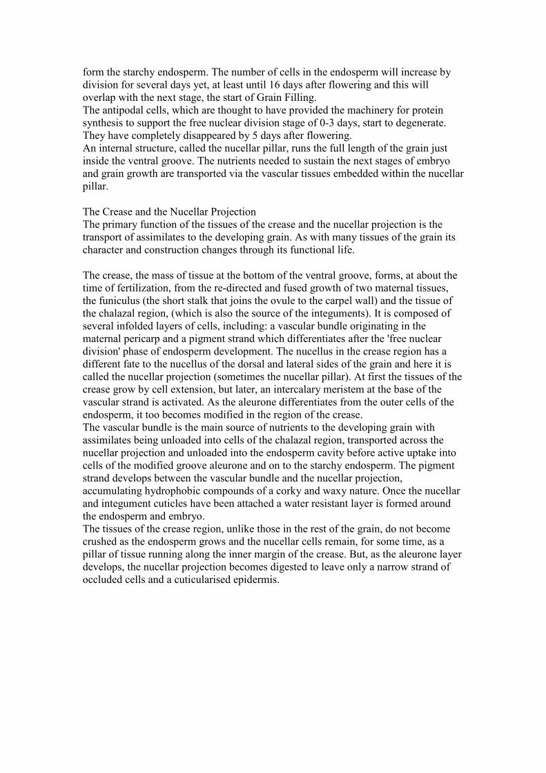
Grain growth 1 to 4 days: Embryo micrographs
The embryo is just 2 days old and may have divided only
once. At this stage it is supported by the cellular
endosperm which is close by at the base of the embryo
sac. The cellular endosperm is a product of divisions of
the second polar event nucleus.
The cellular endosperm had increased markedly by 3
days after fertilization but the embryo has barely
changed.
At higher magnification, the embryo has a characteristic
shape but shows no discernable cell patterning as yet. 3
days after fertilization.
Grain growth 4 to 10 days
The developing grain grows very rapidly in the ten days after fertilisation. We have
divided this time into two sections, 1-4 and 4-10 days after flowering.
Between 4 and 10 days after flowering the grain is sometimes called 'Water-ripe' or
'Pre-milk'. This describes the observation that when squashed or cut open it appears to
contain only water; the inside of the grain has very little in the way of cellular
structure or content. However, the overall size has increased considerably.
In the first stage of grain growth the endosperm cells were in 'free nuclear division';
now, in the second stage, the internal structure of the endosperm is laid down. The
nuclei of the endosperm continue to divide rapidly and cell walls start to grow near
the inner nucellar epidermis. The first full ring of syncytial endosperm is formed by 7
days after flowering.
Depending on their position in the embryo sac, cells of the endosperm have different
fates. The outer cell layer gives rise to aleurone initials whereas the inner cells will
form the starchy endosperm. The number of cells in the endosperm will increase by
division for several days yet, at least until 16 days after flowering and this will
overlap with the next stage, the start of Grain Filling.
The antipodal cells, which are thought to have provided the machinery for protein
synthesis to support the free nuclear division stage of 0-3 days, start to degenerate.
They have completely disappeared by 5 days after flowering.
An internal structure, called the nucellar pillar, runs the full length of the grain just
inside the ventral groove. The nutrients needed to sustain the next stages of embryo
and grain growth are transported via the vascular tissues embedded within the nucellar
pillar.
The Crease and the Nucellar Projection
The primary function of the tissues of the crease and the nucellar projection is the
transport of assimilates to the developing grain. As with many tissues of the grain its
character and construction changes through its functional life.
The crease, the mass of tissue at the bottom of the ventral groove, forms, at about the
time of fertilization, from the re-directed and fused growth of two maternal tissues,
the funiculus (the short stalk that joins the ovule to the carpel wall) and the tissue of
the chalazal region, (which is also the source of the integuments). It is composed of
several infolded layers of cells, including: a vascular bundle originating in the
maternal pericarp and a pigment strand which differentiates after the 'free nuclear
division' phase of endosperm development. The nucellus in the crease region has a
different fate to the nucellus of the dorsal and lateral sides of the grain and here it is
called the nucellar projection (sometimes the nucellar pillar). At first the tissues of the
crease grow by cell extension, but later, an intercalary meristem at the base of the
vascular strand is activated. As the aleurone differentiates from the outer cells of the
endosperm, it too becomes modified in the region of the crease.
The vascular bundle is the main source of nutrients to the developing grain with
assimilates being unloaded into cells of the chalazal region, transported across the
nucellar projection and unloaded into the endosperm cavity before active uptake into
cells of the modified groove aleurone and on to the starchy endosperm. The pigment
strand develops between the vascular bundle and the nucellar projection,
accumulating hydrophobic compounds of a corky and waxy nature. Once the nucellar
and integument cuticles have been attached a water resistant layer is formed around
the endosperm and embryo.
The tissues of the crease region, unlike those in the rest of the grain, do not become
crushed as the endosperm grows and the nucellar cells remain, for some time, as a
pillar of tissue running along the inner margin of the crease. But, as the aleurone layer
develops, the nucellar projection becomes digested to leave only a narrow strand of
occluded cells and a cuticularised epidermis.
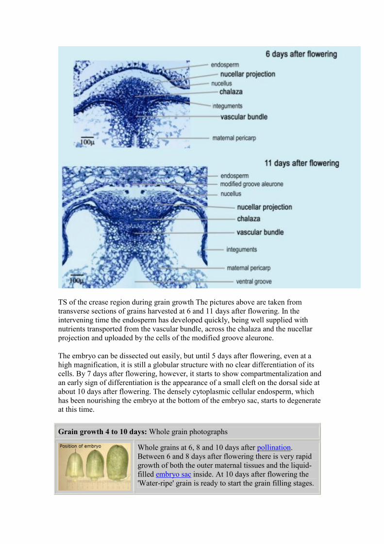
TS of the crease region during grain growth The pictures above are taken from
transverse sections of grains harvested at 6 and 11 days after flowering. In the
intervening time the endosperm has developed quickly, being well supplied with
nutrients transported from the vascular bundle, across the chalaza and the nucellar
projection and uploaded by the cells of the modified groove aleurone.
The embryo can be dissected out easily, but until 5 days after flowering, even at a
high magnification, it is still a globular structure with no clear differentiation of its
cells. By 7 days after flowering, however, it starts to show compartmentalization and
an early sign of differentiation is the appearance of a small cleft on the dorsal side at
about 10 days after flowering. The densely cytoplasmic cellular endosperm, which
has been nourishing the embryo at the bottom of the embryo sac, starts to degenerate
at this time.
Grain growth 4 to 10 days: Whole grain photographs
Whole grains at 6, 8 and 10 days after pollination.
Between 6 and 8 days after flowering there is very rapid
growth of both the outer maternal tissues and the liquid-
filled embryo sac inside. At 10 days after flowering the
'Water-ripe' grain is ready to start the grain filling stages.
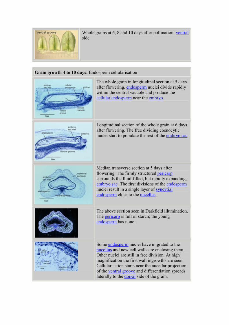
Whole grains at 6, 8 and 10 days after pollination: ventral
side.
Grain growth 4 to 10 days: Endosperm cellularisation
The whole grain in longitudinal section at 5 days
after flowering. endosperm nuclei divide rapidly
within the central vacuole and produce the
cellular endosperm near the embryo.
Longitudinal section of the whole grain at 6 days
after flowering. The free dividing coenocytic
nuclei start to populate the rest of the embryo sac.
Median transverse section at 5 days after
flowering. The firmly structured pericarp
surrounds the fluid-filled, but rapidly expanding,
embryo sac. The first divisions of the endosperm
nuclei result in a single layer of syncytial
endosperm close to the nucellus.
The above section seen in Darkfield illumination.
The pericarp is full of starch; the young
endosperm has none.
Some endosperm nuclei have migrated to the
nucellus and new cell walls are enclosing them.
Other nuclei are still in free division. At high
magnification the first wall ingrowths are seen.
Cellularisation starts near the nucellar projection
of the ventral groove and differentiation spreads
laterally to the dorsal side of the grain.
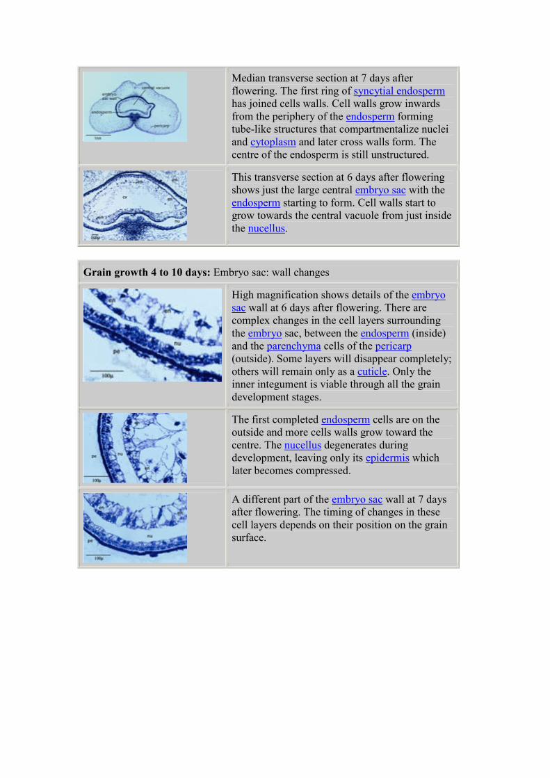
Median transverse section at 7 days after
flowering. The first ring of syncytial endosperm
has joined cells walls. Cell walls grow inwards
from the periphery of the endosperm forming
tube-like structures that compartmentalize nuclei
and cytoplasm and later cross walls form. The
centre of the endosperm is still unstructured.
This transverse section at 6 days after flowering
shows just the large central embryo sac with the
endosperm starting to form. Cell walls start to
grow towards the central vacuole from just inside
the nucellus.
Grain growth 4 to 10 days: Embryo sac: wall changes
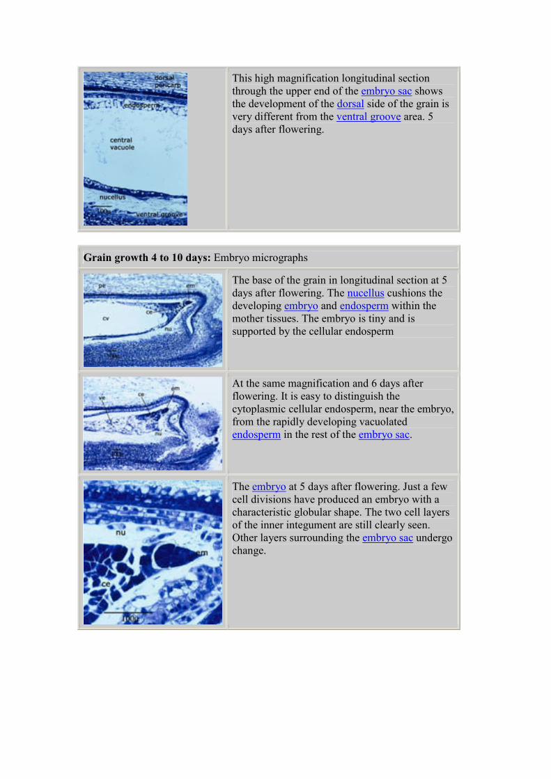
High magnification shows details of the embryo
sac wall at 6 days after flowering. There are
complex changes in the cell layers surrounding
the embryo sac, between the endosperm (inside)
and the parenchyma cells of the pericarp
(outside). Some layers will disappear completely;
others will remain only as a cuticle. Only the
inner integument is viable through all the grain
development stages.
The first completed endosperm cells are on the
outside and more cells walls grow toward the
centre. The nucellus degenerates during
development, leaving only its epidermis which
later becomes compressed.
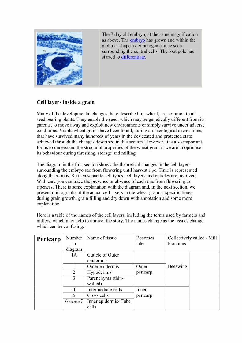
A different part of the embryo sac wall at 7 days
after flowering. The timing of changes in these
cell layers depends on their position on the grain
surface.
This high magnification longitudinal section
through the upper end of the embryo sac shows
the development of the dorsal side of the grain is
very different from the ventral groove area. 5
days after flowering.
Grain growth 4 to 10 days: Embryo micrographs
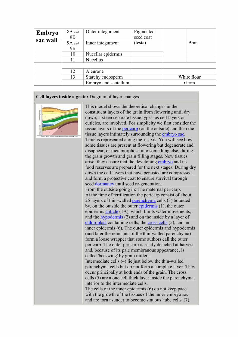
The base of the grain in longitudinal section at 5
days after flowering. The nucellus cushions the
developing embryo and endosperm within the
mother tissues. The embryo is tiny and is
supported by the cellular endosperm
At the same magnification and 6 days after
flowering. It is easy to distinguish the
cytoplasmic cellular endosperm, near the embryo,
from the rapidly developing vacuolated
endosperm in the rest of the embryo sac.
The embryo at 5 days after flowering. Just a few
cell divisions have produced an embryo with a
characteristic globular shape. The two cell layers
of the inner integument are still clearly seen.
Other layers surrounding the embryo sac undergo
change.
The 7 day old embryo, at the same magnification
as above. The embryo has grown and within the
globular shape a dermatogen can be seen
surrounding the central cells. The root pole has
started to differentiate.
Cell layers inside a grain
Many of the developmental changes, here described for wheat, are common to all
seed bearing plants. They enable the seed, which may be genetically different from its
parents, to move away and exploit new environments or simply survive under adverse
conditions. Viable wheat grains have been found, during archaeological excavations,
that have survived many hundreds of years in the desiccated and protected state
achieved through the changes described in this section. However, it is also important
for us to understand the structural properties of the wheat grain if we are to optimise
its behaviour during threshing, storage and milling.
The diagram in the first section shows the theoretical changes in the cell layers
surrounding the embryo sac from flowering until harvest ripe. Time is represented
along the x- axis. Sixteen separate cell types, cell layers and cuticles are involved.
With care you can trace the presence or absence of each one from flowering to
ripeness. There is some explanation with the diagram and, in the next section, we
present micrographs of the actual cell layers in the wheat grain at specific times
during grain growth, grain filling and dry down with annotation and some more
explanation.
Here is a table of the names of the cell layers, including the terms used by farmers and
millers, which may help to unravel the story. The names change as the tissues change,
which can be confusing.
Number
in
diagram
Name of tissue Becomes
later
Collectively called / Mill
Fractions
1A Cuticle of Outer
epidermis
1 Outer epidermis
2 Hypodermis
3 Parenchyma (thin-
walled)
Outer
pericarp
Beeswing
4 Intermediate cells
5 Cross cells
Pericarp
6 becomes7 Inner epidermis/ Tube
cells
Inner
pericarp
8A and
8B
Outer integument
9A and
9B
Inner integument
Pigmented
seed coat
(testa)
10 Nucellar epidermis
Embryo
sac wall
11 Nucellus
Bran
12 Aleurone
13 Starchy endosperm White flour
Embryo and scutellum Germ
Cell layers inside a grain: Diagram of layer changes
This model shows the theoretical changes in the
constituent layers of the grain from flowering until dry
down; sixteen separate tissue types, as cell layers or
cuticles, are involved. For simplicity we first consider the
tissue layers of the pericarp (on the outside) and then the
tissue layers intimately surrounding the embryo sac.
Time is represented along the x- axis. You will see how
some tissues are present at flowering but degenerate and
disappear, or metamorphose into something else, during
the grain growth and grain filling stages. New tissues
arise; they ensure that the developing embryo and its
food reserves are prepared for the next stages. During dry
down the cell layers that have persisted are compressed
and form a protective coat to ensure survival through
seed dormancy until seed re-generation.
From the outside going in: The maternal pericarp.
At the time of fertilization the pericarp consist of about
25 layers of thin-walled parenchyma cells (3) bounded
by, on the outside the outer epidermis (1), the outer
epidermis cuticle (1A), which limits water movements,
and the hypodermis (2) and on the inside by a layer of
chloroplast containing cells, the cross cells (5), and an
inner epidermis (6). The outer epidermis and hypodermis
(and later the remnants of the thin-walled parenchyma)
form a loose wrapper that some authors call the outer
pericarp. The outer pericarp is easily detached at harvest
and, because of its pale membranous appearance, is
called 'beeswing' by grain millers.
Intermediate cells (4) lie just below the thin-walled
parenchyma cells but do not form a complete layer. They
occur principally at both ends of the grain. The cross
cells (5) are a one cell thick layer inside the parenchyma,
interior to the intermediate cells.
The cells of the inner epidermis (6) do not keep pace
with the growth of the tissues of the inner embryo sac
and are torn asunder to become sinuous 'tube cells' (7),
appearing as an incomplete layer or randomly sited
individuals.
Going further in: The cell layers surrounding the embryo
sac.
At the time of fertilization the embryo sac is enclosed in
two integuments, outer (8) and inner (9) surrounding the
nucellus (11) . Each integument consists of two layers of
cells (A and B). About the time the zygote commences
division the outer integument begins to degenerate and a
cuticle develops on the outer surface of the inner
integument. Later the cell-contents of the outer
integument disappear entirely, the delicate walls shrivel
and finally become completely disorganized. The semi-
permeable membrane cuticle on the inner integument
(9B) persists until the grain reaches its maximum size.
The nucellar tissue (11) surrounding the developing
embryo is at first several cells thick but it, too, gradually
becomes disorganized and absorbed. It is widely assumed
that degeneration of the nucellar cells provides nutrients
as well as space for the developing endosperm and
embryo. The nucellar epidermis (10) continues as a
living tissue, its cells dividing and expanding as the ovule
grows until, later, its cell contents also become
disorganized, the remaining cytoplasm collects as a thin
layer across the middle of the cell and the radial walls are
absorbed. The single-celled tissue collapses during
ripening, upper and lower walls are crushed together, and
finally it becomes a delicate covering on the outer
surface of the aleurone cells (12).
Cell layers inside a grain: Cell layer micrographs
The embryo sac wall in ts at high magnification two days
after flowering.
The thin-walled parenchyma (3) , a protective and
supportive structure, is 15-20 cells thick at anthesis. The
cross cells (5), with their axis at right angles to the long
axis of the grain, show a large cross section. The tube
cells (inner epidermis) (7 was 6) have their long axis
parallel to the long axis of the grain so, in TS they show a
small cross section. The outer and inner integuments (8
and 9) are two cells thick. The nucellar epidermis (10) is
a single cell layer.
The nucellus (11) is still a bulky multi-layer tissue
cushioning the newly fertilized embryo and providing
nutrients for its first divisions and those of the coenocytic
endosperm.
About the time the embryo commences division the thin-
walled parenchyma (3) starts to degenerate. At the same
time the outer integument (8), which at first consists of
two cell layers, begins to degenerate; cells lose turgidity,
cytoplasm disappears and elongated nuclei become
thinner. The cross cells (5) persist. They have elongated
nuclei and vacuolated cytoplasm and contain chloroplasts
(4µ), which give the green colour to the young grain, and
minute starch grains 3µ by 1.5µ.The nucellus (11), which
is at first several cells thick, gradually becomes
disorganized and is absorbed. It disappears most rapidly
near the embryo and on the dorsal side of the grain.
TS at four days after flowering.
TS at six days after flowering.
The starch grains in the parenchyma (3) disappear
starting at the top of the grain. They remain longest
where they cover the embryo and in the ventral groove.
The cross cells (5) and tube cells (7) are the tissues that
give strength to the embryo sac at this time. The two
layers of the outer integument (8A and 8B) have
disappeared, as has the outer layer of the inner
integument (9A). The inner layer of the inner integument
(9B) persists and acquires resinous substances. The
nucellus remains only as the nucellar epidermis (10).
Internally, cell walls grow enclosing the nuclei of the
coenocytic endosperm. Completed cells form at the edge
as a result of periclinal divisions and become the
aleurone (12) layer. The aleurone (12) cells are first
distinguishable at about 6 days after flowering and are
later characterized by a large number of small vacuoles.
The rounded vacuoles become filled with reserves to
form aleurone grains, (a form of protein body) lipid
droplets form a ring around the aleurone grains and a
single layer of droplets line the plasmalemma.
The tube cells (7) are an incomplete layer that is
stretched and torn apart by the growth of the endosperm;
they are only a narrow band on the dorsal side, but
provide more complete coverage over the embryo and the
brush end of the grain.
The nucellar epidermis (10) continues as a living tissue
but its cell contents become disorganized, the remaining
cytoplasm collects as a thin layer across the middle of the
cell, and the radial walls are absorbed. The seed coat or
testa develops from the integuments (8 and 9). By six
days after flowering both the layers of the outer
integument (8A and B) had degenerated.
The cell contents of the outer layer of the inner
integument (9A) disappear at about day 11; this layer
develops a semi-permeable cuticle on the outside and its
cells collapse.
The cell cavities of the inner layer of the inner
integument (9B) are recognizable for some time and a
yellow or reddish-brown oily resinous substance forms
inside, visible through the translucent pericarp, giving the
grain its characteristic colour.
Transverse section at eleven days after flowering.
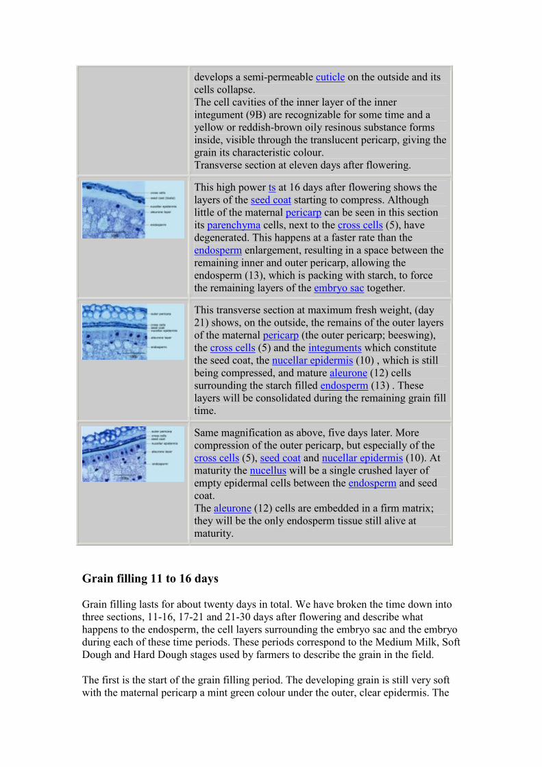
This high power ts at 16 days after flowering shows the
layers of the seed coat starting to compress. Although
little of the maternal pericarp can be seen in this section
its parenchyma cells, next to the cross cells (5), have
degenerated. This happens at a faster rate than the
endosperm enlargement, resulting in a space between the
remaining inner and outer pericarp, allowing the
endosperm (13), which is packing with starch, to force
the remaining layers of the embryo sac together.
This transverse section at maximum fresh weight, (day
21) shows, on the outside, the remains of the outer layers
of the maternal pericarp (the outer pericarp; beeswing),
the cross cells (5) and the integuments which constitute
the seed coat, the nucellar epidermis (10) , which is still
being compressed, and mature aleurone (12) cells
surrounding the starch filled endosperm (13) . These
layers will be consolidated during the remaining grain fill
time.
Same magnification as above, five days later. More
compression of the outer pericarp, but especially of the
cross cells (5), seed coat and nucellar epidermis (10). At
maturity the nucellus will be a single crushed layer of
empty epidermal cells between the endosperm and seed
coat.
The aleurone (12) cells are embedded in a firm matrix;
they will be the only endosperm tissue still alive at
maturity.
Grain filling 11 to 16 days
Grain filling lasts for about twenty days in total. We have broken the time down into
three sections, 11-16, 17-21 and 21-30 days after flowering and describe what
happens to the endosperm, the cell layers surrounding the embryo sac and the embryo
during each of these time periods. These periods correspond to the Medium Milk, Soft
Dough and Hard Dough stages used by farmers to describe the grain in the field.
The first is the start of the grain filling period. The developing grain is still very soft
with the maternal pericarp a mint green colour under the outer, clear epidermis. The
green colour comes from chloroplasts in the 'cross cell' layer. The embryo is easy to
dissect out. The grain is at the Medium Milk stage.
Sectioned grains from 11 and 16 days after flowering illustrate the dynamic changes
in microscopic structure that occur during the first stage of grain filling. The
meristematic cells of the endosperm continue to divide and compartments are starting
to form within the endosperm. The first large, Type 'A' starch grains are seen at about
16 days after flowering. Lipid and protein bodies are also seen at this time.
The cell layers surrounding the embryo sac continue to change their character, cell
walls become thickened and what will become the aleurone layer is recognisable for
the first time. Typical aleurone cells are first visible near the nucellar projection at 12
days after flowering. The aleurone cells close to the ventral groove, which acts as
transfer cells for the up-take of assimilates into the endosperm stop growing early and
develop special characteristics. In comparison the cells of the dorsal aleurone are still
dividing, enabling the grain to continue to expand. The aleurone cells that interface
with the embryo near the scutellum are also changing even though the embryo is still
physically separate.
The embryo is developing rapidly and has an elongated shape. The densely
cytoplasmic cellular endosperm, still present at eleven days after flowering, has been
completely consumed by the embryo by the sixteenth day. At 16 days after flowering
the scutellum is clearly defined and the embryo now uses the endosperm starch
reserves near the scutellum for its own development.
Grain filling 11 to 16 days: Whole grain photographs
An individual spikelet 16 days after pollination.
floret 1 has been cut away and its component
parts are separated. Developing grains are found
in floret one and floret two but only rarely in the
upper florets.
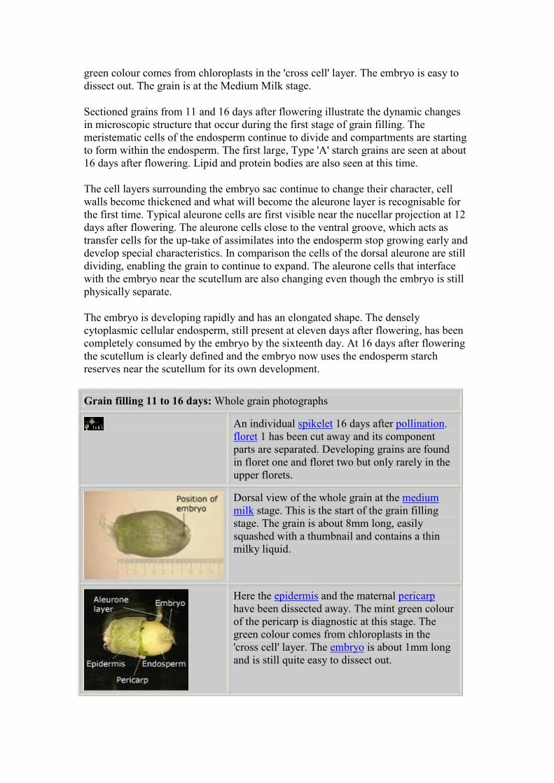
Dorsal view of the whole grain at the medium
milk stage. This is the start of the grain filling
stage. The grain is about 8mm long, easily
squashed with a thumbnail and contains a thin
milky liquid.
Here the epidermis and the maternal pericarp
have been dissected away. The mint green colour
of the pericarp is diagnostic at this stage. The
green colour comes from chloroplasts in the
'cross cell' layer. The embryo is about 1mm long
and is still quite easy to dissect out.
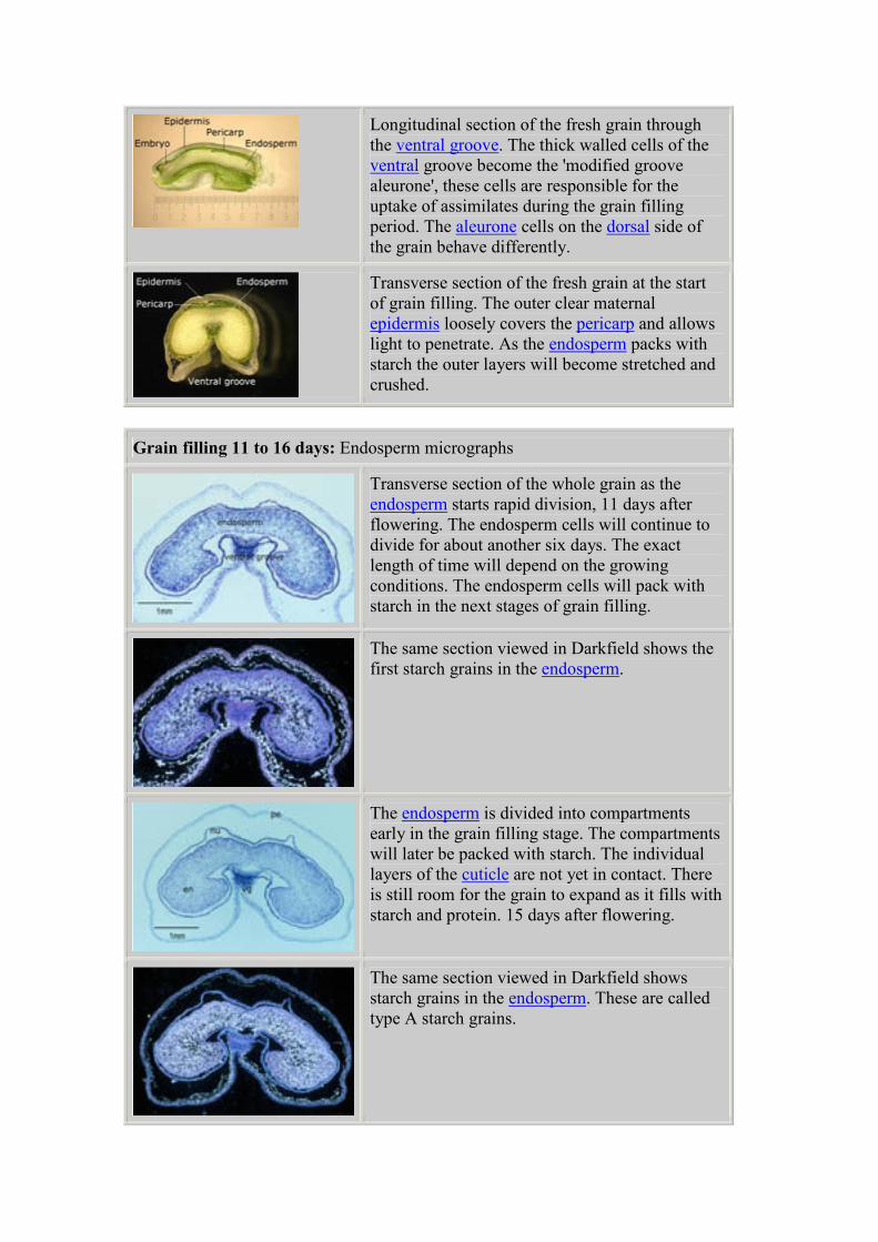
Longitudinal section of the fresh grain through
the ventral groove. The thick walled cells of the
ventral groove become the 'modified groove
aleurone', these cells are responsible for the
uptake of assimilates during the grain filling
period. The aleurone cells on the dorsal side of
the grain behave differently.
Transverse section of the fresh grain at the start
of grain filling. The outer clear maternal
epidermis loosely covers the pericarp and allows
light to penetrate. As the endosperm packs with
starch the outer layers will become stretched and
crushed.
Grain filling 11 to 16 days: Endosperm micrographs
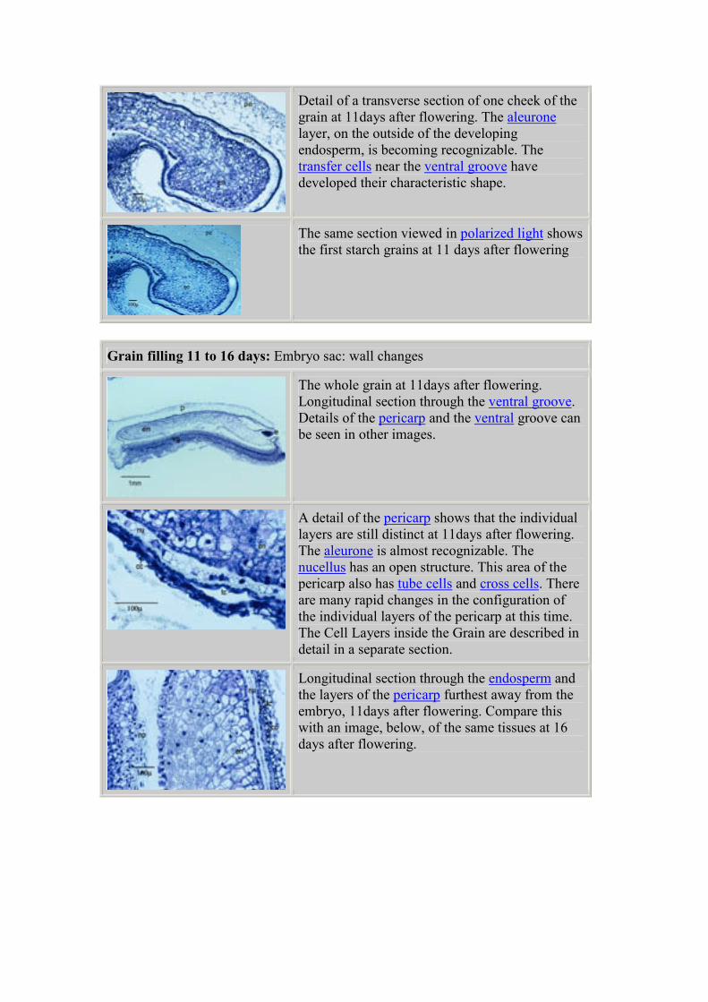
Transverse section of the whole grain as the
endosperm starts rapid division, 11 days after
flowering. The endosperm cells will continue to
divide for about another six days. The exact
length of time will depend on the growing
conditions. The endosperm cells will pack with
starch in the next stages of grain filling.
The same section viewed in Darkfield shows the
first starch grains in the endosperm.
The endosperm is divided into compartments
early in the grain filling stage. The compartments
will later be packed with starch. The individual
layers of the cuticle are not yet in contact. There
is still room for the grain to expand as it fills with
starch and protein. 15 days after flowering.
The same section viewed in Darkfield shows
starch grains in the endosperm. These are called
type A starch grains.
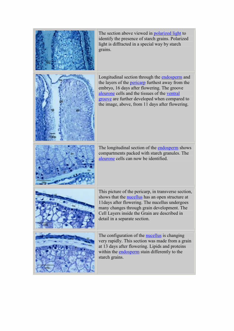
Detail of a transverse section of one cheek of the
grain at 11days after flowering. The aleurone
layer, on the outside of the developing
endosperm, is becoming recognizable. The
transfer cells near the ventral groove have
developed their characteristic shape.
The same section viewed in polarized light shows
the first starch grains at 11 days after flowering
Grain filling 11 to 16 days: Embryo sac: wall changes
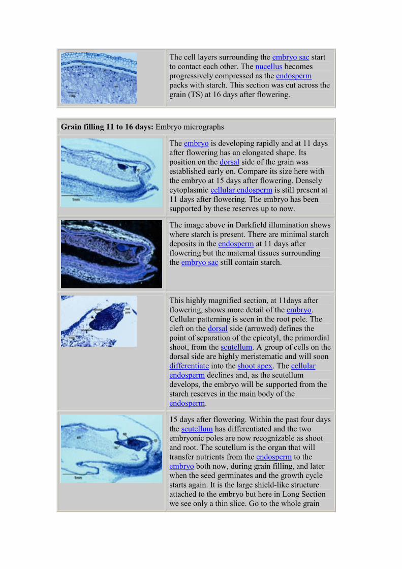
The whole grain at 11days after flowering.
Longitudinal section through the ventral groove.
Details of the pericarp and the ventral groove can
be seen in other images.
A detail of the pericarp shows that the individual
layers are still distinct at 11days after flowering.
The aleurone is almost recognizable. The
nucellus has an open structure. This area of the
pericarp also has tube cells and cross cells. There
are many rapid changes in the configuration of
the individual layers of the pericarp at this time.
The Cell Layers inside the Grain are described in
detail in a separate section.
Longitudinal section through the endosperm and
the layers of the pericarp furthest away from the
embryo, 11days after flowering. Compare this
with an image, below, of the same tissues at 16
days after flowering.
The section above viewed in polarized light to
identify the presence of starch grains. Polarized
light is diffracted in a special way by starch
grains.
Longitudinal section through the endosperm and
the layers of the pericarp furthest away from the
embryo, 16 days after flowering. The groove
aleurone cells and the tissues of the ventral
groove are further developed when compared to
the image, above, from 11 days after flowering.
The longitudinal section of the endosperm shows
compartments packed with starch granules. The
aleurone cells can now be identified.
This picture of the pericarp, in transverse section,
shows that the nucellus has an open structure at
11days after flowering. The nucellus undergoes
many changes through grain development. The
Cell Layers inside the Grain are described in
detail in a separate section.
The configuration of the nucellus is changing
very rapidly. This section was made from a grain
at 13 days after flowering. Lipids and proteins
within the endosperm stain differently to the
starch grains.
The cell layers surrounding the embryo sac start
to contact each other. The nucellus becomes
progressively compressed as the endosperm
packs with starch. This section was cut across the
grain (TS) at 16 days after flowering.
Grain filling 11 to 16 days: Embryo micrographs
The embryo is developing rapidly and at 11 days
after flowering has an elongated shape. Its
position on the dorsal side of the grain was
established early on. Compare its size here with
the embryo at 15 days after flowering. Densely
cytoplasmic cellular endosperm is still present at
11 days after flowering. The embryo has been
supported by these reserves up to now.
The image above in Darkfield illumination shows
where starch is present. There are minimal starch
deposits in the endosperm at 11 days after
flowering but the maternal tissues surrounding
the embryo sac still contain starch.
This highly magnified section, at 11days after
flowering, shows more detail of the embryo.
Cellular patterning is seen in the root pole. The
cleft on the dorsal side (arrowed) defines the
point of separation of the epicotyl, the primordial
shoot, from the scutellum. A group of cells on the
dorsal side are highly meristematic and will soon
differentiate into the shoot apex. The cellular
endosperm declines and, as the scutellum
develops, the embryo will be supported from the
starch reserves in the main body of the
endosperm.
15 days after flowering. Within the past four days
the scutellum has differentiated and the two
embryonic poles are now recognizable as shoot
and root. The scutellum is the organ that will
transfer nutrients from the endosperm to the
embryo both now, during grain filling, and later
when the seed germinates and the growth cycle
starts again. It is the large shield-like structure
attached to the embryo but here in Long Section
we see only a thin slice. Go to the whole grain
photographs and the micrographs in Grain Filling
2 and 3 to see where it fits between the embryo
and the endosperm. The densely cytoplasmic
cellular endosperm has been completely used by
15 days after flowering.
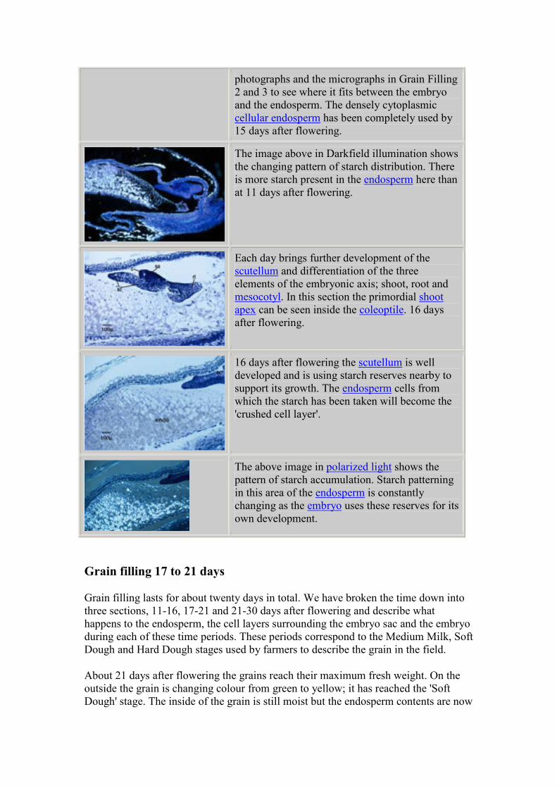
The image above in Darkfield illumination shows
the changing pattern of starch distribution. There
is more starch present in the endosperm here than
at 11 days after flowering.
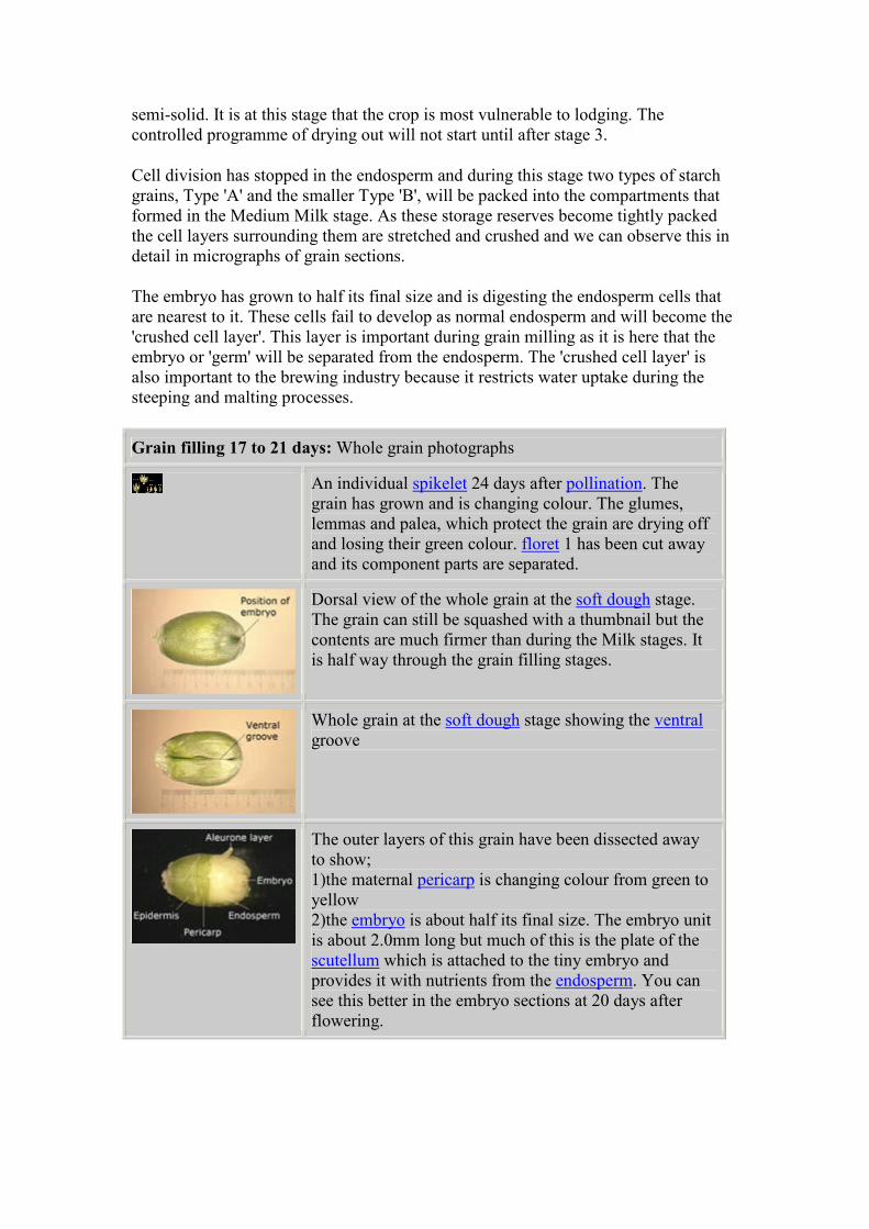
Each day brings further development of the
scutellum and differentiation of the three
elements of the embryonic axis; shoot, root and
mesocotyl. In this section the primordial shoot
apex can be seen inside the coleoptile. 16 days
after flowering.
16 days after flowering the scutellum is well
developed and is using starch reserves nearby to
support its growth. The endosperm cells from
which the starch has been taken will become the
'crushed cell layer'.
The above image in polarized light shows the
pattern of starch accumulation. Starch patterning
in this area of the endosperm is constantly
changing as the embryo uses these reserves for its
own development.
Grain filling 17 to 21 days
Grain filling lasts for about twenty days in total. We have broken the time down into
three sections, 11-16, 17-21 and 21-30 days after flowering and describe what
happens to the endosperm, the cell layers surrounding the embryo sac and the embryo
during each of these time periods. These periods correspond to the Medium Milk, Soft
Dough and Hard Dough stages used by farmers to describe the grain in the field.
About 21 days after flowering the grains reach their maximum fresh weight. On the
outside the grain is changing colour from green to yellow; it has reached the 'Soft
Dough' stage. The inside of the grain is still moist but the endosperm contents are now
semi-solid. It is at this stage that the crop is most vulnerable to lodging. The
controlled programme of drying out will not start until after stage 3.
Cell division has stopped in the endosperm and during this stage two types of starch
grains, Type 'A' and the smaller Type 'B', will be packed into the compartments that
formed in the Medium Milk stage. As these storage reserves become tightly packed
the cell layers surrounding them are stretched and crushed and we can observe this in
detail in micrographs of grain sections.
The embryo has grown to half its final size and is digesting the endosperm cells that
are nearest to it. These cells fail to develop as normal endosperm and will become the
'crushed cell layer'. This layer is important during grain milling as it is here that the
embryo or 'germ' will be separated from the endosperm. The 'crushed cell layer' is
also important to the brewing industry because it restricts water uptake during the
steeping and malting processes.
Grain filling 17 to 21 days: Whole grain photographs
An individual spikelet 24 days after pollination. The
grain has grown and is changing colour. The glumes,
lemmas and palea, which protect the grain are drying off
and losing their green colour. floret 1 has been cut away
and its component parts are separated.
Dorsal view of the whole grain at the soft dough stage.
The grain can still be squashed with a thumbnail but the
contents are much firmer than during the Milk stages. It
is half way through the grain filling stages.
Whole grain at the soft dough stage showing the ventral
groove
The outer layers of this grain have been dissected away
to show;
1)the maternal pericarp is changing colour from green to
yellow
2)the embryo is about half its final size. The embryo unit
is about 2.0mm long but much of this is the plate of the
scutellum which is attached to the tiny embryo and
provides it with nutrients from the endosperm. You can
see this better in the embryo sections at 20 days after
flowering.
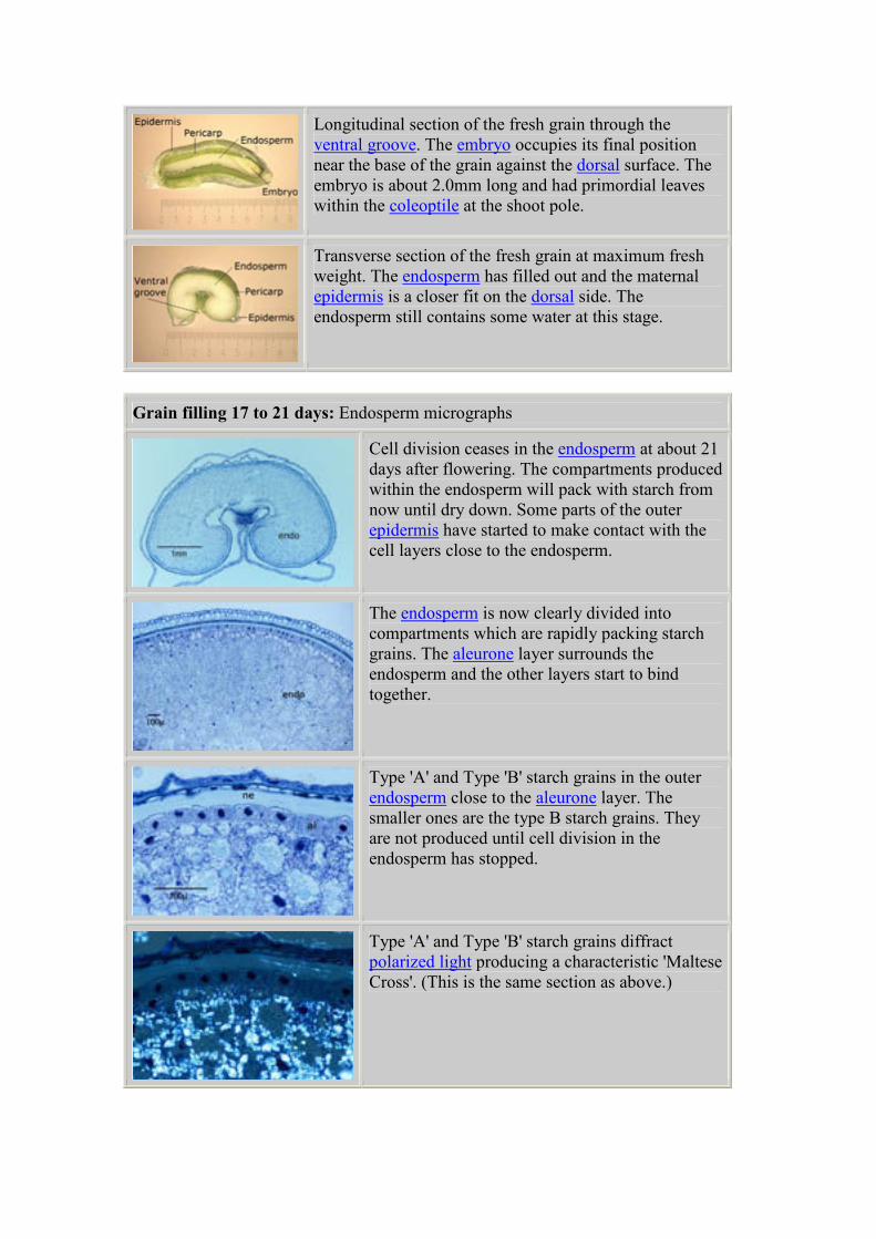
Longitudinal section of the fresh grain through the
ventral groove. The embryo occupies its final position
near the base of the grain against the dorsal surface. The
embryo is about 2.0mm long and had primordial leaves
within the coleoptile at the shoot pole.
Transverse section of the fresh grain at maximum fresh
weight. The endosperm has filled out and the maternal
epidermis is a closer fit on the dorsal side. The
endosperm still contains some water at this stage.
Grain filling 17 to 21 days: Endosperm micrographs
Cell division ceases in the endosperm at about 21
days after flowering. The compartments produced
within the endosperm will pack with starch from
now until dry down. Some parts of the outer
epidermis have started to make contact with the
cell layers close to the endosperm.
The endosperm is now clearly divided into
compartments which are rapidly packing starch
grains. The aleurone layer surrounds the
endosperm and the other layers start to bind
together.
Type 'A' and Type 'B' starch grains in the outer
endosperm close to the aleurone layer. The
smaller ones are the type B starch grains. They
are not produced until cell division in the
endosperm has stopped.
Type 'A' and Type 'B' starch grains diffract
polarized light producing a characteristic 'Maltese
Cross'. (This is the same section as above.)
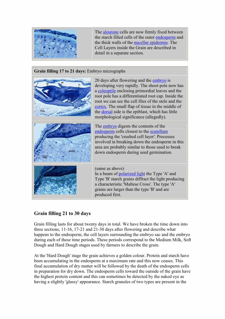
The aleurone cells are now firmly fixed between
the starch filled cells of the outer endosperm and
the thick walls of the nucellar epidermis. The
Cell Layers inside the Grain are described in
detail in a separate section.
Grain filling 17 to 21 days: Embryo micrographs
20 days after flowering and the embryo is
developing very rapidly. The shoot pole now has
a coleoptile enclosing primordial leaves and the
root pole has a differentiated root cap. Inside the
root we can see the cell files of the stele and the
cortex. The small flap of tissue in the middle of
the dorsal side is the epiblast, which has little
morphological significance (allegedly).
The embryo digests the contents of the
endosperm cells closest to the scutellum
producing the 'crushed cell layer'. Processes
involved in breaking down the endosperm in this
area are probably similar to those used to break
down endosperm during seed germination.
(same as above)
In a beam of polarized light the Type 'A' and
Type 'B' starch grains diffract the light producing
a characteristic 'Maltese Cross'. The type 'A'
grains are larger than the type 'B' and are
produced first.
Grain filling 21 to 30 days
Grain filling lasts for about twenty days in total. We have broken the time down into
three sections, 11-16, 17-21 and 21-30 days after flowering and describe what
happens to the endosperm, the cell layers surrounding the embryo sac and the embryo
during each of these time periods. These periods correspond to the Medium Milk, Soft
Dough and Hard Dough stages used by farmers to describe the grain.
At the 'Hard Dough' stage the grain achieves a golden colour. Protein and starch have
been accumulating in the endosperm at a maximum rate and this now ceases. This
final accumulation of dry matter will be followed by the death of the endosperm cells
in preparation for dry down. The endosperm cells toward the outside of the grain have
the highest protein content and this can sometimes be detected by the naked eye as
having a slightly 'glassy' appearance. Starch granules of two types are present in the
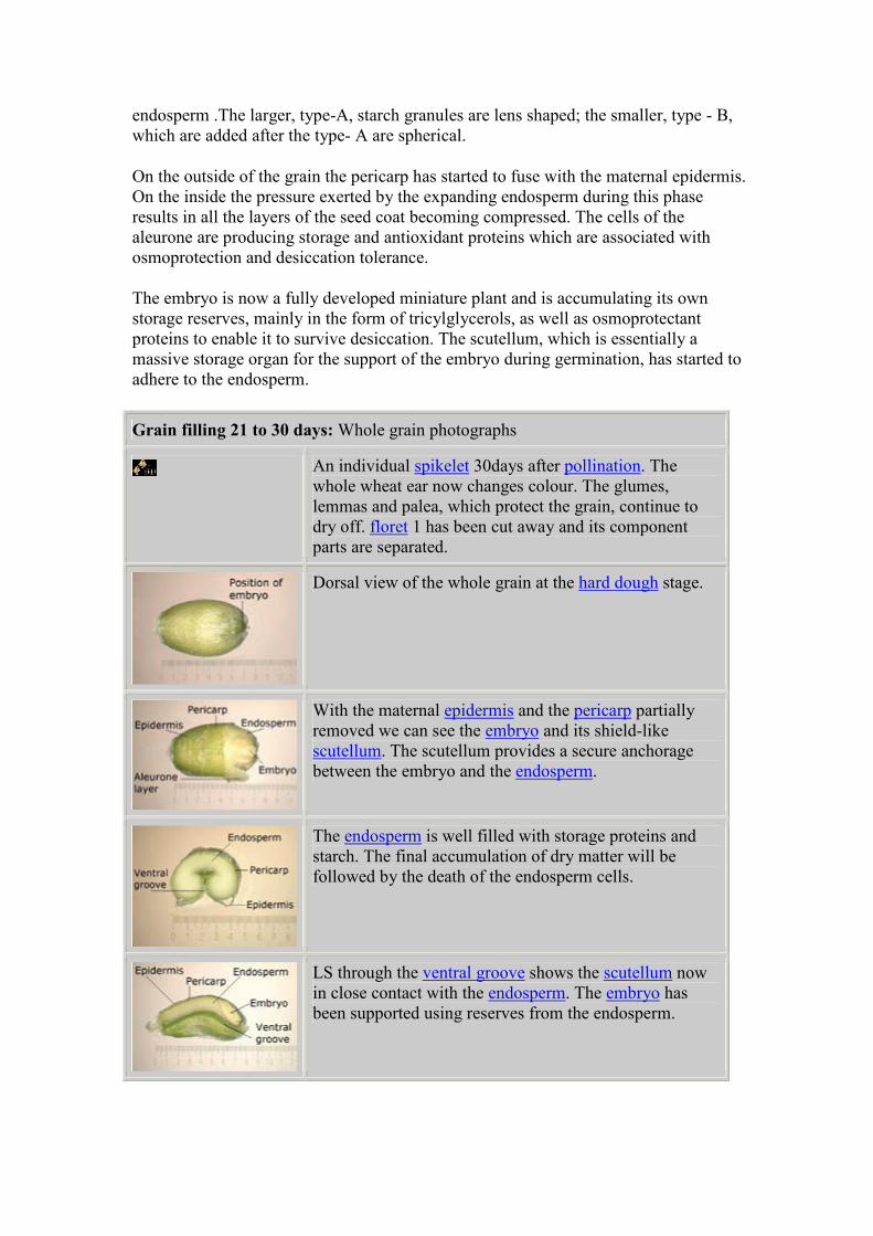
endosperm .The larger, type-A, starch granules are lens shaped; the smaller, type - B,
which are added after the type- A are spherical.
On the outside of the grain the pericarp has started to fuse with the maternal epidermis.
On the inside the pressure exerted by the expanding endosperm during this phase
results in all the layers of the seed coat becoming compressed. The cells of the
aleurone are producing storage and antioxidant proteins which are associated with
osmoprotection and desiccation tolerance.
The embryo is now a fully developed miniature plant and is accumulating its own
storage reserves, mainly in the form of tricylglycerols, as well as osmoprotectant
proteins to enable it to survive desiccation. The scutellum, which is essentially a
massive storage organ for the support of the embryo during germination, has started to
adhere to the endosperm.
Grain filling 21 to 30 days: Whole grain photographs
An individual spikelet 30days after pollination. The
whole wheat ear now changes colour. The glumes,
lemmas and palea, which protect the grain, continue to
dry off. floret 1 has been cut away and its component
parts are separated.
Dorsal view of the whole grain at the hard dough stage.
With the maternal epidermis and the pericarp partially
removed we can see the embryo and its shield-like
scutellum. The scutellum provides a secure anchorage
between the embryo and the endosperm.
The endosperm is well filled with storage proteins and
starch. The final accumulation of dry matter will be
followed by the death of the endosperm cells.
LS through the ventral groove shows the scutellum now
in close contact with the endosperm. The embryo has
been supported using reserves from the endosperm.
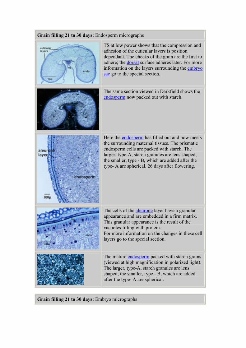
Grain filling 21 to 30 days: Endosperm micrographs
TS at low power shows that the compression and
adhesion of the cuticular layers is position
dependant. The cheeks of the grain are the first to
adhere; the dorsal surface adheres later. For more
information on the layers surrounding the embryo
sac go to the special section.
The same section viewed in Darkfield shows the
endosperm now packed out with starch.
Here the endosperm has filled out and now meets
the surrounding maternal tissues. The prismatic
endosperm cells are packed with starch. The
larger, type-A, starch granules are lens shaped;
the smaller, type - B, which are added after the
type- A are spherical. 26 days after flowering.
The cells of the aleurone layer have a granular
appearance and are embedded in a firm matrix.
This granular appearance is the result of the
vacuoles filling with protein.
For more information on the changes in these cell
layers go to the special section.
The mature endosperm packed with starch grains
(viewed at high magnification in polarized light).
The larger, type-A, starch granules are lens
shaped; the smaller, type - B, which are added
after the type- A are spherical.
Grain filling 21 to 30 days: Embryo micrographs
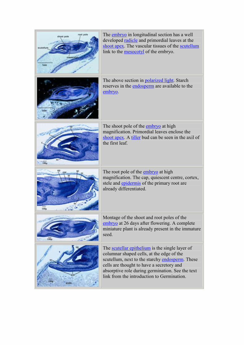
The embryo in longitudinal section has a well
developed radicle and primordial leaves at the
shoot apex. The vascular tissues of the scutellum
link to the mesocotyl of the embryo.
The above section in polarized light. Starch
reserves in the endosperm are available to the
embryo.
The shoot pole of the embryo at high
magnification. Primordial leaves enclose the
shoot apex. A tiller bud can be seen in the axil of
the first leaf.
The root pole of the embryo at high
magnification. The cap, quiescent centre, cortex,
stele and epidermis of the primary root are
already differentiated.
Montage of the shoot and root poles of the
embryo at 26 days after flowering. A complete
miniature plant is already present in the immature
seed.
The scutellar epithelium is the single layer of
columnar shaped cells, at the edge of the
scutellum, next to the starchy endosperm. These
cells are thought to have a secretory and
absorptive role during germination. See the text
link from the introduction to Germination.
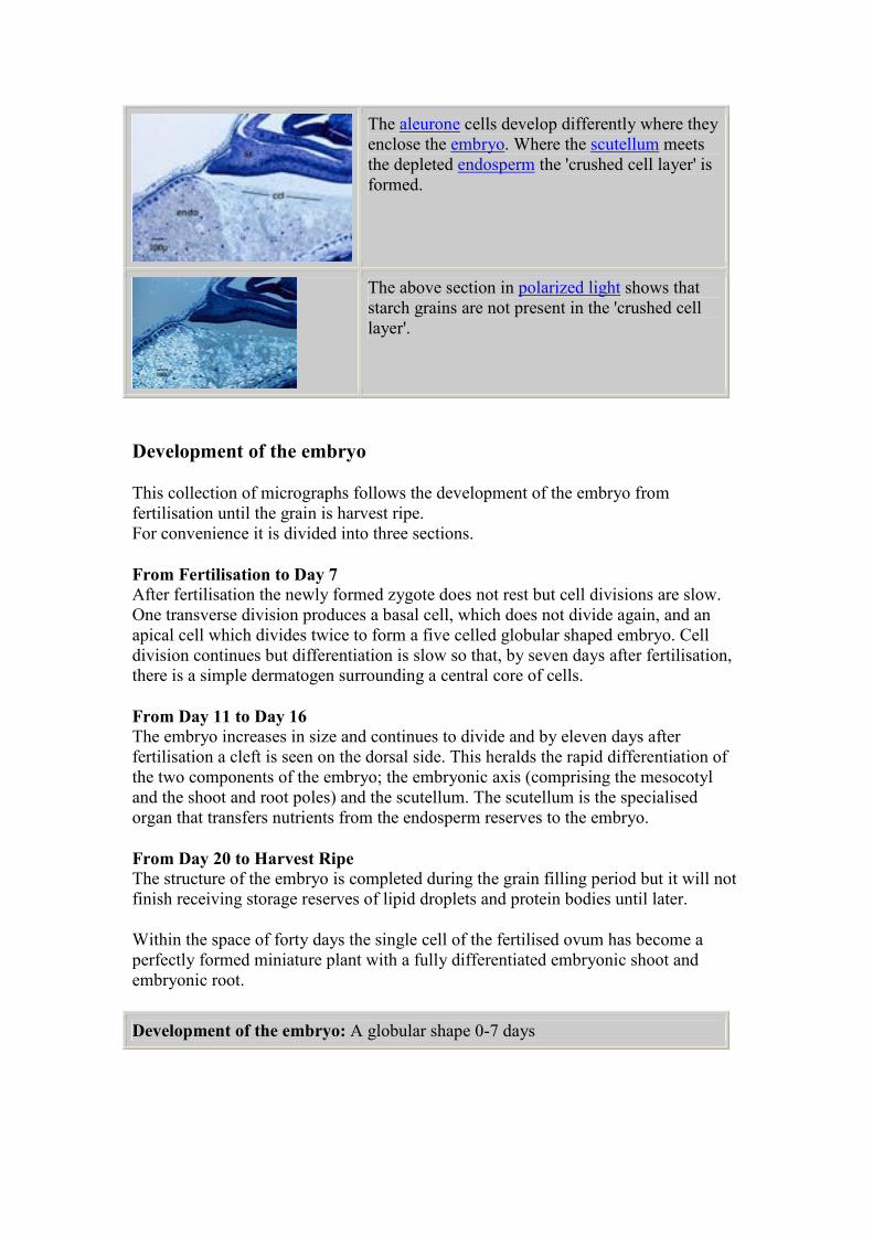
The aleurone cells develop differently where they
enclose the embryo. Where the scutellum meets
the depleted endosperm the 'crushed cell layer' is
formed.
The above section in polarized light shows that
starch grains are not present in the 'crushed cell
layer'.
Development of the embryo
This collection of micrographs follows the development of the embryo from
fertilisation until the grain is harvest ripe.
For convenience it is divided into three sections.
From Fertilisation to Day 7 After fertilisation the newly formed zygote does not rest but cell divisions are slow.
One transverse division produces a basal cell, which does not divide again, and an
apical cell which divides twice to form a five celled globular shaped embryo. Cell
division continues but differentiation is slow so that, by seven days after fertilisation,
there is a simple dermatogen surrounding a central core of cells.
From Day 11 to Day 16 The embryo increases in size and continues to divide and by eleven days after
fertilisation a cleft is seen on the dorsal side. This heralds the rapid differentiation of
the two components of the embryo; the embryonic axis (comprising the mesocotyl
and the shoot and root poles) and the scutellum. The scutellum is the specialised
organ that transfers nutrients from the endosperm reserves to the embryo.
From Day 20 to Harvest Ripe The structure of the embryo is completed during the grain filling period but it will not
finish receiving storage reserves of lipid droplets and protein bodies until later.
Within the space of forty days the single cell of the fertilised ovum has become a
perfectly formed miniature plant with a fully differentiated embryonic shoot and
embryonic root.
Development of the embryo: A globular shape 0-7 days
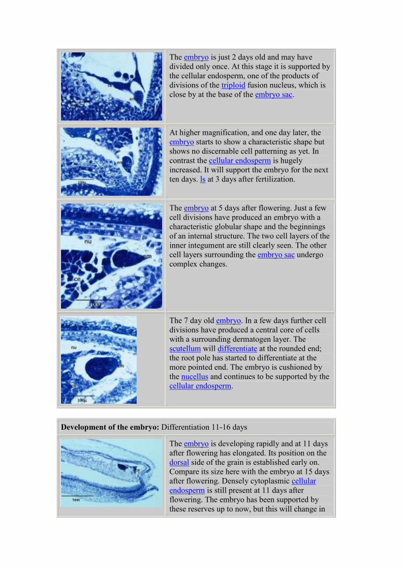
The embryo is just 2 days old and may have
divided only once. At this stage it is supported by
the cellular endosperm, one of the products of
divisions of the triploid fusion nucleus, which is
close by at the base of the embryo sac.
At higher magnification, and one day later, the
embryo starts to show a characteristic shape but
shows no discernable cell patterning as yet. In
contrast the cellular endosperm is hugely
increased. It will support the embryo for the next
ten days. ls at 3 days after fertilization.
The embryo at 5 days after flowering. Just a few
cell divisions have produced an embryo with a
characteristic globular shape and the beginnings
of an internal structure. The two cell layers of the
inner integument are still clearly seen. The other
cell layers surrounding the embryo sac undergo
complex changes.
The 7 day old embryo. In a few days further cell
divisions have produced a central core of cells
with a surrounding dermatogen layer. The
scutellum will differentiate at the rounded end;
the root pole has started to differentiate at the
more pointed end. The embryo is cushioned by
the nucellus and continues to be supported by the
cellular endosperm.
Development of the embryo: Differentiation 11-16 days
The embryo is developing rapidly and at 11 days
after flowering has elongated. Its position on the
dorsal side of the grain is established early on.
Compare its size here with the embryo at 15 days
after flowering. Densely cytoplasmic cellular
endosperm is still present at 11 days after
flowering. The embryo has been supported by
these reserves up to now, but this will change in
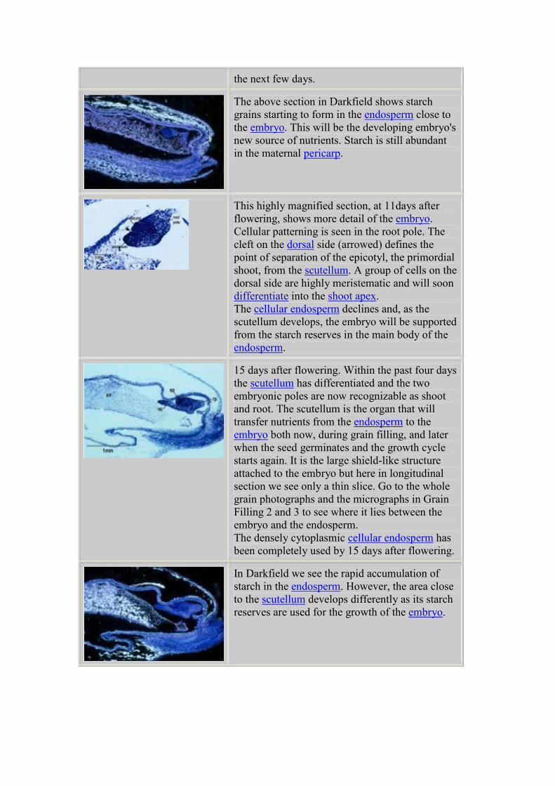
the next few days.
The above section in Darkfield shows starch
grains starting to form in the endosperm close to
the embryo. This will be the developing embryo's
new source of nutrients. Starch is still abundant
in the maternal pericarp.
This highly magnified section, at 11days after
flowering, shows more detail of the embryo.
Cellular patterning is seen in the root pole. The
cleft on the dorsal side (arrowed) defines the
point of separation of the epicotyl, the primordial
shoot, from the scutellum. A group of cells on the
dorsal side are highly meristematic and will soon
differentiate into the shoot apex.
The cellular endosperm declines and, as the
scutellum develops, the embryo will be supported
from the starch reserves in the main body of the
endosperm.
15 days after flowering. Within the past four days
the scutellum has differentiated and the two
embryonic poles are now recognizable as shoot
and root. The scutellum is the organ that will
transfer nutrients from the endosperm to the
embryo both now, during grain filling, and later
when the seed germinates and the growth cycle
starts again. It is the large shield-like structure
attached to the embryo but here in longitudinal
section we see only a thin slice. Go to the whole
grain photographs and the micrographs in Grain
Filling 2 and 3 to see where it lies between the
embryo and the endosperm.
The densely cytoplasmic cellular endosperm has
been completely used by 15 days after flowering.
In Darkfield we see the rapid accumulation of
starch in the endosperm. However, the area close
to the scutellum develops differently as its starch
reserves are used for the growth of the embryo.
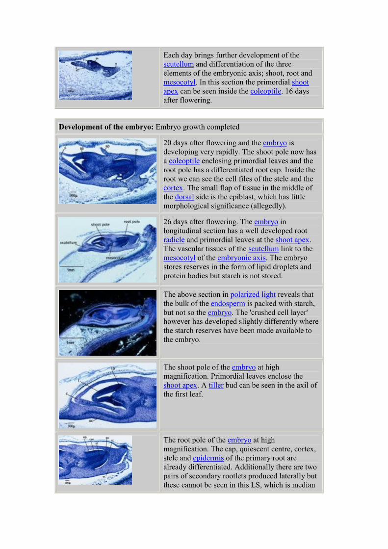
Each day brings further development of the
scutellum and differentiation of the three
elements of the embryonic axis; shoot, root and
mesocotyl. In this section the primordial shoot
apex can be seen inside the coleoptile. 16 days
after flowering.
Development of the embryo: Embryo growth completed
20 days after flowering and the embryo is
developing very rapidly. The shoot pole now has
a coleoptile enclosing primordial leaves and the
root pole has a differentiated root cap. Inside the
root we can see the cell files of the stele and the
cortex. The small flap of tissue in the middle of
the dorsal side is the epiblast, which has little
morphological significance (allegedly).
26 days after flowering. The embryo in
longitudinal section has a well developed root
radicle and primordial leaves at the shoot apex.
The vascular tissues of the scutellum link to the
mesocotyl of the embryonic axis. The embryo
stores reserves in the form of lipid droplets and
protein bodies but starch is not stored.
The above section in polarized light reveals that
the bulk of the endosperm is packed with starch,
but not so the embryo. The 'crushed cell layer'
however has developed slightly differently where
the starch reserves have been made available to
the embryo.
The shoot pole of the embryo at high
magnification. Primordial leaves enclose the
shoot apex. A tiller bud can be seen in the axil of
the first leaf.
The root pole of the embryo at high
magnification. The cap, quiescent centre, cortex,
stele and epidermis of the primary root are
already differentiated. Additionally there are two
pairs of secondary rootlets produced laterally but
these cannot be seen in this LS, which is median
through the ventral groove.
Montage of the shoot and root poles of the
embryo at 26 days after flowering. A complete
miniature plant is already present in the immature
seed.
Dry down 30 to 40 days
During this time the grain loses water at a constant rate. The embryo and aleurone
enter a state of dormancy around the time of grain maximum fresh weight. The stage
is known as Harvest Ripe.
The colour of the grain changes from golden to light brown but there is a wide degree
of variability in the timing of this process both between different grains in the ear and
different ears in the crop. In practise this stage of crop growth is closely monitored by
the farmer. The moisture content of the grain is carefully watched so that the crop is
harvested at the optimum time.
Often the harvested grain is air dried after combining to precisely control its moisture
content during storage. Without this controlled process of desiccation, the internal
biological processes of pre-germination and germination may start and the grain can
be ruined. Pre-harvest sprouting is a major agronomic problem. Weak dormancy
induction and a short period of after-ripening can have detrimental consequences for
malting quality. The understanding of these complex processes, at the genetic level, is
essential to prevent grain spoilage.
More information on dormancy induction, late embryogenesis-abundant (LEA)
proteins, the role of abscisic acid (ABA) is available here.
Dry down 30 to 40 days: Whole grain photographs
Ears are held vertically within the canopy during dry-
down. Individual spikelets dry at different rates; the last
to dry down will be at the base of the ear. When the crop
is completely ripe the will ears bend over to allow the
ripe seed to fall away.
An individual spikelet 40 days after pollination. floret 2
has been cut away and its component parts are separated.
The grain has started to shrink and the colour has
changed to a light brown. The glumes, lemmas and palea,
which have protected the grain, are now dry and brittle.
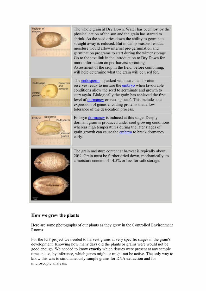
The whole grain at Dry Down. Water has been lost by the
physical action of the sun and the grain has started to
shrink. As the seed dries down the ability to germinate
straight away is reduced. But in damp seasons residual
moisture would allow internal pre-germination and
germination programs to start during the winter storage.
Go to the text link in the introduction to Dry Down for
more information on pre-harvest sprouting.
Assessment of the crop in the field, before combining,
will help determine what the grain will be used for.
The endosperm is packed with starch and protein
reserves ready to nurture the embryo when favourable
conditions allow the seed to germinate and growth to
start again. Biologically the grain has achieved the first
level of dormancy or 'resting state'. This includes the
expression of genes encoding proteins that allow
tolerance of the desiccation process.
Embryo dormancy is induced at this stage. Deeply
dormant grain is produced under cool growing conditions
whereas high temperatures during the later stages of
grain growth can cause the embryo to break dormancy
early.
The grain moisture content at harvest is typically about
20%. Grain must be further dried down, mechanically, to
a moisture content of 14.5% or less for safe storage.
How we grew the plants
Here are some photographs of our plants as they grew in the Controlled Environment
Rooms.
For the IGF project we needed to harvest grains at very specific stages in the grain's
development. Knowing how many days old the plants or grains were would not be
good enough. We needed to know exactly which tissues were present at any sample
time and so, by inference, which genes might or might not be active. The only way to
know this was to simultaneously sample grains for DNA extraction and for
microscopic analysis.
Plants in Controlled Environments
Our chosen variety was Mercia, a bread-making wheat with a hard endosperm. Mercia
is a winter wheat: sown in the field in the autumn and overwintered as a rosette plant
with several tillers. It has a cold requirement; in other words the plants must receive a
winter chill in order to grow and flower naturally the following spring and summer.
We needed to use uniform plants from a reproducible source. For these reasons field
grown plants would not be acceptable. They would not be uniform and we could only
achieve one harvest in a calendar year, so all our plants were grown in artificial, but
reproducible, conditions in large Controlled Environment Rooms. This meant we
could sow and harvest continuously through the year.
Firstly we had to satisfy the chilling requirement and then we had to provide the
correct artificial environment, (temperature, humidity, day length and light intensity)
for the plants to grow steadily through to flowering and then on through the grain
filling stages until harvest ripe. We had several attempts before we got it right! The
eventual success was in no small way due to the skill and experience of our support
staff, Bob Hughes, Richard Parkinson and Godfrey Hardbard to whom we are most
grateful.
The sophisticated 'walk-in' Controlled Environment Rooms allowed us independent
control of day and night temperature, day and night humidity, and both the intensity,
duration and quality of the light.
Details of the schedule are in the table below: the abbreviations are:
Temp. - temperature: degrees Celsius
RH - relative humidity: %
Ppd - photoperiod (daylength): hours
PAR - photosynthetically-active radiation: µmols m-2 s-1
Light was supplied by 56 metal halide lamps (250W), designed to give a spectrum
similar to 'natural daylight'. The lamps were lit daily, in sequence, to provide the
required PAR. The PAR was a minimum of 250 µmols m-2 s-1 as the crop was
established and a maximum of 850 µmols m-2 s-1 as the crop started to ripen.
Additionally, independently switched tungsten lights were provided for 30 minutes
longer than the metal halide lamps, at each end of the photoperiod, to simulate a dawn
and dusk, when the far red part of the natural light spectrum is higher.
If you compare these images with those of the plants grown outside in the field (see
section: Early Growth and Tillering) and you will spot some differences in plant form
as they experience an artificial winter and spring compared to the natural seasons. The
differences disappear once the plants come to full canopy and flowering.
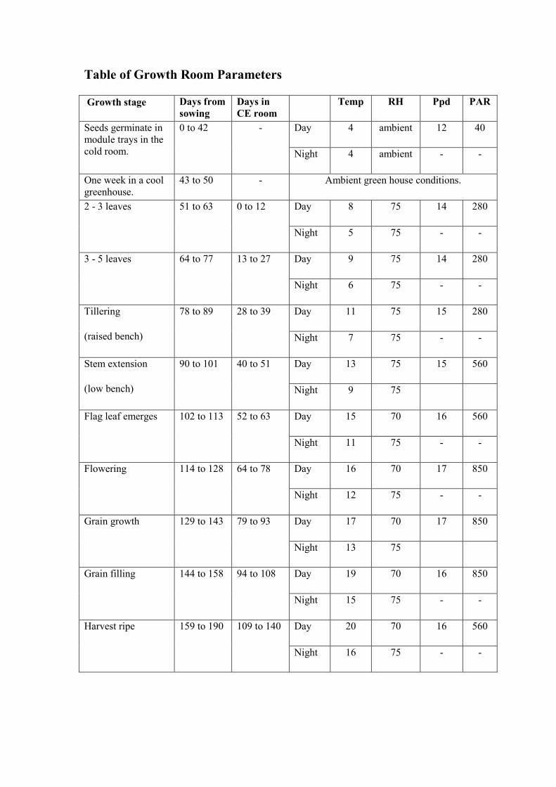
Table of Growth Room Parameters
Growth stage Days from
sowing Days in
CE room Temp RH Ppd PAR
Day 4 ambient 12 40 Seeds germinate in
module trays in the
cold room.
0 to 42 -
Night 4 ambient - -
One week in a cool
greenhouse. 43 to 50 - Ambient green house conditions.
Day 8 75 14 280 2 - 3 leaves 51 to 63 0 to 12
Night 5 75 - -
Day 9 75 14 280 3 - 5 leaves 64 to 77 13 to 27
Night 6 75 - -
Day 11 75 15 280 Tillering
(raised bench)
78 to 89 28 to 39
Night 7 75 - -
Day 13 75 15 560 Stem extension
(low bench)
90 to 101 40 to 51
Night 9 75
Day 15 70 16 560 Flag leaf emerges
102 to 113 52 to 63
Night 11 75 - -
Day 16 70 17 850 Flowering 114 to 128 64 to 78
Night 12 75 - -
Day 17 70 17 850 Grain growth 129 to 143 79 to 93
Night 13 75
Day 19 70 16 850 Grain filling 144 to 158 94 to 108
Night 15 75 - -
Day 20 70 16 560 Harvest ripe 159 to 190 109 to 140
Night 16 75 - -
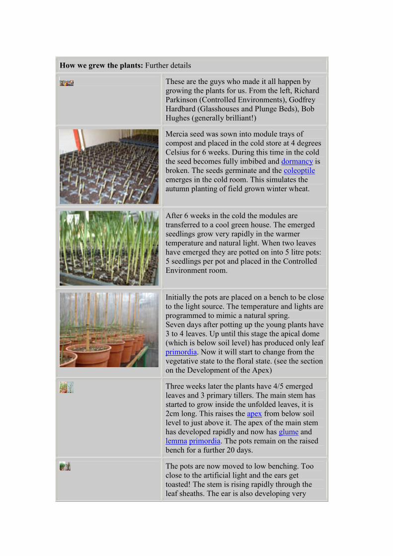
How we grew the plants: Further details
These are the guys who made it all happen by
growing the plants for us. From the left, Richard
Parkinson (Controlled Environments), Godfrey
Hardbard (Glasshouses and Plunge Beds), Bob
Hughes (generally brilliant!)
Mercia seed was sown into module trays of
compost and placed in the cold store at 4 degrees
Celsius for 6 weeks. During this time in the cold
the seed becomes fully imbibed and dormancy is
broken. The seeds germinate and the coleoptile
emerges in the cold room. This simulates the
autumn planting of field grown winter wheat.
After 6 weeks in the cold the modules are
transferred to a cool green house. The emerged
seedlings grow very rapidly in the warmer
temperature and natural light. When two leaves
have emerged they are potted on into 5 litre pots:
5 seedlings per pot and placed in the Controlled
Environment room.
Initially the pots are placed on a bench to be close
to the light source. The temperature and lights are
programmed to mimic a natural spring.
Seven days after potting up the young plants have
3 to 4 leaves. Up until this stage the apical dome
(which is below soil level) has produced only leaf
primordia. Now it will start to change from the
vegetative state to the floral state. (see the section
on the Development of the Apex)
Three weeks later the plants have 4/5 emerged
leaves and 3 primary tillers. The main stem has
started to grow inside the unfolded leaves, it is
2cm long. This raises the apex from below soil
level to just above it. The apex of the main stem
has developed rapidly and now has glume and
lemma primordia. The pots remain on the raised
bench for a further 20 days.
The pots are now moved to low benching. Too
close to the artificial light and the ears get
toasted! The stem is rising rapidly through the
leaf sheaths. The ear is also developing very
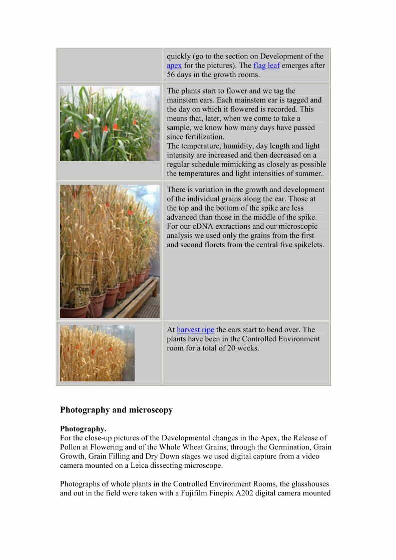
quickly (go to the section on Development of the
apex for the pictures). The flag leaf emerges after
56 days in the growth rooms.
The plants start to flower and we tag the
mainstem ears. Each mainstem ear is tagged and
the day on which it flowered is recorded. This
means that, later, when we come to take a
sample, we know how many days have passed
since fertilization.
The temperature, humidity, day length and light
intensity are increased and then decreased on a
regular schedule mimicking as closely as possible
the temperatures and light intensities of summer.
There is variation in the growth and development
of the individual grains along the ear. Those at
the top and the bottom of the spike are less
advanced than those in the middle of the spike.
For our cDNA extractions and our microscopic
analysis we used only the grains from the first
and second florets from the central five spikelets.
At harvest ripe the ears start to bend over. The
plants have been in the Controlled Environment
room for a total of 20 weeks.
Photography and microscopy
Photography. For the close-up pictures of the Developmental changes in the Apex, the Release of
Pollen at Flowering and of the Whole Wheat Grains, through the Germination, Grain
Growth, Grain Filling and Dry Down stages we used digital capture from a video
camera mounted on a Leica dissecting microscope.
Photographs of whole plants in the Controlled Environment Rooms, the glasshouses
and out in the field were taken with a Fujifilm Finepix A202 digital camera mounted
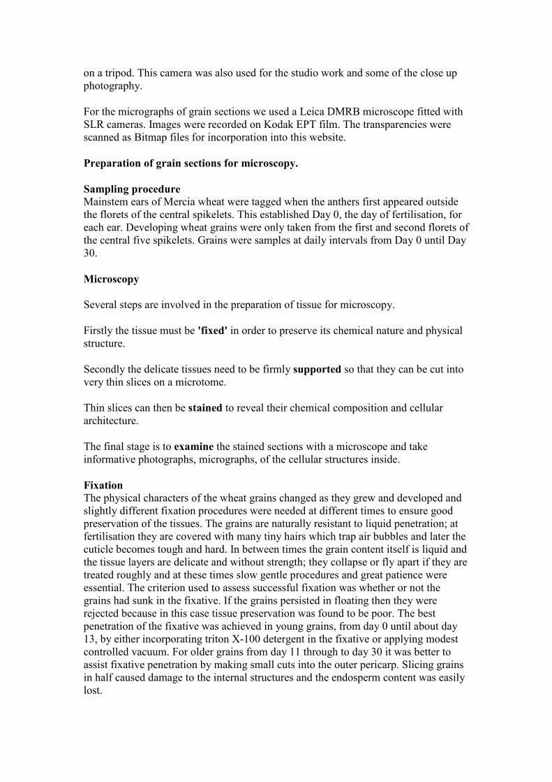
on a tripod. This camera was also used for the studio work and some of the close up
photography.
For the micrographs of grain sections we used a Leica DMRB microscope fitted with
SLR cameras. Images were recorded on Kodak EPT film. The transparencies were
scanned as Bitmap files for incorporation into this website.
Preparation of grain sections for microscopy.
Sampling procedure Mainstem ears of Mercia wheat were tagged when the anthers first appeared outside
the florets of the central spikelets. This established Day 0, the day of fertilisation, for
each ear. Developing wheat grains were only taken from the first and second florets of
the central five spikelets. Grains were samples at daily intervals from Day 0 until Day
30.
Microscopy
Several steps are involved in the preparation of tissue for microscopy.
Firstly the tissue must be 'fixed' in order to preserve its chemical nature and physical
structure.
Secondly the delicate tissues need to be firmly supported so that they can be cut into
very thin slices on a microtome.
Thin slices can then be stained to reveal their chemical composition and cellular
architecture.
The final stage is to examine the stained sections with a microscope and take
informative photographs, micrographs, of the cellular structures inside.
Fixation The physical characters of the wheat grains changed as they grew and developed and
slightly different fixation procedures were needed at different times to ensure good
preservation of the tissues. The grains are naturally resistant to liquid penetration; at
fertilisation they are covered with many tiny hairs which trap air bubbles and later the
cuticle becomes tough and hard. In between times the grain content itself is liquid and
the tissue layers are delicate and without strength; they collapse or fly apart if they are
treated roughly and at these times slow gentle procedures and great patience were
essential. The criterion used to assess successful fixation was whether or not the
grains had sunk in the fixative. If the grains persisted in floating then they were
rejected because in this case tissue preservation was found to be poor. The best
penetration of the fixative was achieved in young grains, from day 0 until about day
13, by either incorporating triton X-100 detergent in the fixative or applying modest
controlled vacuum. For older grains from day 11 through to day 30 it was better to
assist fixative penetration by making small cuts into the outer pericarp. Slicing grains
in half caused damage to the internal structures and the endosperm content was easily
lost.
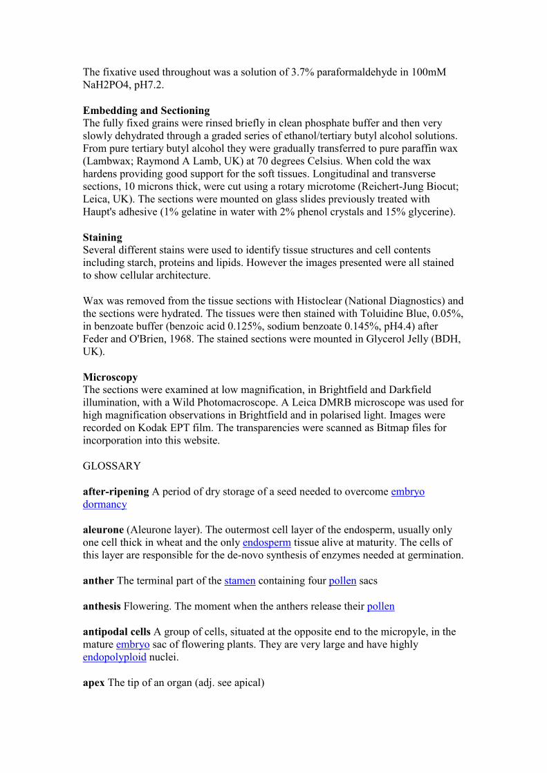
The fixative used throughout was a solution of 3.7% paraformaldehyde in 100mM
NaH2PO4, pH7.2.
Embedding and Sectioning The fully fixed grains were rinsed briefly in clean phosphate buffer and then very
slowly dehydrated through a graded series of ethanol/tertiary butyl alcohol solutions.
From pure tertiary butyl alcohol they were gradually transferred to pure paraffin wax
(Lambwax; Raymond A Lamb, UK) at 70 degrees Celsius. When cold the wax
hardens providing good support for the soft tissues. Longitudinal and transverse
sections, 10 microns thick, were cut using a rotary microtome (Reichert-Jung Biocut;
Leica, UK). The sections were mounted on glass slides previously treated with
Haupt's adhesive (1% gelatine in water with 2% phenol crystals and 15% glycerine).
Staining Several different stains were used to identify tissue structures and cell contents
including starch, proteins and lipids. However the images presented were all stained
to show cellular architecture.
Wax was removed from the tissue sections with Histoclear (National Diagnostics) and
the sections were hydrated. The tissues were then stained with Toluidine Blue, 0.05%,
in benzoate buffer (benzoic acid 0.125%, sodium benzoate 0.145%, pH4.4) after
Feder and O'Brien, 1968. The stained sections were mounted in Glycerol Jelly (BDH,
UK).
Microscopy The sections were examined at low magnification, in Brightfield and Darkfield
illumination, with a Wild Photomacroscope. A Leica DMRB microscope was used for
high magnification observations in Brightfield and in polarised light. Images were
recorded on Kodak EPT film. The transparencies were scanned as Bitmap files for
incorporation into this website.
GLOSSARY
after-ripening A period of dry storage of a seed needed to overcome embryo
dormancy
aleurone (Aleurone layer). The outermost cell layer of the endosperm, usually only
one cell thick in wheat and the only endosperm tissue alive at maturity. The cells of
this layer are responsible for the de-novo synthesis of enzymes needed at germination.
anther The terminal part of the stamen containing four pollen sacs
anthesis Flowering. The moment when the anthers release their pollen
antipodal cells A group of cells, situated at the opposite end to the micropyle, in the
mature embryo sac of flowering plants. They are very large and have highly
endopolyploid nuclei.
apex The tip of an organ (adj. see apical)
apical meristem Growing point. A zone of cell division at the tip of the stem or the
root. Behind the meristem the cells continue to divide and differentiation begins.
awn A projection from the tip of the lemma Less prominent in wheat than in barley.
brush A collection of hairs at the distal end of the wheat grain.
carpel The female part of the flower. A carpel consists of three parts: 1) the ovary, a
swollen basal portion containing the ovule, which becomes the seed after fertilisation,
2) styles, extensions of the ovary and 3) stigmas, specialised filaments on which the
pollen falls and germinates.
caryopsis The fruit, of grasses, in which the ripened ovary wall, the pericarp, is fused
to the seed coats, the testa, at maturity.
cellular endosperm The endosperm formed at the base of the embryo sac from
divisions of the fused polar nucleus. This region of endosperm nurtures the embryo
during its first divisions.
chalaza The basal region of the ovule where the stalk is attached.
chloroplast A chlorophyll-containing organelle (plastid) that gives the green colour to
leaves and stems. Plastids harvest light energy that is used to fix carbon dioxide in the
process called photosynthesis.
chromosomes Thread-shaped structures that occur in pairs in the nucleus of a cell.
Chromosomes contain the genetic information of living organisms.
coenocytic endosperm A multinucleate condition in which nuclei divide within a
common cytoplasm without being separated from each other by a plasmalemma or
cell wall.
coleoptile A protective cylindrical sheath-like structure that surrounds the shoot apex
in cereal and grass embryos.
coleorhiza The protective layers of tissue surrounding the primordial root (radicle)
within the grain.
cortex In the mature wheat embryo, the cylinder of parenchyma cells that surround
the central vascular core in the shoot and root.
crease The longitudinal furrow on the ventral side of the wheat grain.
cross cells These cells are only found in grasses. They are formed from cells of the
pericarp that elongate transversely, lose their chlorophyll and become lignified. They
lie between the tube cells and the parenchyma of the pericarp.
cuticle An extra-cellular material secreted by plant cells that restricts water movement.
It is composed of a fatty substance called cutin.
cytoplasm The living contents of a cell containing different types of organelles and
bounded by a plasma membrane.
differentiate Change from a simple to a more complex structure or function.
Developing tissues and organs become specialized for particular functions. n.
differentiation
diploid A condition in which the chromosomes in the nucleus of a cell exist as pairs,
one set being derived from the female parent and the other from the male. In cereals
such as barley and rice all the cells except the reproductive cells are diploid. When
gametes are formed the diploid number (2n) is reduced by half to the haploid number
(n) by meiosis When gametes fuse at fertilization the diploid condition is restored. In
modern wheat the situation is more complex because it contains three genomes
derived from different ancient diploid ancestors.
dormancy A resting period in the life of a plant during which growth slows or
appears to stop. In cereals this is between the start of seed dry-down and maturity. A
period of 'after-ripening' is often required before dormancy can be successfully broken.
dorsal On or near the back. The side of the organ away from the central axis.
double fertilisation The unique process, in flowering plants, when two male nuclei,
from pollen, fuse separately with different female nuclei. The first male nucleus fuses
with the egg cell to form the zygote that develops into the embryo The second male
nucleus fuses with the two polar nuclei (the second polar event) to form a triploid
nucleus that then develops into the endosperm
double ridge This is the term used to describe the transition from the 'vegetative' to
the 'floral' state in the shoot apex of wheat and barley. Growth of the leaf primordium
(ridge) is suppressed and the floral bud primordium (ridge) begins to grow out, giving
a 'double ridge' appearance to the dissected shoot apex when view under the
microscope. At this time the plant apex no longer produces leaves but is 'florally
initiated' and only flower parts will subsequently develop.
dpa 'Days Post Anthesis' or the number of days after flowering and fertilisation
early milk A description the young grain. When punctured the grain contents are
starting to look milky.
embryo The rudimentary plant present in the seed; composed of the embryonic axis
(shoot end and root end) and the scutellum
embryo sac A large cell that develops in the ovule of flowering plants. It contains a
large number of nuclei derived by division of the megaspore nucleus. At the
micropyle end is the egg nucleus and two synergids; at the opposite end are nuclei
that will become the antipodals and in the centre are two polar nuclei which fuse to
become the primary endosperm nucleus.
embryonic axis The major part of the embryo, consisting of the shoot, mesocotyl and
the root.
endopolyploid when the DNA in the nucleus of a cell is replicated again and again,
without any cell division taking place. The DNA C value goes from 4 to 8 to 16 to 32
etc and the chromosome number (n) goes from 2 to 4 to 8 to 16 etc.
endosperm A nutritive tissue, characteristic of flowering plants, which nourishes the
embryo In most plants it is a transient tissue absorbed by the embryo before maturity
whereas in cereals and grasses it contains storage reserves in the mature grain and is
not absorbed until after germination. (see also coenocytic, cytoplasmic and syncytial
endosperm).
epiblast A small flap of tissue on the side of the grass embryo axis opposite the
scutellum
epicotyl The upper portion of the embryonic axis, above the cotyledons (in cereals:
the scutellar node) and below the first true leaves.
epidermis The outermost protective cell layer of tissue, usually covered by a cuticle
fertilisation The union of male and female gametes (reproductive cells) during the
process of sexual reproduction.
filament The stalk of a stamen
flag leaf The uppermost leaf on the stem.
floret The flower (including the lemma and palea) of cereals and grasses.
funiculus The short stalk that joins the ovule to the carpel wall.
gamete A reproductive cell that fuses with another gamete to form a zygote Gametes
are haploid, (they contain half the normal (diploid) number of chromosomes); thus
when two fuse, the diploid number is restored. Gametes are formed by meiosis
germ The term used by millers to denote the embryo of cereals.
glume A membranous bract at the base of a cereal or grass spike. Chaff.
haploid Describes a nucleus or cell with a single set of unpaired chromosomes The
haploid number is designated as n. Reproductive cells, formed as a result of meiosis,
are haploid. Fusion of two such cells restores the normal (diploid) number.
hard dough A description the grain at maximum dry weight. The grain is hard to
puncture and has started to dry off.
harvest ripe The grain has dried off and is ready to be harvested.
hypodermis The cell layer beneath the epidermis of the pericarp
imbibe Absorb water: re-hydrate. n.imbibition
integuments Two distinct tissue layers that surround the nucellus of the ovule When
mature they become the testa or seed coat
intercalary meristem A meristem found between already differentiated tissues.
Occurs in the internodes of grasses (just above leaf nodes) and enables longitudinal
growth of the stem.
internode The part of a plant stem between two nodes.
lemma The lower of two bracts that protect the floret in grasses.
lodging Term used when the crop canopy becomes flattened by heavy rains. Often
triggered by severe weather at the soft dough stage. The problem is a combination of
heavy heads and weak stems. Modern breeding increases stem strength.
lodicules Two small structures below the ovary which, at flowering, swell up and
force open the enclosing bracts, exposing the stamens and carpel
ls Longitudinal Section. The tissue sections were cut along the long axis of the grain.
The central sections go lengthwise through the embryo and along the length of the
ventral groove
medium milk A description the half-grown grain. When punctured the grain contents
are milky.
meiosis The process of cell division that results in the formation of haploid cells from
diploid cells to produce gametes.
meristem The region of active cell division in plants, from which permanent tissue is
derived. adj. meristematic
mesocotyl The region where vascular tissues of the root join the epicotyl at the
scutellar node
micropyle The small opening in a plant ovule through which the pollen tube passes in
order to effect fertilisation
node The part of a plant stem from which one or more leaves arise. The nodes are
separated on the stem by the internodes.
nucellar epidermis The outer cellular layer of the nucellus
nucellar pillar Also called the nucellar projection. A shaft of tissue that runs along
the top of the crease on the inner side of the pigment strand
nucellus The central portion of the ovule, containing the embryo sac, and surrounded
by integuments
osmoprotection A survival strategy brought about by the synthesis of groups of
proteins (termed dehydrins) in response to water stress. Most tissues produce these
proteins which protect cellular organelles and membranes from the damaging effects
of water loss (desiccation). A notable exception being the cells of the starchy
endosperm which undergo programmed cell death.
ovule The part of the plant that, after fertilisation, develops into the seed. It is
composed of the embryo sac, the nucellus, the integuments and the funiculus
ovum Unfertilised egg-cell containing a haploid nucleus.
palea The upper of two bracts enclosing the flower in cereals and grasses.
parenchyma A primary tissue with polyhedral cells and thin cell walls.
peduncle The final internode of the plant stem, that bears the flower.
pericarp The ovary wall; later the fruit/seed/grain wall.
pigment strand A cylinder of cells that lies between the vascular tissues and the
nucellar projection in the crease region.
plasmalemma The membrane that surrounds a protoplast.
polar nuclei Two haploid nuclei in the embryo sac. These nuclei fuse with a pollen
nucleus to form a triploid endosperm nucleus, which subsequently divides to form the
endosperm.
polarized light A technique used in light microscopy. A beam of polarized light is
passed through thin slices of preserved tissue. The light is diffracted by some parts of
the plant tissue and not others. Starch grains diffract the light in a very characteristic
manner producing an image of the Maltese Cross. This image is diagnostic of the
presence of starch grains.
pollen The male gametes, which are produced in the stamens of flowering seed plants.
pollination The transfer of pollen from an anther (the male reproductive organ) to a
stigma (the receptive part of the female reproductive organ).
primordia A group of cells that represents the initial stages in development of a plant
organ. s primordium
protein body A membrane-bound granule of storage proteins.
rachilla The axis in the centre of a spikelet The secondary axis of the flower spike.
rachis The primary axis of the wheat ear, it bears the spikelets.
radicle The part of a plant embryo that develops into the primary root.
scutellar epithelium A continuous layer of cylindrical cells covering the endosperm
side of the scutellum Thought to act as a secretory and absorptive organ.
scutellum A shield-like structure which represents the monocotyledon of the grass
embryo It is pressed against the endosperm and functions as an absorptive organ.
second polar event The fusion of a male gamete with two haploid female nuclei at
the time of the fertilisation of the ovule The resulting triploid nucleus divides to form
the endosperm
seed coat See integuments
shoot apex See apical meristem
soft dough A description the grain at maximum fresh weight. When punctured the
grain contents are still wet but starting to become floury.
spikelet A sub-unit of the ear consisting of several florets on a thin axis, the rachilla
stamen The structure in a flower that produces pollen grains, consisting of a stalk
(filament) and a terminal lobed head (anther).
stigma The glandular sticky surface at the tip of a carpel of a flower, which receives
the pollen
syncytial endosperm A mass of cytoplasm with several nuclei, not divided into
separate cells.
terminal spikelet This marks the completion of the spikelet initiation phase in wheat.
The terminal spikelet has its plane of symmetry at right angles to the other spikelets.
testa The outer coat of the seed, formed by the hardened integuments
tiller A lateral shoot that develops from the axillary bud of leaves at the base of a
cereal stem.
transfer cells Specialized cells with intricate cell walls and large membrane surface
thought to function in the transport of materials.
triploid Describing a nucleus or cell that has three times (3n) the haploid number (n)
of chromosomes
triploid fusion nucleus The result of the fusion of one male gamete (pollen) with two
haploid female nuclei (polar cells) in the 'second polar event'. This nucleus divides to
form the cells of the endosperm
ts Transverse Section. The tissue sections were cut across the grain. The images
presented are of sections from the middle of the grain (median).
tube cells Cells formed from the inner epidermis of the pericarp in which the cells
elongate parallel to the grain's long axis.

vascular tissue A tissue system which transports water and nutrients (called xylem)
and photosynthates (called phloem) around the plant.
ventral The side of the grain towards the central axis.
ventral groove Also called the crease The furrow on the ventral side of the grain. The
tissues of the ventral groove are adapted for efficient transfer of nutrients.
water ripe (also called Pre-milk). A description the newly pollinated grain. When
punctured the grain contents are very watery.
zygote The cell resulting from the fusion of male and female gametes during sexual
reproduction. Subsequent cell divisions and differentiation result in the production of
the embryo
http://www.wheatbp.net/cgi-bin/grain2.pl?topic=Home