What’s in that wound bed? Slough, Eschar, or Biofilm?€¦ · 3/3/2017 1 ©2017 National Pressure...
14
3/3/2017 1 ©2017 National Pressure Ulcer Advisory Panel | www.npuap.org What’s in that wound bed? Slough, Eschar, or Biofilm? Linda J. Cowan, PhD, ARNP, FNP-BC, CWS Disclosures • Employed as a Research Health Scientist, North Florida/South Georgia Veterans Health System, Gainesville, FL. • Research funding received from: – VA – Biomonde – Healthpoint – Smith & Nephew – Hollister – Medline • This material is the result of work supported with resources and the use of facilities at the VA.
-
Upload
hoangkhanh -
Category
Documents
-
view
216 -
download
0
Transcript of What’s in that wound bed? Slough, Eschar, or Biofilm?€¦ · 3/3/2017 1 ©2017 National Pressure...

3/3/2017
1
©2017 National Pressure Ulcer Advisory Panel | www.npuap.org
What’s in that wound bed? Slough, Eschar, or Biofilm?
Linda J. Cowan, PhD, ARNP, FNP-BC, CWS
Disclosures
• Employed as a Research Health Scientist, North
Florida/South Georgia Veterans Health System,
Gainesville, FL.
• Research funding received from:
– VA
– Biomonde
– Healthpoint
– Smith & Nephew
– Hollister
– Medline
• This material is the result of work supported with
resources and the use of facilities at the VA.

3/3/2017
2
Disclaimers
• Speaker does not endorse any one
particular company’s products, is not
employed by industry, has no financial
interest in the listed commercial
companies.
• Contents of this presentation do not
represent the views of the U.S.
Department of Veterans Affairs or the
United States Government
Participants will describe:
• Key characteristics of chronic non-
healing wounds
• Impediments to wound healing
• Characteristics of slough, eschar, and
biofilm in open wounds
• Evidence-based approaches to address
or remove slough, eschar, and biofilm
from open wounds
• Potential antibiofilm treatment strategies

3/3/2017
3
Chronic Wounds vs. Acute Wounds
• All chronic wounds begin as acute
wounds
• Common chronic wounds
– Venous ulcers of the lower extremities
– Diabetic foot ulcers
– Pressure ulcers
– Complex trauma and surgical wounds
Key characteristics of chronic wounds
• Imbalanced at microcellular level
• Stuck in inflammatory phase1-4
– High MMPs / Low TIMPs (inverse correlation)
– High inflammatory cytokines
– Low growth factors
– Fibroblast inhibition5
• Does not follow expected pathway to
healing (less than 50% improvement in 4
weeks)6
3/3/2017
4
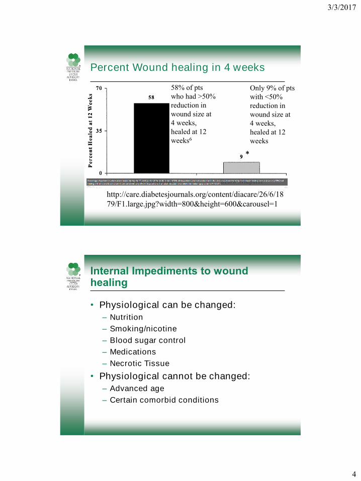
Percent Wound healing in 4 weeks
http://care.diabetesjournals.org/content/diacare/26/6/18
79/F1.large.jpg?width=800&height=600&carousel=1
58% of pts
who had >50%
reduction in
wound size at
4 weeks,
healed at 12
weeks6
Only 9% of pts
with <50%
reduction in
wound size at
4 weeks,
healed at 12
weeks
Internal Impediments to wound healing
• Physiological can be changed:
– Nutrition
– Smoking/nicotine
– Blood sugar control
– Medications
– Necrotic Tissue
• Physiological cannot be changed:
– Advanced age
– Certain comorbid conditions

3/3/2017
5
External impediments to wound healing
• Can be changed:
– Repetitive trauma - due to self or due to
caregiver/clinicians (failure to of-load;
inappropriate shoes/mobility devices; wet-to-
dry dressings)
– Exposure – toxic chemicals, environmental
hygiene, temperature
– Physical barriers – rolled wound edges, non-
viable tissue (slough, fibrin, eschar)
– Invasion – virulent pathogens (biofilm)
T-I-M-E-(s) Principle for WBP1,8,9
• T - Address non-viable tissue in wound
• I - Address infection
• M – Manage moisture
• E – Address wound edges
• S – Address surround periwound skin

3/3/2017
6
Documenting wound assessments
• Location
• Suspected etiology, contributing factors
• Size (W X L X D in cm)
• Undermining, tunneling (clock method)
• Exudate (color, amount, odor)
• Wound bed tissue (color, amount viable)
• Wound edges and surrounding tissue
• Last treatments used, compliance,
wound response, patient/CG education
Describing wound tissue
• Color of wound bed (in percentages)
• Viable (living tissue with good perfusion)
• Non-viable (not living)
• Boggy (wet spongy consistency)
• Fluctuant (moving in waves, movable &
compressible, variable/unstable)
• Friable (bleeds easily with light touch)
• Hypergranulating (overgrowing baseline)
• Pale (anemic looking)

3/3/2017
7
Characteristics of slough in wounds
• What it is
• What it is not
• Slough vs. Fibrin
Slough
• Best ways to remove

3/3/2017
8
Characteristics of eschar in wounds
• What it is
• What it is not
• When not to remove
Eschar
• When to remove
• Best ways to remove
3/3/2017
9
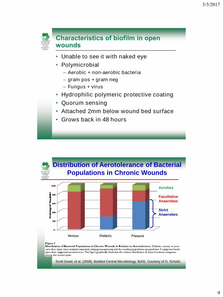
Characteristics of biofilm in open wounds
• Unable to see it with naked eye
• Polymicrobial
– Aerobic + non-aerobic bacteria
– gram pos + gram neg
– Fungus + virus
• Hydrophilic polymeric protective coating
• Quorum sensing
• Attached 2mm below wound bed surface
• Grows back in 48 hours
Scott Dowd, et al. (2008). BioMed Central Microbiology, 8(43). Courtesy of G. Schultz.
Distribution of Aerotolerance of Bacterial
Populations in Chronic Wounds
Aerobes
Facultative
Anaerobes
Strict
Anaerobes
3/3/2017
10
Prevalence of biofilm
More than • 60% of all chronic wounds
with biofilm
More likely more prevalent than we think• 7
Malone et al., 2017.
Problems of biofilm
• Impairs wound healing
• Host often lacking common signs and
symptoms of infection
• Difficult to detect, identify, eradicate
• Normal presumptive antibiotic treatment
may actually encourage biofilm growth
3/3/2017
11
Biofilm
• When to remove
• Best ways to remove
Potential antibiofilm strategies
• Prevention
• Debridement
• Topical products
• Systemic products
• Combined approaches
– “one-two” punch: removal and preventive

3/3/2017
12
Evidence for debridement methods
• Autolytic (exudate/MMPs)
– Pros/Cons
• Enzymatic (collagenase)
• Mechanical (debriding gauze, wet-to-dry)
• Sharp
– Scalpel, scissors, curette
• Ultrasonic (low and high frequency)
– With and without forced water
• Larval/biological
Evidence for Larval Debridement
Mudge• , E., Price, P., Neal, W., & Harding, K. G. (2014). A randomized
controlled trial of larval therapy for the debridement of leg ulcers: Results
of a multicenter, randomized, controlled, open, observer blind, parallel
group study. Wound Repair and Regeneration, 22, 1, 43-51.
Bohova• , Jana, Majtan, Juraj, Majtan, Viktor, & Takac, Peter. (2014).
Selective Antibiofilm Effects of Lucilia sericata Larvae Secretions /
Excretions against Wound Pathogens. Hindawi Publishing Corporation.
• Poppel, A. K., Vogel, H., Wiesner, J., & Vilcinskas, A. (2015).
Antimicrobial peptides expressed in medicinal maggots of the blow fly
Lucilia sericata show combinatorial activity against bacteria.
Antimicrobial Agents and Chemotherapy, 59(5), 2508-14.
Cazander• , G., Pritchard, D. I., Nigam, Y., Jung, W., & Nibbering, P. H.
(2013). Multiple actions of Lucilia sericata larvae in hard-to-heal wounds:
Larval secretions contain molecules that accelerate wound healing,
reduce chronic inflammation and inhibit bacterial infection. Bioessays,
35(12), 1083-1092.

3/3/2017
13
Questions?
• Contact information:
References
1. Schultz, G. S., Sibbald, R. G., Falanga, V., Ayello, E. A., Dowsett, C.,
Harding, K., Romanelli, M., ... Vanscheidt, W. (2003). Wound bed
preparation: a systematic approach to wound management. Wound Repair
and Regeneration: Official Publication of the Wound Healing Society [and]
the European Tissue Repair Society, 11, 1-28.
2. Trengove, N.J., Stacey, M.C., Macaulley, S., Bennett, N., Gibson, J.,
Burslem, F., Murphy, G., Schultz, G. (1999). Analysis of the acute and
chronic wound environments: the role of proteases and their inhibitors.
Wound Repair and Regeneration, 7, 442-452.
3. Demidova-Rice, T., Hamblin, M.R., Herman, I.M. (2012). Acute and Impaired
Wound Healing: Pathophysiology and Current Methods for Drug Delivery,
Part 1: Normal and Chronic Wounds: Biology, Causes, and Approaches to
Care. Adv Skin Wound Care, 25(7), 304–314.
4. Schultz, G. (2014). Molecular and cellular regulation of wound healing: What
goes wrong when wounds fail to heal or heal too much?. London: Henry
Stewart Talks. http://hstalks.com/lib.php?t=HST186.3834&c=252.
5. Harding, K. G., Moore, K., & Phillips, T. J. (2005). Wound chronicity and
fibroblast senescence - implications for treatment. International Wound
Journal, 2(4), 364-368.

3/3/2017
14
References
Sheehan6. , P., Jones, P., Caselli, A., Giurini, J.M., Veves, A. (2003). Percent
Change in Wound Area of Diabetic Foot Ulcers Over a 4-Week Period Is a
Robust Predictor of Complete Healing in a 12-Week Prospective Trial.
Diabetes Care, 26(6), 1879-1882.
Malone7. , M., Barjnsholt, T., McBain, A.J., James, G.A., Stoodley, P.,
Leaper, D., Tachi, M., Shultz, G., Swanson, T., Wolcott, R.D. (2017). The
prevalence of biofilms in chronic wounds: a systematic review and meta-
analysis of published data. Journal of Wound Care, 25(12), 1-5.
Schultz8. , G. S., Barillo, D. J., Mozingo, D. W., & Chin, G. A. (April 01, 2004).
Wound bed preparation and a brief history of TIME. International Wound
Journal, 1(1), 19-32.
Leaper9. , D. J., Schultz, G., Carville, K., Fletcher, J., Swanson, T., & Drake,
R. (January 01, 2012). Extending the TIME concept: what have we learned
in the past 10 years? International Wound Journal, 9, 1-19.
Granick10. , M., Boykin, J., Gamelli, R., Schultz, G., & Tenenhaus, M.
(January 01, 2006). Toward a common language: surgical wound bed
preparation and debridement. Wound Repair and Regeneration, 14.
References
11. Cowan, L., Phillips, P., Stechmiller, J., Yang, Q., Wolcott, R. & Schultz, G.
(2013). Antibiofilm Strategies and Antiseptics (Chapter 4) in Antiseptics in
surgery: Scientific basis, indications for use, evidence based
recommendations, vacuum instillation therapy; Willy, C., & Alt, V. (editors),
33 tables. Berlin: Lindqvist Book Publ.
12. Cowan, L., Phillips, P., Liesenfeld, B., Mikhaylova, A., Moore, D., Stechmiller,
J., & Schultz, G. (June 01, 2011). Caution: When Combining Topical Wound
Treatments, More Is Not Always Better. Wound Practice & Research: Journal
of the Australian Wound Management Association, 19(2), 60-64.