What- W v~cJv? , &ut- cv d.a:t'fV wit'l-v...
70
"What- W ....... ,&ut- cv d.a:t'fV wit'l-v
Transcript of What- W v~cJv? , &ut- cv d.a:t'fV wit'l-v...

"What- W v~cJv? ....... , &ut- cv ~ d.a:t'fV wit'l-v k¥t,ow~'
Introduction
I ntrotfuction
1. INTRODUCTION
Fungal infections in humans range from superficial and cutaneous to deeply invasive and
disseminated forms (Table 1). Though these pathogenic fungi have a worldwide distribution,
particular species may predominate in certain geographical areas. Among them infections
due to Candida and Aspergillus spp. are the most common. Essentially, this rise in the
infections is observed to occur predominantly in people whose immune system is
compromised (due to AIDS, organ transplantation or cancer chemotherapy), who have been
treated with broad-spectrum antifungal drugs, or who have been subject to invasive
procedures (like catheters and prosthetic devices) .
. ~ Table 1. Common fungal pathogens * ~~ ':
FUNGI TYPE SITES OF INFECTION
Aspergillus fumigatus filamentous
Candida albicans dimorphic
Cryptococcus neoformans true yeast
Histoplasma capsulatum dimorphic
Coccidiodes immitis dimorphic
Blastomyces dermatidis dimorphic
Paracoccidiodes brasiliensis dimorphic
Rhizopus arrhizus filamentous
·Compiled from Maricha! et ai., 1999.
1.1 Candida and Candidiasis ~~ ~- . ~~
lungs( aspergillosis), systemic
lungs, systemic candidiasis
meningitis
lung infections
lung infections
lung infections
lung infections
lungs,rhinocerebral, systemic
The history of Candidiasis dates back to the fourth century BC when Hippocrates described
oral apthae in two patients with severe underlying disease in his book Epidemics. The genus
Candida is composed of an extremely heterologous group of organisms that mainly grow as
yeast. C. albicans is the species most frequently associated with fungal infections in humans
(Vanden Bossche et ai., 1998). C. albicans accounts for majority of the systemic infections
with mortality rates ranging from 50% to 100%. This pathogen is the most common cause of
Introduction
septicemia in western hospitals, and accounts for development of oropharyngeal and clinical
thrush in >80% of the HIV -infected population. Candida spp. rank fifth among causes of
nosocomial bloodstream infections. Not only are the bloodstream infections with C. albicans
and non-albicans species occurring more frequently, but the relative proportion due to non
albicans species is increasing, including species such as C. glabrata, C. krusei, C.
parasilopsis, C. tropicalis, C. lusitaniae and (Table 2).
Table 2. Species commonly causing invasive Candidiasis *
SPECIES . FREQUENCY
Candida albicans 50%
Candida tropicalis 15-30%
Candida glabrata 15-30%
Candida parasilopsis 15-30%
Candida krusei ~1%
Candida lusitaniae ~1%
• From: http://www.doctorfungus.orglindex.htm
Cornmon risk factors for blood-stream candidiasis include extremes of age (low-birth-weight
infants and the elderly), immunosupression, malignancy with leukopaenia, major surgery, or
trauma, exposure to multiple antibacterial agents, central venous catheterization as well as
parenteral nutrition (Odds, 1988). Other factors contributing to development of Candida
infection are listed in Table 3.
Table 3. Factors contributing to development of Candida infection in hosts *
,/ Exposure to the fungus
,/ Inoculum delivered during the exposure
,/ Virulence of the fungus
,/ Host's immune status
,/ Underlying disease
,/ Immunosuppressive therapy
,/ Prior exposure to the fungus
• Compiled from Odds, 1988.

Introduction
Although a potential pathogen, C. aibicans is present as a commensal organism in many, if
not most, healthy individuals, where it may be found on the skin and in the oral cavity,
gastrointestinal tract and vagina. C. aibicans, the principal infectious agent of human
infection, is oval yeast of 2-6 11M in diameter. It exists as diploid, asexual polymorphic yeast
with various biochemical abilities, both assimilative and fermentative, but lacks any proper
sexual stage as well as carotenoid pigments. This medically significant fungus has the ability
to undergo phenotypic switching and has 8 chromosomes (Odds, 1988; Prasad, 1991).
1.2 What is antifungal drug resistance?
From a clinical perspective, drug resistance may be defined as the persistence or progression
of an infection despite appropriate drug therapy. The clinical outcome of treatment depends
not only on the susceptibility of the pathogen to a given drug but also on many other factors
including pharmacokinetics, drug interactions, immune status, and patient compliance, as
well as several specific conditions such as the occurrence of biofilms on surfaces of catheters
and prosthetic valves (White et ai., 1998). Antifungal drug resistance has been classified as
either primary, when a fungus is resistant to a drug prior to any exposure, or secondary, when
an initially sensitive fungus becomes resistant after exposure to the drug (Rex et ai., 1995;
White et ai., 1998). ....' . ~ • The therapeutic options for treating fungal infections, often caused by the emerging new
pathogens whose incidence has increased due to the AIDS pandemic and use of
immunosuppressive drugs in transplant and cancer patients, are limited by the relatively low
number and structural variety of antifungals (Kolaczkowski & Goffeau, 1997). Only a few
classes of antifungal agents are available to treat these infections. Figure 1 depicts the sites of
action of some of the common antifungals. Most commonly used antifungals inhibit the
ergosterol biosynthetic pathway and chiefly includes azoles, allylamines and morpho lines
and others such as polyenes and 5-FC impair membrane barrier function and macromolecule
synthesis respectively (Vanden Bossche et ai., 1989; Williams and Wilkins, 2004).
3

MEMBRANE FUNCTION
Polyenes: Amphotericin B Nystatin Primaricin
METABOLIC INmBITORS
Cispentacin Difluoromethyl Ornithine
NUCLUCACID FUNCnON AND
SYNTHESIS
5-fluorocytmine Trimethoprim Sulfomethaxozole Pentamidine
Introduction
CELL WALL SYNTHESIS
Nikkomycins Echlnocandins
/
Polyoxins
ERGOSTEROL SYNTHESIS
Azoles Allylaminesi Thiocarbamates Morphines
NUCLEAR DIVISION
Griseofulvin Benomyl
PROTEIN SYNTHESIS
Blasticidin, Sinefungin
Figure 1. Site and mechanism of action of different classes of drugs on a typical fungal cell. (Courtesy: Adopteei from Georgopapadakou and Walsh, 1994).
1.4 Multi!!rug resistance (MDR)
In the 1990s, drug resistance has become an important problem in a variety of infectious
diseases including human immunodeficiency virus (HIV) infection, tuberculosis, and other
bacterial infections which have profound effects on human health. At the same time, there
have been dramatic increases in the incidence of fungal infections, which are probably the
result of alteration:; in immune status associated with the AIDS epidemic, cancer
chemotherapy, and organ and bone marrow transplantation. The rise in the incidence fungal
infections has exacerbated the need for the next generation of antifungal agents, since many
of the currently available drugs have undesirable side effects, are ineffective against new or
reemerging fungi, or lead to the rapid development of resistance. Although extremely rare 10
years ago, antifungal drug resistance is quickly becoming a major problem in certain
populations, especially those infected with HIV, in whom drug resistance of the agent causing
oropharyngeal candidiasi~; is a major problem (Graybill, 1988; Vanden Bossche et aI., 1994).
Introduction
For instance, 33% of late-stage AIDS patients in one study had drug-resistant strains of C.
aibicans in their oral cavities (Law et ai., 1994). C. aibicans cells have evolved elaborate
molecular mechanisms to protect themselves from injuries caused by environmental exposure
to toxic compounds of different structure and functions. One of these mechanisms termed
multidrug resistance (MDR) or pleiotropic drug resistance also operates in a wide variety of
cell types including bacteria, protozoan, fungi and mammalian cells (Balzi and Goffeau,
1994 and 1995; Gottesman et ai., 1995 and 1996).
MDR can develop after sequential or simultaneous exposure to all the different drugs to
which the cell or microorganism is resistant. However it may also develop surprisingly
before exposure to many of the compounds to which the cell or microorganism is found to be
resistant. It generally involves a network of membrane-associated transporters acting as
multidrug efflux pumps and transcription factors regulating the expression of these pumps.
These multidrug efflux systems present a disturbing clinical threat, since the acquisition of
such a single system by a cell may decrease its susceptibility to a broad spectrum of
chemotherapeutic drugs. The development of drug resistance is a frequent impediment to the
effective treatment of infectious and malignant diseases (Van denbroucke-Grauls, 1993;
Sternberg, 1994; Peters, 1996). A primary goal in the study of chemotherapy is to understand
how cells can become drug resistant by lowering the intracellular concentration of drugs or
altering the ability of the drugs to affect their targets. Mechanisms of multi drug resistance are
opportunistic in their manipulation of the normal pathways of cellular homeostasis. Several
alterations in the cell take place resulting in drug resistance.
A drug-resistant, pathogenic fungus is an organism, according to Kerridge (1985 and 1986)
that will grow and produce clinical symptoms of disease in the presence of the drug at the
maximal concentration, at the site of infection.
From a mycological viewpoint different classifications of resistance are made.
1.4.1 Intrinsic resistance
A species is regarded as intrinsically resistant when it is not included in the normal spectrum
of a given antifungal compound. Primary or innate resistance is characteristics of any species
and is not dependent upon previous exposure to the drug. For example, Candida krusei can
be regarded as intrinsically resistant to fluconazole and C. parapsiiosis to Amphotericin B.
5

Introduction
1.4.2 Acquired resistance
Acquired resistance is found in isolates belonging to a species which is nonnally susceptible
to the compound. Alternately, resistance can develop in previously susceptible isolates
during or after antifungal drug exposure (Klepser et al., 1997). Acquired resistance is also
tenned as secondary resistance and is most familiar in the setting of HIV-infected patients
with oropharyngeal candidiasis due to C. albicans who receive multiple courses of azole
antifungal therapy.
1.4.3 Selective resistance
When a patient is colonized with multiple species or strains, during treatment the most
sensitive isolates are eradicated favoring the growth and selection of less sensitive or
resistant isolates. This type of resistance is called selective resistance.
1.4.4 Phenotypic resistance
This occurs when a strain develops a progressive increase in resistance during continued
incubation in the presence of antifungal compound.
1.4.5 Genotypic resistance
It is a feature of the clone and is inherited by daughter cells.
1.5 Mechanisms of resistance to antifungals
Although the molecular basis of drug resistance III Candida is not very clear, evidence
accumulated so far suggests that MDR is a multifactorial phenomenon where a combination
of mechanisms could contribute to drug resistance (White, 1997a; Franz et al., 1998a; Lopez
Ribot et aI., 1998). A schematic representation of the known molecular mechanisms of
antifungal drug resistance in pathogenic fungi is depicted in Figure 2 and Table 4.
Azoles are most commonly used to treat the fungal infection. But how azoles enter
susceptible fungal cells is not known, although their relative hydrophobicity may facilitate
entry by passive diffusion. Once inside the cells, azoles interact with the 14u-DM enzyme in
the ergosterol biosynthetic pathway, allowing precursors to be incorporated into newly
synthesized regions of plasma membrane (Barrett-Bee and Dixon, 1995). Not all of the azole
e:ntering the cell remains there, because two low-level active efflux systems, the ABC
transporters and the major facilitators, pump free drug from the cell.

......... __ - Substrate
~;-: By-Pass Pathway
I 5 Inactive Drug
Altered Drug Target
Figure 2. Mechanisms of azole resistance in fungi. (1) changes in cell wall/plasma membrane leading to impaired azole uptake. (2) efflux of drugs mediated by the ABC or the MFS class of efflux pumps. (3) over-expression of the drug target or of the efflux pumps. (4) mutation in the drug target (p450l4DM) does not allow the drug to bind due to low affinity. (5) activation of alternate pathways such as ~5 ,6-desaturase.
(6) sequestration of the drug into an organelle-like vacuole by organellar pump. (7) chromosome alterations or changes in chromosome number as a means to maintain more
copies of the required gene. (8) modification of azoles (CYP52) to an inactive form. (Courtesy: modified from Prasad et aI. , 2000) .

Introduction
Table 4. Molecular Mechanisms leading to antifungal resistance *
Alterations in drug import by change in sterol components of the plasma membrane
Alterations in intracellular drug processing Modification
Degradation Genetic changes in target enzyme, ERGll including:
Point mutations Over -expressi on Gene amplification (leads to over-expression) Gene conversion or mitotic recombination
Alterations in other enzymes in ergosterol biosynthetic pathway Increased efflux of drug due to:
ABC transporters (CDR genes) Facilitator
Courtsey: White et at., 1998.
Although the molecular basis of drug resistance in Candida is not very clear, accumulated
evidences suggests that MDR is a multi-factorial phenomenon where various different type
of resistance mechanisms are known to be present in different supplementing combinations
(White et at., 1998). These chiefly include reduction in the import of drug into the cell;
modification or degradation of the drug once it is inside the cell; changes in the interaction of
the drug with the target enzyme; changes in other enzymes in the same biosynthetic pathway;
and an increased efflux of the drug from the cell (Prasad et al., 2000a and 2002) . Some of
the well-known mechanisms of resistance in Candida are discussed below which have been
broadly divided into azole and non-azole mediated resistance.
1.5.1 Mechanisms of azole resistance
Azole antifungal agents are by far the most commonly used drugs to treat infections caused
by C. albicans. The target for azole antifungal agents in yeast is a cytochrome P-4S0. This
enzyme is involved in the 14a-demethylation of lanosterol which is an important step in the
biosynthesis of ergosterol. Azole drugs interact in the active site of the target enzyme by
binding to the heme moiety, acting as competitive inhibitors of the substrate. Figure 2 depicts
molecular mechanisms of azole resistance that are commonly present in the pathogenic yeast
C. albicans which include increased over-expression of multidrug transporters, mutations in
7
Introduction
lanosterol demethylase that reduce azole binding and increased expreSSIOn of ERG11
(Bossche et aI., 1987; Vanden Bossche et al., 1994a and 1994b; Vanden Bossche and
Koymans, 1998; Marichal, 1999). Additional known mechanisms have also been
demonstrated to participate in azole resistance in C. albicans clinical isolates. These include
decreased import, alterations in other enzymes in ergosterol biosynthetic pathway,
chromosomal alterations, modification or degradation of the drug and transcriptional
regulators. Nevertheless, other unknown mechanisms of azole resistance may still be found
in clinical isolates. In the following sections these mechanisms have been discussed in detail.
1.5.1.1 Permeability resistance
Defects in drug import are a common mechanism of drug resistance. Many hydrophilic
drugs, for example the anticancer antimetabolite methotrexate, cannot easily diffuse through
the plasma membrane and have to use specific transporters for this purpose. Alterations in
these transporters often lead to reduced influx of drugs. In the case of methotrexate,
resistance is found to be associated with alterations in the folate transporter (Skovsgaard et
al., 1994).
Decreased toxicity can also be caused due to changes in the lipid composition of the
membranes leading to a decrease in permeability. The decreased permeability and fluidity of
the membranes result from cis- to trans-isomerization of their saturated fatty acids.
Resistance to the polyene systemic antifungal such as amphotericin B, which interacts with
membrane ergosterol to form pores in membrane results in most cases, from defects in the
ergosterol biosynthetic pathway, leading to a decreased ergosterol levels in the plasma
membrane (Kelly et aI., 1997). Several studies have demonstrated that when the ergosterol
component of the plasma membrane is eliminated or reduced in favor of other sterol
components such as 14a-methyl sterols, there are concomitant permeability changes in the
plasma membrane and a lack of fluidity (Bossche et aI., 1987). These changes may lower the
capacity of azole drugs to enter the cell. Most amphotericin B resistant isolates contain
abnormally low number of ergosterol molecules in their plasma membrane thereby limiting
the number of available binding sites for the polyene and thus preventing the membrane
damage. Existence of other mechanisms of polyene resistance not linked to sterol alterations
has also been suggested (Joseph-Home and Hollomon, 1997). Another kind of permeability
Introduction
barrier in C. albicans involves fonnation of biofilms, which are large masses of cells. They
have taken center stage with the increasing recognition of their role in human infections. In
the protected microenvironment ofbiofilms, the pathogens are more resistant to antimicrobial
therapies (Reynolds and Fink, 2001).
However reduction of membrane penneability due to changes in the membrane biophysical
properties is not a very efficient way of resistance unless it is accompanied by another
resistance mechanism, such as active efflux or enzymatic activation, which often is the case.
1.5.1.2 Efflux mediated resistance
In addition to an alteration or an over-expression of 14a-Ianosterol
demethylase involved in sterol biosynthesis, azole resistance in C. albicans is
also elicited by other mechanisms like the reduced intracellular drug
accumulation of drugs due to drug efflux pumps. The most intriguing aspect
of these transporters generally relates to their lack of specificity, wherein a single transporter
can recognize a variety of unrelated xenobiotics. It is apparent from a host of studies that
drug transporters of yeasts, similar to its homologues in mammalian system, are capable of
transporting substrates that are structurally diverse. It is suggested that ABC transporters are
primarily involved in protection against exogenous toxic compounds, whereas MFS
transporters may have evolved primarily as a mechanism to secrete endogenous toxic
metabolites (Del Sorbo et al., 2000). The characterization of ABC proteins e.g. CDR1
(Prasad et aI., 1995), CDR2 (Sanglard et al., 1997), and of CaMDR1 (Fling et aI., 1991; Ben
Yaacov et al., 1994), a MFS transporter, as efflux pumps of C. albicans, and their over
expression in certain instances of azole resistant isolates has confinned that these transporters
represent another mechanism involved in the MDR scenario of C. albicans.
1.5.1.2.1 ABC transporters
The most intensively investigated MDR ABC transporters in this respect are the mammalian
ABC transporter protein P-gp. Since the substrates of P-gps and many related transporters are
hydrophobic they tend to accumulate in membranes. Several lines of evidence suggest that P
gp somehow can directly recognize and export from the cell membrane the variety of
compounds to which it mediates resistance. Studies on mechanisms of MDR transporter
Introduction
functioning have proposed that P-gp recognizes the drugs as they enter the membrane lipid
phase in the manner of a hydrophobic vacuum cleaner model (Raviv et aI., 1990;
Kolaczkowski and Goffeau, 1997). Another idea is that the transporter is essentially a
"flippase" that detects drug within the inner leaflet of the plasma membrane and "flips" it
into the outer leaflet (from which it can diffuse away from the cell) or directly into the
extracellular space Their substrate specificity profiles include a huge variety of hydrophobic
compounds ranging from peptides and steroid hormones to anticancer drugs. Resulting from
the explosion of sequence information, it is now known that ATP-hinding ~assettes (ABC)-,
ATPases constitute one of the largest and most highly conserved superfamilies and are found
in all organisms organisms (Higgins, 1992 and 1993; Ambudkar and Gottesman, 1998;
Holland and Blight, 1999). The fact that these transporters are structurally and functionally
conserved throughout evolution indicates that these proteins could be involved in performing
some indispensable functions in the cell. Some of the important physiological functions
attributable to ABC proteins known to date include roles in absorption, excretion, signal
transduction, bacterial pathogenesis and most importantly in drug and antibiotic resistance
(Kerridge, 1986; Prasad et al., 2000a and 2000b).
The drug efflux pump encoded by CDR1 of C. albicans was the first ABC efflux pump
implicated in conferring resistance to cycloheximide in a PDR5 disruptant hypersensitive
strain of S. cerevisiae (Prasad et aI., 1995). CDR1 encodes a 1501 amino acid long protein
(169.9 kDa) whose predicted structural organization is characterized by two homologous
halves, each comprising a hydrophobic region with a set of six transmembrane stretches,
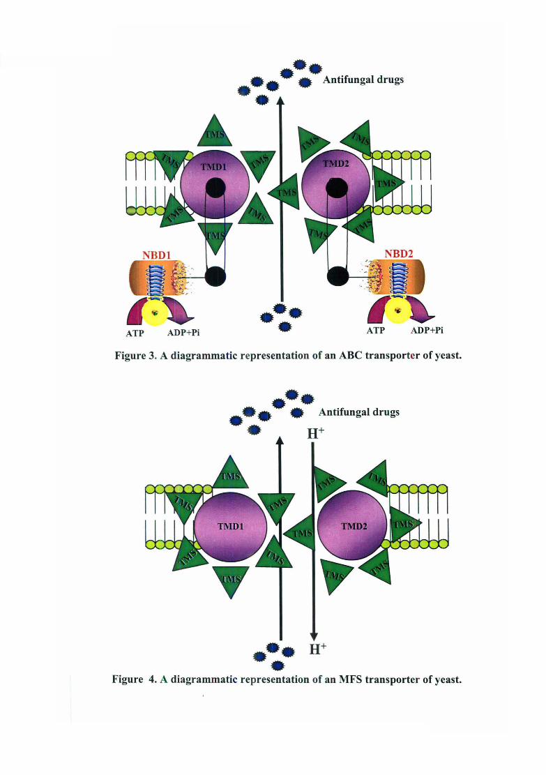
preceded by a hydrophilic nucleotide binding fold (Figure 3). The structure is identical to that
of the S. cerevisiae ABC proteins Pdr5p and Snq2p. It mirrors the architecture of the yeast a
mating pheromone transporter Ste6, as well as mammalian drug resistance P-glycoprotein (P
gp or MDRl) and cystic fibrosis factor CFTR (Dassa and Bouige, 2001). The significance of
inversion of domains in some of the ABC drug transporters is not understood and may be
related to their physiological functions. Cdrl p is remarkably similar to Pdr5p of S. cerevisiae.
The similarity is not limited to only the A TP binding motif but is conserved along the entire
length of the protein. Despite the high homology between CDR1 and PDR5, and their
encoded products, some functional features tend to distinguish them. For example, while
Introduction
single or low copies of CDR] are sufficient to increase drug resistance in S. cerevisiae,
multiple copies of PDR5 are required to yield a similar level of drug resistance.
That efflux pumps other than CDR] could be contributing to drug resistance became
apparent after isolation of its close homolog CDR2. CDR2 exhibits 84 % identity with Cdrlp
and confers resistance to fluconazole and several other drugs (Sanglard et aI., 1997). In spite
of close identity between CDR] and CDR2, their flanking promoter regions exhibit
considerable divergence. Interestingly the promoter of both the genes, though a close
homolog of PDR5, lacks the characteristic PDRIIPDR3 transcription factor binding sites,
which further suggests divergence in the regulation between close homologs of the two
yeasts (Prasad et aI., 1995).
Since azole resistance appears to be a multi-factorial phenomenon this led to search of more
homologs of known CDRs. Using PCR-based cloning, other homologs of CDR] and CDR2
namely CDR3, CDR4, and CDR5, were identified (Alarco et aI., 1997; Balan et aI., 1997;
Franz et al., 1998a and 1998b; Sanglard et aI., 1998a and 1999). Cdr3p and Cdr4p show the
highest homology to Cdrl p and Cdr2p; however, compared to Cdrl p and Cdr2p which are>
90% similar, Cdr3p and Cdr4p are only 75% similar to Cdrlp and Cdr2p. Interestingly over
expression of CDR3 and deletion of CDR3 and CDR4 could only affect drug susceptibilities
(Alarco et al., 1997; Franz et al., 1998a and 1998b). Why some of the known Cdrp's are
unable to elicit a multi drug resistance phenomenon is not yet clear.
1.5.1.2.2 Major facilitators
The major facilitator (MFS) proteins that are involved in symport, antiport, or uniport of
various substrates, have been found to be ubiquitously present from bacteria to higher
eukaryotes. These proteins are proton motive force (PMF)-dependent antiporters which
efflux out substrates in exchange of one or more H+ ion. The MFS has originally been
defined as a superfamily of permeases that are characterized by two structural units of six
transmembrane spanning a-helical segments, linked by a cytoplasmic loop (Figure 4).
These transporters have been classified into five distinct clusters or families of membrane
transport proteins within the MFS involved in (i) drug resistance, (ii) sugar uptake, (iii)
uptake of Krebs cycle intermediates, (iv) phosphate ester/phosphate antiport and (v)
oligosaccharide uptake (Saier and Reizer, 1991; Marger and Saier, 1993; Paulsen et al.,
1996). On the basis of hydropathy and phylogenetic analyses, MFS drug efflux proteins
ATP ADP+Pi
•• •• •• Antifungal drugs ••
• • • ATP ADP+Pi
Figure 3. A diagrammatic representation of an ABC transporter of yeast .
••• •• • Antifungal drugs ••
••• H+
• Figure 4. A diagrammatic representation of an MFS transporter of yeast.

Introduuion
which have mor~ than 100 members. can be divided into two distinct groups containing
either 12 or 14 TMS (Irans Membrane .segments) (Paulsen et aI., 1996).
CaMDRl (previcusly known as BEN!) and FLUI are the two MFS genes identified in C.
albicans. CaMDRl was initially identified as a gene, which conferred resistance to tubulin
binding agent be 10myl and tetrahydrofolate reductase inhibitor methotrexate (Fling et a/ ..
1991; Ben-Yaaeo \' et £I/.. 1994).
Correlation, not cause. It is important to note that the efflux pumps (discussed above),
including the CDR genes and the MDRI gene, show an increased expression that correlates
with resistance in clinical isolates of C. albicans. Mutant strains of C. albicalls and S.
cerevisiae with di~:ruptions in these genes are hyper-susceptible to azole and other drugs, and
over-expreSSIOn 0' these genes in S. cerevi,'iae can cause an increased resistance. However,
there is no definitive proof that over-expression of these genes in C. alb/cans results in a
drug-resistant phenotype.
1.5.1.3 Molecular alteratiomi in other ERG genes
Modifications :,n the ergosterol biosynthetic pathway are likely to generate cross resistance to
a large number of mreIated drugs. In addition to the alterations in lanosterol demethylase, a
common mechanisn of resistance is an alteration in other enzymes of the same biosynthetic
pathway. A ddeclive lanosterol demcthylase will result in the accumulation of 14cx
methylfecosterol and 14a-methylergosta-8,24(28)-dien-3-~,6-a diol. Presence of 140.
methylfecosterol can modify the membrane function and fluidity. Cells with this sterol
product have an inc reased sensitivity to oxygen-dependent microbicidal systems of the ho~t.
This diol causes growth arrest in S. cerevislae but is tolerated in C. alhicans. The tOXIC
effects of this diol i~; eliminated by a mutation in ERG3. To date ERGI. ERG2, ERG3, ERG4,
ERG7, ERG6, ERell, ERG24, ERG25 and ERG26 have been cloned from C. alhicans,
ERG3 and ERGII rave been cloned from C. glabrata, and ERGII has been cloned from C.
krllsei (Burgener-Kairuz et at., 1994). The ERG3 and ERGII genes of C. glahrata have bee;l
studied in some det,,!il. The double deletion mutant of ERG3 and ERG 11 in C. glabrata has
been shown to be a(:("obically viable, accumulates 14-a-methylfecosterol and lanosterol and
is resistant to azoles md AmB (Geber et aI., 1995).

Introduction
Another well ctaracterized mechanism of azole resIstance in both S. ccrc)'isiae and C.
albicans is conferred by loss-of-function mutations in sterol ~5.6-desaturase (Kelly et aI.,
1997). A defect ill this enzyme leads to the accumulation of 14a-methylfecosterol instead of
14a-methylergost1-8, 24(28)-dien-3-~,6-a dio!. Accumulation of suffieient amounts of 14a
methylfecosterol compensates for ergosterol in the membranes and thus contributes to az,)\e
resistance in C. :t!bicans (Vanden Bossche and Koymans, 1998; White et al., 1998;
(Ghannoum and Rice, 1999). Because of the lack of ergosterol in their membranes, these
isolates were cros~-resistant to AmB. In S. cerevisiae, the ~56-desaturase defect can also
suppress the lethality resulting from the inactivation of CYP5IAI and is also linked 10
acquisition of resi:;tance to azole derivatives (Kelly et al., 1997). C. glabrata strains
produced under labc1ratory conditions lacking functional ERG 3 and CYP51 A I genes showed
similar phenotypes (Geber et al., 1995).
Azoles also inhibit he cytochrome P450-catalyzed ~ 22 -desaturase enzyme. ~ 22 -destaurase
(CYP6I and also ERG5) and its homologues have also been identified 111 C. albicans and
Schizosaccharomyces pombe. It has been hypothesized that an increase of this non-essential
enzyme would captUl e sufficient azole to prevent the inhibition of the 14a-demethylation
reaction. So far, however, no biochemical evidence has been presented to support this
hypothesis (Marichal Lt af., 1999).
1.5.1.4 lWolecular alterations of ERG11
As discussed above the predominant target site of the azole drugs is the heme protein,
lanosterol demethylase also called CYP5I A I (encoded by ERG II) which cocatalyzes
cytochrome P450 depetdent 14a-demethylation of lanosterol (Ghannoum and Rice, 1999).
Inhibition of 14a-demel hylatiol1 leads to the depletiol1 of ergosterol and accumulation of
sterol precursors, including 14a-methylated sterols (lanosterol, 4, 14-dimethylzymosterol,
and 24-methylenedihydrolanosterol), resulting in the fonnation of plasma membrane with
altered structure and func\ ion.
Several lines of evidence implicate a modification in the quantity or quality of 14a
demethylase in the expression of resistance to azole antifungal agents. Several genetic
alterations like point mut<ttions, over-expression of the gene, gene amplification and gene
conversion have been identified in ERG II of C. albicans.

Introduction
Point mutatiolls
Alteration of the affinity of azole derivatives for CYP51AI, which has been described in
different post-reatment yeast isolates of C. albicans (Bossche, 1994) is another important
mechanism of resistance. A survey of resistant and sensitive clinical C. albicans isolmes has
identified different point mutations that are associated with azole-resistant isolates. Sequence
data identified Y132H (tyrosine 132 is replaced by a histidine), T135A (threonine 315 is
replaced by alanine) or R476K (arginine 476 is replaced by lysine) mutations that decrease
the affinity of the target for an azole derivative. It has been shown that R467K alone is
sufficient to cause azole resistance (Marichal, 1999).
Another significant change observed in the ERG 11 gene of the resistant isolate was reported
by White, 19Q8, namely loss of allelic variation in the ERG 11 promoter and in the
downstream TlIRI gene (which encodes homosenne kinase, which is involved in threonine
synthesis). Point mutations in ERG 11 have been developed in laboratory strains also that
result in azole resistance. Although these changes may account for resistance development,
they are n01 the only factors involved (Ghannoum and Rice, 1999).
Over-expre<,s;o II
Over-expression of CYP5IA1 as a resistance factor has been mentioned for a few C. albicans
and C. glabrat:l Isolates and has been implicated as a mechanism of resistance to azole
antifungals (V,mden Bossche and KOymans, 1998; Ghannoum and Rice, 1999). 'Over
expression of CYP51A 1 in C. albicans resistant isolates has been measured and was up to
three times hifher than could be measured in azole susceptible isolates, thus probably
accounting for a minor effect on the development of resistance. However, some C. alhicans
strains have up to a IO-fold increase in CYP51 Al mRNA. Over-expression of CYP51 A 1 can
be achieved by gene amplification. as shown in a C. glabrata isolate resistant to azole
derivatives (Marichal et aI., 1997). Hybridization experiments on chromosomal blots indicate
that this increase in copy number is due to amplification of the entire chromosome containing
the CYP5IA1 gene. Azoie dependent upregulation of £"RG11 has been described in several
different clinica isolates of C. albicClIlS (Sanglard et aI., 1995; AlbeI1son et aI., 1996; White
et al., 1997). BJt it is difficult to assess the contribution of ERG1I over-expression to a
resistant phenot>pe, since these limited cases of over-expression have always accompanied
other alteration~; associated with resistance including mutations in ERG 11 and over-
14

Introduction
expression of efflux pumps. Hence over-expression of the target enzyme plays only a limited
role in clinical resistance to the azoles.
Azole dependent up-regulation is not limited to ERG 11 and also involves upregulation of
other ERG genes upstream and downstream of ERG 11. Likewise ERG 11 upregulation was
also not limited to azole drugs and ERG 11 was also inducible by drugs that target other
enzymes of the ergcsterol biosynthetic pathway (Morita and Nozawa, 1985; Barrett-Bee and
Dixon, 1995). Thes;? data suggest a common global ERG upregulation e.g. in response to
ergosterol depletion.
1.5.1.5 Nucleo-cytoplasmic interactions as determinants of drug resistance
Another class of prcteins which are involved in pleiotropic drug resistance in yeast are the
transcription regulators (Balzi and Goffeau, 1994 and 1995). In S. cerevisiae severa:
regulatory protems (Pdr 1 p. Pdr3p, and Yap 1 p) controlling expression of multiple drug
resistance genes of the ABC transporters and MF superfamilies are knO\vn and mutations in
these regulators tha' result in upregulation of their respective target genes have been
identified. Various g,:netic regulators connecting PDR regulators to drug pumps have been
uncovered. As a first example, the regulators PDR1, PDR3, PDR7 and PDR9 have been
shown to control transcription of the multi drug transporter gene PDR5, encoding an ABC
protein (Balzi and Goffeau, 1995). yAP 1 targets include the cadmium resistance ABC
transporter gene rCF r, as well as a number of genes involved in response to oxidative stress
such as GSH1, encoding y-glutamylcysteine synthase, TRX2, one of the two genes encoding
for thioredoxin, and GLR 1, encoding glutathione reductase. Several polymorphic mutant
alleles of PDRl gene ,vhich display altered drug resistance profiles have been isolated (Choi
et aI., 1988; Subik et al., 1986: Carvajal et of., 1997).
Recently functional homologs of these regulators have also been found in C. olbicans. Over
expression of YAP 1 homologue CAP 1 in S. cerevisiae resulted in resistance of the
transforrnants against luconazole and other drugs that was mediated by the transcriptional
activation of the FIR I gene encoding a major facilitator homologous to the C. alhicans
CaMDR1 gene (Alm"co et al., 1997). Over-expression of a mutated form of CAP 1, but not
wild type CAP 1 in CAI4 resulted in actintion of the CaMDR1 gene and concomitantly
resistance against fluconazole and several other drugs, suggesting the possibility that similar
Introduction
mutations might also be responsible for CaMDRI activation in fluconazole resistant clinical
C. alhieans Isolates. On the other hand, disruption of the CAP 1 gene in the CaMDRl over
expressing FR2 strain did not suppress but further increased the level of CaMDRl
expression, and it was therefOle recognized as a negative regulator CaMDRl gene. Similarly
the C. albieans transcriptional regulator FCRl was identified by functional complementation
of pdrl/pdr3 mutant (Talibi a'ld Raymond, 1999). Over-expression of FCRl in this strain
resulted in fluconazole reSj:starce that was mediated by the transcriptional activation of the
ABC transporter PDR5. In contrast disruption of FeRl in C. alhieans resulted in a
hyperresistance against fluconamle, demonstrating that similarly to CAP l, FCRl behaved as
a transcriptional activator when overexpressed in S. cerevisiae but acted as a negative
regulator of drug resistance in C. albieam'. The transcriptional targets of FCRl have not been
reported.
So far, none of the transcriptiol111 regulators of drug resistance identified in C. a/hieans has
been shown to be involved in tre development of fluconazole resistance in clinical isolates
and it was suggested that mutati Jns in the regulatory region of the multiple drug resistance
genes themselves may be respom:ible for their over-expression in resistant isolates. This lack
of knowledge about the molecul,r changes leading to activation of multiple drug reslstance
genes in clinical C. albieans strain is due to the fact that wild type C. albicans strains is not
easily accessible to genetic manipulation. However, using MPA R technique (mycophenolic
acid). Wirsching et al. (2000) ha\e shown that CaMDRl regulation in fluconazole resistant
lsolates that over-express CaMDR I involves trans-regulatory factor. It is likely that such a
mechanism is of general relevance and may be present in other fluconazole resistant C.
albieans strains. The unraveling of this unexpectedly large pleiotropic network for multidrug
resistance control should have implications for the development of new fungicides.
1.5.2 Mechanisms ofnon-azole mediated resistance
1.5.2.1 Resistance to 5-flucytosine
Primary resistance to 5-FC is a common phenomenon, Resistance may occur due to the
deficiency or lack of enzymes invoLed m the uptake or metabolism of 5-FC, or may be due
to the deregulation of the pyrimidine biosynthetic pathway. whose products can compete
with the fluorinated metabolites of 5-FC (Kerridge and Whelan, 1984). Detailed

Introduction
investigations on the molecular mechanisms of resistance to 5-FC have shown that intrin.~ic
resistance in fungi can be due to a defect in cytosine permease (with the exception of C.
albicans), while acquired resistance results from a failure to metabolize 5-FC to 5-FUTP mld
5-F dUMP, or from the loss of feedback control of pyrimidine biosynthesis.
1.5.2.2 Resistance 10 polyenec."
Polyene resistance .1as not been a major clinical problem to date, although polyene resistant
isolates have been i:;olated and characterized. Acquired resistance to AmB is often associated
with alteration of membrane lipids. especially sterols. Most polyene resistant clinical
Candida isolates ha,e a greatly reduced ergosterol content in their membranes (Hitchcock ct
a/., 1987; White et (//., 1998). There is evidence to suggest that alterations in the membran'~
structure or in the sterol-to-phospholipid ratio in the membrane may be associated with
resistance. Recently, clinical isolates of C. albicans resistant to AmB were described lacking
ergosterol and accumulating 3-~-ergosta-7 ,22-dienol and 3-~-ergosta-8-enol, typical for a
defect in the sterol L~ 56 desaturase system (Kelly et af., 1997). Such a defect is known in
laboratory yeasts (s. cerel'isiae) harbouring a defect in the Ll56 desaturase gene ERG3 (Kelly
et al., 1994; Geber et a/. , 1995).
1.5.2.3 Resistance tOfJllylamines
Squalene epoxidase (product of the ERG I gene) is the target enzyme of the allylamines
naftifine and terbinafine (Favre et af., 1999). Both are used mainly to treat dennatophytosis.
The gene encoding sqlalene epoxidase (ERG}) has been cloned in S. cerevisiae. Deletion of
this gene affected via Jility of S. cerevisiae during aerobic growth. Resistance of yeasts to
allylamines has been r :ported only rarely however, the potential to develop resistance by the
action of multidrug eft1ux transporters does exist. For example, the over-expression in S.
cerevisiae of the C. alhicans CDR} and CDR2 genes, and of the CaMDR I gene, can confer
resistance to terbinafine (Sanglard et a/., 1997), showing that this compound is a substrate for
these transporters. MClreover, deletion of the CDR} gene in C. albicans renders cells
hypersusceptible to thl~ same drug (Sanglard et a/., 1996). Several C. albicClns isolates
resistant to azole antifungal agents are also less susceptible to terbinafine. Since such isolates
are resistant to azoIc derivatives by the mechanism of multidrug efflux transporter gene over-

Introduction
expreSSIOn, the cross-resistance to terbinafine could perhaps be explained by this
phenoml;:non.
1.5.2.4 Resistance to morpholines
Amorolfine is the only morpho line derivative in clinical use. It inhibits at least two enzymes
in the post lanosterol ergosterol biosynthesis pathway, 1114 -reductase (product of the ERG 24
gene) and ""g7-isomerase (product of the ERG2 gene). Acquired resistance to rnorpholine
derivatives has not been reported so far in yeast pathogens and this is probably due to the
limited usc of this antifungal for the treatment of superficial fungal infections. However,
resistance to morpho line derivatives in S. ccrel'isiae can be by the over-expres~,ion of the
ERG24 or ERG4 (sterol C-24 (28) reductase) genes (White el al., 1998). Moreover, recent
work has pointed out that amoroltine. like terbinafine, could be a substrate of multidrug
efflux transporters of the ABC -family. This was concluded from results showing that: (i)
over-expr .?ssion of the C. albicans CDR] and CDR2 genes in S. cerevisiae could render cells
resistant to amorolfine (Sanglard el al.. 1997), (ii) C. albicans multidrug transp0l1er mutants
were hypersusceptible to this antifungal agent (Sanglard et aI., 1996), and (iii) C. albicans
clinical isolates resistant to azole antifungal agents over-expressing the CDR] and CDR2
genes were less susceptible to amorolfine (D. Sanglard, unpublished results). Therefore, the
potential for developing resistance to this agent do exist.
1.5.2.5 Resistance to 1,3-f3-glucan ft,ynthase inhibitors
In S. cerevisiae. ~-(L3)-glucan synthase is a multienzyme complex with l:WO subunits
encodec by the FKS] and FKS2 genes. Deletion of both gel1l~s in this yeast results in a lethal
phenotype (Douglas el al.. 1997 ; Kelly et aI., 2000; Douglas, 2001). Resistance to these
compOl.nds is possible, since spontaneous mutants resistant to the echinocardin L-733560
have been isolated in l'itro in S. cerevisiae (Douglas et aI., 1997; Douglas, 2001) and in C.
albica/is (Kurtz et al., 1996). The mechanism of resistance in these two yeast species is
thought to be a lower af1inity of echinocandin to the ~-(1 ,3)-glucan synthase produced in
these nutants. Resistance to echinocandin may not be relevant in clinical situations, since it
was shown that C. albicans resistant mutants exhibited attenuated virulence in animal
experi.11ents (Kurtz et al., 1996).
1 Q
Introduction
1.6 Strategies to combat MDR
Considerable eVIdence has accumulated indicating that the multidrug emux pumps (mainly
CDRIICDR2 and CdvlDRl) plays a role in the development of simultaneous resistance to
multiple antifungal drugs in clinical C albicans isolates. In recent years, research to
overcome this barrie' in treatment of fungal infections has been focused mainly on two areas:
(1) advanced and sensitive detection of multidrug emux pumps mRNA or protein and its
expression regulati01 (2' development of inhibitors/modulators of these multidrug emU)<:
pumps. While the stl dies dealing with development of modulatorslinhibitors have an indireCT
intervention via mhibition of the energy supply for drug efflux, recent understanding of the
transcriptional regulation of plasma membrane efflux pumps provides effective and direct
avenue for the development of broad-spectrum fungicides. Thereby discerning the MOR
regulatory net\vo:~k could help us to hit back the problem of MOR.
In the following section, an overview of regulation for controlling expression of MDR genes
is presented.
1.7 Regulation of MDR genes
The term 'gene expression' refers to all processes that are needed to convert the genetic
information contained in a gene to produce a functional protein. Regulation of gene
expression in an eubryotic cell is a seri,::s of complex control mechanism, starting with the
activation and nuclear transport of the transcription factors, transcription in the nucleus,
followed by modific1tion and splicing of the transcript, export of the mRNA from the
nucleus to the cytoplasm, cytoplasmic distribution of the mRNA, and finally, translation to
protein (Figure 5). Subsequently the activity, multimerization and degradation of the
functional protein are likewise tightly regulated.
1.7.1 Regulation of Human multidrug resistance type 1 gene (hMDRI)
The hMDR 1 gene encodes a 1,280 amino acid protein. termed P-glycoprotein (P-gp). P
glycoproteins were fit'st discovered by Juliano and Ling in 1976 as large cell membrane
proteins which an:: oVt~r-expressed in cancer cells and conferred resistance to a diverse array
of hydrophobic drugs. P-glycoprotein (P-gp) is a 170 kOa A TP-dependent membrane-bound
transporter that is kno Ivn to confer resistance to a variety of structurally unrelated, clinically
19

cytoplasm \ , ...... ------ ..
",,, nucleus ............. .... ," introns exons ",
," / \ I '-- " ,'DNA '-
I \ " + Transcription \ : pre-mRNA_ • _. _ :
" Capping + Polyadenylation : \ TmG- • _ . - {Aln "
" + Splicing ,/ '" mRNA1mG (AI. '-'
"" " ' .... ~ ;;:~:: .. mRNA 7mG (Aln
I
]
Transcriptional control
(DNA binding proteins)
Post·transcriptional control
(RNA.tJinding proteins)
Dfcay I ~anslatJon . Nucleotides A • A Protein ] ' •. P /
Post·translational control
Figure 5. Steps at which eukaryotic gene expression can be controlled. Gene expression is a multi-step process in which the information contained in genomic DNA is converted via mRNA to protein. See text/or detail.

Introduction
important antineoplastic agents (Childs and Ling 1994; Fardel et at. 1996; Sharom, 1997). P
gp is encoded by the mdr 1 genes, which includes MDRI in humans and mdr 1 a and mdr 1 b in
rodents.
Over-expression of the mdrl gene product(s) has been implicated as a primary mechanism of
tumor drug resistance (Riordan et at., 1985; Shen et at., 1986; Lum and Gosland, 1995),
particularly in tumors arising from tissues which normally express P-gp (e.g., the liver,
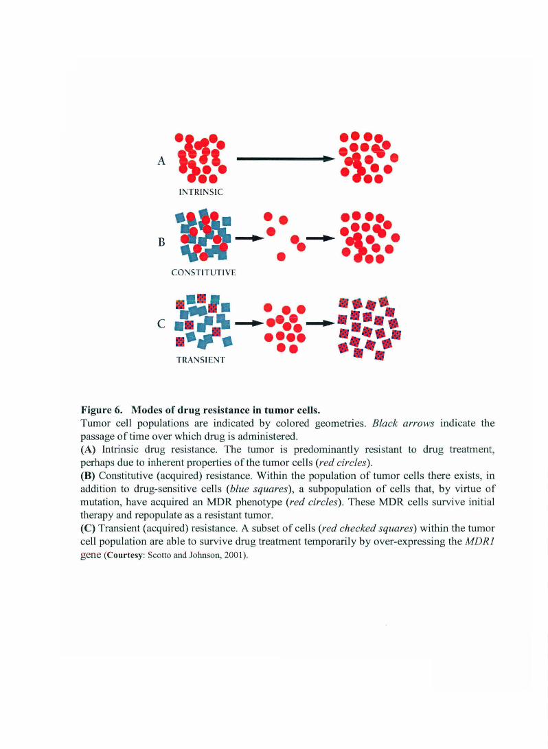
kidney, intestine and blood-brain barrier). Several observations suggest a role for the MDRI
gene in both intrinsic, transiently induced or acquired drug resistance (Figure 6). Amongst
the higher eukaryotes though the expression of P-gp has been frequently examined,
regulatory mechanisms of this gene product are complex and still poorly understood.
Evidence from numerous studies indicate that expression and activity of P-gp can be
controlled either pre- or post-transcriptionally by a myriad of environmental influences. For
instance, protein kinase C activators which increase P-gp activity and drug resistance have
been found to enhance mdr 1 gene expression via both transcriptional and translational
pathways (Chaudhary and Roninson, 1992). Modulation in protein stability, plasma
membrane incorporation, mRNA stability and processing gene transcription and gene
amplification have each been reported for P-gp (Shen et aI., 1986; Gottesman et at., 1995;
Sharom, 1997). Of these, alterations in P-gp expression that occur at the level of mRNA are
perhaps the most frequently observed. The increase in MDRI mRNA levels in tumor cells
can occur via gene amplification of the MDRI gene during selection of cells for resistance to
a single agent, enhanced MDRI transcription rates, transcriptional induction by transcription
factors and upon prolonged cellular exposure to several cytotoxic drug agents and by
stabilization of the MDRI mRNA.
1. 7.1.1 Gene rearrangement
A t(4q;7q) translocation in an adriamycin-selected human cell line resulted in a hybrid
mRNA containing sequences from both MDRI and a novel gene normally located on
chromosome 4. This gene rearrangement resulted in the activation of MDRI expression
mediated by promoter sequences found within the translocated chromosome 4 DNA.
Additional gene rearrangements have been identified in other MDR cell lines and in some
20
·~1· ••••• A · ·1~~· 1i·1• • • • •• •• It\:TRINSIC
•• •• • ••• ..~. .. • •• · ~1t • B ••• • •• CO;-.JSTITUTIVE
•• • • •• • ••• .... . ... \ c .. ~ .. .. ... \ .. •• • • ••• . : .... •• • ••• TRANSIENT
Figure 6. Modes of drug resistance in tumor cells. Tumor cell populations are indicated by colored geometries. Black arrows indicate the passage of time over which drug is administered. (A) Intrinsic drug resistance. The tumor is predominantly resistant to drug treatment, perhaps due to inherent properties of the tumor cells (red circles). (B) Constitutive (acquired) resistance. Within the population of tumor cells there exists, in addition to drug-sensitive cells (blue squares), a subpopulation of cells that, by virtue of mutation, have acquired an MDR phenotype (red circles). These MDR cells survive initial therapy and repopulate as a resistant tumor. (C) Transient (acquired) resistance. A subset of cells (red checked squares) within the tumor cell population are able to survive drug treatment temporarily by over-expressing the MDRI gene (Courtesy: Scotto and Johnson, 2001).

Introduction
patient tumors, suggesting that this may be a frequent mechanism for over-expression of
otherwise silent P-gp genes in acquired drug resistance.
1. 7.1.2 Gene amplification
Several studies have identified amplified MDRI DNA sequences from multidrug resistant
cell lines (Meyers et al., 1985; Fojo et al., 1987; Scotto et aI., 1988). The elevated 4.5-kb
MDRI messenger RNA levels during selection of human KB carcinoma cells as well as other
multidrug-resistant sublines of human leukemia and ovarian carcinoma cells selected in
colchicine, vinblastine, or adriamycin is associated with amplification of the MDRI DNA
sequences (Shen et aI., 1986). Increase in MDRI mRNA expression can also precede gene
amplification (Shen et al., 1986).
1. 7.1.3 Transcriptional regulation
1. 7.1.3.1 Cis-element and cognate binding factors
The MDRI gene contains at least two promoter regions (Veda et al., 1987a; 1987b;
Rothenberg et al., 1989). Veda et al. (1987a; 1987b) demonstrated that MDRI transcription
can be initiated from major downstream promoter (at positions -136 to -140) or from a minor
upstream promoter (at positions -155 to -180) which was found to be the case in colchicines-,
but not doxorubicin- or vinblastine-selected KB cells. The major downstream promoter is
used in most of the normal tissues.
""- MDRI belongs to a group of protein-encoding genes that lack a consensus T AT A box within "'-
c~ the proximal promoter region and this has been associated with the diversity of transcription
r::::. sites in eukaryotic cells (Maniatis et aI., 1987). Instead, basal transcription is directed by an -~:.<). initiator (1m) sequence that encompasses the transcription start site (+ 1) and most likely acts --£. as the position of nucleation of the RNA Pol II preinitiation complex (Figure 7). The first
identification of an 1m element in the MDRI promoter came from in vitro studies, which
~ showed that sequences from -132 to +82 were sufficient for accurate initiation and deletion of i..fi
sequences downstream of +5 decreased elongation of correctly initiated transcripts to I....." .....
~ undetectable levels (Cornwell, 1990). Subsequently, transient transfection studies indicated
\;:" that sequences between -6 and + 11 were sufficient for proper initiation of transcription in
vivo (Van Groenigen et al., 1993; Madden et al., 1993). Like most other "TATA-Iess" genes,
21
\ j , / .• ,- ,r i

Introduction
the MDRI promoter also includes both an inverted CCAAT element (-82 to -73) (Goldsmith
et al., 1993) and a GC-rich element (-56 to -43) that have been shown to interact with
members of the Spl family of transcription factors (Cornwell and Smith, 1993a; Thayer et
aI., 1996; Sundseth et al., 1997). Transfection analyses of promoter constructs mutated in one
or both of these elements indicate the involvement of the two elements in the constitutive
(i.e., operative under normal growth conditions) expression of MDRI in some cell lines.
A second GC-rich element has been identified (-110 to -103) that is unable to interact with
Spl and may be the site for an unidentified protein complex (Cornwell and Smith, 1993a).
Interestingly, immediately downstream and overlapping this GC-rich region is an inverted
MED-l element (Multiple start site Element Downstream 1; iMED; Figure 7), which was
first described in the hamster MDRI homolog, pgpl, and shown to be involved in the
activation of that gene in drug-resistant cells (Thayer, 1999).
Functional disruption of iMED, either through mutation of the element or the use of a
transcriptional decoy to sequester iMED-binding proteins, led to a decrease in constitutive
MDRI transcription in human neuroblastoma (Thayer, 1999) and leukemia (Marthinet et ai., 2000) cell lines; whether this element is also involved in induction of MDRI transcription in
resistant cells is under investigation. Consensus binding sites for AP-l, CEBP and Y -box
proteins in the 5' flanking region of MDRI genes have also been identified (Silverman and
Schrenk, 1997). Potential transcriptional regulatory elements found within the MDRI gene
includes several heat shock consensus elements, evidenced by an enhancement of MDRI
mRNA after heat shock treatment and a phorbol ester response element (Angel et aI., 1987;
Chin et al., 1990a). The mouse and hamster mdr promoter DNA sequences differ
significantly from that of the human. This difference is generally thought to be the reason for
the enhanced responsiveness of the rodent promoters to certain kinds of environmental
stresses. The murine promoters contain both TAT A and CAAT boxes as well as putative SP-
1, AP-l and AP-2 sites (Hsu et aI., 1990; Raymond et al., 1990; Ikeguchi et al., 1991; Cohen
et aI., 1991).
Studies show that there IS more than one mdrla promoter, which, in combination with
alternative polyadenylation sites, may account for the multiplicity of mdrla transcripts
observed in mouse cells (Hsu et aI., 1990). In addition, elevated P-gp expression has been
observed in the gravid mouse uterus which suggests hormonal influence on expression
22

Introduction
(Arceci et at., 1988 and 1990). Interestingly, the mdr 1 b promoter which drives P-gp
expression in the adrenal and secretory glands of the endometrium, contains a progesterone
response element (Cohen et at., 1991).
1.7.1.3.2 Transcriptional Inducers of hMDRl
By far, the most studied regulation of MDRI is at the level of transcriptional induction. From
the earliest studies of P-glycoprotein function and regulation, it has been suggested that P
gp is a 'stress-response gene' since its activity can be modulated by environmental adversity
(Scotto and Egan, 1998). MDRI mRNA level is have been show to be enhanced by various
factors, including hormones and environmental circumstances. Induction is seen after heat
shock, treatment by heavy metal (Chin et at., 1990a), differentiation agents such as sodium
butyrate and retinoic acid, or chemotherapeutic agents (Scotto and Egan, 1998), or III
response to carcinogens in the liver (Fairchild et at., 1987; Burt and Thorgeirsson, 1988;
Bradley et at., 1992). Treatment with cytotoxic agents (Fardel et ai., 1996a), ultraviolet
radiation, or partial hepatectomy has also been shown to increase MDRI expression in both
rodent and human cell lines (Thorgeirsson et ai., 1987; Burt and Thorgeirsson, 1988;
Mickley et at., 1989; Chin et ai., 1990b; Marino et aI., 1990; Uchiumi et ai., 1993;
Chaudhary and Roninson, 1993). Cadmium chloride and sodium arsenite can also induce
MDRI mRNA (Chin et at., 1990a and 1992) and the induction was found to be sensitive to
actinomycin D, therefore it requires synthesis of RNA (Chin et aI., 1990b). The effects of the
increase in MDRI expression seen following exposure to anti-neopiastic agents, including
those which are not P-gp substrates, persisted for several weeks following removal of the
drug (Chaudhary and Roninson, 1993). There have been reports that non-specific protein
kinase inhibitors block drug-mediated MDRI induction. However, recent work by Parissenti
et al. (1999) showed no apparent change in P-gp levels or cellular sensitivity by inhibition of
PKA activity to block MDRI gene expression in adriamycin-resistant MCF-7 breast cancer
cells. MDR-CAT reporter gene constructs have demonstrated that mutant Ras as well as both
mutant and wild type p53 genes may stimulate the MDRI promoter (Chin et at., 1992; Zhou
and Kuo, 1998). There are also suggestions of transcriptional regulation of MDRI by the c
Raf signal transduction pathway (Cornwell and Smith, 1993b).
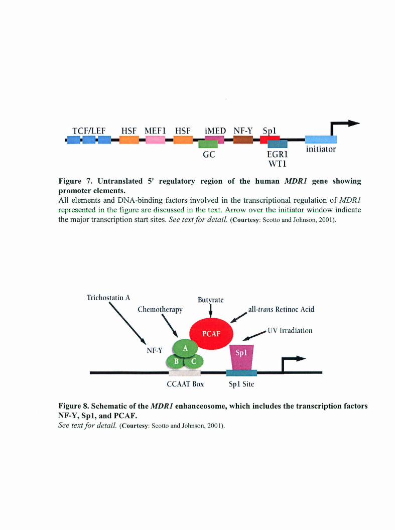
GC EGRl WIl
Figure 7. Un translated 5' regulatory region of the human MDRI gene showing promoter elements. All elements and DNA-binding factors involved in the transcriptional regulation of MDRl represented in the figure are discussed in the text. Arrow over the initiator window indicate the major transcription start sites. See text for detail. (Courtesy: Scotto and Johnson, 2001).
Triehostatin A
NF-Y
CCAAT Box
/ all-trailS Retinoe Acid
~ UV Irradiation
Spl Site
Figure 8. Schematic of the MDRI enhanceosome, which includes the transcription factors NF-Y, Spl, and PCAF. See text for detail. (Courtesy: Scotto and Johnson, 2001) .

Introduction
Furthermore, there are also evidences that MDRl mRNA may be co-induced by 2,3,7,8-
TCDD, which is also a potent inducer of the CYP4501 gene family (Burt and Thorgeirsson,
1988). In addition, treatment of human colon cancer cell lines with differentiating agents
such as dimethyl sulfoxide or sodium butyrate has been show to elevate MDRl gene
expression (Mickley et al., 1989). Recent studies indicate that the signals from all of these
divergent stimuli converge on a region of the MDRl promoter that we refer to as the MDRl
enhanceosome (Jin, 1998; Hu et al., 2000 and unpublished work) (Figure 8). This region
includes binding sites for the trimeric transcription factor NF-Y and the Sp family of GC
binding transcription factors. Together, these DNA-binding proteins recruit the histone
acetyltransferase PCAF to the MDRl promoter, resulting in the acetylation of promoter
proximal histones and subsequent transcriptional activation that is likely mediated by further
chromatin remodeling. Although the mechanism by which each agent transduces the signal
that results in promoter activation has yet to be determined, the role of the MDRl
enhanceosome in the regulation of transcription by a variety of stimuli makes it an attractive
target for therapeutic intervention.
1. 7.1.4 Post-transcriptional regulation: evidence for increased MDRl mRNA stability
While much of the focus on understanding the normal regulation of MDRl gene expression
in tissues is at the transcriptional level, there is growing evidence that suggests enhanced
mRNA stability plays a key role in this regulation, most especially in those tissues which
normally express the MDRl mRNA. The observed association between the localization of
the MDRl gene product, P-gp, to the lumenal surface of hepatocytes, and the enhanced P-gp
expression during rat liver carcinogenesis (Fairchild et aI., 1987; Thorgeirsson et aI., 1987;
Nakatsukasa et aI., 1992; Bradley et aI., 1992) which correlated with the process of tumor
progression (Bradley et aI., 1992; Nakatsukasa et al., 1992) has prompted studies on the
regulation of P-gp in the liver as a model system to identify possible triggers whereby the P
gp system is dysregulated. Several important studies using the rat liver system led to the
realization that mRNA stability was involved in P-gp over-expression. First, results by Lee et
al. (1993) using gene specific probes generated fiom the 3'UTRs of each rat P-gp gene
demonstrated that only one member of the P-gp gene family, class II P-gp, was strongly
induced when hepatocytes were grown in primary culture. In addition, this over-expression
24
Introduction
paralleled that of the cytoskeletal genes: actin and tubulin and suggested a possible common
regulatory mechanism in primary rat hepatocytes. Second, the class II P-gp, which is
expressed at a very low level in normal liver, has also been shown to be over-expressed in
several models of rat liver carcinogenesis. By nuclear run on assays and mRNA decay
studies, it was demonstrated that an increased rnRNA stability is the major mechanism
involved in the increased expression of class II P-gp. Moreover, studies using various drugs
also indicate that the integrity of the cytoskeleton is important for the maintenance of high
expression of class II P-gp. Disruption of the cytoskeleton in cultured hepatocytes with
cytochalasin D did not affect the transcriptional activity of the class II P-gp gene but rapidly
destabilized its rnRNA. It is possible that an association between class II P-gp RNA and
cytoskeletal elements may underlie the mechanism that regulates class II P-gp mRNA
stability (Lee et ai., 1993). Perhaps the strongest evidence which supports mRNA stability
came from the follow-up studies by Lee et ai. (1993) where they directly measured the in
vivo mRNA half-life and performed nuclear run analysis of class II P-gp mRNA and a
diverse group of unrelated genes which they have previously determined to be over
expressed in primary monolayer of adult rat hepatocytes during culturing (Lee et at., 1993).
Interestingly, in 1998 their published results showed that these same transcripts were also
over-expressed and stabilized in primary liver cancers and transplantable tumors (Lee et ai.,
1998). Their findings led them to suggest that increased mRNA stability is a primary
mechanism contributing to over-expression of P-gp and other genes in rat liver tumors.
Although this study focused on the rat counterpart of MDRl, it adds support to the
knowledge that MDRI mRNA stability, although it still remains less characterized, may be a
potential mechanism of MDRI mRNA over-expression in human liver tumors. An important
point emerged from this study is the observed global mRNA stabilization of other genes
besides MDRI. Does stabilization of the mRNA represent a characteristic mechanism for up
regulation of P-gp and other genes in rat liver tumors? If so, does this same mechanism
operate in the human liver system? However, to date, no current knowledge of the human
MDRI mRNA stabilization in vivo has been shown. However, several liver cancer cell lines,
such as the human hepatoma line (HepG2) are now available to address this issue.
1. 7.1.5 Determinants of MDRI mRNA Stability/decay
25

Introduction
1. 7.1.5.1 Cis-elements: RNA sequences
Specific cis-elements contribute to the steady-state level ofmRNA by promoting degradation
or stabilization (Klausner et al., 1993; Ross 1996). Sequences that function as determinants
to promote MDRI mRNA degradation have been identified in all regions of the transcript,
such as the 5' Untranslated region (5' UTR), the protein coding region, and the 3'
Untranslated region (3'UTR) (Figure 9). Among these, best characterized regions of MDRJ
mRNA stability are located in the 3'-untranslated region. In fact, 3'UTR can direct mRNA
decay independent to that of the remaining regions of the mRNA (Ross, 1996). 3'UTRs
consist of sequence motifs which were found to be important in cellular processes such as
sub-cellular localization, translation efficiency and mRNA stability (Ross, 1996).
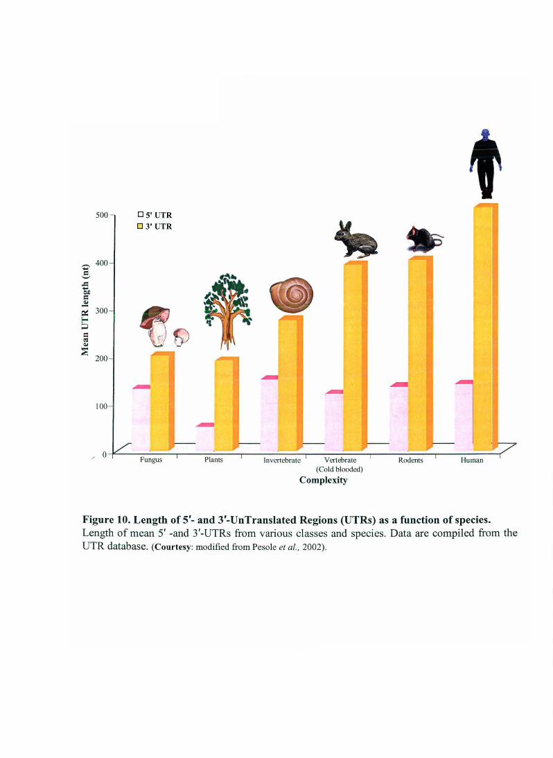
Interestingly, length of 3'- as well as 5'-UTR varies as a function of species (Figure 10). The
3'UTR of the human multidrug resistance type 1 (hMDRl) mRNA is approximately 380
nucleotides long and is very AU-rich (70%). The MDRI 3'UTR shares sequence similarities
to the 3'UTRs of rapidly degraded mRNAs. By far, the best characterized mRNA decay
determinants are adenylate-uridylate (AU) sequences in the 3'UTR. A large number of
unstable mRNAs have been shown to have AU-rich sequences contained in their 3'UTRs
(Rajagopalan and Malter, 1997; Maurer et al., 1999; Bloch, 1999).
However, not all sequences have comparable destabilizing effects on reported mRNAs.
MDRI mRNA contains AU sequences which are similar to rapidly degrading mRNAs of the
c-myc, clos and the lymphokines. A destabilizing function for MDRI has been proposed
(Marino et aI., 1990; Chen and Shyu, 1994) and previously published work has provided the
first test of this hypothesis. Other studies suggested that the MDRI mRNA decays with a
half-life of 10 hours or longer (Muller et aI., 1995).
Previously published work of Prokipcak et al. (1999) has shown that the MDRI 3 'UTR does
not behave as a classic ARE cis-element i.e. it does not function as an active mRNA
destabilizer in human hepatoma cell line, HepG2. MDRI mRNA has an intermediate half life
(8 h) compared to that of the c-myc (30 min) or GAPDH mRNA (> 24 hr). Furthermore, this
half-life was prolonged (>20 h) upon exposure to cycloheximide, a protein synthesis
inhibitor, suggesting the requirement of protein synthesis. Using B-globin chimeric mRNAs,
whereby the 3'UTR of c-myc or MDRI was fused to the B-globin coding region, it was
26

Translational control Subcellular localization Stability
I \ ~ IRES I Polyadenylation
5'
@
Interacting protein
, , "
5' UTR
An~ 11\ ~~ AAUAAA-)
3'
3'UTR
Figure 9. The generic structure of a eukaryotic mRNA, illustrating some post-transcriptional regulatory elements that affect gene expression. Abbreviations (from 5' to 3'): UTR, UnTranslated Region; m7G, 7-methyl-Guanosine cap; hairpin, hairpin-like secondary structures; uORF, upstream Open Reading Frame; IRES, internal ribosome entry site; CPE, Cytoplasmic Polyadenylation Element; AAUAAA, polyadenylation signal (Courtesy: Mignone et. al., 2002).
Z' == '-'
..c: .... Of)
== ~ ~ Eo-< ;J
== 4'S ~
~
500 OS' UTR 03' UTR
400
300
200
100
f))
O~--~~---.----~----r-----~---'----~---''-----~--'---------~ Plants Invertebrate Vertebrate Rodents Human
(Cold blooded)
Complexity
Figure 10. Length of 5'- and 3'-UnTranslated Regions (UTRs) as a function of species. Length of mean 5' -and 3'-UTRs from various classes and species. Data are compiled from the UTR database. (Courtesy: modified from Pesole et aI. , 2002).

Introduction
demonstrated that the c-myc 3'UTR, as predicted, destabilized the globin mRNA. However,
the MDRI 3'UTR had no effect on the stability of the globin mRNA.
1.7.1.5.2 Trans-acting factors: RNA-binding proteins
The cis determinants discussed above playa role in determining the stability or decay of
most mRNAs. However, a number of studies have show that although these elements may
target an mRNA for degradation there are cases whereby a stimulus (such as a drug,
hormone, intracellular factors) can in fact modulate the inherent mRNA half-life (Williams et
al., 1993; Tian et al., 1998; Bloch, 1999; Blaxall et al., 2000; Liu et al., 2000). In majority of
the studies to date, it has been shown that RNA-binding proteins form the primary means of
regulating this half-life by their ability to bind to these elements or structures (Siomi and
Dreyfuss, 1997). The nuclear RNA processing reactions mediated by interactions between
proteins and relatively short cis-acting elements can be found almost anywhere along the
RNA. However, an overriding majority of these interactions which regulate mRNA
expression in the cytoplasm is regulated via the 3'UTRs (Cusack, 1999).
Competition analysis showed that the MDRI 3'UTR had a five fold lower affinity for AU
binding proteins which are known to interact with c-myc AU-rich 3'UTR (Prokipcak et aI.,
1999). Overall the data suggest an association between the affinity of AU-binding proteins to
the 3'UTR and the degree of MDRI mRNA stability. Thus, stability of the MDRI mRNA is
the result of the interaction between specific cis-elements in its 3'UTR and trans-acting
factors (RNA-binding proteins). The uniqueness of this interaction may contribute in part to
the observed differences in mRNA half-life between MDRI and other genes bearing similar
cis elements.
1.7.2 Regulation of MDR in S. cerevisiae
Molecular mechanism underlying the up-regulation of PDR genes in the development of
multi drug resistance is well described in the baker's yeast S. cerevisiae, wherein several drug
extrusion pumps like PDR5 Celeiotropic Drug Resistance), SNQ2 (Sensitivity to 4-
Nitroquinoline N-oxide) and YORI (Yeast Oligomycin Resistance), etc. have been implicated
in the development of drug resistance. In S. cerevisiae MDR is known as PDR O~leiotropic
Drug Resistance). Three networks of genes, PDR (Eleiotropic Drug Resistance), YRR (Yeast
27
Introduction
Reveromycin A Resistance) and YAP (Yeast AP-l like factor) are mainly involved in
multidrug resistance. More recently a repressor of grug resistance 1 (RDRI) has also been
identified. Over-expression of yeast multidrug extrusion pumps results from over-production,
genetic alterations (Y AP network) and or spontaneous point mutations (PDR network) in
regulatory factors governing their expression. Some drug efflux transporters are also induced
in the Msn2p-dependent manner (Wolfger et aI., 2004). Msn2p is the Cys2His2 Zn finger
protein acting as a regulator in the general stress response pathway (Martinez-Pastor et al.
1996). The major determinants of multidrug resistance in S. cerevisiae involve the
transcriptional regulator PDRI and PDR3, which control the expression of ABC pumps,
MFS transporters and other genes. Spontaneous dominant mutations which activate the
master transcription regulators Pdrl p and Pdr3p result in spectacular multidrug resistance
phenotypes (Balzi & Goffeau, 1995) which either actively extrude drugs out of the cell
(Kolaczkowski & Goffeau, 1997) or modify their passive diffusion into the cell by altering
the lipid composition of the cell bilayer.
1.7.2.1 The PDR network
The PDRI and PDR3 genes encode highly homologous transcription factors involved in the
control of multidrug resistance in the yeast S. cerevisiae. Both genes belong to the family of
Gal4p-like proteins characterized by the N-terminal DNA binding domain of the Zn(II)2-Cys6
binuclear cluster DNA-binding motif with the consensus sequence of CySX2CysX6CySXs_
12CysX2CySX6_SCys (Delahodde et aI., 1995). They recognize the consensus sequence
5'TCCGCGGA3' designated as PDRE (PDR Responsive Element) present in the promoters
of target genes. The target genes code for the A TP binding cassette (ABC) transporters
(PDR5, SNQ2, YORI, PDRIO, PDRII, PDRI5, BATJ) and the transporters (HXT9, HXTII,
FLRJ) belonging to the major facilitator super-family (MFS) (Figure 11). Their over
expression due to mutations either in the PDRI or PDR3 genes results in the increased efflux
of cytotoxic compounds from the cells and establishment of multidrug resistance. The PDR
network is currently known to comprise 10 transcription factors regulating about 70 different
target genes. In this network, the Pdrlp transcription factor has the largest set (about 30) of
potential targets.
Despite their structural (36% identity, 57% similarity over the entire amino acid sequence)
and global functional similarities, Pdrl p and Pdr3p play distinct roles. Disruption of PDRI
28

Ngg1p Pdr1Jp 0 hOQ
\ I ) Pdr1p ----.... Pdr3p
Ucp2p War1p Pdrap Yrr1p Yap1p Yapap Rdr1p Msn2p Stp5p Yrm1p
Figure 11. The PDR network. Genes in the centerline represent target genes of transcriptional regulators depicted above and below. Note that the cartoon only lists genes of the ABC family. The yeast PDR network also contains non-ABC genes whose function has not established in many cases (see text for details). Red lines indicate a negative regulatory impact, while black lines ending with an arrow indicate positive regulation (Courtesy: Jungwirth and Kuchler, 2006).

Introduction
has a more pronounced effect on drug sensitivity than disruption of PDR3. However,
sensitivity to a set of drugs, such as rhodamine 6G or diazoborine (Kolaczkowska & Goffeau,
1999) is more affected by the inactivation of the PDR3 gene. The role of PDR1 is well
documented by genetic experiments showing that the PDR1 locus is associated with
resistance to more than 20 structurally unrelated inhibitors of both cytoplasmic and
mitochondrial function (Kolaczkowska & Goffeau, 1999) or with permeability to small
molecules like estradiol. Single point mutations in the PDR1 locus (pdr 1-2, pdr 1-3, pdr 1-6,
pdrl-7, pdrl-8) increase to various extents the mRNA level of downstream target genes,
such as PDR5, SNQ2, YOR1, PDR10, PDR15 and the newly identified PDR16. Similarly,
PDR3 mutant alleles (pdr3-2 to pdr3-1 0) have been shown to activate the expression of the
PDR5, SNQ2, PDR15 and PDR3 genes (Kolaczkowska and Goffeau, 1999).
Recent DNA microarray analysis has shown that the transcription levels of several soluble
proteins (which functions not obviously related to cellular efflux of drugs) are also regulated
by PDRlIPDR3 genes. This signifies the universal role of Pdrlp and Pdr3p in regulating
varied physiological functions of S. cerevisiae. Remarkably, Pdrl p and Pdr3p can positively
or negatively regulate expression of target genes, implying that additional factors modulate
their activity (Martens et al., 1996; Saleh et aI., 1997).
1.7.2.1.1 Molecular mechanism of PDR network activation upon exposure to antifungal drug
In S. cerevisiae, mutation of a single nucleotide in the genes encoding the transcription
factors Pdrl p or Pdr3p could initiate multi drug resistance; these factors control the
expression of several ABC efflux pumps (Carvajal et al., 1997; Nourani et aI., 1997; DeRisi
et aI., 2000). Rapid and transient activation of drug-efflux genes is also observed8 when
fungi are treated with high concentrations of antifungal drugs. Thakur et al. (2008) find that
various antifungal drugs and other xenobiotics (alien substances) can bind directly to a
hydrophobic domain (XBD) in Pdrlp and Pdr3p, and that this binding leads to drug
dependent activation of drug-efflux genes. Thakur et al. (2008) make another intriguing
observation: deletion of the gene encoding the GallI protein decreases drug-dependent
expression of Pdr5p and other drug-efflux pumps. GallI p is one of the 20 subunits of the
yeast Mediator complex, which recruits the RNA polymerase II machinery for the
transcription of specific sets of genes. Thakur and colleagues (2008) show that the activation
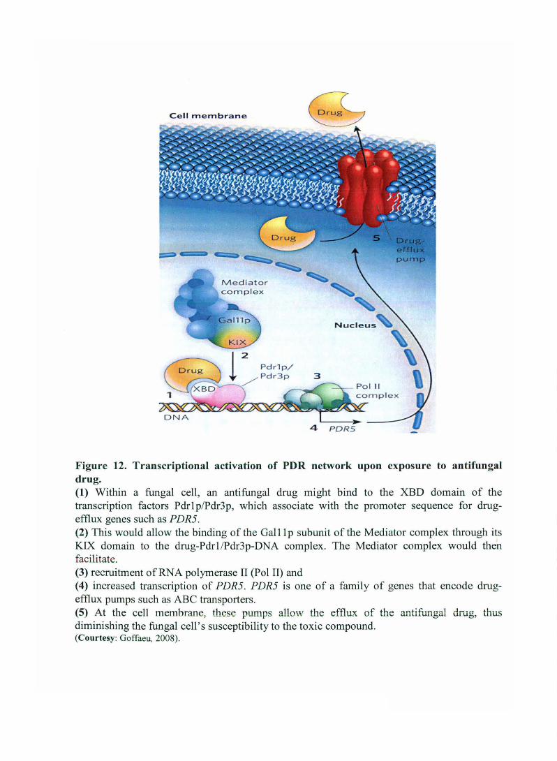
Cell membrane
4 PDRS
Figure 12. Transcriptional activation of PDR network upon exposure to antifungal drug. (1) Within a fungal cell, an antifungal drug might bind to the XBD domain of the transcription factors Pdrl plPdr3p, which associate with the promoter sequence for drugefflux genes such as PDR5. (2) This would allow the binding of the GallI p subunit of the Mediator complex through its KIX domain to the drug-PdrllPdr3p-DNA complex. The Mediator complex would then facilitate. (3) recruitment of RNA polymerase II (Pol II) and (4) increased transcription of PDR5. PDR5 is one of a family of genes that encode drugefflux pumps such as ABC transporters. (5) At the cell membrane, these pumps allow the efflux of the antifungal drug, thus diminishing the fungal cell's susceptibility to the toxic compound. (Courtesy: Goffaeu, 2008).

Introduction
domain of Pdrl p/Prd3p at their carboxyl termini interacts with the KIX domain of GallI p,
and that this interaction is enhanced by antifungal drugs (Figure 12).
More over, they show that the yeast KIX domain is structurally similar to its mammalian
counterpart, which can also bind to yeast Pdrl p. Mammalian KIX domains interact with
specific transcription factors known as nuclear receptors, which affect cholesterol
biosynthesis and other aspects of lipid homeostasis. Similarly, in yeast, several aspects of
lipid homeostasis depend on Pdrl p/Pdr3p (DeRisi et aI., 2000), and this process is now
shown to be regulated by the KIX domain (Thakur et al., 2008). With the discovery of the
involvement of new molecular domains (XBD in Pdrlp/Pdr3p and KIX in Gaillp) in the
pathway leading to antifungal-drug resistance, Thakur and colleagues (2008) argue that there
is now reason to redirect the long-standing search for small molecules that inactivate
resistance (Lamping et aI., 2007). But finding such small molecules to prevent drug
Pdrlp/Pdr3p interaction might be difficult, because this interaction is of limited specificity.
Nonetheless, the successful use of the oestrogen-receptor antagonist tamoxifen in the
treatment of breast cancer indicates that small molecules antagonizing oestrogen-PXR
interaction could be found. The similarity between drug-Pdrl p/Pdr3p interaction and
oestrogen-PXR interaction gives reason to believe that drug-Pdrlp/Pdr3p interaction could
also be blocked with small molecules. And a broad screen, which is not restricted to the
blockage of drug-Pdrlp/Pdr3p interaction but can detect any inhibitor molecule that reduces
Pdrl/Pdr3p activity, has already been reported by Kozovska and Subik (2003).
1.7.2.1.2 Other PDR regulators
Pdrl p is post-transcriptionally regulated by Pdrl3p, which is the unique yeast member of the
Hsp70 protein family (Kolaczkowska and Goffeau, 1999). Pdrl3p regulates the function of
Pdrlp but not ofPdr3p (Figure 11). Over-production or gain-of-function mutation (S295F) of
Pdr13p elevates Pdrl p wild-type activity. Yeast cells carrying inactivated PDRJ3 gene
exhibit cold-sensitive growth defect. Phenotype analysis of the strain lacking the PDR13
gene shows its possible connection with the general-stress response system. In the pdr 13
knockout strain, the induced expression of several genes containing STRE elements being
under the control of the general stress response factors Msn2p and Msn4p, is observed.
Tested genes include CITl (cytoplasmic catalase), HSP 12 (heat shock protein) as well as
30

Introduction
CUP I (metalothionine). PDR6 gene has recently been shown to be a member of an important
RanBP7/importin-BICselp superfamily of Ran GTP-binding proteins. Since these proteins
are involved in nuclear transport, Pdr6p is implicated to be a novel candidate protein
involved in the regulation of nuclear transport of transcription factors involved in multidrug
resistance (Capieaux et ai., 1991; Gorlich et at., 1997; Pennisi, 1998). Two loci, PDR7 and
PDR9, both localized on chromosome II, were reported to affect the transcription of the
PDR5 gene (Dextar et at., 1994). Mutants of both genes, isolated as spontaneous suppressors
of the hypersensitive pdr I knockout derivative restore resistance to cycloheximide and
sulfometuron methyl. Since these genes have not yet been identified within the sequence of
chromosome II, it is difficult to predict their role within the PDR network.
Genomic approaches uncovered another zinc finger regulator, Pdr8p, as involved in the PDR
network (Hikkel et at., 2003). Pdr8p mediates resistance to ketoconazole and oligomycin,
operating mainly through Yrrlp and its respective target genes. Recently another
transcription factor, Rdrlp (Repressor of grug resistance 1; YOR380W), belonging to the
Gal4 family of zinc cluster proteins has been identified to be a repressor of PDR5 gene via
PDRE, a cis-regulatory element also shared by PDRIIPDR3 (Hellauer et at., 2002).
I1rdr I I I1rdr I strain has increased resistance to cycloheximide, as expected from the over
expression of PDR5. In addition, the activity of a PDR5-tacZ reporter is increased in I1rdrl
strain. Also it has been shown that Rdrlp interacts with the PDREs and mutations known to
reduce binding of PdrllPdr3p abolished the induction observed in the I1rdrl strain (Hellauer
et at., 2002). Heterodimers of Rdrlp with Pdrlp or Pdr3p compete with Pdrlp/Pdr3p for
binding to PDREs (Hellauer et ai., 2002). Akache and Turcotte (2002) have done by the
phenotypic analysis of 33 genes encoding yeast zinc cluster proteins in order to better
understand their functional role in MDR phenomenon. In this endeavor, they have identified
that deletion mutants of nine such genes- RDSI, UPC2, STB5, ECM22, HAL9, YILI30W,
YKL222C, RDS2, and RDS3, are either resistant or sensitive to at least one drug. Of these
deletion of STB5 greatly decreased RNA levels of SNQ2 and PDRl6 (and to lesser extent
PDR5), thereby suggesting that Stb5p activates the expression of MDR genes (Akache et ai.,
2004). Stb5p activates transcription by binding to PDREs, the same elements found to be
critical for Pdrlp and Pdr3p activity. Stb5p acts predominantly within a Pdrlp heterodimer,
Introduction
while no interactions occur with Pdr3p or YrrIp, the latter is only present as a homodimer
(Akache and Turcotte, 2002; Akache et aI., 2004).
Recently, yet another novel zinc finger regulator, Warlp, was identified as the mam
modulator of weak acid stress adaptation through transcriptional control of PDRI2 (Kren et
al., 2003). Warlp controls a rather small regulon (Schuller et al., 2004), forms homodimers,
is activated by stress-triggered hyper-phosphorylation (Frohner et aI., unpublished
observations), and decorates a cis-acting weak acid response element in the PDR12 promotor
(Kren et al., 2003, Schuller et ai., 2004). Ecm22p and Umc2p are members of the
Zn(II)2Cys6 transcription factors involved in regulation of membrane sterol homeostasis
(Crowley et al., 1998; Vik and Rine, 2001) through control of ABC transporters. Based on
genome-wide transcriptional profiling of a Upc2-1 gain-of-function mutant strain that
exhibits aerobic sterol influx, PDR11 and A US1 were identified as major determinants
required for uptake of free sterols under conditions of impaired ergosterol biosynthesis
(Wilcox et al., 2002).
The ABC transporter Pdrl5p is induced upon various stresses, including heat shock, high
osmolarity and weak acid stress in an Msn2p-dependent manner (Wolfger et aI., 2004). The
Cys2His2 zinc finger protein Msn2p, a master regulator of general stress response pathway,
modulates a large set of genes in response to a variety of different environmental stimuli
(Gasch et aI., 2000). Interestingly, Pdr15p stress induction requires Msn2p (Wolfger et aI.,
2004), but bypasses upstream components of the HOG pathway (Seet and Pawson, 2004),
suggesting the existence of a novel as yet undisclosed HOG signaling branch converging at
the downstream Msn2p regulator (Wolfger et aI., 2004).
Moreover, Pdrl5p levels are strongly elevated in cells undergoing diauxic shift, while
expression of Pdr5p almost completely disappears (Marnnun et al., 2004; Wolfger et aI.,
2004). Thus, Pdrl5p and Pdr5p may perform non-overlapping functions related to
detoxification in different growth phases or under certain metabolic conditions.
1.7.2.2 YRR1 and YRM network
Yrr 1 was recently identified as a Zn2Cys6 transcription factor involved in control of the PDR
phenomenon (Cui et al., 1998). It was isolated by reveromycin A resistance screening (Cui et
al., 1998). Gain-of-function mutations in its activation domain confer high level resistance to
32
rntroduction
the cell cycle inhibitor reveromycin A, to the DNA-damaging agent 4-nitroquinoline-N-oxide
and to oligomycin (Zhang et al., 2001; Le Crom et aI., 2002). YRRI deletion leads to
hypersensitivity to 4-nitroquinoline-N-oxide (4-NQO) and oligomycin but not to
cycloheximide (Cui et al., 1998). SNQ2 gene is involve,d in YRRI-mediated NQO resistance,
as suggested by observations that SNQ2 confers resistance to 4-NQO (Decottignies et aI.,
1998; Servos et aI., 1993). Promoter deletions have also demonstrated that Yrrlp interacts
with the promoter region of SNQ2 (Zhang et aI., 2001). Yrrl affects oligomycin resistance by
activating YORI expression via a region in the YORI promoter that is very similar, but not
identical, to the PDRE elements recognized by Pdr 1 /Pdr3 and RDRI (Zhang et al., 2001).
Thus, YRRI is involved in complex PDR network regulation, directly activating SNQ2 and
YORl, both of which are common targets of Pdrl p, Pdr3p and another regulator of MDR,
Yaplp. As Pdrlp activates Yrrlp (Zhang et aI., 2001), it seems highly likely that YRRI plays
a major role in PDR regulation network. An intricate interplay of cross-regulation links
YRRl, PDRl, and PDR3. PDRI regulates PDR3 and YRRl, both of which regulate their own
expression (Delahodde et aI., 1995; Zhang et al., 2001), see Figure 13. In addition, different
post-translational processes, including nuclear targeting or heterodimer formation (Mamnun
et aI., 2002), may be involved in different regulatory pathways.
Genome-wide analyses have established that the sets of target genes directly regulated by
these three transcription factors display considerable overlap (DeRisi et aI., 2000; Devaux et
al., 2001; Le Crom et aI., 2002). Most of these co-regulated genes encode for proteins
involved in the structural organization of the plasma membrane. These proteins include ABC
and MFS transporters, together with proteins involved in the control of specific steps in lipid
biosynthesis. Accordingly, the drug resistance phenotypes conferred by the gain-of-function
mutations for the three transcription factors reflect changes in plasma membrane
composition. Thus, discrete changes in plasma membrane properties can be accomplished
through sophisticated cross-regulations between several transcription factors, not all of which
have been identified (Akache & Turcotte, 2002; Moye-Rowley, 2002).
YRM 1 (Yeast Reveromycin resistance Modulator) encodes another Zn(II)2Cys6 regulator
acting as a specific inhibitor of Yrrlp.\ In the absence of Yrrlp, Yrmlp activates the
transcription of most genes regulated by Yrrl p, reflecting the high level of complexity of the
regulatory processes controlling drug resistance phenotypes in yeast (Lucau-Danila et aI.,
33

---------- ..... , , , , , , \
OIlI!t)Pl.\tln
o
S'li 1
\ \ ,
MFS Permease
, ,
YAP I
LRI
Y PI
4-;>o.QO~
t
o
YL
Metabulism
I I
Figure 13. The YRRI regulation network is linked to the PDR network.
I , I ,
All the regulated genes found to be activated by Yrrlp fall into the three main characterized functional groups: MFS permease, ABC transporter, and metabolic genes. For all these genes, the consensus PDRE sequence is shown as an open circle. Most of these genes are also regulated by Pdrl p, Pdr3p, and Yap 1 p. These transcription factors control the expression of other transcription factors (dotted arrows) or, in some cases, their own expression (YRRI and PDR3). Finally, the most common drugs used (4-NQO and oligomycin) are marked next to the genes associated with specific resistance. (Courtesy: Le Crom et at. , 2002) .

Introduction
2003). YRM1 is homologous (41 % identities) to YRR1. The degree of similarity is higher in
the zinc finger domain, but the two proteins display considerable similarity throughout the
entire length of their sequences. The sequences in the Cys regions are quasi-identical
suggesting that the two factors recognize similar DNA binding motifs.
Microarray experiments were carried out to determine whether Yrm1 was a transcription
factor and to identify the corresponding direct target genes. The situation turned out to be
more complex than expected: Yrm1 acts as a transcription factor and interacts with the
promoter of the target genes only in the absence of Yrr 1. The sets of target genes directly
regulated by YRR1 or YRM1 are similar, but not identical (Figure 14). Alternative promoter
occupancy thus plays a key role in this cross-regulation adding a new degree of complexity to
the cell drug response (Akache & Turcotte, 2002).
1.7.2.3 YAP network
Yap 1 p, the first member of the family of Yaps to be described, was initially identified by its
ability to bind and activate the SV-40 AP-l recognition element (ARE: TGACTAA). Based
on its ARE binding capacity, this factor was purified as 90Kda protein and the corresponding
gene was cloned by screening a Agt 11 library with a monoclonal antibody against Yap 1 p
(Harshman et ai., 1988). Subsequently this gene was also found in multi-copy transformants
resistant to the iron chelators 1,1 O-phenanthroline and I-nitroso-2-naphtol (Schnell et ai.,
1992) as well as to a variety of drugs including 4-nitroquinoline-N-oxide, N-methyl-N-nitro
N-itrosoguanine, triaziquone, sulphomethuron methyl and cycloheximide, the locus being
historically designated PARI1SNQ3/PDR4 (Hertle et ai., 1991; Hussain and Lenard, 1991;
Haase et aI., 1992). Yap 1 p have also been implicated in ABC gene regulation, although their
precise role in PDR or stress response has not been unraveled so far (Moye-Rowley, 2003;
Rodrigues-Pousada et aI., 2004). Yaplp decorates promoters of SNQ2 and YCFl to
modulate their expression (Moye-Rowley et ai., 1989; Wemmie et aI., 1994).
Notably, another member of the b-ZIP family, Yap8p, also participates III arsenite
detoxification by regulating Y cfl p expression (Wu et ai., 1993; Bobrowicz et ai., 1997).
Interestingly, recent studies revealed a link between Yap 1 p and the aging process, because
over-expression of YAP 1 significantly improves survival during chronological aging in yeast
cells (Herker et ai., 2004), possibly through Y cfl p expression regulation (Jungwirth et ai.,
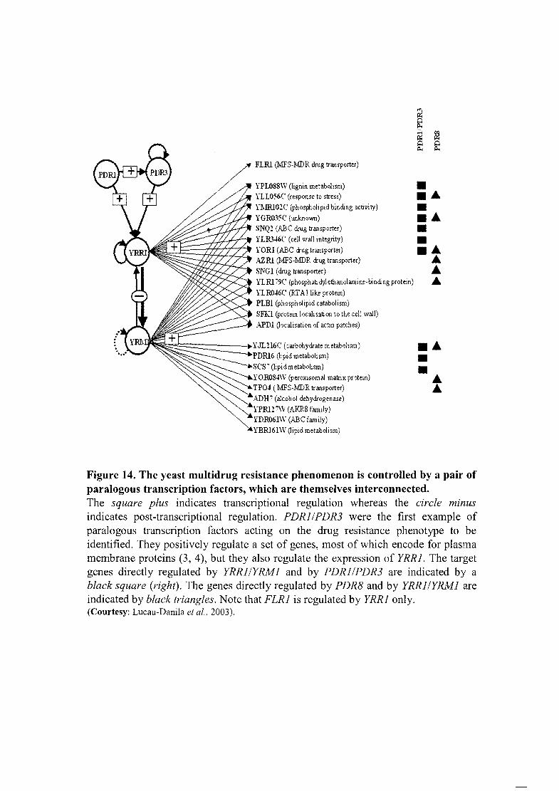
,. FLRl (MFS·MDR drug transporter)
!PLOSS\V (lignin metabolism) YLL056C' (response to stress) Yl\IRl01C (ph osphohpld binding activity) YGRO~5(, (WlknOwn) SNQ! (ABC drug transporter)
I~§~~~~~ YLR'46(, (celt wall integrity) \' YORI (ABC aug transporter) \' AZRl (MFS·MDR aug transporter)
SNGI (drug transporter) YLRl79(, (phosphab dylethanolamine·btnding protein)
HR046C (RTAJlikeprotein) PLBl (phospholipid catabolism)
• SITl (protein locali sati on to the cetl wall) • APDI Oocaltsatlon of actin patches)
~=::::::======:-I~YJL216(, (carbohydrate metabolism) ~PDRl6 Oipid metabohsm) "SC'S7 (tipidmetabolism)
YOROS4\v (peroxisomal matrix protein) TP04 (l:v1FS-MDR transporter)
"ADH' (alcohol dehydrogenase) "YPRlnV (AKR8 family)
YDR061W (ABC family) :4 YBRl6l\V Oipid metabolism)
• .~ • ... • • ... ~ .. ~
... • • .. ..
Figure 14. The yeast multidrug resistance phenomenon is controlled by a pair of paralogous transcription factors, which are themselves interconnected. The square plus indicates transcriptional regulation whereas the circle minus indicates post-transcriptional regulation. PDRlIPDR3 were the first example of paralogous transcription factors acting on the drug resistance phenotype to be identified. They positively regulate a set of genes, most of which encode for plasma membrane proteins (3, 4), but they also regulate the expression of YRRl. The target genes directly regulated by YRRlIYRMl and by PDRlIPDR3 are indicated by a black square (right). The genes directly regulated by PDR8 and by YRRlIYRMl are indicated by black triangles. Note that FLRl is regulated by YRRl only. (Courtesy: Lucau-Danila et aI., 2003).
Introduction
unpublished data). Besides YAP1, a second gene, YAP2, conferring resistance to 1,10-
phenanthroline in transformed cells over-expressing a multicopy yeast library, was also
described (Bossier et al., 1993). This gene encodes a 45 KDa protein that also binds the ARE
cis-acting element. Sequence homologies identified it as CAD1, due to the acquisition of
cadmium resistance in cells over-expressing a multi-copy genomic library (Hirata et aI.,
1994). Later, it was shown that these cells gain resistance to cycloheximide (Bossier et aI.,
1993; Wu et al., 1993). The sequence of the YAP 1 and YAP2 genes revealed the presence of a
bZip family domain in the N-terminus homologous to the true budding yeast AP-l factor
Gcn4p and to c-Jun in mammalian counterpart. Six more factors of b-Zip family YAP3-8
homologous to the YAP1 and YAP2 was identified by sequence alignment. The Yap family
binding site Was thus subsequently characterized as TTAC/GTAA (Fernandes et aI., 1997)
for Yaplp-Yap4p. So far the corresponding binding site for Yap5p-Yap8p has not been
characterized, although incase ofYap8p it appears to be TTAATAA (Wysocki et al., 2004).
1.7.2.3.1 YAP network and its interaction with PDR
Yaplp seems to be involved in stress-dependent activation of certain PDR genes, which
suggests a functional interplay between YAP and PDR networks in S. cerevisiae (Wu et al.,
1993). The PDR5 and SNQ2 genes, possess, in addition to PDRE, YAP response elements
(YRE) in their promoters (Miyahara et al., 1996; Fernandes et aI., 1997). The presence of a
functional YAP 1 gene was reported to be required for the expression of SNQ2 stimulated by
external stress conditions (Wolfger et al., 1997). Thus, at least one PDR target gene, SNQ2,
could be under the control of the general stress response pathway via Yap 1 p.
On the other hand, a S. cerevisiae strain overproducing Cap 1 p, the homologue of Yap 1 p
from C. albicans or its truncated version, not only shows dramatically enhanced mRNA level
of the MFS transporter-FLR1 but also slightly elevated level of the PDR5 transcript. Thus, it
seems that Yaplp could contribute to the control of 4-NQO and cycloheximide resistance via
genes encoding ABC transporters, such as SNQ2 and PDR5 and/or MFS transporters (Figure
15). The YAP1-mediated diazoborine and 4-NQO resistance are mainly dependent on the
PDR3 gene (Wendler et al., 1997). Furthermore the double pdrl pdr3 knockout strain is
hypersensitive to diazoborine even when YAP 1 is over-expressed, demonstrating a
requirement of intact PDR1 and PDR3 loci for Y API-mediated resistance to that drug
35
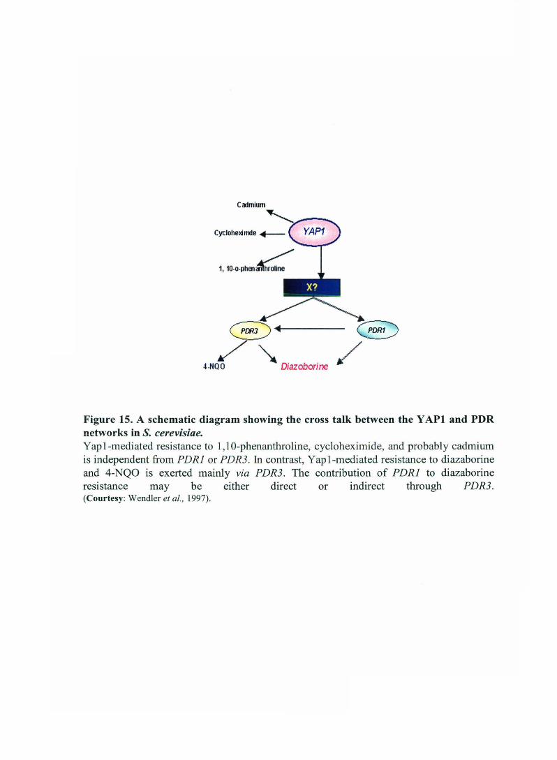
Cadmium
1, 10-0-Phen~
4-NQO ~ D' b ' lazo onne
Figure 15. A schematic diagram showing the cross talk between the Y API and PDR networks in S. cerevisiae. Yapl-mediated resistance to 1, lO-phenanthroline, cycloheximide, and probably cadmium is independent from PDRl or PDR3. In contrast, Yapl-mediated resistance to diazaborine and 4-NQO is exerted mainly via PDR3. The contribution of PDRl to diazaborine resistance may be either direct or indirect through PDR3. (Courtesy: Wendler et aI., 1997).

Table 5: Trans-regulatory factors controlling the PDR in S. cerevisae.
TFs Description Target genes Functions
Pdrl Zn(II)2Cys6 PDR3, PDR5, PDRIO,PDRI5, PDRI6, IPTGI, SNQ2, Activation of PDR genes YORI, HXT9, HXTll
Pdr3 Zn(II)2Cys6 PDR3, PDR5, PDRI5, PDRlO, SNQ2, YORI, HXT9, Activation of PDR genes HXTll
Pdr8 Zn(II)2Cys6 PDRI5, YORI Regulation ofPDR
Pdrl3 HSP 70 PDRI Post-translational Pdr1 p regulation homologue
Stb5 Zn(JI)2Cys6 PDR5,SNQ2 Regulation of PDR, Pdr 1 p-heterodimer
Warl Zn(II)2Cys6 PDR12 Regulation of weak acid stress response
Ecm22/ Zn(II)2Cys6 PDRII, AUSI Regulation of sterol biosynthesis Upc2
Rdrl Zn(II)2Cys6 PDR5, PDRI5, PSDRI6, PH084, YOR049C Repressor of drug resistance genes
Yrrl Zn(U)2Cys6 SNQ2, YORI, FLRI, AZRI, SNG 1, YLLo56C, YLR346C, Activation of PDR and repressor of Yrml YRMI
Yrml Zn(H)2Cys6 YRRI, SNQ2, YORI, AZRI, SNGI, PDRI6, TP04 Activation of PDR and repressor of Yrmi
Yapll bZip PDR5, SNQ2, YORI, YCFI, TRX2, GSHI, GLRI, FLRI, Oxidative stress and heavy metal stress Yap8 ATRI response
Msn2 Cys2His2 PDRI5 Regulation of general stress response
Nggl -- PDRI,PDR3 Inhibition ofPDRlp activity

Introduction
(Wendler et aI., 1997). This, together with the observation that Yaplp indirectly affects
diazoborine resistance suggests the existence of a Yap 1 p-dependent activation of Pdr3p (but
much less of Pdrl p) rather than direct Yap 1 p binding to the gene( s) responsible for multidrug
resistance.
1. 7.2.3.2 Complex interplay among MDR regulators in S. cerevisiae
The Gal4p family of yeast zinc cluster proteins comprises regulators of MDR genes in S.
cerevisiae as described above. These zinc cluster proteins contribute differently to the
regulation of specific PDR genes. For example, induced-expression of FLRl, a gene involved
in PDR (Alarco et aI., 1997), is dependent on Pdr3p but not Pdrlp (Broco et at., 1997).
Conversely, a deletion of Pdrlp greatly diminishes expression of PDR5 while removal of
Pdr3p has marginal effects (Mah€: et aI., 1996). Moreover, deletion of PDRl or PDR3 results
in increased or decreased expression of PDRl5 (a homologue of PDR5), respectively
(Wolfger et aI., 1997). This pattern of regulation is further complicated by the fact that Pdr3p
undergoes positive auto-regulation (Delahodde et aI., 1995), while expression of YRRl is
under the control of Pdrlp/Pdr3p and itself (Zhang et aI., 2001). Growth conditions also
modulate Pdr3p expression (Mamnun et aI., 2004). Moreover, activity ofYrrlp is negatively
regulated by over-expression of the zinc cluster protein Yrml p (Lucau-Danila et aI., 2003).
The observation that zinc cluster proteins form various combinations of dimers will be
invaluable in better understanding the complex regulation of PDR genes. Different
combinations of PDRl or PDR3 homo- and hetero-dimers may regulate the expression of
different genes, which might help explain how these two proteins act differently. For
example, Pdrl p and Pdr3p bind as homo- or hetero-dimers to pleiotropic drug response
elements (PDREs) found in promoters of target genes. Pleiotropic drug response elements
(PDREs) present in the promoters of their target genes, ABC transporters, as well as in the
PDR3 promoter have been shown to be important in the regulation of these genes. Pdrl p,
Pdr3p, Stb5p, and Rdrl p all act through this element, with Pdrl p, Pdr3p, and Stb5p able to
bind to an everted repeat CCGCGG. Characterization of PDREs in the PDR3 promoter
indicates its auto-regulation. Even though they act through the same element, all these zinc
cluster proteins may perform different functions.
To better understand the interplay among these activators, Akache et al.(2004) performed
native co-immunoprecipitation experiments using strains expressing tagged zinc cluster

4'i'U
III
Figure 16. A schematic diagram of interactions among zinc cluster PDR regulator of S. cerevisiae. Arrows indicate zinc cluster protein interactions identified. rhoo denotes petite mutant. See text for detail (Courtesy: modified from Akache et aI. , 2000) .

Introduction
proteins from their natural chromosomal locations as well as glutathione S-transferase pull
down assays and reveals that Stb5p was found predominantly as a Pdr 1 p heterodimer and
shows little homodimerization. No interactions of Stb5p with Pdr3p or Yrrlp was detected.
In contrast to Stb5p, Yrrlp is only detected as a homodimer. Importantly, the purified DNA
binding domains of Stb5p and Pdrlp were also bound to a PDRE as heterodimers in vitro.
These results indicates that the DNA binding domains of Pdrl p and Stb5p are sufficient for
heterodimerization. The interactions among these various transcriptional activators are
summarized in Figure 16.
In conclusion, these results demonstrate that the four zinc cluster protein activators of MDR
genes do not act individually. Instead, there is existence of a complex interplay among these
activators as they form various populations of homo- and hetero-dimers (Figure 16). There
may be differences in the binding specificity or activity of each of these populations,
allowing for a very specific and varied expression of genes involved in PDR. Pdrlp was the
only protein able to show multiple interactions indicating that it is similar to mammalian
nuclear receptor RXR in its ability to recruit various partners. With at least four different zinc
cluster proteins regulating the expression of PDR genes via different PDREs, the cellular
ability to respond to drugs is much more adaptable and flexible to fine tune the regulation of
MDR genes.
1.7.3 Regulation of MDR in C. albicans
C. albicans also harbors Pdr5p-like ABC pumps i.e. CDRl, CDR2 and Flrlp-like MFS
transporters i.e. CaMDRl. Hence it would appear that there would be some similarity in the
transcription regulatory factors governing multi drug resistance between C. albicans and S.
cerevisiae. However this does not appear to be the case as reports show that regulation of
expression is quite different in both the strains. In C. albicans at least two mechanisms lead
to clinical fluconazole resistance: alteration of the drug target (lanosterol 14-a demethylase)
as reviewed in a previous section and a decrease of drug accumulation associated with over··
expression of multidrug ABC transporters, such as CDRl, CDR2 or the MFS transporter
BENr/CaMDRl, see Figure 2 (Fling et al., 1991; Sanglard et al., 1995 and 1997). The latter
mechanism seems to be responsible for an early response developing rapidly after exposure
to fluconazole, whereas genetic alterations in target genes and/or trans-acting regulatory
37

Introduction
factors are also observed in long tenn treatment (Franz et al., 1998a). Several trans-acting
factors of C. albicans identified by functional complementation in I1PDRIII1PDR3 S.
cerevisiae strains were unable to regulate the MDR genes expression in C. albicans (Talibi
and Raymond, 1999). Yeast to hyphal transition has been associated to the virulence of C.
albicans and several regulatory pathways involved in yeast to hyphal transition (EFG] ,
CPHl, TVPl, NRGl) has been established, however no direct evidence for the evolvement
of these regulatory pathways in CDR] regulation has been established yet.
1.7.3.1 ERGll
Mechanisms of resistance to the azole antifungal agents that have been described for C.
albicans include increased expression of the ERG]] gene encoding 14-a lanosterol
demethylase. Mutations in ERG]] that interfere with the ability of the azole to bind to its
target can also confer resistance (White, 1997b; Franz et al., 1998a; Sanglard et aI., 1999b;
Kelly et aI., 1999a and 1999b; Lamb et al., 2000; Kakeya et aI., 2000; Perea et al., 2001).
Previous studies have shown that ERG]] transcript levels vary depending on the growth state
of the culture in both S. cerevisiae and C. albicans (Turi and Loper, 1992; Krishnamurthy et
al., 1998; Henry et al., 2000; Lyons and White, 2000) and in response to azole antifungals
(i.e., fluconazole, itraconazole, and ketoconazole), wild-type C. albicans strains over-express
ERG]] and other genes involved in ergosterol biosynthesis (De Backer et aI., 2001; Copping
et aI., 2005; Liu et al., 2005). In a previous study of transcriptional regulation of the ERG 11
promoter, the full-length C. albicans ERG 11 promoter was fused to the luciferase reporter
gene (Rluc) from Renilla reniformis (PR1uc) shows maximal induction under azole drug
pressure, but not during logarithmic growth but as cells apprQach stlltionary phase, probably
because the cells have exhausted sterol stores, such as lipid droplets, at this time (Song et al.,
2004). This effect was observed for several azole drugs as well as for fenpropimorph and
terbinafine, suggesting that it is related to ergosterol depletion more than any specific
intennediate in the ergosterol pathway. The high levels of azole induction as cells approach
stationary phase were observed only when the cells were grown under continuous drug
pressure. The ERG 11 promoter activity was shown to parallel nascent sterol synthesis and to
be inversely proportionate to total ergosterol levels. Overall, it appears that the ERG} 1
promoter is activated in response to sterol depletion in the cell, regardless of the mechanism
of that depletion (Song et aI., 2004). Subsequent 5'-deletions and linker scan mutations of
38
Introduction
ERG 11 promoter localized the region important for azole induction to a segment from bp -
224 to -251 upstream of the start codon, specifically two 7 -bp sequences separated by 13 bp,
see Figure 17 A (Oliver et aI., 2007). These sequences form an imperfect inverted repeat and
has homology to the §terol response ~lement (SRE; TCGTATA) from S. cerevisiae and was
regulated by the transcription factor Upc2p, which has been implicated in sterol regulation in
both S. cerevisiae and C. albicans (Oliver et al., 2007). This region was recognized by the
transcription factor Upc2p and functions as an enhancer of transcription, as it can be placed
upstream of a heterologous promoter in either direction, resulting in the azole induction of
that promoter (Oliver et al., 2007). This promoter constructs are not azole inducible in the
/1upc2/ /1upc2 homozygous deletion, demonstrating that Upc2p controls the azole induction
of ERG 11. These results identify an azole-responsive enhancer element (ARE) in the ERG 11
promoter that was controlled by the Upc2p transcription factor. No other ARE was present in
the ERG 11 promoter. The activity of the ARE was UPC2 dependent, suggesting that Upc2p
regulates ERG genes through direct binding to these ARE (Oliver et aI., 2007). Thus, this
ARE and Upc2p were necessary and sufficient for azole induction of ERG 11.
Regarding the C. albicans transcription factor Upc2p (!lQtake fontrol 2.), which regulates
sterol metabolism, has been identified and characterized (MacPherson et aI., 2005; Silver et
al., 2004; White and Silver, 2005) based on its homology to the known S. cerevisiae factors
Upc2p and Ecm22p. Gene deletion of UPC2 in C. albicans results in significant increases in
drug susceptibility to azoles and other ~rgosterol Qiosynthesis inhibitor§ (EBls) (Silver et al.,
2004; MacPherson et al., 2005; White and Silver, 2005). The /1upc2//1upc2 homozygous
deletion strain also shows reduced accumulation of radio-labeled cholesterol and reduced
cellular ergosterol levels. Northern blot analysis of mRNA hybridized with several genes in
the ergosterol pathway, including ERG 11, demonstrated that azole induction of these genes
did not occur in the !J.upc2//1upc2 strain (Silver et al., 2004). Finally, the DNA binding
domain (DBD) of the C. albicans Upc2p was shown to bind to a region of the ERG2 gene
containing a sequence with homology to the sterol response element (SRE) previously
identified in S. cerevisiae (MacPherson et aI., 2005). This suggests that Upc2p is an
important transcription factor in the cell's response to azole drugs because of its role in the
regulation of sterol metabolism.
39

A.ERGII
Azole responsive _a .1$I""'K_'",~~ M\!MWk<J;;&;~
-Jk%fihi_~%j~
M\fh-$.1i~ y.§4H,_tfu~!tinl~
•• _*t"~~ ·\ .... bWiM,'_1I§#_~
\\ l*h4\$.~"~'~
fYfu" RLUC\
Azole-Responsive Enhaneer element (ARE) ,
·251 ·224
B. CaMDRl
Oxidative stress responsive
~~ C===:=JIII.I:JIII.£:I_illiClIII.C=II.£====:=j~
11II1IIa._ •
1II--"'ieMl=t-. -_-~-... ..;-. -----'~----"~
dl_.(·~_.~=i"I·c:=====r+=~ ·'·J]IIII_c==i_lt. =====r+=JB
_~.~. ____ ~r+~~
r+~ i.lIt:· ======:j~ ;..-___ ~r+~~
"202 Response Element (liRE)
·56l
BCDomyl Response Element . (BRE) '.
-296 ·260
Figure 17. 5'-deletion analysis of MDR genes of C. albicans.
C. CDRl/2
Drug / Steroid responsive
: [1=.~~==D·;:IvE"£MEI'.IO.ID,q'MlII72,,=·==·:~e="'J'~£'ML'='=:C+~'~ ".~nT¥'
L.1=.,.,,, ~~_ "-1",c
.IM¥¥ . '1st ,?¥".C"~ .W8 .:.,,*k. ~
l[}j?.'e· ===="'::::;;::'2fiim:C· ::z:', =S::j" .~ ... ;. "g.Bi':"'. SH~
·f.:q:;:." .. SiiJ~
In:· S:,~
I S .. \ RLUC\
, Drug Response Element' (ORE)
-417 -357
Identified cis-acting elements and cognate binding factor are shown (A) ERGll; (B) CaMDRI and (C) CDRI12. See text/or details.

Introduction
Recent genome-wide location profiling identifies the in vivo transcriptional targets ofUpc2p,
which includes 12 genes involved in ergosterol biosynthesis (NCP1, ERGll, ERG2 and
others), 18 genes encoding ribosomal subunits (RPS30, RPL32, RPL12, and others), 3 genes
encoding drug transporters (CDR1, MDR1, and YOR1), 4 genes encoding transcription
factors (IN02, ACE2, SUTl and UPC2) , and 6 genes involved in sulfur amino acid
metabolism (MET6, SAM2, SAH1, and others) (Znaidi et at., 2008). Bioinformatics analyses
suggested that Upc2p binds to the DNA motif 5'-VNCGBDTR that includes the previously
characterized Upc2p binding site 5'-TCGTATA (Znaidi et at., 2008). Northern blot analysis
showed that increased binding correlates with increased expression for the analyzed Upc2p
targets (ERGll, MDR1, CDR 1 , YOR1, SUTl, SMF12 and CBPl). The analysis of ERGll,
MDRl, and CDR1 transcripts in wild-type and !':!,.upc2/ !':!,.upc2 strains grown under Upc2p
activating conditions (lovastatin treatment and hypoxia) showed that Upc2p regulates its
targets in a complex manner, acting as an activator or as a repressor depending upon the
target and the activating condition (Znaidi et at., 2008). Taken together, these results indicate
that Upc2p is a key regulator of ergosterol metabolism and contribute to azole resistance by
regulating the expression of drug efflux pump-encoding genes as well as its own
transcription by an auto-regulatory mechanism in addition to ergosterol biosynthesis genes.
More recently, using genome-wide gene expression profiling, it has been shown that UPC2
and other genes involved in ergosterol biosynthesis were coordinately up-regulated with
ERG 11 in a fluconazole-resistant clinical isolate compared with a matched susceptible isolate
from the same patient (Dunkel et at., 2008). Sequence analysis of the UPC2 alleles of these
isolates revealed that the resistant isolate contained a single-nucleotide substitution in one
UPC2 allele that resulted in a G648D exchange in the encoded protein (Dunkel et at., 2008).
Introduction of this mutated allele into a drug-susceptible strain resulted in constitutive up
regulation of ERG 11 and increased resistance to fluconazole (Dunkel et al., 2008). Thus it has
been shown for the first time that a gain-of-function mutation in UPC2 leads to the increased
expression of ERG 11 and imparts resistance to fluconazole in clinical isolates of C. albicans
(Dunkel et al., 2008). More recently, UPC2 promoter was cloned upstream of a luciferase
reporter gene (Rluc) by Hoot et al. (2008). PCaupcrRluc activity was induced in response to
ergosterol biosynthesis inhibitors and in response to anaerobicity. Under both conditions,
induction correlates with the magnitude of sterol depletion (Hoot et al., 2008). Azole
40

Introduction
inducibility in the parental strain was approximately 100-fold, and in a /).upc2//).upc2
homozygous deletion strain was l7-fold, suggesting that, in addition to auto-regulation,
CaUPC2 transcription is controlled by a novel, Upc2p-independent mechanism(s) (Hoot et
aI., 2008). Curiously, basal PCaupc2-Rluc activity was fivefold higher in the deletion strain,
which may be an indirect consequence of the lower sterol level in this strain, or a direct
consequence of repression by an auto-regulatory mechanism (Hoot et aI., 2008). These
results suggest that transcriptional regulation of CaUPC2 expression is important in the
response to antifungal drugs, and that this regulation occurs through Upc2p-dependent as
well as novel Upc2p-independent mechanisms (Hoot et aI., 2008).
1. 7.3.2 CaMDRl
Up-regulation of CaMDRl confers resistance to fluconazole in clinical C. albicans.
Understanding the transcriptional control of CaMDRl, by both cis- and trans-acting
effectors, is therefore important for determining how azole resistance and transport
mechanisms are regulated in C. albicans. CaMDRl expression is controlled by at least two
regulatory promoter cis-acting regions as reported recently by Harry et al. (2002). Deletion
of one of these regions abolished benomyl-induced MDRI expreSSIOn III a C. albicans
laboratory strain indicating that this region affects the activities of corresponding
transcription factors. Recently, Riggle and Kumamoto (2006) exploited the PCaMDRI-yEGFP
reporter system to identify MDRE (Multidrug Response ~lement) which is 80 bp upstream of
TAT A box. Finer deletion of MDRE abolishes its trans-activation and factor binding ability
thereby indicating its importance in CaMDRl transcription activation. Efforts of other group
(Hiller et al., 2006; Rognon et ai., 2006) have also led to the identification of various other
cis-acting elements such as fu02 Responsive ~lement (HRE; -561 to -520 with respect to the
ATG start codon) and Benomyl Responsive ~lement (BRE; -296 to -260 with respect to the
A TG start codon) in CaMDRl promoter region mediating its up-regulation in AS isolates and
its constitutive activation in AR isolates, see Figure 17B. Two potential binding sites
(TT AG/CTAA) for the bZip transcription factor Cap 1 p (Candida AP-l protein) lie within the
HRE and inactivation of CAP 1 abolished the transient response to H202 (Rognon et aI.,
2006). Sequence analysis of the CaMDRl regulatory region in two pairs of matched
fluconazole-susceptible and resistant isolates did not reveal any promoter (cis-) mutations in
41
Introduction
the resistant isolates. However, integration of P CaMDRI-GFP (CaMDRI promoter from the
fluconazole-susceptible strain CAI4) to an ectopic locus into the genome of the clinical
resistant isolates exhibited a fluorescent phenotype, whereas trans formants of the
corresponding susceptible isolates did not express the GFP gene (Wirsching et at., 2000).
These results demonstrate that the CaMDRI promoter was activated by trans-regulatory
factor(s) that was mutated in fluconazole-resistant isolates, resulting in deregulated,
constitutive CaMDRI expression and this is likely to be true also for other fluconazole
resistant C. albicans isolates and other genes encoding efflux pumps as also reported by
Riggle and Kumamoto (2006).
Trans-acting factors regulating CaMDRI were reported recently. One of the cellular MDR
determinants in C. albicans is the CAP 1 gene encoding an AP-l like regulatory protein,
[homologous to the YAP 1 (Yeast activator protein)], was cloned by functional
complementation of MDRIIMDR3 drug susceptible strain of S. cerevisiae (Alarco et al.,
1997). Deletion of CAPlin C. albicans results in hypersensitivity to a limited number of
drugs and oxidants, indicating its involvement in multidrug resistance and oxidative stress
response (Alarco & Raymond 1999). The pleiotropic phenotype of Caplp is likely to result
from modulation of target genes including CaYCFI, CaTRX2, MDRI (a MFS), CaGSHI,
CaGLRI and possibly the CIP2 gene, which has a putative YRE element in its promoter (
Park et al., 1998; Alarco & Raymond 1999). The truncated CAP 1 gene, lacking the C
terminus, over-expresses the CaYCFI, CaTRX2, CaTRRI and MDRI genes and results in
hyper-resistance phenotype. The observation that the hyperactive truncated Cap 1 p does not
display increased resistance to ketoconazole and fluphenazine, two well known substrates of
the Cdrl p ABC transporter, suggests that the CDRI gene is not likely to be a target for
Caplp. Caplp acts as a positive regulator of the FLRI gene expression in S. cerevisiae.
However, inactivation of Cap 1 p in a C. albicans wild type strain as well as in a fluconazole
resistant strain does not affect susceptibility of those cells to fluconazole and other antifungal
agents like cerulenin or brefeldin A (Alarco et al., 1997; Alarco & Raymond, 1999).
Furthermore, the disruption of CAPlin C. albicans results in up-regulation of the CaMDRI
gene, indicated that in C. albicans Caplp acts as a negative transcription factor of CaMDRI
expression and that it is not responsible for the stable fluconazole resistance phenotype
(Alarco and Raymond, 1999).
42
Introduction
Recently, by genome-wide gene expression analysis Morschhauser et al. (2007) identified a
zinc cluster transcription factor, designated as MRRI (Multidrug Resistance Regulator 1),
that was coordinately up-regulated with MDRI in drug-resistant, clinical C. albicans isolates.
Inactivation of MRRI in two such drug-resistant isolates abolished both MDRI expression
and multidrug resistance. Subsequent sequence analysis (Morschhauser et al., 2007) reveals
the existence of homozygous hyperactive MRRI alleles (P683S exchange in one isolate and a
G997V substitution in the other isolate) causing the MDRI over-expression in resistant
isolates. Introduction of these mutated alleles into a drug-susceptible C. albicans strain
resulted in constitutive MDRI over-expression and multidrug resistance. Many of the Mrr1p
target genes encode oxido-reductases, whose up-regulation in fluconazole-resistant isolates
may help to prevent cell damage resulting from the generation of toxic molecules in the
presence of fluconazole and there by contribute to drug resistance (Morschhauser et aI.,
2007).
1.7.3.3 CDR1 / CDR2
Candida grug resistance gene 1 (CDR]) was cloned by functional complementation of a
PDR5 null mutant strain of S. cerevisiae. Introduction of the CDRI gene into this
cycloheximide and azole susceptible PDR5 null mutant of S. cerevisiae resulted in drug
resistant phenotype to the drugs tested (Prasad et aI., 1995). Northern blotting analyses
revealed enhanced level of CDRI transcript in azole resistant clinical isolates of C. albicans
and its in vitro activation was demonstrated by elevated temperature, human steroid
hormones and drugs including azoles (Krishnamurthy et al., 1998). Several elements of CDR
genes are important for the regulation of CDRI and CDR2, see Figure 18. In-silico analysis
revealed the presence of several putative stress inducible cis-regulatory elements (HSE,
MDR-NF1IYB-1, AP-1 etc) in the CDRI promoter (Table 6).
Both SRE 1 and SRE2 were specific for steroids, as they did not respond to other drugs, such
as cycloheximide, miconazole and terbinafine. In silica analysis of SRE 112 with the promoter
sequence of MDR (CDR2 and PDR5) and non-MDR (HSP90) steroid responsive genes
revealed a similarity with respect to conservation of three 5 bp stretches (AAGAA, CCGAA
and ATTGG) (Karnani et al., 2004). Using a CDRlIluciferase fusion in an integrative
plasmid, de Micheli et al. (2002) analysed the upstream regulatory region of CDRI. A basal
43

Table 6: Putative cis-acting elements identified in CDR1 promoter.
Sequence Name Consensus Sequence present * Comments sequence <Ie
1. General promoter and enhancer element
TATA box TATATT TATATA (-59) involved in basal machinery Inverted CCAA T box ATTGG ATTGG(-603) involved in basal machinery CCAATbox CCAAT CCAAT involved in basal machinery
2. Specific promoter and enhancer element
ERE TGACCT TGACCC (-884) Estrogen receptor binding site PRE AGAACA AGAACA (-465) Progesterone receptor binding site HSEI TTCCCGAA TTCCCGAA (-259) Heat shock factor binding site HSEII TTCTTGAA TCTTGAA (-128) Heat shock factor binding site PhRE TGTAAACA TGTAAAGC (-979) Pheromone responsive GRE TGTTCT TGTTCC (-994,-340) General stress response element pHRE TGGGGT TGGG-T (-596,-378) pH response element YAP 1 TTACTAA TTACTCA (-166,-1137) Oxidative stress response AP-l TGACTC TGACCC ( -884) UV stress CPHI TGAAACA TGAAACT Involved in morphogenesis MDR-NF-l CTGATTGG CTGATTGAG (-271) Drug responsive
* Position shown in parenthesis are relative to transcription start point (TSP) of CDR1.
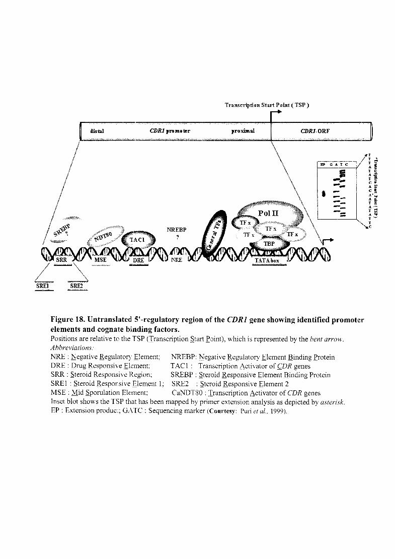
,I !
i !
/ .I
! !
! i
i 1
1 I
I I
! !
I : 1
distal CDR1 proJlloter
Transcription Start Point (TSP )
.nm.~f CDR1-0RF
'\ '. \ \ .
'1\ \ \ ., .,
\ ~~~\ '\ .,
'. '. .,
!P CATC
I ...... -... . ----------"T ! T •
T :;' A I T S. A'S. T " C " A~ C ~ A' ... A So T a A-T ;;l A ... T~
1
! ~~4:,~ } I
I r
NREBP ?
\ '. \ .. , \r
.~~
Figure 18. Untranslated 51-regulatory region of the CDR1 gene showing identified promoter elements and cognate bi,nding factors. Positions are relative to the TSP (Iranscription ~tart £oint), which is represented by the bent arrow. Abbreviations: NRE : ~egative Regulatory Element; NREBP: ~egative Regulatory Element Binding £rotein ORE : Qrug Responsive Element; TAC 1: Iranscription Activator of f;DR genes SRR : ~teroid Responsive Region; SREBP : ~teroid Responsive Element Binding £rotein SRE 1 : ~teroid Responsive Element 1; SRE2 : ~teroid Responsive Element 2 MSE : Mid ~porulation Element; CaNDT80 : Transcription Activator of f;DR genes Inset blot shows the TSP that has been mapped by primer extension analysis as depicted by asterisk. EP : Extension producT; OA TC : Sequencing marker (Courtesy: Puri et aI., 1999),

'ntroduction
response element (BRE) has been identified between nt -860 and -810 in the CDR1 promoter.
The BRE regulates basal expression of CDR1 (de Micheli et aI., 2002). Another BRE
(located between -243 and -234) and a gegative regulatory ~lement (NRE) located within the
-289 region have been reported in CDR1 (Puri et al., 1999; Gaur et aI., 2004). In, silico
analysis reveals that NRE (-ccaaCTGATTGAaact-) harbors A TTGA which shows significant
homology with MDR-NFlIYB-1 binding site (ATTGG) in human MDR1 promoter (inverted
CCAAT) that is known for its involvement in basal as well as stress induced expression of
human MDR1 (Hu et ai. 2000). A purified ~55 kDa nuclear protein was specifically found to
bind to NRE, this report describes purification of the first transacting protein of CDR1
promoter which may control its basal expression (Gaur et al., 2004). Recently, a common
drug responsive element called DRE1/2, which was located between -397 to -376 bp and -
303 to -280 bp in CDR1 promoter (from TSP) was identified, see Figure 17C. (de Michelli et
aI., 2002). DRE sequence (5'-CGGAA/TATCGGATA-3') was present in the promoters of
both CDR1 and CDR2 and has been found to be crucial for the up-regulation of these genes
in azole-resistant strains as well as for the transient up-regulation of both genes in the
presence of different drugs such as 13-estradiol, progesterone, or fluphenazine in azole
susceptible strains (de Michelli et al., 2002). However, sequential or internal deletion of DRE
did not completely abolish the stress responsiveness of CDR1 promoter (de Michelli et aI.,
2002). Recently, Looi et al. (2005) has identified the several hotspot point mutations in
CDR1 promoter of the resistant clinical isolates compared to the susceptible isolates at 39, 49
and 151 nucleotides upstream from the A TG start codon.
Trans-acting factors regulating CDR1 and CDR2 were reported recently. Three C. albicans
genes, designated FCR (!lufonazole resistance), have been isolated by their ability to
complement the fluconazole hypersensitivity of a S. cerevisiae mutant lacking the
transcription factors Pdrl p and Pdr3p. Over-expression of any of the three FCR genes in the
I1pdr 1 I1pdr 3 mutant resulted in increased resistance of the cells to fluconazole and
cycloheximide and increased expression of drug efflux transporter PDR5, whose transcription
is under the control of Pdrlp and Pdr3p. Deletion of PDR5 in the I1pdr111pdrJ strain
completely abrogated the ability of the three FCR genes to confer fluconazole resistance,
demonstrating that PDR5 is required for FCR-mediated fluconazole resistance in S.
cerevisiae (Talibi and Raymond 1999). FCR1 gene (Ca019.6817) encodes a putative 517-
44

Introduction
amino-acid protein with an N-tenninal Zn2C6-type zinc finger motif homologous to that
found in fungal zinc cluster proteins, including S. cerevisiae Pdrlp and Pdr3p. Interestingly,
inactivation of C. albicans Fcrlp, (ortholog of PdrlpIPdr3p of S. cerevisiae) results in
fluconazole resistance (Talibi & Raymond 1999). C. albicans I:!.fcrlll:!.fcr1 knock-out
derivative displays increased resistance to fluconazole and to drugs known to be transported
by Cdrlp and Cdrlp. This hyper-resistance could be reversed to wild-type levels by re
introduction of FCR1 into the I:!.fcr111:!.fcr1 mutant indicating Fcrlp as repressor of CDR1 in
C. albicans (Talibi and Raymond, 1999). However, one cannot exclude that hyperactive,
gain-of-function alleles of Fcrlp (and/or Fcr2p, Fcr3p) could result in constitutive expression
of a set of genes implicated in fluconazole resistance and MDR similarly, as in S. cerevisiae.
Alternatively, other regulatory factors or alterations in cis-acting DNA sequences of the
Yap 1 p target genes FCR1 or CAP 1, could contribute to the development of MDR. Therefore,
detailed characterization of Fcrlp, its target genes as well as the molecular mechanisms
leading to aberrant up-regulation of MDR1 and CDR1 genes is needed for the understanding
of multi drug resistance in C. albicans and fluconazole resistance in clinical isolates
(Kolaczkowska & Goffeau, 1999). According to a recent report by Shen et al. (2007), Fcrlp
inhibits development of fluconazole resistance in C. albicans by abolishing CDR1 function.
Chen et al. (2003, unpublished data) has also identified another regulator of C. albicans
tenned as Repressor of .Efflux rump (REP2). Over-expression of REP2 leads to six fold
increase in the ~-galactosidase activity of the PcDRrlacZ construct in S. cerevisiae.
Furthennore, I:!.rep2/ !:!.rep2 mutant of C. albicans appears to be more susceptible to antifungal
drugs than the parental strain. Quantitative real-time PCR reveals the decreased CDR1
expression in !1rep2/ !1rep2 mutant in the presence of azole. Recently, Chen et al. (2004)
described another potential activator of CDR1 identified by screening of a C. albicans
genomic library expressed in a S. cerevisiae strain, which contained a P CDRI-IacZ fusion.
This factor, CaNDT80 (Non DiIyrosine 80), is a homolog to a meiosis-specific transcription
factor in S. cerevisiae (Chen et al., 2004). Deletion of CaNDT80 in C. albicans conferred
hypersensitivity to azoles and decreased the inducible expression of CDR1. Interestingly, the
global repressor, Ihymidine !!Qtake 1 (Tup1) acts as a negative regulator of CDR1 expression
(Murad et al., 2001; Yang et al., 2006). A recent genome wide location profiling (ChIP-chip)
show that a TF of the zinc cluster family, !lQtake fontrol ~ (Upc2) which regulates ERG
45

Introduction
genes, also targets CDR1 (Znaidi et al., 2008). Recently, another transcription factor
belonging to the family of zinc-finger proteins with a Zn2Cys6 motif namely TAC1
(Iranscriptional ~ctivator of CDR) was identfied (Coste et al., 2004). Tac1 p is a 980 amino
acid protein of the Zn2-Cys6 family, it contains a typical DNA binding domain and an
activation domain rich in acidic residues at the C-terminus. Tac1 p binds to the DRE, which
contains two CGG triplets typical of the DNA-binding sites of Zn2Cys6 transcription factors
(Coste et al. 2004). Tac1p is responsible for transient up-regulation of both CDR genes in
azole-susceptible strains in the presence of inducers (Coste et al., 2004). Till now, only
Tac 1 p has been experimentally proved to function as a transcriptional activator of C.
albicans drug efflux transporter genes, such as CDR1 and CDR2 (Coste et al., 2004).
Interestingly, TAC1 locus is on chromosome 5 and is located close to (within ~14 kb) the
mating-type-like (MTL) locus where ERGJ1 is also located. Previous studies reported a
strong correlation between homozygosity at the mating-type locus and azole resistance in a
number of clinical isolates (Rustad et aI., 2002). In a previous study, Coste et al. (2004)
showed that a clinical azole-resistant strain (DSY296) that is homozygous at the mating-type
locus contains a TAC1 allele that is sufficient to confer fluconazole resistance to a laboratory
strain lacking TAC1. This type of allele was defined as 'hyperactive' because it caused
constitutive high expression of CDR1 and CDR2 in a f:.tac1!f:.tac1 mutant. In contrast, TAC1
alleles of the matched azole-susceptible clinical strain (DSY294) or of a laboratory strain
(CAF2-1), which are strains heterozygous at the mating-type locus, were not able to confer
azole resistance to a f:.tac1! Mac1 mutant. These alleles were defined as 'wild-type' alleles.
Analysis of TAC1 alleles of matched azole-susceptib1e and azole-resistant clinical C.
albicans reveals a single point mutation (N977D) in both hyperactive TAC1 alleles, which is
sufficient to confer hyperactivity as measured by up-regulation of CDR1 and CDR2 and
levels of drug resistance (Coste et al., 2006). These hyperactive alleles carrying this mutation
are co-dominant with other wild-type alleles such that only strains homozygous for
hyperactive alleles show high expression levels of CDR1 and CDR2. Later it has been shown
that homozygosity at MTL accompanies the acquisition of TAC1 homozygosity by either
through mitotic recombination between copies of chromosome 5 or through the presence of
extra copies of chromosome 5 harboring a gain-of-function TAC1 allele and loss of
46

A. Saccharomyces cerevisiae B. Candida albicans
PDR12p .. tl •• ~.
Figure 19. MDR regulatory circuit . . Identified transcription factor( s) factor and corresponding target genes of MDR regulatory circuit are shown. See text for details.
(A) s. cerevisiae (B) c. albicans. Abbreviations: N-Nucleus; ER-Endoplasmic reticulum; V- Vacuole; M-Mitochondrion; G-Golgi bodies.
lntroduction
chromosome 5 with the wild-type TA C1 allele (Coste et al., 2006), but MTL homozygosity
itself does not contribute to the azole resistance phenotype (Coste et ai., 2006).
Recently, genome-wide location (ChIP-chip) analysis (a procedure combining chromatin
immuno-precipitation with hybridization to DNA intergenic micro arrays) eight genes were
identified, whose expression was modulated in the four azole-resistant clinical isolates in a
TAC1-dependent manner and whose promoters were bound by Tac1p, qualifying them as
direct Tac1p targets: CDR1, CDR2, GPX1 (putative glutathione peroxidase), LCB4 (putative
sphingosine kinase), RTA3 (putative phospholipid flippase) and orf19.1887 (putative lipase),
as well as IFU5 and orf19.4898 of unknown function (Liu et ai., 2007). In a separate study, it
was shown that PDR16, encoding a putative phosphatidylinositol transfer protein
contributing to clinical azole resistance, is also a target of Tac1 p (Saidane et al., 2006; Znaidi
et al., 2007). Interestingly, all of these Tac 1 p-regulated genes contain a putative DRE in their
promoters. These results show that Tac1 p binds under non-activating conditions to the
promoters of its targets, including to its own promoter. Role of Tac1 p in regulating lipid
metabolism (mobilization and trafficking) and oxidative stress response in C. albicans has
also been proposed (Liu et al., 2007).
Therefore, observations from these studies suggest that multiple cis-elements and cognate
binding activators/ repressors regulate the expression of MDR network in S. cerevisiae and
C. albicans (Figure 19).
1.8 Aims and scope of the present work
The most well-characterized and frequently over-expreSSIOn of the multidrug resistance
protein in azole resistant clinical C. albicans isolate is Candida drug resistance 1 protein
(Cdrlp), encoded by the CDR1 gene, belonging to ATP-binding cassette (ABC) family
(Figure 20). Research to overcome this barrier in treatment has been focused mainly on two
areas: (1) advanced, sensitive detection of CDR1 mRNA or Cdrlp and its expression
regulation (2) development of Cdrl p modulators/inhibitors.
Keeping in view the importance of CDR1 in development of drug resistance, in this study we
undertook the first possibility to dissect the molecular mechanisms of CDR1 expression in
matched set of isolates, i.e. sensitive and resistant versions of the same strain, as determined
by RFLP or karyotyping (Figure 21). CDR1 gene is not detectably expressed in azole-
47

SUSCEPTIBLE Azoles
.. J_, MDR
... <!
+ 1:) -Y. ~ . e" ,..~ . 0-': 0" ~ ~
<o/. (>
Q -.J -+ -+ -+ ~ Erg II -+ Ergosterol
RESISTANT Azoles
.. ~
X
.J,J.J -+ -+ -+ -+ -+ Ergosterol
Erg II
Figure 20. Molecular mechanisms of azole resistance in clinical isolates of C. albicans. In a susceptible cell, azole drugs enter the cell through an unknown mechanism, perhaps by passive diffusion. The azole then inhibits Ergll (red circle), blocking the formation of ergosterol. Two types of efflux pumps are expressed at low levels. The CDR proteins are ABC transporters with both a membrane protein (green tubes) and two ABC domains (green circle). The MDR protein is a MFS with a membrane pore (red tubes). In a "model" resistant cell, the azoles also enter the cell through an unknown mechanism. The azoles drugs are less effective against Erg11 for two rea30ns; the enzymes has been modified by specific point mutations (dark circle in pink circle) and the enzyme is over-expressed. Modification in other enzymes in the ergosterol biosynthetic pathway contribute to azole resistance (dark slices in blue spheres). The sterol components of the plasma membrane are modified (dark orange of membrane). Finally. the azoles are removed fmm the cell by over-expression of the CDR genes (ABC transporters) and MDR (MFS). The CDR genes are effective against many azole drugs, while MDR appears to be specific for fluconazole.

A.
3.5- ..;,
~.O-
1.5-
1.0-
B.
-.
Figure 21. CARE-2 hybridization pattern of multiple successive oral C. albicans isolates (used in this study) from two different HIV+-AIDS patients. DNA Fingerprinting patterns shows no alteration in genomic organisation in matched AS and AR isolates. The positions of reference molecular size markers (in kb) are shown to the left of each blot. (A) Gu4 versus Gu4 and (B) DSY294 versus DSY296. (Courtesy: Franz et aI., 1999 and Boerlin et aI., 1996.)

Introduction
susceptible isolates but is strongly activated after the development of azole resistance and
also up-regulated by transient drug inductions (Krishnamurthy et aI., 1998). CDR] over
expression correlates well with increasing azole MICs in resistant clinical isolates. The
comparison of CDR] gene expression in matched azole susceptible and -resistant isolates has
proved to be a powerful tool to identify the resistance mechanisms of clinical C. albicans
isolates. This is required because C. albicans is mostly clonal; that is, it grows vegetatively
without sexual reproduction, indicating that mating is not a common event (Pujol et al.,
1993). Most strains of C. albicans contain a large number of balanced lethals (recessive null
mutations) in which the cell relies on one functional copy of the gene for activity (Whelan
and SolI, 1982). These recessive lethal mutations could not persist in a population that mated
frequently. The clonal nature of C. albicans makes it imperative that matched sets of
sensitive and resistant versions of a single strain should be characterized when determining
the molecular mechanisms of resistance.
Specific aims:
Regulating gene expression from DNA to protein is a complex multi-stage process with
multiple control mechanisms (Figure 5). Transcriptional regulation has been considered the
major control point of protein production in eukaryotic cells; however, there is growing
evidence of pivotal post-transcriptional regulation for many genes. The mRNA level of
particular gene is undoubtedly determined by the balance between synthesis and degradation.
This has prompted us to do extensive investigations to elucidate the possibility as well as
mechanism controlling mRNA stability/turnover along with transcriptional control of CDR].
Over-expression of the CDR] drug efflux pump has been demonstrated by the increased
mRNA levels which can be due to:
1. Increased CDR] transcription from endogenous copies of gene due to mutation( s) in the
CDR] promoter or alterations in the levels or activity of corresponding transcriptional
activator(s) that interact with the CDR] promoter that might result in deregulated CDR]
expression and ultimately increased CDR] mRNA level in AR isolates.
2. Molecular changes associate with possible CDR] mRNA decay i.e. increased half-life of
the CDR] mRNA as a result of a mutation(s) in the 3'UTR of the mRNA.
48

Introduction
The mam aim of the present study was to characterize the possibility of above said
mechanism of differential expression regulation of CDR1 in two different clinical matched
pairs of AS and AR isolates viz. Gu4 versus GuS and DSY294 versus DSY296 as well as
between uninduced and transiently induced resistance (lR) of AS isolates viz. Gu4 versus
Gu4-IR and DSY294 versus DSY294-IR.
The work in the thesis is embodied in two chapters:
1. Transcriptional activation and increased mRNA stability contribute to over-expression of
CDR1 in azole-resistant C. albicans.
2. Differential RNA-protein interaction and poly(A) tail length contributes to increased
CDR1 mRNA stability in azole resistant clinical isolates of C. albicans.
In the emerging scenario of increased incidence of development of drug resistance in C.
albicans, it is pre-requisite to understand the molecular basis leading to MDR. Hence, a
complete knowledge of physiological regulation of MDR is a key to design strategies for the
prevention of MDR and improve the therapeutic efficacy of drugs widely used to combat
these fungal infections. Our following study would pave the way for further investigation, if
CDR1 dependent transcriptional and post-transcriptional processes can become potential
novel targets for a new class of antifungal agents.


![AZOLES - [DePa] Departamento de Programas Audiovisualesdepa.fquim.unam.mx/amyd//archivero/SINTESISYREACCIONESAZOLES-1,2Y1... · Dakin-West utilizando piridina como el disolvente,](https://static.fdocuments.in/doc/165x107/5bcc0c4009d3f2e1348d30cc/azoles-depa-departamento-de-programas-2y1-dakin-west-utilizando-piridina.jpg)
![Chemistry of azoles [compatibility mode]](https://static.fdocuments.in/doc/165x107/568bf3161a28ab893399007a/chemistry-of-azoles-compatibility-mode.jpg)
![AZOLES - [DePa] Departamento de Programas …depa.fquim.unam.mx/amyd/archivero/SINTESISYREACCIONESAZOLES … · EN SOLUCIÓN SE PRESENTAN LOS SIGUIENTES EQUILIBRIOS . TAREA: ... Dakin-West](https://static.fdocuments.in/doc/165x107/5bb288b609d3f2272e8d1201/azoles-depa-departamento-de-programas-depafquimunammxamydarchiverosintesisyreaccionesazoles.jpg)