WHAT IS THE ROLE OF As an aside…. PATHOLOGY...
19
1 PATHOLOGIST’S ROLE IN THE DIAGNOSIS OF INTERSTITIAL LUNG DISEASE Thomas V. Colby, M.D. Mayo Clinic Arizona Mayo Clinic Arizona WHAT SHOULD THE CLINICIAN EXPECT FROM THE PATHOLOGIST? A diagnosis A diagnosis that fits with the clinical-radiologic findings An engaged pathologist A dynamic interchange of ideas Confidence in the pathologist As an aside…. Are you a technician or a consultant? – Dx = nonnecrotizing granulomas. – Dx = nonnecrotizing granulomas with a discussion of the possible diagnoses As physicians we should be consultants whenever we can. WHAT IS THE ROLE OF PATHOLOGY IN THE DIAGNOSIS OF ILD? In some cases, no role. In some cases, the pivotal role. In most cases, part of the data base that must be correlated with the clinical and radiologic features. ATS/ERS 2002 Amyloid on Congo Red Stain UIP/IPF
Transcript of WHAT IS THE ROLE OF As an aside…. PATHOLOGY...

1
PATHOLOGIST’S ROLE IN THE DIAGNOSIS OF INTERSTITIAL LUNG DISEASE
Thomas V. Colby, M.D.Mayo Clinic Arizona
Mayo Clinic Arizona
WHAT SHOULD THE CLINICIAN EXPECT FROM THE PATHOLOGIST?
� A diagnosis� A diagnosis that fits with the clinical-radiologic findings� An engaged pathologist� A dynamic interchange of ideas� Confidence in the pathologist
As an aside….� Are you a technician or a consultant?– Dx = nonnecrotizing granulomas.– Dx = nonnecrotizing granulomas with a discussion of the possible diagnoses
� As physicians we should be consultants whenever we can.
WHAT IS THE ROLE OF PATHOLOGY IN THE DIAGNOSIS OF ILD?� In some cases, no role.
� In some cases, the pivotal role.
� In most cases, part of the data base that must be correlated with the clinical and radiologic features.
ATS/ERS 2002
Amyloid on Congo Red Stain
UIP/IPF

2
WHAT IS THE ROLE OF SPECIMEN TYPE IN PATHOLOGIC DIAGNOSIS AND CLINICAL MANAGEMENT OF ILD?� Expectations of TBBx� Expectations of SLBx (VATS)
� Some guidelines for TBBx and SLBx1X Magnification
TBBx VATS Bx
INTERSTITIAL LUNG DISEASES (ILDs): Background
>100 are recognized with a diversity of pathologic features…… that can be grouped as follows:
– Histologically unique (No Dx/Dx)– Histologically characteristic (small Dx/Dx)– Patterns of injury (much larger Dx/Dx)
� Clinical and radiologic correlation is most important in the last group
HISTOLOGICALLY UNIQUE ILD’S: Examples� Pulmonary Langerhans Cell Histiocytosis (PLCH)� Lymphangioleiomyomatosis (LAM)� Diffuse Alveolar Septal Amyloidosis� Infections (eg. pneumocystis)� Neoplasms (many types)
HISTOLOGICALLY UNIQUE ILD’S (No Dx/Dx)� Small specimens may be adequate for diagnosis (eg. TBBx)
1.

3
26F Asymptomatic smoker with“diffuse infiltrates”PFT’s: Mixed obstructive/restrictive
Unique diagnostic histology: PLCH
S-100 CD1a
�
DIAGNOSTIC TBBx
Unique histology of Lymphangioleiomyomatosis/LAM
“cyst”
HMB45
ILD’S WITH CHARACTERISTICHISTOLOGY (relatively small Dx/Dx)
� Examples– Sarcoidosis– Pulmonary Alveolar Proteinosis
Correlation of Clinical, Radiologic, and Pathologic findings is important …
And a small Bx (eg TBBx) may be adequate
2. SARCOIDOSISGranulomatous inflammation
But there are many causes of granulomas…

4
SARCOIDOSISGranulomas along lymphatic routes
This distribution is very characteristic(and correlates with HRCT) c/w Sarcoidosis
TBBx
ILD’s WITH NON-SPECIFIC PATTERNS OF LUNG INJURY (and a larger Dx/Dx)� Acute Lung Injury: Diffuse Alveolar Damage (DAD), Organizing pneumonia (OP/BOOP pattern)� Chronic inflammation and interstitial fibrosis: seen in the chronic interstitial pneumonias, especially UIP and NSIP.These cases have a larger differential diagnosis, and Clinical-Radiologic correlation is necessary for diagnosis.Often require SLBx to identify the pattern
3.THE IDIOPATHIC INTERSTITIAL
PNEUMONIAS FALL INTO THIS CATEGORY OF NONSPECIFIC REACTION PATTERNS
(2002 ATS/ERS CLASSIFICATION OF IIPs*)
Clinicopathologic Diagnosis Pathologic Pattern
Idiopathic pulmonary fibrosis (IPF) UIPDesquamative interstitial pneumonia (DIP) DIPRespiratory bronchiolitis interstitial lung disease (RBILD) RB
Cryptogenic organizing pneumonia (COP) OP (BOOP)Acute interstitial pneumonia (AIP) DADNonspecific interstitial pneumonia (NSIP) NSIPLymphocytic interstitial pneumonia (LIP) LIP
*ATS/ERS International Consensus Panel; Am J Respir Crit Care Med 2002; 165:277

5
Inflammation and Fibrosis in chronic IP’s: UIP and NSIP
NSIP:Spatially uniformTemporally uniform
UIP:Spatially and temporallyheterogeneous
Fbroblast Foci
UIP vs NSIP is the major clinical problem- we will
come back to this
FOR THE CLINICIAN FACED ACHOICE OF BIOPSY IN ILD
� Histologically unique ILD - small specimens may suffice.� Histologically characteristic - small
specimens may suffice with Clin-Radiol correlation � Non-specific patterns of lung injury
individualize each case; eg.– TBBx is sufficient in COP with typical Clin-Rad– SLBx required to distinguish NSIP from UIP if radiology not diagnostic
Consider TBBx
Consider SLBx
CHOICE OF BIOPSY IN ILDRadiologic patterns-Lymphangitic: consider TBBxGround glass: SLBxUIP-like: SLBx
“High yield” TBBx’s (from Churg):Suspected malignancies, sarcoidosis, infections, transplant rejection
APPROACH TO BRONCHOSCOPIC BIOPSIES
(from TV Colby MD in Pathologica 2011)

6
�
SILICOSIS IDENTIFIED ON TBBx
TBBxSLBx
Silicosis often follows lymphatic routes and has unique histology
TBBx: DIAGNOSIS CATEGORIES1. Diagnostic of . . .2. Histologic changes consistent with . . .3. Nonspecific histologic abnormalities
(e.g. focal inflammation, scarring, alveolar macrophages, et al.)
4. Normal/negative/inadequate(Note: negative information may be useful!)
How can we improve the usefulness of TBBx ??
NONDIAGNOSTIC TBBx’sAbnormal but not diagnostic
From a case of PLCH From a case of COP
A list of pathologic findings (ie. a descriptive diagnosis) is not a clinicopathologic
diagnosis.
The Pathologist and ILD� Even when you cannot make a specific diagnosis you can still be a consultant.
� Be part of the discussion

7
TBBx: DIAGNOSIS CATEGORIES1. Diagnostic of . . .2. Histologic changes c/w. . .3. Nonspecific histologic abnormalities
(e.g. focal inflammation, scarring, alveolar macrophages, et al.)
4. Normal/negative/inadequate(Note: negative information may be useful!)
Can move from 3 → 2 with clin-rad-path correlation and/or additional studies
Can move from 2 → 1 or 3 → 1 with special studies (e.g. IPOX, PCR, genetics, et. al.)
1 and 2 allow a clinicopathologic diagnosis
�
TTBx: Solitary granuloma
Sputum cultures: M. Avium
..A nonspecific findingA nonspecific finding has become c/w
atypical mycobacterial infection
TBBx IN ILDAuthor Diagnostic Bx Comment
Anderson 1978N=939(rev’d by Churg)
31%Nonspec- 44%Normal/Inad- 25%
Poletti 1988N=801 29%
c/w Clin Dx- 37%Nonspec- 34%
Ensminger 2006N=603 38%
TBBx “helpful” in 76%Not “helpful” in 24%
How do you get good at interpreting TBBx’s?
� Know lung pathology as seen in SLBx’s, resections and at autopsy� Know something about clinical pulmonary disease� Know something about HRCT of the lung� Trust your brain!

8
HISTOLOGIC DIAGNOSIS OF LUNG DISEASE1. The larger the specimen the greater the
likelihood of diagnosisAdditive information from cytology/BAL, culture
2. Addition of clinical and radiologic information increases the likelihood of diagnosis
3. An experienced (pulmonary) pathologist increases the likelihood of diagnosis
The clinician can affect #'s 1 and 2The pathologist can affect #’s 2 and 3
SURGICAL LUNG BIOPSIES
Usual Interstitial Pneumonia Chronic bronchiolitis
SURGICAL LUNG BIOPSY (VATS AND OLBx)Surgical lung biopsy allows pattern recognition (and correlation with HRCT)
SURGICAL LUNG BIOPSY (VATS AND OLBx)Diagnostic Usefulness:~95% fit with clinical findings~90% diagnostic in chronic diffuse disease~35-75% rate of specific diagnosis in acute diffuse disease (most of these show DAD)

9
Nondiagnostic SLBx done for “pulmonary fibrosis”
*
* Atalectasis
Problem CasesRadiologic and pathologic interpretation are not black and white.Interpretations are subjective and there are shades of gray.Your clinician is always assessing your confidence and the specificity of pathologic interpretations.
SELECTED ILD’s
� Acute lung injury� Alveolar hemorrhage� Idiopathic interstitial pneumonias
DAD and OP represent….. ACUTE LUNG INJURY PATTERNS(Concept introduced by Katzenstein)
� Diffuse alveolar damage – acute and organizing� Organizing pneumonia (BOOP pattern)
� “Acute” here means injury days to weeks in age
Represent the most common findings in biopsy material

10
DAD
Acute
Organizing
Uniform temporal appearanceAlveolar septal thickeningAirspace organizationHyaline membranes
Organizing pneumonia
Intraluminal organization Patchy distributionPreserved architectureUniform age of lesionsMild cellular infiltrates
Acute Lung Injury: Overlap of Organizing DAD and OP
Org DAD OP
DIFFERENTIAL DIAGNOSIS OF: DAD OP
Organizing infections Allergic reactions: EP, HP Collagen vascular diseaseDrug reactionsOrganizing DADAspirationDistal to: Obstruction, bronchiectasis, COPD
Association with other lesionsIdiopathic
Localized: Focal OPWidespread: COP
InfectionsToxic inhalationsDrug reactionsCollagen Vascular DisRadiationDiffuse alveolar hemShockAcute allergic rxnsNeurologic diseaseMiscellaneousIdiopathic (ie. AIP)
Main Lesions to consider:InfectionConnective tissue diseaseDrug reactionAllergic/hypersensitivityIdiopathic*Many unsolved*
11
Is TBBx useful in ALI?
AJRCCM 2003 IPF
�
Cryptogenic Organizing Pneumonia (COP): TBBx
TBBx findings can be used to support the diagnosis
DIFFUSE ALVEOLAR HEMORRHAGE (DAH)� Pulmonary hemorrhage not due to trauma, airway disease, tumors, or heart failure � Usually recurrent; may be acute or chronic� Typically with dyspnea, hemoptysis, airspace infiltrates, and anemia� Associated renal disease common
This is a medical emergency
Diffuse Alveolar Hemorrhage (DAH)Stereotyped histologic reaction pattern*
RBC’s, fibrin, hyaline membranes
Hemosiderin depositionAirspace organizationWhen chronic
Interstitial thickening/slight fibrosis
Type II cell proliferation
+/- other changes (e.g. WG); +/- positive immunofluorescence
*Specific histologic diagnosis may not be possible!

12
(Anti-GBM Disease)
Diffuse Alveolar
Hemorrhage
Organizing alveolar hemorrhage overlaps
with OP
CAPILLARITIS IN DAH
� Capillaritis is common in DAH� Lung capillaritis is analogous to leukocytoclastic vasculitis� Capillaritis is not specific� Capillaritis is not a disease
AN IMMUNOLOGIC CLASSIFICATION OF DAH IS CONCEPTUALLY USEFUL
ANCA-associated: WG, MPA, pulmonary renal syndromes, isolated alveolar hemorrhageAntibasement membrane antibody:Goodpasture's syndrome, isolated alveolar hemorrhageImmune complex deposition: Collagen vascular diseases, IgA disease, pulmonary renal syndromes, isolated alveolar hemorrhageImmunologic mechanism not identified:IPH, isolated alveolar hemorrhage
HISTOLOGIC EVALUATION OF DAHConfirm presence of alveolar hemorrhageHistologic analysis often stops here!
Look for histologic features of WGCorrelate with serology, clinical pattern of disease, EM/IF studies (if done)
Clinicopathologic diagnosisBe a consultant.

13
Diffuse Alveolar Hemorrhage in WG(91F with fulminant course)
Diffuse Alveolar
Hemorrhage in WG
(91F with fulminant course)
Granulomatous foci diagnostic of
WG
Is TBBx useful in DAH? Transbronchial biopsy
Let’s add some history….

14
20F with dyspnea, hemoptysis, patchy radiologic infiltrates, and + c-ANCA
She also had hematuria
� Diagnosis: ANCA positive diffuse alveolar hemorrhage (compatible with WG)� Follow-up: The patient responded to therapy for WG and was well several years later
THE IDIOPATHIC INTERSTITIAL PNEUMONIAS FALL INTO THE CATEGORY OF
NONSPECIFIC REACTION PATTERNS(2002 ATS/ERS CLASSIFICATION OF IIPs*)
Clinicopathologic Diagnosis Pathologic Pattern
Idiopathic pulmonary fibrosis (IPF) UIPDesquamative interstitial pneumonia (DIP) DIPRespiratory bronchiolitis interstitial lung disease (RBILD) RB
Cryptogenic organizing pneumonia (COP) OP (BOOP)Acute interstitial pneumonia (AIP) DADNonspecific interstitial pneumonia (NSIP) NSIPLymphocytic interstitial pneumonia (LIP) LIP
*ATS/ERS International Consensus Panel; Am J Respir Crit Care Med 2002; 165:277
DIAGNOSIS OF UIP� Surgical lung biopsy is necessary to identify patterns of inflammation and fibrosis � Is biopsy always necessary?
NO! HRCT is an acceptable surrogate in
some situations
Usual Interstitial Pneumonia (UIP)
In ~50% of cases IPF can be diagnosed with HRCT
15
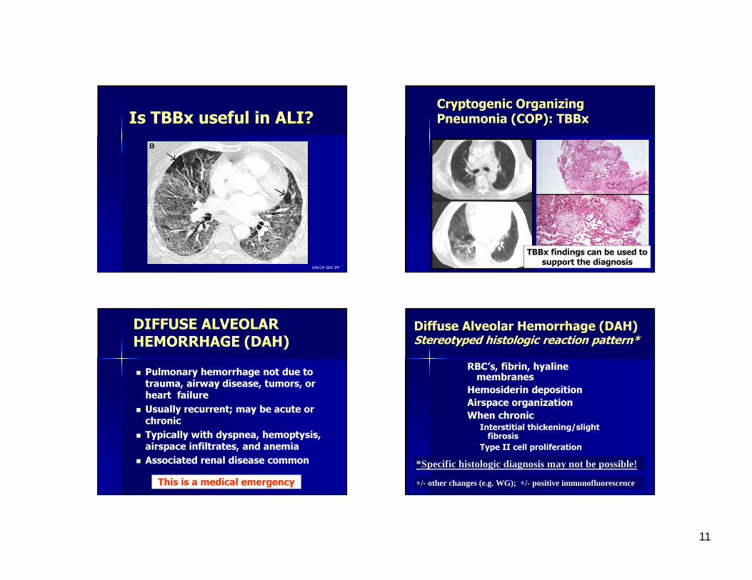
UIP vs NSIP
� For clinicians, radiologists, and pathologists UIP vs NSIP causes the most problems.
NSIP vs UIP: there are significant survival differences
Many other studies have confirmed these findings
Daniil ZD et.al. In AJRCCM 1999; 160: 899-905
DO NSIP AND IPF OVERLAP?OF COURSE!
UIP NSIPClinically
Radiologically
Pathologically
HOW DOES ONE DEAL WITH OVERLAP CASES?� Individualize each case� Clinical-radiologic-pathologic
correlation� “The essential assumption… is that there is no gold standard for … diffuse lung disease, merely the silver standards of clinical, radiologic, and histopathologic evaluation….” (Wells AU. In Am J Respir Crit Care Med 2004;170:827-831)

16
Histology=NSIP
HRCT TRUMPS HISTOLOGY(Sampling error has occurred)
HRCT=UIP
Diagnosis = UIP (IPF)
Biopsy is not the Gold Standard
Why isn’t this UIP?
Healed Pulm Langerhans cell histiocytosis
Scarring is stellate and centrilobular
Why isn’t this UIP?
Chronic hypersensitivity pneumonitis
Cental scarring with peribronchiolar metaplasia, granulomas
FEATURES TO ADDRESS IN FIBROSING INTERSTITIAL PNEUMONIAS� Distribution of the fibrosis (esp. central vs peripheral)� Evidence of active fibrosis (e.g. fibroblast foci)� Associated findings (e.g. dust, granulomas, foreign material)� Pertinent history (e.g. CVD, radiation, birds?, chemotherapy)

17
Is TBBx useful in IIP’s?The idiopathic interstitial pneumonias represent histologic patterns of inflammation and fibrosis.
Transbronchial biopsies are usually too small to allow recognition of patterns of injury.
Transbronchial biopsies do not allow confident diagnosis of UIP or NSIP (the major problem area)
TBBx is of limited value in the IIP’s
DAD c/w AIP OP c/w COPTBBx’s
RB on TBBx
It could be incidental or part of RBILD or DIP
Potential TBBx Sites
UIP as seen on trichrome stained section
Scar
Fibroblast focus
Normal
18
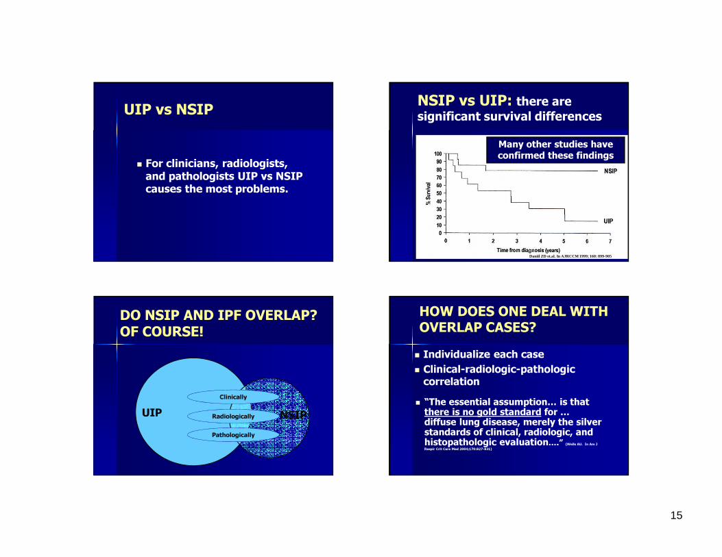
TBBx IN USUAL INTERSTITIAL PNEUMONIA(Berbescu et al. in Chest 2006;129:1126-1131)Summary: “… histologic features of UIP can be identified on TBBx specimens…and……support the diagnosis of UIP in the appropriate clinical and radiographic setting.”But not proven useful in prospective studies
TBBx is not currently accepted for the diagnosis of UIP
TBBx in the diagnosis of Idiopathic Interstitial Pneumonias (IIP’s)
UIP/IPF….DIP ……….RBILD ……COP ………DAD/AIP ..NSIP ………LIP………...
Not recommended
Not recommended
Possible with clin-radiol correlation
Possible with clin-radiol correlation
Possible with clin-radiol correlation
Insufficient data
Insufficient data
WHAT TO EXPECT FROM THE PATHOLOGIST IN ILD CASES?On a transbronchial biopsy?
On a surgical lung biopsy?
On agreeing with his colleagues?
On agreeing with himself/herself?
~35% Dx rate in chronic diffuse disease
~90-95% diagnosis in diffuse disease
Kappas from 0.4 - 0.9
Kappas from 0.4 - 0.8
ROLE OF THE PATHOLOGIST IN ILD’s ?� Be part of a dynamic interchange of ideas and observations with clinical-pathologic-radiologic correlation� Report a pathologic diagnosis that fits with the clinical and radiologic findings!–Does the Bx answer the question ??
� Be a consultant

19
THANK-YOU FOR YOUR ATTENTION!