What is New in Gynecologic Cancer?
71
What is New in Gynecologic Cancer? Professor, Division of Gynecologic Oncology Neag Cancer Center University of Connecticut Research Professor Electrical and Computer Engineering Molly A. Brewer, DVM, MD, MS
Transcript of What is New in Gynecologic Cancer?

What is New in Gynecologic Cancer?
Professor, Division of Gynecologic Oncology
Neag Cancer Center University of Connecticut
Research Professor Electrical and Computer Engineering Molly A. Brewer, DVM, MD, MS

Objectives:
• New screening and diagnostic approaches for ovarian cancer
• How to best triage patients with a pelvic mass
• Updates for cervical cancer screening
• Updates on endometrial cancer

I have no conflicts

Ovarian Cancer

Challenges: Ovarian Cancer
Major obstacles:
• Undefined premalignant lesions
• Poor access to evaluate the ovary
• Poor screening technology
• Little known about prevention
• Early metastasis
• Symptoms usually occur after metastasis
• Ovarian cancer is diagnosed 70% of the time in
Stage III or IV where the survival is 30% or less
Relatively rare cancer: 1.5-1.7% lifetime risk

Ovarian Cancer: Epidemiology • Increased risk/ increased
ovulation:
– early menarche/late menopause
– fertility drugs (?)
– nulliparity
– breast cancer
– family history (breast/ovarian cancer)
– genetic mutations (BRCA1/2)
– Endometriosis (clear cell)
• Decreased risk/ decreased ovulation:
– oral contraceptive use
– breastfeeding
– multiparity
– late menarche/ early menopause
– retinoids
Natural History: unknown

Do serous ovarian cancers originate from the ovary or the tube?
• 2-12% of BRCA 1/2+ women have cancer at the time of BSO
• 75% originate in the tubal epithelium
• By the time most ovarian cancers are diagnosed, there is involvement of both ovaries and tubes
• How to best identify these patients?

h
a
b
c
j e
d
i
f
EB, MB, UU
g
ovarian surface epithelium
and stroma
Structure of the Ovary
Brewer et al, JBO

Ovarian Inclusion Cysts

Fallopian Tube
• Identification of "dysplastic" epithelial changes in the fallopian tubes of women with BRCA1 or BRCA2 mutations (Piek et al, 2003)
• SEE-FIM protocol with routine examination of the fimbria assumed to be the site of origin for many HGSCs with identification of serous tubal intraepithelial carcinomas (STICs) in risk-reducing salpingo-oophorectomies (Medeiros et al 2006)
• STICs seen in 20-60% of HGSCs in sporadic cancers established STIC and the distal tube as an important source of these tumors (Kindelberger et al, 2007)
• p53 signatures - bland-appearing cell outgrowths with p53 mutations - in the tube and their genetic similarity to STICs, a morphologically, genetically and histochemically defined serous carcinogenic sequence (Xian et al, 2010)

Serous Tubal Intraepithelial Neoplasia
P53 IHC

Serous Tubal Intraepithelial Neoplasia

Current state of the art
• Precursors in the tube, which likely remain localized for months to a few years, cannot be detected by current technology.
• Based on frequency of a co-existing STIC, the tube is implicated in HGSC –including ovarian and peritoneal – in from 19 to 59 per cent of cases (Carlson et al, Roh et al, Pryzbycin et al, Tang et al)
• Should we be removing all tubes even though there is no data?
• Does the STIC resemble DCIS of breast where there is limited probability of progression to invasive cancer and has been overtreated?
• If a subset of tumors emerge de novo from the ovarian or peritoneal surfaces, they may never be preventable by screening.

Taking A Family History: The Basics
• When do you take a family history? – Always!
• Why? 1) to identify patients with an increased risk for cancer 2) to develop specific screening and management guidelines based
on their cancer risk
• Who do you include? – First, second and third degree relatives, – Both maternal and paternal sides of the family
• What do you ask? – Type of cancer (primary site) AND age of onset
• Where can patients get additional information about relatives? – Death certificates, tumor registry

How to Take a Family History
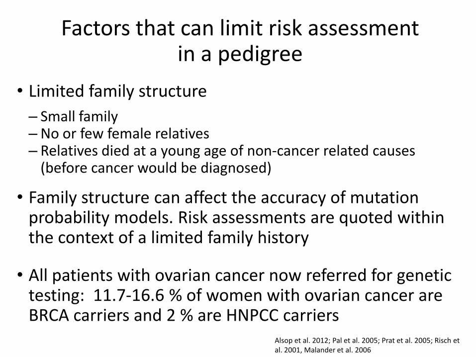
Factors that can limit risk assessment in a pedigree
• Limited family structure
– Small family – No or few female relatives – Relatives died at a young age of non-cancer related causes
(before cancer would be diagnosed)
• Family structure can affect the accuracy of mutation probability models. Risk assessments are quoted within the context of a limited family history
• All patients with ovarian cancer now referred for genetic testing: 11.7-16.6 % of women with ovarian cancer are BRCA carriers and 2 % are HNPCC carriers
Alsop et al. 2012; Pal et al. 2005; Prat et al. 2005; Risch et al. 2001, Malander et al. 2006

Risk Assessment Criteria
• Any individual < 40yo with breast cancer, first or second degree relative
• One case of ovarian cancer
• One case of male breast cancer
• > 2 cases of breast cancer in first or second degree relatives if one is diagnosed at < 50 or is bilateral
• Ashkenazi Jewish families
• Patient with both breast and ovarian cancers

Ovary/Fallopian tube removal in the BRCA population
• 792 women: 509 underwent RRSO
• 85% reduction in BRCA1-associated gynecologic cancer risk (hazard ratio [HR] = 0.15; 95% CI, 0.04 to 0.56)
• 72% reduction in BRCA2-associated breast cancer risk (HR = 0.28; 95% CI, 0.08 to 0.92).
Kauff et al, J Clin Oncol. 2008

Bilateral Oophorectomy
Women not receiving estrogen who underwent PO <45 years of age were 2X more likely to die:
OR
• Estrogen related cancer 5.71
• Non-cancer 1.71
• Vascular 1.41
• Neurological/mental 2.34
Rocca, B et al R Lancet Oncol 2006; 7: 821–28

Metastasis • Recent evidence suggests that metastasis is an earlier
event than previously thought
• Only very small numbers of shed malignant cells are capable of metastasizing (0.01%)
• The persistence of cancer cells in the vasculature does not necessarily result in seeding to distant sites
• Emerging evidence in breast cancer suggests that early tumors may already hold the genetic profile needed for metastasis
Landen et al, JCO 26;6 995-1005 2008

Screening for Ovarian Cancer

Low risk women
Do not screen

High Risk Women
Screen with multiple caveats

Sporadic Familial Hereditary
Single occurrence in family
Two or more first-degree or second-degree affected relatives
Multiple affected individuals in multiple generations
Late age of onset (after 60 y)
Onset typically after 50 y Early age of onset (often less than 50 y)
Unilateral Primarily unilateral or late-onset bilateral
Bilateral/multifocal disease
Lack of other cancers in the family, or only common cancers of late onset
Evidence of skipped generations, no clear inheritance pattern
Presence of ovarian cancer, male breast cancer, Jewish ancestry, multiple other unusual and/or early onset malignancies
Primarily caused by age an other nongenetic factors
Multiple minor/moderate inherited genetic factors interacting with environment
Single major cancer susceptibility gene mutation, autosomal dominant inheritance
Obstet Gynecol Clin North Am. 2013 Sep;40(3):475-512

Ultrasound
• 1,072 premenopausal ovarian masses
• 570 were functional cysts and corpus lutea (53%)
• 264 were endometriotic cysts (25%)
• 192 were benign neoplasms (18%)
• 46 malignancies (4%)
Osmers et al., AMJOG; 175:428-34 1996

Ultrasound
• 5/236 (2.1%) complex adnexal masses were cystic ovarian tumors with thick septa but no solid areas.
• 14% had ascites present • 67% had benign tumors, 30% had ovarian cancer, 3%
had LMP tumors • High risk of cancer=complex/solid morphology, CA
125 value > 35 units/mL • PPV of 84.7% and NPV of 92.4% and correctly
identified 77.3% of patients with stage I and stage II ovarian cancer and 98.6% of patients with stage III and stage IV ovarian cancer.
McDonald et al, Ob Gyn 115(4), 2010, pp 687-694

Ovarian Masses
• Age 20–29, cystic teratomas, serous cystadenomas, and mucinous cystadenomas are responsible for 72, 15, and 11% of benign ovarian neoplasms, respectively
• At age 40–49, the same 3 tumor types make up 43, 46, and 8%, of benign neoplasms
• The risk of a neoplasm being borderline is 2.4% at 20–29 and 6.3% at 40–49
• The risk for malignancy is 4% at 20–29 and 35% at 40–49
Koonings et al, Obstet Gynecol 1989;74:921

Diagnostic ability of CT
• Low-dose unenhanced computed tomography (CT) in asymptomatic women
• 118 (4.1%), mean age 56.2 had an adnexal mass (108 unilateral, 10 bilateral; mean size, 4.1 cm) all benign
• 4 cancers developed in 15-44-month interval among the remaining 2751
• CT is a poor screening technology for ovarian cancer
Pickhardt et al, Radiology. 2010

Diagnostic ability of MRI
• Combination of US and MR findings was false positive for malignancy in only 7 of the 72
• No false negative cases of invasive ovarian malignancy
• 19 had ‘malignant’ findings at US downgraded correctly by MR imaging to surgically correlated complex benign diagnoses.
• MR imaging was most valuable for women who had normal or only slightly elevated levels of CA-125
Soaib et al, Clin Radiol 2005;60:340–8.

CA-125 testing
• Premenopausal women with a known adnexal mass
• Elevations can be seen with
– Endometriosis
– Pelvic inflammatory disease
– Leiomyomas
– Cystic teratomas
– Pleural infections
– Menstrual cycle
Cohen, J Assist Reprod Genet (2007) 24:507–512

HE4 (human epididymis secretory protein 4 ) and Risk for Malignancy (ROMA)
• Premenopausal women: sensitivity and specificity were
– 92.3% and 59.4% for CA125,
– 84.6% and 94.2% for HE4
– 84.6% and 81.2% for ROMA,
• Postmenopausal women: sensitivity and specificity :
– 94.3% and 82.3% for CA125,
– 78.2% and 99.0% for HE4
– 93.1% and 84.4% for ROMA.
Bandiera et al CEBP 2011

CA125 and HE4 Premenopausal Postmenopausal Premenopausal Postmenopusal
HE4 CA
125
HC En Cy OC HC En OC HC En Cy OC HC En OC
Bandiera et al CEBP 2011
HC= healthy controls En= endometriosis Cy= cyst OC= ovarian cancer

• HE4, CA 125 and ROMA results were abnormal
– in 1.5%, 13.6%and 25.8%of healthy women
– in 1.1%, 30.2% and 12.3% of patients with benign diseases
• HE4 had significantly higher concentrations in ovarian cancer than in other malignancies (p<0.001).
• Tumor marker sensitivity in ovarian cancer was 79.3% for HE4, 82.9% for CA 125 and 90.1% for ROMA
Molina et al, Tumor Biol. (2011) 32:1087–1095

OVA1
• CA 125-II, transthyretin [prealbumin], apolipoprotein A1, beta 2 microglobulin, and Transferrin
• Results are interpreted by proprietary software, and an OVA1 score is given, which differs based on menopausal status.
• A high probability of malignancy is defined as at least 5.0 in premenopausal women and at least 4.4 in postmenopausal women.
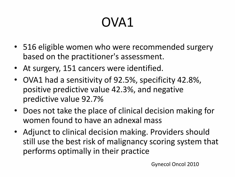
OVA1
• 516 eligible women who were recommended surgery based on the practitioner's assessment.
• At surgery, 151 cancers were identified.
• OVA1 had a sensitivity of 92.5%, specificity 42.8%, positive predictive value 42.3%, and negative predictive value 92.7%
• Does not take the place of clinical decision making for women found to have an adnexal mass
• Adjunct to clinical decision making. Providers should still use the best risk of malignancy scoring system that performs optimally in their practice
Gynecol Oncol 2010

ROCA (Risk of Ovarian Cancer Algorithm)
• Postmenopausal women with annual CA125
• If low risk based on algorithm CA125 1 yr
• If moderate risk based on algorithm repeat CA125 3 months
• If high risk based on algorithm TVS
• Positive predictive value of 40%
• Specificity was 99.9%
Lu et al, Cancer. 2013

Management Guidelines for Adnexal Masses
• In asymptomatic women with pelvic masses TVUS is the
imaging modality of choice
• No alternative imaging modality has demonstrated
sufficient superiority to TVUS to justify its routine use
• Any CA 125 elevation in a postmenopausal woman with a
pelvic mass is highly suspicious for malignancy
ACOG Practice Bulletin

Management Guidelines for Adnexal Masses
• Women with ovarian cancer whose care is managed by
physicians who have advanced training and expertise in
the treatment of women with ovarian cancer, such as
gynecologic oncologists, have improved overall survival
rates compared with those treated without such
collaboration
ACOG Practice Bulletin # 83, July 2007

Management Guidelines for Adnexal Masses
• Simple cysts up to 10 cm in diameter on ultrasound findings are almost universally benign and may safely be followed without intervention, even in postmenopausal patients
• Unilateral salpingo-oophorectomy or ovarian cystectomy in patients with germ cell tumors, stage I stromal tumors, tumors of low malignant potential, and stage IA, grade 1–2 invasive cancer who undergo complete surgical staging and who wish to preserve fertility does not appear to be associated with compromised prognosis
ACOG Practice Bulletin

Clinical assessment, CA125, US
Adnexal mass, signs of intraperitoneal
spread
Staging CT
Surgery appropriate
Surgery inappropriate
Image guided biopsy
Adnexal mass, Probably malignant
Staging CT
Indeterminate mass
MR imaging
Adnexal mass, Probably benign
Manage symptoms
Spencer et al, Gyn Oncology 108
(2008) 262–264
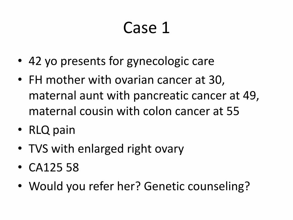
Case 1
• 42 yo presents for gynecologic care
• FH mother with ovarian cancer at 30, maternal aunt with pancreatic cancer at 49, maternal cousin with colon cancer at 55
• RLQ pain
• TVS with enlarged right ovary
• CA125 58
• Would you refer her? Genetic counseling?

Case 2
• 65 yo with cystic complex ovarian mass found on exam, assymptomatic
• No family h/o cancer
• CA125 85
• Would you refer her?

Cervical cancer

The paradigm for screening has changed dramatically

• Screening by Papanicolau (Pap) test should not be used for women aged <21 years, regardless of initiation of sexual activity,
• Screening interval of 3 years should be maintained for women aged 21-30 years.
• ACS and ACOG explicitly recommend against yearly screening.
MMWR Morb Mortal Wkly Rep. 2013 Jan 4;61:1038-42.

• Among those aged 22-30 years, the proportion reporting having had a Pap test within the preceding 12 months decreased from 78.1% to 67.0%.
• Among women aged 22-30 years, who should be screened every 3 years, the proportion who reported never having had a Pap test increased from 6.6% to 9.0%
MMWR Morb Mortal Wkly Rep. 2013 Jan 4;61:1038-42.

• The current model of cervical carcinogenesis suggests that HPV infection results is either transient or persistent.
• Most HPV infection is transient and poses little risk of progression.
• Persistent infection at 1 year and 2 years strongly predicts subsequent risk of CIN 3 or cancer regardless of age

• In a cohort of untreated patients with CIN 3, the cumulative incidence of invasive cancer was reported to be 30.1% at 30 years.
• Most HPV-related types of cervical neoplasia are very slow to progress. On average, a severe dysplasia may take 3–7 years to progress to invasive cervical cancer.

Screening Methods for Cervical Cancer: Joint Recommendations of the American Cancer Society, the American Society for Society Colposcopy and Cervical Pathology, and the American for Clinical Pathology
Population Recommended Screening Comment
Women younger than 21 years No screening
Women aged 21–29 years Cytology alone every 3 years
Women aged 30–65 years Human papillomavirus and cytology co-testing (preferred) every 5 years Cytology alone (acceptable) every 3 years
Screening by HPV testing alone is not recommended
Women older than 65 years No screening is necessary after adequate negative prior screening results
Women with a history of CIN 2, CIN 3 or adenocarcinoma in situ should continue routine age-based screening for at least 20 years
Women who underwent total hysterectomy
No screening is necessary Applies to women without a cervix and without a history of CIN 2, CIN 3, adenocarcinoma in situ, or cancer in the past 20 years
Women vaccinated against HPV Follow age-specific recommendations (same as unvaccinated women)

Women with negative cytology and positive HPV co-testing results who are aged 30 years and older
Level A Evidence
• Repeat co-testing in 12 months. If the repeat cervical cytology test result is LSIL or higher or the HPV test result is still positive, the patient should be referred for colposcopy. Otherwise, the patient should return to routine screening – OR
• Immediate HPV genotype-specific testing for HPV-16 alone or HPV-16/18 should be performed. If positive should be referred directly for colposcopy. Women with negative results from tests for HPV-16 or HPV-16/18 should be retested in 12 months

Case 1
• 42 yo, no h/o abnormal Pap smears, last Pap 1.5 years ago
• ASCUS Pap, HPV +
• Colposcopy negative, 6 cervical biopsies -,
• ECC SCC
• Exam erythematous area anterior cervix
• Bx adeno-squamous carcinoma

Case 2
• 45 yo with ASCUS HPV+, last Pap 3 years ago negative
• Biopsies CIN3, ACIS
• Cone adenocarcinoma 5X9 mm invasion
• Radical hysterectomy 3 mm residual disease

Endometrial Cancer

• Who should be considered for genetic testing?
• To stage or not to stage?
• Can these patients with grade 1 be treated by a generalist?
• Do patients with grade 3 histology need to be staged?

HNPCC: Endometrial Cancer
• 1.8% of all endometrial cancer have HNPCC
• 7/10 did not meet standard criteria for HNPCC
• Mean age 54 years
• 8.6% diagnosed with endometrial cancer < 50 years old had HNPCC
Hampel et al, Cancer Research, 2006. Behrens et al, JCO 2003

HNPCC: Lifetime cancer risk
• Colorectal cancer (men) 28 – 75%
• Colorectal cancer (women) 24 – 52%
• Endometrial cancer 27 – 71%
• Ovarian cancer 3 – 13%
• Gastric cancer 2 – 13%
• Urinary tract cancer 1 – 12%
• Brain tumor 1 - 4%
• Bile duct/gallbladder cancer 2%
• Small bowel cancer 4 - 7%

Mutation Carriers
MLH1 MSH2 MSH6 Total
Mutation carriers 70
67 109 246
Subjects with colorectal cancer (%) 51 31 24 34
Subjects with endometrial carcinoma 10 13 19 15
Subjects with other Lynch associated cancer (%)*
Ovarian carcinoma 1 4 6 4
Small bowel cancer 1 3 0 1
Transitional cell carcinoma 0 4 3 2
Ramsoekh et al, Hered Cancer Clin Pract. 2009 23;7(1):17.

Median Age/Range of Diagnosis of HNPCC Cancer
MLH1 MSH2 MSH6
Colorectal cancer 47 (25-79) 44 (20-82) 53 (32-84)
Endometrial cancer 51 (46-54) 46 (36-55) 56 (47-67)
Ovarian cancer 52 (52-52) 47 (45-48) 49 (35-51)
Ramsoekh et al, Hered Cancer Clin Pract. 2009 Dec 23;7(1):17., Ramsoekh et al, Hered Cancer Clin Pract. 2009 Dec 23;7(1):17

Diagnosis of HNPCC
• Family h/o colon/endometrial cancer at young age
• IHC on tumor tissue with absence of expression of MLH1, MSH2, MSH6 or PMS2
• PCR for definitive diagnosis
• Sporadic endometrial cancers may be associated with epigenetic silencing of MLH1
• Gene sequencing based on IHC

Clinical Staging, FIGO 1971
Stage I Confined to corpus
Ia Cavity less than 8cm
Ib Cavity greater than 8 cm
Stage II Corpus & Coli(cervix)
Stage III Outside the corpus but not outside pelvis
Stage IV Outside the true pelvis, or obvious
involvement of bladder or rectal mucosa.

Surgical Staging, FIGO 1988 Stage Description Ia Tumor limited to endometrium Ib Invasion to less than 50% myometrium Ic Invasion to more than 50% myometrium IIa Endocervical gland involvement only IIb Cervical stromal invasion IIIa Uterine serosa, adnexa, positive cytology IIIb Vaginal Metastases IIIc Metastases to pelvic or periaortic nodes IVa Invasion of bladder and/or bowel mucosa IVb Distant metastases, intraperitoneal, inguinal

Surgical Staging, FIGO 2010
Stage Description
Ia Invasion < 50% myometrium + cytology
Ib Invasion > 50% myometrium + cytology
IIa Endocervical gland involvement only
IIb Cervical stromal invasion
IIIa Uterine serosa, adnexa
IIIb Vaginal Metastases
IIIc Metastases to pelvic or periaortic nodes
IVa Invasion of bladder and/or bowel mucosa
IVb Distant metastases, intraperitoneal, inguinal

Staging vs No Staging
• Low risk 109 (62.6) 90 (51.7)
• Intermediate risk 17 (9.8) 34 (19.5)
• High risk 48 (27.6) 50 (28.7)
• Total 174 174
Papadia et al, IJC 2009
Frozen Section Permanent Section
Authors’ conclusions: If staging was based on frozen section, 12.8% of patients would be undertreated
My conclusion: rely on the permanent section for treatment decisions, management should be with gynecologic oncology

Atypical Endometrial Hyperplasia
Stage
IA IB IC IIB IIIC Total
Grade 1 4 14 2 2 1 23
Grade 2 0 1 1 0 0 2
Total 4 15 3 2 1
Whyte et al, AJOG 2009
Authors’ Conclusion: 25/88 (28.4%) of AEH had carcinoma on final pathology: All patients with AEH should be staged My conclusion: low incidence of high grade cancer in AEH but they should be managed by gynecologic oncology

Role of Lymphadenectomy
• 191 women (88 standard surgery group, 103 lymphadenectomy group) had died, with a hazard ratio (HR) of 1·16 (95% CI 0·87–1·54; p=0·31) in favor of standard surgery
• Conclusion: lymphadenectomy does not alter survival
• My conclusion: UK radiates many more patients than us
The ASTEC/EN.5 writing committee on behalf of the ASTEC/EN.5 Study Group*, Vol 373, 2009

Radiotherapy 227 (33%) 228 (33%)
External beam
+/-brachytherapy 173 (25%) 165 (23%)
Brachytherapy only 54 (8%) 63 (9%)
No Radiotherapy 471 (67%) 469 (67%)
Unknown 6 7
Lymphadenectomy
(N=704)
Standard
surgery (N=704)
The ASTEC/EN.5 writing committee on behalf of the ASTEC/EN.5 Study Group*, Vol 373, 2009
Authors’ conclusion: lymphadectomy does not reduce rate of XRT My conclusion: UK radiates patients that we would either not treat or who would receive chemotherapy

Treatment 2004
• Surgery: TAH/BSO
– Staging: Grade 1 deep invasion, grade 2 superficial invasion, all grade 3
• Radiation
– Positive lymph nodes
– High risk: deep invasion, LVSI
• Chemotherapy
– Papillary serous: Taxol/Platinum +/- WART
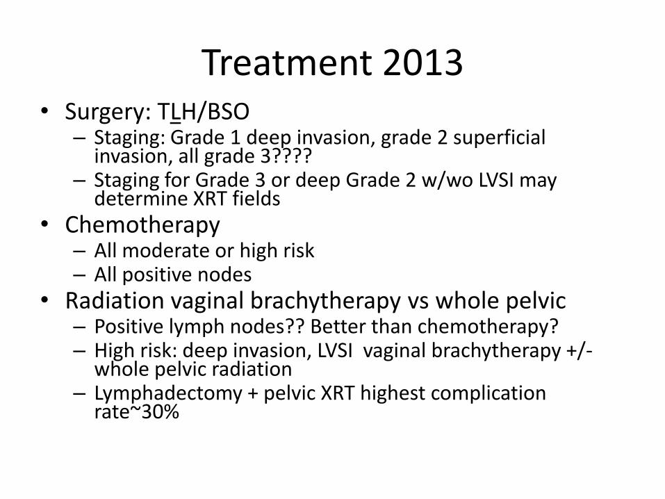
Treatment 2013 • Surgery: TLH/BSO
– Staging: Grade 1 deep invasion, grade 2 superficial invasion, all grade 3????
– Staging for Grade 3 or deep Grade 2 w/wo LVSI may determine XRT fields
• Chemotherapy – All moderate or high risk – All positive nodes
• Radiation vaginal brachytherapy vs whole pelvic – Positive lymph nodes?? Better than chemotherapy? – High risk: deep invasion, LVSI vaginal brachytherapy +/-
whole pelvic radiation – Lymphadectomy + pelvic XRT highest complication
rate~30%

Treatment in 2015
• Less invasive surgery: role of sentinal nodes?
• Chemotherapy for intermediate/high risk disease
• ? Role of XRT
• Targeted therapy: avastin, EGFR inhibitors, other target blockers

Can Generalists Safely Treat Endometrial Cancer?
• Too many patients are upstaged at the time of surgery
• Cytoreductive surgery improves prognosis
• Grade I cancers are Grade 3 on final pathology 30% of the time
• General surgeons do not do adequate lymph node sampling
• Recurrent disease is rarely cured unless it is a vaginal recurrence.


![Advocacy at Home - Ovarian Cancer Research Alliance · 2018-09-28 · [gynecologic/ovarian] cancer patients. (3) Determining any unmet needs of persons with [gynecologic/ovarian]](https://static.fdocuments.in/doc/165x107/5f71893b71cf3e62e42f3d4d/advocacy-at-home-ovarian-cancer-research-alliance-2018-09-28-gynecologicovarian.jpg)