Ocular Immunology and Uveitis Foundation - Uveitis is the third

What do you need to knowabout posterior uveitis
Dr. Anthony Hall MD FRANZCO
Director of OphthalmologyAlfred Hospital,
Melbourne, Australia

Alfred Hospital

Disclosures
• Off label treatments• Paid advisory board Bayer• Paid research support Allergan (makers of Ozurdex)• Paid research support B and L (makers of Retisert)• Paid research support Novartis

My aim: to give you an easy diagnosticapproach to posterior uveitis

Your aims of assessment
•Make a descriptive anatomical diagnosis•Recognise specific named uveitis entities•Diagnose significant underlying systemicinflammatory/infective disease
•Diagnose intra-ocular/systemicinfections/masquerade syndromes
•Diagnose the cause of visual loss

Diagnostic possibilities inintermediate or posterior uveitis
•Isolated ocular disease•Idiopathic•Named•Infectious
•Ocular disease as part of systemic disease•Non-infectious•Infectious•Malignant
•Masquerade syndromes

• Attempt to make an anatomic and descriptive diagnosis• Use recognised nomenclature

Anatomical classification of uveitis
• Anterior uveitis• Intermediate uveitis• Posterior uveitis• Panuveitis
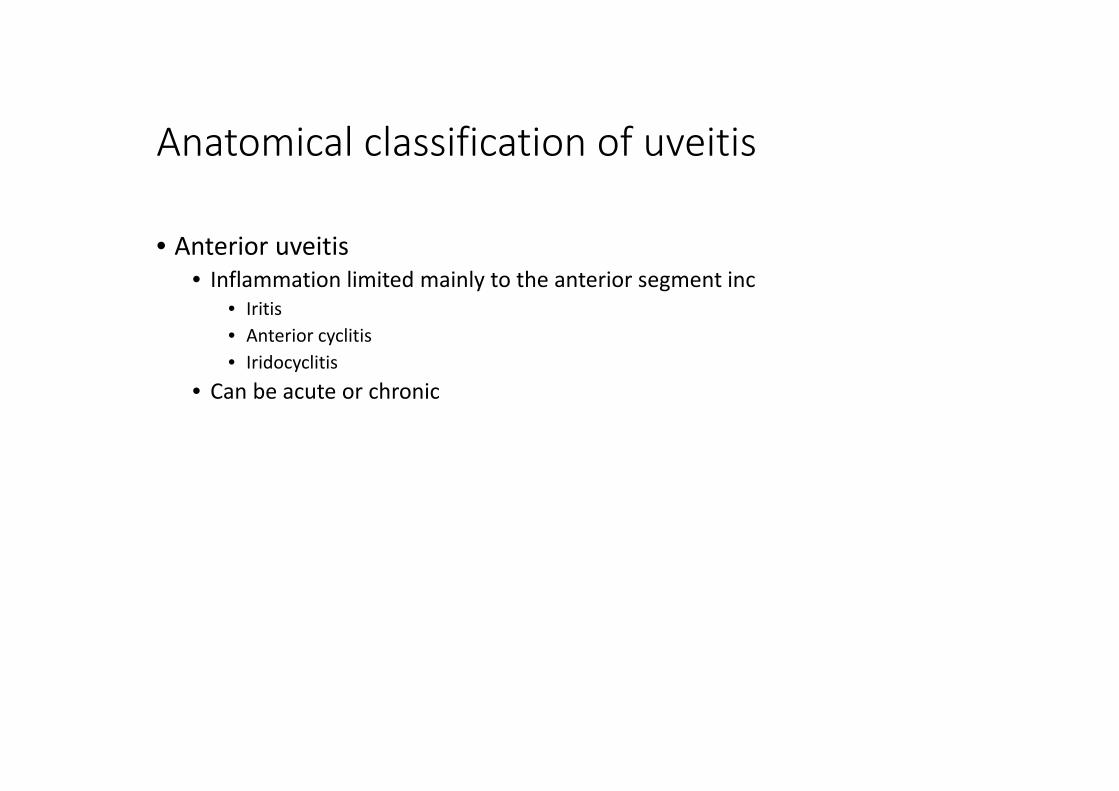
Anatomical classification of uveitis
• Anterior uveitis• Inflammation limited mainly to the anterior segment inc
• Iritis• Anterior cyclitis• Iridocyclitis
• Can be acute or chronic
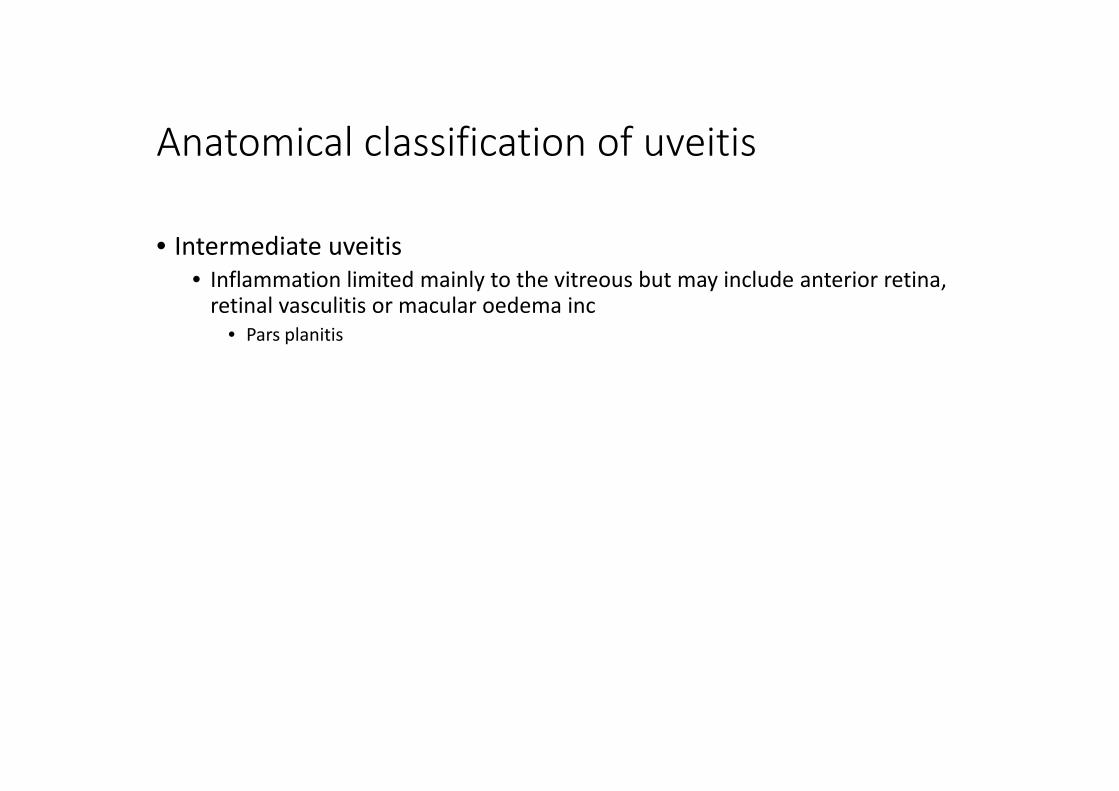
Anatomical classification of uveitis
• Intermediate uveitis• Inflammation limited mainly to the vitreous but may include anterior retina,
retinal vasculitis or macular oedema inc• Pars planitis

Anatomical classification of uveitis
• Posterior uveitis• Inflammation limited mainly to the retina, choroid or optic nerve inc
• Focal, diffuse or multifocal choroiditis• Chorioretinitis• Neuroretinitis• Serous detachment

Anatomical classification of uveitis
• Panuveitis• Inflammation involving the whole eye without preference for anterior or
posterior segment

Different clinical pictures inintermediate/posterior/panuveitis
• Diffuse uveitis/vitritis• Unifocal chorioretinitis• Multifocal chorioretinitis• Confluent chorioretinitis• Retinal vasculitis• Optic papillitis• Serous retinal detachment

Different clinical pictures inintermediate/posterior/panuveitis
• Diffuse uveitis/vitritis• Unifocal chorioretinitis• Multifocal chorioretinitis• Confluent chorioretinitis• Retinal vasculitis• Optic papillitis• Serous retinal detachment
• Isolated ocular disease• Idiopathic• Named• Infectious
• Ocular disease as part ofsystemic disease
• Non-infectious• Infectious• Malignant
• Masquerade syndromes

Non specific/diffuse uveitis
•Vitritis•+/- macular oedema•+/- retinal vasculitis•No focal signs
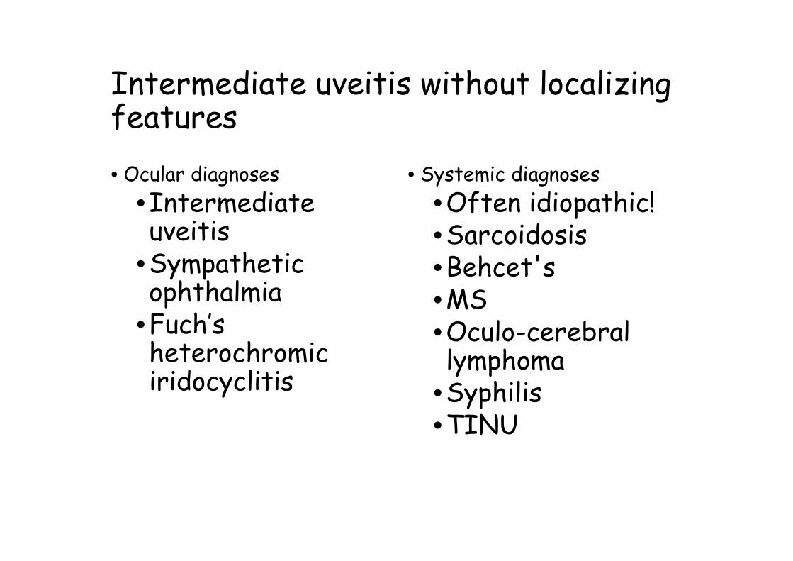
Intermediate uveitis without localizingfeatures• Ocular diagnoses
•Intermediateuveitis
•Sympatheticophthalmia
•Fuch’sheterochromiciridocyclitis
• Systemic diagnoses•Often idiopathic!•Sarcoidosis•Behcet's•MS•Oculo-cerebrallymphoma
•Syphilis•TINU

Fuch’s heterochromic iridocyclitis
• Chronic unilateraluveitis
• Iris changes• Atrophy• Heterochromia
• Widespread non pig KP• Anterior vitritis• High incidence ofcataract and glaucoma
• Poor response to topicalsteroids
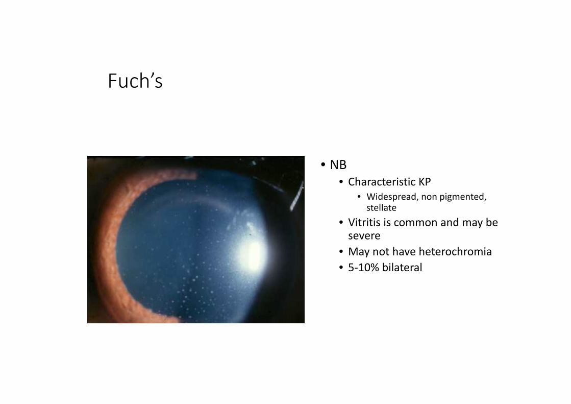
Fuch’s
• NB• Characteristic KP
• Widespread, non pigmented,stellate
• Vitritis is common and may besevere
• May not have heterochromia• 5-10% bilateral


Intermediate uveitis• Common• Young and middle aged• +/- macular oedema• +/- retinal vasculitis• +/- inferior snow balls• +/- snow bank
• 1/3 require topical treatmentonly
• 1/3 intermittent orbital orsystemic therapy
• 1/3 more than steroids

Investigation of intermediate uveitis withoutlocalising features
• History • FBE• U&E, LFT• Syph serology• ACE• If indicated
• MR brain• LP
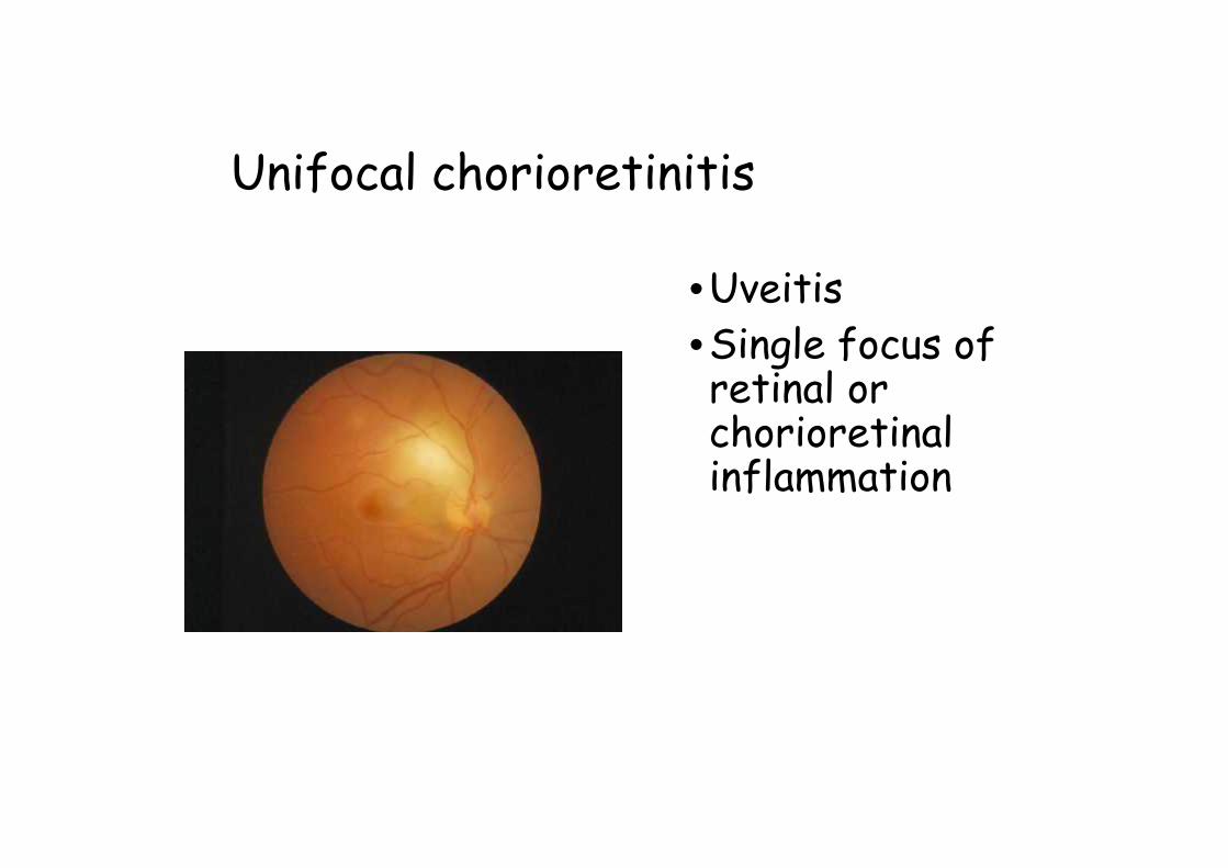
Unifocal chorioretinitis
•Uveitis•Single focus ofretinal orchorioretinalinflammation

Unifocal chorioretinitis
• Ocular diagnoses•Toxoplasmosis•Toxocara•Candida
• Systemic diagnoses•Sarcoidosis•Candida• (other fungal)

Toxoplasmosis • Unilateral uveitis• Active focus ofchorioretinitis – usuallyadjacent to an old scar
• AC activity• Raised pressure
• Otherwise well patient• Clinical diagnosis!• Can be confirmed byvitreous PCR if needed

Candida
• Unilateral uveitis• Single (or multiple) focus of
chorioretinitis with noadjacent scar
• May have vitreous puffballsor preretinal lesions
• At risk patient• Recent IV access• IVDU• Sick inpatients
• Diagnosed clinically andconfirmed with systemiccultures or vit tap/Vx

Toxocara• Uncommon cause of
unilateral uveitis andfocal chorioretinitis
• Mainly young children• Focal chorioretinitis
• Peripheral• Posterior pole
• Rarelyendophthalmitis

Investigation of unifocal chorioretinitis
• History • Toxoplasmosis serology• FBE• ACE• If indicated
• Toxocara serology• Blood/urine cultures• Vit tap
• Toxoplasma pcr• Fungal cultures
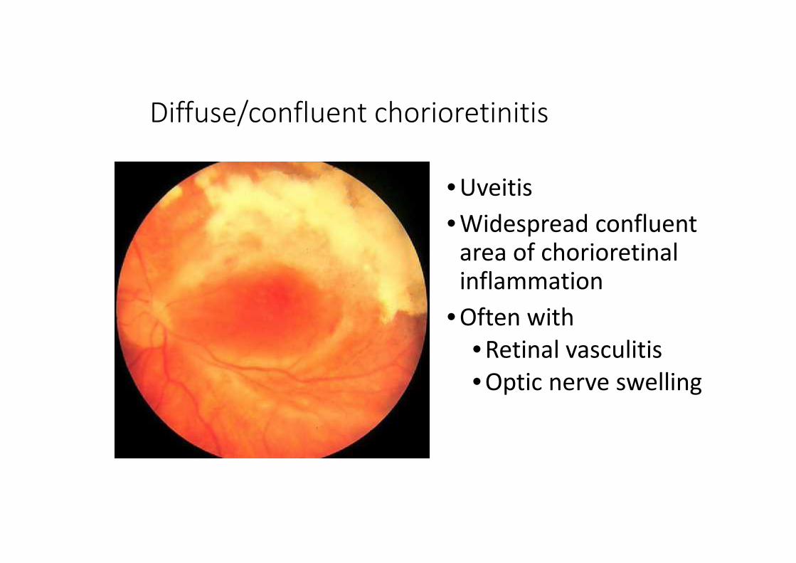
Diffuse/confluent chorioretinitis
• Uveitis• Widespread confluent
area of chorioretinalinflammation
• Often with• Retinal vasculitis• Optic nerve swelling

Diffuse chorioretinitis
• Ocular diagnoses•Viral retinitis•Serpiginouschoroidopathy
•Fungal retinitis
• Systemic diagnoses•Oculo-cerebrallymphoma
•Syphilis•Immuno-suppression/AIDS(and viral retinitis)

CMV retinitis
• Confluent retinitis• Haemorrhage andprogressive scarring
• Mild vitritis• HIV positive with lowCD4 count
• Treated with IV orintravitreal antiviralsand with immunerestoration

Acute retinal necrosis
•Rapidly progressiveperipheral retinitis
•Well patients•VZV or HSV•High incidence ofretinal detachment
•Treated withIV/intravitrealantivirals

Serpiginous choroiditis
• Relapsing remittingslowly progressivechorioretinitis withquiet vitreous
• Typical serpentineshape
• Posterior pole• Beware TB

Serpiginous in Australia
• ¼ had TB

Investigation of diffuse chorioretinitis
• History • FBE, U&E, LFT• HIV viral load, CD4 count• CMV viral load• Consider
• Quantiferon• Vit tap
• Herpes pcr• Fungal cultures
• MRI

Multifocal choroiditis
•Multifocalchoroiditis iscommon in manyforms of uveitis andoften does not carrydiagnosticsignificance

Multifocal choroiditis
• Ocular diagnoses• No inflammation• Mild inflammation• Significant inflammation
• Systemic diagnoses•Sarcoidosis•Behcet's•Oculo-cerebrallymphoma
•Infection• TB• Cryptococcus• PCP• syphillis
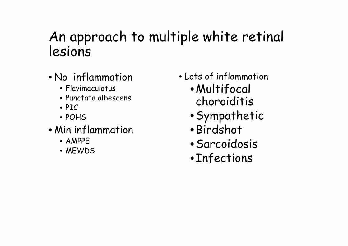
An approach to multiple white retinallesions
• No inflammation• Flavimaculatus• Punctata albescens• PIC• POHS
• Min inflammation• AMPPE• MEWDS
• Lots of inflammation•Multifocalchoroiditis
•Sympathetic•Birdshot•Sarcoidosis•Infections

Punctate inner choroidopathy (PIC)
• Myopic women• Small pale atrophic
lesions around theposterior pole
• Minimal or no vitritis• CNVM common

POHS
• Atrophic peripheralscars and streaks
• Peripapillary atrophy• Macular CNVM very
common• More common in the
USA
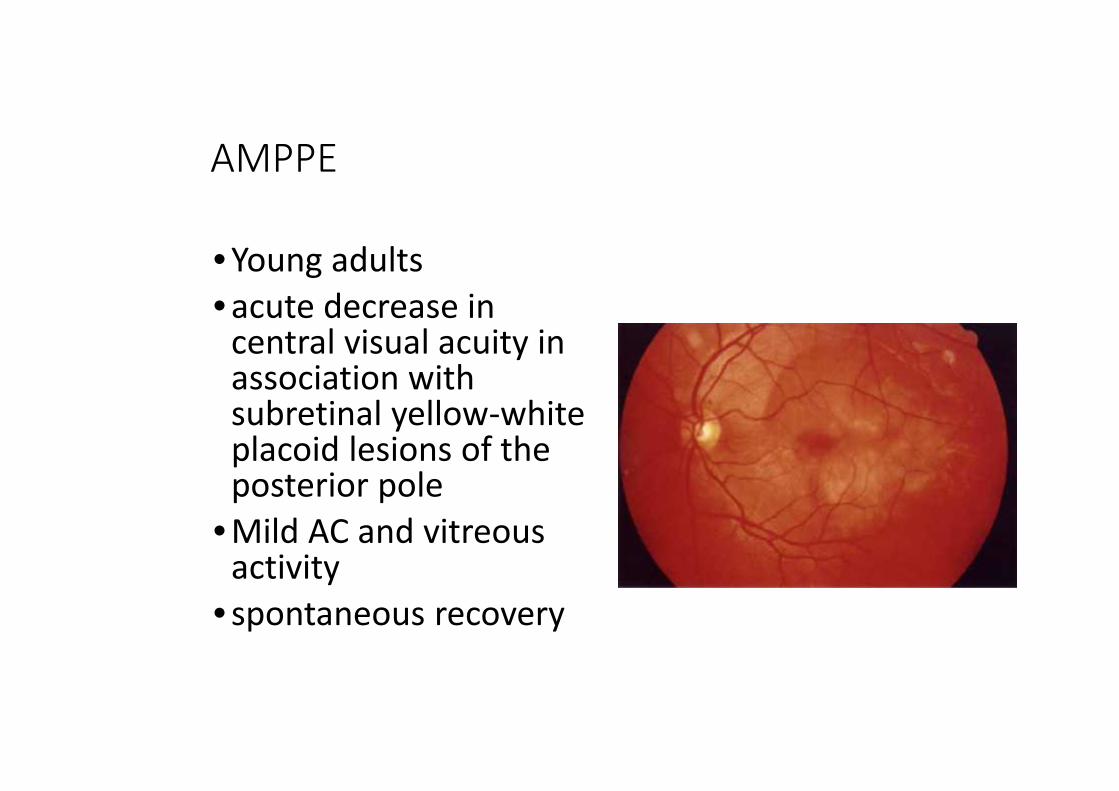
AMPPE
• Young adults• acute decrease in
central visual acuity inassociation withsubretinal yellow-whiteplacoid lesions of theposterior pole
• Mild AC and vitreousactivity
• spontaneous recovery

MEWDS
• Young women• Sudden onset unilateral
visual loss withphotopsia
• Small pale evanescentwhite outer retinal dots
• Mild AC and vitreousactivity
• Spontaneous recovery

Sympathetic ophthalmia•Bilateral uveitisfollowingpenetrating trauma•Surgery•Accidental trauma
•Uveitis with•multifocalchoroiditis
•Diagnosis ofexclusion

Incidence of sympathetic ophthalmia afterpenetrating trauma (cases/1,000 injuries)
0
0.5
1
1.5
2
2.5
3
WW I(5,300
injuries)
WW II(2,600
injuries)
Korean(3200
injuries)
Vietnam(119
injuries)
Iran/Iraq(4,179
injuries)

Aetiology of sympathetic ophthalmia
0
5
10
15
20
25
Haddassah (1970-83) NEI (1982-92) Bascom (1976-97)
Traumatic
Surgical
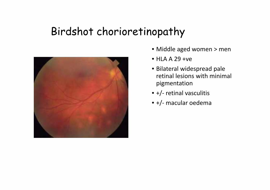
Birdshot chorioretinopathy• Middle aged women > men• HLA A 29 +ve• Bilateral widespread pale
retinal lesions with minimalpigmentation
• +/- retinal vasculitis• +/- macular oedema

Non infectious multifocal choroiditis
• Is still generallyidiopathic
• Think of• Sarcoid• Behcet’s• SO

Infectious multifocal choroiditis
• Aetiology• Mycobacterial
• Tuberculous• Non-tuberculous
• Cryptococcus andother fungus
• Pneumocystis carinii

• Pneumocystis choroiditis• Exclusively in AIDS patients on
inhaled prophylaxis forPneumocystis pneumonia

• Cryptococcal choroiditis• occurs in patients with
cryptococcal meningitis
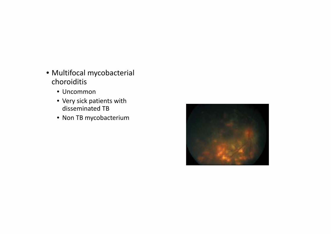
• Multifocal mycobacterialchoroiditis
• Uncommon• Very sick patients with
disseminated TB• Non TB mycobacterium

Syphilitic retinitis
• Punctate inner retinitis• Secondary syphilis

Sarcoidosis
• Focal granulomatousinflammation
• Non necrotising granulomas• Generalised
immunosuppression

“Caseating” granulomas
• Usually infectious• TB• Cat scratch

Sarcoidosis
• Focal granulomatousinflammation
• Generalisedimmunosuppression
• Lung• Skin• Nodes• Eyes• Liver• Heart• CNS• kidneys
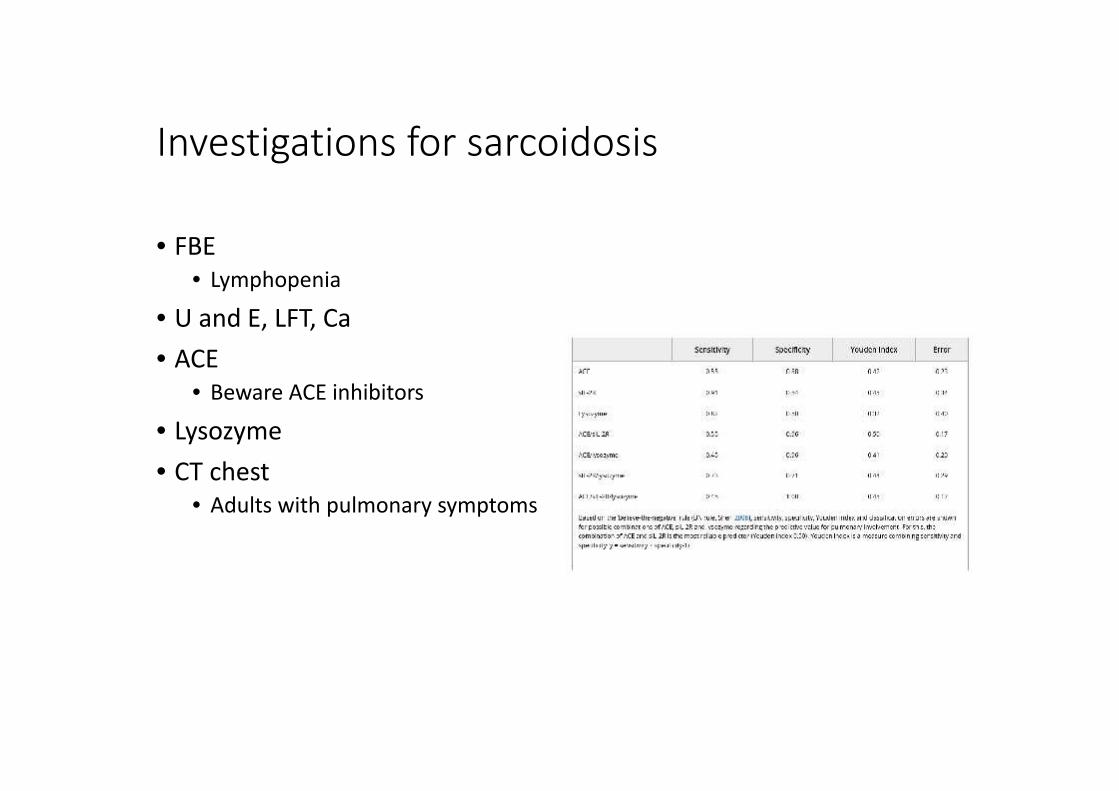
Investigations for sarcoidosis
• FBE• Lymphopenia
• U and E, LFT, Ca• ACE
• Beware ACE inhibitors• Lysozyme• CT chest
• Adults with pulmonary symptoms

Investigation of multifocal chorioretinitis
• History • FBE, U&E, LFT• ACE• If indicated
• HLA A 29• Quantiferon• Blood/urine cultures• FA• Syph serology
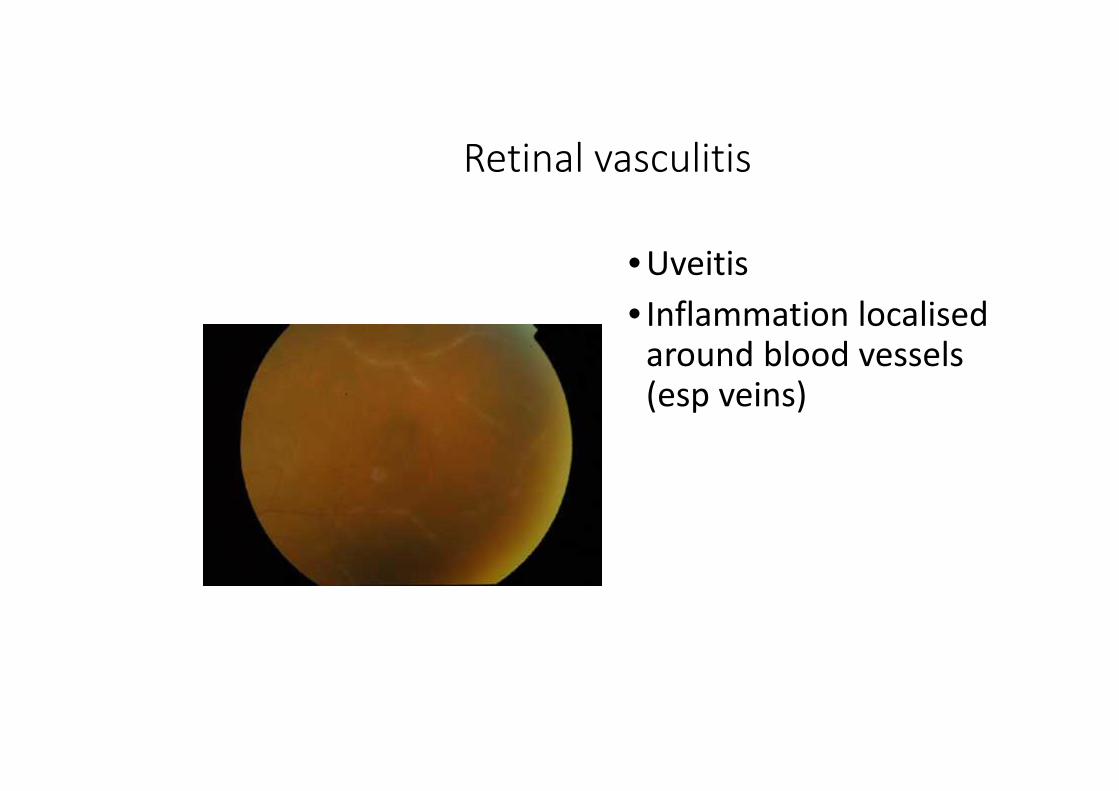
Retinal vasculitis
• Uveitis• Inflammation localised
around blood vessels(esp veins)

Retinal vasculitis - aetiology
• Aetiology in 103 patients in anAustralian uveitis clinic

Development of MS in intermediate uveitis(with or without vasculitis)
• At 10 yrs the chance of MS is28%

Uveitis in MS treatment trials
• All patients with MS ontreatment were followedregularly
• 4735 unique patients in 6different trials
• All with reg ophthalmic followup
• 34/4735 (0.7%) developeduveitis

Retinal vasculitis as part of systemic vasculitis
• 1286 patients with systemic vasculitis• PAN (393)• GPA (343)• MPA (280)• EGPA/CSS (270)
• Rothschild PR1, Pagnoux C, Seror R, Brézin AP,Delair E, Guillevin L. Ophthalmologicmanifestations of systemic necrotizingvasculitides at diagnosis: a retrospective studyof 1286 patients and review of the literature.Semin Arthritis Rheum 2013 Apr;42(5):507-14.

Investigation of “typical” retinal vasculitis
• History• Examination
• Tests• ACE (CXR CT chest)• Quantiferon• T P IgG• Consider MRI

Investigation of “Atypical” retinal vasculitis –acute retinal microangiopathy
• History• Examination
• Tests• FBE• U&E, LFT,• ANA
• Lupus anticoagulant• Anti-cardiolipin antibodies
• Se Igs and cryoglobulins• Se viscosity

Retinal vasculitis - aetiology
• Ocular diagnoses• Intermediate uveitis• Secondary to other
uveitis• Especially retinal infections• Toxo• Viral retinitis
• Eale’s disease
• Systemic diagnoses• Often idiopathic• Sarcoidosis• Behcet’s• MS• Rarely systemic
vasculitis

Eale's disease • Occlusive retinal vasculitis• Veins > arteries• Secondary neovascularisation
and vitreous haemorrhage• Most common in patients from
south Asia• Diagnosis of exclusion
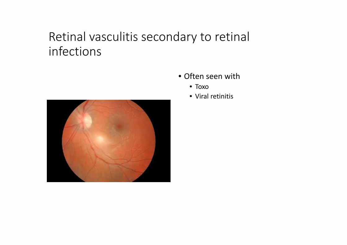
Retinal vasculitis secondary to retinalinfections
• Often seen with• Toxo• Viral retinitis
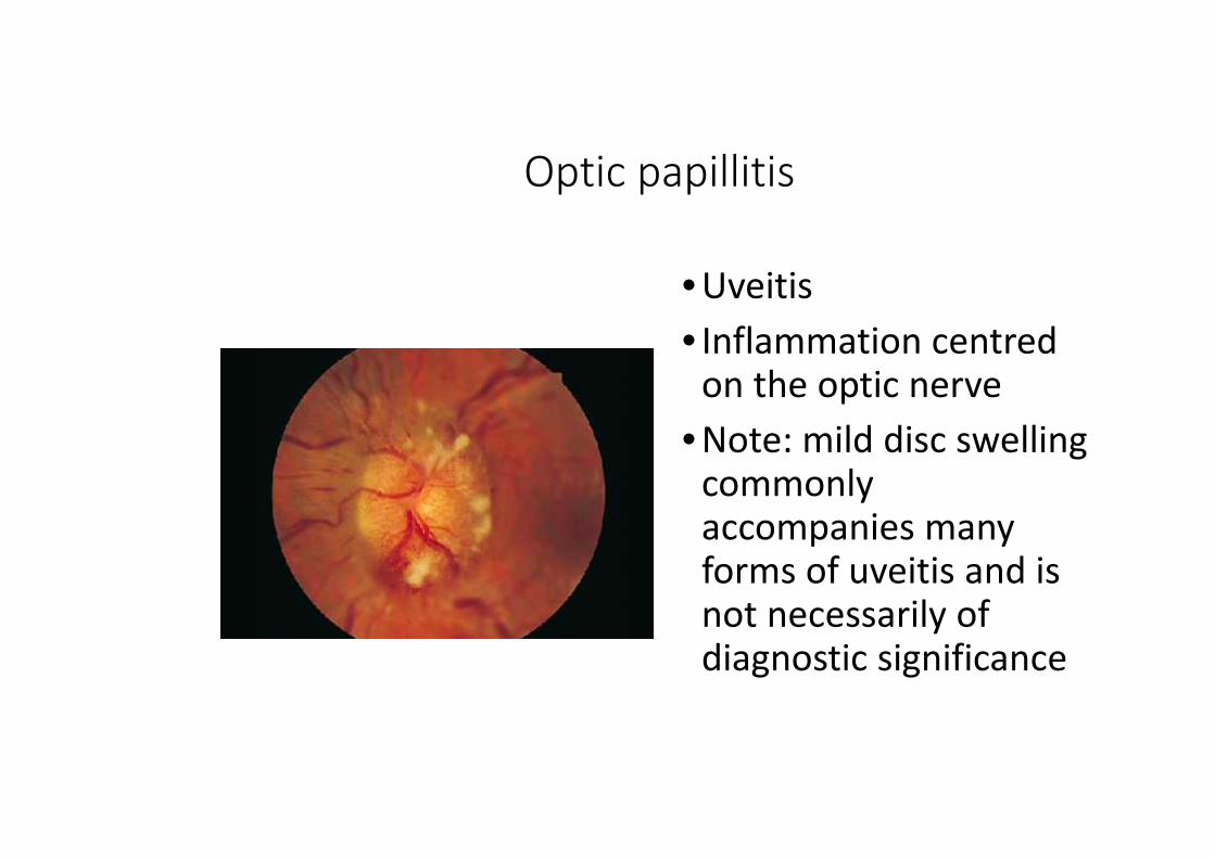
Optic papillitis
• Uveitis• Inflammation centred
on the optic nerve• Note: mild disc swelling
commonlyaccompanies manyforms of uveitis and isnot necessarily ofdiagnostic significance
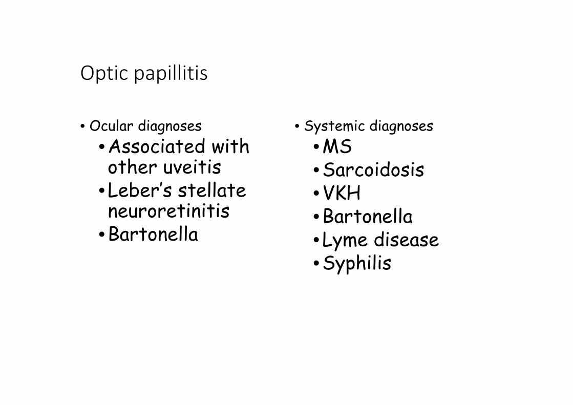
Optic papillitis
• Ocular diagnoses•Associated withother uveitis
•Leber’s stellateneuroretinitis
•Bartonella
• Systemic diagnoses•MS•Sarcoidosis•VKH•Bartonella•Lyme disease•Syphilis

Bartonella
• Systemic illness following catscratch with lymphadenopathy
• Disc swelling• Stellate maculopathy
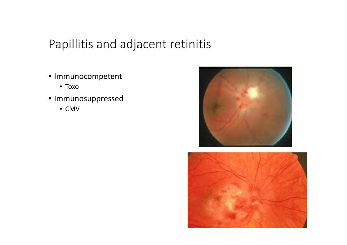
Papillitis and adjacent retinitis
• Immunocompetent• Toxo
• Immunosuppressed• CMV

Investigation of optic papillitis
• History • Syph serology• Bartonella serology• ACE• MRI• If indicated
• Lyme serology• Toxo serology• Quantiferon• Lebers mitochondrial gene testing

Uveitis and serous retinal detachment
• Serous detachment isuncommon in uveitis
• Usually cariesdiagnostic significance
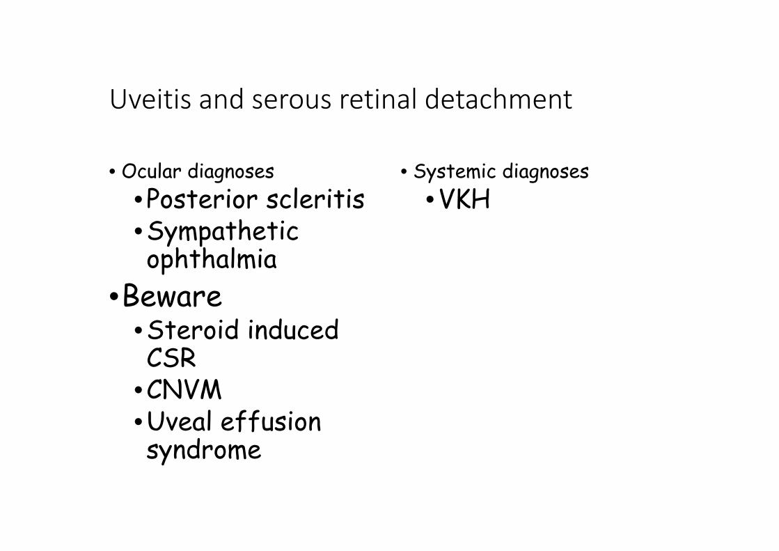
Uveitis and serous retinal detachment
• Ocular diagnoses•Posterior scleritis•Sympatheticophthalmia
•Beware•Steroid inducedCSR
•CNVM•Uveal effusionsyndrome
• Systemic diagnoses•VKH

Posterior scleritis
• Posterior scleritis• Pain• Disc swelling• Choroidal folds• Serous detachment• Minimal uveitis

VKH
• Headaches• CSF lymphocytosis
• Hearing changes• Uveitis
• Serous detachment• Disc swelling• Multifocal leak and disc swelling
on FA

VKH
• 3 out of• Bilateral anterior uveitis• Bilateral posterior uveitis with
• Serous ret det• Disc swelling• RPE changes
• CNS involvement• Pleocytosis• HA• Tinnitus/CN involvement
• Skin involvement

VKH
• 3 phases• Meningo-encephalitic phase• Uveitis/auditory phase• Skin/hair phase
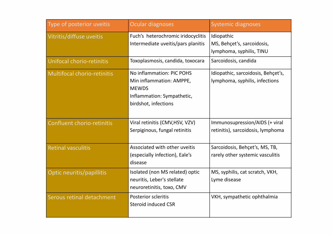
Type of posterior uveitis Ocular diagnoses Systemic diagnoses
Vitritis/diffuse uveitis Fuch’s heterochromic iridocyclitisIntermediate uveitis/pars planitis
IdiopathicMS, Behçet’s, sarcoidosis,lymphoma, syphilis, TINU
Unifocal chorio-retinitis Toxoplasmosis, candida, toxocara Sarcoidosis, candida
Multifocal chorio-retinitis No inflammation: PIC POHSMin inflammation: AMPPE,MEWDSInflammation: Sympathetic,birdshot, infections
Idiopathic, sarcoidosis, Behçet’s,lymphoma, syphilis, infections
Confluent chorio-retinitis Viral retinitis (CMV,HSV, VZV)Serpiginous, fungal retinitis
Immunosupression/AIDS (+ viralretinitis), sarcoidosis, lymphoma
Retinal vasculitis Associated with other uveitis(especially infection), Eale’sdisease
Sarcoidosis, Behçet’s, MS, TB,rarely other systemic vasculitis
Optic neuritis/papillitis Isolated (non MS related) opticneuritis, Leber's stellateneuroretinitis, toxo, CMV
MS, syphilis, cat scratch, VKH,Lyme disease
Serous retinal detachment Posterior scleritisSteroid induced CSR
VKH, sympathetic ophthalmia

Important concepts in making a diagnosis inposterior uveitis
• Limited number of diagnostic possibilities for eachtype of posterior uveitis and a limited number ofsystemic diseases seen commonly in posterioruveitis
• Most posterior uveitis is not associated withsystemic disease
• A good history is more important than untargetedinvestigation
• Many more mistakes are made in the treatment ofuveitis than in its diagnosis
• The underlying diagnosis rarely guides treatment

• Handful of important systemic diagnoses• Sarcoidosis• Behcets• MS• Syphilis• Oculo-cerebral lymphoma
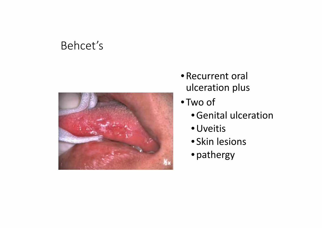
Behcet’s
• Recurrent oralulceration plus
• Two of• Genital ulceration• Uveitis• Skin lesions• pathergy

Syphilis
Stage Clinical features
Primary Chancre
Secondary SkinLiverJointsEyes (2.5-5%)
Latent
Tertiary Neuro-syphilisCardiac

Ocular syphilis
•Anterior uveitis Posterior Uveitis Panuveitis Total patients
HIV positive 27 (96.4%) 48 (60.8%) 18 (50%) 93 (65%)
HIV negative 1 (3.6%) 31 (39.2%) 18 (50%) 50 (35%)
Total 28 (20%) 79 (55%) 36 (25%) 143
Amaratunge BC Camuglia JE Hall AJ Syphilitic Uveitis: A review of clinical manifestations andtreatment outcomes of syphilitic uveitis in HIV positive and HIV negative patients. Clin Exp Ophth inpress

0.1
1
10
100
2007 2008 2009 2010 2011
Age
stan
dard
ised
rate
per
100
000
Year of diagnosis
NSW NT QLD SA TAS VIC WA
Infectious syphilis, 2007 – 2011, by State/Territory and year
Source: National Notifiable Diseases Surveillance System

Primary ocular lymphoma
• Elderly patients (>50 yo)• Persistent and progressive and
treatment resistant uveitis with:• Prominent vitritis• +/- multifocal choroiditis, retinitis,
retinal vasculitis

Diagnosis
• Systemic• MRI• LP• Brain biopsy
• Ocular• Vit biopsy• +/- retinal biopsy
Intraocular lymphoma: a series of 14 patients withclinicopathological features and treatment outcomes.Hoffman PM, McKelvie P, Hall AJ, Stawell RJ, Santamaria JD.Eye 2003 May;17(4):513-21.
Ocular history taking in uveitis
• Trauma/surgery• Infection/SO
• Persistent unilaterality• infection

Systemic history taking in uveitis
System Possible diseases
Joints B27, Sarcoid, RA, Lupus…
Respiratory Sarcoid, GPA, TB, …
GIT IBD, Behcet's, Whipples…
GUT Behcet's, Reactive arthritis, GPA, syphilis, TINU…
Skin Ps arthritis, syphilis, Sarcoid, SLE…
Mouth Behcet’s, HSV…
CNS Lymphoma, MS, neuro-Behcets, infections, VKH…
Drugs Bisphosphonates, Rifabutin, vaccines, tattoos.

Investigation in uveitis
• Limited untargetedinvestigations for when thereare no specific features tosuggest a diagnosis
• FBE, U&E, LFT, RBG, ACE, CXR,VDRL
• More intensive/expensive/invasive investigations onlywhen clinically indicated

Underlying disease Useful investigations
Sarcoidosis Most useful: ACE, CR, HRCT chest, targeted biopsy, BALLess useful: Gallium scan, blind biopsy
Systemic vasculitis ANA, ENA, ANCA, Rh Factor, MSU, HLA A29, Hep serology
Birdshot HLA A29
VKH LP
MS MRI and LP
Primary oculo-cerebral lymphoma MRI brain, LP, brain biopsy, vitrectomy +/- retinal biopsy
Ocular syphilis (usually 2ry) RPR, TP IgG
Toxocara Toxocara serology
Lyme disease Borelia serology
Cat scratch disease Bartonella serology
Brucellosis Brucella serology
Viral retinitis Most useful: AC or vitreous PCR, systemic viral loadConsider HIV serology/CD4 countLess useful: viral serology (may be useful if negative)
TB Most useful: IFN gamma release assay (Quantiferon), CXRLess useful: mantoux
West Nile West Nile virus serology
Metastatic endophthalmitis Blood cultures, culture site of suspected primary infection eg catheter tips, central lines,chest, urine, etc. Vitreous tap

Summary
• Make a descriptive diagnosis ofthe inflammation
• Determine clinically if possiblethe cause of the patients visualloss
• Take a targeted history
Summary
• Investigate aggressively anyfeatures identified on history
• If no clues on history then alimited number ofinvestigations targeting thelikely causes and possible sideeffects of treatment

Thank you