
WHA T’S THE CONNECTION? Sharon Winter · WHA T’S THE CONNECTION? ... Aplysia. In humans and...
16
What’s the Connection? 59 WHAT’S THE CONNECTION? Directions for Teachers Getting Ready See sidebars for additional information regarding preparation of this lab. Directions for Setting Up the Lab General: ■ Make an “X” on the chalkboard for the teacher-led introduction. ■ Photocopy the Directions for Students pages. Teacher Background A reflex is an involuntary neural response to a specific sensory stimulus that threatens the survival or homeostatic state of an organism. Reflexes exist in the most primitive of species, usually with a protective function for animals when they encounter external and internal stimuli. A primitive example of this protective reflex is the gill withdrawal reflex of the sea slug Aplysia. In humans and other vertebrates, protective reflexes have been maintained and expanded in number. Examples are the gag reflex that occurs when objects touch the sides or the back of the throat, and the carotid sinus reflex that restores blood pressure to normal when baroreceptors detect an increase in blood pressure. A second type of reflex, the stretch reflex, has evolved to help maintain muscle tone that is important for posture and movement. An understanding of the neural circuitry underlying each type of reflex also has clinical significance. When physicians test for potential damage to various components of the nervous system, they often begin by attempting to elicit reflex responses to the appropriate stimuli. Examples of reflexes with protective and diagnostic importance are the flexor withdrawal reflex, the corneal (blink) reflex, and the accommodation reflex. The loss of the pupillary light reflex in a comatose patient, described under the section Protective reflex, often indicates extreme damage deep in the brain. Loss of the knee-jerk response may indicate problems with muscle tone, chronic diabetes, or damage at the L2, L3, or L4 vertebral level. Sharon Winter Lake Washington High School 12033 NE 80th Street Kirkland, WA 98033 SYNOPSIS Students elicit and observe reflex responses and distinguish between types of reflexes. They then design and conduct experiments to learn more about reflexes and their control by the nervous system. LEVEL Exploration, Concept/Term Introduction Phases Application Phase STUDENT PRIOR KNOWL- EDGE Before participating in this activity students should be able to: ■ Describe the parts of a neuron and explain their functions. ■ Distinguish between sensory and motor neurons. ■ Describe briefly the organization of the nervous system. INTEGRATION Into the Biology Curriculum ■ Health ■ Biology I, II ■ Human Anatomy and Physiology ■ AP Biology Across the Curriculum ■ Mathematics ■ Physics ■ Psychology OBJECTIVES At the end of this activity, students will be able to: ■ Identify common reflexes of the human body and the neural circuitry involved in these reflexes. ■ Distinguish between stretch and protective reflexes. ■ Describe the role of reflexes.
Transcript of WHA T’S THE CONNECTION? Sharon Winter · WHA T’S THE CONNECTION? ... Aplysia. In humans and...
What’s the Connection? 59
WHAT’S THE CONNECTION?
Directions for Teachers
Getting ReadySee sidebars for additional information regarding preparation of this lab.
Directions for Setting Up the LabGeneral:
■ Make an “X” on the chalkboard for the teacher-led introduction.
■ Photocopy the Directions for Students pages.
Teacher BackgroundA reflex is an involuntary neural response to a specific sensory stimulusthat threatens the survival or homeostatic state of an organism. Reflexesexist in the most primitive of species, usually with a protective function foranimals when they encounter external and internal stimuli. A primitiveexample of this protective reflex is the gill withdrawal reflex of the sea slugAplysia. In humans and other vertebrates, protective reflexes have beenmaintained and expanded in number. Examples are the gag reflex thatoccurs when objects touch the sidesor the back of the throat, and the carotid sinus reflex that restores bloodpressure to normal when baroreceptors detect an increase in blood pressure.A second type of reflex, the stretch reflex, has evolved to help maintainmuscle tone that is important for posture and movement.
An understanding of the neural circuitry underlying each type of reflex alsohas clinical significance. When physicians test for potential damage tovarious components of the nervous system, they often begin by attemptingto elicit reflex responses to the appropriate stimuli. Examples of reflexeswith protective and diagnostic importance are the flexor withdrawal reflex,the corneal (blink) reflex, and the accommodation reflex. The loss of thepupillary light reflex in a comatose patient, described under the sectionProtective reflex, often indicates extreme damage deep in the brain. Lossof the knee-jerk response may indicate problems with muscle tone, chronicdiabetes, or damage at the L2, L3, or L4 vertebral level.
Sharon WinterLake Washington High School
12033 NE 80th StreetKirkland, WA 98033
SYNOPSISStudents elicit and observe reflex responses and distinguish between typesof reflexes. They then design and conduct experiments to learn more aboutreflexes and their control by the nervous system.
LEVEL
Exploration, Concept/Term Introduction Phases
Application Phase
STUDENT PRIOR KNOWL-EDGEBefore participating in thisactivity students should beable to:■ Describe the parts of a
neuron and explain theirfunctions.
■ Distinguish betweensensory and motorneurons.
■ Describe briefly theorganization of the nervoussystem.
INTEGRATIONInto the Biology Curriculum■ Health■ Biology I, II■ Human Anatomy and
Physiology■ AP Biology
Across the Curriculum■ Mathematics■ Physics■ Psychology
OBJECTIVESAt the end of this activity,students will be able to:■ Identify common reflexes
of the human body and theneural circuitry involved inthese reflexes.
■ Distinguish betweenstretch and protectivereflexes.
■ Describe the role ofreflexes.
60 Neuroscience Laboratory Activities Manual
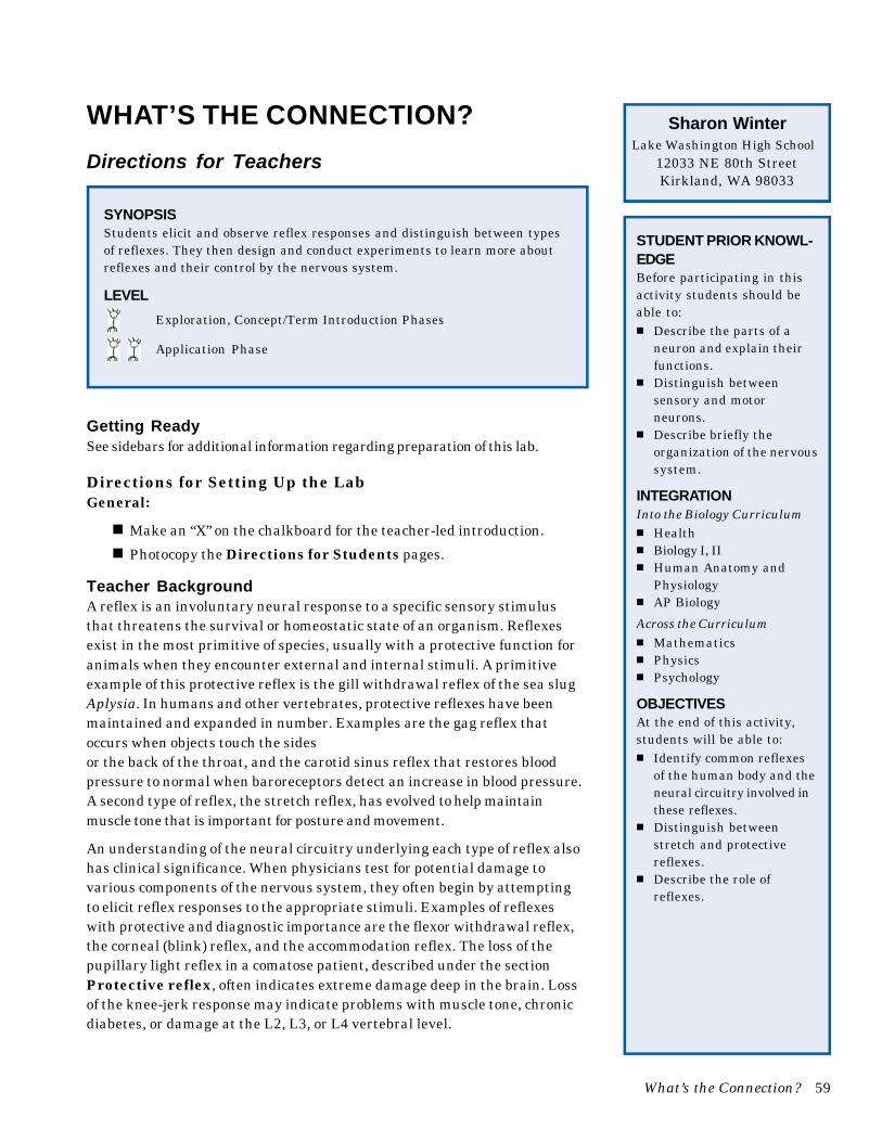
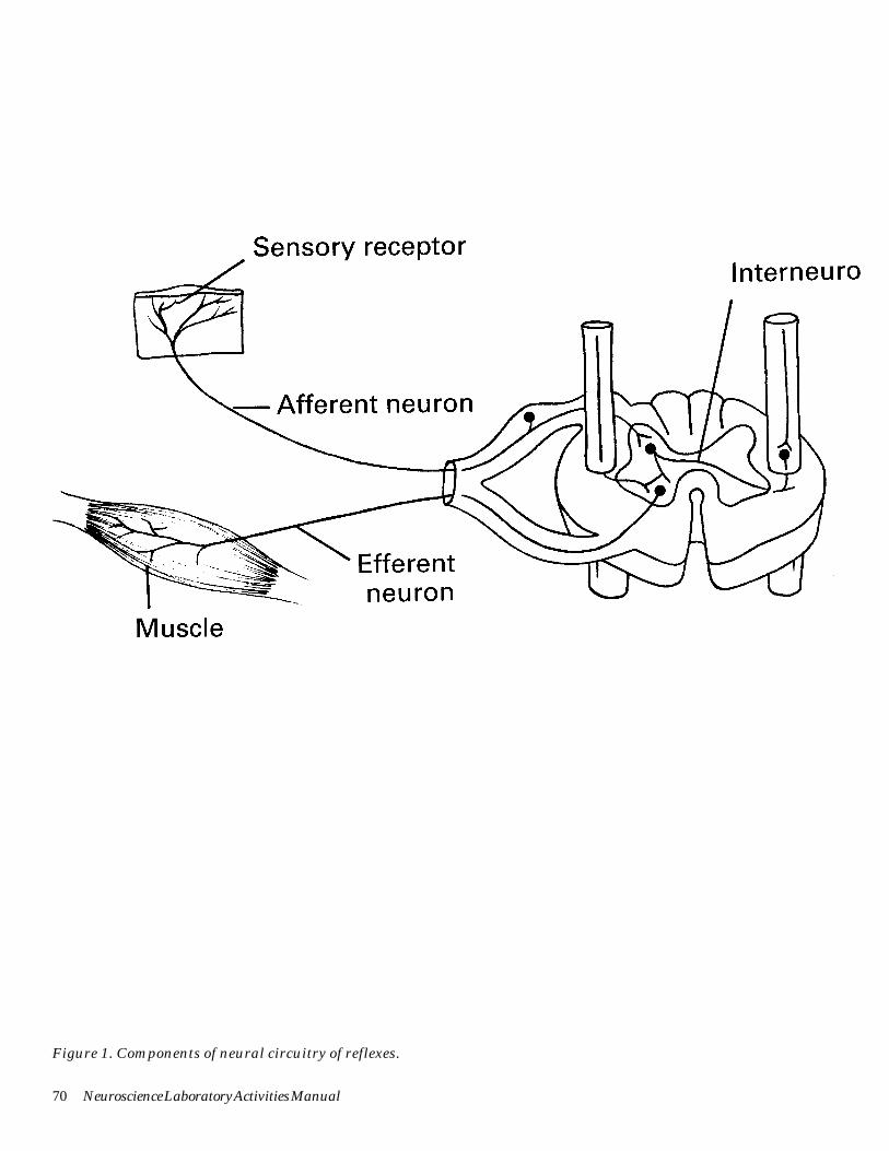
For each reflex resulting in a specific motor output, the neural pathwaylinking the sensory receptor to the central nervous system (CNS) may bestudied. In this activity, components of the neural circuitry of reflexes willbe studied. These components include a sensory receptor, a sensory (afferent)neuron, a reflex center in the CNS that may involve one or more levels ofintegration, a motor (efferent) neuron, and a muscle (effector). See Figure 1.
Students will investigate the following reflexes to learn about these compo-nents:
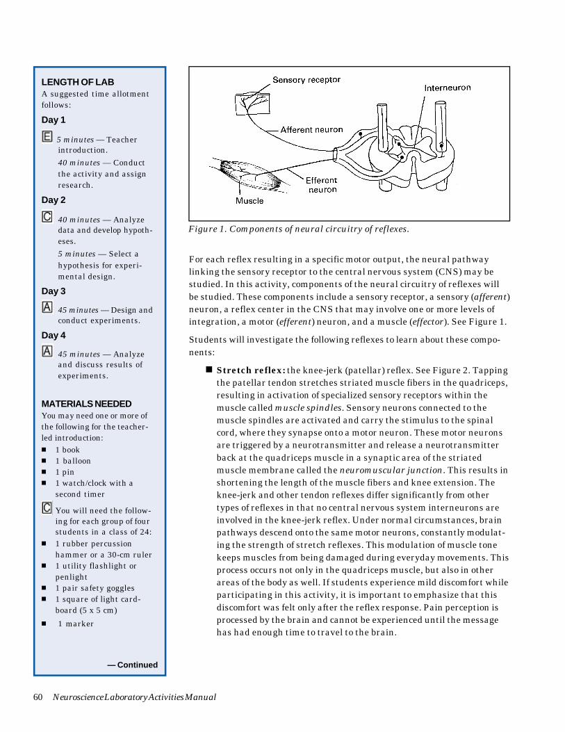
■ Stretch reflex: the knee-jerk (patellar) reflex. See Figure 2. Tappingthe patellar tendon stretches striated muscle fibers in the quadriceps,resulting in activation of specialized sensory receptors within themuscle called muscle spindles. Sensory neurons connected to themuscle spindles are activated and carry the stimulus to the spinalcord, where they synapse onto a motor neuron. These motor neuronsare triggered by a neurotransmitter and release a neurotransmitterback at the quadriceps muscle in a synaptic area of the striatedmuscle membrane called the neuromuscular junction. This results inshortening the length of the muscle fibers and knee extension. Theknee-jerk and other tendon reflexes differ significantly from othertypes of reflexes in that no central nervous system interneurons areinvolved in the knee-jerk reflex. Under normal circumstances, brainpathways descend onto the same motor neurons, constantly modulat-ing the strength of stretch reflexes. This modulation of muscle tonekeeps muscles from being damaged during everyday movements. Thisprocess occurs not only in the quadriceps muscle, but also in otherareas of the body as well. If students experience mild discomfort whileparticipating in this activity, it is important to emphasize that thisdiscomfort was felt only after the reflex response. Pain perception isprocessed by the brain and cannot be experienced until the messagehas had enough time to travel to the brain.
LENGTH OF LABA suggested time allotmentfollows:
Day 1
5 minutes — Teacherintroduction.
40 minutes — Conductthe activity and assignresearch.
Day 2
40 minutes — Analyzedata and develop hypoth-eses.
5 minutes — Select ahypothesis for experi-mental design.
Day 3
45 minutes — Design andconduct experiments.
Day 4
45 minutes — Analyzeand discuss results ofexperiments.
MATERIALS NEEDEDYou may need one or more ofthe following for the teacher-led introduction:■ 1 book■ 1 balloon■ 1 pin■ 1 watch/clock with a
second timer
You will need the follow-ing for each group of fourstudents in a class of 24:
■ 1 rubber percussionhammer or a 30-cm ruler
■ 1 utility flashlight orpenlight
■ 1 pair safety goggles■ 1 square of light card-
board (5 x 5 cm)
■ 1 marker
— Continued
Figure 1. Components of neural circuitry of reflexes.
What’s the Connection? 61
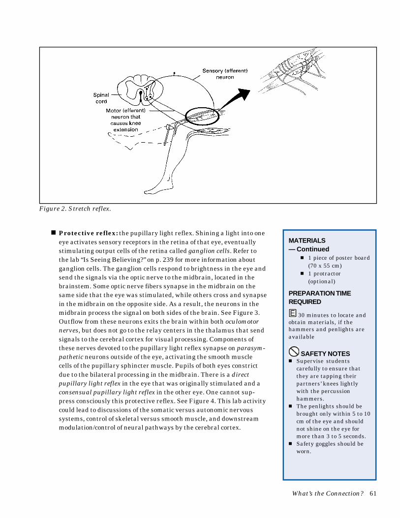
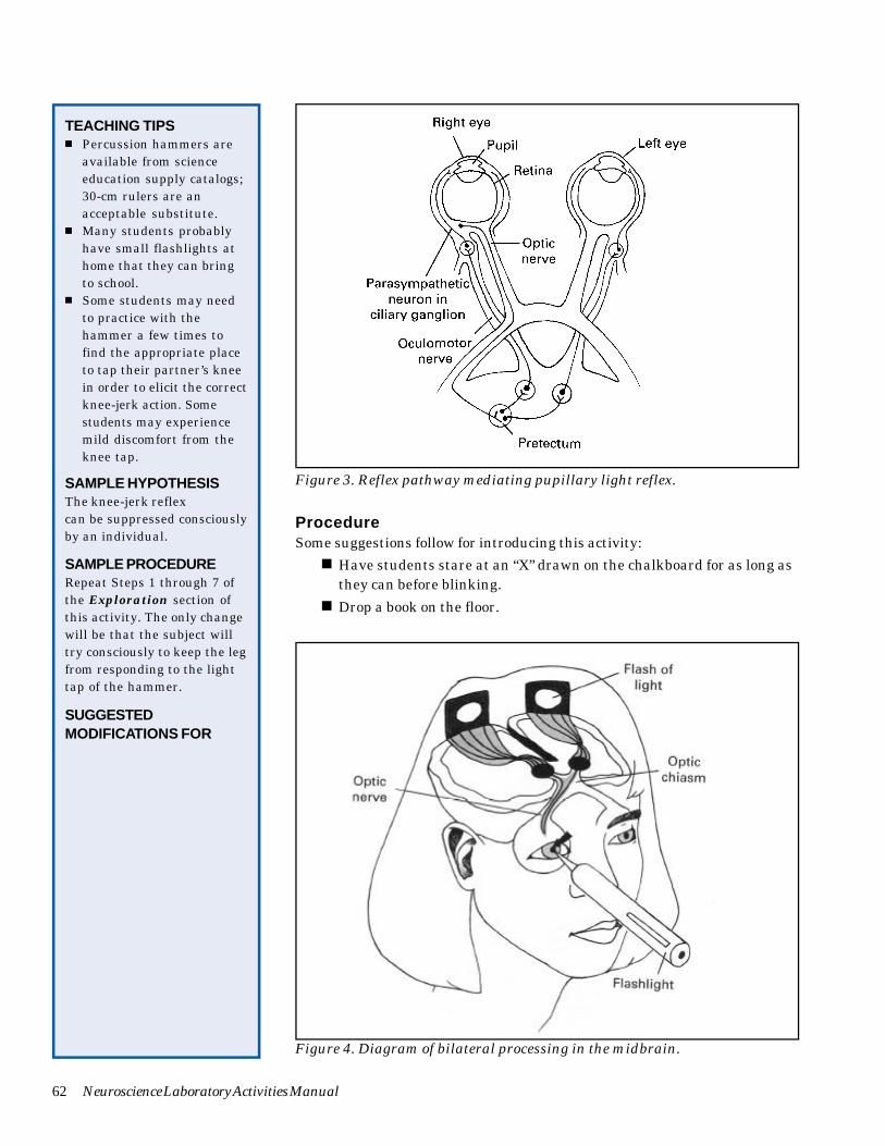
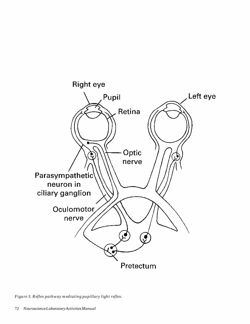
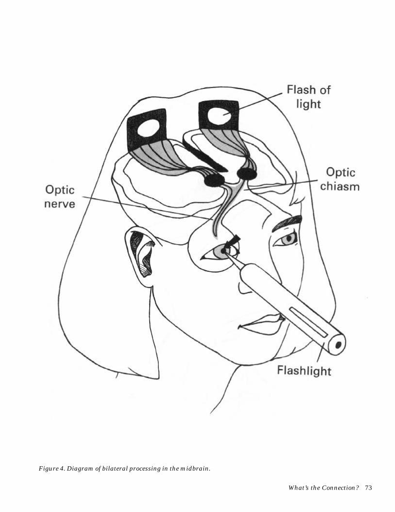
■ Protective reflex: the pupillary light reflex. Shining a light into oneeye activates sensory receptors in the retina of that eye, eventuallystimulating output cells of the retina called ganglion cells. Refer tothe lab “Is Seeing Believing?” on p. 239 for more information aboutganglion cells. The ganglion cells respond to brightness in the eye andsend the signals via the optic nerve to the midbrain, located in thebrainstem. Some optic nerve fibers synapse in the midbrain on thesame side that the eye was stimulated, while others cross and synapsein the midbrain on the opposite side. As a result, the neurons in themidbrain process the signal on both sides of the brain. See Figure 3.Outflow from these neurons exits the brain within both oculomotornerves, but does not go to the relay centers in the thalamus that sendsignals to the cerebral cortex for visual processing. Components ofthese nerves devoted to the pupillary light reflex synapse on parasym-pathetic neurons outside of the eye, activating the smooth musclecells of the pupillary sphincter muscle. Pupils of both eyes constrictdue to the bilateral processing in the midbrain. There is a directpupillary light reflex in the eye that was originally stimulated and aconsensual pupillary light reflex in the other eye. One cannot sup-press consciously this protective reflex. See Figure 4. This lab activitycould lead to discussions of the somatic versus autonomic nervoussystems, control of skeletal versus smooth muscle, and downstreammodulation/control of neural pathways by the cerebral cortex.
MATERIALS— Continued
■ 1 piece of poster board(70 x 55 cm)
■ 1 protractor(optional)
PREPARATION TIMEREQUIRED
30 minutes to locate andobtain materials, if thehammers and penlights areavailable
SAFETY NOTES■ Supervise students
carefully to ensure thatthey are tapping theirpartners’ knees lightlywith the percussionhammers.
■ The penlights should bebrought only within 5 to 10cm of the eye and shouldnot shine on the eye formore than 3 to 5 seconds.
■ Safety goggles should beworn.
Figure 2. Stretch reflex.
62 Neuroscience Laboratory Activities Manual
TEACHING TIPS■ Percussion hammers are
available from scienceeducation supply catalogs;30-cm rulers are anacceptable substitute.
■ Many students probablyhave small flashlights athome that they can bringto school.
■ Some students may needto practice with thehammer a few times tofind the appropriate placeto tap their partner’s kneein order to elicit the correctknee-jerk action. Somestudents may experiencemild discomfort from theknee tap.
SAMPLE HYPOTHESISThe knee-jerk reflexcan be suppressed consciouslyby an individual.
SAMPLE PROCEDURERepeat Steps 1 through 7 ofthe Exploration section ofthis activity. The only changewill be that the subject willtry consciously to keep the legfrom responding to the lighttap of the hammer.
SUGGESTEDMODIFICATIONS FOR
Figure 4. Diagram of bilateral processing in the midbrain.
Figure 3. Reflex pathway mediating pupillary light reflex.
ProcedureSome suggestions follow for introducing this activity:
■ Have students stare at an “X” drawn on the chalkboard for as long asthey can before blinking.
■ Drop a book on the floor.
What’s the Connection? 63
STUDENTS WHO AREEXCEPTIONALBelow are possible ways tomodify this specific activity forstudents who have specialneeds, if they have not alreadydeveloped their own adapta-tions. General suggestions formodification of activities forstudents with impairmentsare found in the AAAS Barrier-Free in Brief publications.Refer to p. 19 of the introduc-tion of this book for informa-tion on ordering FREE copiesof these publications. Some ofthese booklets have addressesof agencies that can provideinformation about obtainingassistive technology, such asAssistive Listening Devices(ALDs); light probes; andtalking thermometers,calculators, and clocks.
Blind or Visually Impaired■ Using a Sewell Drawing
Kit, provide raised linedrawings of the stretchreflex and protective reflexfor students who are blind.Use photo-enlarged copiesof these diagrams forstudents with reducedvision.
■ Assign a student with avisual impairment to actas either the subject orexperimenter for thestretch reflex exercise. Forthe protective reflexactivity, the studentprobably would be mostcomfortable as an observeraided by a sighted partnerwho carefully describes theevents.
■ Use one of the auditorystimuli to introduce theactivity if the classincludes a student withlimited vision.
Deaf or Hard-of-HearingIntroduce the activity withone of the visual stimuli thatare suggested. If the student
— Continued
■ Close a book with a loud smack.■ Pop a balloon that is sitting on the teacher’s demonstration table.
■ Pretend to throw an imaginary object at the class.
Whatever stimulus you choose to use, take care not to compromise the safetyof your students. Immediately following the reflex response, ask questionssuggested in the list below:
■ What was your reaction to the stimulus?
■ How did your body respond?
■ How quickly did it respond?
■ Can you control your reaction?
■ What is the function of this response?
■ What is this response called? If students use the word “reflex” inanswering this question, ask them to name other reflexes.
Tell students they are going to do activities examining this phenomenon.
Exploration
The Exploration activity demonstrates the concept of the reflex arc. Thestudent procedures for conducting the activity with small groups are listedbelow.
1. Divide the class into six groups of four students each and have eachgroup conduct the activity. If possible, give each student the opportunityto be the subject.
2. Assign each student in the group one of the following roles:
■ Subject
■ Experimenter
■ Timer
■ Data recorder.
3. Have each group do the knee-jerk reflex. The subject should relax andsit on a desk or table with his/her legs dangling loosely over the edge.
4. The experimenter should apply the stimulus by tapping, firmly butcarefully, the area just below the kneecap with a percussion hammer.He/she should do this when the data recorder tells the timer to begintiming the reaction.
5. The data recorder should indicate to the timer when the subject hasresponded and the timer should note the time.
6. The data recorder should describe the response and record the time.
7. The group should repeat Steps 4–5 with the other leg.
8. Have the timer and data recorder exchange roles with the subject andthe experimenter to do the pupillary light reflex.
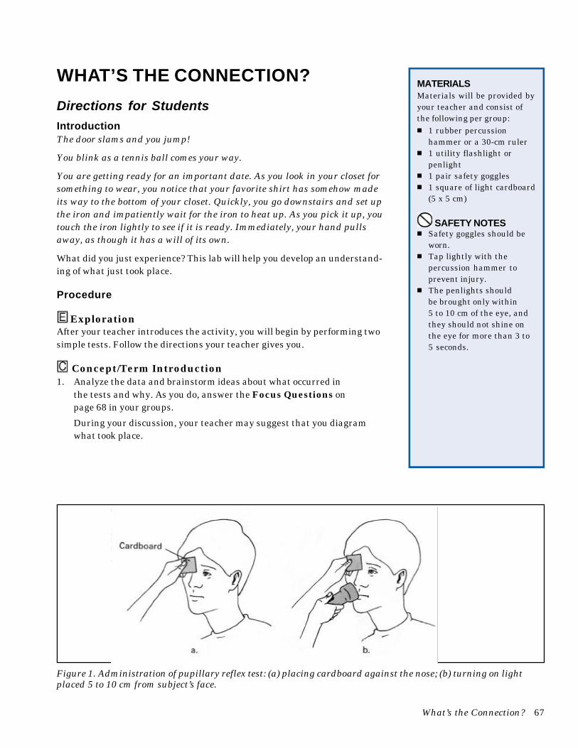
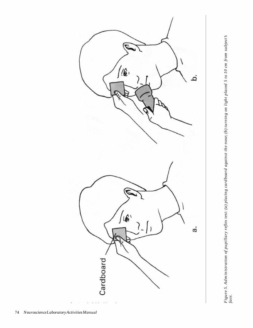
9. In a dimly lit room, the subject should look out toward a wall until his/her eyes dilate. The experimenter should place a 5 x 5 cm square of lightcardboard on the bridge of the subject’s nose to separate each eye’s fieldof vision. See Figure 5. Then the experimenter should bring a flashlight
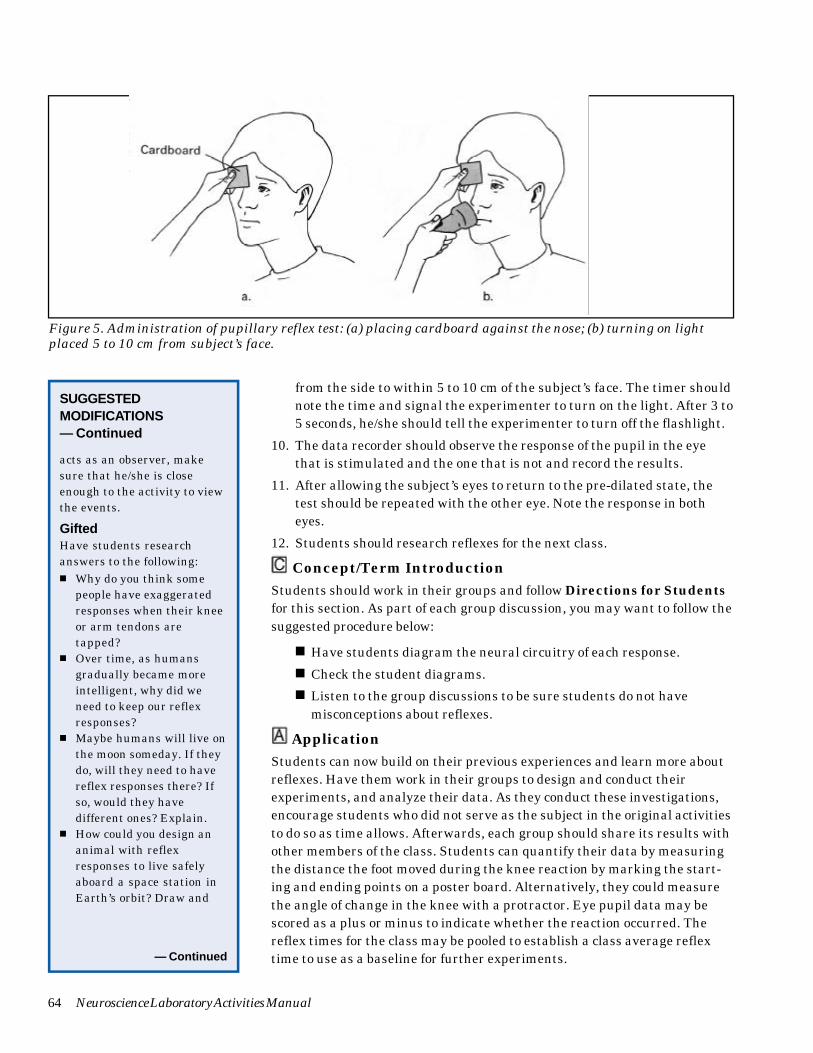
64 Neuroscience Laboratory Activities Manual
from the side to within 5 to 10 cm of the subject’s face. The timer shouldnote the time and signal the experimenter to turn on the light. After 3 to5 seconds, he/she should tell the experimenter to turn off the flashlight.
10. The data recorder should observe the response of the pupil in the eyethat is stimulated and the one that is not and record the results.
11. After allowing the subject’s eyes to return to the pre-dilated state, thetest should be repeated with the other eye. Note the response in botheyes.
12. Students should research reflexes for the next class.
Concept/Term Introduction
Students should work in their groups and follow Directions for Studentsfor this section. As part of each group discussion, you may want to follow thesuggested procedure below:
■ Have students diagram the neural circuitry of each response.
■ Check the student diagrams.
■ Listen to the group discussions to be sure students do not havemisconceptions about reflexes.
Application
Students can now build on their previous experiences and learn more aboutreflexes. Have them work in their groups to design and conduct theirexperiments, and analyze their data. As they conduct these investigations,encourage students who did not serve as the subject in the original activitiesto do so as time allows. Afterwards, each group should share its results withother members of the class. Students can quantify their data by measuringthe distance the foot moved during the knee reaction by marking the start-ing and ending points on a poster board. Alternatively, they could measurethe angle of change in the knee with a protractor. Eye pupil data may bescored as a plus or minus to indicate whether the reaction occurred. Thereflex times for the class may be pooled to establish a class average reflextime to use as a baseline for further experiments.— Continued
Figure 5. Administration of pupillary reflex test: (a) placing cardboard against the nose; (b) turning on lightplaced 5 to 10 cm from subject’s face.
acts as an observer, makesure that he/she is closeenough to the activity to viewthe events.
GiftedHave students researchanswers to the following:■ Why do you think some
people have exaggeratedresponses when their kneeor arm tendons aretapped?
■ Over time, as humansgradually became moreintelligent, why did weneed to keep our reflexresponses?
■ Maybe humans will live onthe moon someday. If theydo, will they need to havereflex responses there? Ifso, would they havedifferent ones? Explain.
■ How could you design ananimal with reflexresponses to live safelyaboard a space station inEarth’s orbit? Draw and
SUGGESTEDMODIFICATIONS— Continued
What’s the Connection? 65
Suggested questions students may wish to investigate include the following:
■ Can you suppress the knee-jerk reflex consciously? The eye reflex?
■ How is each of the following reactions controlled by the nervoussystem? In each case can the reflex be controlled consciously by thenervous system?
❏ Flexor (biceps) withdrawal reflex
❏ Corneal (blink) reflex
❏ Gag reflex
❏ Accommodation reflex
■ Does the timing or intensity of each of the reflexes vary amongindividuals? Do these characteristics change with age?
■ Is the response different between males and females?
■ What factors affect the timing and intensity of the withdrawal reflexof the land snail, Helix, sp.?
■ How are the reflexes of other invertebrate animals, such as thecockroach, controlled by their nervous systems?
■ Your students probably will develop other questions related to re-flexes. In the sidebar on p. 62 is a sample hypothesis and procedurethat students might derive related to this activity. This example hasbeen included as a suggested outcome of the activity and is not meantto be given to the students. Students should develop their own hypoth-eses and procedures. Make sure they understand that there is not justone correct hypothesis and procedure.
Answers to Questions in “Directions for Students”
Concept/Term Introduction
Focus Questions
1. Protection and survival.
2a. Yes, in the pupillary reflex.
2b. In the pupillary reflex, some fibers of the optic nerve cross over in thenervous system, while others stay on the same side; midbrain processingof mixed signals results in the pupils on both sides of the body respond-ing instead of one.
3a. Both are involuntary.
3b. The pupillary response is a protective reflex and the knee-jerk reaction isa stretch reflex.
4a. You would not be able to plan and execute complex activities. You wouldonly be able to respond to the environment.
4b. You would have to think consciously about every action and function ofyour body. For example, blinking your eyes to maintain moisture,yawning, making the heart pump, breathing. All of your time would bespent with basic survival. You also would lack the ability to measureand react to many stimuli.
SUGGESTEDMODIFICATIONS— Continued
label your animal and itsreflex arcs. Explain howthe various reflexes enableyour animal to survive(Solomon, Schmidt &Adragna, 1990).
Mobility Impaired■ Use a large flashlight that
is easy to hold and turn onfor students with certainphysical impairments sothat they can participateas experimenters in theeye reflex activity.
■ Assign one of severalroles, depending upon thetype of impairment. Astudent with limited useof his/her legs couldassume any role exceptsubject for the knee-jerkstimulus. One withlimited use of his/herarms might need a largerflashlight that can begrasped easily to act asthe experimenter for theprotective responseactivity. A student with aphysical impairmentshould be able to assumeroles of subject, timer, ordata recorder for thelatter activity.
66 Neuroscience Laboratory Activities Manual
5a. No; they both occurred immediately after the stimulus was given. (Note:There are measurable differences, but these cannot be detected by thetechniques used in this lab. The pupillary response has more synapses,so it is slower, but the neurons are shorter than those of the knee-jerkreflex.)
5b. Both are involuntary and involve a reflex arc of the nervous system.
6. Yes; protection and survival.
7. The nervous system.
8a. It helps them react quickly to potential danger.
8b. An insect running into a dark area of the room when a person entersand turns on the light.
Application
Analysis1–5. Answers will vary depending on experiments students conduct.
ReferencesSolomon, E.P., Schmidt, R.R. & Adragna, P.J. (1990). Human anatomy andphysiology. 2nd ed. Ft. Worth, TX: Saunders College Publishing.
Suggested ReadingKandel, E.R., Schwartz, J.H. & Jessell, T.M. (Eds.). (1991). Principles ofneural science. 3rd ed. New York: Elsevier Science Publishing Company.
What’s the Connection? 67
WHAT’S THE CONNECTION?
Directions for StudentsIntroductionThe door slams and you jump!
You blink as a tennis ball comes your way.
You are getting ready for an important date. As you look in your closet forsomething to wear, you notice that your favorite shirt has somehow madeits way to the bottom of your closet. Quickly, you go downstairs and set upthe iron and impatiently wait for the iron to heat up. As you pick it up, youtouch the iron lightly to see if it is ready. Immediately, your hand pullsaway, as though it has a will of its own.
What did you just experience? This lab will help you develop an understand-ing of what just took place.
Procedure
ExplorationAfter your teacher introduces the activity, you will begin by performing twosimple tests. Follow the directions your teacher gives you.
Concept/Term Introduction1. Analyze the data and brainstorm ideas about what occurred in
the tests and why. As you do, answer the Focus Questions onpage 68 in your groups.
During your discussion, your teacher may suggest that you diagramwhat took place.
MATERIALSMaterials will be provided byyour teacher and consist ofthe following per group:■ 1 rubber percussion
hammer or a 30-cm ruler■ 1 utility flashlight or
penlight■ 1 pair safety goggles■ 1 square of light cardboard
(5 x 5 cm)
SAFETY NOTES■ Safety goggles should be
worn.■ Tap lightly with the
percussion hammer toprevent injury.
■ The penlights shouldbe brought only within5 to 10 cm of the eye, andthey should not shine onthe eye for more than 3 to5 seconds.
Figure 1. Administration of pupillary reflex test: (a) placing cardboard against the nose; (b) turning on lightplaced 5 to 10 cm from subject’s face.
68 Neuroscience Laboratory Activities Manual
FOCUS QUESTIONS
1. What functions do the responses in this test serve?
2a. Was more than one side of the body involved in either or bothresponses?
2b. Explain.
3a. How are these two responses similar?
3b. How are they different?
4a. What would your life be like if you were only capable of respondingin this fashion?
4b. What would life be like without these types of responses?
5a. Did the speed of the two responses vary?
5b. Explain.
6. Does the speed of the response perform a special function?
7. What system of the body is involved in this response?
8a. How does this system contribute to the survival of other animals?
8b. Give an example.
2. Make a list of possible hypotheses to explain what the data showed
or to gather further information. Then select the hypothesis you feel isbest to share with the rest of the class.
3. Research individually the group hypothesis and return to the class withinformation for developing a procedure to test the hypothesis.
ApplicationThink of questions that arose as you conducted your Exploration activity,discussed your results as a class, and answered the Focus Questions. Youmay wish to draw on information you gathered to develop your hypothesis.Decide as a group what question you wishto test. Then design a simple experiment to test that question. Write yourprocedure in a numbered list. Make sure that your group does the following:
■ Writes the question as a hypothesis or in the form of an “if... then”statement.
■ Gathers quantifiable data.
■ Decides what variables must be controlled, and plans howto control those variables.
Teacher approval must be obtainedbefore you begin this activity!

What’s the Connection? 69
Analysis1. Did your group obtain the results you expected? How do you explain your
results in terms of what you learned during group sharing?
2. Draw a concept map to explain your results.
3. How did you express your data quantitatively?
4. If you were to repeat this experiment, what would you dodifferently?
5. What might have been sources of error in your experiment?
70 Neuroscience Laboratory Activities Manual
Figure 1. Components of neural circuitry of reflexes.

What’s the Connection? 71
Fig
ure
2. S
tret
ch r
efle
x.
72 Neuroscience Laboratory Activities Manual
Figure 3. Reflex pathway mediating pupillary light reflex.
What’s the Connection? 73
Figure 4. Diagram of bilateral processing in the midbrain.
74 Neuroscience Laboratory Activities Manual
Fig
ure
5. A
dm
inis
trat
ion
of
pupi
llar
y re
flex
tes
t: (
a) p
laci
ng
card
boar
d a
gain
st t
he
nos
e; (
b) t
urn
ing
on l
igh
t pl
aced
5 t
o 10
cm
fro
m s
ubj
ect’s
face
.


