WESTERN BLOTTING - The ScientistTheScientist 2017 3 the-scientist.com WESTERN BLOTTING S ample...
7
WESTERN BLOTTING Page 2 Before the Blot: Western Blot Sample Prep Page 3 The Anatomy of a Protein Gel Page 4 Membrane Memo: A Guide to Selecting the Right Membrane for Your Transfer Page 5 5 Tips for Choosing the Right Antibodies for Your Next Western Blot Custom Publishing From: Sponsored By:
Transcript of WESTERN BLOTTING - The ScientistTheScientist 2017 3 the-scientist.com WESTERN BLOTTING S ample...
WESTERN BLOTTING
Page 2Before the Blot: Western Blot Sample Prep
Page 3The Anatomy of a Protein Gel
Page 4Membrane Memo: A Guide to Selecting the Right Membrane for Your Transfer
Page 55 Tips for Choosing the Right Antibodies for Your Next Western Blot
Custom Publishing From: Sponsored By:
RethinkWesternBlottingExercise your mem-brain.
Exercise your choice.No single transfer membrane gives you the best signal-to-noise ratio for all your proteins of interest. That’s why MilliporeSigma, the original Western blotting experts and inventors of Immobilon® PVDF membrane, gives you multiple membrane options.
Immobilon® membranes provide:• Stronger protein signals due to high adsorption
and retention of protein in the membrane
• Prolonged shelf life due to higher tensile strength
• Easier stripping and reprobing
Help your brain make the right membrane choice. Visit our web site to order Immobilon® membranes:SigmaAldrich.com/westernblot
The life science business of Merck KGaA, Darmstadt, Germany operates as MilliporeSigma in the U.S. and Canada.
Copyright © 2017 EMD Millipore Corporation. All Rights Reserved. MilliporeSigma and the Vibrant M are trademarks and Immobilon is a registered trademark of Merck KGaA, Darmstadt, Germany.
2017 - 0446604/17
WesternBlotting
No single transfer membrane gives you the best signal-to-noise ratio for all your proteins of interest. That’s why MilliporeSigma, the original Western blotting
PVDF membrane,
membranes provide:• Stronger protein signals due to high adsorption
2017 - 04466 - TheScientst eBook Ad_MSIG.indd 1 04/05/17 3:25 PM
TheScientist 2017 the-scientist.com3
WESTERN BLOTTING
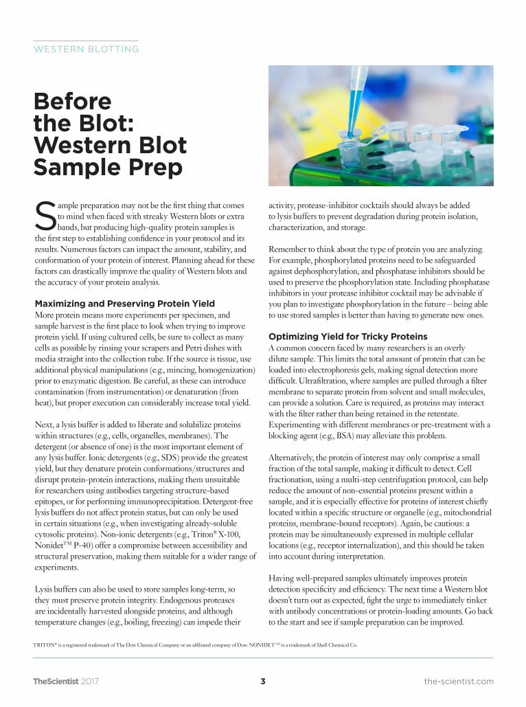
Sample preparation may not be the first thing that comes to mind when faced with streaky Western blots or extra bands, but producing high-quality protein samples is
the first step to establishing confidence in your protocol and its results. Numerous factors can impact the amount, stability, and conformation of your protein of interest. Planning ahead for these factors can drastically improve the quality of Western blots and the accuracy of your protein analysis.
Maximizing and Preserving Protein YieldMore protein means more experiments per specimen, and sample harvest is the first place to look when trying to improve protein yield. If using cultured cells, be sure to collect as many cells as possible by rinsing your scrapers and Petri dishes with media straight into the collection tube. If the source is tissue, use additional physical manipulations (e.g., mincing, homogenization) prior to enzymatic digestion. Be careful, as these can introduce contamination (from instrumentation) or denaturation (from heat), but proper execution can considerably increase total yield.
Next, a lysis buffer is added to liberate and solubilize proteins within structures (e.g., cells, organelles, membranes). The detergent (or absence of one) is the most important element of any lysis buffer. Ionic detergents (e.g., SDS) provide the greatest yield, but they denature protein conformations/structures and disrupt protein-protein interactions, making them unsuitable for researchers using antibodies targeting structure-based epitopes, or for performing immunoprecipitation. Detergent-free lysis buffers do not affect protein status, but can only be used in certain situations (e.g., when investigating already-soluble cytosolic proteins). Non-ionic detergents (e.g., Triton® X-100, NonidetTM P-40) offer a compromise between accessibility and structural preservation, making them suitable for a wider range of experiments.
Lysis buffers can also be used to store samples long-term, so they must preserve protein integrity. Endogenous proteases are incidentally harvested alongside proteins, and although temperature changes (e.g., boiling, freezing) can impede their
activity, protease-inhibitor cocktails should always be added to lysis buffers to prevent degradation during protein isolation, characterization, and storage.
Remember to think about the type of protein you are analyzing. For example, phosphorylated proteins need to be safeguarded against dephosphorylation, and phosphatase inhibitors should be used to preserve the phosphorylation state. Including phosphatase inhibitors in your protease inhibitor cocktail may be advisable if you plan to investigate phosphorylation in the future – being able to use stored samples is better than having to generate new ones.
Optimizing Yield for Tricky ProteinsA common concern faced by many researchers is an overly dilute sample. This limits the total amount of protein that can be loaded into electrophoresis gels, making signal detection more difficult. Ultrafiltration, where samples are pulled through a filter membrane to separate protein from solvent and small molecules, can provide a solution. Care is required, as proteins may interact with the filter rather than being retained in the retentate. Experimenting with different membranes or pre-treatment with a blocking agent (e.g., BSA) may alleviate this problem.
Alternatively, the protein of interest may only comprise a small fraction of the total sample, making it difficult to detect. Cell fractionation, using a multi-step centrifugation protocol, can help reduce the amount of non-essential proteins present within a sample, and it is especially effective for proteins of interest chiefly located within a specific structure or organelle (e.g., mitochondrial proteins, membrane-bound receptors). Again, be cautious: a protein may be simultaneously expressed in multiple cellular locations (e.g., receptor internalization), and this should be taken into account during interpretation.
Having well-prepared samples ultimately improves protein detection specificity and efficiency. The next time a Western blot doesn’t turn out as expected, fight the urge to immediately tinker with antibody concentrations or protein-loading amounts. Go back to the start and see if sample preparation can be improved.
Before the Blot: Western Blot Sample Prep
TRITON® is a registered trademark of The Dow Chemical Company or an affiliated company of Dow. NONIDETTM is a trademark of Shell Chemical Co.
4 the-scientist.comTheScientist 2017
SAMPLE PREPARATION
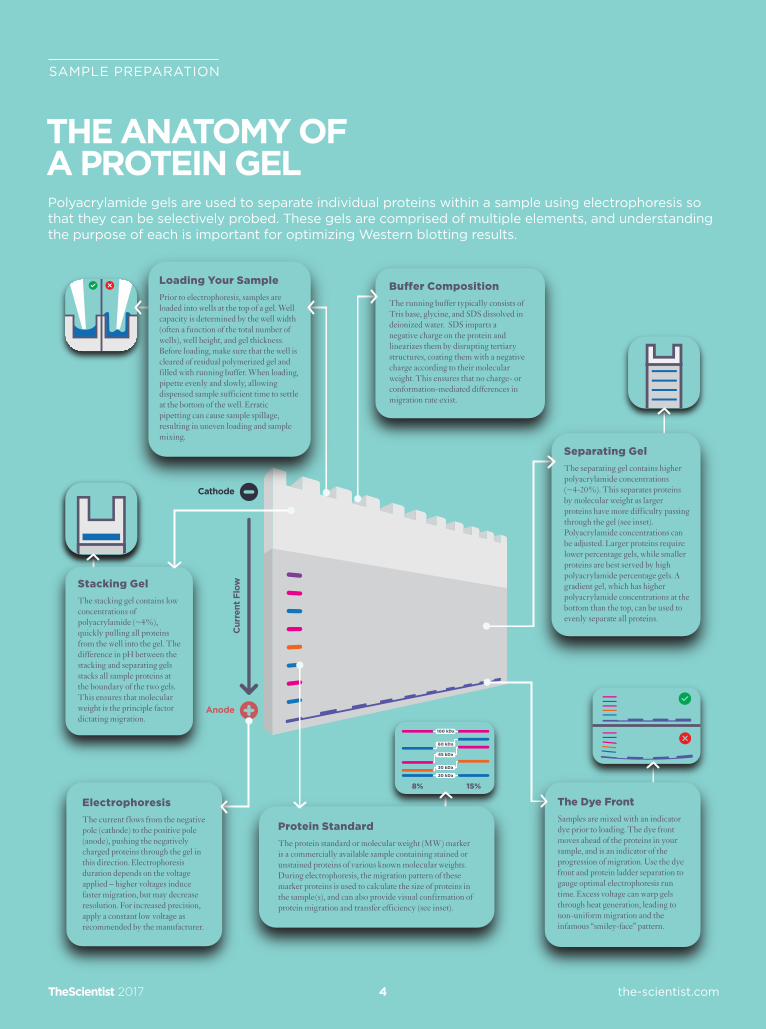
THE ANATOMY OFA PROTEIN GELPolyacrylamide gels are used to separate individual proteins within a sample using electrophoresis so that they can be selectively probed. These gels are comprised of multiple elements, and understanding the purpose of each is important for optimizing Western blotting results.
ElectrophoresisThe current flows from the negative pole (cathode) to the positive pole (anode), pushing the negatively charged proteins through the gel in this direction. Electrophoresis duration depends on the voltage applied – higher voltages induce faster migration, but may decrease resolution. For increased precision, apply a constant low voltage as recommended by the manufacturer.
Loading Your SamplePrior to electrophoresis, samples are loaded into wells at the top of a gel. Well capacity is determined by the well width (often a function of the total number of wells), well height, and gel thickness. Before loading, make sure that the well is cleared of residual polymerized gel and filled with running buffer. When loading, pipette evenly and slowly, allowing dispensed sample sufficient time to settle at the bottom of the well. Erratic pipetting can cause sample spillage, resulting in uneven loading and sample mixing.
Separating GelThe separating gel contains higher polyacrylamide concentrations (~4-20%). This separates proteins by molecular weight as larger proteins have more difficulty passing through the gel (see inset). Polyacrylamide concentrations can be adjusted. Larger proteins require lower percentage gels, while smaller proteins are best served by high polyacrylamide percentage gels. A gradient gel, which has higher polyacrylamide concentrations at the bottom than the top, can be used to evenly separate all proteins.
The Dye FrontSamples are mixed with an indicator dye prior to loading. The dye front moves ahead of the proteins in your sample, and is an indicator of the progression of migration. Use the dye front and protein ladder separation to gauge optimal electrophoresis run time. Excess voltage can warp gels through heat generation, leading to non-uniform migration and the infamous “smiley-face” pattern.
Protein StandardThe protein standard or molecular weight (MW) marker is a commercially available sample containing stained or unstained proteins of various known molecular weights. During electrophoresis, the migration pattern of these marker proteins is used to calculate the size of proteins in the sample(s), and can also provide visual confirmation of protein migration and transfer efficiency (see inset).
Bu�er CompositionThe running buffer typically consists of Tris base, glycine, and SDS dissolved in deionized water. SDS imparts a negative charge on the protein and linearizes them by disrupting tertiary structures, coating them with a negative charge according to their molecular weight. This ensures that no charge- or conformation-mediated differences in migration rate exist.
Stacking GelThe stacking gel contains low concentrations of polyacrylamide (~4%), quickly pulling all proteins from the well into the gel. The difference in pH between the stacking and separating gels stacks all sample proteins at the boundary of the two gels. This ensures that molecular weight is the principle factor dictating migration.
Cathode
Anode
Cur
rent
Flo
w
TheScientist 2017 the-scientist.com5
WESTERN BLOTTING
Gel electrophoresis is used to spatially separate proteins within a sample for selective probing. However, antibodies are unable to bind proteins locked within
polyacrylamide gels. They are therefore transferred to microporous membranes prior to antibody addition. The porosity of Western blot membranes facilitates protein-membrane interactions by increasing the surface-area available for binding, with smaller pores providing more surface area than larger pores.Membranes are typically available in 0.2- and 0.45-µm pore sizes. Whereas the 0.45-µm size is optimal for most purposes, a smaller pore size may increase transfer yield for lower-molecular-weight proteins (<20 kDa).
It can be tempting to think that all membranes are alike, but the materials used to make membranes not only differ regarding physical properties (e.g., durability), they also vary with respect to protein transfer efficacy, detection sensitivity, and background signal. The two most common materials used for Western blotting membranes are nitrocellulose and polyvinylidene fluoride (PVDF).
The Star of the Show: Nitrocellulose versus Polyvinylidene FluorideNitrocellulose was one of the earliest materials used for Western blotting membranes, and it remains popular today. Protein binding to nitrocellulose involves electrostatic and hydrophobic interactions.1 Nitrocellulose membranes have a binding capacity of around 100-200 µg/cm2, and are both physically brittle and susceptible to chemicals. It is therefore not recommended for stripping and reprobing, as the harsh treatments used to remove bound antibody tend to also remove transferred protein. Supported nitrocellulose membranes offer a solution by applying nitrocellulose onto a robust synthetic support structure, thus increasing membrane strength without diminishing protein-binding properties.
Polyvinylidene fluoride (PVDF) is a durable, non-reactive polymer that is resistant to solvents, acids, and bases. It binds proteins through hydrophobic and dipole interactions,2 and has higher protein retention than nitrocellulose, resulting in generally higher signal.3 This combination of physical resilience and exceptional protein affinity makes PVDF membranes ideal for stripping and reprobing. Although the high protein retention of PVDF membranes increases detection sensitivity,
it may also increase background and non-specific signal when immunodetection methods have not been sufficiently optimized.
The Rest of the Cast: Other Factors Impacting Protein TransferBoth nitrocellulose and PVDF membranes are affected by transfer-buffer composition: specifically, the presence/absence of methanol and SDS. Polyacrylamide gels have the ability to absorb water, resulting in physical expansion. Higher concentrations of acrylamide result in more expansion, meaning that a gradient gel could change from a rectangle to a trapezoid. The presence of methanol serves to stabilize gel dimensions.4 In addition, PVDF membranes are highly hydrophobic, and they therefore require alcohol priming (also called prewetting) prior to transfer to facilitate penetration by aqueous transfer buffers.
SDS plays an important role in facilitating effective protein-separation within the gel and enhances protein-elution from the gel, but the high negative charge it imparts upon proteins impedes protein-membrane binding. As such, the addition of methanol to remove SDS post-electrophoresis typically yields the best results, although in some situations, some level of SDS is required to maintain protein solubility (e.g., very hydrophobic or high-MW proteins).4
Protein-transfer efficiency from gel to membrane is integral to successful Western blotting. Even the best antibody will have difficulties compensating for insufficient target-protein transfer. Nitrocellulose and PVDF membranes each have their own unique characteristics which may confer advantages or disadvantages depending on experimental conditions and goals. Each membrane material also has specific protocol tweaks and alterations designed to maximize its capabilities. Picking the right membrane for your particular experiment is essential to getting the clearest and most accurate result possible.
Membrane Memo: A Guide to Selecting the Right Membrane for Your Transfer
References: 1. S.C. Low et al. “Electrophoretic interactions between nitrocellulose membranes and
proteins: Biointerface analysis and protein adhesion properties.” Colloids Surf, B, 110:248-53, 2013.
2. D.W. Speicher. "Microsequencing with PVDF membranes: efficient electroblotting, direct protein adsorption and sequencer program modifications." In: T. Hugli, editor. Techniques in protein chemistry. San Diego, CA: Academic Press; 1989.
3. M.G. Pluskal et al, "ImmobilonTM PVDF Transfer Membrane: A New Membrane Substrate For Western Blotting of Proteins." Biotechniques, 4(3):272-282, 1986
4. "Membrane selection." In: Protein blotting handbook: Tips and tricks (6th ed.). Billerica, MA: MilliporeSigma; 2012.
TheScientist 2017 the-scientist.com6
WESTERN BLOTTING
After all the effort to harvest and isolate your protein of interest, you want to be able to trust the detection result. However, you can’t have confidence in your probing if you
don’t trust the probe. With all of the options out there, picking the right antibody can be daunting, but there are a few key things to note which will set you on the path to success.
1. The Product Sheet is Your Best FriendA good general principle is to consult the product information sheets, which contain details such as antibody source species, antibody specificity and cross-reactivity, whether the antibody has been tested for Western blot and other applications, and expected band size (migration distance). They should be your go-to reference at every step of your Western-blot journey. Consult them before purchase, when planning your experiment, and when interpreting your results. However, be sure to also validate the antibody using your own equipment and protocol. Experimental conditions and lot-to-lot variation can result in differences between product sheet data and your own results.
2. Antibodies 101Antibodies provide a quantifiable signal correlating to the abundance of the protein of interest. Primary antibodies directly bind proteins (and, depending on clonality, are highly specific), while secondary antibodies bind primary antibodies for signal amplification (and are species-specific rather than protein-specific). To limit non-specific binding, the primary antibody should be produced in a different species than the experimental model, and the secondary antibody should target the primary-antibody host species. For example, when targeting mouse GAPDH, you can use a rabbit-anti-mouse primary and a goat-anti-rabbit secondary antibody.
3. Understanding Your ModelThe popularity and flexibility of transgenic models and fusion proteins means that your protein of interest may not be from the same species as your experimental model (e.g., human cells expressing mouse protein). Your epitope – the antibody binding site – may even be from a different species than the rest of the protein (e.g., jellyfish GFP-conjugated to a mouse protein). Antibodies can have cross-species reactivity, which is affected by both epitope location and sequence homology. If you’re trying to identify a transgenic human protein in a mouse model, make sure not to use an antibody that cross-reacts with the mouse analog.
Understanding your model system can explain unexpected probing results.
4. Understanding Your EpitopePolyclonal antibodies, which target multiple epitopes on a single protein, are useful when high antibody affinity is an advantage, such as when the target is sparse or for immunoprecipitation assays. When additional specificity is required, monoclonal antibodies, which bind a single epitope, can delineate between multiple proteins with significant sequence homology (e.g., receptor isoforms). Antibodies selectively targeting post-translational modifications such as phosphorylation sites and epitopes formed by geometry can identify protein state and conformation. In many of these situations, sample preparation can be just as important as your antibody (see Article 1 in this eBook for tips on protecting target conformation and modification during sample prep.)
5. Optimizing Signal DetectionThe most popular methods for primary antibody quantification involve using secondary antibodies conjugated to an enzyme (e.g., horseradish peroxidase) that reacts with a chemical agent to generate chemiluminescence or chromogenic signal. This system is simple and effective, but signal arises from the secondary-enzyme conjugate, so this method cannot distinguish between multiple antibodies probed on the same membrane. Fluorescent tags can be directly applied to primary or secondary antibodies, and multi-wavelength imaging then permits the identification of discrete signals from multiple antibodies on a single membrane. Fluorescence is more stable and consistent than enzymatically-generated chemiluminescence, allowing data to be compared across experimental runs. Shifting to fluorescence requires some specialized equipment and the selection of appropriate reagents such as fluorescence-compatible membranes, but can significantly increase throughput and efficiency.
Knowledge is PowerThe accurate and reproducible identification and quantification of protein levels relies heavily on the right antibody. Understanding the ins-and-outs of your model, your antibody, and your detection methodology is vital for selecting the right antibody for your experimental needs. Keep in mind that antibody performance will vary based on application suitability, experimental conditions, and production standards, so it is important to always validate the effectiveness of an antibody within your own laboratory.
5 Tips for Choosing the Right Antibodies for Your Next Western Blot

The right Western choices can take the “ugly” out of your next blot.
Western blotting tools from MilliporeSigma help tailor your choices to your target:• Ultradurable, tear-resistant TruPAGE™ precast gels • Complete selection of PVDF and nitrocellulose membranes to help you make the right choice
for transferring your protein of interest• 30 minute immunodetection with the SNAP i.d.® 2.0 system for cleaner, more consistent data• Ready-to-use reagents like the ultrasensitive Luminata™ substrate• Application-specific antibody manufacturing expertise, with over 70,000 tested in Western blot
Show us your ugliest blots. We’ll provide tips and tricks for revealing beautiful Western data in our Protein Blotting Handbook, 6th edition. sigma-aldrich.com/westernblot P.S. If your blot has a big fingerprint in the middle… it might actually be you.
it’s not you. it’s your tools.
The life science business of Merck KGaA, Darmstadt, Germany operates as MilliporeSigma in the U.S. and Canada.MilliporeSigma, Luminata, TruPAGE and the vibrant M are trademarks and Millipore and SNAP i.d. are registered trademarks of Merck KGaA, Darmstadt, Germany. All marks are the property of their respective owners. Copyright © 2016 EMD Millipore Corporation. All Rights Reserved. LSo-16-13326 | 10/2016


![Western Blotting BCH 462[practical] Lab#6. Objective: -Western blotting of proteins from SDS-PAGE.](https://static.fdocuments.in/doc/165x107/56649dc85503460f94abe06c/western-blotting-bch-462practical-lab6-objective-western-blotting-of.jpg)