Welcome To Opal
5
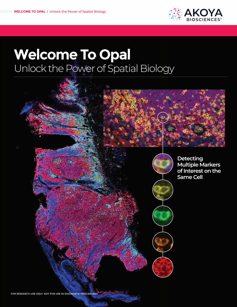
Welcome To Opal Unlock the Power of Spatial Biology FOR RESEARCH USE ONLY. NOT FOR USE IN DIAGNOSTIC PROCEDURES. WELCOME TO OPAL | Unlock the Power of Spatial Biology Detecting Multiple Markers of Interest on the Same Cell
Transcript of Welcome To Opal
FOR RESEARCH USE ONLY. NOT FOR USE IN DIAGNOSTIC PROCEDURES.
Welcome To Opal Unlock the Power of Spatial Biology
FOR RESEARCH USE ONLY. NOT FOR USE IN DIAGNOSTIC PROCEDURES.
WELCOME TO OPAL | Unlock the Power of Spatial Biology
Detecting Multiple Markers of Interest on the Same Cell

FOR RESEARCH USE ONLY. NOT FOR USE IN DIAGNOSTIC PROCEDURES.
Welcome To Opal
Opal Multiplex assays are based on traditional IHC staining workflows.
WELCOME TO OPAL | Unlock the Power of Spatial Biology
AKOYABIO.COM 2
What is Opal?
Opal™ is a method for multiplex fluorescent immunohistochemistry in forma-
lin-fixed, paraffin-embedded (FFPE) tissue that can be performed manually
or via automation. It allows the use of standard unlabeled primary antibodies,
including multiple antibodies raised in the same species. The method involves detection
with Opal reactive fluorophores that covalently label the epitope. After labeling is com-
plete, antibodies are removed in a manner that does not disrupt the Opal fluorescence
signal. This allows the next target to be detected without fear of antibody cross-reactivity.
Opal enables the development of multiplexed assays with balanced, quantitative signal for
rare and abundant targets, imaged in a single final scan with the Akoya portfolio of multi-
spectral imagers, including the Vectra Polaris.
Step 1
Step 3
Step 2
Step 4
Step 5
Step 6
Step 7
Repeat Steps 3-7 for Each Primary
AB
Slide Prep
Epitope Retrieval
Primary AB Incubation
Opal Polymer HRP
Incubation
Signal Amplification
Antibody Stripping
Blocking
Iterative Staining Diagram for Opal Multiplex IHC Assays

FOR RESEARCH USE ONLY. NOT FOR USE IN DIAGNOSTIC PROCEDURES.
Welcome To Opal
WELCOME TO OPAL | Unlock the Power of Spatial Biology
AKOYABIO.COM 3
Opal Enables You To...• Use the best primary antibodies, regardless of species – with no cross-
reactivity
• Identify multiple cell phenotypes while retaining spatial and morphological context that is lost with bulk measurements and flow cytometry
• Get more information from precious and scarce samples
• Image once, without coverslip removal
Standard Detection TSA Detection
Opal Uses Tyramide Signal Amplification (TSA) as Part of the Multiplexing Workflow to:• Improve sensitivity by 10 to 100-fold
• Achieve excellent resolution with low background
• Higher dynamic range (up to 4 logs) compared to chromogenic (up to 1 log)
• Reduce antibody consumption
• Better correlation between protein expression and signal intensity (better quantification, e.g. phenotypic scoring)
• Increase plexing for multiple biomarker detection strategies
• Add signal amplification to almost any immunoassay

FOR RESEARCH USE ONLY. NOT FOR USE IN DIAGNOSTIC PROCEDURES.
WELCOME TO OPAL | Unlock the Power of Spatial Biology
AKOYABIO.COM 4
Contact us today to learn more about
in-person and virtual Akoya
Academy sessions!
OPAL MOTIF KITSOpal™ MOTiF™ Antibody Panel Kits are pre-optimized, ready-to-use primary and secondary antibody panels designed for use with the Leica BOND RX and Vectra Polaris systems.
Opal MOTiF Kits
Name Size Part #
MOTiF™ PD-1/PD-L1 Panel: Pan I/O Kit 50 slides OP-000001
MOTiF™ PD-1/PD-L1 Panel: Auto Melanoma Kit 50 slides OP-000003
LEARN MORE: www.akoyabio.com/contact-a-sales-representative/
OPAL MANUAL MULTIPLEX IHC DETECTION KITSOpal™ kits come complete with all your necessary secondary antibodies and detection reagents for use on Akoya imaging platforms.
Opal Kits & Reagents
Name Size Part #
Opal Polaris 7 Color Manual IHC Detection Kit 50 slides NEL861001KT
Opal 7-Color Manual IHC Kit 50 slides NEL811001KT
Opal 4-Color Manual IHC Kit 50 slides NEL810001KT
Opal 7 Solid Tumor Immunology Kit 50 slides OP7TL4001KT
Opal 7 Tumor Infiltrating Lymphocyte Kit 50 slides OP7TL3001KT
Opal 7 Immunology Discovery Kit 50 slides OP7DS2001KT
Opal 4 Lymphocyte Kit 50 slides OP4LY2001KT
Opal 4-Color Anti-Rabbit Manual IHC Kit 50 slides NEL840001KT
LEARN MORE: www.akoyabio.com/contact-a-sales-representative/
OPAL AUTOMATION MULTIPLEX IHC DETECTION KITSWith our Opal Automation IHC kits you can perform Opal multiplex staining on one of the leading research automated staining platforms – the BOND RX™ by Leica Biosystems. Automation provides you with the flexibility to support the dynamic demands of translational research.
Opal Kits & Reagents
Name Size Part #
Opal Polaris 7 Color Automation IHC Detection Kit 50 slides NEL871001KT
Opal 7-Color Automation IHC Kit 50 slides NEL821001KT
Opal 4-Color Automation IHC Kit 50 slides NEL820001KT
Opal 4-Color Anti-Rabbit Automation IHC Kit 50 slides NEL830001K
LEARN MORE: www.akoyabio.com/contact-a-sales-representative/
WELCOME TO OPAL | Unlock the Power of Spatial Biology
© 2020 Akoya Biosciences, Inc. All rights reserved. Akoya Biosciences and Codex are registered trademarks of Akoya Biosciences, Inc. A Delaware corporation.
To learn more visit A K O Y A B I O . C O Mor email us at I N F O @ A K O Y A B I O . C O M
DN-00062
Additional ResourcesPlease visit www.akoyabio.com/support for additional resources, including FAQs
and publications.
Opal, as part of the Phenoptics solution from Akoya Biosciences, is the most flexible and dynamic workflow to interrogate the spatial relationships between different phenotypes within the context of the tissue microenvironment.
For more information on the Opal methodology and workflow, please visit our website to view support resources, including our new Opal Assay Development Guide and Opal Quick Start Tool, an interactive form with guidance based on published data from top researchers using Opal as their spatial biology platform.
Reach out to us at [email protected] with any questions or to request a quote today.
Contact us today to learn more about
in-person and virtual Akoya
Academy sessions!