Welcome to Olympus Fluoview FV500 Training · •Nikon C1si spectral 13 Snyder Hall. Confocal ... C...
64
Olympus Fluoview FV500 Training Welcome to... Cancer Center Confocal Core
Transcript of Welcome to Olympus Fluoview FV500 Training · •Nikon C1si spectral 13 Snyder Hall. Confocal ... C...

OlympusFluoview FV500
Training
Welcome to...
Cancer Center Confocal Core

1)Tomakestateoftheartimagingtechnologiesavailableto1)Tomakestateoftheartimagingtechnologiesavailabletothelargerscientificcommunityofusersthelargerscientificcommunityofusers
2)Topromotemoreinteractionbetweentechnology2)Topromotemoreinteractionbetweentechnologydevelopersandusersdevelopersandusers
3)Tobringexpertiseinnewimagingtechnologiestostudents3)TobringexpertiseinnewimagingtechnologiestostudentstheUMNtheUMN

Contentofthelecture
Principlesofconfocalimaging.Differentimplementations/modes.
Primeronusage
Imageprocessingandrestoration

LaserScanningConfocalMicroscope

•High quality, multi-wavelengthfluorescence images•3-Dimensional information
•Optical sections through thick sample•Microscope Techniques:
•Colocalization•Fluorescence Recovery afterPhotobleaching (FRAP)•FRET•Time Course
Why use Why use ConfocalConfocal??????
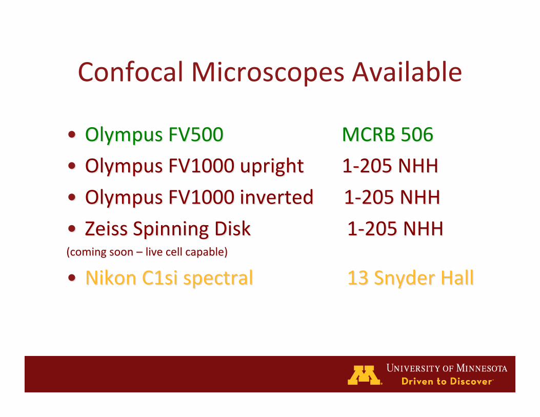
ConfocalMicroscopesAvailable
•• OlympusFV500OlympusFV500 MCRB506MCRB506
•• OlympusFV1000uprightOlympusFV1000upright 1‐205NHH1‐205NHH
•• OlympusFV1000inverted1‐205NHHOlympusFV1000inverted1‐205NHH
•• ZeissSpinningDiskZeissSpinningDisk 1‐205NHH1‐205NHH(comingsoon(comingsoon––livecellcapable)livecellcapable)
•• NikonC1sispectralNikonC1sispectral 13SnyderHall13SnyderHall
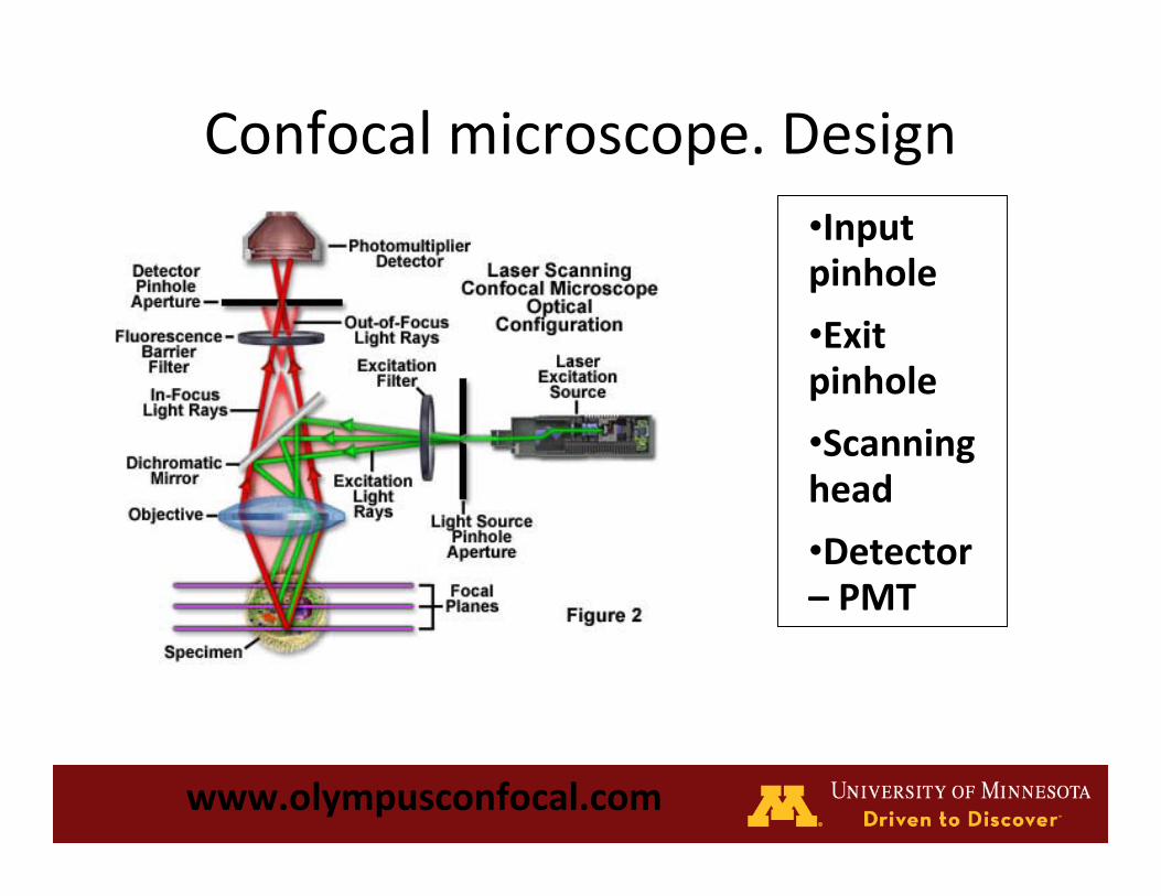
Confocalmicroscope.Design
www.olympusconfocal.com
•Inputpinhole
•Exitpinhole
•Scanninghead
•Detector–PMT

WidefieldvsConfocalImages
http://www.meyerinst.com/confocals/tcs-spe/side-by-side.jpg
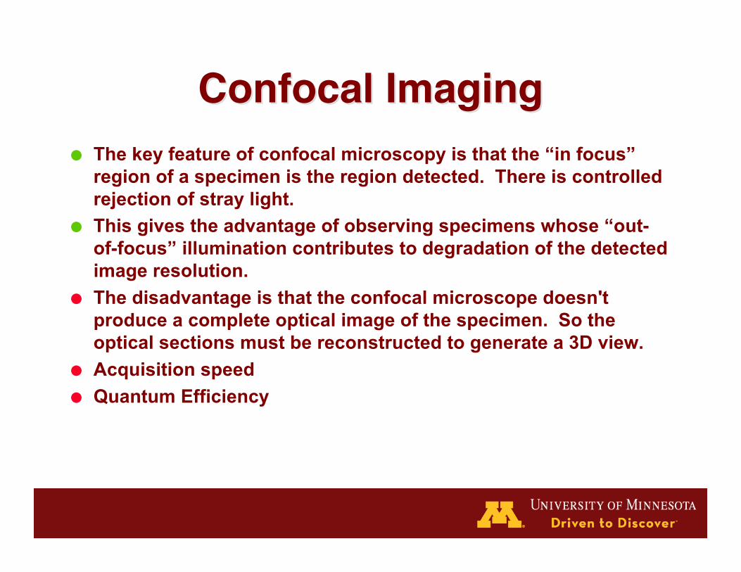
Confocal ImagingConfocal Imaging The key feature of confocal microscopy is that the “in focus”
region of a specimen is the region detected. There is controlledrejection of stray light.
This gives the advantage of observing specimens whose “out-of-focus” illumination contributes to degradation of the detectedimage resolution.
The disadvantage is that the confocal microscope doesn'tproduce a complete optical image of the specimen. So theoptical sections must be reconstructed to generate a 3D view.
Acquisition speed Quantum Efficiency

Confocalmicroscopevs.widefield.
www.olympusconfocal.com
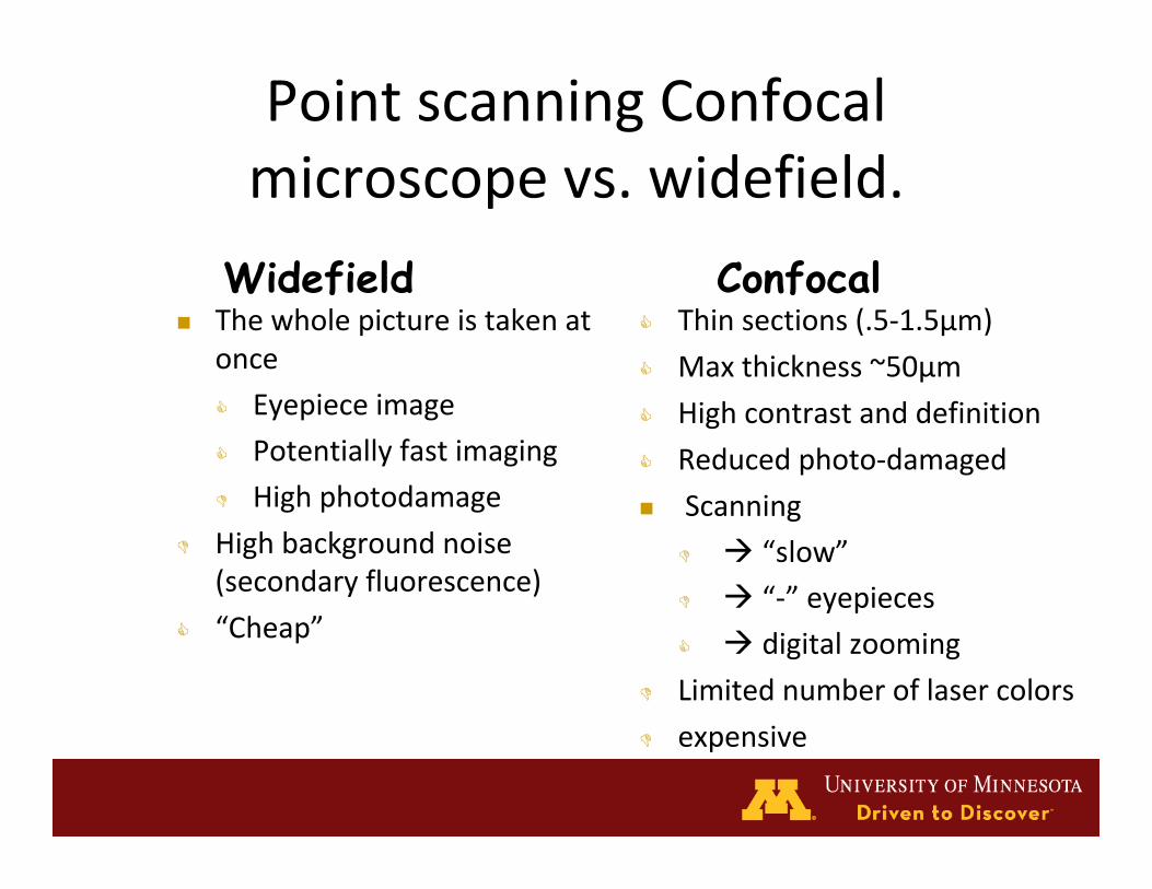
PointscanningConfocalmicroscopevs.widefield.
Thewholepictureistakenatonce
Eyepieceimage
Potentiallyfastimaging
Highphotodamage
Highbackgroundnoise(secondaryfluorescence)
“Cheap”
Thinsections(.5‐1.5μm)
Maxthickness~50μm
Highcontrastanddefinition
Reducedphoto‐damaged
Scanning
“slow” “‐”eyepieces
digitalzooming
Limitednumberoflasercolors
expensive
Widefield Confocal

Depth of FieldDepth of Field
4x 10x
40x20x
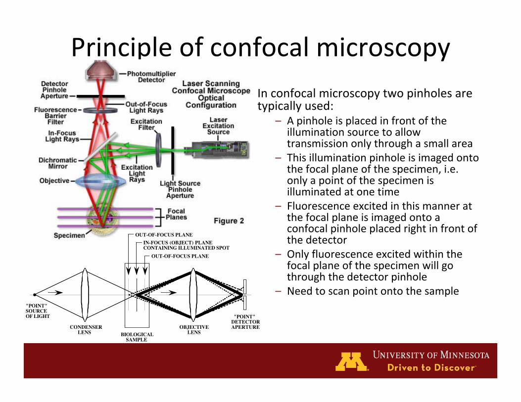
PrincipleofconfocalmicroscopyInconfocalmicroscopytwopinholesaretypicallyused:
– Apinholeisplacedinfrontoftheilluminationsourcetoallowtransmissiononlythroughasmallarea
– Thisilluminationpinholeisimagedontothefocalplaneofthespecimen,i.e.onlyapointofthespecimenisilluminatedatonetime
– Fluorescenceexcitedinthismanneratthefocalplaneisimagedontoaconfocalpinholeplacedrightinfrontofthedetector
– Onlyfluorescenceexcitedwithinthefocalplaneofthespecimenwillgothroughthedetectorpinhole
– Needtoscanpointontothesample
CONDENSER LENS
OBJECTIVE LENSBIOLOGICAL
SAMPLE
OUT-OF-FOCUS PLANE
OUT-OF-FOCUS PLANE
"POINT" SOURCE OF LIGHT "POINT"
DETECTOR APERTURE
IN-FOCUS (OBJECT) PLANE CONTAINING ILLUMINATED SPOT
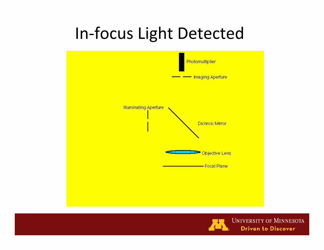
In‐focusLightDetected

Out‐of‐FocusLightBlocked


Before Using the Confocal
• Fill in access form with user information, proof of
HazardousChemicalWastetrainingandIDcardnumber.
• Turnintotheconfocaladministrator(Margaret,470CCRB,
322CRorMMC806).
• Readmanualandpolicydocuments.Handbooksoon.
• CompleteadvancedtrainingcoursegivenbyMarkSanders
or otherwise demonstrate familiarity with the FV500
system.You Are Here

The goals of a shared instrument grant areto provide access to state-of-the-arttechnology that will facilitate interactionand enhance scientific productivity.
A confocal microscope typifies thattechnology, which because of cost is usuallybeyond the scope of a single laboratory toeither purchase or maintain.
NCRRSHAREDINSTRUMENTATIONGRANT(#1NCRRSHAREDINSTRUMENTATIONGRANT(#1S10RR16851)S10RR16851)

UNIVERSITY OF MINNESOTA
BONE TUMOR BIOLOGY LABORATORY
DEPARTMENT OF ORTHOPAEDIC SURGERY
AND NCI DESIGNATED COMPREHENSIVE
CANCER CENTER
Room 506 CCRB
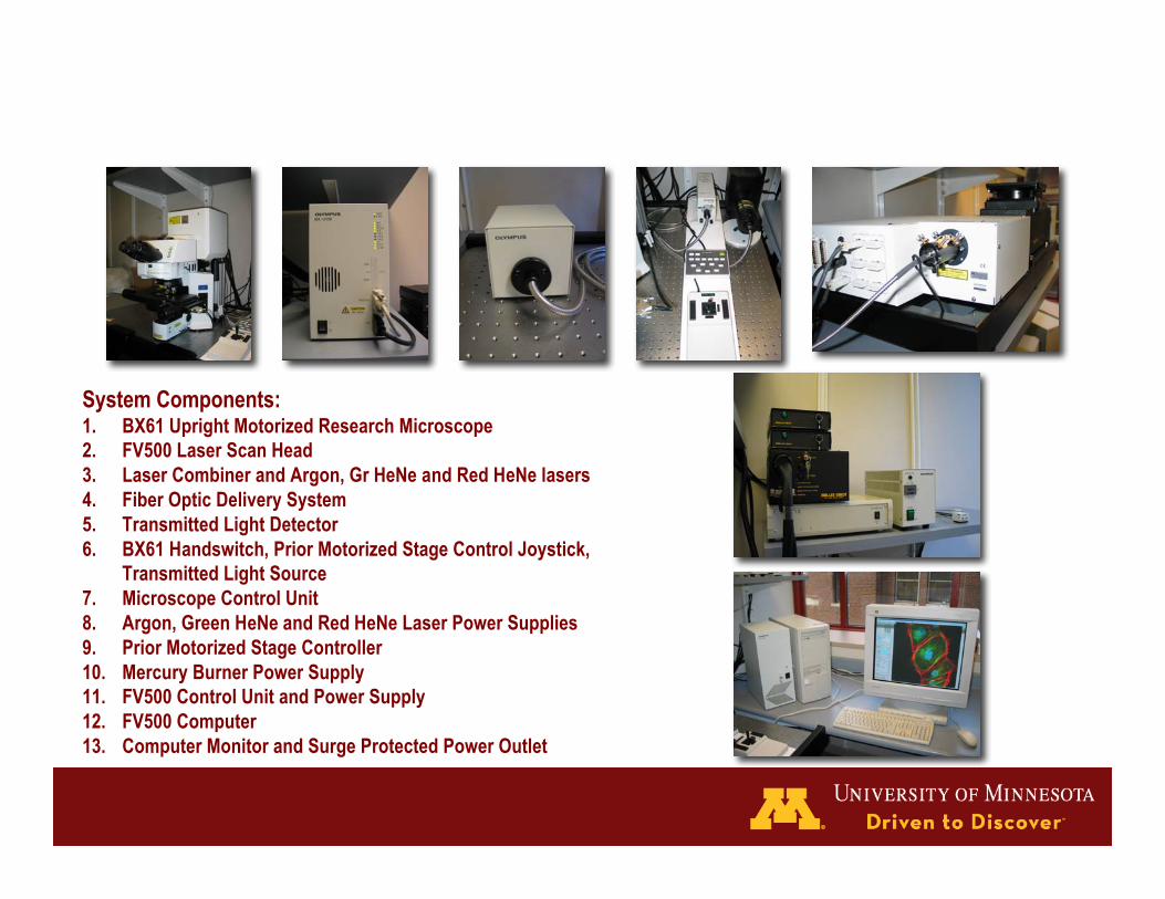
System Components:1. BX61 Upright Motorized Research Microscope2. FV500 Laser Scan Head3. Laser Combiner and Argon, Gr HeNe and Red HeNe lasers4. Fiber Optic Delivery System5. Transmitted Light Detector6. BX61 Handswitch, Prior Motorized Stage Control Joystick,
Transmitted Light Source7. Microscope Control Unit8. Argon, Green HeNe and Red HeNe Laser Power Supplies9. Prior Motorized Stage Controller10. Mercury Burner Power Supply11. FV500 Control Unit and Power Supply12. FV500 Computer13. Computer Monitor and Surge Protected Power Outlet
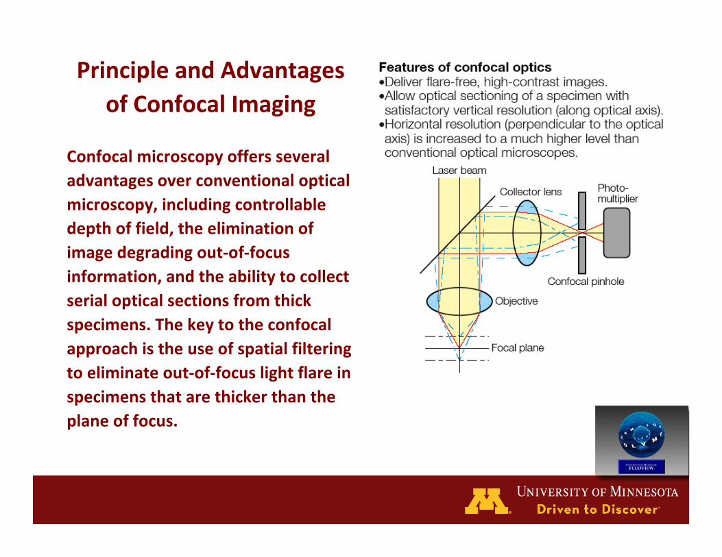
PrincipleandAdvantagesofConfocalImaging
Confocalmicroscopyoffersseveraladvantagesoverconventionalopticalmicroscopy,includingcontrollabledepthoffield,theeliminationofimagedegradingout‐of‐focusinformation,andtheabilitytocollectserialopticalsectionsfromthickspecimens.Thekeytotheconfocalapproachistheuseofspatialfilteringtoeliminateout‐of‐focuslightflareinspecimensthatarethickerthantheplaneoffocus.
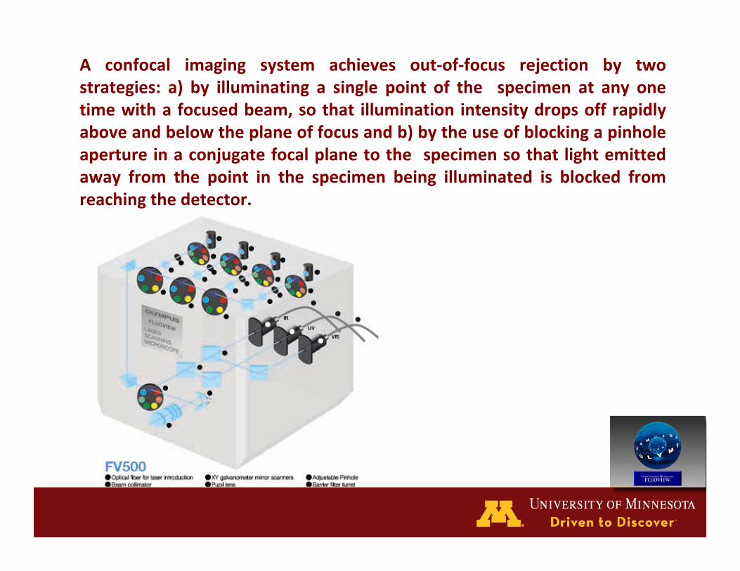
A confocal imaging system achieves out‐of‐focus rejection by twostrategies: a) by illuminating a single point of the specimen at any onetimewithafocusedbeam,sothat illumination intensitydropsoffrapidlyaboveandbelowtheplaneoffocusandb)bytheuseofblockingapinholeapertureinaconjugatefocalplanetothespecimensothatlightemittedaway from the point in the specimen being illuminated is blocked fromreachingthedetector.
FV550VisiblelasersBlueDiode[3mW,405nm]yieldsgoodexcitationforthefollowingfluorochromes:
• DAPI–nuclearstain
Argon[10mW,488nm]yieldsgoodexcitationforthefollowingfluorochromes:
• GFP–greenfluorescentprotein
• FITC–antibodylabeling
• YOYO‐1–DNA
• Calcein–bonegrowth
GreenHeliumNeon (HeNe) [1mW,543nm]yields goodexcitation for the followingfluorochromes:
• PropidiumIodide(PI)–DNA,RNA
• TRITC–antibodylabeling
• Cy3–antibodylabeling
• Rhodamine‐Phalloidin–actinfibers
RedHeNe[10mW,633nm]yieldsgoodexcitationforthefollowingfluorochromes:
• Cy5–antibodylabeling
• AlexaFluor633–antibodylabeling

DYE [excitation/emission] Laser DYE [excitation/emission] Laser
Ca++ Crimson [590/615] Red HeNe eGFP [488/509] ArgonCa++ Green-1 [506/531] Argon FITC [494/518] ArgonCa++ Green-2 [503/536] Argon Fluo-3 [506/526] ArgonCa++ Green-5N [506/532] Gr HeNe Fura Red [472/657] Red HeNeCa++ Orange [549/576] Gr HeNe MitoTracker [490/516] ArgonCa++ Orange-5N [___/580] Gr HeNe PI [536/617] Gr HeNeCy 3 [550/565] Gr HeNe Rhodamine-Phalliodin [542/565] Gr HeNeCy 3.5 [581/596] Gr HeNe SNARF-1 (pH !) [488/530] Red HeNeCy 5 [650/670] Red HeNe Texas Red [595/615] Red HeNeCy 5.5 [675/695] Red HeNe TRITC [496/520] Gr HeNeDil [549/565] Gr HeNe YFP [514/527] Gr HeNeDiO [484/501] ArgonNOT on list:
AlexaFluor-1 [632/647] Red HeNe Calcein [494/517] ArgonAPC [650/660] Red HeNe DS Red [558/583] Red HeNeAPC/Cy7 [650;755/767] Red HeNe YoYo-1 [491/509] Argon
TableListingtheDyesinthe<AvailableDyes>ListandSomePopularDyesNotontheList
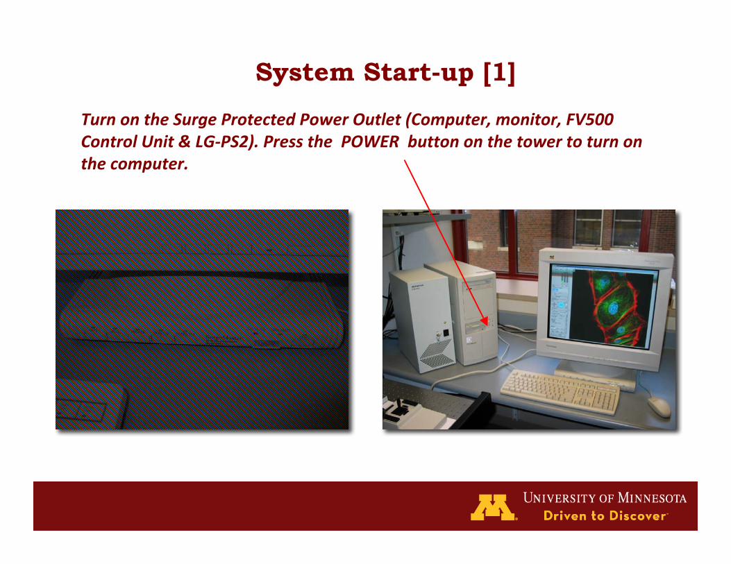
System Start-up [1]
TurnontheSurgeProtectedPowerOutlet(Computer,monitor,FV500ControlUnit&LG‐PS2).PressthePOWERbuttononthetowertoturnonthecomputer.

System Start-up [2]
TurnonLaserPowerSuppliesONLYasNEEDED(Argon,GreenHeNe,RedHeNe)and“ON”fortheArgonlaser.Therecommendedwarm‐upis10minfortheArgonand30minfortheGrHeNe.
RedHeNe
GreenHeNe
Argon
TurnontheMercuryBurner
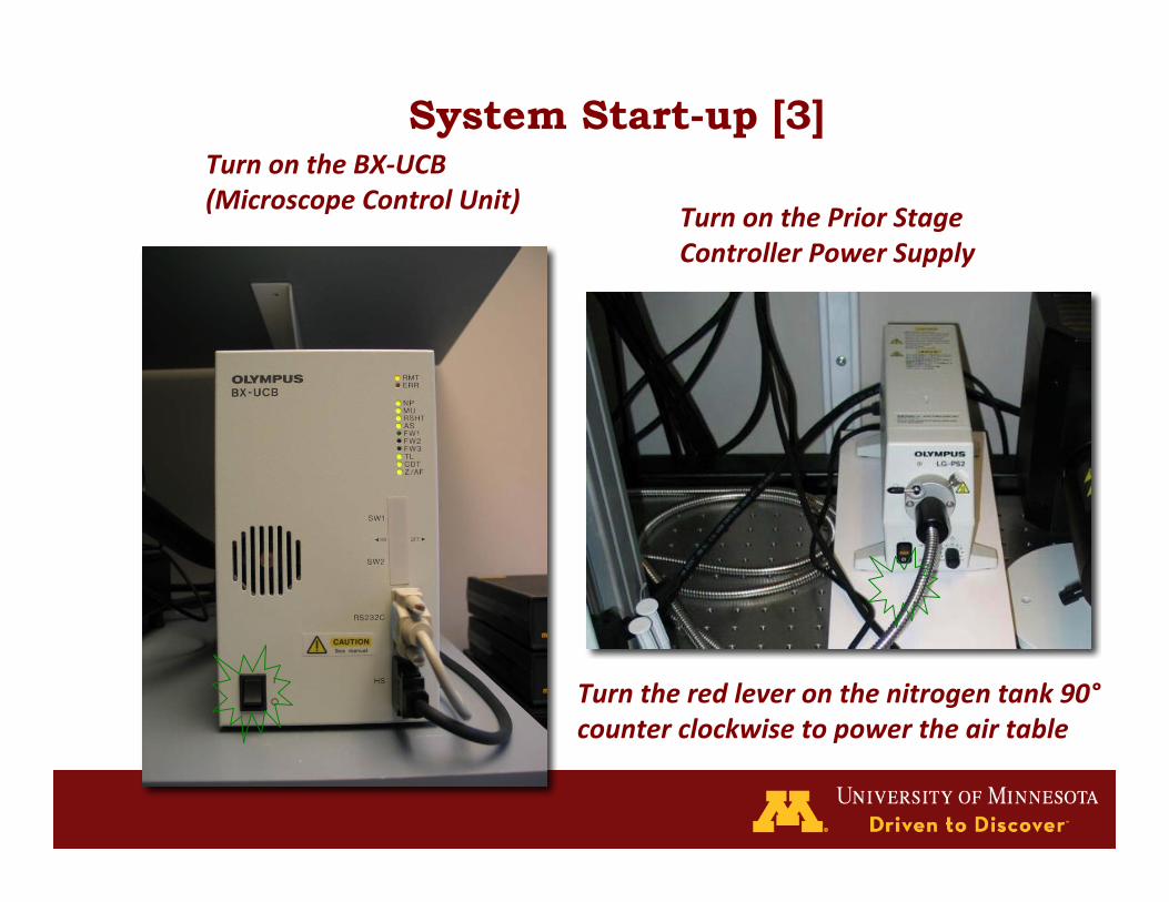
System Start-up [3]TurnontheBX‐UCB(MicroscopeControlUnit)
TurnonthePriorStageControllerPowerSupply
Turntheredleveronthenitrogentank90°counterclockwisetopowertheairtable

Waitat~2minforthemicroscopesystemstoinitializethendouble‐clicktheFluoviewIcontolaunchtheconfocalprogram.Note:ittakesabout2minutestolaunchafterclickingontheFluoviewIcon
LogontothecomputerusingUSERNAMEandPASSWORD
You will see the Main Fluoview Software Window:
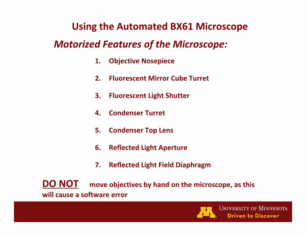
DONOT moveobjectivesbyhandonthemicroscope,asthiswillcauseasoftwareerror
UsingtheAutomatedBX61Microscope
MotorizedFeaturesoftheMicroscope:
1. ObjectiveNosepiece
2. FluorescentMirrorCubeTurret
3. FluorescentLightShutter
4. CondenserTurret
5. CondenserTopLens
6. ReflectedLightAperture
7. ReflectedLightFieldDiaphragm
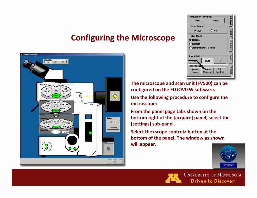
ConfiguringtheMicroscope
Themicroscopeandscanunit(FV500)canbeconfiguredontheFLUOVIEWsoftware.
Usethefollowingproceduretoconfigurethemicroscope:
Fromthepanelpagetabsshownonthebottomrightofthe[acquire]panel,selectthe[settings]sub‐panel.
Selectthe<scopecontrol>buttonatthebottomofthepanel.Thewindowasshownwillappear.

GraphicUserInterface(GUI)[Light Path] box
selects the light path <BI> is for directobservation, <LSM> is for scanning,<TV> not used.
[Mirror Unit] box
clicking the cube automaticallyswitches the turret.
[Shutter] box
click to open (9:00) / close (12:00) theEPI shutter.
[Nosepiece] box
click to choose the objective.
[Top Lens] box
clicking the engages (centered icon) ordisengages (to side) the lens into thelight path.
[Condenser] box
selects the condenser used.
[Aperture Iris] box
changes the AS value.

Whythe20xobjectivewasnotherefor9months
EscapethestageBEFOREBEFOREchangingobjectives

Thissoftwareusespanel‐typewindows.Functionsareexecutedbyselectingthepanelpagetab(attheedgeofthepanel)ofthefunctiontobeexecutedandthepanelwillmovetothefront.
TheFLUOVIEWsoftwareisorganizedbytwokindsofpanels,thefunctionpanels(left,downside)anddisplaypanel(bigoneonright)
UsingtheFLUOVIEWSoftware

Thepanelstructureofthesoftwareisset‐upaccordingtothetreebelow
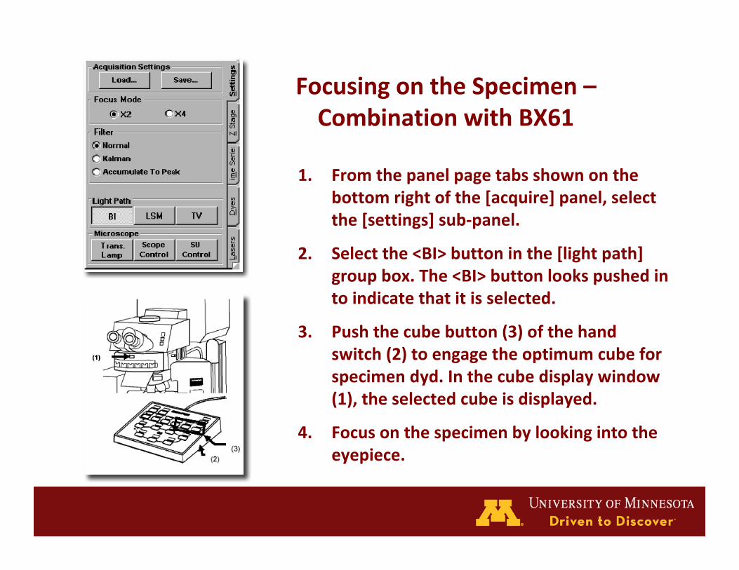
FocusingontheSpecimen–CombinationwithBX61
1. Fromthepanelpagetabsshownonthebottomrightofthe[acquire]panel,selectthe[settings]sub‐panel.
2. Selectthe<BI>buttoninthe[lightpath]groupbox.The<BI>buttonlookspushedintoindicatethatitisselected.
3. Pushthecubebutton(3)ofthehandswitch(2)toengagetheoptimumcubeforspecimendyd.Inthecubedisplaywindow(1),theselectedcubeisdisplayed.
4. Focusonthespecimenbylookingintotheeyepiece.
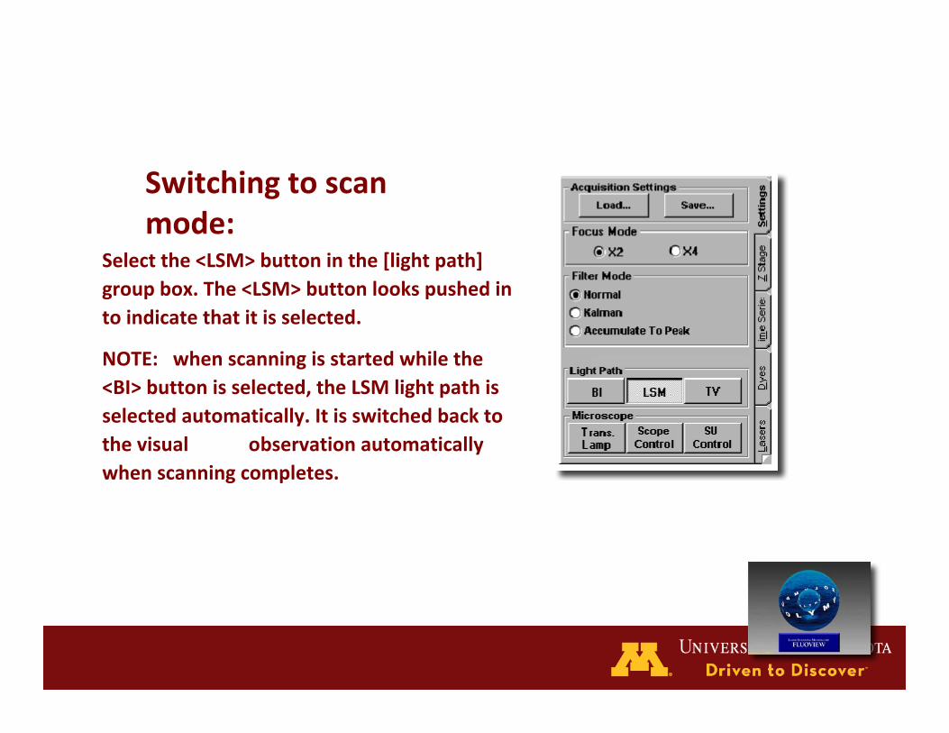
Switchingtoscanmode:
Selectthe<LSM>buttoninthe[lightpath]groupbox.The<LSM>buttonlookspushedintoindicatethatitisselected.
NOTE: whenscanningisstartedwhilethe<BI>buttonisselected,theLSMlightpathisselectedautomatically.Itisswitchedbacktothevisual observationautomaticallywhenscanningcompletes.

SelectingtheDyingMethod

SettingtheChannels

SettingtheScanSpeed
SettingtheXYObservationMode

SettingtheCross‐sectiontobeObserved

StoppingRepeatedScanning
AcquiringImage

SavingImage[1]

SavingImage[2]


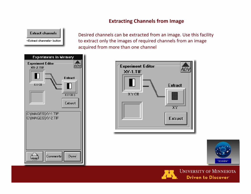
Saving, Opening and Shredding Images Saving,OpeningandShreddingImages
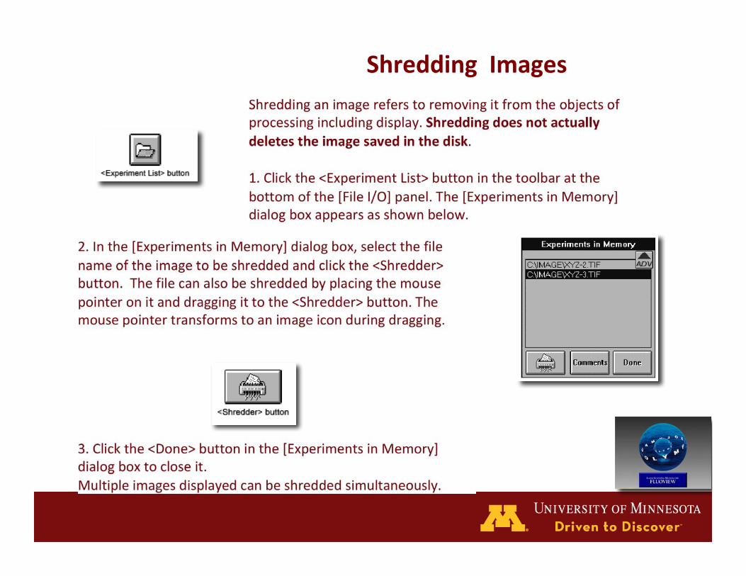
ShreddingImages
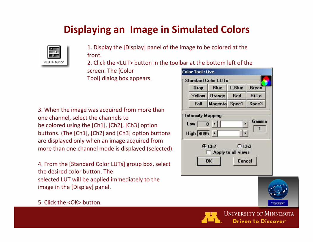
DisplayinganImageinSimulatedColors


ImageProcessing
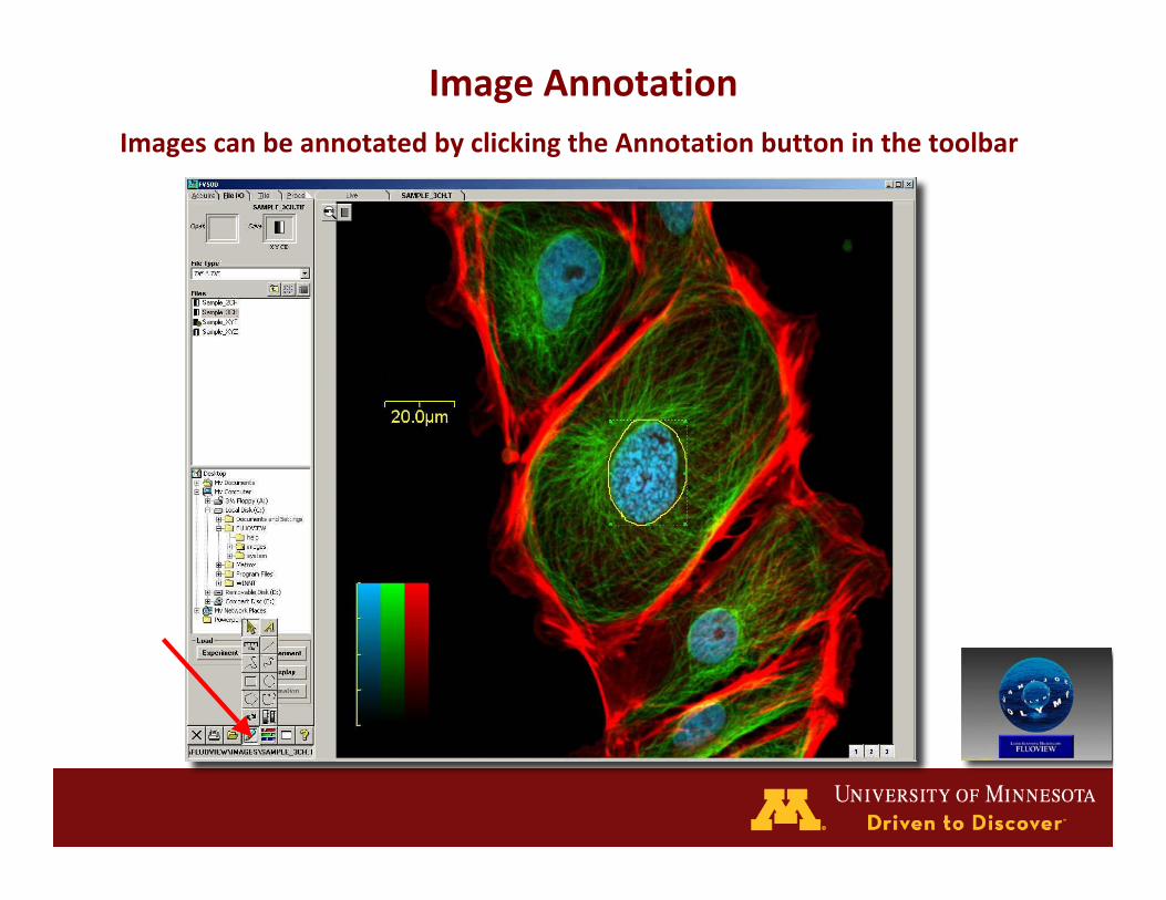
ImageAnnotationImagescanbeannotatedbyclickingtheAnnotationbuttoninthetoolbar
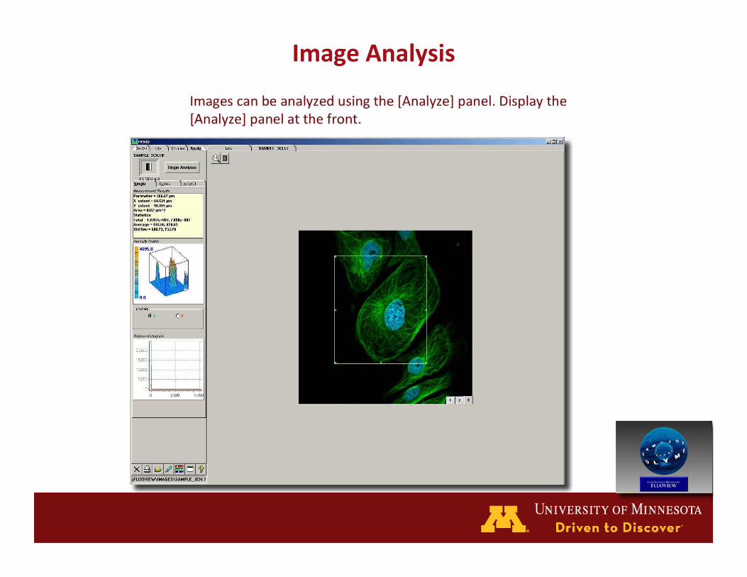
ImageAnalysis
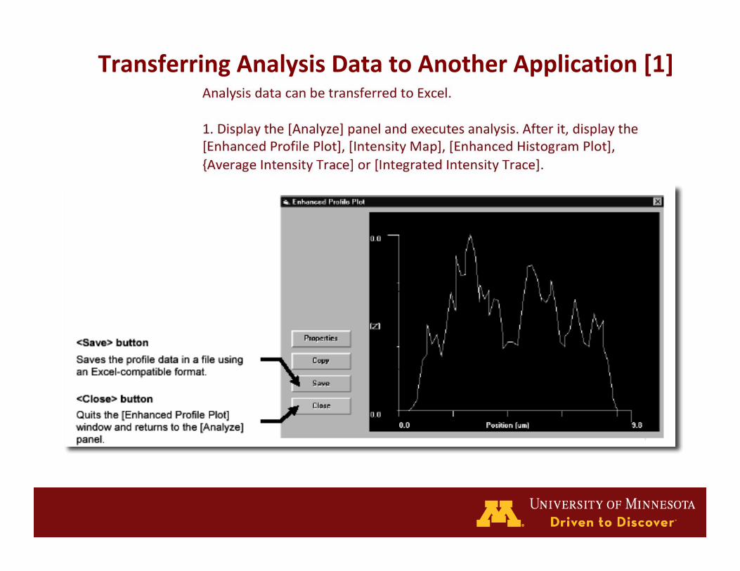
TransferringAnalysisDatatoAnotherApplication[1]

TransferringAnalysisDatatoAnotherApplication[2]

Viewing3DImage
SystemShut‐down[1]
• Cleanobjectivesofanyresidualoilusinglenspaper
• Returnmicroscopestagetonon‐escapedposition(UP)
• Returnobjectivesto4x• ExitfromtheFVprogram.Thismaytakeawhile,howeverifyouturnoffthemicroscopebeforeexiting,asystemerrorwilloccur
• Transferyourfilestoremovablemedia.Only1.5GBofstorageisallowed
• TurnoffthePriorStageControllerPowerSupply• TurnofftheBX‐UCB(MicroscopeControlUnit)
• TurnofftheMercuryBurner.
• TurnoffLaserPowerSupplies
• Select<shutdown>onthecomputerandselectLOGOFFfromthedrop‐downmenu.Thecomputerwilllogyournameoutandreturntotheloginmenubox.
• Selecttheshutdownbuttontoshutdownthecomputer.Note:powerwillgooff.
• TurnofftheSurgeProtectedPowerOutlet.Donotturnthemonitor,FV500ControlUnitorLG‐PS2off.
• Turnredleverontank90°clockwisetostopflowofgastotable.
• Signoutonthelogsheet.
• Reportanyproblems.
SystemShut‐down[2]

On-line Reservations for Confocal Microscope [1]
Signupusingtheon‐linereservationscheduleat:
<http://www.umncc.calendarhost.com/confocal/cgi-bin/calweb/calweb.cgi>
Reservationsareonafirst‐come,first‐servebasis.
Ifyoumustcancel,dosoinadvance.
LoginwithIDandpassword,bothcasesensitive.Thesameonesareusedforlog‐intotheFV500.

On-line Reservations for Confocal Microscope [2]Selectdateandapproximatetimebyclickingneartime

On-line Reservations for Confocal Microscope [3]Fill‐inuserinformation.NOTE:theUSERIDwillfillinthePIfieldandappearontheweeklycalendarintheappropriatetimeslot

Huh?! What?!
Hands-on session to follow...
• Youmaywanttouseourlaserscanningconfocalmicroscopes
• Toreservetime:– ContactMark([email protected])toscheduleatrainingsession.
– 612‐624‐3454
• ImagingCenterhours9am‐5pmM‐F
UseoftheUniversity‐wideImagingUseoftheUniversity‐wideImagingCentersCenters
• Youarewelcometoconsultwithourstaffanduseouropticalimagingcapabilitiesandexpertise.– www.cbs.umn.edu/ic– www.bipl.umn.edu
– www.uic.umn.edu
• Togetstarted:– ContactMarkSanders([email protected])
– 612‐624‐3454
• Imagingfacilitiesarestaffed9am‐5pmM‐F


![User’s Manual · User’s Manual [Safety Guide] FLUOVIEW FV1000 CONFOCAL LASER SCANNING BIOLOGICAL MICROSCOPE ... While the LED lamp on the FV10-SU is illuminated, the laser beam](https://static.fdocuments.in/doc/165x107/5f0598127e708231d413bc0b/useras-useras-manual-safety-guide-fluoview-fv1000-confocal-laser-scanning.jpg)