Distributed by Conrad Electronic SE • Klaus-Conrad-Str. 1 ...

JPET # 262790
1
Distinct regulation of sigma-1 receptor multimerization by its agonists and antagonists in
transfected cells and rat liver membranes
Weimin Conrad Hong
Department of Pharmaceutical Sciences, Butler University, Indianapolis, IN 46208
This article has not been copyedited and formatted. The final version may differ from this version.JPET Fast Forward. Published on February 14, 2020 as DOI: 10.1124/jpet.119.262790
at ASPE
T Journals on January 19, 2022
jpet.aspetjournals.orgD
ownloaded from
This article has not been copyedited and formatted. The final version may differ from this version.
JPET Fast Forward. Published on February 14, 2020 as DOI: 10.1124/jpet.119.262790 at A
SPET
Journals on January 19, 2022jpet.aspetjournals.org
Dow
nloaded from
This article has not been copyedited and formatted. The final version may differ from this version.JPET Fast Forward. Published on February 14, 2020 as DOI: 10.1124/jpet.119.262790
at ASPE
T Journals on January 19, 2022
jpet.aspetjournals.orgD
ownloaded from

JPET # 262790
2
a) Running title: Regulation of σ1R multimerization by ligands
b) Corresponding author:
Weimin Conrad Hong Ph.D.,
Department of Pharmaceutical Sciences, Butler University, PB351,
4600 Sunset Avenue, Indianapolis, IN 46208
Email: [email protected]
Phone: 317-940-9580
Fax: 317-940-6172
c) Text pages:
Figures: 8
Tables: 1
References: 62
Words in Abstract: 249 (250 max)
Words in Introduction: 750 (750 max)
Words in Discussion: 1479 (1500 max)
d) Nonstandard abbreviations:
ANOVA: analysis of variance
BD1008: N-[2-(3,4-dichlorophenyl)ethyl]-N-methyl-2-(1-pyrrolidinyl)ethylamine
dihydrobromide
This article has not been copyedited and formatted. The final version may differ from this version.JPET Fast Forward. Published on February 14, 2020 as DOI: 10.1124/jpet.119.262790
at ASPE
T Journals on January 19, 2022
jpet.aspetjournals.orgD
ownloaded from

JPET # 262790
3
BD1047: N-[2-(3,4-dichlorophenyl)ethyl]-N-methyl-2-(dimethylamino)ethylamine
dihydrobromide
BD1063: 1-[2-(3,4-dichlorophenyl)ethyl]-4-methylpiperazine dihydrochloride
BRET: Bioluminescence resonance energy transfer
CM304: 3-(2-(azepan-1-yl)ethyl)-6-(3-fluoropropyl)-1,3-benzothiazol-2-one hydrochloride
DSP: dithiobis(succinimidyl propionate)
DTG: 1,3-di-o-tolylguanidine
FRET: Förster resonance energy transfer
GDN: glyco-diosgenin
HEK293: human embryonic kidney 293
kD: kilodalton
MW: molecular weight
NE-100: 4-methoxy-3-(2-phenylethoxy)-N,N-dipropylbenzeneethanamine monohydrochloride
PAGE: polyacrylamide gel electrophoresis
PBS: phosphate-buffered saline
PBSCM: PBS with CaCl2 and MgCl2
(+)Pent: (+)-pentazocine
PFO: perfluorooctanoic acid
PRE-084: 2-(4-morpholinethyl) 1-phenylcyclohexanecarboxylate hydrochloride
SEM: standard error of mean
SLS: sodium lauroyl sarcosinate
(+)SKF 10047: (+) N-allylnormetazocine (NANM) hydrochloride
TCEP: tris(2-carboxyethyl)phosphine
This article has not been copyedited and formatted. The final version may differ from this version.JPET Fast Forward. Published on February 14, 2020 as DOI: 10.1124/jpet.119.262790
at ASPE
T Journals on January 19, 2022
jpet.aspetjournals.orgD
ownloaded from

JPET # 262790
4
TM: transmembrane domain
σ1R: sigma1 receptor
FH-σ1R: sigma1 receptor with N-terminal Flag and 2xHis8 tags
e) Recommended section assignment: Cellular and Molecular
This article has not been copyedited and formatted. The final version may differ from this version.JPET Fast Forward. Published on February 14, 2020 as DOI: 10.1124/jpet.119.262790
at ASPE
T Journals on January 19, 2022
jpet.aspetjournals.orgD
ownloaded from

JPET # 262790
5
Abstract
Extensive studies have shown that the sigma-1 receptor (σ1R) interacts with and
modulates the activity of multiple proteins with important biological functions. Recent crystal
structures of σ1R as a homo-trimer differ from a dimer-tetramer model postulated earlier. It
remains inconclusive whether ligand binding regulates σ1R oligomerization. Here novel non-
denaturing gel methods and mutational analysis were used to examine σ1R oligomerization. In
transfected cells σ1R exhibited as multimers, dimers and monomers. Overall σ1R agonists
decreased, whereas σ1R antagonists increased σ1R multimers, suggesting that agonists and
antagonists differentially affect the stability of σ1R multimers. Endogenous σ1R in rat liver
membranes also showed similar regulation of oligomerization as in cells. Mutations at key
residues lining the trimerization interface (Arg119, Asp195, Phe191, Trp136, and Gly91)
abolished multimerization without disrupting dimerization. Intriguingly, truncation of the N-
terminus reduced σ1R to apparent monomer. These results demonstrate that multiple domains
play crucial roles in coordinating high-order quaternary organization of σ1R. The E102Q σ1R
mutant implicated in juvenile amyotrophic lateral sclerosis formed dimers only, suggesting that
dysregulation of σ1R multimeric assembly may impair its function. Interestingly, oligomerization
of σ1R was pH dependent and correlated with changes in [3H](+)-pentazocine binding affinity
and Bmax. Combined with mutational analysis, it is reasoned that σ1R multimers possess high-
affinity and high-capacity [3H](+)-pentazocine binding, whereas monomers likely lack binding.
These results suggest that σ1R may exist in interconvertible oligomeric states in a dynamic
equilibrium. Further exploration of ligand-regulated σ1R multimerization may provide novel
approaches to modulate the function of σ1R and its interacting proteins.
This article has not been copyedited and formatted. The final version may differ from this version.JPET Fast Forward. Published on February 14, 2020 as DOI: 10.1124/jpet.119.262790
at ASPE
T Journals on January 19, 2022
jpet.aspetjournals.orgD
ownloaded from

JPET # 262790
6
Significance statement
The σ1R modulates the activities of various partner proteins. Recently crystal structures
of σ1R were elucidated as homotrimers. This study used novel non-denaturing gel methods to
examine σ1R oligomerization in transfected cells and rat liver membranes. Overall agonist
binding decreased whereas antagonist binding increased σ1R multimers, which comprised
trimers and larger units. σ1R multimers were shown to bind [3H](+)-pentazocine with high-
affinity and high-capacity. Further, mutational analysis revealed a crucial role of its N-terminal
domain in σ1R multimerization.
This article has not been copyedited and formatted. The final version may differ from this version.JPET Fast Forward. Published on February 14, 2020 as DOI: 10.1124/jpet.119.262790
at ASPE
T Journals on January 19, 2022
jpet.aspetjournals.orgD
ownloaded from

JPET # 262790
7
Introduction
The sigma receptor (σR) was named after the distinct behavioral signs induced by
SKF10047 (N-allylnormetazocine) in a chronic spinal dog preparation (Martin et al., 1976).
However, molecular cloning identified a 25-kD membrane protein as the sigma-1 receptor (σ1R)
(Hanner et al., 1996; Jbilo et al., 1997). Its sequence is highly conserved in evolution, but distinct
from opioid receptors as originally proposed. Multiple alternative splice variants of σ1R have
been characterized (Pan et al., 2017), including an isoform lacking exon 3 (ΔE3) which encodes
amino acids (aa) 119–149 (Ganapathy et al., 1999).
Extensive studies have shown that σ1R can interact with and modulate the activity of a
plethora of partner proteins, including channels, receptors, and transporters (Hayashi and Su,
2001; Aydar et al., 2002; Hayashi and Su, 2007; Wu and Bowen, 2008; Carnally et al., 2010;
Kim et al., 2010; Navarro et al., 2010; Balasuriya et al., 2012; Kourrich et al., 2013; Srivats et al.,
2016; Hong et al., 2017; Sambo et al., 2017; Thomas et al., 2017; Schmidt and Kruse, 2019) that
play important roles in cellular homeostasis and neuronal signaling. The majority of σ1R protein
is located in endoplasmic reticulum, particularly mitochondria-associated ER membranes
(Hayashi and Su, 2007), which are critical sites to modulate energy balance, calcium regulation,
and stress response. Several σ1R mutations have recently been implicated in juvenile
amyotrophic lateral sclerosis (ALS) and distal hereditary motor neuropathies. Molecular
mechanisms underlying such motor neuron deficits have been studied intensively, and aberrant
σ1R expression and function appear to be crucial in these conditions (Al-Saif et al., 2011;
Bernard-Marissal et al., 2015; Li et al., 2015; Gregianin et al., 2016; Watanabe et al., 2016;
Dreser et al., 2017).
This article has not been copyedited and formatted. The final version may differ from this version.JPET Fast Forward. Published on February 14, 2020 as DOI: 10.1124/jpet.119.262790
at ASPE
T Journals on January 19, 2022
jpet.aspetjournals.orgD
ownloaded from

JPET # 262790
8
Many clinical drugs and synthetic compounds with diverse structures exhibit varying
affinities for σ1R (Matsumoto, 2007; Cobos et al., 2008; Maurice and Su, 2009; Chu and Ruoho,
2016), and appear to share a limited pharmacophore consensus (Walker et al., 1990; Ablordeppey
and Glennon, 2007; Newman and Coop, 2007; Weber and Wunsch, 2017). Several candidate
endogenous ligands have been proposed over the years, including neurosteroids (Su et al., 1988;
Bergeron et al., 1996), sphingosine (Ramachandran et al., 2009) and N,N-dimethyltryptamine
(Fontanilla et al., 2009), but these hypotheses have not been conclusively confirmed.
Traditionally σ1R ligands have been classified as agonists or antagonists, depending upon
whether they produce or block certain cellular, physiologic or behavioral responses. Although
affinities of these ligands for σ1R have been extensively studied using traditional binding
techniques, molecular mechanisms for agonists or antagonists to induce distinct changes of σ1R
remain largely unknown.
The ability to modulate σ1R function with different ligands has made it an attractive
target for developing novel therapeutic strategies. It has been shown that σ1R agonists have
ameliorative effects in several animal models of neurodegenerative disorders, such as
Alzheimer’s disease (Lahmy et al., 2013; Maurice and Goguadze, 2017; Ryskamp et al., 2019),
Parkinson’s disease (Francardo et al., 2014), Huntington’s disease (Ryskamp et al., 2017), and
retinal degeneration (Wang et al., 2016), whereas σ1R antagonists have pain-relief effects
(Merlos et al., 2017). Accumulating evidence also suggest that σRs are critically involved in
cellular adaptive mechanisms elicited by psychostimulants (Cai et al., 2017; Katz et al., 2017)
and alcohol (Sabino and Cottone, 2017). Therapeutic potentials of σ1R antagonists have been
explored in rodent models of cocaine or methamphetamine addiction (Hiranita et al., 2011;
Robson et al., 2014; Sambo et al., 2017).
This article has not been copyedited and formatted. The final version may differ from this version.JPET Fast Forward. Published on February 14, 2020 as DOI: 10.1124/jpet.119.262790
at ASPE
T Journals on January 19, 2022
jpet.aspetjournals.orgD
ownloaded from

JPET # 262790
9
Whether σ1R possesses one or two transmembrane domain (TM) has been controversial
(Hanner et al., 1996; Aydar et al., 2002; Hayashi and Su, 2007). In recent years atomic force
microscopy and solution nuclear magnetic resonance methods were employed to explore σ1R
structures (Carnally et al., 2010; Balasuriya et al., 2012; Ortega-Roldan et al., 2013).
Breakthrough on the crystal structures of σ1R has elucidated its homo-trimer organization, with
each protomer containing a single TM and a cytoplasmic ligand-binding pocket (Schmidt et al.,
2016). Such structural architecture differs from a dimer-tetramer model postulated by early work
(Chu and Ruoho, 2016). Further, crystal structures of σ1R bound with agonist (+)-pentazocine or
antagonist haloperidol showed similar homo-trimer organization, with limited conformational
rearrangement (Schmidt et al., 2018), suggesting that trimers may be the lowest free energy state
of σ1R during crystallization. It remains unclear whether ligand binding affects the native high-
order organization of σ1R. This study examined σ1R oligomerization using molecular,
biochemical and pharmacological techniques. The results show that multiple domains on σ1R
coordinate its multimerization. Further, agonists and antagonists dynamically regulate σ1R
oligomerization in distinct manners, and quaternary structures of σ1R significantly impact ligand
binding.
This article has not been copyedited and formatted. The final version may differ from this version.JPET Fast Forward. Published on February 14, 2020 as DOI: 10.1124/jpet.119.262790
at ASPE
T Journals on January 19, 2022
jpet.aspetjournals.orgD
ownloaded from

JPET # 262790
10
Materials and Methods
Chemicals, radioligands and antibodies. Sources of reagents are as follows: (-)-cocaine
HCl, (+)-pentazocine succinate, (+)-SKF 10,047, National Institute on Drug Abuse (NIDA) Drug
Supply Program; PRE-084, Tocris (Minneapolis, MN); DTG, BD1008, BD1047, BD1063, NE-
100, gifts from Dr. Jonathan L. Katz, CM304, gift from Dr. Christopher R. McCurdy; d-erythro-
sphingosine, dehydroepiandrosterone, haloperidol, Cayman chemicals (Ann Arbor, MI); BCA
protein kit, Pierce (Rockford, IL); PFO, SLS, TCI America (Portland, OR); GDN, Anatrace
(Maumee, OH); all other chemicals, Sigma-Aldrich (St. Louis, MO) or Fisher Scientific
(Pittsburgh, PA); glutathione-conjugated Sepharose beads, GE Healthcare (Pittsburgh, PA);
[3H](+)-Pentazocine (NET-1056, 26.9 Ci/mmol), Perkin Elmer (Boston, MA); anti-Flag rat mAb
L5, anti-HA, mAb HA11, anti-Myc mAb 9E10, HRP-conjugated secondary antibodies,
Biolegend (San Diego, CA); anti-σ1R mAb clone B5, Santa Cruz Biotechnology (Santa Cruz,
CA).
DNA subcloning and stable expression cell lines. The coding sequences of human σ1R
cDNA were subcloned into CMV promoter-based mammalian expression plasmids expressing
N-terminal fusion of HA, Myc or FLAG-2xHis8 tags (Hong et al., 2017). σ1R mutants were
generated using the QuikChange method, verified by standard DNA sequencing procedures.
Plasmids DNA were linearized, transfected into cells using TransIT LT1 reagent (Mirus Bio,
Madison, WI) to isolate G418-resistant clones. Alternatively, cells were transiently transfected
using PolyJet (SignaGen, Rockville, MD). Cells were then cultured in DMEM with 10% FBS
(Sigma), penicillin-streptomycin in humidified incubators with 5% CO2 at 37°C. G418 (0.5
mg/ml) was included for stable lines, and expression of σ1R was verified by immunoblot using
antibodies against σ1R or epitope tags.
This article has not been copyedited and formatted. The final version may differ from this version.JPET Fast Forward. Published on February 14, 2020 as DOI: 10.1124/jpet.119.262790
at ASPE
T Journals on January 19, 2022
jpet.aspetjournals.orgD
ownloaded from

JPET # 262790
11
Rat liver membrane preparations. Fresh liver tissues from mixed gender Sprague-Dawley
rats (BioIVT, Hicksville, NY) were rinsed with cold PBS, cut into small pieces with a razor
blade, washed with ice-cold sucrose-phosphate buffer (SPB, 0.32 M sucrose, 7.74 mM Na2HPO4,
2.26 mM NaH2PO4, pH 7.4), and resuspended in SPB (1:10 ratio, w/v in g:ml). Tissues were
homogenized in a glass homogenizer with a motor-driven Teflon pestle at 2,000 rpm for 20
strokes, and centrifuged at 1,000 g for 10 min at 4°C. The supernatant was centrifuged at 10,000
g for 10 min at 4°C. The resulting pellet was resuspended in SPB by vortexing (1:4 w:v),
aliquoted, and frozen in liquid N2.
Drug treatment. Confluent cells in 12-well plates were incubated at 37°C in culture
medium with σ1R ligands for 1 h (Fig. 1A, B, C, Fig. 2A, B), or antagonists for 0.5h, followed
by agonist for 1 h (Fig. 1D, E). Cells were then washed with cold PBSCM, harvested and
incubated with GDN-Tris lysis buffer (0.1% GDN, NaCl 150 mM, EDTA 1 mM, Tris 10 mM,
pH 7.5, and protease inhibitors) for 2 h at 4°C, followed by centrifugation at 20,000 g for 20 min.
The resulting supernatants were used as GDN lysates. In Fig. 3, GDN lysates of cells were
incubated with drugs overnight on ice before gel analysis.
Liver membranes were thawed and divided into equal portions in microfuge tubes. Drugs
were added to tubes and incubated at 37°C for 2 h. Tubes were centrifuged at 15,000 g for 10
min at 4°C, the pellets were lysed similarly as cells, except the GDN-HEPES lysis buffer used 20
mM HEPES to replace Tris.
Analysis of σ1R multimeric states by PFO-PAGE and SLS-PAGE. GDN lysates were
mixed with an equal volume of 2x sample buffer (40% glycerol, bromophenol blue 0.005%, Tris
100 mM, 8% PFO or 4% SLS, pH 7.5) to a final concentration of 4% PFO or 2% SLS, and
heated at 37°C for 10 min. Samples were run in 5-15% polyacrylamide Tris-glycine gels
This article has not been copyedited and formatted. The final version may differ from this version.JPET Fast Forward. Published on February 14, 2020 as DOI: 10.1124/jpet.119.262790
at ASPE
T Journals on January 19, 2022
jpet.aspetjournals.orgD
ownloaded from

JPET # 262790
12
(running buffer: 0.1% PFO or 0.1% SLS, 25 mM Tris, 192 mM glycine, pH 8.3). Proteins were
transferred to PVDF membranes and immunoblotted with Flag, HA or σ1R antibodies.
Chemiluminescent signals were captured with a MultiImage III device (Alpha Innotech, San
Leandro, CA) as digital TIFF images without pixel saturation. Integrated densities of bands were
quantified using the NIH ImageJ software and normalized to percent of vehicle. The following
proteins (from Sigma-Aldrich) were mixed with PFO or SLS sample buffer and used as
molecular standards: egg white lysozyme (14 kD), ovalbumin (45 kD), BSA (66 kD monomer,
132 kD dimer), bovine liver catalase (240 kD) and equine apoferritin (443 kD). Due to stable
expression of tagged σ1R, variances of σ1R levels in cell lysates within each experiment were
usually minimal. GDN lysates were also mixed with 4 x SDS sample buffer, heat at 85°C for 10
min, run in SDS-PAGE, transferred and blotted to verify equal loading of total σ1R.
Crosslinking of σ1R. GDN-HEPES lysates from cells or rat liver were incubated with 0.2
to 1 mM DSP (a bifunctional, primary amine-reactive crosslinker with a cleavable disulfide bond)
at 25°C for 1 h, mixed with SDS sample buffer without DTT, heated at 85°C for 10 min, and run
in SDS-PAGE. A parallel set of lysates were treated with DSP, and further incubated with 20
mM TCEP at 25°C for 0.5 h to reduce the disulfide bond in DSP.
[3H](+)-Pentazocine binding in GDN-solubilized cell lysates. Transfected HEK293 cells
were harvested from 150 mm dishes, lysed in GDN-Tris buffer at 4°C with gentle shaking, and
centrifuged at 20,000 g for 30 min. Supernatants were collected and adjusted to final pH values
of approximately 6, 7.5, or 9, by diluting with appropriate combinations of 1 M solutions of Tris-
HCl (pH 7.5), Tris base (pH 10.4) or Tris-HCl (pH 4.7) to final 10 mM Tris, and GDN was
supplemented to 0.02%. Binding reactions were set up in polystryrene tubes, containing 200 µl
diluted lysates and 50 µl drugs (5 nM [3H](+)-pentazocine and various concentrations of
This article has not been copyedited and formatted. The final version may differ from this version.JPET Fast Forward. Published on February 14, 2020 as DOI: 10.1124/jpet.119.262790
at ASPE
T Journals on January 19, 2022
jpet.aspetjournals.orgD
ownloaded from

JPET # 262790
13
competing ligands). After overnight incubation on ice, 4 ml ice-cold Tris buffer (10 mM, pH 7.5)
were added to each tube, and the mixture was rapidly filtrated onto 0.05% polyethylenimine-
soaked GF/B filter papers using an M-24 harvester (Brandel Instruments, Gaithersburg, MD),
followed by three washes of 3 ml Tris buffer. Dried filters were soaked with 3 ml liquid
scintillation cocktail overnight, and measured for radioactivity using a Tri-Carb 2900TR liquid
scintillation counter (Perkin-Elmer) at 45% efficiency. Non-specific binding measured in the
presence of 10 µM haloperidol was generally <10% of total binding for WT σ1R, and subtracted
from total counts to obtain specific binding. Data were analyzed using GraphPad Prism (San
Diego, CA) for non-linear regression to derive Bmax or Kd values.
This article has not been copyedited and formatted. The final version may differ from this version.JPET Fast Forward. Published on February 14, 2020 as DOI: 10.1124/jpet.119.262790
at ASPE
T Journals on January 19, 2022
jpet.aspetjournals.orgD
ownloaded from

JPET # 262790
14
Results
Previously we reported a non-denaturing gel method which detected σ1R monomer,
dimer, or high molecular weight (MW) multimers, based on their apparent electrophoretic
mobility. The proportion of σ1R multimers was decreased by the agonist (+)-pentazocine, but
increased by the antagonist CM304 (Hong et al., 2017). In this assay, during gel electrophoresis
SDS was replaced by perfluorooctanoic acid (PFO), a mild detergent shown to preserve protein
oligomers (Ramjeesingh et al., 1999; Penna et al., 2008).
Following literature review σ1R drugs that are well-characterized as agonists or
antagonists were selected for this study. HEK293 cells were stably transfected with the wild-type
(WT) human σ1R containing N-terminal Flag and 2xHis8 tags (FH-σ1R, predicted MW of 32 kD:
25 kD σ1R + 7 kD tags with linker), and cultured to confluency in multi-well plates to minimize
samples variability. Cells then were incubated with drugs in culture medium at 37°C for 1 h,
washed, and solubilized using a mild detergent glyco-diosgenin (GDN). Lysates were run in
PFO-PAGE and immunoblotted with Flag antibodies. The two lower-MW bands matched the
estimated size of σ1R monomer (comprising one σ1R polypeptide or protomer) and dimer
(comprising two protomers). High-MW diffused bands likely represented multiple forms,
apparently larger than trimers (three promoters), thus termed as “multimers” in this study.
Oligomerization of σ1R is defined as assembly from monomers to any form containing at least
two σ1R protomers, whereas multimerization specifically refers to formation of σ1R complexes
containing three and more protomers.
σ1R agonists (+)-pentazocine, 1,3-Di-o-tolylguanidine (DTG), (+)-SKF10047, and
cocaine all significantly decreased σ1R multimer band density, with (+)-pentazocine producing
largest effect (32 ± 4% of vehicle, Fig. 1A). In contrast, σ1R antagonists (BD1063, BD1047,
This article has not been copyedited and formatted. The final version may differ from this version.JPET Fast Forward. Published on February 14, 2020 as DOI: 10.1124/jpet.119.262790
at ASPE
T Journals on January 19, 2022
jpet.aspetjournals.orgD
ownloaded from

JPET # 262790
15
BD1008, and haloperidol) all significantly increased the band density of σ1R multimers above
two-fold of that in vehicle (Fig. 1B), resembling effects of CM304. As they mostly have
nanomolar affinities for σ1R, low micromolar concentrations of these drugs were likely sufficient
to permeate through cell membranes and occupy most of intracellular σ1R sites during
incubation, and induce significant effects on its multimerization.
Distinct effects of (+)-pentazocine were dose-dependent, ranging from 0.1 to 2 µM (Fig.
1C). Further, pre-exposure of antagonists (haloperidol, BD1008 or BD1063) in these cells for 0.5
h blocked (+)-pentazocine’s effects (Fig. 1D&E). Notably, these drugs did not change the total
pool of σ1R (shown in SDS-PAGE), but altered the proportion of multimers to dimers and
monomers. Dose-dependent effects of haloperidol (0.1 to 1 µM) on reversing (+)-pentazocine’s
effects were also shown in PFO-PAGE (Yano et al., 2018). These data showed that generally
σ1R agonists and antagonists induced opposite effect on σ1R multimerization. PRE-084 and NE-
100 appeared to produce effects as agonist or antagonist, respectively, although not statistically
significant.
Other mild detergents were then explored. If lysates were mixed with 2% sodium
deoxycholate, only high-MW σ1R multimeric bands were seen (data not shown). Fortuitously,
replacing 4% PFO with 2% sodium lauroyl sarcosinate (SLS) (Reichel, 2012), an ionic detergent
less stringent than SDS, yielded remarkable results. σ1R mainly showed as monomer and
multimer bands, with the dimer band largely absent. Consistent with results in PFO-PAGE, (+)-
pentazocine decreased, whereas haloperidol increased σ1R multimers in SLS-PAGE (Fig. 2A).
However, this assay had a higher sensitivity. Haloperidol increased σ1R multimer band densities
to approximately four-fold of vehicle, and (+)-pentazocine decreased σ1R multimer bands to 23
± 4% of vehicle. Further, in SLS-PAGE significant effects on σ1R multimeric band densities by
This article has not been copyedited and formatted. The final version may differ from this version.JPET Fast Forward. Published on February 14, 2020 as DOI: 10.1124/jpet.119.262790
at ASPE
T Journals on January 19, 2022
jpet.aspetjournals.orgD
ownloaded from

JPET # 262790
16
NE-100 (157 ± 19% of control) and PRE-084 (67 ± 7% of control, Fig. 2B) were revealed.
Interestingly, σ1R multimers appeared as multiple high-MW smeared bands, but apparently
larger than 100 kD, suggesting that transfected σ1R might exist in multimeric forms larger than
homotrimers.
As most σ1R is located on intracellular membranes, during incubation drugs permeated
through cell membranes to bind σ1R, and most likely they remained bound to σ1R during cell
lysis, since (+)-pentazocine and haloperidol were shown to dissociate very slowly from σ1R
(dissociation t1/2 > 3 h) using traditional radioligand off-rate method (Bowen et al., 1993) or
scintillation proximity assay (Schmidt et al., 2018). Further, if lysates from drug-naïve cells were
incubated with drugs overnight on ice, similar effects as those in preincubation were seen
(Supplemental Fig. 1). Lastly, FH-σ1R cell lysates exhibited robust binding of [3H](+)-
pentazocine (Fig. 7), suggesting that GDN solubilization, to a large extent, preserved active
conformations of σ1R capable of ligand binding.
These features facilitated examination of effects by potential endogenous ligands of σ1R.
Several candidates have been proposed, including progesterone (Su et al., 1988),
dehydroepiandrosterone (Bergeron et al., 1996), and d-erythro-sphingosine (Ramachandran et al.,
2009). Due to their limited water solubility, it was difficult to incubate cells at concentrations
close to their binding affinities for σ1R (high nM to low µM). However, these lipids could be
dissolved in ethanol at 10 mM, then mixed with GDN-solubilized FH-σ1R cell lysates to achieve
final concentrations of 10 to 100 µM. Following overnight incubation on ice, lysates were then
subjected to SLS-PAGE. Compared with vehicle treatment (0.5% ethanol), progesterone (10 and
50 µM) significantly increased σ1R multimers (144 ± 19% and 207 ± 26% of vehicle, Fig. 3).
Dehydroepiandrosterone and d-erythro-sphingosine appeared to induce a slight, dose-dependent
This article has not been copyedited and formatted. The final version may differ from this version.JPET Fast Forward. Published on February 14, 2020 as DOI: 10.1124/jpet.119.262790
at ASPE
T Journals on January 19, 2022
jpet.aspetjournals.orgD
ownloaded from
JPET # 262790
17
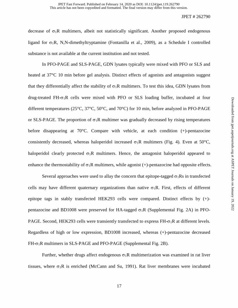
decrease of σ1R multimers, albeit not statistically significant. Another proposed endogenous
ligand for σ1R, N,N-dimethyltryptamine (Fontanilla et al., 2009), as a Schedule I controlled
substance is not available at the current institution and not tested.
In PFO-PAGE and SLS-PAGE, GDN lysates typically were mixed with PFO or SLS and
heated at 37°C 10 min before gel analysis. Distinct effects of agonists and antagonists suggest
that they differentially affect the stability of σ1R multimers. To test this idea, GDN lysates from
drug-treated FH-σ1R cells were mixed with PFO or SLS loading buffer, incubated at four
different temperatures (25°C, 37°C, 50°C, and 70°C) for 10 min, before analyzed in PFO-PAGE
or SLS-PAGE. The proportion of σ1R multimer was gradually decreased by rising temperatures
before disappearing at 70°C. Compare with vehicle, at each condition (+)-pentazocine
consistently decreased, whereas haloperidol increased σ1R multimers (Fig. 4). Even at 50°C,
haloperidol clearly protected σ1R multimers. Hence, the antagonist haloperidol appeared to
enhance the thermostability of σ1R multimers, while agonist (+)-pentazocine had opposite effects.
Several approaches were used to allay the concern that epitope-tagged σ1Rs in transfected
cells may have different quaternary organizations than native σ1R. First, effects of different
epitope tags in stably transfected HEK293 cells were compared. Distinct effects by (+)-
pentazocine and BD1008 were preserved for HA-tagged σ1R (Supplemental Fig. 2A) in PFO-
PAGE. Second, HEK293 cells were transiently transfected to express FH-σ1R at different levels.
Regardless of high or low expression, BD1008 increased, whereas (+)-pentazocine decreased
FH-σ1R multimers in SLS-PAGE and PFO-PAGE (Supplemental Fig. 2B).
Further, whether drugs affect endogenous σ1R multimerization was examined in rat liver
tissues, where σ1R is enriched (McCann and Su, 1991). Rat liver membranes were incubated
This article has not been copyedited and formatted. The final version may differ from this version.JPET Fast Forward. Published on February 14, 2020 as DOI: 10.1124/jpet.119.262790
at ASPE
T Journals on January 19, 2022
jpet.aspetjournals.orgD
ownloaded from

JPET # 262790
18
with drugs, and solubilized with GDN lysis buffer. Lysates were then subjected to non-
denaturing gel analysis, and immunoblot were probed with a mouse monoclonal antibody for
σ1R (clone B5, Santa Cruz Biotechnology). Different from cell lysates, in PFO-PAGE σ1R were
detected as almost exclusively high MW smear bands, very faint signals of dimer bands based on
apparent electrophoretic mobility, and no monomers. Compared with vehicle, (+)-pentazocine
and BD1008 appeared to increase or decrease the dimer signal respectively (Fig. 5A). In SDS-
PAGE σ1R in rat liver lysates only showed as a single band of 25 kD, if lysates were mixed with
SDS (final 1%) and heated to 85°C. If the mixture was incubated at room temperature (RT) for 1
h, a faint band near 75 kD appeared in BD1008-treated samples (Fig. 5B). The apparent MW of
this band was consistent with a σ1R trimer, suggesting that in liver membranes BD1008-induced
σ1R trimers were stable enough to partially resist SDS treatment at RT. Absence of non-specific
bands validated this antibody for detecting native σ1R.
Most importantly, drug effects were convincingly shown when samples were run in SLS-
PAGE (Fig. 5C). Compared with vehicle, (+)-pentazocine significantly decreased the proportion
of multimers in total σ1R proteins, with a concomitant increase in σ1R monomers. In contrast,
BD1008 had a significant effect opposite to (+)-pentazocine (Fig. 5C&D). These drug effects
were very similar to those seen in transfected HEK293 cells (Fig. 2), but the difference in σ1R
multimer MW was worth noting. In rat liver lysates these bands appeared to range from
approximately 70 kD (possible trimer) to beyond 400 kD, with the highest density near
approximate 100 kD, but those from cells appeared to have larger MW sizes (Fig. 2). This
suggested that high-order quaternary organization of native or heterologously overexpressed σ1R
might not be the same, but drug effects on σ1R multimerization were preserved overall. It should
be cautioned that MW estimation of these bands was limited, due to the nature of these gels.
This article has not been copyedited and formatted. The final version may differ from this version.JPET Fast Forward. Published on February 14, 2020 as DOI: 10.1124/jpet.119.262790
at ASPE
T Journals on January 19, 2022
jpet.aspetjournals.orgD
ownloaded from

JPET # 262790
19
Crosslinking assays were done to validate the presence of σ1R multimers. DSP is a
bifunctional crosslinker that selectively reacts with primary amines, with a cleavable disulfide
bond in its 12Å spacer arm. DSP crosslinking in rat liver GDN lysates induced a 50 kD band
after non-reducing SDS-PAGE, which disappeared with TCEP treatment to reduce disulfide-
bonds (Supplemental Fig. 3A). Because rat σ1R has a sole lysine (Lys142) with its side chain
solvent accessible, based on human σ1R crystal structures (Schmidt et al., 2016), the 50 kD band
was most likely a dimer formed through crosslinking at Lys142. FH-σ1R has three more lysine
residues in its epitope and linker. DSP crosslinking induced multiple high-MW bands. Beside a
clear dimer band, two discernible bands were detected at positions corresponding to trimer and
tetramer, and smeared bands above 150 kD (Supplemental Fig. 3B). These data suggested that in
transfected cells trimers of σ1R could exist, yet they might undergo further assembly, thus trimer
bands were not detected in non-denaturing gels (Fig.1 & 2).
Recent breakthrough on crystal structures of σ1R shed new light on key residues
mediating homotrimer formation (Schmidt et al., 2016). Mutants of σ1R at these pivotal positions
were examined in non-denaturing gels. In WT σ1R, the benzyl side chains of Phe191 in three
protomers form aromatic interaction with each other (Fig. 6A). Mutation of Phe191 to Gly
(F191G) removed this interaction and abolished σ1R multimerization, but appeared to retain
dimerization (lane 1, Fig. 6B). The hydrophobic side chain of Trp136 in WT σ1R interacts
extensive with several residues in the neighboring protomer. Substitution of Trp136 with Gly
(W136G) showed severely reduced multimerization, and a dimer but no monomer band (lane 2,
Fig. 6B). Crystal structures show that Arg119 and His116 of a WT protomer form a network of
hydrogen bonds with Asp195 and Thr198 of its neighboring protomers, which is critical in
maintaining trimerization interface. Alanine substitution at either positions (R119A or D195A)
This article has not been copyedited and formatted. The final version may differ from this version.JPET Fast Forward. Published on February 14, 2020 as DOI: 10.1124/jpet.119.262790
at ASPE
T Journals on January 19, 2022
jpet.aspetjournals.orgD
ownloaded from
JPET # 262790
20
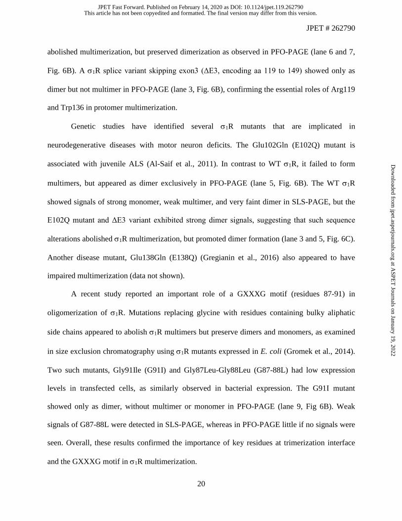
abolished multimerization, but preserved dimerization as observed in PFO-PAGE (lane 6 and 7,
Fig. 6B). A σ1R splice variant skipping exon3 (ΔE3, encoding aa 119 to 149) showed only as
dimer but not multimer in PFO-PAGE (lane 3, Fig. 6B), confirming the essential roles of Arg119
and Trp136 in protomer multimerization.
Genetic studies have identified several σ1R mutants that are implicated in
neurodegenerative diseases with motor neuron deficits. The Glu102Gln (E102Q) mutant is
associated with juvenile ALS (Al-Saif et al., 2011). In contrast to WT σ1R, it failed to form
multimers, but appeared as dimer exclusively in PFO-PAGE (lane 5, Fig. 6B). The WT σ1R
showed signals of strong monomer, weak multimer, and very faint dimer in SLS-PAGE, but the
E102Q mutant and ΔE3 variant exhibited strong dimer signals, suggesting that such sequence
alterations abolished σ1R multimerization, but promoted dimer formation (lane 3 and 5, Fig. 6C).
Another disease mutant, Glu138Gln (E138Q) (Gregianin et al., 2016) also appeared to have
impaired multimerization (data not shown).
A recent study reported an important role of a GXXXG motif (residues 87-91) in
oligomerization of σ1R. Mutations replacing glycine with residues containing bulky aliphatic
side chains appeared to abolish σ1R multimers but preserve dimers and monomers, as examined
in size exclusion chromatography using σ1R mutants expressed in E. coli (Gromek et al., 2014).
Two such mutants, Gly91Ile (G91I) and Gly87Leu-Gly88Leu (G87-88L) had low expression
levels in transfected cells, as similarly observed in bacterial expression. The G91I mutant
showed only as dimer, without multimer or monomer in PFO-PAGE (lane 9, Fig 6B). Weak
signals of G87-88L were detected in SLS-PAGE, whereas in PFO-PAGE little if no signals were
seen. Overall, these results confirmed the importance of key residues at trimerization interface
and the GXXXG motif in σ1R multimerization.
This article has not been copyedited and formatted. The final version may differ from this version.JPET Fast Forward. Published on February 14, 2020 as DOI: 10.1124/jpet.119.262790
at ASPE
T Journals on January 19, 2022
jpet.aspetjournals.orgD
ownloaded from

JPET # 262790
21
Further mutational analyses were conducted to explore the dimeric interface. Very weak
expression of double mutants (D195A/G91I, D195A/G87-88L, E102Q/G91I, and E102Q/G87-
88L) prevented their signal detection in PFO-PAGE, despite faint bands in SLS-PAGE (data not
shown). Surprisingly, removing the N-terminal 36 residues (Δ36aa, including the TM region of
aa 8 to 32) of σ1R resulted in a mutant that showed as a distinct monomer in PFO-PAGE and
SLS-APGE (lane 11, Fig. 6B&C). The mutant lacking N-terminal 10 residues (Δ10aa) displayed
severely impaired oligomerization (lane 16). However, a mutant lacking first 5 residues (Δ5aa)
but sparing the TM, retained substantially multimerization (lane 15). Moreover, the E102Q
mutant with Δ5aa remained as dimer (lane 14), but was converted to apparent monomer if
combined with Δ36aa (land 13). Similarly, Δ36aa deletion also changed dimeric G91I to
monomer (lane 12).
These data suggest that in addition to key residues in the trimerization interface and
GXXXG motif, the N-terminal (NT) domain of σ1R play a crucial role in its multimerization by
potentially linking two homotrimers to form a hexamer, or multiple homotrimers to high-order
oligomers. In fact, the unit cell organization of σ1R crystals shows a pair of homotrimers linked
together through interactions of two parallel NT domains, each from a protomer of the two
neighboring homotrimers (Schmidt et al., 2016). Further evidence was obtained in mutational
analysis on σ1R homomeric interaction by co-transfection of Myc-tagged WT σ1R and a series of
deletion mutants of σ1R with N-terminal glutathione S-transferase (GST) fusion. Even upon
gradual truncations of more than 100 residues in the C-terminus of GST-σ1R, Myc-σ1R was co-
enriched by glutathione beads pull-down. However, this interaction was substantially diminished
if the NT of σ1R was deleted (Supplemental Fig. 4).
This article has not been copyedited and formatted. The final version may differ from this version.JPET Fast Forward. Published on February 14, 2020 as DOI: 10.1124/jpet.119.262790
at ASPE
T Journals on January 19, 2022
jpet.aspetjournals.orgD
ownloaded from

JPET # 262790
22
Next, the impact of σ1R oligomerization on ligand binding was studied. GDN-solubilized
lysates from FH-σ1R cells showed robust [3H](+)-pentazocine binding, with a Kd value (37.8 ±
3.3 nM, average ± SEM) in a similar range to that of σ1R expressed in Sf9 cell membranes
(Schmidt et al., 2016), and a Bmax value (20.3 ± 2.3 pmol/mg protein) several-fold higher than
those in native tissues (McCann and Su, 1991; Bowen et al., 1993). Remarkably, σ1R binding
exhibited exquisite sensitivity to pH values in the buffer. Compared with normal buffer of pH 7.5,
total binding (in the absence of unlabeled ligands) was markedly enhanced in a basic buffer of
pH 9, but reduced in an acidic buffer of pH 6 (Fig. 7A). Kinetic analysis of these homologous
competition binding data (Fig. 7B) revealed that in pH 9 there was a significant decrease in Kd
value (i.e., increase in affinity) of (+)-pentazocine, rather than an increase in binding Bmax (Fig.
7C,D). In contrast, acidic buffer (pH 6) significantly decreased not only binding affinity but also
Bmax values of [3H](+)-pentazocine. These changes in ligand binding appeared to correlate with
the oligomeric states of σ1R revealed in PFO-PAGE. Compared with control (pH 7.5), there was
a significant increase in σ1R multimers at pH 9, with a concomitant decrease in dimers and
monomers (Fig. 7E). An opposite effect was observed at pH 6. These data suggest that ligand
binding to σ1R is significantly affected by its quaternary structures.
Considering multiple oligomeric states of σ1R, ideally [3H](+)-pentazocine binding
should be analyzed using a multi-state model. However, computer-assisted non-linear regression
of binding data showed that a simple, one-site model would suffice (Fig. 7A,B), and Scatchard
plot appeared to be linear (Fig. 7A inset). Statistical comparisons of one-site versus two-site
models did not consistently yield clear-cut conclusions.
Reduced capacity to bind [3H](+)-pentazocine were observed in GDN lysates of σ1R
mutants deficient in multimerization, including Δ36aa, ΔE3, E102Q, R119A and D195A (Table
This article has not been copyedited and formatted. The final version may differ from this version.JPET Fast Forward. Published on February 14, 2020 as DOI: 10.1124/jpet.119.262790
at ASPE
T Journals on January 19, 2022
jpet.aspetjournals.orgD
ownloaded from

JPET # 262790
23
1). Bmax values measured in untransfected HEK293 cells was negligible (2% of WT σ1R-
transfected cells, in pmol/mg protein), presumably due to a low level of endogenous σ1R. The
weak expression levels of these mutants in transfected cells confounded the interpretation of
their low Bmax. For instance, D195A σ1R still showed approximately 20% of binding Bmax of WT
σ1R despite its low expression, suggesting that (+)-pentazocine binding was not fully
compromised in this dimer-forming mutant. Nevertheless, the monomer-only mutant Δ36aa σ1R
did not bind (+)-pentazocine, despite its sufficient expression. Together with observations that
pH-sensitivity of (+)-pentazocine binding correlated with changes of σ1R multimerization (Fig.
7), these data support the hypothesis that σ1R multimers exhibit most active conformation for
high-affinity (+)-pentazocine binding, whereas its monomers likely do not bind (+)-pentazocine.
This article has not been copyedited and formatted. The final version may differ from this version.JPET Fast Forward. Published on February 14, 2020 as DOI: 10.1124/jpet.119.262790
at ASPE
T Journals on January 19, 2022
jpet.aspetjournals.orgD
ownloaded from

JPET # 262790
24
Discussion
This study used two non-denaturing gel methods to examine σ1R oligomerization. In
general, agonists decreased σ1R multimers, whereas antagonists increased multimers (Fig. 1-5).
Antagonist binding appeared to stabilize σ1R multimers, as a higher temperature was required to
dissociate σ1R multimers (Fig. 4).
Although these methods detected multiple high-MW smear bands, they could not
determine the stoichiometry of σ1R multimers. Detergent solubilization might introduce artificial
aggregates of σ1R. However, distinct changes in band signals by ligands argue against this.
Further, existence of high-MW σ1R complex was supported by early purification studies using
[3H]azido-DTG or [3H](+)SKF10047 as affinity ligands, in which the labeled protein complex
under non-denaturing conditions appeared to be approximately 150 or 450 kD (Kavanaugh et al.,
1988; McCann and Su, 1991). In blue native gels, purified σ1R showed as multiple smear bands
from 60 to 480 kD (Schmidt et al., 2016).
In rat liver membranes very little dimer and no monomer of σ1R were present in PFO-
PAGE (Fig. 5A), suggesting that native σ1R multimeric complex was more resistant to extraction
by PFO. Distinct drug effects on σ1R multimerization were optimally demonstrated by SLS-
PAGE (Fig. 5C). σ1R multimers appeared as high-MW bands including possible trimer, tetramer,
and beyond. All σ1R multimers disappeared in denaturing SDS-PAGE. BD1008 induced a weak
band of σ1R trimer if samples were not heated with SDS (Fig. 5B).
Notwithstanding its limitations, these non-denature gel approaches offered a relatively
straightforward readout of σ1R oligomerization. Results on G91I mutant were consistent between
PFO-PAGE (Fig. 6B) and size exclusion chromatography (Gromek et al., 2014). Two recent
This article has not been copyedited and formatted. The final version may differ from this version.JPET Fast Forward. Published on February 14, 2020 as DOI: 10.1124/jpet.119.262790
at ASPE
T Journals on January 19, 2022
jpet.aspetjournals.orgD
ownloaded from

JPET # 262790
25
studies utilized Förster or bioluminescence resonance energy transfer (FRET or BRET) assays to
examine ligand effects on σ1R oligomerization (Mishra et al., 2015; Yano et al., 2018). These
assays are advantageous in monitoring the distance between donor- and acceptor-tagged σ1R in
live cells, while PFO-PAGE and SLS-PAGE appear to be more sensitive in detecting effects by
some ligands such as BD1047 and DTG.
To induce maximal effects on σ1R multimerization, most drugs were used at low
micromolar concentrations, approximately 100 to 1000-fold of their Ki values for σ1R. (+)-
Pentazocine showed dose-dependent effects on decreasing σ1R multimers (Fig. 1C). With a
subnanomolar affinity for σ1R (James et al., 2012), CM304 induced significant effects at 0.1 µM.
Although 0.45 µM drugs were sufficient to stabilize oligomers of σ1R purified from bacteria
(Gromek et al., 2014), 100 µM (+)-pentazocine or haloperidol was used in COS-7 cells for FRET
analysis (Mishra et al., 2015). Thus, higher concentrations of drugs were necessary to permeate
across membranes to bind intracellular σ1R in cells. Most ligands tested here are relatively
selective for σ1R, but haloperidol also has a high affinity for dopamine D2 receptors. This action
was unlikely involved, because haloperidol had effects on σ1R multimerization in cold cell
lysates (Supplemental Fig. 1).
It is worth noting that effects of PRE-084 and NE-100 on σ1R multimerization did not
correlate with their binding affinities. The binding pocket in σ1R can accommodate diverse
ligands with a charged nitrogen as the central pharmacophore (Ablordeppey and Glennon, 2007).
Comparison between σ1R structures bound with (+)-pentazocine and NE-100 revealed limited
conformational rearrangement (Schmidt et al., 2018). These structures likely provided snapshots
of σ1R in its lowest free energy state. However, dynamic conformational changes induced by
This article has not been copyedited and formatted. The final version may differ from this version.JPET Fast Forward. Published on February 14, 2020 as DOI: 10.1124/jpet.119.262790
at ASPE
T Journals on January 19, 2022
jpet.aspetjournals.orgD
ownloaded from

JPET # 262790
26
different ligands and impact on σ1R quaternary structures have not been thoroughly examined.
Further, drug binding to σ1R may exhibit different cooperativity. Although (+)-pentazocine fully
occupied the binding pocket of each σ1R protomer in crystal structures, binding kinetic analysis
of Flag-tagged σ1R and molecular dynamics simulation supported a multi-step model of (+)-
pentazocine binding (Schmidt et al., 2018). Whether PRE-084 or NE-100 induces different
conformational changes or binding cooperativity of σ1R from (+)-pentazocine will require more
sophisticated techniques.
Stabilization of σ1R multimer by antagonists may be explained by their preferential
higher affinity for σ1R multimers than dimers/monomers. In WT σ1R haloperidol’s IC50 value
for competing against [3H](+)-pentazocine binding was approximately 1/10 of those in R119A or
D195A mutants (data not shown), which forms only dimers and monomers. However, similar Kd
values (Table 1) was not sufficient to explain why (+)-pentazocine dissociated σ1R multimers.
Other potential mechanisms, such as negative protomer cooperativity for (+)-pentazocine
binding, will be pursued in future studies.
As σ1R interacts with many protein partners and regulates their function (Schmidt and
Kruse, 2019), efficacies of σ1R drugs in functional assays are likely determined by multiple
factors at molecular, cellular, and higher integrative levels. (+)-Pentazocine decreased σ1R’s
association with BiP (Hayashi and Su, 2007), whereas haloperidol decreased its association with
acid sensing ion channels (Carnally et al., 2010), suggesting that different partners may
preferentially interact with specific oligomeric forms of σ1R. Changes in σ1R oligomerization by
ligands can modulate the availability of σ1R to associate with its partners, but does not fully
account for ligands’ efficacies in functional assays.
This article has not been copyedited and formatted. The final version may differ from this version.JPET Fast Forward. Published on February 14, 2020 as DOI: 10.1124/jpet.119.262790
at ASPE
T Journals on January 19, 2022
jpet.aspetjournals.orgD
ownloaded from

JPET # 262790
27
Data in this study suggest that multiple domains coordinate σ1R oligomerization. Crystal
structures of σ1R have pinpointed critical residues mediating interactions for homotrimerization
(Schmidt et al., 2016). Mutation of Arg119 or Asp195 abolished multimerization in non-
denaturing gels, confirming a crucial role of hydrogen bonds at these positions. Further,
hydrophobic interactions and van der Waals forces involving Phe191 and Trp136 are also pivotal,
as removing their aromatic side chains severely impaired multimerization (Fig. 6B,C). The
importance of the GXXXG motif (Gromek et al., 2014) was also substantiated by results of G91I
σ1R in PFO-PAGE. As Gly91 is in close proximity (4-5 Å) to Trp136 of a neighboring protomer
in σ1R crystal structures, mutation to bulky side chain disrupted multimerization.
In non-denaturing gels multiple high-MW smeared bands appeared larger than σ1R
homotrimers, suggesting possible high-order organization of σ1R trimers. A corollary of this
scenario would require additional domains to mediate non-covalent assembly of trimeric
building blocks. Current data support the hypothesis that the NT of σ1R serves such a role.
Unlike WT σ1R and the Δ5aa mutant, Δ10aa and Δ36aa mutants yielded only monomers in PFO-
PAGE (Fig. 6B), despite their intact trimerization domains. It is tempting to speculate that intact
NT interactions are required to initiate σ1R high-order assembly.
This provocative hypothesis is partly corroborated by σ1R crystal structures, in which
each unit cell comprises two pairs of homotrimers (Schmidt et al., 2016). Both pairs are linked
together through interactions of two parallel NT domains, each from a protomer in the two
homotrimers (Fig. 6A). The opposite orientation of the two pairs requires their embedding into
two lipid bilayers in native membranes, and is possibly an artifact in crystallization. However,
dimerization of homotrimers may resemble a form of native σ1R oligomerization.
This article has not been copyedited and formatted. The final version may differ from this version.JPET Fast Forward. Published on February 14, 2020 as DOI: 10.1124/jpet.119.262790
at ASPE
T Journals on January 19, 2022
jpet.aspetjournals.orgD
ownloaded from

JPET # 262790
28
Dimer formation after dissociation of multimers may involve assembly of two protomers
from neighboring homotrimers via their NT interactions, or rearrangement of two cognate
protomers within a homotrimer. Current results on NT truncations favor the first scenario, but do
not rule out the latter. Dimer interactions appeared to be preserved by PFO, but disrupted by the
more stringent detergent SLS. Persistence of E102Q or ΔE3 σ1R dimers in SLS-PAGE is
perplexing and requires further investigation. The E102Q mutant was shown to be prone to
aggregation and have aberrant cellular location (Wong et al., 2016; Dreser et al., 2017). In co-
immunoprecipitation assays, ΔE3 σ1R exhibited altered association with the dopamine
transporter than WT σ1R (Hong et al., 2017), and exerted dominant-negative effects in disrupting
association of WT σ1R with µ opioid receptors (Pan et al., 2017). These results suggest that
dysregulation of σ1R quaternary structure impairs its physiological function.
Intriguingly, buffer pH significantly altered the oligomeric states of σ1R (Fig. 7E).
Although the underlying mechanism and physiological significance are beyond the scope of this
study, a rudimentary inference is that σ1R multimers dissociate in acidic lysosomes to facilitate
degradation. This serendipitous finding helped to examine how σ1R oligomerization affected
ligand binding. Results (Fig. 7) indicated that σ1R multimers possess high-affinity and high-
capacity binding of [3H](+)-pentazocine. Because Δ36aa σ1R formed monomer exclusively, but
lacked binding (Table 1), it was inferred that monomeric σ1R could not bind (+)-pentazocine.
This hypothesis is consistent with a previous study using σ1R mutants from bacterial expression
(Gromek et al, 2014).
In summary, this study demonstrated that multiple domains coordinate the
oligomerization of σ1R. The equilibrium balance between monomers, dimers and multimers of
σ1R is dynamically regulated by agonists or antagonists in distinct manners. Whereas antagonists
This article has not been copyedited and formatted. The final version may differ from this version.JPET Fast Forward. Published on February 14, 2020 as DOI: 10.1124/jpet.119.262790
at ASPE
T Journals on January 19, 2022
jpet.aspetjournals.orgD
ownloaded from

JPET # 262790
29
promote σ1R multimerization, agonists facilitate its dissociation (Fig. 8). σ1R mutants implicated
in neurodegenerative diseases displayed aberrant multimerization, suggesting that balance in σ1R
oligomerization is important in its physiological function. Extensive studies have shown that σ1R
associates with a plethora of partner proteins that are involved in diverse cellular signaling
pathways. Distinct regulation of σ1R multimerization by agonists and antagonists may
selectively modulate activities of clientele proteins within its interactome network. Future work
will shed light on whether σ1R oligomerization may be precisely controlled by cells’ adaptive
responses to physiochemical changes in the environment, which in turn may impact on these
signaling mechanisms.
This article has not been copyedited and formatted. The final version may differ from this version.JPET Fast Forward. Published on February 14, 2020 as DOI: 10.1124/jpet.119.262790
at ASPE
T Journals on January 19, 2022
jpet.aspetjournals.orgD
ownloaded from

JPET # 262790
30
Acknowledgments
I thank Drs. Jonathan L. Katz, Mark R. Macbeth, and Jie Jiang for advice and comments
on this manuscript, and generous gifts of various σ1R ligands from Jonathan L. Katz, Christopher
R. McCurdy, and NIDA Drug supply program.
This article has not been copyedited and formatted. The final version may differ from this version.JPET Fast Forward. Published on February 14, 2020 as DOI: 10.1124/jpet.119.262790
at ASPE
T Journals on January 19, 2022
jpet.aspetjournals.orgD
ownloaded from

JPET # 262790
31
Author contributions
Participated in research design: Hong
Conducted experiments: Hong
Performed data analysis: Hong
Wrote the manuscript: Hong
This article has not been copyedited and formatted. The final version may differ from this version.JPET Fast Forward. Published on February 14, 2020 as DOI: 10.1124/jpet.119.262790
at ASPE
T Journals on January 19, 2022
jpet.aspetjournals.orgD
ownloaded from

JPET # 262790
32
References
Ablordeppey SY and Glennon RA (2007) Pharmacophore models for sigma-1 receptor binding, in Sigma receptors: chemistry, cell biology and clinical implications (Matsumoto RR, Bowen WD and Su TP eds) pp 71-98, Springer, New York, NY.
Al-Saif A, Al-Mohanna F and Bohlega S (2011) A mutation in sigma-1 receptor causes juvenile amyotrophic lateral sclerosis. Ann Neurol 70:913-919.
Aydar E, Palmer CP, Klyachko VA and Jackson MB (2002) The sigma receptor as a ligand-regulated auxiliary potassium channel subunit. Neuron 34:399-410.
Balasuriya D, Stewart AP, Crottes D, Borgese F, Soriani O and Edwardson JM (2012) The sigma-1 receptor binds to the Nav1.5 voltage-gated Na+ channel with 4-fold symmetry. J Biol Chem 287:37021-37029.
Bergeron R, de Montigny C and Debonnel G (1996) Potentiation of neuronal NMDA response induced by dehydroepiandrosterone and its suppression by progesterone: effects mediated via sigma receptors. J Neurosci 16:1193-1202.
Bernard-Marissal N, Medard JJ, Azzedine H and Chrast R (2015) Dysfunction in endoplasmic reticulum-mitochondria crosstalk underlies SIGMAR1 loss of function mediated motor neuron degeneration. Brain 138:875-890.
Bowen WD, de Costa BR, Hellwell SB, Walker JM and Rice KC (1993) [3H]-(+)Pentazocine: a potent and highly selective benzomorphan-based probe for sigma-1 receptors. Molecular Neuropharmacology 3:117-126.
Cai Y, Yang L, Niu F, Liao K and Buch S (2017) Role of Sigma-1 Receptor in Cocaine Abuse and Neurodegenerative Disease, in Sigma Receptors: Their Role in Disease and as Therapeutic Targets (Smith SB and Su T-P eds) pp 163-175, Springer International Publishing, Cham.
Carnally SM, Johannessen M, Henderson RM, Jackson MB and Edwardson JM (2010) Demonstration of a direct interaction between sigma-1 receptors and acid-sensing ion channels. Biophys J 98:1182-1191.
Chu UB and Ruoho AE (2016) Biochemical Pharmacology of the Sigma-1 Receptor. Mol Pharmacol 89:142-153.
Cobos EJ, Entrena JM, Nieto FR, Cendan CM and Del Pozo E (2008) Pharmacology and therapeutic potential of sigma(1) receptor ligands. Curr Neuropharmacol 6:344-366.
Dreser A, Vollrath JT, Sechi A, Johann S, Roos A, Yamoah A, Katona I, Bohlega S, Wiemuth D, Tian Y, Schmidt A, Vervoorts J, Dohmen M, Beyer C, Anink J, Aronica E, Troost D, Weis J and Goswami A (2017) The ALS-linked E102Q mutation in Sigma receptor-1 leads to ER stress-mediated defects in protein homeostasis and dysregulation of RNA-binding proteins. Cell Death Differ 24:1655-1671.
Fontanilla D, Johannessen M, Hajipour AR, Cozzi NV, Jackson MB and Ruoho AE (2009) The hallucinogen N,N-dimethyltryptamine (DMT) is an endogenous sigma-1 receptor regulator. Science 323:934-937.
Francardo V, Bez F, Wieloch T, Nissbrandt H, Ruscher K and Cenci MA (2014) Pharmacological stimulation of sigma-1 receptors has neurorestorative effects in experimental parkinsonism. Brain 137:1998-2014.
Ganapathy ME, Prasad PD, Huang W, Seth P, Leibach FH and Ganapathy V (1999) Molecular and ligand-binding characterization of the sigma-receptor in the Jurkat human T lymphocyte cell line. J Pharmacol Exp Ther 289:251-260.
This article has not been copyedited and formatted. The final version may differ from this version.JPET Fast Forward. Published on February 14, 2020 as DOI: 10.1124/jpet.119.262790
at ASPE
T Journals on January 19, 2022
jpet.aspetjournals.orgD
ownloaded from

JPET # 262790
33
Gregianin E, Pallafacchina G, Zanin S, Crippa V, Rusmini P, Poletti A, Fang M, Li Z, Diano L, Petrucci A, Lispi L, Cavallaro T, Fabrizi GM, Muglia M, Boaretto F, Vettori A, Rizzuto R, Mostacciuolo ML and Vazza G (2016) Loss-of-function mutations in the SIGMAR1 gene cause distal hereditary motor neuropathy by impairing ER-mitochondria tethering and Ca2+ signalling. Human Molecular Genetics 25:3741-3753.
Gromek KA, Suchy FP, Meddaugh HR, Wrobel RL, LaPointe LM, Chu UB, Primm JG, Ruoho AE, Senes A and Fox BG (2014) The oligomeric states of the purified sigma-1 receptor are stabilized by ligands. J Biol Chem 289:20333-20344.
Hanner M, Moebius FF, Flandorfer A, Knaus HG, Striessnig J, Kempner E and Glossmann H (1996) Purification, molecular cloning, and expression of the mammalian sigma1-binding site. Proc Natl Acad Sci U S A 93:8072-8077.
Hayashi T and Su TP (2001) Regulating ankyrin dynamics: Roles of sigma-1 receptors. Proc Natl Acad Sci U S A 98:491-496.
Hayashi T and Su TP (2007) Sigma-1 receptor chaperones at the ER-mitochondrion interface regulate Ca(2+) signaling and cell survival. Cell 131:596-610.
Hiranita T, Soto PL, Kohut SJ, Kopajtic T, Cao J, Newman AH, Tanda G and Katz JL (2011) Decreases in cocaine self-administration with dual inhibition of the dopamine transporter and sigma receptors. J Pharmacol Exp Ther 339:662-677.
Hong WC, Yano H, Hiranita T, Chin FT, McCurdy CR, Su TP, Amara SG and Katz JL (2017) The sigma-1 receptor modulates dopamine transporter conformation and cocaine binding and may thereby potentiate cocaine self-administration in rats. J Biol Chem 292:11250-11261.
James ML, Shen B, Zavaleta CL, Nielsen CH, Mesangeau C, Vuppala PK, Chan C, Avery BA, Fishback JA, Matsumoto RR, Gambhir SS, McCurdy CR and Chin FT (2012) New positron emission tomography (PET) radioligand for imaging sigma-1 receptors in living subjects. J Med Chem 55:8272-8282.
Jbilo O, Vidal H, Paul R, De Nys N, Bensaid M, Silve S, Carayon P, Davi D, Galiegue S, Bourrie B, Guillemot JC, Ferrara P, Loison G, Maffrand JP, Le Fur G and Casellas P (1997) Purification and characterization of the human SR 31747A-binding protein. A nuclear membrane protein related to yeast sterol isomerase. J Biol Chem 272:27107-27115.
Katz JL, Hiranita T, Hong WC, Job MO and McCurdy CR (2017) A Role for Sigma Receptors in Stimulant Self-Administration and Addiction. Handb Exp Pharmacol.
Kavanaugh MP, Tester BC, Scherz MW, Keana JF and Weber E (1988) Identification of the binding subunit of the sigma-type opiate receptor by photoaffinity labeling with 1-(4-azido-2-methyl[6-3H]phenyl)-3-(2-methyl[4,6-3H]phenyl)guanidine. Proc Natl Acad Sci U S A 85:2844-2848.
Kim FJ, Kovalyshyn I, Burgman M, Neilan C, Chien CC and Pasternak GW (2010) Sigma 1 receptor modulation of G-protein-coupled receptor signaling: potentiation of opioid transduction independent from receptor binding. Mol Pharmacol 77:695-703.
Kourrich S, Hayashi T, Chuang JY, Tsai SY, Su TP and Bonci A (2013) Dynamic interaction between sigma-1 receptor and Kv1.2 shapes neuronal and behavioral responses to cocaine. Cell 152:236-247.
Lahmy V, Meunier J, Malmstrom S, Naert G, Givalois L, Kim SH, Villard V, Vamvakides A and Maurice T (2013) Blockade of Tau hyperphosphorylation and Abeta(1)(-)(4)(2) generation by the aminotetrahydrofuran derivative ANAVEX2-73, a mixed muscarinic
This article has not been copyedited and formatted. The final version may differ from this version.JPET Fast Forward. Published on February 14, 2020 as DOI: 10.1124/jpet.119.262790
at ASPE
T Journals on January 19, 2022
jpet.aspetjournals.orgD
ownloaded from

JPET # 262790
34
and sigma(1) receptor agonist, in a nontransgenic mouse model of Alzheimer's disease. Neuropsychopharmacology 38:1706-1723.
Li X, Hu Z, Liu L, Xie Y, Zhan Y, Zi X, Wang J, Wu L, Xia K, Tang B and Zhang R (2015) A SIGMAR1 splice-site mutation causes distal hereditary motor neuropathy. Neurology 84:2430-2437.
Martin WR, Eades CG, Thompson JA, Huppler RE and Gilbert PE (1976) The effects of morphine- and nalorphine- like drugs in the nondependent and morphine-dependent chronic spinal dog. J Pharmacol Exp Ther 197:517-532.
Matsumoto RR (2007) Sigma receptors: Historical Perspective and Background, in Sigma receptors: chemistry, cell biology and clinical implications (Matsumoto RR, Bowen WD and Su TP eds) pp 1-24, Springer, New York, NY.
Maurice T and Goguadze N (2017) Sigma-1 (sigma1) Receptor in Memory and Neurodegenerative Diseases. Handb Exp Pharmacol 244:81-108.
Maurice T and Su TP (2009) The pharmacology of sigma-1 receptors. Pharmacol Ther 124:195-206.
McCann DJ and Su TP (1991) Solubilization and characterization of haloperidol-sensitive (+)-[3H]SKF-10,047 binding sites (sigma sites) from rat liver membranes. J Pharmacol Exp Ther 257:547-554.
Merlos M, Romero L, Zamanillo D, Plata-Salaman C and Vela JM (2017) Sigma-1 Receptor and Pain. Handb Exp Pharmacol 244:131-161.
Mishra AK, Mavlyutov T, Singh DR, Biener G, Yang J, Oliver JA, Ruoho A and Raicu V (2015) The sigma-1 receptors are present in monomeric and oligomeric forms in living cells in the presence and absence of ligands. Biochem J 466:263-271.
Navarro G, Moreno E, Aymerich M, Marcellino D, McCormick PJ, Mallol J, Cortes A, Casado V, Canela EI, Ortiz J, Fuxe K, Lluis C, Ferre S and Franco R (2010) Direct involvement of sigma-1 receptors in the dopamine D1 receptor-mediated effects of cocaine. Proc Natl Acad Sci U S A 107:18676-18681.
Newman AH and Coop A (2007) Medicinal chemistry: new chemical classes and subtype-selective ligands, in Sigma receptors: chemistry, cell biology and clinical implications (Matsumoto RR, Bowen WD and Su TP eds) pp 25-44, Springer, New York, NY.
Ortega-Roldan JL, Ossa F and Schnell JR (2013) Characterization of the human sigma-1 receptor chaperone domain structure and binding immunoglobulin protein (BiP) interactions. J Biol Chem 288:21448-21457.
Pan L, Pasternak DA, Xu J, Xu M, Lu Z, Pasternak GW and Pan YX (2017) Isolation and characterization of alternatively spliced variants of the mouse sigma1 receptor gene, Sigmar1. PLoS One 12:e0174694.
Penna A, Demuro A, Yeromin AV, Zhang SL, Safrina O, Parker I and Cahalan MD (2008) The CRAC channel consists of a tetramer formed by Stim-induced dimerization of Orai dimers. Nature 456:116-120.
Ramachandran S, Chu UB, Mavlyutov TA, Pal A, Pyne S and Ruoho AE (2009) The sigma1 receptor interacts with N-alkyl amines and endogenous sphingolipids. Eur J Pharmacol 609:19-26.
Ramjeesingh M, Huan LJ, Garami E and Bear CE (1999) Novel method for evaluation of the oligomeric structure of membrane proteins. Biochem J 342 ( Pt 1):119-123.
Reichel C (2012) SARCOSYL-PAGE: a new electrophoretic method for the separation and immunological detection of PEGylated proteins. Methods Mol Biol 869:65-79.
This article has not been copyedited and formatted. The final version may differ from this version.JPET Fast Forward. Published on February 14, 2020 as DOI: 10.1124/jpet.119.262790
at ASPE
T Journals on January 19, 2022
jpet.aspetjournals.orgD
ownloaded from

JPET # 262790
35
Robson MJ, Turner RC, Naser ZJ, McCurdy CR, O'Callaghan JP, Huber JD and Matsumoto RR (2014) SN79, a sigma receptor antagonist, attenuates methamphetamine-induced astrogliosis through a blockade of OSMR/gp130 signaling and STAT3 phosphorylation. Exp Neurol 254:180-189.
Ryskamp D, Wu J, Geva M, Kusko R, Grossman I, Hayden M and Bezprozvanny I (2017) The sigma-1 receptor mediates the beneficial effects of pridopidine in a mouse model of Huntington disease. Neurobiol Dis 97:46-59.
Ryskamp D, Wu L, Wu J, Kim D, Rammes G, Geva M, Hayden M and Bezprozvanny I (2019) Pridopidine stabilizes mushroom spines in mouse models of Alzheimer's disease by acting on the sigma-1 receptor. Neurobiol Dis 124:489-504.
Sabino V and Cottone P (2017) Sigma Receptors and Alcohol Use Disorders. Handb Exp Pharmacol 244:219-236.
Sambo DO, Lin M, Owens A, Lebowitz JJ, Richardson B, Jagnarine DA, Shetty M, Rodriquez M, Alonge T, Ali M, Katz J, Yan L, Febo M, Henry LK, Bruijnzeel AW, Daws L and Khoshbouei H (2017) The sigma-1 receptor modulates methamphetamine dysregulation of dopamine neurotransmission. Nat Commun 8:2228.
Schmidt HR, Betz RM, Dror RO and Kruse AC (2018) Structural basis for sigma1 receptor ligand recognition. Nat Struct Mol Biol 25:981-987.
Schmidt HR and Kruse AC (2019) The Molecular Function of sigma Receptors: Past, Present, and Future. Trends Pharmacol Sci 40:636-654.
Schmidt HR, Zheng S, Gurpinar E, Koehl A, Manglik A and Kruse AC (2016) Crystal structure of the human sigma1 receptor. Nature 532:527-530.
Srivats S, Balasuriya D, Pasche M, Vistal G, Edwardson JM, Taylor CW and Murrell-Lagnado RD (2016) Sigma1 receptors inhibit store-operated Ca2+ entry by attenuating coupling of STIM1 to Orai1. J Cell Biol 213:65-79.
Su TP, London ED and Jaffe JH (1988) Steroid binding at sigma receptors suggests a link between endocrine, nervous, and immune systems [see comments]. Science 240:219-221.
Thomas JD, Longen CG, Oyer HM, Chen N, Maher CM, Salvino JM, Kania B, Anderson KN, Ostrander WF, Knudsen KE and Kim FJ (2017) Sigma1 Targeting to Suppress Aberrant Androgen Receptor Signaling in Prostate Cancer. Cancer Res 77:2439-2452.
Walker JM, Bowen WD, Walker FO, Matsumoto RR, De Costa B and Rice KC (1990) Sigma receptors: biology and function. Pharmacol Rev 42:355-402.
Wang J, Saul A, Roon P and Smith SB (2016) Activation of the molecular chaperone, sigma 1 receptor, preserves cone function in a murine model of inherited retinal degeneration. Proc Natl Acad Sci U S A 113:E3764-3772.
Watanabe S, Ilieva H, Tamada H, Nomura H, Komine O, Endo F, Jin S, Mancias P, Kiyama H and Yamanaka K (2016) Mitochondria-associated membrane collapse is a common pathomechanism in SIGMAR1- and SOD1-linked ALS. EMBO Mol Med 8:1421-1437.
Weber F and Wunsch B (2017) Medicinal Chemistry of sigma1 Receptor Ligands: Pharmacophore Models, Synthesis, Structure Affinity Relationships, and Pharmacological Applications. Handb Exp Pharmacol 244:51-79.
Wong AY, Hristova E, Ahlskog N, Tasse LA, Ngsee JK, Chudalayandi P and Bergeron R (2016) Aberrant Subcellular Dynamics of Sigma-1 Receptor Mutants Underlying Neuromuscular Diseases. Mol Pharmacol 90:238-253.
This article has not been copyedited and formatted. The final version may differ from this version.JPET Fast Forward. Published on February 14, 2020 as DOI: 10.1124/jpet.119.262790
at ASPE
T Journals on January 19, 2022
jpet.aspetjournals.orgD
ownloaded from

JPET # 262790
36
Wu Z and Bowen WD (2008) Role of sigma-1 receptor C-terminal segment in inositol 1,4,5-trisphosphate receptor activation: constitutive enhancement of calcium signaling in MCF-7 tumor cells. J Biol Chem 283:28198-28215.
Yano H, Bonifazi A, Xu M, Guthrie DA, Schneck SN, Abramyan AM, Fant AD, Hong WC, Newman AH and Shi L (2018) Pharmacological profiling of sigma 1 receptor ligands by novel receptor homomer assays. Neuropharmacology 133:264-275.
This article has not been copyedited and formatted. The final version may differ from this version.JPET Fast Forward. Published on February 14, 2020 as DOI: 10.1124/jpet.119.262790
at ASPE
T Journals on January 19, 2022
jpet.aspetjournals.orgD
ownloaded from

JPET # 262790
37
Footnotes
This study was supported by Butler University Faculty Startup Fund and Holcomb Research
Award (WCH). Portions of the work herein were presented at Experimental Biology (2017, 2019)
and Society for Neuroscience (2017) meetings.
This article has not been copyedited and formatted. The final version may differ from this version.JPET Fast Forward. Published on February 14, 2020 as DOI: 10.1124/jpet.119.262790
at ASPE
T Journals on January 19, 2022
jpet.aspetjournals.orgD
ownloaded from

JPET # 262790
38
Figure legends
Fig. 1 Distinct effects on σ1R multimerization by agonists or antagonists in PFO-PAGE.
HEK293 cells stably transfected with FH-σ1R were treated with drugs at 37°C (in A, B, and C:
agonists or antagonists for 1 h; in D & E: antagonists 0.5 h, then (+)-pentazocine 1 h), lysed in
GDN-Tris buffer, and run in PFO-PAGE. A) Agonists decreased high-MW multimers. B)
Antagonists increased high-MW multimers. C) Dose-dependent effects by agonist (+)-
pentazocine or antagonist CM304. D & E) Interaction of agonist and antagonists on σ1R
multimerization, with representative immunoblots. All panels show column graphs (mean ±
SEM) from multiple experiments (n): A, n = 4 – 7; B, n = 4 – 11; C, n = 3 – 6; D, n = 3; E, n = 4
- 7. * P < 0.05, ** P < 0.01, one-way ANOVA and post-hoc Dunnett’s test, compared with
vehicle. (+)Pent: (+)-pentazocine; (+)SKF: (+)-SKF10047.
Fig. 2 Distinct effects on σ1R multimerization by agonists or antagonists, detected with SLS-
PAGE which showed a higher sensitivity than PFO-PAGE. A and B) Samples from FH-σ1R
cells pretreated with drugs as in Fig. 1. Representative immunoblots and summary column
graphs (mean ± SEM) from multiple experiments: A, n = 3 - 6; B, n = 4 – 8. * P < 0.05, ** P <
0.01, one-way ANOVA and Fisher’s least significant difference test, compared with vehicle.
(+)Pent: (+)-pentazocine.
Fig. 3 Effects of putative endogenous ligands on σ1R multimerization. GDN lysates from drug-
naïve FH-σ1R cells were incubated with ligands on ice overnight, then run in SLS-PAGE.
Representative immunoblots and summary (mean ± SEM) from n = 4 – 8 experiments. * P <
This article has not been copyedited and formatted. The final version may differ from this version.JPET Fast Forward. Published on February 14, 2020 as DOI: 10.1124/jpet.119.262790
at ASPE
T Journals on January 19, 2022
jpet.aspetjournals.orgD
ownloaded from

JPET # 262790
39
0.05, ** P < 0.01, one-way ANOVA and Fisher’s least significant difference test, compared with
vehicle. SPG: d-erythro-sphingosine; PROG: progesterone; DHEA: dehydroepiandrosterone.
Fig. 4 (+)-Pentazocine and haloperidol differentially affected the thermostability of σ1R
multimers. FH-σ1R cells were treated with vehicle or drugs (10 µM) at 37°C for 1 h before lysis.
Cell lysates were mixed with 2X concentrated PFO or SLS loading buffer, incubated for 10 min
at 25°C, 37°C, 50°C, and 70°C, run in PFO-PAGE or SLS-PAGE, and immunoblotted with Flag
antibody. Representative blots from n = 3 experiments. (+)Pent: (+)-pentazocine; Halo:
haloperidol.
Fig. 5 Analysis of drugs effects on σ1R multimerization in rat liver membranes. Rat liver
membranes were treated with drugs for 2 h at 37°C, followed by lysis with GDN-HEPES buffer.
A) In PFO-PAGE σ1R predominantly showed as smear bands above approximately 100 kD,
while a very weak dimer band appeared to be increased following (+)-pentazocine treatment, and
decreased by BD1008. B) In SDS-PAGE only 25 kD σ1R monomer was present if lysates were
heated with SDS. However, σ1R trimer band (75 kD, ►) was revealed in BD1008-treated
samples following RT incubation with SDS. C) Significant effects by (+)-pentazocine and
BD1008 in SLS-PAGE. σ1R multimer bands ranged from approximately 70 kD (possible trimer,
►) to above 300 kD, with most density near 100 kD. D) Summary graph showed σ1R multimer
band densities as % of total σ1R (mean ± SEM, n = 5 experiments) in SLS-PAGE. ** P < 0.01,
one-way ANOVA and post-hoc Dunnett’s test.
This article has not been copyedited and formatted. The final version may differ from this version.JPET Fast Forward. Published on February 14, 2020 as DOI: 10.1124/jpet.119.262790
at ASPE
T Journals on January 19, 2022
jpet.aspetjournals.orgD
ownloaded from

JPET # 262790
40
Fig. 6 Mutational analyses show that multiple domains on σ1R are critical for its multimerization.
A) Structure cartoons of σ1R with mutated residues annotated. Left: side view of σ1R
homotrimer, with transparent surface presentation and peptide backbone in ribbon. Side chains of
specific residues are highlighted. Right: unit cell organization of four σ1R homotrimers in its
crystal structure. Zoomed view: parallel alignment of N-terminal 36 aa from two protomers in
neighboring homotrimers. Cartoons are based on PDB code 5HK1 (Schmidt et al., 2016), and
generated using UCSF Chimera software. B and C) Distinct electrophoretic migration pattern of
σ1R constructs. GDN lysates from HEK293 cells transiently transfected with WT and mutant
FH-σ1R were run in PFO-PAGE and SLS-PAGE, followed by immunoblot detection using Flag
antibodies. Representative blots from n > 3 experiments. Constructs are numbered and aligned in
both blots, except lanes 1 and 2 were switched in SLS-PAGE. Lack of signals in lanes 8 and 10
of PFO-PAGE was likely due to low expression of these mutants.
Fig. 7 Correlation of [3H](+)-pentazocine binding with σ1R quaternary structures in different pH
buffers. Lysates from GDN-solubilized FH-σ1R cells were incubated overnight on ice in Tris
buffer of pH 6, 7.5 or 9, and analyzed in binding assays. Representative raw data in A (inset
shows Scatchard plot) and binding isotherms in B. Both: duplicate samples (average ± SD). Tot:
total binding. C and D) Comparisons of Bmax and Kd values (mean ± SEM, n = 4 experiments,
each with duplicates). E) Modulation of σ1R oligomerization by buffer pH values, as examined
in PFO-PAGE. Top: graph showing σ1R multimer band densities as % of total σ1R (mean ±
SEM, n = 4 experiments) in PFO-PAGE; bottom, representative blot. * P < 0.05, ** P < 0.01,
one-way ANONA and post hoc Dunnett’s test, versus pH 7.5.
This article has not been copyedited and formatted. The final version may differ from this version.JPET Fast Forward. Published on February 14, 2020 as DOI: 10.1124/jpet.119.262790
at ASPE
T Journals on January 19, 2022
jpet.aspetjournals.orgD
ownloaded from
JPET # 262790
41
Fig. 8 Hypothesis cartoon based on results in this study and previous reports. Agonists and
antagonists differentially affect σ1R multimerization. High-order organization of σ1R likely
comprises multiple (≥2) units of homotrimers. Current data suggest a critical role of the N-
terminus in mediating formation of multimers beyond homotrimers.
This article has not been copyedited and formatted. The final version may differ from this version.JPET Fast Forward. Published on February 14, 2020 as DOI: 10.1124/jpet.119.262790
at ASPE
T Journals on January 19, 2022
jpet.aspetjournals.orgD
ownloaded from

JPET # 262790
42
Table 1. Bmax and Kd values of [3H](+)-pentazocine binding in HEK293 stably transfected with
σ1R constructs. Bmax values were adjusted to total protein concentrations in lysates. Shown
values are mean ± SEM from n = 3 experiments, each with duplicates, except WT (n = 4),
E102Q and ΔE3 (n = 2). Bmax of endogenous σ1R in untransfected HEK293 cells was 2% of that
in WT transfected cells, and was subtracted from these values. Note small Bmax values of mutants
except Δ36aa were partly attributed to their lower expression levels than WT (Fig. 6). All had N-
Myc tag except FH-σ1R Δ36aa.
σ1R WT Δ36aa R119A D195A E102Q ΔE3
Bmax (pmol/mg protein)
16.6 ±3.5
0.08 ±0.05
1.83 ±0.13
3.67 ±0.26
0.99 ±0.06
0.21 ±0.22
Kd (nM)
16.4 ±7.5
14.0 ±3.2
9.8 ±1.4
12.9 ±2.7
5.0 ±0.6
17.0 ±14.1
This article has not been copyedited and formatted. The final version may differ from this version.JPET Fast Forward. Published on February 14, 2020 as DOI: 10.1124/jpet.119.262790
at ASPE
T Journals on January 19, 2022
jpet.aspetjournals.orgD
ownloaded from
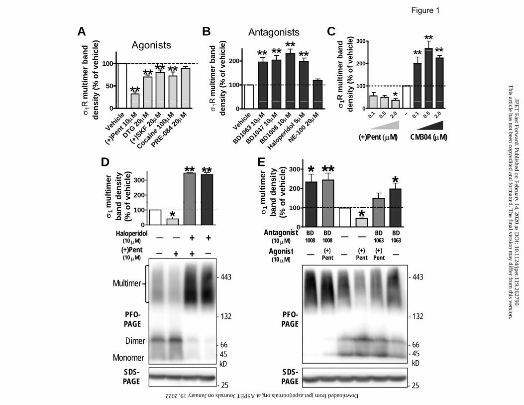
Agonists
nd cle)
Antagonists
** ****nd cle)
A B C
Figure 1
300
****
**and
icle
)
50
100
*** *****
R m
ultim
er b
anity
(% o
f veh
ic100
200 **** **
R m
ultim
er b
ansi
ty (%
of v
ehic
100
200
*
** **
1R m
ultim
er b
ans
ity (%
of v
eh
Vehicl
e Mμ
(+)Pen
t 10
Mμ
DTG 20
Mμ
(+)SKF 20
Mμ
Cocaine 1
00
Mμ
PRE-084 2
0
0σ1R
dens
Vehicl
e Mμ
BD1063
10
Mμ
BD1047
10
Mμ
BD1008
10
Mμ
Haloperi
dol 5
Mμ
NE-100 2
0
0σ1R
dens
(+)Pent (μM) CM304 (μM)
0.1 0.5 2.0 -- 0.1 0.5 2.0
0σ1
den
200
300****
mul
timer
d de
nsity
f veh
icle
)
D E
200
300 * ** *
mul
timer
d de
nsity
of v
ehic
le)
Haloperidol (10 μM) — — + +
(+)Pent — + + —
0
100 *σ1
mba
nd(%
of
Antagonist (10 μM)
BD 1008
BD 1008 — — BD
1063 BD 1063
Agonist (+)
(+) (+)
0
100 *σ1
mba
n(%
o
( )(10 μM)
— + + —
- 443 Multimer
Agonist (10 μM)
— ( )
Pent — ( )
Pent ( )
Pent —
- 443
- 66 - 45
- 132
Dimer
M
PFO-PAGE
- 66 - 45
- 132PFO-PAGE
- 45kD
Monomer 45kD
- 25 - 25SDS-PAGE
SDS-PAGE
This article has not been copyedited and form
atted. The final version m
ay differ from this version.
JPET
Fast Forward. Published on February 14, 2020 as D
OI: 10.1124/jpet.119.262790
at ASPET Journals on January 19, 2022 jpet.aspetjournals.org Downloaded from

Figure 2
400 ** **
r ban
dve
hicl
e)
150
200 **
band
ehic
le)
A B
0
100
200
300
*σ1R
mul
timer
dens
ity (%
of v
0
50
100
150
***
σ1R
mul
timer
bde
nsity
(% o
f ve
d
Vehicl
e Mμ
PRE-084 2
0
Mμ
DTG 20
Mμ
NE-10
0 20
0d
Haloperidol — — + +(+)Pent — + + —
(10 μM)
Multimer- 443
443 Multimer
SLS-PAGE
- 443 - 240
SLS-PAGE
- 25
- 66 - 45kDMonomer
SDS-
- 66 - 45kD
- 25SDS-PAGEPAGE 25PAGE
This article has not been copyedited and form
atted. The final version m
ay differ from this version.
JPET
Fast Forward. Published on February 14, 2020 as D
OI: 10.1124/jpet.119.262790
at ASPET Journals on January 19, 2022 jpet.aspetjournals.org Downloaded from

Figure 3
200
250 ***ba
ndve
hicl
e)
Figure 3
0
50
100
150 *
σ1R
mul
timer
en
sity
(% o
f v
SPG PROG DHEA ▬▬▬▬▬▬ ▬▬▬▬▬▬ ▬▬▬▬▬▬
(μM) 10 100 50 10 — 10 50
0σ d- 443
SLS-PAGE - 66
443 - 240
SDS-PAGE 25
- 45kD
PAGE - 25
This article has not been copyedited and form
atted. The final version m
ay differ from this version.
JPET
Fast Forward. Published on February 14, 2020 as D
OI: 10.1124/jpet.119.262790
at ASPET Journals on January 19, 2022 jpet.aspetjournals.org Downloaded from

Figure 4
(+)P
ent
—
Halo
(+)P
ent
—
Halo
(+)P
ent
—
Halo
(+)P
ent
—
Halo
———————— ———————— ———————— ———————— 25°C 37°C 50°C 70°C
- 443 - 240
- 66
Multimer
SLS-PAGE
- 45 Monomer
Multimer- 443
Multimer
PFO-PAGE
Dimer
- 240
- 66 Monomer - 45
kD
This article has not been copyedited and form
atted. The final version m
ay differ from this version.
JPET
Fast Forward. Published on February 14, 2020 as D
OI: 10.1124/jpet.119.262790
at ASPET Journals on January 19, 2022 jpet.aspetjournals.org Downloaded from

CBD1008 +
BD1008 (2 μM) — — + A
Figure 5
-443
132
BD1008 (2 μM) — — +
(+)Pent (5 μM) + — —
Multimer
-443
(2 μM)
(+)Pent (5 μM) + — —
-132
-66
-45SLS-PAGE
MultimerPFO-PAGE
Dimer
-132
-66
-45
-14kD
Monomer
Enhance contrast
kD
-66
-45kD
DB D
50
75
100
****
timer
ban
d%
of t
otal
σ1R
)BSDS-PAGE
-25 kD
-250
-150
0
25
σ1R
mul
dens
ity (%
BD1008 (10 μM) — — +
-100-75
-50
-37
SDS-PAGE
RT sample
(10 μM) (+)Pent (20 μM) + — —
-25kD
pincubation
This article has not been copyedited and form
atted. The final version m
ay differ from this version.
JPET
Fast Forward. Published on February 14, 2020 as D
OI: 10.1124/jpet.119.262790
at ASPET Journals on January 19, 2022 jpet.aspetjournals.org Downloaded from

-t 36
aa
AFigure 6
E102E102
F191
N-t 5 aaN-t 10 aa
N-
W136
F191
G
W13
6G
ΔE3
WT
E102
Q
R119
A
D195
A
D195
A / G
91I
G91I
G87-
88L
Δ36a
a
Δ36a
a / G
91I
▬▬▬ ▬▬▬ ▬▬▬ ▬▬▬ ▬▬▬ ▬▬▬ ▬▬▬ ▬▬▬ ▬▬▬ ▬▬▬ ▬▬▬ ▬▬▬1 2 3 4 5 6 7 8 9 10 11 12
Δ36a
a / E
102Q
Δ5aa
/ E10
2Q
E102
Q
WT
Δ5aa
Δ10a
a
▬▬▬ ▬▬▬ ▬▬▬ ▬▬▬ ▬▬▬ ▬▬▬13 14 5 4 15 16
B1 2 3 4 5 6 7 8 9 10 11 12 13 14 5 4 15 16
- 66 -
- 443 -
- 240 -Multimer
Dimer
PFO-PAGE
- 45 -
- 443 -- 240 -
Monomer
MultimerSLS-PAGE
kD
C
▬▬▬ ▬▬▬ ▬▬▬ ▬▬▬ ▬▬▬ ▬▬▬ ▬▬▬ ▬▬▬ ▬▬▬ ▬▬▬ ▬▬▬ ▬▬▬2 1 3 4 5 6 7 8 9 10 11 12
▬▬▬ ▬▬▬ ▬▬▬ ▬▬▬ ▬▬▬ ▬▬▬13 14 5 4 15 16
- 66 -
- 45 -Dimer
Monomer
PAGE
kD
This article has not been copyedited and formatted. The final version may differ from this version.JPET Fast Forward. Published on February 14, 2020 as DOI: 10.1124/jpet.119.262790
at ASPE
T Journals on January 19, 2022
jpet.aspetjournals.orgD
ownloaded from

8e ) Bmax σ1R multimer100
A E
Figure 7
C1.5ee
4
6
8
pH 6
pH 9pH 7.5
+)-P
enta
zoci
neng
(x10
00 c
pm) Bmax
50
100
**
Bm
ax a
t pH
7.5
50
75
100(n = 4) *
of to
tal σ
1R
0 10 200.0
0.5
1.0
1.5
Bound
Bou
nd /
Fre
n)
Tot0
2
-9 -8 -7Log [(+)-pentazocine] (M)
[3 H](+
bind
in
pH 6pH 7.
5pH 9
0% o
f B
pH 7.5
pH 6pH 9
0
25 **% o
B D
10
15
20
pH 7.5pH 9
Pent
azoc
ine
mol
/mg
prot
ein
Kd
50
75 **(n
M)
- 443
- 132
Multimer
PFO
B D
0 25 50 75 1000
5pH 6
(+)-Pentazocine (nM)
[3 H](+
)-Pbi
ndin
g (p
m
pH 6pH 7.
5pH 9
0
25
**K
d(
- 66
- 45kD
Dimer
Monomer
PFO-PAGE
( ) ( ) p kD
This article has not been copyedited and form
atted. The final version m
ay differ from this version.
JPET
Fast Forward. Published on February 14, 2020 as D
OI: 10.1124/jpet.119.262790
at ASPET Journals on January 19, 2022 jpet.aspetjournals.org Downloaded from

Figure 8
AntagonistDimer
(two protomers)
+N
Agonist+
Monomer(one protomer)
Multimer (≥ three protomers)
C
Multimer (≥ three protomers)High-affinity (+)-pentazocine binding
This article has not been copyedited and form
atted. The final version m
ay differ from this version.
JPET
Fast Forward. Published on February 14, 2020 as D
OI: 10.1124/jpet.119.262790
at ASPET Journals on January 19, 2022 jpet.aspetjournals.org Downloaded from

Supplemental Data JPET # 262790
Distinct regulation of sigma-1 receptor multimerization by its agonists and antagonists in transfected cells and rat liver membranes
Weimin Conrad Hong
Department of Pharmaceutical Sciences, Butler University, Indianapolis, IN 46208
Journal of Pharmacology and Experimental Therapeutics

zocin
e
dol zocin
e
dol
Supplemental Figure 1JPET # 262790
(+)-P
entaz
oVe
hicle
BD10
08Ha
loperi
do(+)
-Pen
tazo
Vehic
leBD
1008
Halop
erido
▬▬▬▬▬▬▬ ▬▬▬▬▬▬▬Cell incubation Lysate incubation
- 443 Multimer
PFO-PAGE
- 66 - 45kD
Dimer
Monomer
Blot: anti-Flag
Suppl. Fig.1 Same effects on σ1R oligomerization by specific σ1R ligands, either pre-treatment before cell lysis or incubation with cell lysates, suggesting that in GDN-lysates the binding activity of σ1R was largely preserved. Left: FH-σ1R cells were treated with 10 μM drugs at 37°C for 1 h before cell lysis. Right: FH-σ1R cells were lysed with GDN lysis buffer first, then lysates were incubated with 10 μM drugs on ice overnight. Samples were run in PFO-PAGE and μ g g pimmunoblotted with Flag antibodies. Note the opposite effects by antagonists (BD1008 and haloperidol) and agonist (+)-pentazocine on the proportion of σ1R multimer in each lane.

Transfect FH-σ1 0.5 μg DNA —————
0.1 μg DNA ————— A B
Supplemental Figure 2JPET # 262790
HA-σ1R
25
50
75
100 **
R m
ultim
eran
d de
nsity
of to
tal σ
1R)
BD1008 (10 μM) — — + — — + (+)Pent (20 μM) + — — + — —
- 443 - 248Multimer
BD1008 (10 μM) + — — (+)Pent (20 μM)
— — +
443
0
25σ1 ba (%
SLS-PAGE
- 66 - 45kD
- 132
Monomer- 443
- 132PFO-PAGE PFO-
- 443
- 132
Multimer
Multimer
- 66 - 45kD
- 25SDS-PAGE
SDS-PAGE
PAGE- 66 - 45kD
25
DimerMonomer
DimerMonomer
PAGE - 25
Suppl. Fig. 2 Distinct effects by agonist and antagonist on σ1R multimerization wereindependent of epitope tags and expression levels in transfected cells.A) N-terminal HA-tagged σ1R (predicted MW: 28 kD) was stably transfected into HEK293 cells.Confluent cells in 12-well plates were treated with drugs for 2h at 37°C before lysis. Shownwere representative blots of GDN lysates run in PFO-PAGE and SDS-PAGE, and quantitativep y , qsummary in column graph (average ± SEM, n = 4. * p < 0.05, one way ANOVA and post-hocDunnett’s comparison with vehicle). σ1R multimers in PFO-PAGE was quantified as % of totalσ1R signals (including multimer, dimer and monomer).B) HEK293 cells transiently transfected with FH-σ1R (MW: 32 kD), treated with drugs for 2h at37°C before lysis. GDN lysates were run in SLS-PAGE, PFO-PAGE, and SDS-PAGE inparallel. Shown are representative blots from n = 2 experiments. Opposite effects of (+)-
t i d BD1008 R lti i ti i il t hi h l i l lpentazocine and BD1008 on σ1R multimerization were similar at high or low expression levels.

A DSP (mM) — 0.2 — 0.2
FH-σ1R transfected HEK293 cellsB
DSP (mM) 1.0 0.2 — — 0.2 1.0 TCEP
Rat liver – endogenous σ1R
Supplemental Figure 3 JPET # 262790
Non-reducing
TCEP (20 mM) — — + +
-250
-150
-100-75
Blot: Flag
TCEP (20 mM) + + +
— — —
250-
150-
100-75-
Blot: σ1R
reducingSDS-PAGE
-50
-37
-25kD
Monomer
Dimer
Monomer
Dimer
75
50-
37-
25-kD
Ponceau Sstaining
Suppl. Fig. 3 Crosslinking of σ1R. Rat liver membranes and FH-σ1R cells were lysed in HEPES-GDN buffer (0.1% GDN, NaCl 150 mM, EDTA 1 mM, HEPES 20 mM, pH 7.5, protease inhibitors).Lysates were incubated with DSP, a homobifunctional, primary amine-reactive crosslinker with al bl di lfid b d) t 25°C f 1 h f ll d b i b ti ith 20 M TCEP t 25°C f 0 5cleavable disulfide bond) at 25°C for 1 h, followed by incubation with 20 mM TCEP at 25°C for 0.5
h, and mixed with SDS sample buffer without DTT, heated at 85°C for 10 min, and run in SDS-PAGE. Native σ1R has one lysine residues (K142), while FH-σ1R has three more lysine residues inits tag and linker.A) In rat liver lysates DSP crosslinking induced a dimer, likely mediated by K142 which is exposedon the surface of σ1R in its crystal structures (Schmidt et al., 2016). Blot with mAb antibody for σ1R(clone B5, Santa Cruz Biotechnology).(clone B5, Santa Cruz Biotechnology).B) Crosslinked FH-σ1R lysates showed multiple immuno-reactive bands. Based on calculated MWof FH-σ1R (32 kD), monomer band (~30 kD) was predominant, with weak dimer band (~60 kD)seen after long exposure (from left: lane 1). DSP treatment induced more dimer formation,apparent trimer (~90 kD,►), tetramer (~120 kD,►►), and high-MW oligomers (lane 2), whichdisappeared after TCEP reduction (lane 4). Note faint bands at 37 and 50 kD were likely non-specific bands which were also seen in non-transfected cells.
S f f f f fPanels below show Ponceau S staining of membranes after transfer. Marked upward shift ofprotein bands showed efficient crosslinking. DSP at 0.2 mM induced optimal crosslinking results incell lysates, as higher concentrations (DSP≥ 0.5mM) reduced total σ1R signals without inducingmore high-MW bands, likely due to crosslinking of σ1R with other large protein complexes thatfailed to migrate properly into gels.

ASupplemental Figure 4
JPET # 262790
N-t TM Cytosolic domainWT (223 aa)ΔC1 (delete aa 211 - 223)ΔC2 (delete aa 185 - 223)ΔC3 (delete aa 158 - 223)ΔC4 (delete aa 132 223)
A
GST R WT Δ Δ Δ Δ
ΔC4 (delete aa 132 - 223)ΔC5 (delete aa 117 - 223)ΔN1 (delete aa 1 - 36)ΔN2 (delete aa 1 - 67)ΔE3 (missing exon 3, aa 119 - 149)
WT Δ Δ Δ Δ B GST-σ1R WT Δ
C1 Δ C2
Δ C3
Δ C4
Myc-σ1R + + + + +GST pulldown
Myc blot - 25 -
WT Δ C5
Δ N1
Δ N2
Δ E3
+ + + + + B
Myc blot
GST blot
1/10 lysateinput
- 40 -kD
- 25 -- 50 -
S l Fi 4 D l ti t ti l l i h d i t t l f th N t i lSuppl. Fig. 4 Deletion mutational analysis showed an important role of the N-terminaldomain of σ1R in its protomer-protomer interaction.A) Schematic of wild-type (WT) and deletion mutants of σ1R. These mutants were taggedwith N-terminal glutathione S-transferase tag (GST, ~ 25 kD).B) HEK293 cells were transiently co-transfected with GST-σ1R deletion mutants and N-Myc-tagged WT σ1R. Cell lysates were incubated with glutathione beads to enrich GSTfusion proteins After multiple washes eluates from the beads were run in SDS-PAGE andfusion proteins. After multiple washes, eluates from the beads were run in SDS PAGE, andblotted with tag-specific antibodies (see method details of GST pulldown in Hong et al.,2017). Deletion of large segments of GST-σ1R cytoplasmic domain, including key residueslocated at homotrimeric interface, did not abolish co-enrichment of Myc-σ1R. However,deletion of N-terminal 36 aa substantially decreased the amount of co-enriched Myc-σ1R.