WEDNESDAY SLIDE CONFERENCE 2010-2011 Conference 4 1 ... · reported as lethal epileptic syndrome or...
12
CASE I: PFIZER SND CASE 1 GP (AFIP 3164302). Signalment: Adult, female, transgenic β-actin luciferase mouse on an FVB strain background ( Mus musculus ). History: Animals were supplied by a commercial vendor and held as stock animals with routine husbandry for approximately 1 month without undergoing any procedure or treatment. Animals were found during mornin g health checks to be non-responsive with a hunched posture, and were subsequently euthanized. Four stock animals in this batch were found moribund within a 48 hour period. Clinical signs recorded in animals included a hunched posture, piloerection, dehydration and inappetance, a reddish nasal discharge, and a wet chin and chest. Gross Pathology: No significant abnormalities were noted at necropsy in animals submitted from this batch. Histopathologic Description: Sections of the brain show multifocal acute neuronal necrosis in many areas. Necrotic neurons are shrunken and angular with pyknotic nuclei and red cytoplasm. Necrotic neurons can be found scattered in most nuclei; however, regions of the cerebral cortex, thalamus and hippocampus are severely affected, but the distribution varies between individuals. Half of the affected animals from this batch additionally had multifocal random areas of hepatic coagulative necrosis. Contributor’s Morphologic Diagnosis: Brain: Severe acute neuronal necrosis. Contributor’s Comment: The presentation of these animals clinically, associated with lesions of widespread neuronal necrosis and occasional hepatic coagulative necrosis, is classic for a strain related entity in FVB mice reported as lethal epileptic syndrome or ‘Space cadet’ syndrome. 1,2 The FVB is an inbred mouse strain used extensively in transgenics research because of its superior reproductive performance and large pronuclei which facilitate microinjection of genomic material. 2 However, the strain is recognized as seizure prone, with both spontaneous 1 The Armed Forces Institute of Pathology Department of Veterinary Pathology Conference Coordinator Matthew Wegner, DVM WEDNESDAY SLIDE CONFERENCE 2010-2011 Conference 4 1 September 2010 Conference Moderator: Sarah L. Hale, DVM, Diplomate ACVP 1-1. Brain, mouse. In multiple sections of brain (cerebral cortex, thalamus, hippocampus), there is multifocal neuronal necrosis characterized by neurons which are shrunken, angular, and have pyknotic nuclei and hypereosinophilic cytoplasm. (HE 400X)
Transcript of WEDNESDAY SLIDE CONFERENCE 2010-2011 Conference 4 1 ... · reported as lethal epileptic syndrome or...

CASE I: PFIZER SND CASE 1 GP (AFIP 3164302).
Signalment: Adult, female, transgenic β-actin luciferase mouse on an FVB strain background (Mus musculus).
History: Animals were supplied by a commercial vendor and held as stock animals with routine husbandry for approximately 1 month without undergoing any procedure or treatment. Animals were found during mornin g health checks to be non-responsive with a hunched posture, and were subsequently euthanized. Four stock animals in this batch were found moribund within a 48 hour period. Clinical signs recorded in animals included a hunched posture, piloerection, dehydration and inappetance, a reddish nasal discharge, and a wet chin and chest.
Gross Pathology: No significant abnormalities were noted at necropsy in animals submitted from this batch.
Histopathologic Description: Sections of the brain show multifocal acute neuronal necrosis in many areas. Necrotic neurons are shrunken and angular with pyknotic nuclei and red cytoplasm. Necrotic neurons can be found scattered in most nuclei; however, regions of the cerebral cortex, thalamus and hippocampus are severely affected, but the distribution varies between individuals. Half of the affected animals from this batch additionally had multifocal random areas of hepatic coagulative necrosis.
Contributor’s Morphologic Diagnosis: Brain: Severe acute neuronal necrosis.
Contributor’s Comment: The presentation of these animals clinically, associated with lesions of widespread neuronal necrosis and occasional hepatic coagulative necrosis, is classic for a strain related entity in FVB mice reported as lethal epileptic syndrome or ‘Space cadet’ syndrome.1,2 The FVB is an inbred mouse strain used extensively in transgenics research because of its superior reproductive performance and large pronuclei which facilitate microinjection of genomic material.2 However, the strain is recognized as seizure prone, with both spontaneous
1
T h e A r m e d F o rc e s I n s t i t u t e o f P a t h o l o g yD e p a r t m e n t o f Ve t e r i n a r y P a t h o l o g y
C o n f e re n c e C o o rd i n a t o rM a t t h e w We g n e r, D V M
WEDNESDAY SLIDE CONFERENCE 2010-2011
C o n f e r e n c e 4 1 September 2010
C o n f e re n c e M o d e r a t o r : S a r a h L . H a l e , D V M , D i p l o m a t e A C V P
1-1. Brain, mouse. In multiple sections of brain (cerebral cortex, thalamus, hippocampus), there is multifocal neuronal necrosis characterized by neurons which are shrunken, angular, and have pyknotic nuclei and hypereosinophilic cytoplasm. (HE 400X)

and induced seizures (tail tattooing, fur clipping, and audiogenic) being reported.2 Additionally, FVB mice are more prone to neuronal death with kainate-induced seizures, compared with C57Bl/6 mice, even with seizures of equal duration and intensity scores.8 In some colonies, spontaneous mortality rates in animals 4-12 months of age may approach 20%, with many investigators reporting that female mice are more prone to seizures than males.1,2,6 Observed seizures include facial grimace, chewing automatism, ptyalism with matting of ventral neck and forelimbs, and clonic convulsions that may progress to tonic convulsions and death.2 When seizures are not observed clinical signs are generally non-specific, including lethargy, moribundity, matting of fur, or being found dead.2 Syndrome-associated histologic lesions include neuronal necrosis in the cerebral cortex, hippocampus and thalamus; astrocytosis, gliosis, and poliomalacia are reported in animals with a longer post-seizure survival interval.1,2 Acute coagulative necrosis of hepatocytes has also been reported, which Goelz et al. interpret as resulting from terminal hypoxia in seizuring animals.2
The neuronal necrosis observed in these mice is reminiscent of that observed in cases of status epilepticus. Excitotoxicity is a proposed mechanism of neuronal death in status epilepticus, though hyperthermia, hypoxia, hypotension and hypoglycaemia associated with prolonged seizuring may exacerbate brain injury.5 Excitotoxic neuronal death is caused by elevated levels of excitatory neurotransmitters, particularly glutamate, with the subsequent opening of the glutamate receptor-associated ion channels causing prolonged depolarization of neurons and increased intracellular calcium levels which lead to cell death.7 The cause of FVB epileptic syndrome has not been determined to our knowledge, although genes involved in hippocampal excitability, hyperexcitability and glutamate release have been proposed as likely candidates.4 Work by Schauwecker et al has determined that the susceptibility of FVB mice compared to C57Bl/6 mice to seizure-associated neuronal death is linked to the galanin receptor 1 (GalR1) gene.8 FVB mice have increased levels of GalR1 in the hippocampus by quantitative real time PCR relative to C57Bl/6 mice, although these authors report no polymorphisms between the two mouse strains in the regions of the gene analyzed.4 GalR1 and its ligand, the neuropeptide galanin, reduce seizure threshold by modulating the excitatory tone in the hippocampus by a hyperpolarizing action, and can inhibit the excitatory neurotransmitter glutamate.7 GalR1 null mutation mice have spontaneous seizures and enhanced susceptibility to excitotoxin-induced neuronal injury.4,7 The relationship of increased GalR1 to the mechanisms of seizure susceptibility and increased neuronal death FVB mice is undetermined, though it may reflect a compensatory change to increased excitatory neurotransmitter levels or increased excitatory tone in the hippocampus.
AFIP Diagnosis: Brain, cerebral cortex, hippocampus, and pyriform plexus: Neuronal necrosis, acute, multifocally
extensive.
Conference Comment: The moderator and conference participants commented on the well-preserved sections of brain with minimal tissue artifact. The moderator observed that this case presents an excellent example of neuronal necrosis. A focal point of discussion was differentiation between dark neurons and necrotic neurons. Dark neurons have been reproducibly induced by exerting pressure on fresh, unfixed tissues (i.e. post-mortem manipulation) or inadequate perfusion-fixation. The histologic characteristics of dark neurons are contracted, darkly stained neurons with an indistinct nucleus and a corkscrew-shaped apical dendrite.3 In contrast, necrotic neurons, colloquially characterized as “red and dead,” are shrunken and angular with pyknosis or karyorrhexis and vacuolization of the surrounding neuropil.
Differentiation of dark neurons from necrotic neurons can be extremely difficult, especially considering some conditions causing acute neuronal necrosis, e.g hypoglycemia, status epilepticus and ischemia reperfusion can appear histologically similar.3 Knowledge of post-mortem tissue handling, circumstances surrounding the animal’s death, and expected lesions based on historical data of previous toxicologic studies provide a clearer interpretation of the lesion and corroborative evidence. The chart below outlines some of the histologic differences and corroborative evidence to help distinguish between the two:
WSC 2010-2011
2
Dark Neurons Necrotic Neurons
1. Appear contracted, darkly stained neurons with an indistinct nucleus and a corkscrew-shaped apical dendrite
2. Collapsed brain microvessels indicating poor or inadequate perfusion-fixation
3. Consistent histomorpholgic features among all affected neurons
4. No reported clinical signs
1. Appear shrunken and angular with pyknosis or karyorrhexis and vacuolization of the surrounding neuropil
2. Sequence of degradative changes indicating the changes occurred at different times
3. Evidence of an inflammatory response: microglial cells, reactive astrocytes, etc.
4. Associated clinical signs

Contributor: Pfizer Global Research and Development, Sandwich Laboratories, Sandwich, Kent, United Kingdom.
References: 1. Donnelly TM. What’s your diagnosis: ‘Space cadet’ syndrome in female FVB/n mice. Lab An. 2007;36:16.2. Goelz MF, Mahler J, Harry J, et al. Neuropathologic findings associated with seizures in FVB Mice. Lab An Sc. 1998;48:34-37.3. Jortner BS. The return of the dark neuron. A histological artifact complicating contemporary neurotoxicologic evaluation. Neurotoxicology. 2006;27:628-634.4. Kong S, Lorenzana A, Deng Q, McNeill TH, Schauwecker PE. Variation in Galr1 expression determines susceptibility to excitotoxin-induced cell death in mice. Genes, Brain, Behav. 2008;7:587-598.5. Lowenstein DH, Alldredge BK. Status epilepticus. N Engl J Med. 1998;338:970-976.6. Mahler JF, Stokes W, Mann PC, Takaoka M, Maronpot RR. Spontaneous lesions in aging FVB/N mice. Toxicol Pathol. 1996;24:710-716.7. Mitsukawa K, Lu X, Bartfai T. Galanin, galanin receptors and drug targets. Cell Mol Life Sci. 2008;65:1796-1805.8. Schauwecker PE. Genetic basis of kainate-induced excitotoxicity in mice: phenotypic modulation of seizure-induced cell death. Epilepsy Res. 2003;55:201-210.
CASE II: 081090-99 (AFIP 3136041).
Signalment: 3-year-old, female, equine “French trotter” (Equus caballus).
History: For the past five months the mare showed poor condition with severe weight loss and generalized exfoliative dermatitis, easily epilated hair, and multifocal severe alopecia with pruritus. The pruritus decreased with dexamethasone treatment. In spite of treatment, the body condition of the mare remained poor. Clinically, a suspicion of an immune-mediated skin disease was made. The animal was euthanized for humane reasons.
Gross Pathology: Generalized severe amyotrophy and severe weight loss (generalized cachexia) was noted at necropsy. The examined animal presented with generalized, exfoliative dermatitis characterized by diffuse hypertrophy of skin, dry scales, serous exudates, and severe multifocal profound ulceration and alopecia involving the head, neck, flanks and hind limbs. Further, multiple deep ulcers, measuring 1 to 3 cm of diameter with fibrotic margins, were observed on the lips, upper and lower gingiva, and tongue. Two masses measuring 15 cm were observed near the duodenum and the right dorsal colon; these masses replaced the pancreas and were whitish-yellow with nodular, firm, fibrous appearance and dry cut section.
The gastric mucosa contained multifocal chronic ulcerations of the non-glandular mucosa, the fundus, and the margo plicatus and were characterized by elevated fibrous margins
and a dry appearance. Moderate hyperplasia of the mucosa was noticed in the proximal duodenum and right dorsal colon.
Moderate, diffuse, lymphadenomegaly, with a wet and whitish cut surface indicating a reactive lymphadenitis, was noticed in the gastric, pancreatic and mesenteric lymph nodes.
Macroscopic diagnoses:- Dermatitis, hyperplastic, exfoliative, and parakeratotic, diffuse, chronic, severe, with multifocal alopecia. - Pancreatitis, sclerosing, diffuse, chronic, severe.- Cheilitis, stomatitis and gastritis, ulcerative, multifocal, chronic, severe.
Laboratory Results (clinical pathology, microbiology, PCR, ELISA, etc.):
WSC 2010-2011
3
Parameter Value Normal value
Hematocrit (%) 5 32-52
Hemoglobin (g/100ml) 17.3 11-19
Leucocytes (cells/mm3) 12,040 6,000-12,500
Neutrophils (cells/mm3) 8,416 2,000-5,500
Eosinophils (cells/mm3) 626 0-700
Basophils (cells/mm3) 24 0-100
Monocytes (cells/mm3) 253 0-600
Lymphocytes (cells/mm3) 2,661 1,600-4,600
Thrombocytes (cells/ mm3) 175,000 90,000-350,000
Complete Blood Count:

Histopathologic Description: Skin: The skin lesions are characterized by focal ulceration and covered by fibrinous exudate admixed with cellular debris. The adjacent epidermis presents with parakeratotic hyperkeratosis and diffuse hyperplasia characterized by proliferation of keratinocytes (acanthosis) and formation of irregular rete ridges into the superficial dermis. Moderate multifocal lymphocytic exocytosis and multifocal vacuolar degeneration of keratinocytes are observed. The superficial dermis is severely infiltrated by lymphocytes, plasma cells, less numerous eosinophils, and in the ulcerated areas by numerous degenerate neutrophils (suppuration); it shows moderate multifocal fibrosis, vascular congestion with lymphatic ectasia, and perivascular lymphoplasmacytic and eosinophilic infiltrates. There is a slight lymphocytic infiltration of hair follicles and sebaceous glands.
Pancreas: The architecture of the pancreatic parenchyma is completely distorted and characterized by severe, diffuse, fibrosis containing thick mature collagen fibers and few fibroblasts. Only a few pancreatic epithelial cells and islets of Langerhans are observed in the sections. Severe ductular hyperplasia with columnar to cubioidal monostratified epithelium and abundant intraluminal amphophilic amorphous material (mucus) can be noticed. Severe multifocal infiltration by numerous non-degranulated and degranulated eosinophils, as well as lymphocytes and plasma cells, is one of the main features of this lesion. Vascular fibrosis and lymphocytic eosinophilic transcytosis are further observed features.
WSC 2010-2011
4
2-1. Haired skin, horse. ( a ) C a c h e x i a a n d generalized exfoliative dermatitis with multifocal alopecia involving the head, neck, flanks, and hind legs. (b) Head, dermal lesions: dry scales, serous exudates, multifocal ulceration and alopecia. (c) Oral cavity: cheilitis, stomatitis and gingivitis, ulcerative, multifocal, chronic, severe. (d) Detail of the skin lesions, h i n d l i m b : d i f f u s e hypertrophy of the skin, dry scales, serous exudates and severe multifocal profound ulcerations and alopecia. Photographs c o u r t e s y o f E c o l e Nationale Veterinaire D ’ A l f o r t , U n i t e d’Histologie et d’Anatomi Pathologique, 7 avenue du General de Gaulle, 94704 Maisons-Alfort Cedex, France, www.vet-alfort.fr
Biochemical Analysis:
Parameter Value Normal value
Total bilirubin (mg/l) 7.8 0-35
Calcium (mg/l) 117.6 107–129
Creatinine (mg/l) 11.2 8-22
GGT (Gamma glutamyl transferase) (UI/l)
23 0-40
Total protein (g/l) 66 56-79
Urea (g/l) 0.43 0.1–0.24
AST (UI/ l) 193 100-600
Fibrinogen (g/l) 3.25 2-4.5

WSC 2010-2011
5
2-2. Haired skin, horse. Focally within the epidermis is an area of ulceration. Diffusely, the adjacent epidermis is hyperplastic and hyperkeratotic and there is an inflammatory cellular infiltrate in the dermis. (HE 40X)
2-3. Pancreas, horse. Diffusely, the pancreatic parenchyma is distorted and replaced by extensive fibrous connective tissue and inflammatory cells; there is loss of exocrine pancreatic acini and remnant ducts are hyperplastic, ectatic and filled with mucinous material. (40X)

Colon (histoslides not submitted): The colonic crypts are separated by severe infiltration by lymphocytes, plasma cells and a few eosinophils, and are multifocally atrophic and degenerate with superficial enterocyte loss. Further, the middle sized arterioles exhibit vacuolar degeneration of the medial cells with formation of multifocal intimal and intramedial calcification foci (intimal bodies).
Contributor’s Morphologic Diagnosis: 1. Skin: Dermatitis, interface and perivascular, lymphoplasmacytic and eosinophilic, diffuse, chronic, severe, with focal ulceration and suppuration.2. Pancreas: Pancreatitis, eosinophilic and sclerosing, diffuse, chronic, severe with subtotal epithelial atrophy, ductular hyperplasia and moderate multifocal lymphocytic infiltration.3. Colon: Colitis, lymphoplasmacytic and eosinophilic, diffuse, severe, chronic with superficial epithelial erosion.
Contributor’s Comment: Equine multisystemic eosinophilic epitheliotropic disease (MEED) is a rare, sporadic disease of horses characterized by eosinophilic infiltration into several organs.10,12 The condition is characterized by exfoliative dermatitis, ulcerative stomatitis, wasting, and infiltration of epithelial tissues by eosinophils and lymphocytes. Scaling and crusting usually begin on the face or coronary bands.3,9 Histopathologic findings in the dermis include superficial and deep perivascular to diffuse dermatitis or granulomatous dermatitis.4,6,9,11 Similar inflammatory lesions, often associated with varying degrees of fibrosis, are seen in other epithelial organs, especially the pancreas, salivary glands, oral mucosa, gastrointestinal tract, esophagus, biliary tracts and bronchial mucosa.1,2,10,11 There is no breed or sex predisposition.11 Although horses can be affected at any age, most cases involve young animals (mean age of 3 to 4 years).12 There is no specific clinical diagnostic test for this disease.13 The etiopathogenesis of the disease is unknown; a hypersensitivity reaction to larvae of Strongylus equinus or food allergens is suspected, as well as an epitheliotropic cell-associated virus or a genetic basis.3,4,7,11,12 The differential diagnosis includes dermatophilosis, dermatophytosis, pemphigus foliaceous, systemic lupus erythematosus, sarcoidosis, bullous pemphigoid, pemphigus vulgaris, vasculitis, erythema multiforme, epitheliotropic lymphoma, drug eruption and various toxicoses. The definitive diagnosis is based in the histopathologic examination.11,12 The condition is generally associated with a poor prognosis.7,8
Idiopathic hypereosinophilic syndrome has been reported in humans, dogs, cats, horses, and ferrets.2,7,13 The hypereosinophilic syndrome in man is characterized by marked peripheral eosinophilic infiltrates in various organs of which the skin is the second most common. The most frequently affected organ is the heart.8 Immunosuppressive doses of systemic glucocorticoids (dexamethasone) may be effective in horses if administered early in the course of the disease (before wasting).3,7,11 Hydroxyurea is an orally
active antineoplastic agent whose mode of action is to inhibit DNA synthesis without affecting RNA or protein synthesis; it has been used successfully for treating humans with hypereosinophilic syndrome and has been suggested for the treatment of cats. An improvement of the clinicopathological signs was noticed in a horse treated with corticosteroid and hydroxyurea and suggests that this therapy could be effective in attempts to treat this condition.7
AFIP Diagnosis: 1. Pancreas: Pancreatitis, sclerosing, lymphoplasmacytic and eosinophilic, diffuse, marked with ductular hyperplasia, duct ectasia, and mucinous metaplasia.2. Haired skin and mucocutaneous junction, lip: Dermatitis and cheilitis, lymphoplasmacytic and eosinophilic, perivascular, multifocal, mild to moderate, with ulceration, epidermal hyperplasia and parakeratotic hyperkeratosis.
Conference Comment: Conference participants agreed the histopathologic findings in the submitted sections of pancreas and haired skin with lip are most consistent with the contributor’s diagnosis of equine multisystemic eosinophilic epitheliotropic disease (MEED). Most conference participants characterized the inflammatory pattern in the skin and lip as perivascular, rather than interface, based on the observation that most of the inflammation centers around vessels in the superficial dermis and subepithelial connective tissue. In contrast, interface dermatitis is typified by inflammatory cells that obscure the dermal-epidermal junction and associated damage to the basal layer of keratinocytes;5 with the exception of the areas of ulceration, participants did not observe these features.
The moderator provided several useful guidelines for approaching the histologic evaluation of the tissues submitted for this case to arrive at the most accurate diagnosis. Many conference participants struggled with tissue identification for the pancreas due to the extensive
WSC 2010-2011
6
2-4. Pancreas, horse. Reactive fibrosis admixed with lymphocytes, plasma cells, macrophages, and eosinophils separate and surround ectatic hyperplastic ducts that are lined by columnar to cuboidal epithelium and filled with mucin. (HE 200X)

fibrosis that replaces almost all of the glandular components. To address this difficulty, the moderator suggested first evaluating the section of skin to identify the histologic features of the lesion and develop a histomorphologic diagnosis. Knowledge of the key histologic features in the skin lesion then provides insight into the underlying disease process and pathogenesis, and then leads the astute diagnostic pathologist to consider the other tissues likely to be affected in equine multisystemic eosinophilic disease, such as the pancreas. The moderator noted that lymphoplasmacytic and eosinophilic pancreatitis and exfoliative dermatitis are classic lesions of MEED.
The contributor indicates the pathogenesis of MEED is unknown, and lists some potential etiologies. In addition to the etiologies listed, it has also been suggested that clonal proliferation of epitheliotropic T-lymphocytes in affected tissues results in secretion of interleukin-5, thereby attracting eosinophils. Another potential etiology is repeated type I hypersensitivity reactions that occur in response to inhaled, ingested (dietary), or parasitic antigens.5 The contributor provides excellent information on MEED. For additional information on other causes of eosinophilic dermatitis in horses the reader is referred to WSC 2009-2010, Conference 4, Case I.
Contributor: Ecole Nationale Veterinarie D’Alfort, Unité d’Histologie et d’Anatomie Pathologique, 7 avenue du Général de Gaulle, 94704 Maisons-Alfort Cedex, Francewww.vet-alfort.fr
References 1. Breider MA, Kiely RG, Edwards JF. Chronic eosinophilic pancreatitis and ulcerative colitis in a horse. J Am Vet Med Assoc. 1985;186:809-811.2. Carmalt J. Multisystemic eosinophilic disease in a Quarter horse. Equine Veterinary Education. 2004;16:231-234.3.Fadok VA. An overview of equine dermatoses characterized by scaling and crusting. Vet Clin North Am Equine Pract. 1995;11:43-51.4. Gibson KT, Alders RG. Eosinophilic enterocolitis and dermatitis in two horses. Equine Vet J. 1987;19:247-252.5. Ginn PE, Mansell JEKL, Rakich PM. Skin and appendages. In: Maxie MG, ed. Jubb, Kennedy and Palmer’s Pathology of Domestic Animals. 5th ed., vol. 1. Philadelphia, PA: Elsevier Ltd; 2007:739-741. 6. Henson FMD, Milner PÏ, Sheldon O. Multisystemic eosinophilic epitheliotropic disease in a welsh pony. Equine Veterinary Education. 2002;14:176-178.7. Hillyer MH, Mair TS. Multisystemic eosinophilic epitheliotropic disease in a horse: attempted treatment with hydroxyurea and dexamethasone . Vet Rec . 1992;130:392-395.8. McCue ME, Davis EG, Rush BR, Cox JH, Wilkerson MJ. Dexamethasone for treatment of multisystemic eosinophilic epitheliotropic disease in a horse. J Am Vet Med Assoc. 2003;223:1320-1323.
9. Nimmo Wilkie JS, Yager JA, Nation PN, Clarck EG, Townsend HG, Baird JD. Chronic eosinophilic dermatitis: a manifestation of a multisystemic, eosinophilic, epitheliotropic disease in five horses. Vet Pathol. 1985;22: 297-305. 10. Sanford SE. Multisystemic eosinophilic epitheliotropic disease in a horse. Can Vet J. 1989;30: 253-254.11. Scott DW. Unusual immune-mediated skin diseases in the horse. Equine practice. 1991;13:10-18.12. Scott DW, Miller WH. Miscellaneous skin diseases: multisystemic eosinophilic epitheliotropic disease. In: Equine dermatology. Philadelphia, PA: WB Saunders Company; 2003:661-695. 13. Singh K, Holbrook TC, Gilliam LL, Cruz RJ, Duffy J, Confer AW. Severe pulmonary disease due to multisystemic eosinophilic epitheliotropic disease in a horse. Vet Pathol. 2006;43:189-193.
CASE III: 4039 (AFIP 3067196).
Signalment: 10-month-old, male, castrated, Red Angus calf, (Bos taurus).
History: A disease characterized by loss of body weight, dehydration, deep depression, blindness and, in some cases, opisthotonos, aggressiveness and ataxia, was observed in a herd of 87 cattle with ages between ten to fourteen months during October 2003. The herd had been transferred from another paddock some days before the observation of the first cases. The clinical manifestation period of the disease was of 14 to 15 days. Four animals were affected; three died and one apparently recovered.
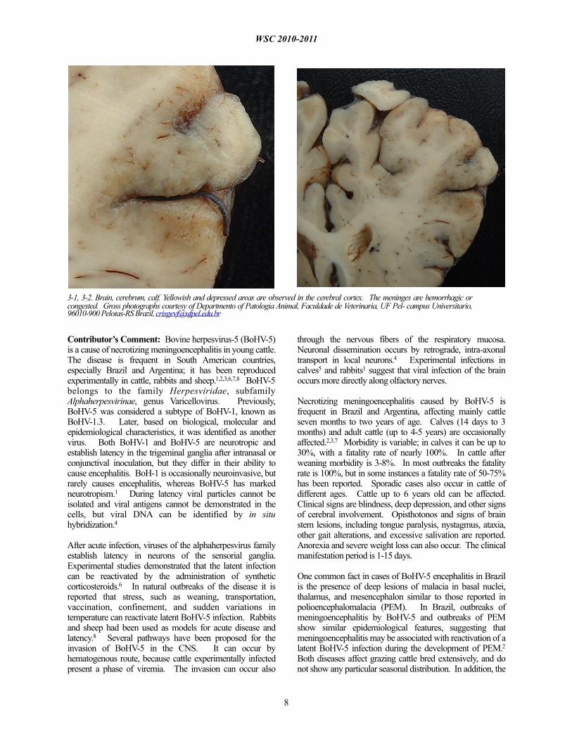
Gross Pathology: One calf showing emaciation, dehydration, severe depression and blindness was euthanized and necropsied. Yellowish and depressed areas were observed in the cerebral cortex. The meninges were hemorrhagic or congested.
Histopathologic Description: Brain, cerebral cortex: There were extensive areas of malacia in many regions of the cerebral cortex with infiltration of mononuclear cells mainly macrophages and gitter cells. Perivascular cuffing with many layers of mononuclear cells and multifocal hemorrhages was also observed. Blood vessels in the areas of malacia had hyperptrophied endothelial cells. Intranuclear inclusion bodies were observed within some astrocytes and neurons. Similar lesions of malacia were observed on the basal nuclei and thalamus. Discrete lesions were also observed in pons and cervical medulla (not included).
Contributor’s Morphologic Diagnosis: 1. Brain, cerebral cortex: Malacia, focally extensive, and encephalitis, subacute, accentuated with intranuclear inclusion bodies in astrocytes and neurons, etiology consistent with Bovine herpesvirus type-5, red Angus, calf.2. Brain, meninges: Meningitis, diffuse, moderate.
WSC 2010-2011
7
Contributor’s Comment: Bovine herpesvirus-5 (BoHV-5) is a cause of necrotizing meningoencephalitis in young cattle. The disease is frequent in South American countries, especially Brazil and Argentina; it has been reproduced experimentally in cattle, rabbits and sheep.1,2,3,6,7,8 BoHV-5 belongs to the family Herpesviridae, subfamily Alphaherpesvirinae, genus Varicellovirus. Previously, BoHV-5 was considered a subtype of BoHV-1, known as BoHV-1.3. Later, based on biological, molecular and epidemiological characteristics, it was identified as another virus. Both BoHV-1 and BoHV-5 are neurotropic and establish latency in the trigeminal ganglia after intranasal or conjunctival inoculation, but they differ in their ability to cause encephalitis. BoH-1 is occasionally neuroinvasive, but rarely causes encephalitis, whereas BoHV-5 has marked neurotropism.1 During latency viral particles cannot be isolated and viral antigens cannot be demonstrated in the cells, but viral DNA can be identified by in situ hybridization.4
After acute infection, viruses of the alphaherpesvirus family establish latency in neurons of the sensorial ganglia. Experimental studies demonstrated that the latent infection can be reactivated by the administration of synthetic corticosteroids.6 In natural outbreaks of the disease it is reported that stress, such as weaning, transportation, vaccination, confinement, and sudden variations in temperature can reactivate latent BoHV-5 infection. Rabbits and sheep had been used as models for acute disease and latency.8 Several pathways have been proposed for the invasion of BoHV-5 in the CNS. It can occur by hematogenous route, because cattle experimentally infected present a phase of viremia. The invasion can occur also
through the nervous fibers of the respiratory mucosa. Neuronal dissemination occurs by retrograde, intra-axonal transport in local neurons.4 Experimental infections in calves5 and rabbits1 suggest that viral infection of the brain occurs more directly along olfactory nerves.
Necrotizing meningoencephalitis caused by BoHV-5 is frequent in Brazil and Argentina, affecting mainly cattle seven months to two years of age. Calves (14 days to 3 months) and adult cattle (up to 4-5 years) are occasionally affected.2,3,7 Morbidity is variable; in calves it can be up to 30%, with a fatality rate of nearly 100%. In cattle after weaning morbidity is 3-8%. In most outbreaks the fatality rate is 100%, but in some instances a fatality rate of 50-75% has been reported. Sporadic cases also occur in cattle of different ages. Cattle up to 6 years old can be affected. Clinical signs are blindness, deep depression, and other signs of cerebral involvement. Opisthotonos and signs of brain stem lesions, including tongue paralysis, nystagmus, ataxia, other gait alterations, and excessive salivation are reported. Anorexia and severe weight loss can also occur. The clinical manifestation period is 1-15 days.
One common fact in cases of BoHV-5 encephalitis in Brazil is the presence of deep lesions of malacia in basal nuclei, thalamus, and mesencephalon similar to those reported in polioencephalomalacia (PEM). In Brazil, outbreaks of meningoencephalitis by BoHV-5 and outbreaks of PEM show similar epidemiological features, suggesting that meningoencephalitis may be associated with reactivation of a latent BoHV-5 infection during the development of PEM.2 Both diseases affect grazing cattle bred extensively, and do not show any particular seasonal distribution. In addition, the
WSC 2010-2011
8
3-1, 3-2. Brain, cerebrum, calf. Yellowish and depressed areas are observed in the cerebral cortex. The meninges are hemorrhagic or congested. Gross photographs courtesy of Departmento of Patologia Animal, Faculdade de Veterinaria, UF Pel- campus Universitario, 96010-900 Pelotas-RS Brazil, [email protected]

age of affected cattle is similar. The hypothesis that BoHV-5 can be reactivated in cattle which develop PEM, and this reactivation results in severe encephalitis and PEM, was demonstrated, at least experimentally, in cattle infected experimentally by BoHV-5 that received ammonium sulphate from days 114 to 180 after inoculation. One out of 3 cattle developed BoHV-5 meningoencephalitis with lesions of PEM. Other animals recovered after the development of clinical signs but continued to manifest chronic signs of PEM. Histologically, chronic lesions of PEM and mild meningoencephalitis were observed.2 In Argentina BoHV-5 infections also have the same characteristics as PEM. In a retrospective study from 1972 to 1999, of 89 cases previously diagnosed as PEM, 12 were caused by BHV-5 infections.7
In reports from other countries malacia of the cerebral cortex is absent or rarely reported.2 Malacia of the cerebral cortex had also been reproduced in cattle inoculated with BoHV-5, but in most experimental reproductions of the disease this lesion is not reported. Nevertheless, some of these authors mentioned neuronal degeneration and necrosis or the presence of focal rarefaction necrosis. Deep malacia had not been reported in cases of BoHV-5 infections in other countries or in the experimental reproduction of the disease.2
The differential diagnosis of BoHV-5 infection in Brazil includes the different causes of PEM (sulphur intoxication, sodium chloride intoxication/water deprivation, lead intoxication, and thiamin deficiency). Other infectious diseases, like rabies, malignant catarrhal fever (MCF), and listeriosis, have to be included in the differential diagnosis list. In rabies, clinical signs and distribution of the lesions
more frequently affect the spinal cord, brain stem and cerebellum, but some animals also show signs of cerebral involvement. Inclusion bodies are found in approximately 90% of the cases, mainly in the cerebellum, but also in brain stem, spinal cord and cerebrum. Some cases of MCF have signs of cerebral disease, but most also have keratitis and corneal opacity, as well as ulcerative lesions in the oral cavity and r e s p i r a t o r y s y s t e m .
Listeriosis is a sporadic disease with characteristic lesions and signs affecting the brain stem.
AFIP Diagnosis: Brain, cerebrum: Encephalitis, necrotizing and lymphohistiocytic, subacute, multifocally extensive, moderate to severe, with meningitis and eosinophilic intranuclear inclusion bodies.
Conference Comment: Conference participants commented on the variability of the lesions among the examined slides, including the absence of extensive necrosis in some sections. Additionally, the moderator noted the uneven distribution of viral inclusions, which tended to occur in small clusters. The moderator also emphasized that the most remarkable histologic lesion is the irreparable large area of necrosis in the cerebrum.
One reference text on veterinary neuropathology describes three features of inflammation within the central nervous system: 1) perivascular cuffing; 2) gliosis; and 3) neuronal satellitosis and neuronophagia.9 Perivascular cuffing and extension of inflammatory cells into the adjacent neuropil is a significant histologic finding that should propel the pathologist to search for the underlying cause of the lesion. The leukocyte populations surrounding the vessels may offer clues as to the type of inflammatory process and associated etiology, e.g. lymphoplasmacytic for viral infections, suppurative for bacterial infections, etc.9
Gliosis is characterized by the increased prominence of glial cells due to hyperplasia, hypertrophy or both. Glial cells can: undergo transformation to macrophages; surround degenerating neurons (satellitosis); and begin phagocytosis
WSC 2010-2011
9
3-3. Brain, cerebrum, calf. Multifocally, there is malacia and cavitation of the neuropil and replacement with mononuclear cells, gitter cells, and cellular debris (necrosis). Additionally, there is moderate mononuclear perivascular cuffing. (HE 200X)

of the neuron (neuronophagia).5 Astrocytes also undergo reactive changes in response to tissue damage characterized by cell swelling, cytoplasmic eosinophilia, large vesiculate nuclei, and, rarely, multiple nuclei. Reactive astrocytes are frequently referred to as gemistocytes or gemistocytic astrocytes. In addition to the reactive astrocytic changes, there is often hyperplasia of astrocytes. As the severity of the lesion worsens, the number of astrocytes typically increases resulting in more pronounced and dense astrocytosis; these cells frequently remain at the affected site after the lesion is resolved (glial scar).5
The contributor provides a thorough discussion of the epidemiology, clinical presentation, pathogenesis, lesions and differential diagnosis list for BoHV-5 infection.
Contributor: Departamento de Patologia Animal/Laboratório Regional de Diagnóstico, Faculdade de Veterinária, UFPel. 96010-900, Pelotas- RS, Brazilwww.ufpel.edu.br/fvet/oncovet; www.ufpel.edu.br/fvet/lrd. References: 1. Chowdhury SI, Onderci M, Bhattacharjee PS, Al-Mubarak A, Weiss ML, Zhoou Y. Bovine herpesvirus 5 (BHV-5) Us9 is essential for BHV-5 neuropathogenesis. J Virol. 2002;6:3839-3851.2. David N, Hubner SO, Riet-Correa F, Halfen D, Lemos RA. Reactivation of latent bovine herpesvirus type 5 in cattle with polioencephalomalacia induced by ammonium sulphate. Pesq Vet Bras. 2007;27:445-441.3. Elias F, Schild AL, Riet-Correa F. Meningoencefalite e encefalomalacia por herpes vírus bovino-5: distribuição das lesões no sistema nervoso central de bovinos naturalmente infectados. Pesq Vet Bras. 2004;24:123-131.4. Engels M, Ackermann M. Pathogenesis of ruminant herpesvirus infections. Vet Microbiol. 1996;53:3-15. 5. Maxie MG, Youssef S. Nervous system. In: Maxie MG, ed. Jubb, Kennedy and Palmer’s Pathology of Domestic Animals. 5th ed., vol. 1. Philadelphia, PA: Elsevier Ltd; 2007:292-295.6. Perez SE, Bretschneider MR, Leunda MR, Osorio FA, Flores EF, Odeón AC. Primary infection, latency, and reactivation of bovine herpesvirus type 5 in the bovine nervous system. Vet Pathol. 2002;39:437-444.7. Perez SE, Vagnozzi A, Sur JH, Odriozola E, Campero CM, Odeón AC. Análisis retrospectivo de casos con diagnóstico de necrosis cerebrocortical y su relación com herpesvirus bovino tipo 5. Rev Arg Microbiol. 2003;35:69-73.8. Silva AM, Weiblen R, Irigoyen LF, et al. Experimental infection of sheep with bovine herpesvirus type-5 (BHV-5): acute and latent infection. Vet Microbiol. 1999;66:89-99.9. Summers BA, Cummings JF, de Lahunta A. Principles of neuropathology. In: Veterinary Neuropathology. St. Louis, MO: Mosby; 1995:39-42.
CASE IV: 09-2735 (AFIP 3164124).
Signalment: 15-year-old, male, neutered, domestic shorthair, feline (Felis catus).
History: This 15-year-old castrated male domestic shorthair cat had a history of diabetes mellitus since two years of age; it was well controlled. The cat was diagnosed with mild to moderate hypertrophic cardiomyopathy. The cat presented due to acute onset of seizures and obtundation. There was severe white matter swelling/edema of unknown etiology found on an MRI. The blood pressure was 174 mmHg. There was partial cerebellar herniation. Treatment included prednisone and mannitol that resulted in clinical improvement for a period of approximately two weeks prior to reoccurrence of the clinical signs. Despite aggressive medical management with mannitol, hypertonic saline, steroids and antibiotics the cat's condition worsened and the owner elected euthanasia.
Gross Pathology: Gross necropsy findings reported by the submitting veterinarian were confined to partial herniation of the cerebellum. There was partial herniation of the cerebellum.
Histopathologic Description: There is marked diffuse edema of the white matter. Clear space and small eosinophilic lakes of edema fluid separate nerve fibers. Astrocytes in the white matter often have increased amounts of eosinophilic cytoplasm. In the meninges, arteries exhibit changes characterized by either myointimal hyperplasia and adventitial proliferation or fibrinoid necrosis. Around vessels with fibrinoid necrosis there are perivascular and mural infiltrates of neutrophils along with a few macrophages. The tunica media contains many pyknotic nuclei. A few of the vessels have concentric layers of proliferating fibrocytes in the tunica adventitia. Mild to moderate white matter edema is also present in the pons, medulla and cerebellar white matter. There are no vascular changes in those regions.
Contributor’s Morphologic Diagnosis: 1. Edema, diffuse, severe, cerebral white matter. 2. Fibrinoid necrosis and adventitial hyperplasia, multifocal, arteries, meninges.
Contributor’s Comment: The white matter edema and the vascular changes are consistent with hypertensive encephalopathy. This condition has been reported in cats with chronic renal disease and following renal transplantation.1 However, any condition that results in hypertension could theoretically cause the brain pathology.2 These conditions include hyperthyroidism, primary hyperaldosteronism, diabetes mellitus, pheochromocytoma and erythropoietin therapy. About 20% of cases of hypertension in cats have no identifiable cause (i.e. idiopathic hypertension). The cat had a history of diabetes mellitus that could have caused the hypertension. However, a full necropsy was not performed and the other conditions could not be entirely ruled-out.
WSC 2010-2011
10

The pathogenesis of hypertensive encephalopathy is thought to involve the development of vasogenic edema as a result of sudden increases in blood pressure that exceed the autoregulatory capacity of the vasculature in the brain resulting in endothelial injury and breakdown of the blood-brain barrier.2 The edema is primarily in the white matter where it results in separation of axons and myelin sheaths and pallor of the tissue. There is sometimes increased perivascular space around white matter vessels. Vascular changes, usually in the pia, are consistent with hypertension and include onion-skinning of the vessels (hyperplastic arteriosclerosis) and, occasionally, fibrinoid or hyaline change (hyaline arteriosclerosis).
AFIP Diagnosis: 1. Brain, cerebrum, white matter: Edema, diffuse, moderate, with reactive astrocytosis.2. Brain, meninges: Hyaline vascular necrosis, multifocal, moderate with perivascular sclerosis.
Conference Comment: Almost all conference participants observed the histologic lesions confined to the white matter of the cerebrum and interpreted the findings as consistent with edema; but only a few noted and diagnosed the vascular lesions. When evaluating nervous tissue, the differentiation of edema and preparation artifact is often difficult. Rough handling and autolysis often exacerbates the degree of artifact seen in tissue sections. In the tissue sections of this case, in addition to the physical separation of nerve fibers by clear space, many astrocytes are enlarged with increased eosinophilic cytoplasm, presumably as the result of edema. Recognizing the vascular lesions and their association with feline hypertension provides an identifiable explanation for diffuse white matter edema in this cat.
In general, there are four mechanisms of edema:41. Increased microvascular permeability (e.g.
vasculitis secondary to endotoxemia)
2. Increased intravascular hydrostatic pressure (e.g. pulmonary hypertension)
3. Decreased intravascular osmotic pressure (e.g. protein-losing nephropathy/enteropathy)
4. Decreased lymphatic drainage (e.g. intestinal lymphangectasia)
Because the structural qualities of the brain differ from other tissues, including the absence of a lymphatic system in the parenchyma, the mechanisms and pathogenesis of edema in the central nervous system are somewhat unique; and if not promptly treated, it is often fatal. Two mechanistic categories of edema of the brain are recognized and summarized in chart 1.4
McGavin and Zachary’s Pathologic Basis of Veterinary Disease expands the mechanistic list by including hydrostatic (interstitial) and hypo-osmotic types of brain edema. Briefly, hydrostatic edema is characterized by extracellular periventricular fluid accumulation as a result of increased hydrostatic pressure within the ventricles. Hypo-osmotic edema occurs with water and salt intoxication in which the differences in osmotic pressure between the brain and the plasma results in movement of fluid from the vasculature into the brain.5
In this case, partial herniation of the cerebellum through the foramen magnum (colloquially referred to as “cerebellar coning”) was noted at necropsy. In addition to the foramen magnum, there are two other potential locations of herniation during brain swelling. The occipital cortex may herniate caudally beneath the tentorium cerebelli or, in cases of unilateral swelling, the cingulate gyrus of the unaffected hemisphere may be forced laterally under the falx cerebri.4
Participants also discussed the differential diagnosis for white matter spongiosis, which can be broadly classified as either
WSC 2010-2011
11
4-1. Brain, cerebrum, white matter, cat. Diffusely within the white matter there is edema. Multifocally, astrocytes contain increased amounts of eosinophilic cytoplasm. Photograph courtesy of Arizona Veterinary Diagnostic Laboratory, 2831 N. Freeway, Tuscon, Arizona 85705, [email protected]
4-2. Brain, meninges, cat. In the meninges, the arteries exhibit arterial myointimal hyperplasia with adventitial proliferation or fibrinoid necrosis. Photograph courtesy of Photo courtesy of Arizona Veterinary Diagnostic Laboratory, 2831 N. Freeway, Tuscon, Arizona 85705, [email protected]

idiopathic or toxic/metabolic. A more detailed discussion of the various idiopathic spongiform myelinopathies is available within the selected references.3,4 Toxic and metabolic causes of status spongiosis include, but are not limited to, hepatic encephalopathy, renal encephalopathy, branched chain α-ketoacid decarboxylase deficiency (i.e. maple syrup urinary disease), hexachlorophene toxicosis, halogenated salicylanilide toxicosis, bromethalin toxicity and several plant species.3
Contributor: Arizona Veterinary Diagnostic Laboratory, Agricultural Experiment Station, The University of Arizona.
References: 1. Brown CA, Munday JS, Mathur S and Brown SA. Hypertensive encephalopathy in cats with reduced renal function. Vet Pathol. 2005;42:642-649.2. Kent M. The cat with neurological manifestations of systemic disease; key conditions impacting on the CNS. Journal of Feline Med Surg. 2009;11:395-407.3. Maxie MG, Youssef S. Nervous system. In: Maxie MG, ed. Jubb, Kennedy and Palmer’s Pathology of Domestic Animals. 5th ed., vol. 1. Philadelphia, PA: Elsevier Ltd; 2007:385-388. 4. Summers BA, Cummings JF, de Lahunta A. Principles of neuropathology. In: Veterinary Neuropathology. St. Louis, MO: Mosby; 1995:36-36.5. Zachary JF. Nervous system. In: McGavin MD, Zachary JF, eds. Pathologic Basis of Veterinary Disease. 4th ed. St.
Louis, MO: Elsevier; 2007:862-865.
WSC 2010-2011
12
Vasogenic Edema Cytotoxic Edema
Pathogenesis Damage to cerebral vasculature fluid and proteins leave vessel under hydrostatic pressure edema
Glial cell injury disturbed cellular osmoregulation acute cell swelling with maintenance of vascular integrity
Gross findings Flattened gyri and narrowed sulciHerniation (i.e. cerebellar coning)Edematous swelling of the white matter
Similar to findings in vasogenic edema with possible displacementMay be grossly normal
Distribution of the edema White matter; may affect grey matter if severe
Grey matterWhite matterBoth
Histologic Findings Generalized pallorSwelling and necrosis of astrocytesAstrocyte hypertrophyEosinophilic lakes of edema fluid
Astrocyte swellingW h i t e m a t t e r s p o n g i o s i s ( i f oligodendrocytes affected)Intracellular fluid accumulation
Chart 1.