
weaning ventilation intensive care gastric reflux - Respiratorytherapy
58
WEANING VENTILATION INTENSIVE CARE GASTRIC REFLUX SPECIAL SECTION: SLEEP INTERNATIONAL
Transcript of weaning ventilation intensive care gastric reflux - Respiratorytherapy

WEANINGVENTILATION
INTENSIVE CAREGASTRIC REFLUX
SPECIAL SECTION:SLEEP INTERNATIONAL

Introducing the Rapidlab® 1200 analyzer, the latest addition to the Rapidsystems™ blood gas product portfolio. This cartridge-basedanalyzer helps keep testing straightforward and throughput high in the critical care environment. With a full test menu, intuitiveinterface, automatic quality control and our advanced connectivity solutions, it’s one analyzer that can increase your testingcapabilities without increasing your workload. Brought to you by the inventors of automated blood gas analysis, the innovativeRapidlab 1200 analyzer simply works.
Invented in 1964. Innovating ever since. Call 800-255-3232.
© 2004 Bayer HealthCare LLC. All rights reserved.Rapidlab is a registered trademark and Rapidsystems is a trademark of Bayer HealthCare LLC.
Getting her resultsshouldn’t test your patience

Emergency Care · Perioperative Care · Critical Care · Perinatal Care · Home Care Because you care
What’s one way to dramatically impact Critical Care?
Achieve
100%weaning protocol compliance.
Ventilation weaning protocols have been shown to reduce lengthof stay*; unfortunately, they can also be labor intensive for clinicians.But with Dräger Medical’s SmartCare™ system, they’re automaticallysupported. Think of what that can mean to your patients… yourproductivity… and your bottom line. Yet it’s just one aspect of ourintegrated CareArea™ Solutions for Critical Care… and the entirecare process.
To discover how all our innovative solutions can impact your careprocess, visit www.draegermedical.com.
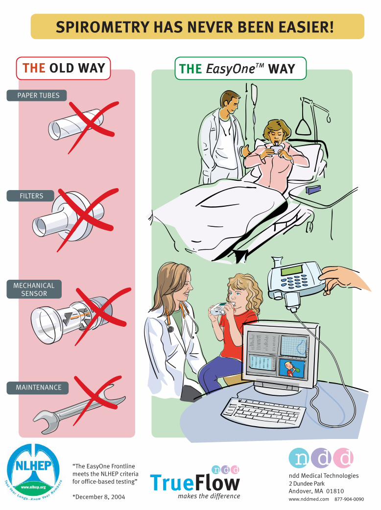
FILTERS
PAPER TUBES
MECHANICAL SENSOR
MAINTENANCE
SPIROMETRY HA S N E V ER B EE N E A SIER !
TrueFlo w makes the difference
T H E OLD WAY T H E E a s y O n e WAY
2 Dundee Park Andover, MA 01810
ndd Medical Technologies
www.nddmed.com 877-904-0090
“The EasyOne Frontlinemeets the NLHEP criteriafor office-based testing”
*December 8, 2004

Pressure. PressureEasy®
ET tube cuff pressure controllerA traditional manometer is
difficult to set up, hard to use,
inconvenient to transport, and
can be costly.
The cost-effective, convenient and easy-to-usePressureEasy® Cuff Pressure Controller.
There’s an easier way to check your patients’
pressure than a traditional manometer. The
PressureEasy® Cuff Pressure Controller is a
cost-effective, stand-alone device that’s easy to
set up and monitor, prevents blow-by and works
simply by looking at it. So, for a better way to
monitor cuff pressure, choose the PressureEasy®
Cuff Pressure Controller.
For more information, call 1-800-848-1757 or visit
www.smiths-medical.com.
Smiths Medical • 2231 Rutherford Road • Carlsbad, CA 92008
1.800.848.1757 • www.smiths-medical.com
Smiths design mark and PressureEasy are trademarks of the Smiths Medical family of companies. The symbol ® indicates the trademark is registered in the U.S. Patent and Trademark Office and certain other countries.
©2006 Smiths Medical family of companies. All rights reserved.

6 Respiratory Therapy Vol. 1 No. 6 � October-November 2006
Effective, convenient, comfortable...Available for in-home and institutional use
Smaller, lighter, withexpanded features
Vest sizes fit smalltoddlers to large adults
ELECTROMED, INC.creating superior care through innovation ®
502 Sixth Ave. N.W. New Prague, MN 56071
Phone: 1-800-462-1045Web: www.electromed-usa.com
New
“TrimlineTM”Model
Published six times each year byGoldstein and Associates, Inc.10940 Wilshire Blvd.,Suite 600Los Angeles, CA 90024 USATel: 310-443-4109Fax: 310-443-4110E-mail: [email protected]: www.respiratorytherapy.ca
PublisherSteve Goldstein
EditorLes Plesko
Senior EditorCarol Brass
Design and Production Managementhttp://accugraphics.net
Circulation, Coverage, Advertising Rates: Complete detailsregarding circulation, coverage, advertising rates, space sizes, and similarinformation are available to prospective advertisers. Closing date is 45days preceding date of issue.
Change of Address notices should be sent promptly to CirculationDepartment. Provide old mailing label as well as new address. Allow twomonths for change.
Editorial Contributions will be handled with reasonable care.However, publishers assume no responsibility for the safety of artwork,photographs or manuscripts. A hardcopy of all contributions must bemailed or faxed to our editorial offices. No e-mail submissions will beaccepted. Every precaution is taken to ensure accuracy, but the publisherscannot accept responsibility for the correctness or accuracy of informationsupplied herein or for any opinion expressed. Editorial closing date is thefirst day of the month preceding month of issue.
©2006 by Goldstein & Associates, Inc. All rights reserved. Reproduction inwhole or in part without written permission is strictly prohibited.
Russel A. Acevedo, MD, FAARC, FACP,FCCM, FCCP
Medical Director,Respiratory Care DepartmentCrouse Hospital, Syracuse, NY
Mohammed Al Ahmari, BSRT, MSc., RRTPrince Sultan Military College
of Health SciencesAl-Khobar, Saudi Arabia
Antonio Esquinas, MD, PhD, FCCP Intensive Care Unit
Hospital Morales MeseguerMurcia, Spain
Larry Conway, RRTNorth Mississippi Medical Center
Tupelo, MS
Edwin Coombs, MA, RRTDirector of Marketing/Product Management
Maquet, Inc. Critical Care DivisionBridgewater, NJ
Dr. Javier FernandezDirector of Clinical Affairs & Education
Respiratory Division Latin AmericaMiami, FL
Gerardo N. Ferrero, PTClinical Specialist, Latin America
Buenos Aires, Argentina
Surinder K. Jindal, MDPostgraduate Institute of Medical
Education & ResearchChandigarh, India
Rebecca A. MabryGeneral Sleep Manager
Viasys Healthcare, Yorba Linda, CA
Paul Mathews, PhD, RRT, FCCM,FCCP, FAARC
Associate Professor, Respiratory CareUniversity of Kansas Medical Center
Kansas City, KS
Hossein Razavi, MD, FCCPPulmonary, Critical Care &
Sleep MedicineSt. Helena, CA
Daniel D. Rowley, BS, RRT-NPS, RPFTSurgical/Trauma/Burn ICU
University of Virginia Medical CenterCharlottesville, VA
J. Kyle Schwab, MDMedical Director
Louisiana Sleep FoundationBaton Rouge, LA
Editorial Advisory Board

We’ve been pioneering healthcare improvements for
over 100 years, and have bui l t a reputat ion for del iver ing
innovation and qual i ty. Today, we develop products
that continual ly advance the science of respiratory
care. Guided by a world-class aerosol research lab,
we approach the future with the expert ise necessary to
adapt to a changing healthcare market.
at pari, our job is to help patients breathe easier.
PARI RESPIRATORYEQUIPMENT, INC.
1.800.FAST.NEB (327.8632)www.PARI.com
R E U S A B L E N E B U L I Z E R
call 1-800-FAST-NEB formore information on how
to access pari products
ML-44-012 Rev 6/06

The VAR will function in anyposition as long as the ad-justments are made in a se-cured position (strapped ortaped to the patient). Tiltingmovements of less than 45°have no significant effect onthe PIP setting.The working mechanism ofthe VAR consists of a movingpiston or diaphragm whichhas mass (weight). The massadds an additional springforce or a subtraction ofspring force when the VAR ispositioned with the modula-tor vertically up or verticallydown.
The VAR ispositional sensitive
If the VAR is moved from ahorizontal to a vertical posi-tion, the addition or subtrac-tion of spring force will affectthe PIP setting by 1 to 3 cm-H2O.
The positional effect on PIPis an education and trainingissue. The VAR will functionin any position as long as thefinal adjustments are madein its secured position.
The VAR is ideal withchanging compliance
[1]Kallet, Richard H MS RRT: Implementation of a LowTidal Volume Ventilation Protocol for Patients withAcute Lung Injury or Acute Respiratory DistressSyndrome. Respir Care 2001:46(10):1024-1037
VARTM - VORTRANAutomatic Resuscitator
Preparefor aventilatoremergency
Modulatorvertically
UP
ModulatorverticallyDOWN
VAR-PlusTM
VARTM
The VAR is not a time-cycled ventila-tor and will respond automatically tochanges in compliance, as seen inARDS. The VAR self-adjusts byincreasing respiratory rate anddecreasing tidal volume (TV) anddelivers a stable minute ventilation(MV) when compliance decreases froma healthy 0.07 to a stiff 0.02 L/cm H2O(Fig 1 & Fig 2).
This is recommended by the ARDSNetwork’s publication, funded by theNational Heart and Lung Institute,which showed a 22% lower mortalitywith low TV ventilation strategy inpatients with ALI or ARDS.(1)
The VAR automatically delivers a lowerTV and a higher rate and is ideal forARDS patients with decreasing com-pliance.
VAR VAR-Plus
PIPincreased 2.4 1.0UP positionPIPdecreased -2.9 -0.9DOWN
VAR-Plus for pediatricto adult patients(10 kg and above)
Automatically lowers TVand increases rate inARDS with a decreasingcompliance
Hands-free operation
Gas-powered, automaticpressure cycled
Single patient use
For breathing andapneic patients
Safe for MRI / CT scan
Meets ASTMrequirements
FREE CEU OnlineContinuing Education1 contact hourfor RRT/RCP, RN and MDwww.mededcenter.com
Tid
al Volu
me (
mL)
Compliance (L/cm-H2O)
Fig 2 - TV Automatically Changeswith Changing Compliance
0
100
200
300
400
500
600
700
800
900
1000
0.010.020.030.040.050.060.07
PEEP 5
PEEP10
PEEP 15
Fig 1 - Rate Automatically Changes|with Changing Compliance(VAR-Plus with in-line PEEP Valve)
Compliance (L/cm-H2O)
Resp
irato
ry R
ate
(BPM
)
0
5
10
15
20
25
30
35
40
45
50
0.010.020.030.040.050.060.07
PEEP 5
PEEP 10
PEEP 15
7/06-C
1804 Tribute Road, Suite F, Sacramento, CA 95815Tel: (800) 434-4034 E-mail: [email protected]: (916) 648-9751 Website: www.vortran.com
MEDICAL TECHNOLOGY 1, INC.VORTRAN

Respiratory Therapy Vol. 1 No. 6 � October-November 2006 9
Vol. 1 No. 6October-November 2006
ARTICLES19 Tailoring Respiratory Therapies
25 NAVA – A New Generation
29 Protocols for Weaning
33 Longitudinal Measurement of FEV1
36 Management of Tracheal Stenosis
39 Trends in Outcomes With HFOV
45 Emergency Ventilatory Support
48 Product Case Study: VAR
50 Oxygen Conservation During Transport
57 RSBI and Weaning Success
DEPARTMENTS9 Editorial: Facing a Crisis?
10 News
13 Products
15 Executive Profile
17 Clinical Trials Update
SPECIAL SECTION59 Sleep International
Table of Contents
Editorial
FACING A CRISIS?
In the latest issue of Hamilton Medical’s newsletter, respiratory therapist PaulGarbarini asks, are RT departments facing a crisis? Says Garbarini, “RTdepartments are facing the same staffing shortages seen in other healthcareprofessions. This need is further exacerbated by the legitimate push to implementICU process improvement strategies which include ventilator bundles, dailyscreening for spontaneous breathing trials, reduced errors etcetera.”
To back up his assertion, Garbarini cites a recent issue of Critical Care Medicinethat reports Bureau of Labor statistics which estimate a 43 percent increase indemand for respiratory therapists by 2008. Respiratory therapy is now said to beamong the top 10 growth fields through the end of this decade. What does thisportend for effective levels of care?
Let’s look at the evidence. The Critical Care study by Paul Mathews, et al, sought toexplore respiratory therapy manpower needs in critical care practice. The authorsdelineated the historical development of respiratory care, as well as itscredentialing system, and collected data from AARC, NBRC and CoARC. Theauthors conducted a thorough survey about the use of mandatory overtime in RTdepartments, followed up by a questionnaire from a wide range of institutionswhere RTs worked.
Critical Care’s study found a stable attrition rate of about 30 percent, and varyingenrollments, and found that half the hospitals had a policy addressing mandatoryovertime, while a lesser number had instituted disciplinary procedures for RTs whorefused overtime. Seven of the 30 hospitals queried indicated that they usedmandatory overtime frequently to keep up staffing levels. The actual ratio of bedsand RCPs wasn’t too far off recommended levels, but with an exponential increaseof need on the way, it’s only a matter of time before the need for RTs is bound to hitthe critical point. According to the study abstract, “by two focused surveys, wewere able to show that while mandatory overtime is a common practice inrespiratory care departments, it was not overwhelming utilized. We also learnedthat in most hospitals, regardless of bed size, there is a perceived need for 1.3 RCPsmore than the actual staff and that it appears that the critical staffing level betweenactual to preferred RCP to beds is between 9 and 11 beds.” Another study in thesame issue of Critical Care Medicine noted that “productivity, practice patterns, theaging of the workforce and patients, and other major determinants are onlyminimally affected by most government policy. Despite several attempts throughoutthe 1980s and 1990s, demand for health care has been particularly difficult tocontrol for policymakers… There are many barriers to successful workforcepolicy… The task before those concerned about workforce issues is to educatepolicymakers about how changes in the physician workforce will affect cost,access, and quality, and to impress upon them that serious efforts to improvequality of care and reduce costs will not be effective unless qualified physicians arethere to provide that care.”
In his article, Garbarini notes, by way of example, that the California Code ofRegulations states that the ratio of therapists to ventilators should be no greaterthan one to four. The average respiratory therapist is assigned to about 10 ventilatorpatients. Obviously, eventually, something’s going to give. As recent historicalevents show, there’s a marked tendency to deal with problems after the horses arealready out of the barn. So why not start dealing with this now?
Les Plesko, Editor
For more information see the study, Respiratory Care Manpower Issues, Model and Workforce, Critical CareMedicine, Interface of Public Policy and Critical Care Medicine. 34(3) Suppl:S32-S45, March 2006. Mathews,Paul PhD, RRT, FCCM; Drumheller, Lois BS, RRT; Carlow, John J. EdD; with the assistance of the AmericanAssociation for Respiratory Care; The National Board for Respiratory Care; The Council on Accreditation ofRespiratory Care.

10 Respiratory Therapy Vol. 1 No. 6 � October-November 2006
LETTERLast night I finished reading your journal (Vol 1, No 5) and
thought that I would send a note of congratulations to you – it isexcellent! I read the editorial and the entire News section whichis better than most trade magazines to keep up on what’s goingon. I was [also] surprised by the topic coverage and number ofarticles. Nice job. P.S. Our ad looked good, too.
Norman H Tiffin BSc, RRT, MSAVice President, MarketingPari Respiratory Equipment
PLACEBO EFFECT A study carried out by researchers from Manchester
University, UK, found that codeine is statistically no better thana placebo for treating COPD patients for cough. The Journal ofAllergy and Clinical Immunology reported that researcherstracked 24 COPD patients’ coughing by placing a microphone ontheir lapels. Half the patients were given codeine while the otherhalf received a placebo. Their coughing was tracked for a periodof ten hours after taking their medication. At the beginning ofthe period both groups of patients had an average coughing
total of 8.27 seconds per hour. After taking their medications,the placebo group’s coughing dropped to 7.22 seconds per hourand the codeine groups coughing dropped to 6.41 seconds perhour. There was no difference between the codeine and theplacebo from a statistical standpoint, even though the codeinedose their volunteers received was far higher than any OTCdose found in standard cough remedies. Very little had beenknown about its impact on patients with chronic lung diseases.
EXACERBATEDNew data from a multinational, interview-based patient study,
published in the medical journal Chest, shed light on COPDpatients’ comprehension, recognition, and experience ofexacerbations and the burden associated with these events.Exacerbations are known to impair health-related quality of lifein patients with COPD and increase the risk of mortality. Thestudy showed that physicians often underestimate thepsychological impairment experienced by patients during anexacerbation. Exacerbations cause substantial anxiety, patientsreported; 12% stated they worry about dying, 10% that theyworry about suffocating, 10% that they will experience apermanent worsening of their condition and 8% that they will behospitalized. A majority of patients reported that besidesinfluencing their activities in daily life, a significant worseningof their mood causing a variety of negative feelings, such asdepression, irritability/bad temper, anxiety, isolation, anger, andguilt. Moreover 42% stated that exacerbations affected theirpersonal relationships. According to the study, the observationthat physicians fail to appreciate the considerable changes tothe patient’s emotional well-being demonstrates acommunications gap between patients and their doctors andrepresents a dilemma in COPD management that may lead to
News� October-November 2006
Respiratory Therapy Vol. 1 No. 6 � October-November 2006 11
undertreatment. The study was conducted with 125 patientsdiagnosed with COPD from France, Germany, Spain, Swedenand the UK. The patients were age 50 or younger and hadexperienced a minimum of two exacerbations during theprevious year. Sixty five percent were male COPD patients who,during the previous 12 months, had experienced a mean of 4.6exacerbations with an average duration of 2 weeks and a meanrecovery time of 10 days. Notably, 20% felt that they had notreturned to their previous state of health after an exacerbation,demonstrating the importance of reducing these events. As asidenote, the study reported that only 1.6% of patientsunderstood what was meant by the widely used clinical term“exacerbations.” The term used most often by patients todescribe an exacerbation was “crisis,” underscoring theseriousness with which patients view the worsening of theircondition. Two-thirds of patients stated they were aware of thesymptoms associated with their condition getting worse. Mostpatients (85%) experienced the same symptoms from oneexacerbation to another, breathlessness being predominant. Atthe onset of an exacerbation, 33% of patients reported that theyreact by self-administering their medication while only aminority contact their physician. Reported in Medical NewsToday.
FLUTE OR FLUKE?Researchers at Karolinska University Hospital, Solna,
Stockholm, Sweden investigated whether bronchialresponsiveness to leukotriene D4 is reduced by fluticasonepropionate. The researchers conducted their research by havingthe study’s 13 volunteers participate in an inhalation challengewith methacholine and leukotriene D4 on consecutive daysbefore and after two weeks of treatment with inhaledfluticasone twice daily. Study results showed that although thefluticasone propionate therapy vs placebo for two weeks causeda significant reduction in methacholine sensitivity and inexhaled nitric oxide, it had no effect in blocking thebronchoconstriction that occurs when leukotrienes are inhaled,nor does it influence the production of leukotrienes, asmeasured in the urine. The clinical implication of the study,according to the authors, supports synergistic therapy with aninhaled corticosteroid as well as an oral antileukotriene forcertain, persistent asthmatic patients.
MISDIAGNOSISA new study shows that more than half of patients with COPD
– chronic obstructive pulmonary disease – may be misdiagnosedas having asthma. COPD is a progressive condition that leads toa worsening of respiratory symptoms, a decline in lung functionand increased disability; however, it tends to be under-diagnosed and under-treated. The study results, published in theJournal of Asthma, are from the most recent prospective,patient-reported, objectively documented COPD study toexamine COPD misdiagnosis.
COPD, which includes chronic bronchitis and emphysema, ischaracterized by a loss of lung function over time. Primarily adisease of current and former smokers, COPD affects nearly 12million Americans. Unlike asthma, COPD is associated with acascade of decline that leads to a diminished quality of life overtime. The study, conducted in Denver and Aberdeen, Scotland,and sponsored by Boehringer Ingelheim Pharmaceuticals, Incand Pfizer Inc, analyzed data from 597 patients age 40 and olderwith a history of lung disease or recent treatment withrespiratory medications. Patients were then screened usingspirometry, a lung function test, to confirm their diagnosis of
COPD. In this study, a COPD diagnosis was defined inagreement with American Thoracic Society and EuropeanRespiratory Society guidelines as the presence of obstruction,the inability to get air out of the lungs, based on spirometryresults. Of the 235 patients diagnosed with COPD by spirometry,51.5% reported a prior diagnosis of asthma only. Only 37.9% ofparticipants diagnosed with COPD based on the study testsreported a previous diagnosis of the disease, while 10.6%reported no prior diagnosis of COPD or asthma. Researcherssaid the findings were surprising given the availability ofcredible diagnosis and treatment guidelines specifically forCOPD, and noted that patients could benefit from lifestylemodification, pulmonary rehabilitation and properpharmacotherapy.
CHRONICA Europe-wide trial involving premature babies is
investigating whether the risk of chronic lung disease can behalved if they are given nitric oxide gas to breathe shortly afterbirth. Medics from the University of Leicester, King’s CollegeLondon and Medway hospitals are involved in the trial. Resultsof a similar US based study have recently been published in theNew England Journal of Medicine. Doctors involved in the studysaid the research is potentially very important but that therewas a long way to go before an understanding could be reachedabout the best way to use nitric oxide and the babies that couldbe most helped. The intervention is very costly and this wasonly the second study to show a positive effect in prematurebabies, while half a dozen other studies have had either noeffect or a negative effect. It also appeared not to have helpedthe sickest, most premature babies. Another similar study in theUS won’t be ready for two more years.
SIGHA new study in the online edition of the American Journal of
Physiology-Lung Cellular and Molecular Physiology shows thatlow tidal volume combined with periodic deep inflationprovides the best balance between keeping the lung open andpreventing VILI in mice. And, using mice, these researchershave shown for the first time that although deep inflation isnecessary, it can be overdone. There is still a lot of controversyand uncertainty about how best to ventilate the lung, said thestudy’s senior author. One controversy is whether deepinflations, the sighs that each of us takes periodically, shouldever be given, and if so, how frequently. The researchers dividedmice into three experimental groups. All three groups receivedPEEP and low tidal volume air. Each group was ventilated fortwo hours. The experimental groups differed according to howmany deep inflations they received. They were as follows: HVreceived one deep inflation each breath, LV received two deepinflations each hour, and LVDI received two deep inflations eachminute. In addition, there were two control groups, a surgicalsham, which received no ventilation, and a group that receiveddeep inflation every breath and no PEEP. The study found thatthe lungs of the mice given two big breaths every minute (LVDI)remained more open and functioned better than the LV and HV.The lungs of the mice that received only two deep breaths perhour (LV) became stiff and portions of the lungs collapsed.However, lung function returned briefly to normal when themice received their infrequent deep inflations. This suggeststhat the lungs self-repair after the deep inflation, at least overthe course of the first two hours. The lungs of the mice thatreceived deep inflation every breath (HV) sufferedoverdistention injury to their lungs. This group was akin to a

high tidal volume group, once again demonstrating that lowtidal volume is safer. The control group that received high tidalvolume but no PEEP showed the highest evidence of injury,even higher than the high tidal volume group. This indicates thatPEEP helps reduce the negative effects of frequent deepinflation. The researchers said they demonstrated that it’spossible to give deep breaths too frequently and too seldom.The middle ground, two deep inflations per minute, providedthe most benefit to the mice without injuring the lungs.
LUNG RECRUITMENT Writing in Hamilton Medical’s latest newsletter, authors Justin
Tse, RRT-NPS and Jeff Borrink, BS, RRT, discuss lungrecruitment in patients with ARDS: Acute respiratory distresssyndrome (ARDS) was first described in 1967 by Ashbaugh, whodescribed a syndrome of severe respiratory failure associatedwith pulmonary infiltrates, similar to infant hyaline membranedisease. In 1994, the American-European Consensus Committeedefined ARDS as the acute onset of bilateral infiltrates on chestx-ray, a partial pressure of arterial oxygen to fraction of inspiredoxygen ratio of less than 200 mm Hg and a pulmonary arteryocclusion pressure of less than 18, or the absence of clinicalevidence of left arterial hypertension.
The development of ARDS starts with damage to the alveolarepithelium and vascular endothelium resulting in increasedpermeability to plasma and inflammatory cells into theinterstitium and alveolar space. Damage to the surfactantproducing cells and the presence of protein-rich fluid in thealveolar space disrupts the production of pulmonary surfactantleading to microatelectasis and impaired gas exchange.Mechanical ventilation is usually initiated to restore adequateoxygen to the tissues. However, we have learned thatmechanical ventilation itself can cause or enhance the effects ofARDS. Because of this, management has changed frommaintaining “normal gas exchange” to decreasing the possibilityof ventilator induced lung injury. Lung protection strategiesgenerally utilize two main components: low tidal volumes toprevent stress and strain to lung parenchyma, and higher levelsof PEEP to prevent derecruitment injury. Some studies utilizinglow tidal volume strategies have been shown to increasesurvivability. Other studies have shown improved patientoutcomes with higher levels of PEEP. A recent study byGattinoni, et al looked at the relationship between thepercentage of potentially recruitable lung, as indicated bycomputed tomography (CT), and the clinical and physiologicaleffects of PEEP.
The study looked at 68 patients with acute lung injury (ALI)or ARDS. Nineteen patients had ALI without ARDS and 49 hadARDS. The overall mortality rate was 28 percent. Each patientunderwent whole-lung CT during breath-holding sessions atairway pressures of 5, 15, and 45 cm of water. The percentage ofrecruitable lung was defined as the proportion of lung tissue inwhich aeration was restored at airway pressures between 5 and45 cm H2O. The percentage of potentially recruitable lung variedwithin the study population. The average was 13 +/- 11 percentof the lung weight, which corresponded to an absolute weight of217+/- 232 g of recruitable lung tissue and correlated with thepercentage of lung tissue which was recruited after theapplication of PEEP. On average, 24 percent of the lung couldnot be recruited. Gattinoni, et al reported that patients with ahigher percentage of potentially recruitable lung had greatertotal lung weights, poorer oxygenation, higher levels of deadspace, and higher rates of death than patients with a lowerpercentage of potentially recruitable lung. They concluded that
in patients with ARDS, the percentage of potentially recruitablelung is extremely variable and is strongly associated with theresponse to PEEP. The study by Gattinoni et al generatedseveral responses by physicians who wrote letters to the editorof the NEJM. One such response came from Drs Amato, Borges,and Carvalho, from the University of Sao Paulo. They believedthat the study by Gattinoni et al suggested that the potential forlung recruitment in patients with acute lung injury is generallylow and extremely variable among patients, and that this wasdue to a suboptimal recruitment maneuver that was used in theGattinoni study. Dr. Amato et al referred to other studies thatdemonstrate a much larger potential for recruitment and greaterhomogeneity of response. They argued that Gattinoni et al usedan inspiratory plateau pressure of 45cmH2O, which is lower thanthe critical opening pressures reported in other recent studies inhumans. They also argued that Gattinoni et al. allowedpressures to repeatedly fall to 5cmH2O, which is a pressurebelow the closing pressures of most lung units. They go on tosay that this procedure was likely to have cyclically forced theenergy provided by the next pulse of inspiratory pressure to bewasted in the opening of the recollapsed units, instead ofpromoting the recruitment of units. They suggest that theapplication of the same inspiratory pressure, but a higherexpiratory pressure (20 to 25cmH2O) could have greatlyenhanced the efficacy of the maneuver, thus affecting the mainconclusion of the study by Gattinoni et al.
Another response by Dr Kacmarek et al expressed surprise atthe level of lung recruitment by Gattinoni. Based on previousstudies of recruitment maneuvers, they would have expected ahigher level of recruitable lung than what was demonstrated inthe Gattinoni study. They suggest that the lack of response mayhave been due to the length of time for which patients receivedmechanical ventilation (5+/-6 days) before being studied, andrefer to studies that recommend lung recruitment be performedas early as is feasible in the course of ARDS.
LET IT STINKNew research shows that a chemical compound found in
many air fresheners, toilet bowl cleaners, mothballs and otherdeodorizing products, may be harmful to the lungs. Humanpopulation studies at the National Institute of EnvironmentalHealth Sciences (NIEHS), a part of the National Institutes ofHealth, found that exposure to a volatile organic compound(VOC), 1,4 dichlorobenzene, may cause modest reductions inlung function. “Even a small reduction in lung function mayindicate some harm to the lungs,” said NIEHS researcherStephanie London. The researchers examined the relationshipbetween blood concentrations of 11 common volatile organiccompounds and lung function measures in a representativesample of 953 adults. VOCs are a diverse set of compoundsemitted as gases from thousands of commonly used products,including tobacco smoke, pesticides, paints, and cleaningproducts. VOCs are also released through automotive exhaust.The researchers found that of the common VOCs analyzed,which included benzene, styrene, toluene, and acetone, only thecompound 1,4 DCB was associated with reduced pulmonaryfunction and this effect was seen even after careful adjustmentfor smoking, The researchers found that 96 percent of thepopulation samples had detectable 1,4 DCB blood concentrationlevels. African Americans had the highest exposure levels andnon-Hispanic whites the lowest. The particular VOC tested, 1,4DCB, is a white solid compound with a distinctive aroma, usedprimarily as a space deodorant in products such as roomdeodorizers, urinal and toilet bowl blocks, and as an insecticide
12 Respiratory Therapy Vol. 1 No. 6 � October-November 2006

Respiratory Therapy Vol. 1 No. 6 � October-November 2006 13
fumigant for moth control. The researchers used data from thethird National Health and Nutrition Examination Survey(NHANES) and a special component of the study specificallydesigned to assess the level of common pesticides and VOCs inthe US population. Data from 953 adults 20-59 years old who hadboth VOC blood measures and pulmonary function measures areincluded in the study published in the August issue ofEnvironmental Health Perspectives. Four pulmonary functionmeasures were used in the analyses. The researchers foundmodest reductions in pulmonary function with increasing bloodconcentrations of 1,4 DCB. There was approximately a 4%decrease in the test which measures FEV1 between the highestand lowest levels of exposure. The research suggested that 1,4-DCB may exacerbate respiratory diseases.
PRODUCTS
SPOTLIGHT ON SPIROMETRY: KOKO LEGENDFerraris Respiratory introduces the all-inclusive KoKo Legend
portable spirometer. KoKo Legend redefines accuracy throughtechnology and simplicity with Legend’s intuitive color touchscreen walking both patient and physician through standardtesting procedures promoting superior patient test results. KoKoLegend utilizes a unique flexible orifice pneumotach which isextraordinarily precise at the low flow rates common in bothpediatric and COPD, exceeding the 2005 ATS standards forspirometry. Choose built-in-printing or external office printingfor 8 1/2 x 11 reports. Easily transfer data into our KoKo PFTSpirometry software via a standard USB cable. For moreinformation call 800-574-7374, ferrarisrespiratory.com.
SPOTLIGHT ON SPIROMETRY: COSMED Cosmed is a manufacturer of cardio-pulmonary diagnostic
equipment. The product line ranges from simple spirometers tocomplete pulmonary function and metabolic systems. Thespirometer product line consist of two units, the PC-basedMicroQuark USB and the Pony FX which is a portable bedsidespirometer. Cosmed spirometers meet all ATS/ERSrecommendations for acceptability. The spirometers use theturbine technology that ensures maximum precision at low flowrates up to 20 l/s and at a very low resistance of 0.7 cmH2O/l/s.Options include: screening spirometry ( FVC, SVC, MVV), userdefined predicteds, methacholine challenge, automatic BTPScorrection and a 1 to 3 year warranty on the systems. Contactcosmed.it.
GOING SOFTNonin announced a new Soft Sensor line to complement its
comprehensive family of PureLight pulse oximetry sensors. Thenew line of soft sensors features a durable design that deliversconsistent performance. The soft sensors perform like twosensors in one for spot-checks or continuous monitoring, withpatient comfort in mind. Available in three sizes, the soft sensorsare flexible for use in many medical settings including EMS,hospital and sleep. Soft sensors are easy to clean, latex-free andcome with a one-year warranty. For more information contactnonin.com, (800) 356-8874.
KEEP IT IN PROPORTIONPuritan Bennett, maker of the world’s best-selling critical care
ventilator, the 840 ventilator, is pleased to introduce an
innovative, advanced software option for mechanical ventilationto the U.S. market. The new PAV+ Proportional AssistVentilation Plus software option delivers positive airwaypressure that is directly related to a patient’s inspiratory effortduring spontaneous breathing. Ventilator support increases aspatient demand increases, thereby optimizing the patient’scontribution to the total work of breathing. The original conceptof providing breathing assistance in proportion to patientinspiratory effort was invented by Dr Magdy Younes at theUniversity of Manitoba. The basic technique amplifies thepatient’s inspiratory effort by increasing airway pressure duringinspiration. The new PAV+ software feature, availableexclusively from Puritan Bennett for its flagship 840 ventilator,augments the benefits of the basic technique by employing asophisticated software algorithm – the “plus” – thatautomatically adjusts the positive airway pressure based onairway pressure measurements taken throughout the inspiratorycycle. This maintains a targeted degree of support pressurethroughout each of the patient’s spontaneous breaths. Anappropriate support pressure is maintained based on theworkload a patient needs to overcome to breathe. A cliniciansimply sets the target level of support desired (shown on themonitor as % Support). At a setting of 60%, for example, theventilator performs 60% of the work of inspiration and thepatient performs 40%. The PAV+ software option also graphicallydisplays real-time assessments of patient work, airwayresistance and lung compliance. This information can assist theclinician in assessing the best treatment approach for theindividual patient at any given time. “The ability to turn asophisticated clinical concept into an innovative, easy-to-useproduct feature is one of Puritan Bennett’s core strengths,” saidBrent Boucher, Vice President, General Manager. “Clinicians relyon our design expertise to incorporate exceptional features intoour ventilator products that can facilitate patient management.Our new PAV+ product is a very powerful tool to improvepatient care.” The PAV+ software option is intended for use onany patient weighing more than 25 kg who is spontaneouslybreathing during mechanical ventilation. Contacttycohealthcare.com, puritanbennett.com.
SMALL BUT SMARTRespironics announces the release of its new MiniElite
Compressor Nebulizer System for active patients who want totake their aerosol treatments wherever or whenever. The smalland lightweight MiniElite compressor is easy-to-use andprovides efficient nebulizer treatments. This stylish, elegantcompressor provides portability and can be powered by threeversatile power source options, AC, car adapter and battery. Theoptional rechargeable lithium ion battery operates the MiniElite
compressor for up to 90 minutes between charges. This batterytechnology offers easy and fast re-charging in a lightweightdesign. The MiniElite compressor weighs less than 1 pound andless than 1.4 pounds with the optional battery attached. “Thepatient-friendly design of the MiniElite fits the lifestyle needs ofpatients,” said Matt Conlon, Director of Sales and Marketing forRespironics Respiratory Drug Delivery business, “while meetinghomecare providers need for cost effectiveness and productreliability.” Respironics Respiratory Drug Delivery is advancingthe science of respiratory care with innovative drug deliverytechnologies for the treatment of respiratory and non-respiratory diseases. Through ongoing research, RespironicsRDD provides breakthrough solutions for pharmaceuticalcompanies, healthcare providers and patients. Respironics is aleading developer, manufacturer and distributor of innovative
products and programs that serve the global sleep andrespiratory markets. Focusing on emerging market needs, theCompany is committed to providing valued solutions to helpimprove outcomes for patients, clinicians and health careproviders. Respironics markets its products in 131 countries andemploys over 4,700 associates worldwide. Further informationcan be found at respironics.com.
DEDICATEDNova Biomedical announced the addition of a new model to
its series of Stat Profile pHOx Analyzers providing a dedicatedtest menu for respiratory care. The new Stat Profile pHOxincorporates a seven-test menu, including pH, PO2, SO2%,hemoglobin, hematocrit, and lactate, to provide acomprehensive diagnostic picture of oxygen transport. Thecompact analyzer uses a liquid-only calibration system thateliminates bulky compressed tanks, gas regulators, gas tubinglines and humidifiers. To promote blood conservation, the pHOxuses just 125 uL of whole blood to measure the full seven-testmenu, and only 60 uL of whole blood for a three-test micro-sample. Contact novabio.com.
BREATHE DEEPLYSince its inception, Inogen has been dedicated to the design,
development and manufacture of clinically efficacious oxygentechnologies. The lack of substantial research in the area oflong-term oxygen therapy and oxygen technologies has longmade the provision of home LTOT a blend of art and science.Inogen recognizes that its technology is part of a paradigm shiftand is leading the industry in changing the provision of LTOT ina new direction. Inogen discarded existing paradigms to designand build an oxygen concentrator that redefines how oxygentherapy is delivered. Described by industry experts as “atechnological breakthrough,” the Inogen One is a completedeparture from current mainstream technologies, both thestandard large, bulky, stationary concentrator systems and theinefficient and impractical portable devices. Working withexperts in PSA and conserver technology, Inogen’s engineersdeveloped, tested and delivered a technology that eliminates theneed to choose between a stationary or portable oxygensystem.The Inogen One enhances the clinical efficacy of pulse-dosed oxygen delivery in a myriad of clinical applications,including sleep, and for patients with COPD. The Inogen One’stechnology has been thoroughly bench-tested and clinicallyvalidated through original science and work. Contactinogen.net.
LEGACYB&B Medical Technologies carries on the legacy of Stephen
Briggs III. A new team has assumed ownership and managementof B&B Medical Technologies, a leading designer of specialtyairway management devices and nebulizers for infants,pediatrics and adults. Continuing B&B’s legacy as a respiratorytherapist-owned company are David Thompson and Beth Keifer,who together bring more than 50 years in clinical, educationaland technical expertise in the respiratory care field. Thompsonand Keifer are supported on the core team by Robert Sprowls, aCarlsbad, CA businessman. B&B products are designed for easy,one person application, helping to minimize risk of accidentaldisconnects and unplanned extubations. B&B’s StabilTube,LockTite, ET Tape for Adults and Infants and Bite Block provideclinicians simple solutions for comfortably securing theendotracheal tube, prevention of ventilator disconnects and aconvenient answer to prevent ET tube biting. B&B’s TrachGuard
and TrachStay comfortably secure the ventilator circuit to thetracheostomy tube while preventing accidental disconnects.B&B’s patented Hope nebulizer technology provides efficientdelivery of continuous medication combined with the ability toblend gases such as Heliox without affecting medicationdelivery. The Hope Nebulizer is the first nebulizer specificallycleared by FDA for Heliox administration. Stephen Briggs IIIalong with Dr. Ernie Bodai created B&B Medical Technologiesin 1985 to ensure that specialty airway related products had apathway to the clinical community. Briggs’ legacy, earnedthrough his lifetime in the respiratory therapy community willbe carried on in the new ownership and management. Contactbandb-medical.com.
14 Respiratory Therapy Vol. 1 No. 6 � October-November 2006

EXECUTIVE PROFILES
Fluke BiomedicalJerry Zion
Jerry Zion is Product Manager for Fluke Biomedical.
Who is responsible within your company, by title or nameor job description, for training and education of your staffand your customers?
Product managers are responsible for training the technicalsupport and sales staff (including all worldwide channelpartners) as part of the release of any new product or productenhancement. After that, the technical support associatesprovide help-desk and refresher training for end-users, and thesales staff (including channel partners) provide initial trainingfor end-users when they take delivery of products purchasedfrom Fluke Biomedical.
What types of education do you provide?Fluke Biomedical provides initial training for end-users, and
educational seminars and presentations at local biomedicalsociety and respiratory therapy society and associationmeetings. The subject matter is always related to testing theperformance of medical devices against their manufacturer’sspecifications and (in the case of medical device manufacturers)against the IEC standards and/or the national standards(whether harmonized with IEC or not).
How do you manage “off-hours” assistance for clinicalquestions?
Our Technical Assistance Center (TAC) is open Mondaythrough Friday from 7 AM to 4 PM Pacific Time. Voice mail ande-mail are checked promptly after 7 AM and are responded to ina timely matter. When questions require research and cannot beanswered within 24 hours, the customer is notified that we areworking on the answer and given our best estimate when wewill have the answer.
Do you provide technical service support, and of whatnature?
Fluke Biomedical provides a Telephone Assistance Center(TAC or help desk) that can be reached via phone or e-mail.Fluke Biomedical also has two best-in-class service andcalibration labs for product-support issues that requireadditional attention.
What formal education programs does your companyprovide for biomedical training and service?
Fluke Biomedical regularly presents at local biomedical andrespiratory therapy meetings on topics related to testing theperformance of medical devices against their manufacturer’sspecifications, IEC, and/or national standards.
What do you feel is important to support thecustomer/end-user of your product?
End-users need to understand the medical device under test,the clinical setting in which that medical device is used, theprinciples of the base measurement technologies used in the
medical device, the testing device, and any specialconsiderations that need to be addressed in setting up thetesting to ensure accuracy of test results.
How does your company reach out to its customersregarding product performance and R&D?
Fluke Biomedical is committed to listening to its customersand providing solutions to make each customer’s job easier.Sales staff, product managers, and technical support associatesreceive input from customers on their satisfaction with productdesigns and functionality. There are also regular internal effortsto gather customer feedback and data .
What mechanisms are in place to assist hospitals in theireducational requirements and ongoing education?
Fluke Biomedical encourages customers to attend their localbiomedical and/or respiratory therapy society/associationmeetings. Sales staff notify customers of any presentationswhich will be provided by Fluke Biomedical. Customers canrequest and host special seminars and are encouraged to invitetheir colleagues from their local area.
Where do you see the future of your product in relation toend-user requirements?
We depend heavily on the “voice of our customers” to guideour product development and enhancement. Fluke Biomedicalcan only deliver great products when we understand the end-user’s work-model and application needs. The better educatedthe end-user on the product as applied to the specific testingthey need to perform; the more they will use the product.
eVent Medical Ltd.David Bennett
David Bennett is Global Service Director, eVent Medical.
Who is responsible within your company, by title or jobdescription, for training and education of your staff andyour customers?
Clinical training is provided by our clinical sciencedepartment, field based clinical specialists, and our sales team.Service training is provided through our service organization,service specialists, and partners.
What types of education do you provide?eVent provides education related to the clinical and technical
application of our devices as well as assists in the education ofour communities at large. Clinical, technical, and educationalmaterials are available upon request.
How do you manage “off-hours” assistance for clinicalquestions?
eVent Medical provides customers with a 24 hour hotline (1-888-454-VENT).
Do you provide technical service support, and of whatnature?
eVent Medical provides our customers a free 24 hourtechnical assistance hotline (1-888-454-VENT). Questions related
Respiratory Therapy Vol. 1 No. 6 � October-November 2006 15

to service problems or product performance are easilyanswered. Additionally service courses are taught locally and atour San Diego facility on a quarterly basis.
What, if any, formal education programs does yourcompany provide for biomedical training and service?
eVent Medical offers quarterly scheduled biomedical classes.On-site classes are also available with the purchase of ourproducts.
What do you feel is important to support thecustomer/end-user of your product?
eVent Medical believes that the customer needs intelligent,immediate, and responsive support of their products. With thefree hotline (1-888-454-VENT) eVent Medical can immediatelydirect customers to the department they need. Parts can beshipped within 24 hours of request and service is available 365days a year. Our US standard 5-year parts warranty provides ourcustomers with peace of mind.
What activities does your company undertake to promotethe product?
eVent Medical is active in all areas of promotion. We activelysupport tradeshows, periodicals, web casts, and the creation ofpeer reviewed material (abstracts, white-papers, clinicalstudies). eVent is a clinician focused company and we go out ofour way to help our fellow caregivers.
How does your company reach out to its customersregarding product performance and R&D?
eVent Medical continually asks customers via marketing
analysis, customer satisfaction surveys, and simply by askingthem to speak their mind. We regularly meet in a think tankformat with leading clinicians to gain feedback on our productsand ask about their future needs. Feedback is formally enteredinto our development process. Clinical needs, costrequirements, and ease of use implications are carefullybalanced to assure our products meet our customer’s needs. Weare a clinician focused company that pride ourselves onsupporting current clinical strategies and trends in respiratorycare. We take pride in our unique ability to build our products tomeet our customer’s needs.
What mechanisms are in place to assist hospitals in theireducational requirements and ongoing education?
eVent Medical offers “in-house” clinical and biomedicaltraining courses to our customers. Super user training andbiomedical courses are available regularly in our San Diegooffices. We also offer a number of clinical and technicaleducation tools to our customers.
Where do you see the future of your product in relation toend-user requirements?
eVent Medical Ltd was started because of the need forclinician focused respiratory products. What resulted wereinnovative, value-based products that offered state-of-the-artresults. As the fast-paced critical needs of respiratory carechange, eVent Medical will continue to lead the way in products,service, support, and in the unique way of hearing ourcustomers – we listen.
16 Respiratory Therapy Vol. 1 No. 6 � October-November 2006

CLINICAL TRIALS UPDATEFor information about the latest clinical trials, respiratory
caregivers can now turn to clinicaltrials.gov. A recent searchrevealed more than a thousand active studies. A randomsampling includes the following studies:
• Long-Term Results in Mechanically Ventilated IndividualsWith Acute Lung Injury/Acute Respiratory DistressSyndromeCondition: Respiratory Distress Syndrome, Adult
• C-Reactive Protein (CRP)-Guided Management Algorithmfor Adults With Acute CoughCondition: Cough
• Computer-Based Decision Support in Managing Asthma inPrimary CareCondition: Asthma
• Pharmaceutical Care for Asthma Control Improvement(PHARMACI)-StudyCondition: Asthma
• Evaluate the Effect of Nebulized Budesonide and OralCorticosteroids on Asthma Relapse in Children FollowingDischarge From the ER/Outpatient Care FacilityCondition: Asthma
• Improving Asthma Care for Very Low Birth Weight InfantsCondition: Asthma
• Evaluation and Treatment of Severe Acute RespiratorySyndrome (SARS)Condition: Severe Acute Respiratory Syndrome
• Sedation Management in Pediatric Patients Supported onMechanical VentilationCondition: Respiratory Failure
SAMPLE LISTINGBelow is a sample of information provided on the website:Computer-Based Decision Support in Managing Asthma in
Primary Care.This study is currently recruiting patients. Verified by McGill
University September 2005.Sponsors and Collaborators: McGill University, Canadian
Institutes of Health Research (CIHR).Information provided by: McGill University, ClinicalTrials.gov
identifier: NCT00170248.
PURPOSEAsthma is a health problem that afflicts many Canadians.
Better methods are needed to provide primary care physicianswith ways of implementing current guidelines into regularpractice for optimal disease management. This study will testthe benefits of providing computer-based decision-support forasthma to primary care physicians, with links to homemonitoring for their patients. To add value and to ensure regularuse for the physician for all of his/her patients, thesecomputerized decision-support tools will be linked to anelectronic prescribing and drug management system. We willevaluate the effectiveness of the computer-based decision-support system by determining whether asthma patients ofphysicians who receive computer-assisted management toolshave better disease control after 18 months of implementationcompared to asthma patients of physicians who have theelectronic prescription and drug management system alone. Toanswer this question we will conduct a cluster randomizedcontrolled trial in a population of 52 physicians in 24 clinics inWest Montreal, and a total of 2,880 of their patients with asthma.
Condition: Asthma / Intervention: Device: computer-baseddecision support for asthma management
MedlinePlus related topics: AsthmaStudy Type: InterventionalStudy Design: Treatment, Randomized, Single Blind,
Placebo Control, Parallel Assignment Official Title: Evaluating the Impact of Computer-Based
Decision Support for the Management of Asthma in PrimaryCare.Further study details as provided by McGill University:
Primary Outcomes: poor asthma controlSecondary Outcomes: quality of care indicators (inhaledcorticosteroid to beta2-agonist ratio, prescription of an actionplan); patient outcome: self-reported symptom control.Expected Total Enrollment: 2880.Study start: March 2003Background: Asthma is a chronic condition that is responsible
for substantial morbidity. Direct costs for physicians, hospitalcare and medications in Canada are conservatively estimated at$306 million per year for persons with asthma. Existingevidence suggests that considerable reductions in morbiditycould be achieved by early prevention and timely treatment.Much of the costs of asthma care are related to poor diseasecontrol due to under-use of effective prophylactic therapies,inadequate monitoring of disease severity, and insufficientpatient education. A recent Canadian survey found that only64% of patients with poor asthma control had been prescribedan inhaled corticosteroid, and of these only 52% made use of themedication on a daily basis. Further, although asthma self-management has been shown to reduce the relative risk ofhospitalization for asthma by 39%, only 21% of asthma patientsare provided with an action plan to institute for diseaseexacerbation and only 22% of primary care physicians provideaction plans for their asthma patients on a regular basis.Computerized decision-support systems have provided a newset of tools for enabling integrated evidence-based care, byproviding physicians with patient-specific reminders and alertsfor needed preventive care and management, and timelyfeedback from patients. However, there has been limited use ofcomputer-enabled decision-support in primary care, and onlyone reported study in chronic disease management. A keybarrier to success has been the challenge of providing primarycare physicians with a computerized solution that will producevalue-added benefits and can be integrated easily into theirroutine workflow. Our prior research has shown that anintegrated electronic prescribing and drug management systemcan provide value-added benefits for physicians because it islinked to information on dispensed medications, and alerts forprescribing problems. Early uptake and utilization of thiscomputerized drug management system by primary carephysicians provides an opportunity to develop and evaluate theeffectiveness of an integrated asthma management decision-support system to enhance the use of prophylactic therapiesand timely monitoring of asthma severity in primary care.
Objective: To determine if computerized decision-support andhome-monitoring systems for asthma that is integrated into anelectronic prescription and drug management system can: a)increase quality of disease management, b) improve treatmentoutcomes for patients with asthma.
Research Plan: A cluster-randomized trial with 18 months offollow-up will be conducted in a population of 52 primary carephysicians in full-time private fee-for-service practice in 24clinics in West Montreal, and an estimated 2,880 participatingasthma patients within their practices. Enrolled physicians will
Respiratory Therapy Vol. 1 No. 6 � October-November 2006 17

receive the MOXXI electronic prescription and drugmanagement software, equipped with wireless modem to accessthe central databases and application server, and wirelessprinter. This system allows physicians to write and sendprescriptions electronically, provides alerts for potentialprescribing errors, a profile of current and past medicationsthrough automated links with the provincial drug insuranceplan and community-based pharmacies, a medicationcompliance calculator based on dispensed prescriptions, andautomated problem list creation based on treatment indicationand verification of diagnostic codes on medical services claimsfiles. Clinics will be randomized to receive a) computerizeddecision-support and home-monitoring for asthma integratedwith the MOXXI system or b) the MOXXI system alone. Theasthma management decision support system uses data fromthe patient problem and medication list to provide patient-specific management recommendations based on CanadianConsensus guidelines for asthma management. Computerizedtelephony technology is used to collect home-monitoringinformation from patients between visits and feedback toprimary care physicians in accordance with options selected bythe physician for each patient.
The primary outcome, measured in the last 6 months offollow-up will be poor asthma control, defined as an ER visit orhospitalization for asthma in the last 6 months of follow-up orthe dispensing of > 500 doses of short-acting beta2-agonists.Secondary outcomes will include two evidence-based quality ofcare indicators (inhaled corticosteroid to beta2-agonist ratio,prescription of an action plan) and one secondary patientoutcome (self-reported symptom control). Primary andsecondary outcomes will be measured using data from themedical chart, patient questionnaire, records of prescribed anddispensed drugs, and Ministry of Health beneficiary, medicalservices and hospitalization databases. Effectiveness ofcomputer-based decision support will be assessed bymultivariate hierarchical modeling to take into account multiplemeasurements for the same patient, clustered within physicianand clinic, and to adjust for baseline differences in patientcharacteristics.
ELIGIBILITYAges Eligible for Study: 5 Years - 45 Years, Genders Eligible for
Study: Both Inclusion Criteria: Physicians are eligible for inclusion if they
are general practitioners or family physicians in full-time (≥ 4days/week), fee-for-service practice in the West Island ofMontreal patients where the study physician has written ordispensed prescriptions for beta2-agonists, anti-leukotrienes,or inhaled corticosteroids, and has verified the diagnosis ofasthma.
Location and Contact Information: Please refer to this study byClinicalTrials.gov identifier NCT00170248
Study chairs or principal investigators: Robyn Tamblyn,PhD, Principal Investigator, McGill University
More information: global study websiteStudy ID Numbers: ISRCTN58726678; 21448Last Updated: November 21, 2005Record first received: September 13, 2005ClinicalTrials.gov Identifier: Health Authority: Quebec: CAI-
Provincial Freedom of Information Office
18 Respiratory Therapy Vol. 1 No. 6 � October-November 2006
For just $80 a year, Respiratory Therapy brings you original articles,worldwide coverage of respiratory therapy, the latest clinical papers,works-in-progress, news, profiles of RTs, guest commentaries and more.
Respiratory Therapy brings you what you want to know now. Our fast-track editorial process means you get the information you need quicklyand efficiently. Subscribe to RT and find out about: THE LATESTPROTOCOLS, VENTILATION, MONITORING, APNEA, RT MANAGEMENT,COPD, BLOOD-GAS, SPIROMETRY, LEGISLATIVE ISSUES, GOVERNMENTREGULATIONS, ETHICS, PRODUCT CASE STUDIES, ASTHMA, SLEEP,CRITICAL CARE, NEBULIZERS, CPAP, NEW TREATMENT MODALITIES,AND MUCH MORE. Each issue of Respiratory Therapy is edited for itsexclusive audience of RTs, RT managers and supervisors, RT nurses,researchers, students, and others allied to the field.
The journal’s international clinical focus highlights all aspects ofpulmonology and related subjects, as well as issues dealing withmanagement of the RT function. Our regular departments include guestcommentaries, editorials, executive profiles and product reviews.
Start your subscription now. Simply fill out and mail the enclosed cardin this issue and don’t miss the next issue of Respiratory Therapy,published six times each year exclusively for respiratory therapyprofessionals. (Note: overseas subscriptions are $110 per year.)
SUBSCRIBE NOW
www.respiratorytherapy.ca
Respiratory Therapy Vol. 1 No. 6 � October-November 2006 19
Articles
The award-winning Robert Wood Johnson University Hospital ofNew Brunswick, New Jersey has undergone rapid expansion inrecent years to accommodate treatment of more than 200,000patients annually. This academic medical center provides stateof the art care including cardiac care and transplantation,emergency medicine, neurosurgery, and pediatric and neonatalcritical care. The institution is also home to The Bristol-MyersSquibb Children’s Hospital, with the region’s largest pediatricintensive care unit, and a Level One Trauma Center with apediatric commitment.
The Respiratory Care Department at Robert Wood Johnson hasencountered many challenges during this rapid expansion, andhas developed and standardized work processes and protocolsto meet these challenges. This team effort includes members ofthe respiratory therapy department, together with medicaldirectors, intensivists, and nurses in the different critical caredepartments.
How has the respiratory care department expanded withthe institution in recent years, and how have routines andprocedures been adapted to accommodate this expansion? Gerald Schlette, MS, RRT, RPFT, Director Respiratory CareServices: We currently have 66 staff members, and will hopefullybe going up to 77. We staff seven ICUs, one stepdown unit, ourNICU and our level one trauma center as well.
Bernadette Lewis, RRTNPS, Supervisor, Respiratory CareServices: There have been some substantial changes. Theinstitution had undergone a reorganization and reengineeringphase, which was subsequently abandoned, and we arecurrently going back to the way respiratory care was initiallydesigned, in the late 80s and early 90s. We now have a structurewith a Director, Supervisors and Coordinators, with theobjective of improving patient care. The respiratory caredepartment now clearly demonstrates its benefit to theinstitution by means of reducing lengths of stay, developing andinitiating various protocols, which help improve patient careand outcomes.
How do you manage education and training with such alarge department and staff? Bernadette Lewis: In the past, we have had an educator role, butnow the supervisors and coordinators all take responsibility foreducation and training. We schedule, develop and implement allrespiratory therapy inservicing throughout the institution; notonly for our own staff, but for the physicians, residents andnurses as well. In the past year, staff members have also takenan active role in inservicing their peers and colleagues onvarious topics.
Gerald Schlette: We have redefined our professional roles andjob descriptions; for example, we have combined specialty areassuch as being certified for both pediatric advanced life supportas well as cardiac advanced life support. Roles from that haveadvanced to where we are offering different courses to theresidents in both of the medical schools, teaching them not onlyabout respiratory care but cardiopulmonary respiratory therapy,ventilation and weaning, right from the start. This makes our job
Tailoring and Improving RespiratoryTherapies In An Expanding UniversityHospital Environment
Reprinted with the permission of Critical Care News,www.criticalcarenews.com. CRITICAL CARE NEWS is published by MAQUETCritical Care. Maquet Critical Care AB, Solna, Sweden, ©Maquet CriticalCare AB 2006. All rights reserved. The designations Servo-i, Automode andOpen Lung Tool are registered trademarks of Maquet Critical Care. GeraldSchlette is Director of Respiratory Care Services at Robert Wood JohnsonUniversity Hospital. Bernadette Lewis is Supervisor, Respiratory CareServices; Louis Fuentes, RRT is one of four Critical Care Coordinators. DrJagadeeshan Sunderram is Medical Director and head of the MICU; KumarDeZoysa, BS, RRT, is Critical Care Coordinator, Surgical ICU and ServillanoDerikito, RRT is Staff Respiratory Therapist. Doug Campbell is Assistant VicePresident of Operations. Jacqueline Williams-Phillips, MD, FAAP, is MedicalDirector of the Pediatric Intensive Care Unit at The BristolMyers SquibbChildren’s Hospital at Robert Wood Johnson University Hospital. She is anAssistant Professor at Robert Wood Johnson Medical School and has beenan attending physician in the PICU since completing her fellowship inpediatric critical care at Children’s National Medical Center in WashingtonDC in 1994.
20 Respiratory Therapy Vol. 1 No. 6 � October-November 2006
now, three years later, much easier, and it has worked out verywell for us.
Bernadette Lewis: This past year we instituted our first SICUresidents’ lecture. We inserviced the residents on mechanicalventilation modes, ventilatory strategies, and the weaningprocess; we also conducted hands-on training. It was a series oftwo-hour sessions over six weeks, and we received very goodreviews from the residents. It improves patient care andoutcomes, if we can get to the intensive care unit and there isno question about the availability of modes and technology.Everyone working with the patient will be versed in ventilatormodes, strategies and weaning protocols.
The other important aspect in these new procedures is thatrespiratory caregivers actually go on rounds with thephysicians, so we have a lot of autonomy in the units. They starttheir rounds by 08.00 am, and have their plan of action for theday, for each patient.
Gerald Schlette: We want to work closely in the beginning of theday with the physicians, nurses, residents and respiratorytherapists, to deliver a plan of action and course of therapy forthat day for each individual patient and set goals, that can bemonitored over a twenty-four hour period. The next day,everyone can check whether we are achieving the goals onschedule, or if we are ahead or have fallen back. The objectiveis transparency and teamwork, for the patient’s benefit.
Louis Fuentes, RRT, Critical Care Coordinator: One of the thingswe try to institute in rounds is clinical dialogue with thephysicians. We want all of our respiratory therapy staffmembers to have clinical input regarding their patients in theICU. Ideally, our goal is to decrease ventilator length of stay bypassing pertinent information to each other as healthcareproviders, and incorporating this information into a plan foreach patient that needs our intervention on a daily basis. Ascoordinators and supervisors, we need to be a resource for ourstaff and show our support. We aspire to educate our therapistsby looking at x-rays, interpreting hemodynamics, and utilizingdifferent ventilator strategies so that we maintain a dynamicmindset towards patient care. Together with the nursing staff,the therapists are the eyes and ears of the patient, when thephysicians are not in the ICU. So when the physicians arepresent, if there is a problem, we can address it as descriptivelyas possible for them and give our clinical opinion for thesesituations in these areas.
How did you start to change the way you had beenmanaging ventilation therapy? Gerald Schlette: We started having a lot of problems with ourolder ventilators. There was no battery back up, and there werea couple of instances where there was risk of danger. I had usedthe Servo-i in another institution and it was something I thoughtwas promising. I had an overnight decision to make withminimal input from the physicians here. We had an opportunityto replace our old fleet of 29 ventilators and purchase 35 newunits. We currently have 41 Servo-i ventilators. That was thestart of uniformity. We started to hire clinical coordinators, ofwhich we currently have four; in the NICU, surgical ICU,medical ICU and PICU.
The coordinators play a pivotal role in our objectives forsynchronization and efficiency. Our Critical Care Coordinator,Louis Fuentes, oversees everyone. We have instituted weaningprotocols. Because each of the ICU departments is different, wehave implemented protocols that are very similar, but somewhatcustomized to each individual department. Louis does this incollaboration with each of the Medical Directors for theindividual units. Our objective is to establish an institutionaldatabase. We can now accurately track ventilator length of stay,which we were not previously able to do.
This lack of correct data became evident as an extremelyimportant parameter that we used in a process with the UHCUnited Hospitals Consortium, which led us to discover that thedata from the past was inaccurate. The charge initially came tous that our ventilator length of stay was a ridiculous number.Upon investigation, we discovered that their manner ofestimating and accounting our ventilator days was first day onventilator until day of discharge, which couldn’t possibly bemore inaccurate. So we decided to institute our own database,which Louis updates with the other coordinators every singleday. Our ventilator length of stay is now marvelous for aninstitution this large.
What is your current ventilator length of stay? Louis Fuentes: Presently in our trauma unit, our surgical unitand neurosurgical unit, our ventilator days average at 6 days,which is very satisfactory in these units.
In our open-heart recovery unit, the ventilator length of stay isefficient with our fast track extubation and weaning protocol.The average length of ventilator stay there is approximately 8hours, postsurgery for fasttrack patients. We are currently
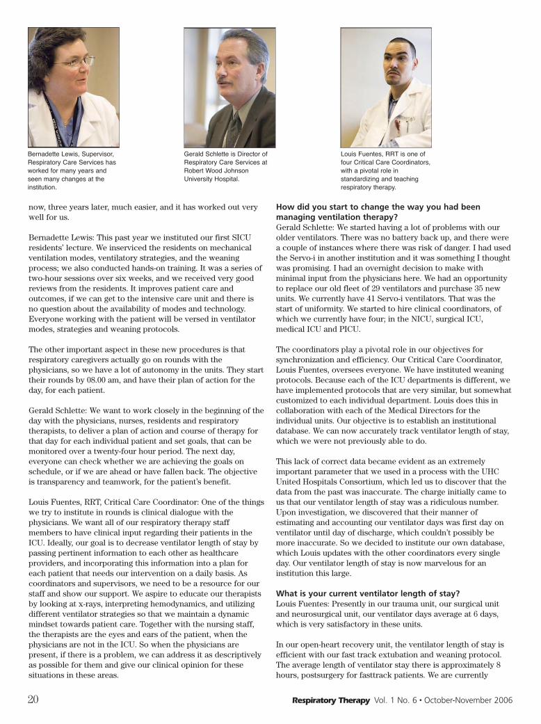
Bernadette Lewis, Supervisor,Respiratory Care Services hasworked for many years andseen many changes at theinstitution.
Gerald Schlette is Director ofRespiratory Care Services atRobert Wood JohnsonUniversity Hospital.
Louis Fuentes, RRT is one offour Critical Care Coordinators,with a pivotal role instandardizing and teachingrespiratory therapy.

Respiratory Therapy Vol. 1 No. 6 � October-November 2006 21
discussing protocols for the medical ICU and our long-termventilatory unit, where the patients tend to be very complicatedmedical patients including renal dialysis issues. Sometimes theycannot be put in a long-term facility, since these facilities arelimited in New Jersey, without dialysis capabilities. Those aresome of the projects we are working on now. In thisdepartment, we are considering implementation of a VolumeSupport/PRVC protocol, which we are discussing with ourMedical Director Dr. Jagadeeshan Sunderram.
How has the ventilation technology supported the focusedeffort in this institution? Gerald Schlette: Our therapists had some experiences of theServo 300 Ventilator, some with Automode and some not. Anoverwhelming majority of the therapists were reluctant to everuse Automode, and I feel this was a huge disservice to ourpatient population.
When we implemented the Servo-i fleet with the new userinterfaces and graphics, it was much easier to introduce andinstruct about Automode. In our openheart recovery unit,nurses were initially very reluctant and resistant to this change.But they realized the benefit of Automode—how fast it will helpthe patient get off the ventilator, the interaction with theanesthesia and medication postop, and now they don’t wantanything else. And this has expanded from the openheartrecovery unit into the SICU heart department and other areas ofthe hospital as well. The physicians see the sequence of eventsand the benefits of them.
What other types of ventilation therapies are used inaddition to standard therapies? Bernadette Lewis: We have five nitric oxide units, two in theNICU, one in the PICU and the other two units are available forthe additional patient population. We also provide highfrequency oscillation (HFO) when appropriate, in all patientcare populations. HFO should not be used as a last resort; itsuse is encouraged prior to the patient’s decline. We have begunto educate physicians and staff to monitor certain physiologicaldisease states, such as ARDS that is difficult to treat, and toimplement HFO before the patient reaches life-threateningcircumstances.
I understand that more expansion is planned for TheBristol-Myers Squibb Children’s Hospital, as well? Gerald Schlette: Yes, Children’s Specialized Hospital inMountainside will be part of our campus in New Brunswick,
joining The Bristol-Myers Squibb Children’s Hospital and TheChild Health Institute of New Jersey. This acute care pediatricrehab facility is scheduled to open in 2008.
Can you describe the other ICU units here? Louis Fuentes: Our neurosurgical unit is a seven bed ICU. Ourtrauma unit accommodates ten critical care beds. There we seeeverything from uncomplicated general surgery patients toARDS. We have an active trauma helicopter service and landtransport trauma service. The cardiac ICU comprises eighteenbeds, where they care for postop open-heart cases, valvereplacements, aortic valve replacements, robotic surgery, andwe have done transplant cases as well. Dr. Mark Anderson andDr. Peter Scholz are the chiefs of Cardiac and CardiothoracicSurgery. We perform rounds with the cardiac intensivists, andwork closely together with them regarding patient goals. OurMICU has 16 critical care beds and incorporates a dailyreadiness to wean assessment on every patient requiringmechanical ventilation. Our MICU coordinator Mr. WilliamTwaddle, RRT has worked intensely to help facilitate weaning inthe MICU and CCU. Recently, we have approval on a newmechanical ET tube holder to help us prevent inadvertentextubation and potential pressure ulcers from having anartificial airway. The CCU consists of 15 critical care beds withpatients suffering from a multitude of cardiac conditions.
Bernadette Lewis: The trauma unit keeps us very busy on somedays. Just last week, we had seven cases come in within theperiod of one to two hours. It can be anything from stabwounds, to construction worker accidents, to traffic accidentswith multiple victims. Numerous highways surround us, and weare a heavily populated urban area.
Gerald Schlette: In terms of acuity, the institution is pretty muchat the 100% mark. We have a total of 584 beds, but we haveexceeded 600 on occasion. They are looking at redesigning ourholding area for the ER to accommodate these patients, as acombined solution for medical, cardiac and surgery patients.
ROBERT WOOD JOHNSON MEDICAL ICUCritical Care News met up with Dr Jagadeeshan Sunderrambetween rounds at the Robert Wood Johnson Medical ICU todiscuss new procedures for weaning and outcome tracking. DrSunderram is a pulmonologist and Medical Director and Head ofthe MICU at Robert Wood Johnson University Hospital.
Can you describe the impact recent development has had
Doug Campell, Assistant VicePresident of Operationsdescribed disaster coordinationactivities.
Dr. Jagadeeshan Sunderram isMedical Director and head ofthe MICU at Robert WoodJohnson University Hospital.
Kumar DeZoysa and ServillanoDerikito.

on the medical intensive care unit and staff? I have had this position since 2001 and there has been a lot ofexpansion since that time. The MICU has grown and is a state-of-the-art unit, with one nurse for every two patients and a podsystem where the patients and monitors can be followed closely.There is a central nursing station, but this is not used as oftensince the pod system came into effect, which enables the nursesto be closer to the patient.
How is your cooperation with respiratory therapy? It has been a fantastic coordination. The implementation of aCritical Care Coordinator function has enabled us to do anumber of things to improve our outcomes. For example, wehad very high rates of ventilator-associated pneumonia in thepast, and what we have done is to institute something calledmultidisciplinary rounds. This includes about thirty questionsthat look at prevention strategies for ventilator-associatedpneumonia, for gastrointestinal bleeds, and so on asrecommended by the Society of Critical Care Medicine. We dothat three days a week and cover the whole ICU to make surethat these recommendations are in place. We have been able toreduce our rates of 10 VAPS per 1,000 patient ventilator days,which was about the national average, down to 3 to 4 VAPS per1,000 patient ventilator days. This is a substantial reduction. Weimplemented this program only five months ago, so we haveseen these dramatic reductions in a very short period of time. Infact, in some months we have had no ventilator-associatedpneumonias at all.
Are you implementing a series of weaning protocols? We have a series of standard questions that the therapists askeach morning. They ask if the patient is ready for a trial ofspontaneous breathing, they conduct a rapid-shallow breathingindex; they look at high FiO2 levels and high PEEP levels. Theyestablish whether the patient is on less than 8 cm H2O of PEEP,if the patient is at less than 60% of O2 concentration, andwhether the patient is hemodynamically stable and has nocontraindications to rapid-shallow breathing index. If theanswer to those three questions is yes, they conduct a rapid-shallow breathing trial. If the patient passes, the therapistsinform the physician that the patient is ready to come off theventilator. The physician may determine whether or not a trialof spontaneous breathing should be done. We have that protocolin place, and it has become very streamlined, but everythingdepends on patient response. It is much easier for the patientand the staff.
Will you be looking at length of stay on the ventilator? We will definitely be looking at that data. It is not currentlyavailable, however the respiratory department will be collectingthe data in a database for analysis. We have some preliminarydata showing that almost 90% of the patients, who we thoughtwere ready, did come off the ventilator. And about 50% of thepatients who we did not think were ready actually came off theventilator as well! I think it had to do with the issue of sedation.Once we examined sedation management, we changed theprotocols and routines, and I think this group of patients will bereclassified in the future. That is what is so fascinating aboutimplementing these protocols; you look at one or two aspects indetail, which may illustrate how a full change of events canoccur. There are a series of links here, and at some point thereis always a failure. So we are trying to identify where the failurehappens, and with the data in hand we can identify that pointand fix it. That is a benefit of standardizing our processes.
ROBERT WOOD JOHNSON SURGICAL ICU ANDNEUROTRAUMA ICU Critical Care News spoke with Kumar DeZoysa, BS, RRT,Critical Care Coordinator, Surgical ICU and Servillano Derikito,RRT, Staff Respiratory Therapist, to discuss how the newprocedures are impacting respiratory therapy in the Surgicaland Neurotrauma ICU departments.
Can you generally describe some of the cases you aretreating today? Kumar DeZoysa: We have a patient who was involved in a motorvehicle accident. The patient has a nonsignificant aortic tear,and is being stabilized for the OR. He has been at 70% O2
concentration and 14 cm H2O of PEEP for the last two weeks,and we have been waiting for him to be stabilized. He took aturn for the worse during transport to the CAT scan. Afterreturning he went from SIMV/VC to AC/VC, and the patientcontinued to deteriorate. He was put on AC/PRVC, and thepatient started to improve, which we hypothesized was due tothe fact that the patient had an autoPEEP (=+20 cm H2O) withPRVC. I am capturing the data, using the Servo-i data retrieval,and it makes for an interesting case study.
Is your neurotrauma ICU running basically at full capacitymost of the time? Servillano Derikito: Yes, basically every day of the week is busy,but we tend to get the most patients on Fridays and theweekend, when the accidents are more prevalent.
What are your experiences of implementing the weaningprotocol for standardization? Kumar DeZoysa: We were introduced to the concept of lungrecruitment approximately 8 years ago, via a Siemen’sinstructional video on lung recruitment, and we realized thatthis is a way to improve patient outcomes. Dr. BurchardLachmann’s contributions have formed the cornerstone to ourlung recruitment maneuver philosophy, and we have beenrefining our technique since then. We are trying to implementthis philosophy into our ventilation management and weaningprotocols to facilitate and improve the weaning process. Lungrecruitment maneuvers include monitoring the PaO2: FiO2 ratioand appropriate PEEP titration to maintain this ratio. Webelieve that once the lung is optimally recruited with notedimprovement in ABGs and other indicators of pulmonary status,it will be much easier to facilitate weaning. The newlyimplemented protocol has its basis in routine weaningmaneuvers, but emphasizes the need to have an improvedunderstanding of lung physiology and the pathophysiology ofatelectasis, and incorporates monitoring of the pulmonarystatus.
What about your postextubation strategies for maintaininglung recruitment? Kumar DeZoysa: We have started a research projectincorporating biofeedback in conjunction with lung recruitment.Initially, we educate the patient about atelectasis and correctiveactions to include incentive spirometry (IS) and deep breath andhold (DB&H) maneuvers. We provide each study patient with apulse oximeter within the patient’s visual line of sight, usually atthe foot of the bed. We ask that the patient maintain a certainsaturation level, such as SpO2 > 95%, by performing extra boutsof IS and DB&H exercises in addition to the requisite hourlyregimen. We believe that once patients are armed with thisinformation and a pulse oximeter, they should be able to follow
22 Respiratory Therapy Vol. 1 No. 6 � October-November 2006

their own progress and help themselves improve by means ofthis biofeedback. We started this research project about twomonths ago and we have captured data on approximately 20patients so far, but our objective is 500 patients. It will beinteresting to see a final analysis of the data from this numberof patients. Anecdotally, I can state confidently that patientswith compromised pulmonary status who are compliant withthe instructions improve within hours of implementation ofpulse oximetric biofeedback. As the institution grows, therespiratory therapy department has more opportunities to getinvolved in various aspects of research. We have a verysupportive anesthesia department that has volunteered to guideus through the intricacies and subtle nuances of the processesof research and publication, in addition to our primary goal ofimproved pulmonary and patient care.
ROBERT WOOD JOHNSON UNIVERSITY HOSPITALOPERATIONS Robert Wood Johnson University Hospital is situated at thecrossroads of New Jersey’s chemical, biochemical andpharmaceutical industries, and is in proximity to the Northeastrail corridor, large international airports and New York City.Disaster coordination and preparedness is a significantconsideration for this university hospital. Critical Care Newsspoke with Doug Campbell, Assistant Vice President ofOperations.
You started at this institution in 1985. Have you seenconsiderable expansion since then? It is amazing what has happened in the past twenty years, notonly for Robert Wood Johnson but also for the city of NewBrunswick. The hospital has benefited from the addition of theChildren’s Hospital, the Cancer Building and five new buildingsare currently in the planning process. We have also benefitedfrom our affiliation with the University of Medicine andDentistry of New Jersey-Robert Wood Johnson Medical School,which has expanded on the New Brunswick campus as well.
What are the most significant developments in recentyears? The establishment and growth of The Bristol-Myers SquibbChildren’s Hospital is one of the most significant developments.When we built the Children’s Hospital in early 2000, we shelledthe top two floors for future use, thinking that we would notneed them for some time. However, due to the growth ofprograms we needed the resources of additional space muchquicker than we thought, so all seven floors are now completedand occupied. That development has been enormous, with theaddition of the NICU and Special Care Nursery and the growthof other pediatric units, as well as the pediatric oncologyservice. Additionally, the construction of the Cancer Building,the expansion of the Cancer Institute of New Jersey, all havebeen significant developments that have affected the hospital ina positive way.
Disaster coordination and preparedness must be asignificant consideration for this university hospital, withyour proximity to metropolitan areas and chemicalindustries in New Jersey? That’s correct, and New Brunswick is at the center of the Stateof New Jersey. We have a major university; the Northeastcorridor train line runs along the east side of our campus, thereare oil refineries, the petroleum industries, the chemicalindustries, and pharmaceutical production, all within a close
geographical area to the hospital, which is only 30 minutes fromNew York City. Our sister hospital, Robert Wood Johnson atHamilton, is 30 miles to the south. This facility dealt with thePostal Service Building anthrax cases that developed in 2001.Robert Wood Johnson University Hospital really is at thecrossroads for the need for statewide emergency preparedness.
We participated in Top Off 3 last year, which was acongressionally mandated series of emergency preparednessexercises, lead by the Department of Homeland Security. Weparticipated at the top level, what we call Level Three, and thedrill was conducted over three days.
We had over 380 volunteer patients over three days comethrough our facility, while we were operating the hospital atnearly 100% occupancy. Our courtyard was transformed into a125 bed surge capacity area, and an Incident Command Centerwas set up in our boardroom. The Respiratory Therapydepartment did a great job; since the scenario was thedispensing of plague, they were taxed beyond what they couldimagine. We established a Victim Control center, and it was oneof the toughest experiences the institution has ever undertaken,but an extremely valuable experience to us. Participation in thisdrill was done at 100% occupancy, so we were busy treating ourreal patients as usual, in parallel with the simulation exercises.It taxed all of our resources but we are much better for havingconducted this exercise.
It must be extremely valuable as an institution to have thatexperience in hand. Yes, we know now that we can handle a certain number ofextreme incidents: chemical, biochemical, and a surge ofpatients. These were all very important experiences in terms ofreference for the future responses. The exercise went extremelywell, and we continue to work towards being one of the leadingfacilities in the state. So much so that we received a grant of 1.5million US dollars to construct the medical coordination centeron our campus. The Medical Coordination Center willcoordinate the health care emergency response in a five-countyarea in central New Jersey. This facility is also established toassist with the coordination of responses to emergenciesanywhere in the state.
THE BRISTOL-MYERS SQUIBB CHILDREN´SHOSPITAL Mobile intensive care unit The BristolMyers Squibb Children’s Hospital operates a fullyequipped transport service. Gerald Schlette described it:
The transport service provides a fully equipped mobile intensivecare unit. Patients can be intubated or have chest tubes placedduring transport. The unit provides compressed oxygen, a fullrange of IV therapy equipment, a crash cart including an aorticballoon pump, cardioverters, and two ventilators, one for theincubator and one for older patients.
We have a broad geographical area for patient uptake: northernNew Jersey, Albany New York, south New Jersey, Philadelphiaand other points in Pennsylvania, Washington DC, and Virginia.These are planned emergency patient transports – the patient isusually stabilized but needs to come in for specialized care.
The critical care transport team usually consists of at least onecritical care nurse from the appropriate unit, one critical care
Respiratory Therapy Vol. 1 No. 6 � October-November 2006 23
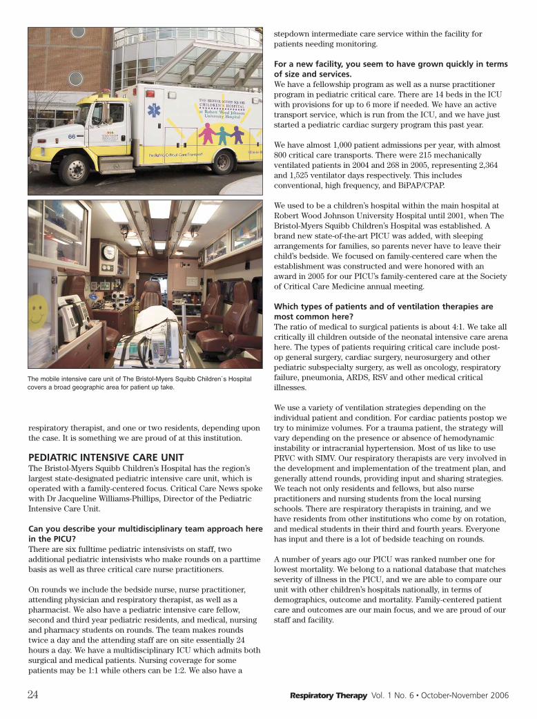
respiratory therapist, and one or two residents, depending uponthe case. It is something we are proud of at this institution.
PEDIATRIC INTENSIVE CARE UNIT The Bristol-Myers Squibb Children’s Hospital has the region’slargest state-designated pediatric intensive care unit, which isoperated with a family-centered focus. Critical Care News spokewith Dr Jacqueline Williams-Phillips, Director of the PediatricIntensive Care Unit.
Can you describe your multidisciplinary team approach herein the PICU? There are six fulltime pediatric intensivists on staff, twoadditional pediatric intensivists who make rounds on a parttimebasis as well as three critical care nurse practitioners.
On rounds we include the bedside nurse, nurse practitioner,attending physician and respiratory therapist, as well as apharmacist. We also have a pediatric intensive care fellow,second and third year pediatric residents, and medical, nursingand pharmacy students on rounds. The team makes roundstwice a day and the attending staff are on site essentially 24hours a day. We have a multidisciplinary ICU which admits bothsurgical and medical patients. Nursing coverage for somepatients may be 1:1 while others can be 1:2. We also have a
stepdown intermediate care service within the facility forpatients needing monitoring.
For a new facility, you seem to have grown quickly in termsof size and services. We have a fellowship program as well as a nurse practitionerprogram in pediatric critical care. There are 14 beds in the ICUwith provisions for up to 6 more if needed. We have an activetransport service, which is run from the ICU, and we have juststarted a pediatric cardiac surgery program this past year.
We have almost 1,000 patient admissions per year, with almost800 critical care transports. There were 215 mechanicallyventilated patients in 2004 and 268 in 2005, representing 2,364and 1,525 ventilator days respectively. This includesconventional, high frequency, and BiPAP/CPAP.
We used to be a children’s hospital within the main hospital atRobert Wood Johnson University Hospital until 2001, when TheBristol-Myers Squibb Children’s Hospital was established. Abrand new state-of-the-art PICU was added, with sleepingarrangements for families, so parents never have to leave theirchild’s bedside. We focused on family-centered care when theestablishment was constructed and were honored with anaward in 2005 for our PICU’s family-centered care at the Societyof Critical Care Medicine annual meeting.
Which types of patients and of ventilation therapies aremost common here? The ratio of medical to surgical patients is about 4:1. We take allcritically ill children outside of the neonatal intensive care arenahere. The types of patients requiring critical care include post-op general surgery, cardiac surgery, neurosurgery and otherpediatric subspecialty surgery, as well as oncology, respiratoryfailure, pneumonia, ARDS, RSV and other medical criticalillnesses.
We use a variety of ventilation strategies depending on theindividual patient and condition. For cardiac patients postop wetry to minimize volumes. For a trauma patient, the strategy willvary depending on the presence or absence of hemodynamicinstability or intracranial hypertension. Most of us like to usePRVC with SIMV. Our respiratory therapists are very involved inthe development and implementation of the treatment plan, andgenerally attend rounds, providing input and sharing strategies.We teach not only residents and fellows, but also nursepractitioners and nursing students from the local nursingschools. There are respiratory therapists in training, and wehave residents from other institutions who come by on rotation,and medical students in their third and fourth years. Everyonehas input and there is a lot of bedside teaching on rounds.
A number of years ago our PICU was ranked number one forlowest mortality. We belong to a national database that matchesseverity of illness in the PICU, and we are able to compare ourunit with other children’s hospitals nationally, in terms ofdemographics, outcome and mortality. Family-centered patientcare and outcomes are our main focus, and we are proud of ourstaff and facility.
24 Respiratory Therapy Vol. 1 No. 6 � October-November 2006
The mobile intensive care unit of The Bristol-Myers Squibb Children´s Hospitalcovers a broad geographic area for patient up take.

Respiratory Therapy Vol. 1 No. 6 � October-November 2006 25
Intensive Care Units around the world have been providing theirpatients with mechanical ventilation for the past thirty years,ever since the first electronic ventilator technology appeared inthe early seventies. And ventilation therapy, regardless of mode,has been delivered in the same manner – a clinician hasregulated the pressures and volumes provided to the patient. Agroup of researchers in Toronto have established a newrespiratory methodology, based on the neural signals providedby the patient. Research in the concept of Neurally AdjustedVentilator Assist (NAVA) is rapidly gaining the interest of thescientific community.
Can you describe what NAVA (Neurally AdjustedVentilatory Assist) is, and how it functions? NAVA is a new mode of mechanical ventilation, where theventilator is controlled directly by the patient’s own neuralcontrol of breathing. Since the introduction of the mechanicalventilator over thirty years ago, ventilation treatment has
traditionally been provided by means of adjusting airwaypressure, flow and volume. With NAVA, the ventilator pressureis adjusted by the patient’s own neural control system. Theneural control of respiration originates in the respiratory center,and signals are transmitted through the phrenic nerve to excitethe diaphragm. With NAVA, these signals are monitored bymeans of bipolar electrodes mounted on a nasogastric feedingtube and positioned in the esophagus at the level of thediaphragm. As respiration increases and the respiratory centerrequires the diaphragm for more effort, the degree of NAVAventilatory support needed is immediately provided.
This means that the patient’s respiratory center is in directcontrol of the mechanical support required on a breath bybreath basis, and any variation in the neural respiratory demandis responded to by the appropriate corresponding change inventilatory assistance.
Besides being a distinct mode of ventilation, NAVA also offers acomplete evaluation of the neural respiratory control in thecontext of a respiratory monitor. For example, if no assist isprovided, the drive will be very large, but if the patient isprovided with assist, the drive decreases. These are rapid reflexloops that are constantly ongoing.
This provides both the neural monitoring signal and how muchassist is to be delivered, at the same time. It also shows theextent of how the respiratory drive has been regulated, and thepressures used in this respect. There are no other modes thatcan provide these capabilities today. A pulse oximeter may tellus: “The saturation is going down to 80% and affecting PaO2,and if FiO2 is increased, the saturation increases back up to91%.” But that is only a monitor, and the clinician is in charge ofmaking the required adjustments. With NAVA, the patient is “incharge” of adjusting the ventilation. We do not make anyinterventions; only observe how the patient is regulatinghimself. One interesting aspect is in emergency situations; wehave found that it is always safest when we apply NAVA. Weseem to have much better control than by any other means. Infact, we have found that the sicker the patient is, the better
NAVA – A New Generation in Respiratory TherapyAn interview with Christer Sinderby, PhD
Christer Sinderby, PhD is Assistant Professor at the Department ofMedicine, University of Toronto, Canada, and Research Staff Scientist at St.Michael’s Hospital, Department of Critical Care Medicine, Toronto. Hereceived his MS degree from the Karolinska Institute in Stockholm,Sweden, and Doctor in Medical Science degree at the Department ofNeurosurgery, Sahlgrenska Hospital, University of Gothenburg, Sweden. Hehas had numerous academic committee assignments, including theUniversity of Western Australia, as a peer reviewer for American Journal ofRespiratory and Critical Care Medicine and Journal of Applied Physiology,and as a member of the American Thoracic Society/European RespiratorySociety task force for the ATS/ERS statement on standardization ofrespiratory muscle tests. He has won numerous research awards and grantsincluding the Montreal Chest Hospital Research Institute Annual Award,the Parker B. Francis Fellowship in Pulmonary Research and Fonds de laRecherche en Santé du Québec Chercheurs Boursier Scholarship. Hisresearch has been extensively published in peer review journals andmonographies. Reprinted with the permission of Critical Care News,www.criticalcarenews.com. CRITICAL CARE NEWS is published by MAQUETCritical Care. Maquet Critical Care AB, Solna, Sweden, ©MAQUET CriticalCare AB 2006. All rights reserved. The designations Servoi, Automode andOpen Lung Tool are registered trademarks of MAQUET Critical Care.
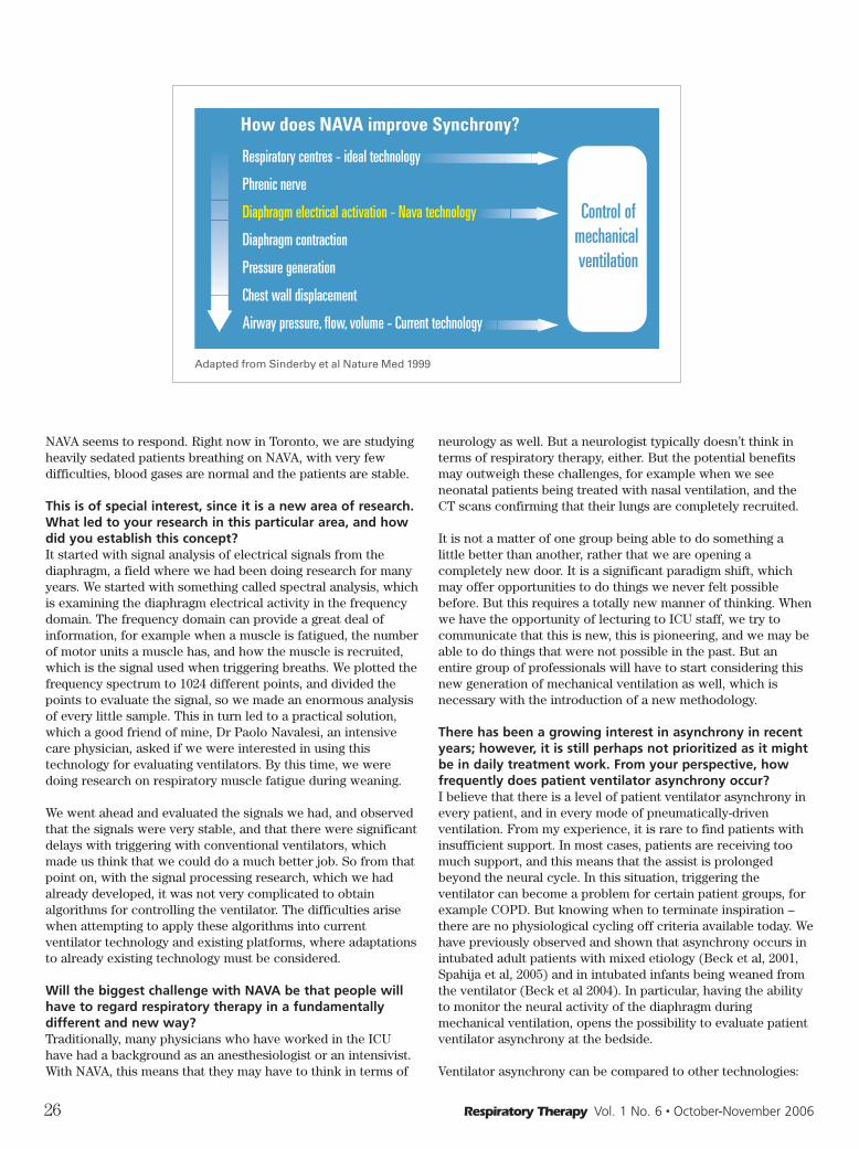
26 Respiratory Therapy Vol. 1 No. 6 � October-November 2006
NAVA seems to respond. Right now in Toronto, we are studyingheavily sedated patients breathing on NAVA, with very fewdifficulties, blood gases are normal and the patients are stable.
This is of special interest, since it is a new area of research.What led to your research in this particular area, and howdid you establish this concept? It started with signal analysis of electrical signals from thediaphragm, a field where we had been doing research for manyyears. We started with something called spectral analysis, whichis examining the diaphragm electrical activity in the frequencydomain. The frequency domain can provide a great deal ofinformation, for example when a muscle is fatigued, the numberof motor units a muscle has, and how the muscle is recruited,which is the signal used when triggering breaths. We plotted thefrequency spectrum to 1024 different points, and divided thepoints to evaluate the signal, so we made an enormous analysisof every little sample. This in turn led to a practical solution,which a good friend of mine, Dr Paolo Navalesi, an intensivecare physician, asked if we were interested in using thistechnology for evaluating ventilators. By this time, we weredoing research on respiratory muscle fatigue during weaning.
We went ahead and evaluated the signals we had, and observedthat the signals were very stable, and that there were significantdelays with triggering with conventional ventilators, whichmade us think that we could do a much better job. So from thatpoint on, with the signal processing research, which we hadalready developed, it was not very complicated to obtainalgorithms for controlling the ventilator. The difficulties arisewhen attempting to apply these algorithms into currentventilator technology and existing platforms, where adaptationsto already existing technology must be considered.
Will the biggest challenge with NAVA be that people willhave to regard respiratory therapy in a fundamentallydifferent and new way? Traditionally, many physicians who have worked in the ICUhave had a background as an anesthesiologist or an intensivist.With NAVA, this means that they may have to think in terms of
neurology as well. But a neurologist typically doesn’t think interms of respiratory therapy, either. But the potential benefitsmay outweigh these challenges, for example when we seeneonatal patients being treated with nasal ventilation, and theCT scans confirming that their lungs are completely recruited.
It is not a matter of one group being able to do something alittle better than another, rather that we are opening acompletely new door. It is a significant paradigm shift, whichmay offer opportunities to do things we never felt possiblebefore. But this requires a totally new manner of thinking. Whenwe have the opportunity of lecturing to ICU staff, we try tocommunicate that this is new, this is pioneering, and we may beable to do things that were not possible in the past. But anentire group of professionals will have to start considering thisnew generation of mechanical ventilation as well, which isnecessary with the introduction of a new methodology.
There has been a growing interest in asynchrony in recentyears; however, it is still perhaps not prioritized as it mightbe in daily treatment work. From your perspective, howfrequently does patient ventilator asynchrony occur? I believe that there is a level of patient ventilator asynchrony inevery patient, and in every mode of pneumatically-drivenventilation. From my experience, it is rare to find patients withinsufficient support. In most cases, patients are receiving toomuch support, and this means that the assist is prolongedbeyond the neural cycle. In this situation, triggering theventilator can become a problem for certain patient groups, forexample COPD. But knowing when to terminate inspiration –there are no physiological cycling off criteria available today. Wehave previously observed and shown that asynchrony occurs inintubated adult patients with mixed etiology (Beck et al, 2001,Spahija et al, 2005) and in intubated infants being weaned fromthe ventilator (Beck et al 2004). In particular, having the abilityto monitor the neural activity of the diaphragm duringmechanical ventilation, opens the possibility to evaluate patientventilator asynchrony at the bedside.
Ventilator asynchrony can be compared to other technologies:
Respiratory centres - ideal technology
Phrenic nerve
Diaphragm electrical activation - Nava technology
Diaphragm contraction
Pressure generation
Chest wall displacement
Airway pressure, flow, volume - Current technology
Control ofmechanical ventilation
How does NAVA improve Synchrony?
Adapted from Sinderby et al Nature Med 1999

Respiratory Therapy Vol. 1 No. 6 � October-November 2006 27
who wants a car that has a five second delayed reaction whenyou accelerate, and sometimes brakes, and sometimes does notbrake at all?
Or a computer mouse that reacts long after you haveclicked on it, or with lightening speed so you can not catchup at all? Exactly – this is basic supply and demand. A methodology andsystem should deliver a support in the amount and time that thepatient needs it, which allows the level of support to decreaseover time. This is the support we are striving to obtain, which iswhy synchrony is important.
In terms of patient risks in connection with asynchrony,what do you believe is most significant? One of the physiological effects of patient ventilator asynchronyis that it interferes with the patient’s natural breathing pattern.As we have shown in intubated infants, excessive delivery ofassist (beyond the neural inspiration) prolongs expiration ascompared to an unassisted breath (Beck et al 2004). In addition,if there is a delay to trigger the ventilator, this will prolonginspiratory time. With NAVA, the ventilator is cycled on whenneural inspiration begins, it provides assist during inspirationproportionally, and it cycles off when the neural expirationbegins. It does not interfere with the natural breathing pattern,because it follows the natural breathing pattern.
One of the greatest problems arising from excessive delivery ofassist (delayed cycling off) is that extreme pressures andvolumes can cause lung injury. Our most recent experience inexperimental research suggests that NAVA is lung protective
(Brander et al, 2006). Our clinical data also indicates thepotential for lung protection as the spontaneously chosen tidalvolumes are low and similar to those recommended by theARDSNet study for reducing ventilator induced lung injury. Inthree preclinical studies and two human studies, we have triedto increase the NAVA assist to levels comparable tooverdistention. And we have not been able to come up to theselevels – there is a neurological reflex that regulates and closespressures that are too high. Physiologically, when a volumeincreases to a certain level where you can no longer inspire, thebody has a neurological mechanism that stops the process. Thisis a fundamental difference from mechanical ventilation, wherea third party regulates volumes and pressures. But synchrony istwo dimensional: a time scale where it might be too late to startto inspire or too early or too late to cycle off, and the otherdimension is amplitude. But the protective component is notonly in amplitude. For example, when we have studied neurallytriggered pressure support, and deliver high pressure supportlevels, in these cases the patient time scale cycles off so earlythat it is not possible to go over a certain level. If the volume isobtained too rapidly, it stops, from a neural perspective. This isthe same pattern of normal respiration, you cannot inspireabove a certain level that the body does not accept. With NAVA,a ventilator would function in the same manner as the body inthis case – you cannot prolong a breath. If a breath wereprolonged, there would be excessive volume, resulting in lunginjury. So NAVA covers both dimensions, the time scale and theamplitude. And the benefit is that you have a methodology thatdoes not lead to a run away situation, but allows the body’sphysiological defense mechanism to regulate the ventilator.General asynchrony can be compared to an example: if a patient
Christer Sinderby was the recipient of a Parker B Francis Fellowship in Pulmonary Research.

tries to breathe and get insufficient support, the inspiration isinsufficient. If the ventilator then should start when the patienthas stopped inspiring, they receive the wrong level of assist andbecome agitated. If I thereafter increase the support from theventilator to supply larger pressures, the patient might miss afew breaths, and we have initiated the downward spiral.
If patient ventilator asynchrony is so severe that the patient“fights” the ventilator, the caregiver usually resorts to sedationand or neuromuscular paralysis of the patient. However, thecurrent trend in mechanical ventilation is to give less sedationand to help the patient breathe spontaneously. If we considerspontaneous breathing to be a true objective in ventilationtherapy, we must help the patient to receive a level of supportthat is adapted to spontaneous breathing, and interfere as littleas possible. Respiratory support should only be delivered whenthe patient wants it. Traditionally, it has been a matter ofsupplying a ventilation support that sustains a certain level ofminute ventilation, but the ventilator must take care of the lung,chest wall and abdomen, and the work of breathing increases.The patient does not receive what he needs.
What considerations should be taken with NAVA in regardto intubated as well as noninvasively ventilated patients? If you are treating an intubated patient, you have a patient whohas lost some level of autonomous breathing control functions(you have blocked the upper airway defense mechanisms).Perhaps you want to protect the airways if the patient is verysedated. With neural control, it does not matter how youventilate; we can ventilate with a mask, with a helmet, nasalprongs, a mouthpiece or an endotracheal tube. We see the sameresults however we deliver ventilation, the fact that we aredelivering neurally controlled ventilation is not an issue.However, this difference between invasive versus noninvasive isa traditional way of thinking in regard to delivery of ventilationtherapy. But for example, some patients must be sedated, if theyare in pain or very ill, and NAVA can still support ventilation inthese cases. NAVA delivers the support when the patient neuralsignals demands a breath, and NAVA delivers support that isproportional to the patient’s neural request, and stops when thepatient has received enough. It works if the patient is intubatedor ventilation is delivered by other means.
This is a matter of disconnecting pneumatics in how weconsider this: the tube, whether it is in the nose or in the throat,is only there to deliver ventilation. Today, physicians usepressure and flow as feedback for supplying support, whichmeans a lot of information is needed from the same tube. WithNAVA, we take the information from another source, directlyfrom the brain, if you will, and that information comes directlyoutside any other routes.
Are any particular patient categories of special interest interms of NAVA, at this point in time? In regard to patient categories, I believe it is very important togain experience in as many types of intensive care patients anddisease categories as possible. Heart-lung transplant patientshave many organs where the neural connection has been cutand replaced by new organs, and I do not think they would begood candidates for NAVA ventilation at this stage; it is perhapstoo early yet. In regard to other different patient categories, weare in the pioneering stages.
We are entering into a new generation in mechanical ventilation.
For myself as a researcher and for the team I work with, we aredoing as much as we can to obtain as much good information aswe can at this stage. Our total focus right now is on safety. Wehope that other intensivists and research centers with apioneering spirit will want to join us in the development of anew methodology, something fundamentally different thaneveryone is used to. There is an exciting journey ahead of us,with experiences that we may have not expected, but alsogroundbreaking therapeutic discoveries in the near future. Thiswill require an enormous amount of communication, andforums for clinicians to share experiences with each other.
When we try something new for the first time, it may easilyhappen that we experience that it does not work according toour expectations, which means it is a question of diagnosticsand translation, in the process of learning the patterns oftreatment with a new methodology. What is important to note isthat the initial physicians working with this will be truepioneers. We have a first solution how NAVA can be applied, butwe will collectively need to share all of our experiences as wego forward.
A new way of thinking and a new perspective on ventilationmeans not only potentially new solutions, but potentially newchallenges as well within the future in critical care.
REFERENCES 1 Beck J, Gottfried SB, Navalesi P, Skrobik Y, Comtois N,
Rossini M, Sinderby C. Electrical activity of the diaphragmduring pressure support ventilation in acute respiratoryfailure. Am J Respir Crit Care Med 2001; 164(3): 419-424.
2 Beck J, Spahija M, DeMarchie N, Comtois C, Sinderby C.Unloading during neurally adjusted ventilatory assist(NAVA). Eur Respir J 2002; 20(38):627S.
3 Beck J, Tucci M, Emeriaud G, LaCroix J, Sinderby C.Prolonged neural expiratory time induced by mechanicalventilation in infants. Pediatr Res 2004; 55: 747-754.
4 Sinderby C, Navalesi P, Beck J, Skrobik Y, Comtois N,Friberg S, Gottfried SB, Lindström L. Neural control ofmechanical ventilation in respiratory failure. NatureMedicine 1999; 5: 1433-1436.
5 Sinderby C, Beck J, Spahija J, Weinberg J, Grassino A.Voluntary activation of the human diaphragm in health anddisease. J Appl Physiol 1998; 85(6): 2146-2158.
6 Navalesi P, Costa R. New modes of mechanical ventilation:proportional assist ventilation, neurally adjusted ventilatoryassist, and fractal ventilation. Curr Opin Crit Care 2003; 9:51-58.
7 ARDSNetwork. Ventilation with lower tidal volumes ascompared with traditional tidal volumes for acute lunginjury and the acute respiratory distress syndrome. N EnglJ Med 2000; 342(18):1301-1308.
28 Respiratory Therapy Vol. 1 No. 6 � October-November 2006

Respiratory Therapy Vol. 1 No. 6 � October-November 2006 29
Few in medicine would deny the immense benefits provided bymechanical ventilators. Ever since the introduction of the ironlung in the 1920s, the ability to help a patient overcomebreathing difficulties while physicians took the time to curethem has saved countless lives throughout the world. And thepopulation of ventilated patients is likely to grow in the comingyears with the aging of the baby boom generation and theproliferation of life-prolonging medical advances. Butmechanical ventilation also carries risks of its own, such as a
higher incidence of pneumonia or lung damage. And thediscomfort to patients of the endotracheal tube can beconsiderable.
These issues, coupled with the considerable cost of maintaininga patient on a ventilator long term (up to $1,200 per day in anICU by some estimates) provide a compelling need for patientsto be weaned off ventilators at the earliest opportunity.
To address this need, Northeast Specialty Hospital haspartnered with physicians from Caritas Saint Elizabeth’s MedicalCenter and the Lahey Clinic to provide a ventilator weaningprogram that has dramatically reduced weaning times andenabled numerous patients previously thought to be lifelongdependents to regain functionality and return home to leadnormal lives.
The three doctors, Bartolome Celli, MD, and Miguel Divo, MD ofCaritas Saint Elizabeth’s and David Neumeyer, MD of the LaheyClinic are currently drawing up a set of weaning protocols thatwill enable them to provide a measure of uniformity to theprocess and allow patients to wean even more quickly and witha higher success rate than with the more traditional physician-directed weaning.
Long-renowned for pulmonary excellence, Northeast SpecialtyHospital is also an acute care hospital that specializes inproviding comprehensive care for patients with complexmedical, short-term rehabilitation and long-term medical andpulmonary needs. Deb Williams, Vice President of the hospital,sees the partnership as an opportunity for the Braintree-basedhospital to become a true regional leader in the highlyspecialized field of ventilator weaning. “We are privileged tohave three such high-caliber and well-respected pulmonologistsworking with us to support our mission in developing theregion’s center of excellence in pulmonary and ventilatorweaning care and rehabilitation.”
At the heart of the program is a multidisciplinary approach thatinvolves not only the referring physician and the staff doctors at
Specialists Team Up To Set Protocols ForWeaning Patients From Ventilators
Special Hospital Feature – Northeast Specialty Hospital
Stephen T. Sweriduk, MD
David Neumeyer, MD; Miguel Divo, MD; and Bartolome Celli, MD.

the hospital, but several team members who bring numerousother areas of expertise to the process. Dr Divo, one of thehospital’s Medical Directors, explains the team approach. “Whatwe do here, which is a big difference from a typical acute carehospital, is that I sit with a case manager, a social worker, anutritionist, a physical therapist, an occupational therapist, arespiratory therapist, a speech pathologist, the nurses and thepatient,” he says. “So, on multidisciplinary grounds we discussthe case. We try to coordinate, to look for goals; goals on ashort, medium and long term. Everybody is trying to derive andunderstand what the care plan is. We address the issues as ateam.”
Dr Neumeyer, who is a board certified pulmonologist, intensivistand sleep specialist at the Lahey Clinic in Burlington and servesas Medical Director, along with Dr Divo, at Northeast SpecialtyHospital in Waltham, concurs. “It’s not just a single specialist,”he says, “it’s a group of people who are looking at differentaspects of a patient’s health and trying to determine what isactually impairing their ability to wean.” Dr Neumeyer, whocompleted his residency in Internal Medicine and a fellowship inpulmonary medicine at New England Deaconess Hospital, aswell as fellowships in critical care at Brigham and Women’sHospital and sleep disorders at the Rhode Island Hospital, hasbeen with the Lahey Clinic Since 1997 and Northeast SpecialtyHospital since the Waltham site opened in April of 2004. One ofthe most appealing features of the program at NortheastSpecialty Hospital, was the state-of-the-art facility that has beenput together at Waltham. The physicians provide coverage there24 hours a day, seven days a week and work closely withreferring physicians, as well as the patients and their families, toidentify health concerns and assess each patient’s strengths andlimitations.
One challenge all the doctors face is that the kind of caredelivered in a typical acute care setting can sometimes becounterproductive to weaning a patient from a ventiator.
“In an acute care hospital,” says Dr Divo, “we are very good atdelivering care regarding medication, treatment, procedures, butin a secondary phase that is rehabilitation, regaining yourfunction, reevaluating everything that makes you transition togoing back home and being a functional patient, this is the placeto do it.”
The place he refers to is the 60-bed rehabilitation unit atChildren’s Hospital’s suburban location on the campus of whatused to be Waltham Hospital, where Northeast SpecialtyHospital opened its newest location in the spring of 2004.
Northeast Specialty Hospital also has sites in Braintree,Stoughton and Natick and is in good company in Waltham,where other tenants include Newton-Wellesley Hospital andBeth Israel Deaconess Medical Center.
Dr Divo’s mentor, and his Division Director at Caritas SaintElizabeth’s is Bartolome Celli, M.D., the Chief of PulmonaryCritical Care. Born in Venezuela, Dr Celli has been in Boston for35 years. He completed his internal medicine training at BostonCity Hospital, where he was Chief Medical Resident between1975 and 1976, and his pulmonary fellowship and critical carefellowship at Boston University Medical Center. After a five-yearstint back in his native country, he came back to New Englandto Tufts University, first as an Associate Professor and then as a
Full Professor. Today he is a professor of Medicine at TuftsUniversity School of Medicine, in addition to his role at CaritasSt. Elizabeth’s.
Dr Celli believes there are two important factors referringphysicians must consider when assessing a patient’s suitabilityfor weaning. “We must be optimistic,” he says. “Physicians whowork in critical care environments, when a patient is on aventilator and can’t come off the ventilator, they’re deemedimpossible to be weaned or impossible to go home. However,the evidence is that about 40% of those patients will ultimatelycome off the ventilator and about half of them will be able to gohome. Therefore, we should have an optimistic approach andsay, okay, even though we have reached the end of what we cando in an acute care setting, we are only beginning what we cando in a more rehab-oriented facility.
“Number two is to not just think of the acute problems butbegin to think as soon as the patient is intubated, ventilated orseverely ill, of what would be the possible outcome if thatpatient was prevented from deteriorating too much and couldgo to one of these rehab units. “So optimism and foresight,those are the two recommendations I would make to thereferring physicians. And getting them to us early enough thatwe can have maximum capacity to rehab them.”
On the issue of timeliness, Dr. Divo agrees. “Time is of theessence here,” he says. “I compare this, and I use this analogywith the patients, this is like fishing. So you throw your line, youhave your bait, and you’re waiting for the right time. And the
30 Respiratory Therapy Vol. 1 No. 6 � October-November 2006
Miguel Divo, MD, has gained national recognition as a specialist in how to bestwean patients from ventilators. Photo by Debra Troyanos.

Respiratory Therapy Vol. 1 No. 6 � October-November 2006 31
right time is what you measure. Too much fluid in the body andyou need to get rid of that. But you can’t do it in one step, youdo it on a weekly basis. For people recovering from bigcomplicated surgeries, what you need is a little bit of the healingprocess. Some of those wounds can affect some of the breathingmuscles. Surgery in the chest, when you crack the chest, youneed to have that strong enough that you can use it as a way ofbreathing again.
“In the middle, you look for quality of life, also. So you’re tryingto make them talk, you’re getting them to communicate. Thoselittle things make a huge difference to the patient. My ultimategoal may be three, four or five months away.”
Along the way, Dr Divo has to manage the expectations, both ofhis patients and their family members.
“I look for a milestone, and I share that with the patient, to go tothe next one, in order to go to the final goal. And it’s one of thevery difficult things to get people to understand here, we needto look for little changes and little gains, not the whole ultimategoal. And we cannot think about immediate gratification andimmediate results. Here everything takes a little bit more time.”
A typical patient is referred from an intensive care unit of anacute care hospital and is either a healthy person who hassuffered some kind of catastrophe that has caused them tobecome so ill that they need to be on a ventilator, or patientssuffering from a multitude of chronic conditions, one of whichhas resulted in a pulmonary crisis that has required assistancewith their breathing. Very often, previous attempts to weanthem in the ICU they were referred from have failed.
“If they have failed originally, we look at why they are stillfailing,” explains Dr Divo. “What are the little things we canimprove? We look for the reversal process here. That is the dailyand constant thinking from the medical perspective on the
pulmonology of how can I reverse the situation: the nutrition,the rehabilitation, the re-exercising. Some of the drugs that yougive in the intensive care unit, by that time, are not beneficial.By now you’re trying to clean up all of that. We look for thepatient to rest, recover, and get good nutrition. Basically, it’sthat, with a lot of science behind it.”
The latest challenge is the creation of weaning protocols thatwill replace the physician-directed approach that most otherhospitals use. “Protocols allow for a uniform approach tochronic weaning in an expeditious manner,” Dr Neumeyerexplains. “If you can do things on a consistent basis, thenpatients tend to wean faster.”
For patients who need home care or other long-term careservices, Northeast Specialty Hospital’s parent company,Commonwealth Communities, owns and operates a number ofskilled nursing and rehabilitation centers, assisted livingresidences and home health care services throughoutMassachusetts.
Dr. Divo’s parting words appropriately sum up the mission ofthe company. “My goal is to identify those people who can comeout of the ventilator and to determine what is the shortestperiod of time that I can do this?”
For more information about the vent weaning program atNortheast Specialty Hospital, please call (800) 500-5715,extension 8679 or visit their website atcommonwealthcommunities.com.
Dr Neumeyer is a board-certifiedpulmonologist at the Lahey Clinic inBurlington and serves as Medical Director,along with Dr Divo, at Northeast SpecialtyHospital in Waltham.
Bartolome Celli, MD is the Chief ofPulmonary Critical Care at Caritas StElizabeth’s Medical Center, as well asProfessor of Medicine at Tufts UniversitySchool of Medicine.


Respiratory Therapy Vol. 1 No. 6 � October-November 2006 33
ABSTRACT The variability of 5 new pulmonary function test (PFT)instruments (Collins CPL, JaegerUSA Masterscreen, MedicalGraphics Profiler DX System, Morgan Transflow Test PFTSystem, SensorMedics Vmax 22D) was established using 24standard waveforms with a waveform simulator. Testing wasperformed on Days 0, 30, 60, and 90. At each test session, eachwaveform was injected 5 times into each PFT instrument (480measurements per instrument). Each instrument was also usedto test 11 healthy subjects at Weeks 0, 12, and 24. At each testsession, each subject performed three acceptable spirometricmaneuvers. The highest FEV1 for each subject was recorded (66measurements per instrument). Overall variability issummarized in the table.
These data demonstrate that the variability associated withlongitudinal measurement of FEV1 assessed by repetitivewaveform simulations is consistently small. Human subjectvariability is much larger. Differences in variability of themagnitude observed in this study have implications for thedesign of clinical studies that utilize FEV1 as an endpoint.
BACKGROUND • Longitudinal measurement of pulmonary function is an
integral component of assessing the safety and efficacy ofdrugs delivered by inhalation. An understanding of thesources of variability associated with the longitudinalmeasurement of pulmonary function is essential for clinicaltrial design, as it is in interpreting longitudinal lung functionmeasurements in disease states. However, methods forassessing the sources of test variability using modernpulmonary function test (PFT) instruments are not readilyavailable.
• This study was performed to assess the sources of variabilityin the longitudinal measurement of forced expiratory volumein 1 second (FEV1) as obtained on 5 commercially available
PFT instruments. The impact of the observedvariability on sample size determinations forcontrolled clinical trials utilizing FEV1 as anendpoint is discussed.
METHODS • One new instrument model from 5 different PFT
equipment manufacturers was purchased toperform these studies. These models, and theirbasic FEV1 measurement technology, were: – Collins CPL (volume-based)
– JaegerUSA Masterscreen Diffusion TP (flow-based) – Medical Graphics Profiler DX System (flow-based) – Morgan Transflow Test PFT System (volume-based) – SensorMedics Vmax 22D (flow-based).
• Each instrument was set up and maintained throughout thestudy according to manufacturer’s specifications.
• Each instrument remained powered on for the duration of thestudy.
• Testing was done at LDS Hospital in Salt Lake City, UT, USA,and performed by 2 experienced technicians.
• On test days, instruments were calibrated approximately 1hour prior to the first test. – Instruments would be calibration checked or recalibrated if
there were long delays between subjects or when
Performance of 5 PFT Instruments in theLongitudinal Measurement of FEV1R.L. Jensen, J.G. Teeter, R.D. England, H.J. White, E.H. Pickering, R.O. Crapo
Authors Jensen and Crapo are with LDS Hospital and University of Utah,Salt Lake City; England, White and Pickering are with Pfizer GlobalResearch and Development, Groton, CT. This study was funded by Pfizer Incand Aventis Pharmaceuticals Inc.
Instrument Simulator testing Human subject testingRMS CVa Meanb Rangeb RMS CVa Range CV
19.4 ,78.008.225.5 ,94.226.373.0snilloC
13.4 ,12.147.246.5 ,65.266.395.1regeaJ
87.9 ,49.081.484.5 ,91.294.347.0scihparG lacideM
58.4 ,98.065.285.5 ,35.266.324.0nagroM
66.11 ,99.042.414.5 ,83.264.358.0scideMrosneS
aRMS CV = Root Mean Square Coefficient of Variation (square root of the average squared coefficient of variation for all waveforms orsubjects across all time points); bMeasured FEV1 in liters BTPS.
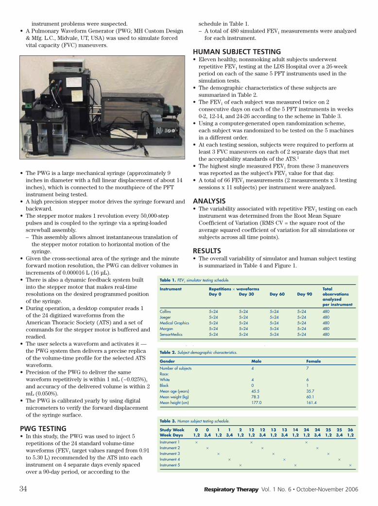
34 Respiratory Therapy Vol. 1 No. 6 � October-November 2006
instrument problems were suspected. • A Pulmonary Waveform Generator (PWG; MH Custom Design
& Mfg. L.C., Midvale, UT, USA) was used to simulate forcedvital capacity (FVC) maneuvers.
• The PWG is a large mechanical syringe (approximately 9inches in diameter with a full linear displacement of about 14inches), which is connected to the mouthpiece of the PFTinstrument being tested.
• A high precision stepper motor drives the syringe forward andbackward.
• The stepper motor makes 1 revolution every 50,000-steppulses and is coupled to the syringe via a spring-loadedscrewball assembly. – This assembly allows almost instantaneous translation of
the stepper motor rotation to horizontal motion of thesyringe.
• Given the cross-sectional area of the syringe and the minuteforward motion resolution, the PWG can deliver volumes inincrements of 0.000016 L (16 µL).
• There is also a dynamic feedback system builtinto the stepper motor that makes real-timeresolutions on the desired programmed positionof the syringe.
• During operation, a desktop computer reads 1of the 24 digitized waveforms from theAmerican Thoracic Society (ATS) and a set ofcommands for the stepper motor is buffered andreadied.
• The user selects a waveform and activates it —the PWG system then delivers a precise replicaof the volume-time profile for the selected ATSwaveform.
• Precision of the PWG to deliver the samewaveform repetitively is within 1 mL (~0.025%),and accuracy of the delivered volume is within 2mL (0.050%).
• The PWG is calibrated yearly by using digitalmicrometers to verify the forward displacementof the syringe surface.
PWG TESTING • In this study, the PWG was used to inject 5
repetitions of the 24 standard volume-timewaveforms (FEV1 target values ranged from 0.91to 5.30 L) recommended by the ATS into eachinstrument on 4 separate days evenly spacedover a 90-day period, or according to the
schedule in Table 1. – A total of 480 simulated FEV1 measurements were analyzed
for each instrument.
HUMAN SUBJECT TESTING • Eleven healthy, nonsmoking adult subjects underwent
repetitive FEV1 testing at the LDS Hospital over a 26-weekperiod on each of the same 5 PFT instruments used in thesimulation tests.
• The demographic characteristics of these subjects aresummarized in Table 2.
• The FEV1 of each subject was measured twice on 2consecutive days on each of the 5 PFT instruments in weeks0-2, 12-14, and 24-26 according to the scheme in Table 3.
• Using a computer-generated open randomization scheme,each subject was randomized to be tested on the 5 machinesin a different order.
• At each testing session, subjects were required to perform atleast 3 FVC maneuvers on each of 2 separate days that metthe acceptability standards of the ATS.1
• The highest single measured FEV1 from these 3 maneuverswas reported as the subject’s FEV1 value for that day.
• A total of 66 FEV1 measurements (2 measurements x 3 testingsessions x 11 subjects) per instrument were analyzed.
ANALYSIS • The variability associated with repetitive FEV1 testing on each
instrument was determined from the Root Mean SquareCoefficient of Variation (RMS CV = the square root of theaverage squared coefficient of variation for all simulations orsubjects across all time points).
RESULTS • The overall variability of simulator and human subject testing
is summarized in Table 4 and Figure 1.
Table 1. FEV1 simulator testing schedule.
Instrument Repetitions × latoTsmrofevawDay 0 Day 30 Day 60 Day 90 observations
analyzedper instrument
Collins 5×24 5×24 5×24 5×24 480Jaeger 5×24 5×24 5×24 5×24 480Medical Graphics 5×24 5×24 5×24 5×24 480Morgan 5×24 5×24 5×24 5×24 480SensorMedics 5×24 5×24 5×24 5×24 480
Table 2. Subject demographic characteristics.
elameFelaMredneG
74stcejbus fo rebmuNRace:
64etihW10kcalB
7.535.54)sraey( ega naeM1.063.87)gk( thgiew naeM4.1610.771)mc( thgieh naeM
g p j
Table 3. Human subject testing schedule.
Study Week 0 0 1 1 2 12 12 13 13 14 24 24 25 25 26Week Days 1,2 3,4 1,2 3,4 1,2 1,2 3,4 1,2 3,4 1,2 1,2 3,4 1,2 3,4 1,2
Instrument 1 ×××Instrument 2 ×××Instrument 3 ×××Instrument 4 ×××Instrument 5 ×××
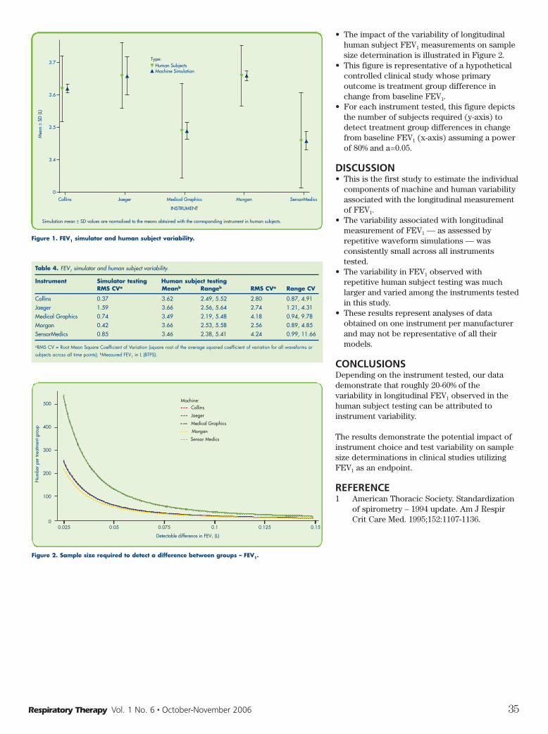
Respiratory Therapy Vol. 1 No. 6 � October-November 2006 35
• The impact of the variability of longitudinalhuman subject FEV1 measurements on samplesize determination is illustrated in Figure 2.
• This figure is representative of a hypotheticalcontrolled clinical study whose primaryoutcome is treatment group difference inchange from baseline FEV1.
• For each instrument tested, this figure depictsthe number of subjects required (y-axis) todetect treatment group differences in changefrom baseline FEV1 (x-axis) assuming a powerof 80% and a=0.05.
DISCUSSION • This is the first study to estimate the individual
components of machine and human variabilityassociated with the longitudinal measurementof FEV1.
• The variability associated with longitudinalmeasurement of FEV1 — as assessed byrepetitive waveform simulations — wasconsistently small across all instrumentstested.
• The variability in FEV1 observed withrepetitive human subject testing was muchlarger and varied among the instruments testedin this study.
• These results represent analyses of dataobtained on one instrument per manufacturerand may not be representative of all theirmodels.
CONCLUSIONS Depending on the instrument tested, our datademonstrate that roughly 20-60% of thevariability in longitudinal FEV1 observed in thehuman subject testing can be attributed toinstrument variability.
The results demonstrate the potential impact ofinstrument choice and test variability on samplesize determinations in clinical studies utilizingFEV1 as an endpoint.
REFERENCE 1 American Thoracic Society. Standardization
of spirometry – 1994 update. Am J RespirCrit Care Med. 1995;152:1107-1136.
Table 4. FEV1 simulator and human subject variability.
Instrument Simulator testing Human subject testingRMS CVa Meanb Rangeb RMS CVa Range CV
Collins 0.37 3.62 2.49, 5.52 2.80 0.87, 4.91Jaeger 1.59 3.66 2.56, 5.64 2.74 1.21, 4.31Medical Graphics 0.74 3.49 2.19, 5.48 4.18 0.94, 9.78Morgan 0.42 3.66 2.53, 5.58 2.56 0.89, 4.85SensorMedics 0.85 3.46 2.38, 5.41 4.24 0.99, 11.66
aRMS CV = Root Mean Square Coefficient of Variation (square root of the average squared coefficient of variation for all waveforms orsubjects across all time points); bMeasured FEV1 in L (BTPS).
Collins Jaeger Medical Graphics Morgan SensorMedics
3.7
3.6
3.5
3.4
0
Mea
n±
SD (L
)
Human SubjectsMachine Simulation
Simulation mean ± SD values are normalised to the means obtained with the corresponding instrument in human subjects.
INSTRUMENT
Type:
Figure 1. FEV1 simulator and human subject variability.
0.025 0.05 0.075 0.1 0.125 0.15
Detectable difference in FEV1 (L)
100
0
200
300
400
500
Num
ber
per
treat
men
t gro
up
Collins
Jaeger
Medical Graphics
Morgan
Sensor Medics
Machine:
Figure 2. Sample size required to detect a difference between groups – FEV1.

36 Respiratory Therapy Vol. 1 No. 6 � October-November 2006
ABSTRACTWe present here a 4 year old child with severe tracheal stenosisand respiratory failure. The patient was not responding toconventional ventilation settings and had significant hypercarbia.The difficulty in mechanical ventilation was handled successfullywith specific ventilatory strategy: use of low respiratory rate, longinspiratory time and normal inspiratory time : expiratory timeratio. Thereafter the child was managed surgically and thestenosis was corrected. The child was discharged after aMontgomery T-tube placement.
Laryngotracheal stenosis is a congenital or acquired narrowing ofthe airway that may affect the glottis, subglottis or trachea. Itcauses severe symptoms and should be suspected in children lessthan 1 year of age with either multiple episodes of croup or croupwhich fails to respond to medical management or requiresendotracheal intubation.1 The term subglottic stenosis waspreviously used as the subglottic region is the most common siteof airway stenosis, mostly secondary to prolonged endotrachealintubation. The onset may be acute or it may develop over aperiod of time and a thorough assessment, radiologic andendoscopic evaluation may be necessary to guide further therapyand management. We present a case of acquired tracheal stenosisthat required prolonged ventilatory support with adaptations forthe obstructive pathology and discuss the implications forventilation of a child with upper airway obstruction.
CASE REPORTA 4-year-old boy presented with history of intermittent high gradefever and cough without expectoration of 3 months’ duration.After 5 days of onset of fever, he developed noisy breathing andprogressively increasing respiratory distress. There was nohistory of cyanosis, wheezing episodes or any history of foreignbody aspiration. There was no history of contact withtuberculosis and no past history of any significant illness. Therespiratory distress and the intensity of noisy breathing increasedgradually over 3 months and were now also persisting duringsleep. There was no history suggestive of diphtheria and the childwas immunized for age.
On presentation, the child had marked respiratory distress, wassitting up bending forward and had marked suprasternalrecessions and severe biphasic stridor. He had a respiratory rateof 48/minute, and had good volume pulses with no cyanosis and
SpO2 of 99% on facemask oxygen. Chest examination showedhyperinflation of the chest wall with symmetrical movements ofboth sides of the chest wall, centrally placed trachea on palpationand equally resonant percussion note on both sides. Chestauscultation revealed equal air entry on both sides and conductedbreath sounds. Examination of cardiovascular and other bodysystems was unremarkable. Initial arterial blood gas analysisshowed a pCO2 of 48.6 mm Hg and Pa2 of 198.4 mm Hg with pH of7.391.
At presentation the possibility of severe upper airway obstructionwas kept and radiographs of soft tissue of neck (lateral view) didnot show any evidence of epiglottitis or narrowing of the cervicalportion of the trachea. Chest X ray showed bilateral hyperinflatedlung fields with no lung parenchymal abnormalities and normalcardiothoracic ratio (48%). However, over the next few hourschild showed progressive deterioration with rising pCO2 valuesup to 80 mm Hg and had to be intubated for respiratory supportand was therefore transferred to the Pediatric Intensive Care Unit(PICU) for further management.
On arrival to the PICU he was ventilated with Siemens Servo 300ventilator on SIMV (volume controlled) mode with initialconventional settings but did not show adequate chest rise andhad progressive CO2 accumulation (values up to 150 mm Hg) onblood gas analysis. The initial settings were: rate 30/min, tidalvolume 80 ml, Ti 0.7 seconds, I:E ratio of 1:2 and positive endexpiratory pressure of 4 cm H2O. On increasing the tidal volumealone, there was no further improvement in PaCO2 levelsassessed after 1 hour. The peak pressure requirement was 38 cmH2O. On bagging with self-inflating bag, high resistance was feltand adequate chest rise could be achieved only if long inspiratorytime was used. Subsequently the settings were modified inkeeping with the requirements so that the ventilator now had lowrates (10/min) and a prolonged inspiratory time (Ti) of 1.8seconds, an I:E ratio of 1:2 and PEEP of 5 cm H2O. With thesesettings, he maintained oxygenation and showed improvement inPaCO2 levels. The peak pressure requirement also fell to 23 cmH2O. The rationale for these settings is discussed below.
Subsequently, a fibreoptic bronchoscopy was done through theendotracheal tube in the PICU after initial stabilization and itshowed severe narrowing at the distal end of the trachea justabove the carina. A diagnosis of supracarinal tracheal stenosiswas established and the child underwent endoscopic balloondilatation of trachea under general anaesthesia the next morning.Intraoperatively, the stenotic area showed some granulationtissue with inflamed mucosa and loss of tracheal rings, and fromthis a biopsy specimen was obtained and dilatation was done
Ventilatory Management of SevereTracheal StenosisLodha Rakesh, Guglani Lokesh, S.C. Sharma, S.K. Kabra
Authors Rakesh, Lokesh and Kabra are with the Department of Pediatrics;Sharma is with the Departments of Otorhinolaryngology, AIIMS, NewDelhi, India. Reprinted from the Indian Journal of Pediatrics, Vol 73, Issue5, pages 441-444, © Indian Journal of Pediatrics.

Respiratory Therapy Vol. 1 No. 6 � October-November 2006 37
with Fogarty’s Catheter. An endotracheal tube of 4.5 Fr size wasinserted under bronchoscopic guidance with tip extendingbeyond the stenotic segment and lying above the carina. He wassubsequently continued on ventilatory support for the next 10days with gradual weaning. A diagnosis of tubercular trachealstenosis was considered and antitubercular therapy was startedbut subsequent work up did not reveal any evidence of tubercularinfection. However, there was satisfactory clinical response as thefever subsided and tracheal granulations on repeat examinationwere absent. The biopsy from the lesion showed mild non-specific inflammatory changes with no granulomas.
A repeat fibreoptic bronchoscopy done 8 days after the dilatationprocedure in the PICU showed no significant stenosis butpersistence of inflamed mucosa. He was then weaned offventilatory support but endotracheal tube was left in situ as itwas bypassing the stenosed segment. He had recurrence ofrespiratory distress, which required resumption of ventilatorysupport and he then underwent repeat dilatation after 20 days ofadmission, which showed collapse and re-stenosis of trachea,which could be easily dilated. Tracheostomy and Montgomery Ttube insertion was done for maintaining long-term patency of theairway. He was discharged from the hospital 2 months afteradmission and is on regular follow up.
DISCUSSIONThe importance of appropriate ventilatory management of severeproximal airway obstruction is highlighted by this case and it isimportant to understand the rationale for the ventilatory settingsdescribed above. The trachea and the upper airway althoughconsidered to be relatively rigid conducting airways, do showsome changes in caliber during the normal respiratory cycle.There is expansion of the intrathoracic airways along with theexpanding lungs while the extrathoracic airway diminishes incaliber due to their intraluminal pressure being lower than
atmospheric pressure. The reverse of this process occurs duringexpiration. If intrathoracic trachea is soft (tracheomalacia), thenarrowing will accentuate during expiration due to positiveintrathoracic pressure. The flow in these large airways is usuallyturbulent due to the high flow rates and this turbulence increasesin the presence of an obstruction in the airway. This turbulencefurther increases the airway resistance.
The mechanics of critical tracheal stenosis is such that it wouldseverely compromise delivery of gases beyond the obstructionand it would not allow adequate emptying out of the lungs aswell. Other obstructive upper airway anomalies like subglotticstenosis could be overcome by use of tracheostomy, which wouldbypass the site of stenosis, but in more distal lesions of thetrachea (as in this case, just above the carina) ventilatorymanagement is more challenging.
With conventional ventilator settings in the patient with severetracheal stenosis, there would be inadequate delivery of gasesbeyond the site of obstruction and there would be a build up ofpressure proximal to it. In this scenario, there would beprogressive CO2 retention and inadequate lung expansion. Alsobecause of inadequate emptying of the lungs during expirationdue to limited airflow across the site of stenosis, there would be aprogressive air-trapping and subsequent decrease in cardiacoutput. Increasing the airway pressures or frequency alone willnot help overcome these effects of the obstruction. Increase inthe airway pressure may increase the risk of barotraumas. Inorder to ensure adequate delivery of gases beyond theobstruction, it is necessary to prolong the inspiratory time (Ti) sothat adequate lung expansion can be achieved. In view of themarkedly increased resistance the time constant will also beincreased justifying the need for high Ti. For this, the frequencyof breaths would have to be kept at low values in order to allowadequate time for expiration also and at the same time avoid

progressive gas trapping (auto-PEEP). It is unlikely that use ofCPAP alone would have led to adequate delivery of volumesacross such severe tracheal stenosis.
There have been reports of use of percutaneous transtracheal jetventilation for cases with tracheal stenosis and other forms ofairway obstruction in the more proximal airways.2 For patientsundergoing surgical repair of the tracheal lesions, extracorporealmembrane oxygenation has also been used.3 In animal models oftracheal stenosis, a comparison of conventional ventilation versustranstracheal jet ventilation showed that although there were nosignificant differences in PaCO2 but mean peak airway pressurevalues, both at the distal portion of stenosis and at the proximalportion, decreased more significantly during jet ventilation thanduring conventional mechanical ventilation.4 Overall the meanarterial pressure, mean pulmonary arterial pressure, centralvenous pressure, and cardiac output did not change significantlybetween conventional mechanical ventilation and jet ventilationwith the stenosis. Pulmonary Artery Occlusion Pressure (PAOP)increased significantly more during conventional mechanicalventilation than during jet ventilation in animal models withstenosis.
During ventilation, the use of flow-volume loops woulddemonstrate plateaus in both inspiratory and expiratory limbsdue to the limitation in both phases caused by the obstruction.Miller and Hyatt5 showed that in patients with fixed airwayobstruction, the FEV1 diminishes progressively as the resistanceincreases but the FVC may remain unchanged, suggesting that theflow rates will be reduced and the ratio of maximal expiratoryand inspiratory flow at 50% of vital capacity (MEF50 /MIF50 ) mayapproach unity (0.9 to 1).
Another contentious issue was the underlying cause of trachealstenosis in this child since the duration of symptoms was 3months and he had no respiratory symptoms prior to this illness.Initial impression of granulation tissue being visible onbronchoscopy directed towards a diagnosis of tuberculosis butthe work up was negative and the biopsy from the lesion wasinconclusive. However, the tracheal inflammation could be due toa tubercular lymph node in the mediastinum. Despite no objectiveevidence of tuberculosis, response to antitubercular therapysupports the diagnosis of tuberculosis. A possibility of acquiredtracheal stenosis secondary to an initial episode of bacterialtracheitis was considered.
The most common cause of acquired laryngotracheal stenosis isprolonged endotracheal intubation, accounting for 90% of cases.Other acquired causes include postinfectious scarring,autoimmune disorders (Wegener’s granulomatosis, sarcoidosis),inhalation injuries, blunt trauma to the neck, previoustracheostomy or cricothyrotomy, and gastroesophageal reflux.6
Prior to the advent of antibiotics, scarring from infections likediphtheria and syphilis were usually a common cause of stenosis.In the trachea, the narrowing may be extrinsic or intrinsic, anddiffuse or localized. Among the extrinsic lesions, disorders of thethyroid gland and great vessels are the most common while theintrinsic ones may be due to infectious, granulomatous,neoplastic, traumatic, immunologic and post-inflammatoryconditions.
There are several case series of laryngotracheal stenosis inchildren, with majority of reports being that of surgical repairtechniques. The ventilatory management of these patients has not
been highlighted but it does pose problems in the perioperativeand post-operative period. There have been reports whereunstable children with critical stenoses requiring high airwaypressures for ventilation and still having CO2 retention, haverequired initiation of extracorporeal support, which was followedby diagnostic endoscopy and then finally definitive surgicalrepair.3 But these facilities may not exist at all institutions andinitial optimal ventilatory support in such sick children may becrucial before they can be shifted to centers where surgical repaircan be done. Therefore, it is only with a understanding of themechanics of tracheal stenosis and its optimal ventilatorymanagement that we can ensure adequate treatment and outcomefor these patients by surgical or non-surgical (eg balloondilatation) means.
In conclusion, this case demonstrates the importance ofappropriate ventilatory management of airway obstructioncaused by tracheal stenosis and highlights the fact that criticalstenosis of trachea may be ventilated with low rates and highinspiratory times. Management of tracheal stenosis in children iscomplex and requires the teamwork of specialists involved inemergency management, intensive care andotorhinolaryngology.12
REFERENCES1 Lesperance MM, Zalzal GH. Assessment and management of
laryngotracheal stenosis. Pediatr Clin North Am 1996; 43:1413-1427.
2 Spoerel WE, Narayanan PS, Singh NP. Transtrachealventilation. Br J Anaesth 1971; 43: 932-938.
3 Walker LK, Wetzel RC, Haller JA. Extracorporeal membraneoxygenation for perioperative support during congenitaltracheal stenosis repair. Anesth Analg 1992; 75: 825-829.
4 Shinozaki M, Sueyoshi A, Morinaga T, Tsuda H, Muteki T.Effect of conventional mechanical ventilation and jetventilation on airway pressure in dogs and plastic modelswith tracheal stenosis. Crit Care Med 1996: 24; 658-662.
5 Miller RD, Hyatt RE. Obstructing lesions of the larynx andtrachea: Clinical and physiologic characteristics. Mayo ClinProc 1969; 44: 145.
6 Courey MS. Airway obstruction - the problem and its causes.Otolaryngol Clin North Am 1995; 28: 673-684.
7 Kumar P, Bush AP, Ladas GP, Goldstraw P. Tracheobronchialobstruction in children: Experience with endoscopic airwaystenting. Ann Thoracic Surg 2003; 75: 1579-1586.
8 Anton-Pacheco JL, Cano I, Garcia A, Martinez A, Cuadros J,Berchi FJ. Patterns of management of congenital trachealstenosis. J Pediatr Surg 2003; 38: 1452-1458.
9 Dunham ME, Holinger LD, Backer CL, Mavroudis C.management of severe congenital tracheal stenosis. Ann OtolRhinol Laryngol 1994; 103: 351-356.
10 Har-El G, Chaudhary R, Saha A et al. Resection of trachealstenosis with end-to-end anastomosis. Ann Otol RhinolLaryngol 1993; 102: 670-674.
11 Loeff DS, Filler RM, Vinograd I et al. Congenital TrachealStenosis: a review of 22 patients from 1965 to 1987. J PediatrSurg 1988; 23: 744-748.
12 Weber TR, Connors RH, Tracy TF et al. acquired trachealstenosis in infants and children. J Thoracic Cardiovasc Surg1991; 102: 29-35
38 Respiratory Therapy Vol. 1 No. 6 � October-November 2006
Respiratory Therapy Vol. 1 No. 6 � October-November 2006 39
ABSTRACTA consensus as to the relative benefit of high frequency
ventilation in different populations and indications has not been
reached. Some have recently hypothesized that improvements in
conventional ventilation and improvements in other standards
of neonatal care have eliminated the potential of the relative
advantages of high frequency ventilation. However, results from
recent mega trials of high frequency ventilation have yielded
inconsistent results, rather than consistent results of no relative
benefit. Rather this suggests a possibility of differences in high
frequency devices, treatment strategies or treatment
populations. We updated our earlier meta-analysis with data
from recent trials in order to address these issues. This analysis
reports on the results of seven randomized controlled trials of
the 3100A HFOV in 951 preterm infants. In this group of trials
with this device we found a clinically significant advantage of
high frequency oscillatory ventilation with regard to reducing
mortality and chronic pulmonary morbidity. We found no
evidence that the relative benefit of HFOV was significantly
attenuated with improvements in conventional ventilator
strategy or other baseline standards of care. We also found no
evidence that the relative benefits were significantly different in
preterm infants of different size or severity.
INTRODUCTIONOur meta-analysis in 2002 demonstrated a clear reduction in
acute and chronic pulmonary adverse effects in infants
associated with the use of the 3100A HFOV (3100A).1 Since that
time two additional randomized controlled trials of that device
have more than doubled the number of preterm infants
studied.2,3 In addition the results of several other large
randomized controlled trials of other high frequency ventilators
in preterm infants have also been published.4-6
In spite of the increasing number of randomized controlled
trials of high frequency ventilation, more recent meta-analysis
reviews of the evidence of its potential benefit in preterm
infants have only highlighted a continuing lack of clarity.
Henderson-Smart and colleagues found some overall advantage
of oscillatory high frequency ventilation for elective use, but
were troubled by the inconsistency among trials.7 Several meta-
analyses have suggested that evolution of ventilator strategies
explains differences in outcomes.8,9 Bollen and colleagues, in a
cumulative meta-analysis of all types of high frequency
ventilation devices and strategies, reported a trend toward no
benefit as conventional ventilation strategies improved, but did
report significant advantage associated with the use of the
3100A. Finally meta-analyses of the rescue indication of high
frequency ventilation is compromised by the diverse population
and a lack of any new prospective rescue studies in over a
decade.
Most believe that the meta analysis of randomized controlled
trials is the highest level of evidence.10 It seems reasonable that
the use of meta-analysis in combining the results from smaller
trials might reduce the need for mega trials. This approach,
however, has been found to be only somewhat predictive.11
Bollen’s cumulative meta-analysis of HFV trials8 would suggest
subsequent mega trials would show no benefit. However the
four recent mega trials reported significantly conflicting results,
ranging from significant benefit3 to potential harm.6,7 This lack
of a consistent pattern of “no relative benefit” suggests the
potential for inconsistencies in the patient populations,
treatment strategies, or treatment devices.
We chose to update our earlier meta-analysis of the randomized
controlled trials of the 3100A in preterm infants with the
information from new trials. In doing so, we wanted to address
two questions. First, have enhancements in conventional
ventilation strategy and use of surfactant replacement and
antenatal steroid therapy reduced the relative benefit of high
frequency ventilation? And second, were the results of a recent
mega trial of high frequency ventilation consistent with previous
trials of the 3100A?
Trends In Expected Outcomes In PretermInfants Treated With the 3100A HFOV: An Updated Meta-AnalysisThomas E. Bachman, MSHA; James A. Ashurst, PhD
Tom Bachman is an Administrator at Mountains Community Hospital, Lake
Arrowhead and Jim Ashurst is with Research Support Services, Seal Beach
CA. This work was supported in part by Viasys Healthcare, Yorba Linda, CA.

40 Respiratory Therapy Vol. 1 No. 6 � October-November 2006
-0.5
-0.4
-0.3
-0.2
-0.1
0.0
0.1
0 200 400 600 800 1,000
Patients
Ris
k D
ifferen
ce D
eath
or C
LD
favors CMV
favors 3100 HFO
NNT
4.2
NNT
9.7
NNT
10.1
0.1
0.2
0.3
0.4
0.5
0.6
0.7
0.8
0.9
1.0
1.1
1.2
1.3
1.4
0 200 400 600 800 1,000
Patients
Od
ds R
atio
Death
or C
LD
favors CMV
favors 3100 HFO
RR
.53
RR
.92RR
.80RR
.70
Figure 1a. Meta analysis showing the stepwise chronological inclusion of 7 RCTs. The solid line is the oddsratio of the death or chronic lung disease outcome. The dotted lines are the 95% confidence limits of theodds ratio. RR is the relative risk. Odds ratios and relative risks less than 1 favor the 3100A.
Figure 1b. Meta analysis showing the stepwise chronological inclusion of 7 RCT’s. The solid line is therisk difference of the death or chronic lung disease outcome. The dotted lines are the 95% confidencelimits of the risk difference. NNT is the number needed to treat to have one less infant die or survive withchronic lung disease. Risk differences less than 1 favor the 3100A.

Respiratory Therapy Vol. 1 No. 6 � October-November 2006 41
METHODSWe conducted a Medline and Google Scholar search to identifyall randomized controlled trials of high frequency ventilation inpreterm infants reported since the search for our earlierpublication.1 While our earlier analysis included 6 outcomevariables, we limited this analysis to two outcomes. The first,the traditional metric, was death or chronic lung disease.Chronic lung disease was defined as the need for supplementaloxygen at 36 weeks post-menstrual age. The second was deathor severe chronic lung disease, the latter being defined as theneed for greater than 30% inspired oxygen or ventilation at 36weeks post-menstrual age. The second outcome was included toaddress concerns that the current definition of chronic lungdisease, while an important benchmark, includes many infantswith minimal long-term morbidity and thus less appropriatelypooled with death.
As in our earlier meta-analysis1 we chose to include onlyrandomized controlled trials of the 3100A. We believe that sucha restriction reduces variation among trials associated withtreatment strategies and device differences, and thus permits amore robust analysis. Unlike surfactant therapy and perhapsnasal CPAP where there are clear prophylactic opportunities,timing of the use of high frequency ventilation is elective.Application of high frequency ventilation can range from firstintention to use at varying degrees of respiratory distress whileon conventional ventilation. We therefore did not exclude anystudies based on elective verses rescue indications. We didexclude studies in term infants and children, studies that did nothave long term outcomes as part of their design and studieswith nitric oxide treatment as part of the investigational arm.
The meta-analysis was conducted in a stepwise chronologicalfashion with the addition of each randomized controlled trial(ie, cumulative meta-analysis) so that trends in effect with timecould be visualized. The odds ratio of the risk and riskdifference were calculated along with their 95% confidencelimits at each step. Relative Risk (RR) and Number Needed toTreat (NNT) were calculated to facilitate interpretation of theresults.12, 13 The homogeneity of the trial results at each step wasalso tested. SPSS12 for Windows was used for all the statisticalcalculations. A p<0.05 was considered statistically significant.
RESULTSWe identified six randomized controlled trials that compared theoutcomes of preterm infants treated with high frequencyventilation and conventional ventilation that were publishedbetween 2001 and 2006.2-6,14 The characteristics of these trialsare summarized in table 1. Two of these trials used the 3100Aexclusively,2,3 two used a mixture of other types of highfrequency devices5,6 and two used a different high frequencydevice exclusively.4,14 The results from the two new studies,which used the 3100A exclusively, were added to the pretermdatabase from our earlier meta-analysis.1
We thus are reporting on the results of 7 trials and 951 preterminfants.2,3,15-19 The summary of the characteristics of these seventrials is shown in table 2. They were reported between 1992 and2002. These trials were conducted in patient populations ofvarying sizes and severity. The latter conclusion is based on theaverage age at intervention, which ranged between 2 and 20hours. The standard of practice of neonatology has also evolvedconsiderably during this decade. In the first study, completed in1989, only 13% of the subjects received antenatal steroids andpresumably none received surfactant.14 In contrast in the laststudy, which began nearly a decade later, 80% of the infantsreceived antenatal steroids and 100% received surfactant priorto enrollment.3 The conventional ventilation strategies used inthese two studies also evolved from time cycled pressurelimited ventilation in 1989 to flow triggered SIMV with a focuson lung protective 5-6 ml per kilogram tidal volumes in 1999. Inspite of this tremendous difference in size and severity andevolution of conventional therapies the relative benefits of the3100A were found to be homogenous with p=0.34 for the deathor chronic lung disease outcome and p=0.95 for death or severechronic lung disease.
Figures 1a and 1b show the results of the cumulative meta-analysis for the outcome of death or chronic lung disease. Theend point, that is inclusion of all the trials, shows a statisticallysignificant (p<0.01) reduction in the risk of death or chroniclung disease associated with use of the 3100A. This is quantifiedas a Relative Risk of 0.80 and the effect size as a NumberNeeded to Treat of 9.7 to expect one less infant to have died orsurvived with chronic lung disease. As can be seen, after the
0.0
0.2
0.4
0.6
0.8
1.0
1.2
0 200 400 600 800 1,000
Patients
Odds R
atio D
eath
or
severe
CLD
favors CMV
favors 3100 HFO
RR
.53
RR
.72
Figure 2a. Meta analysis showing the stepwisechronological inclusion of 7 RCTs. The solid line is theodds ratio of the death or severe chronic lung diseaseoutcome. The dotted lines are the 95% confidencelimits of the odds ratio. RR is the relative risk. Oddsratios and relative risks less than 1 favor the 3100A.

42 Respiratory Therapy Vol. 1 No. 6 � October-November 2006
second trial the size of the benefit is relatively constant rangingbetween a Relative Risk of 0.70 and 0.92 (figure 1a) and a NNTof 10.1 and 7.9 (figure 1b).
Figures 2a and 2b show the results of the cumulative metaanalysis for the outcome of death or severe chronic lungdisease. The end point, that is inclusion of all the trials, shows astatistically significant (p<0.05) reduction in the risk of death orsevere chronic lung disease associated with use of the 3100A.This is quantified as a Relative Risk of 0.72 and Number Neededto Treat of 15.9 to expect one less infant who died or survivedwith severe chronic lung disease. As can be seen, after thesecond trial the size of the benefit is relatively constant rangingbetween a Relative Risk of 0.64 and 0.72 (figure 2a) and NNT of7.9 and 15.9 (figure 2b).
Prior to the inclusion of the final large trial, Courtney et al, thismeta-analysis projects a relative risk of death or chronic lungdisease associated with the use of HFOV of 0.70 (i.e., a 30%reduction). The risk difference translates to a NNT of 7.9 infantsto avoid one death or survivor with chronic lung disease. Thatfinal large trial of the 3100A, Courtney et al, identified a similaradvantage of the 3100A (RR= .83, NNT= 10.9).3
DISCUSSIONWe conducted a cumulative meta-analysis of all the randomizedcontrolled trials which compared treatment of the 3100A HFOVwith conventional ventilation in preterm infants. The results ofthis analysis show a rather consistent pattern of reduction indeath and chronic pulmonary morbidity associated with highfrequency oscillatory ventilation. The size of the resultingprojected benefit was statistically and clinically significant(NNT= 7.9 for death or chronic lung disease and 15.9 for deathor severe chronic lung disease). This analysis was conducted toexplore 1) whether the benefit of high frequency ventilation hasbeen attenuated with the evolution of neonatal respiratory careand 2) whether a meta-analysis of the first 6 trials of the 3100Awould have predicted the results of subsequent large megatrials.
During the period that spans these studies surfactantreplacement therapy has changed from an investigational
treatment to a standard of care. The adoption of the use ofantenatal steroids to enhance fetal lung maturation has alsobecome a standard of care. Likewise the concept of barotraumahas evolved into volu/bio-trauma and conventional ventilationuse has correspondingly been enhanced with widespreadadoption of triggered SIMV and low tidal volume ventilation.Bollen and others suggested that the benefit of high frequencyventilation in preterm infants has been eliminated with theintroduction of lung protective strategies in conventionalventilation.8,9 That conclusion is inconsistent with the data fromthese trials of the 3100A. In the last trial in our series, Courtneyand colleagues tested that very hypothesis and found astatistically significant advantage associated with the 3100A. Incontrast, while Bollen’s evaluation of 14 trials does show acontinual reduction of the reported relative benefit of highfrequency ventilation, our series of trials show no such trend. Infact 4 of the 7 trials of the 3100A reported a statisticallysignificant reduction in chronic lung disease and all showed atrend toward a reduction in one of the two measures ofmortality and pulmonary morbidity we selected. Thisconsistency is not likely to be due to chance. In stark contrastnone of the 14 trials included in Bollen’s analysis, except thoseusing exclusively the 3100A, showed a statistically significantreduction in death or chronic lung disease. This is also unlikelyto be a result of chance.
There are several potential reasons why the trials of the 3100Amight have consistently shown benefit, while those of otherhigh frequency devices have consistently shown no benefit.First, the 3100A HFOV is a different device. There are threecategories of high frequency ventilators, oscillatory (HFOV), jet(HFJV) and flow interrupter (HFFI). The 3100A is significantlydifferent from other HFOV devices because it provides a uniqueasymmetrical oscillatory pattern with a longer exhalationperiod. It is also not sinusoidal and produces harmonics thatcan provide better ventilation at lower peak pressures.20 Finally,it offers considerably more oscillatory power, offering moretherapeutic flexibility/control. Second, it is likely that users ofthe 3100A implement a more consistent effective high/optimumlung volume strategy. This conclusion is based on the fact thatthey receive extensive formal training well beyond any hospitalin-service. In contrast one large trial of another device designed
-0.4
-0.3
-0.2
-0.1
0.0
0.1
0.2
0 200 400 600 800 1,000
Patients
Ris
k D
ifferen
ce D
eath
or s
evere C
LD
favors CMV
favors 3100 HFO
NNT
9.3
NNT
15.9
Figure 2b. Meta analysis showing the stepwisechronological inclusion of 7 RCTs. The solid line is therisk difference of the death or severe chronic lungdisease outcome. The dotted lines are the 95%confidence limits of the risk difference. NNT is thenumber needed to treat to have one less infant die orsurvive with chronic lung disease. Risk differences lessthan 1 favor the 3100A.

Respiratory Therapy Vol. 1 No. 6 � October-November 2006 43
to use a high volume strategy discovered in a post hoc analysisthat about half of those enrolled were not exposed to even theproscribed initial lung recruitment protocol.21 The post hocanalysis did suggest benefit to the higher lung volume strategy.Finally, it is also possible that there are device/strategyinteractions that are not understood. Any of these three issuesmight explain the outcome difference, but no data is available toappropriately speculate.
Systematic reviews of high frequency ventilation conducted bythe Cochrane Collaboration have stratified analyses betweenearly and delayed (ie, rescue) intervention, while others haveexcluded rescue studies to focus on the elective indication.8,9 Inour earlier meta-analysis we chose to include all studies of onedevice, the 3100A, to reduce the disparate potential effects ofdevice and strategy differences. In addition we included studiesranging from rescue to first intention so that we could seedifferences associated with timing of intervention.1,22 In thismeta-analysis the first two studies, at least by today’s standardof practice, should be characterized as rescue studies. 15,16 Thetiming of intervention (8 and 20 hrs) as compared to 3 hours inthe Courtney trial, and initial mean airway pressures of 10 and14 as compared to 8 cm H2O in the Courtney trial support thiscontrast. Yet the projected benefit seen in the combination ofthese two rescue trials is slightly better, but not statisticallysignificantly different, from the results reported in the Courtneytrial. This suggests potential benefit for both rescue and firstintention. Earlier analyses of the relative benefit of the 3100Aand its interaction with severity at intervention did suggest abenefit of reducing the severity of severe air leak syndromes
associated with earlier intervention, but no significant relativebenefit of reduced chronic pulmonary morbidity.1,22
It has been suggested that benefits of high frequency ventilationmight also be related to differing maturity of infants. Such aspeculation is not consistent with our results. Three of theseven trials in our analysis series 2,3,19 enrolled the smallestinfants, and each showed a trend toward reduced death andchronic lung disease associated with HFOV. The one study inour series, which enrolled significantly larger infants, alsoreported a statistically significant advantage of the 3100A.18 In arandomized controlled trial of the 3100A in children, Arnold alsoreported a reduction in the need for supplemental oxygen at 30days associated with 3100A, reinforcing the potential for lungprotection ventilation across the spectrum of infant size.23
The meta-analysis of the first 6 trials of the 3100A in preterminfants accurately projected the benefits seen in the subsequentmega trial of Courtney and colleagues.2 In contrast the otherthree large trials reported between 2001 and 2006 found noadvantage of either high frequency ventilation or conventionalventilation. While Johnson’s study suggests a comparability ofhigh oscillatory frequency ventilation and conventionalventilation,5 the other two trials identified trends suggesting adisadvantage. 4,6 Clearly if one combined the outcomes of theselast four trials, it would appear that high frequency ventilationoffered no advantage, on average. However, considering theconsistency of positive results with the 3100A across 7 trials, itis also reasonable to assume that its use with standardstrategies offers significant potential benefit. In contrast the useof other high frequency devices is likely to offer no benefit.
Use of high frequency ventilation to treat infants doing poorlyon conventional ventilation has become widely accepted acrossthe world. Information from the Vermont-Oxford databasewould suggest it is probably a standard of care, with 24% of allventilated very low birth weigh infants in 325 hospitals receivinghigh frequency ventilation. In contrast only about 5% received itelectively (ie, exclusively).24 Another survey, however, suggeststhat the early use of high frequency ventilation is much morecommon in tertiary care centers.25 Our meta-analysis suggests asignificant potential benefit of adoption of the use of the 3100AHFOV across the continuum from rescue to first intention. InTable 3 this relative advantage is compared to other commonlyutilized surfactant therapies.26
This data on the magnitude of benefit seems in conflict with thegeneral use of these therapies, however. The common use of theoutcome metric combining death with chronic lung diseasecame about because of the fear that some therapies mightincrease survival but increase lung disease among survivors.Though the data is not presented in this paper, the primarybenefit seen with use of the 3100A is a reduction of chronic lungdisease among survivors, while a reduction in mortality is themore pronounced effect with surfactant therapies. There is atrend towards a reduction in mortality associated with the3100A but it is small (NNT= 64) and not statistically significant.In contrast the reduction in mortality associated with theintroduction of surfactant is significant (NNT= 20). Themortality benefit associated with prophylactic versus electivesurfactant use is similarly large, though its adoption has beenslow. However the mortality benefit associated with the use ofnatural rather than synthetic surfactant (NNT=50) iscomparable to that of the 3100A, and its use is essentially
Table 1 Recent High frequency trials in preterm infantsAuthor patients sites HF Device(s)
Morriette 2001 281 10 Dufour HFO
Durand 2001 50 7 3100A HFO
Johnson 2002 797 25 3100A, SLE HFO
Courtney 2002 498 26 3100A HFO
Craft 2003 46 1 Infant Star HFFI
Van Reempts 2003 300 1 3100A HFO, Infant Star HFFI
HFV = high frequency, HFO = high frequency oscillation, HFFI = high frequency
flow interruptor
Table 2 Studies included in meta-analysisAuthor Patients sites Age (hr) Weight (kg)
Clark 1992 83 1 8 1.1
HiFO 1993 84 4 20 1.0
Ramanathan 1995 70 1 < 2 1.0
Gerstmann 1996 125 3 2 1.5
Plavka 1999 41 1 3 .8
Durand 2001 50 7 < 4 .8
Courtney 2002 498 26 < 4 .9
*HiFO preterm strata
Table 3 Relative Benefits Meta Analysis NNT (95% CL)
Death or CLD
3100A 7.9 (5.9 - 27.0)
Surfactant
vs none 16.7 (9.1 - 20.0)
Prophylactic vs Elective 25.0 (14.3 - 100)
Natural vs Synthetic 33.3 (16.7 - >100)
NNT = number needed to treat, CL = confidence limits,
CLD = chronic lung disease

44 Respiratory Therapy Vol. 1 No. 6 � October-November 2006
universal. So neither the mortality or chronic pulmonarymorbidity outcomes seem to explain the differences in theclinical adoption of these therapies. More subjective factors,other than the evidence of long term outcomes, however, seemto explain this disparity.25
CONCLUSIONSThe relative benefit of the use of the 3100A HFOV in preterminfants has remained relatively consistent since its introductionin the early 90’s. There is no evidence that it has significantlyeroded with the enhancement of neonatal care, includingimprovements in conventional ventilator strategy, or that thereis an important difference in the relative benefits in differentpatient populations or for different treatment indications. Incontrast it does not seem prudent at this time to anticipatesimilar benefits when using other high frequency devices.Studies should be conducted to better understand thelimitations of other devices and their application.
REFERENCES1 Null DM, Bachman TE, Ashurst JT. Improved pulmonary
outcomes with HFOV: a meta-analysis of the 3100A trials.Neonatal IC. 2002, 15(6):10-14.
2 Durand DJ, Asselin JM, Hudak ML, et al. Early highfrequency oscillatory ventilation versus synchronizedintermittent mandatory ventilation in very low birth weighinfants: a pilot study of two ventilation protocols. JPerinatol. 2001, 31:221:229
3 Courtney SE, Durand DJ, Asselin JM, et al. High frequencyoscillatory ventilation versus conventional mechanicalventilation for very low birth weight infants. N Engl J Med.2002, 347(9): 643-652
4 Morriette G, Paris-Llado J, Walti H, et al. Prospectiverandomized multicenter comparison of high frequencyoscillatory ventilation and conventional ventilation inpreterm infants < 30 weeks gestational age with RDS.Pediatrics. 2001,107:363-372
5 Johnson AH, Peacock JL, Greenough A, et al. High-frequency oscillatory ventilation for the prevention ofchronic lung disease of prematurity. N Engl J Med. 2002,347(9):633-42.
6 Van Reempts P, Borstlap C, Laroche S, Van der Auwera JC.Early use of high frequency ventilation in the prematureneonate. EJ Pediatrics. 2003. 162(4):219-226.
7 Henderson-Smart DJ, Bhuta T, Cools F, Offringa M. Electivehigh frequency oscillatory ventilation versus conventionalventilation for acute pulmonary dysfunction in preterminfants (Cochrane Review). In: The Cochrane Library, IssueIssue 4, 2003. Oxford: Update Software.
8 Bollen CW, Viterwaal CS, Van Vught. Cummulativemetaanalysis of high frequency versus conventionalventilation in preterm infants. AM J Respir Crit Med.2003.168:1150-1155.
9 Thome UH, Carlo WA, Pohlandt F. Ventilation strategies andoutcome in randomised trials of high frequency ventilation.Arch Dis Child Fetal Neonatal Ed. 2005, 90(6):466-473.
10 Soll RF, Andruscavage L. The principles and practice ofevidence-based neonatology. Pediatrics. 1999, 103(1):215-224.
11 LeLorier J, Gregoire G, Benhaddad A, et al. Discrepanciesbetween meta-analyses and subsequent large randomized,controlled trials. N Engl J Med. 1997, 337(8):536-542.
12 Brachen MB, Statistical methods for analysis of effects oftreatment in overviews of randomized trials. In: Sinclair JC,
Brachen MB, eds. Effective care of the newborn infant.Oxford: Oxford University Press. 1992.
13 Gigerenzer G. Calculated risks: how to know whennumbers deceive you. Simon & Schuster. 2002.
14 Craft AP, Bhandari V, Finer NN. The sy-fi study: arandomized prospective trial of synchronized intermittentmandatory ventilation versus a high-frequency flowinterrupter in infants less than 1000 g. J Perinatol. 2003,23(1):14-19.
15 Clark RH, Gerstmann DR, Null DM Jr., deLemos RA.Prospective randomized comparison of high-frequencyoscillatory and conventional ventilation in respiratorydistress syndrome. Pediatrics. 1992, 89:5-12
16 Anonymous. Randomized study of high-frequencyoscillatory ventilation in infants with severe respiratorydistress syndrome. HiFO Study Group. J Pediatr. 1993,122:609-619
17 Ramanathan R, Ruiz I, Tantivit P, et al. High-frequencyoscillatory ventilation compared to conventionalmechanical ventilation in preterm infants with respiratorydistress syndrome. Pediatric Res. 1995, 37:347A
18 Gerstmann DR, Minton SD, Stoddart RA, et al. The provomulticenter high-frequency oscillatory ventilation trials:improved pulmonary and clinical outcomes in respiratorydistress syndrome. Pediatrics. 1996, 98: 1044-1057
19 Plavka R, Kopecky P, Sebron V, et al. A prospectiverandomized comparison of conventional mechanicalventilation and very early high frequency oscillatoryventilation in extremely premature newborns withrespiratory distress syndrome. Intensive Care Med. 1999,25: 68-75
20 Pillow JJ, Wilkinson MH, Neil HL, Ramsden CA. In vitroperformance characteristics of high-frequency oscillatoryventilators. Am J Respir Crit Care Med. 2001, 164:1019-1024.
21 Kessler M, Modanlou HD, Brudno DS. A multicentercontrolled trial of high frequency jet ventilation in preterminfants with uncomplicated respiratory distress syndrome.Pediatrics. 1997, 100:593-599
22 Bachman TE , Gerstmann DR, Ashurst JA. Assessment ofrelative benefit of HFOV in preterm infants of differingseverity and maturity: a logistic regression meta-analysis.Presented Snowbird High Frequency Conference, 2000.
23 Arnold JH, Hanson JH, Toro-Figuero LO, et al. Prospective,randomized comparison of high-frequency oscillatoryventilation and conventional mechanical ventilation inpediatric respiratory failure. Crit Care Med. 1994, 22:1530-1539
24 Horbar JD, Badger GJ, Carpenter JH, et al. Trends inmortality and morbidity for very low birth weight infants,1991-1999. Pediatrics. 2002,110(1):143-151.
25 Bachman TE. Level of adoption of new neonatal ventilationtechnologies: a survey of thought leaders. Presentation:European Pediatric and Neonatal Ventilation Conference;2006.
26 www.nichd.nih.gov/cochraneneonatal.

Respiratory Therapy Vol. 1 No. 6 � October-November 2006 45
Recently, I read with fervent interest, the article “PositivePressure Ventilation Equipment for Mass Casualty RespiratoryFailure” written by Lewis Rubinson, MD, PhD, Richard Branson,MS, RRT, et al. The authors raise the issue of planning for andproviding mass casualty ventilatory support during large-scale,high impact and catastrophic incidents and events, and offerguidance in application and selection of ventilatory supportequipment. As a clinician, emergency planner, instructor andconsultant in the health care, emergency preparedness andemergency response fields for over 30 years, I have consistentlyvoiced my concerns regarding inadequate disaster planning andweak emergency response capabilities, and have been veryproactive in enhancing emergency preparedness and responsecapabilities on multiple fronts, including national andinternational arenas.
Among my main concerns over the years, have been healthcare/public health readiness and the development, availabilityand reliability of medical and public health countermeasuresand assets, including the implementation of a strategic civilianhealth care system designed to meet the challenges ofunconventional and asymmetric threats and catastrophic events.The inducement of mass Acute Respiratory Distress Syndrome(ARDS) and subsequent Acute Respiratory Failure (ARF) from avariety of etiologies, including pandemic health threats, isplausible and worrisome.
The healthcare, public health and emergency planning andresponse communities must utilize and adopt innovativeconcepts and medical devices to maximize the success of a
major emergency response. While the stockpiling, deploymentand application of full-feature and critical care ventilators mayseem appropriate and optimal, the realities of mass casualtyresponse and medical management dictate that the real-timeactivation, deployment, distribution and actual implementationof full-feature ventilators from the Strategic National Stockpile(SNS) will require substantial time, even if regional resourcesare tapped. Furthermore, key human assets, in the form ofDisaster Medical Assistance Teams (DMATs), members of localMedical Reserve Corps and other HHS assets such as theproposed “rapid deployment force” of specially trainedrespiratory therapists designed to augment local medicalresponse assets, will also require time before they are “boots onthe ground.”
In our business as health professionals, “time is tissue.” The oldand consistent emergency management adage, that initially, “alldisasters are local events” must be remembered in ourcollective emergency planning efforts. What does the isolated,local health care system do for the first 72 hours of a criticalincident or event?
While the selection and use of full-feature and critical careventilators are the optimal choices, we must not discount theuse of pneumatic driven automatic resuscitators/ventilators andother ancillary devices to provide emergent and short-term,interim ventilatory support in both pre-hospital and hospitalenvironments, while awaiting the transition to more optimal,sophisticated ventilatory support equipment.
The authors advocate bag-valve mask ventilation (BVM) in lieuof pneumatic, automatic resuscitators/ventilators such asVortran Automatic Resuscitator (aka SureVent) devices. As amass casualty planner and clinician, I cannot envision providingMANUAL resuscitation and/or ventilatory support for a patientsurge of 50-100 patients, simultaneously, and for an extendedperiod during a critical incident or event, let alone wherepatient surges could easily exceed the thousands in large scale,high-impact events. The following two case scenarios aresubmitted for the readers’ consideration:
Frank G. Rando, PA, RCP, CRT, CVT/CCP, EMT-P, CHMS is Consultant/SME,Senior Faculty, Project Manager/Coordinator Nuclear-Radiological,Biological, Chemical Defense-Medical/Public Health CountermeasuresSpecialist, HAZMAT Specialist & DHS-Certified Weapons of MassDestruction Instructor-SME US Department of Homeland Security NationalEmergency Training Center Emmitsburg, Maryland. Faculty/SME USDepartment of Homeland Security, Noble Training Center, Ft.McClellan/Anniston, Alabama, Faculty/SME. For more information [email protected].
Automatic Resuscitators Can Serve AsEffective “Force Multipliers” For EmergencyVentilatory Support In Mass CasualtyScenariosFrank G. Rando

Case Scenario A: A local industrial processing facility has anaccidental release of a highly irritant gas in a highlyconcentrated area of an urbanized “mixed zoning area” during aheavy thermal inversion. The facility is located adjacent to amajor elevated freeway, an inner city neighborhood, severalschools and child care centers, and a major internationalairport. Three hospitals are located in the vicinity, and one isimpinged and impacted by the plume. The release occurs on aWednesday morning during a heavily congested and stalledfreeway commute and hundreds of community residents,including school children are en route to work and school.
Case Scenario B: On a busy subway platform, severalpassengers suddenly become unconscious, while others areactively seizing or vomiting. An incoming train arrives andhundreds are exposed to some airborne contaminant. Withinseconds to minutes, others become violently ill, collapse andlose consciousness. Not only are both scenarios plausible, theyare real world case studies that have actually occurred and canoccur again.
Scenario A is based on what has been described by the USAttorney’s Office as “the most dangerous two miles in the US,”along the Pulaski Skyway which is a major conduit from NewYork City to New Jersey and is dotted by gas and oil pipelines,petroleum storage facilities, chemical processing andmanufacturing complexes, and major port areas. It is also basedon major industrial releases, the most serious occurring inDecember of 1984 in Bhopal, India where a Union Carbidepesticide manufacturing plant released a large quantity ofmethyl isocyanate, a highly toxic irritant gas. This was a classicexample of mass chemical exposure with ensuing ARDS/non-carcinogenic pulmonary edema and Acute Respiratory Failure,and has served as a frequent “lessons learned” model foremergency planners.
Scenario B is also based on the 1995 Aum Shinrikyo Sarin nerveagent attack on the Tokyo subway system. During the first hour
of this terrorist act, 500 patients arrived at St. Lukes’ HospitalEmergency Department. Within 24 hours, 5,500 patients wereseen and treated at area hospitals.
For argument’s sake let us model a scenario based on a mixedpatient population of 100 actual/frank and impending cases ofrespiratory failure presenting at two separate emergencydepartments.
Is manual bag-valve mask ventilation for those requiringventilatory assistance really a viable option given the abovescenario? Many of these patients may also present withconcomitant physical injuries and require other clinicalinterventions in addition to decontamination.
Is manual ventilation utilizing a BVM labor intensive? Is thisreally practical or good practice in a mass casualty event? Canwe, as human operators, better overcome airway resistanceutilizing a BVM, rather than an automatic resuscitator? Can we,and should we, really entrust manual ventilatory supports tonon-medical augmentees, i.e. untrained individuals? If we do, weas clinicians MUST closely supervise them. Does this not detractclinicians from conducting critical functions such astriage/clinical decision-making and treatment?
Is clinical observation and patient monitoring also requiredduring BVM use, as it is for the SureVent, for example? Are we,as clinicians really capable, and assured, of delivering a more“consistent” tidal volume using a BVM as opposed to anautomatic resuscitator?
Are blood gas parameters, which indicate the efficacy/adequacyof ventilation and oxygenation, consistently better than usingBVMs over automatic resuscitators? How about overall clinicaloutcomes?
Can we treat the causes of airway resistance by utilizingpharmacotherapeutics such as aerosolized bronchodilators andadequate, rapid atropinization coupled with suctioning for nerveagent toxicity, for example, and still utilize an automaticresuscitator device?
Does utilizing a PEEP (Positive-End Expiratory Pressure) valvewith a BVM actually provide a more consistent and optimal levelof PEEP than using a PEEP valve with an automaticresuscitator?
The authors claiming that medical gas supplies are a preciousand highly consumable commodity in mass casualty events istrue, however, this can also be mitigated if clinicians take thetime to be innovative and think outside the box, which usuallyconsists of the controlled-chaos of a sophisticated, state-of-theart critical care unit or emergency department.
The SureVent/VAR automatic resuscitators, for example come,with a mass casualty “Event-Case” capable of providingventilatory support to seven patients via a built-in manifoldsystem. The manifold system can be connected to a single ormultiple gas supply, such as a portable H-cylinder “oxygenfarm”, which in many municipalities has become part of aregional or municipal mass casualty cache, or even aircompressors utilizing clean air or cylinders/wall sources withmedical grade air. Three (3) of these systems, placed in a triageand treatment area of a hospital, for example, would be able to
46 Respiratory Therapy Vol. 1 No. 6 � October-November 2006
The exposure risks can be drastically minimized, if clinicians wear appropriatepersonal protective equipment and clothing.

provide emergent and short-term ventilatory support to 21patients simultaneously.
Of course, clinical observation and patient monitoring must beprovided by a properly trained clinician, however, in a triageand treatment area, the deployment of personnel in thosecritical areas are a matter of standard protocol. This also allowsclinicians to maximize their clinical performance to possiblyperform other therapeutic and diagnostic procedures, especiallyduring critical personnel shortages, as may be expected duringan initial medical response.
Also, the single patient use feature of the disposable VORTRANAutomatic Resuscitator (VAR™) eliminates any cross-infectivity,cross-contamination and equipment sterilization ordecontamination issues. We must also remember that whenthese patients are eventually interfaced with full-feature andsophisticated critical care ventilators, the patient-ventilatorinterface must be closely and carefully monitored by a trainedclinician, usually the respiratory therapist or critical care nurse.
The patient-ventilator interface is a complex, multifactorialclinical problem, and despite the sophistication of themechanical ventilator, the responsible and conscientiousclinician can NEVER really “walk away” from the intubated,critically ill or injured, ventilator dependent patient, despite allof the “bells and whistles”. Even in the modern critical care unit,it is not uncommon for a respiratory therapist or critical careprofessional to conduct periodic “vent checks” and assessmentsof physiological and ventilator parameters and settings.
While the automatic resuscitators are not designed or intendedfor use as full-feature critical care ventilators or for long-termmechanical ventilatory support, they have been proven to behighly useful in a variety of clinical settings and under austereconditions.
These devices do have limitations, such as pressure limits,making them an inappropriate choice for pathology associatedwith decreased chest wall or lung compliance. The authors alsoreport operational failures from positional changes that can becompensated for when it occurs, and the manufacturers havebeen consistent in addressing this issue through training,publications and other means.
Also, automatic resuscitators, such as the VAR device are beingre-engineered to provide more efficient gas consumption and aPEEP valve and intrinsic alarm system are also planned forincorporation into future models.
Clinical trials, and possibly animal models utilizing a lunginjury/ARDS model, are being planned to fully evaluate themechanics and efficacy of providing limited mechanicalventilatory support for ARDS. A pediatric model has beenapproved by the FDA, and has been on the market for at leasttwo years.
The authors maintain that the close clinical observationrequired with the use of the automatic resuscitator will poseoccupational exposure risks involving environmental oretiologic agents. This risk can be drastically minimized, ifclinicians wear appropriate personal protective equipment andclothing such as a full-facial air purifying respirator or poweredair purifying respirator with the appropriate cartridges, P-100
filters, or HEPA filtration system, Ty-Vek clothing, boots andinner and outer gloves. This level of protection (Level C) issufficient to protect against a variety of chemical, biological orradiological agents, bioaerosols and airborne transmissibleviruses and other microorganisms. Also, for high-risk airbornebiological hazards, such as bioaerosols the use ofbiocontainment such as negative-pressure rooms, is highlyrecommended.
In a mass casualty event, compassionate, yet, realistic triagecriteria and paradigms must also be in place, readily availableand implemented as an integral and critical component of thehealth care facility’s disaster plan. There should be astandardized and unwavering protocol for selective resourceallocation and a diversion of critical resources, such asmechanical ventilators, from elective and clinically non-viablecases.
The deployment of mechanical ventilators for mass casualtymanagement should utilize a tiered clinical decision-makingsystem coupled with the strategic use of a “hybrid” system ofventilators which could be a mix of available critical care,transport and automatic resuscitator/ventilator types toaugment and maximize the response and optimize clinicaloutcomes.
Automatic resuscitators can serve as effective “forcemultipliers” for emergent and short-term ventilatory support inmass casualty scenarios. Also, non-invasive ventilatorystrategies should be considered in augmenting mechanicalventilator reserves that will be in high demand during large-scale, high impact, mass casualty events. Nevertheless,providing austere care in overtaxed health care delivery systemswill still require efficient triage systems, so that “the most goodcan be done for the most people.”
Accurate triage reduces the acute burden on health carefacilities and organizations. On average, only 10-15% of disastercasualties present in “serious” categories to warrant overnighthospitalization. This may change significantly for pandemichealth threats, for example.
Therefore, effective triage cannot be overemphasized and iscrucial in providing equitable and rational casualty distributionto receiving medical facilities and reducing health care facilityoverloads to manageable, even “pre-disaster” levels. For the“expectant”, or dead and dying category, palliative care andcomfort should be provided for dying patients or care should beprovided on an “as-available” basis so that they do not consumescarce resources for those who have a chance of long-termsurvival. These victims would include 100% total body surfacearea burn (TBSA), lethal, and some sub-lethal radiation injury,overwhelming sepsis, disseminated intravascular coagulation(DIC), persistent vegetative states, chronic or end-stage renalfailure, end-stage cardiopulmonary disease, etc.
All of these factors and components need to interplay for aneffective national health care preparedness strategy and tooptimize the delivery of emergent and critical care, includingmass casualty mechanical ventilatory support. We are allmedical foot soldiers serving on an unfamiliar andunconventional battlefield; therefore unconventional medicalthreats require unconventional medical approaches andunconventional thinking.
Respiratory Therapy Vol. 1 No. 6 � October-November 2006 47

48 Respiratory Therapy Vol. 1 No. 6 � October-November 2006
PRODUCT REVIEW
The Vortran AutomaticResuscitator The Vortran Automatic Resuscitator (VAR) is not a time-cycledventilator and will respond automatically to changes incompliance, as seen in Acute Respiratory Distress Syndrome(ARDS). The VAR self-adjusts by increasing respiratory rate(RR) and decreasing tidal volume (TV) and delivers a stableminute ventilation (MV) when compliance decreases from ahealthy 0.07 to a stiff 0.02 L/cm H2O (Figure 1 & Figure 2). Thisis recommended by the ARDS Network’s publication, funded bythe National Heart and Lung Institute, which showed a 22%lower mortality with low TV ventilation strategy in patients withALI or ARDS.1 The VAR automatically delivers a lower TV and ahigher respiratory rate (RR) and is ideal for ARDS patients withdecreasing compliance.
INTRODUCTIONVAR-Plus (Model PCM) manufactured by Vortran MedicalTechnology1 is the latest breakthrough in the family ofdisposable automatic resuscitators. It meets and exceeds ASTMguidelines for automatic resuscitators (F 920 - 93) for patientswith a body mass of 10 kg and above.2 The unique feature of theVAR family of products is its ability to respond to changingcompliance at any selected pressure setting. Because the VARcycles at a settable PIP with intrinsic PEEP, the constant(Delta-P is the difference between PIP and PEEP) produces areduced TV at a higher RR as compliance decreases.
METHODSThe Test and Training Lung (TTL, Model 3600i, by MichiganInstruments, Inc.) with PneuView Software was used in thebench top evaluation of VAR-Plus (Model PCM-5011). An in-line
PEEP valve (Model BE 142, by Instrumentation Industries, Inc.)was used to elevate the PEEP from the baseline. The VAR-Pluswas initially set to deliver approximately 800 mL TV for apatient with a compliance of 0.07 L/cm-H2O by adjusting the PIP,intrinsic PEEP and inspiratory flow to achieve a respiratory rateof 10 BPM and I/E ratio of 1:2. After establishing the initialsetting, the compliance setting on the TTL was steadilydecreased by 0.01 and the VAR-Plus was left untouched.Changes in TV, RR and the resulting MV were recorded, at eachcompliance setting, for a PEEP of 5, 10 and 15 cm-H2O.
DISCUSSIONSThe VAR automatically delivered a reduced tidal volume (TV) ata faster respiratory rate (as shown in Figures 1 and 2) from ahealthy 0.07 to a stiff 0.02 L/cm H2O setting, maintaining the lowTV ventilation protocol as recommended by the ARDS Network.Without making any adjustments on the VAR-Plus, it will delivera stable minute ventilation (MV = TV X RR) as shown in Figure3.
CONCLUSIONSThe ARDS Network study found a 22% lower mortality in acutelung injury and ARDS patients who were ventilated with lowertidal volumes than in those ventilated with traditional tidalvolumes ventilation.1 The VAR-Plus automatically delivers alower TV at a higher rate and is ideal for ARDS patients withdecreasing compliance.
REFERENCES1 Kallet, Richard H, MS, RRT: Implementation of a Low TidalVolume Ventilation Protocol for Patients with Acute Lung Injuryor Acute Respiratory Distress Syndrome. Respir Care2001:46(10):1024-1037.
2 Designation: F 920 - 93 (Reapproved 1999) An AmericanNational Standard. Standard Specification for MinimumPerformance and Safety Requirements for ResuscitatorsIntended for Use With Humans.
Table 1 – Results of VAR-Plus with and without in-line PEEP Valve at Set PIP
PIP 17.2 PIP 21.0 PIP 26.5
PEEP 5.1 PEEP 9.7 PEEP 14.9
P 12.1 P 11.3 P 11.6COMPLIANCE
(L/cm-H2O) BPM TV MV BPM TV MV BPM TV MV
0.07 11.5 815 9.4 8.0 855 6.8 10.2 794 8.1
0.06 13.4 671 9.0 9.1 722 6.6 12.1 675 8.2
0.05 16.4 532 8.7 11.4 590 6.7 14.1 564 8.0
0.04 20.7 402 8.4 14.2 451 6.4 17.1 445 7.6
0.03 27.9 285 7.9 18.6 316 5.9 22.0 318 7.0
0.02 41.9 178 7.5 28.1 197 5.6 31.7 204 6.4
0
5
10
15
20
25
30
35
40
45
50
0.010.020.030.040.050.060.07
Compliance (L/cm-H 2O)
Respirato
ry R
ate
(BPM
)PEEP 5
PEEP 10
PEEP 15
0
100
200
300
400
500
600
700
800
900
1000
0.010.020.030.040.050.060.07
Compliance (L/cm-H 2O)
Tid
al Volu
me (
mL)
PEEP 5
PEEP10
PEEP 15
0
5
10
15
20
25
30
35
40
45
50
0.010.020.030.040.050.060.07
Compliance (L/cm-H 2O)
Respirato
ry R
ate
(BPM
)PEEP 5
PEEP 10
PEEP 15
Figure 1 Rate Automatically Changes withChanging Compliance
Figure 2 TV Automatically Changes withChanging Compliance
Figure 3 Delivered Minute Ventilation (MV)with Changing Compliance

Respiratory Therapy Vol. 1 No. 6 � October-November 2006 49
The VAR is PositionalSensitiveThe Vortran Automatic Resuscitators (VAR) will function in anyposition as long as the adjustments are made in a securedposition (strapped or taped to the patient). Tilting movements ofless than 45° have no significant effect on the PIP setting. Theworking mechanism of the VAR consists of a moving piston ordiaphragm which has mass (weight). The mass adds anadditional spring force or a subtraction of spring force when theVAR is positioned with the modulator vertically up or verticallydown. If the VAR is moved from a horizontal to a verticalposition, the addition or subtraction of spring force will affectthe PIP setting by 1 to 3 cm-H2O. The positional effect on PIP isan educational and training issue.1,2 The VAR will function in anyposition as long as the final adjustments are made in its securedposition.
INTRODUCTIONThe Vortran Automatic Resuscitators (VAR(tm)) arepneumatically driven, flow controlled devices unlike theconventional electro-mechanical ventilators. The VARmodulator functions like a piston or a diaphragm system whichcycles at PIP and PEEP. The cycling thresholds are controlledby a spring force on the piston or diaphragm. Because thepiston, diaphragm and spring have mass, the position of themodulator, relative to the vertical direction, causes an increaseor a decrease in the set PIP setting as the modulator is rotated.Because of this effect, final adjustments to the VAR’s modulatorshould be made in the restrained position. However, it is ofinterest to know how much the set PIP pressure changes as themodulator is rotated.
METHODSThree each of the VAR-Plus (PCM-5011) and VAR (RCM-4011)from the production lots were selected for bench top evaluationusing the Test and Training Lung (TTL, Model 3600i, byMichigan Instruments, Inc.) with PneuView Software. Eachdevice was setup initially with the modulator in the vertical upposition (Fig-2) with the I/E ratio set to approximately 1:2. The
PIP data wasrecorded. The devicewas then rotated toposition themodulatorhorizontally (Fig-1),and the resulting PIPwas recorded. Themodulator was thenturned to the downposition (Fig-3), andthe final PIP wasrecorded.
The initial PIPtargets, with themodulator in thevertical up position,were 45, 40, 35, 30,25, 20, 15 and 10 cm-H2O for the VAR-Plusand 45, 40, 35, 30, 25
and 20 cm-H2O, for the VAR-RCM. This yielded a matrix of PIPand PEEP values from which the pressure change from thehorizontal position could be calculated.
RESULTSThe averaged PIP for the horizontal position are listed in Table1. The changes in pressure when the device is rotated fromvertically up to vertically down are calculated from PIPrecordings, and the results illustrate the gravitational effect onthe modulator components and the resultant effect. Table 1shows that the VAR-RCM does have PIP change effects of up to3.1 cm-H2O when the device is rotated from horizontal up ordown. The VAR-Plus exhibits significantly less positionalsensitivity with a change of up to 1.1 cm-H2O. This is a result ofthe improved diaphragm modulator design.
CONCLUSIONSAlthough the VAR experiences PIP setting changes of 1 to 3 cm-H2O when the device is moved, the impact should besignificantly less than the inconsistencies that occur duringmanual resuscitation with a Bag-Valve-Mask (BVM).3,4 Thepositional effect on PIP is an educational and training issue.1,2
Users need to know that the VAR will function in any position,as long as final adjustments to the device are made after it hasbeen positioned securely. The manufacturer of the VAR iscontinuing to improve the design and function of the VAR and ismaking every effort to reduce the changes in the PIP setting.
REFERENCES1 Babic M, Branson R, Stoller J. Evaluation of the SureVent
Emergency Transport Ventilator (abstract). Respir Care2005(50):1531.
2 Mellor S, Holland D, Estetter R, Boynton J, Hawkins K.Effects of positioning on the reliability and effectiveness ofthe Vortran Automatic Resuscitator (abstract). Respir Care2002;47:1008.
3 Auble TE, Menegazzi JJ, Nicklas KA. Comparison ofautomated and manual ventilation in a prehospital pediatricmodel. Prehosp Emerg Care 1998;2:108-111.
4 Silbergleit R, Lee DC, Blankreid C, McNamara RM. Suddensevere barotrauma from self-inflating bag valve devices. JTrauma 1996;40:320-322.
RESULTS
Table 1 – Results of VAR-Plus Set PIP
Devic
e
PIP
with
Modula
tor
positio
ned
horizonta
lly
PIP
incre
ase
Modula
tor
inth
e v
ert
ically
up p
ositio
n
PIP
decre
ase
Modula
tor
invert
ically
dow
npositio
n
44 1.1 -1.1
39 1.0 -1.0
34 1.2 -0.9
29 1.1 -0.8
24 1.1 -0.9
19 1.1 -0.9
14 1.0 -1.0
9 0.8 -1.0VA
R-P
lus
(PC
M)
AVG 1.0 0.9
43 2.1 -3.1
38 2.2 -3.0
33 2.7 -2.9
28 2.4 -2.9
23 2.6 -2.7
18 2.6 -3.0VA
R (
RC
M)
AVG 2.4 -2.9
Figure 1
Modulator in the horizontal position
Figure 2
Set PIP with
Modulator in the
vertically up
position
Figure 3
Rotate Modulator
to vertically down
position

50 Respiratory Therapy Vol. 1 No. 6 � October-November 2006
INTRODUCTIONThe Royal Adelaide Hospital Mediflight Retrieval Service inAdelaide, Australia, undertakes several internationalretrievals/repatriations of critically ill patients each year.Currently the standard management for ventilated patients iscontinuous mandatory ventilation (CMV), facilitated by sedationwith the addition of a nondepolarizing muscle relaxant (NDMR)as appropriate. The ventilator in current use is the Oxylog 1000(Dräger Medical, Lübeck, Germany), which is the standardventilator for all in-hospital transfers (eg, for radiologicalimaging) and also interhospital and roadside-to-hospitaltransportation. The amount of oxygen that has to be carriedduring international transfers is considerable. The size andweight of the cylinders have safety and significant costimplications.
Many of the patients transported overseas receive a form ofpressure or volume assist/support ventilation, rather than CMV,before the transfer. In addition, they likely have been eitherlightly sedated or received no sedation at all. Increasingsedation and paralyzing patients, even for relatively shortperiods, may prolong the time of respiratory weaning. The useof steroid-based NDMRs in critically ill patients with regards tocritical care myopathy/neuropathy may be anotherconsideration.
OXYLOG 1000 VENTILATORThe Oxylog 1000 is a simple, pneumatic driven, time-cycled,volume constant ventilator. Either an adjustable (0-10 cm H2O)or fixed value positive end expiratory pressure (PEEP) valuemay be attached to the expiratory valve of the ventilator. The
ventilator is gas driven and so does not require an electricalsupply or internal battery. With no internal electrical circuitboards, less can go wrong.
For the vast majority of intrastate transfers, oxygen use is notan issue as ample supplies are carried in road ambulances,Royal Flying Doctor Service planes, and rescue helicopters.During long distance transfers, however, the use of oxygen as adriving gas in addition to that delivered to the patient becomes anegative point. When traveling by commercial aircraft, alloxygen required for the journey must be carried in cylinderswithin the cabin. To safely secure these cylinders requires theuse of several seats, which has considerable cost implications.To address these issues we will first discuss what the oxygenrequirements are and how to reduce them. Second, we willpresent the requirements of a suitable transport ventilator alongwith a review of those currently available.
OXYGEN REQUIREMENTSOur initial assessment suggested that the ventilator problemsare twofold: approximately 1 L per minute of oxygen is requiredas the driving gas in addition to that delivered (Drägerdatasheet), and there are only 2 options for oxygenconcentration: no air mix (100% O2) and air mix (60% O2).Oxygen is unnecessarily wasted if patients require less than 60%O2, which would generally be the case for elective transfers.However, when looked at more closely, only the extra liter perminute to drive the ventilator needs to be considered, for thefollowing reason.
When estimating oxygen requirements for transportation, theworst-case scenario must be taken into account. Although itwould be unusual to transfer a patient who required a FiO2
greater than 0.5, there is always the possibility with anycritically ill patient that the oxygen requirement may increaseduring transfer. This may only be a small increase because ofdependent lung atelectasis as a result of sedation and NDMR asoccurs in patients during general anesthesia. The possibility ofmore significant deterioration, however, must be considered(eg, mucus plugging and lobar collapse, pulmonary embolus, or
Oxygen Conservation During Long DistanceTransport of Ventilated PatientsTim Lowes, MB BS, FRCA, Dip IMC.RCS (Ed); Peter Sharley, OAM, MB BS, Dip Obs RACOG, FANZCA, FJFICM
Tim Lowes is with the Royal Adelaide Hospital Mediflight Retrieval Serviceand the Royal Army Medical Corp, UK. Peter Sharley is with the RoyalAdelaide Hospital Mediflight Retrieval Service. This article was provided foruse in Respiratory Therapy by Draeger Medical. Reprinted from Air MedicalJournal, © 2005 by Air Medical Journal Associates. The authors thank GpCapt George Merridew (RAAF) and ex-chief flight nurse Linda Gormly(Pacific Air Ambulance), who assisted in our research into the developmentand use of the Modified Circle System.
pneumothorax). The ability to deliver up to 100% oxygen to thepatient for the whole journey must be allowed for becauseadverse events may occur at any time during the transfer. Areserve of oxygen should be carried in addition to this to coverdelays. Therefore, the minimum “safe” oxygen requirements fora 24-hour transfer with the Oxylog, ventilating with a tidalvolume of 700 mL and rate of 10 bpm (i.e. MV 7.0 l/min), wouldbe: 7.0+1.0 (incl. driving gas) = 8.0 l/min = 480 l/hr = 11,520 L(almost 8 Australian D size cylinders [standard D size cylindervolumes: Australia = 1500 L, UK = 340 L, US = 420-450 L]).
Using a ventilator powered solely by electricity savesapproximately 1.0 L per minute immediately. Using the same 24-hour example, an electric ventilator would require 7.0 l/min =10,080 L (7 D size cylinders). This saves 1 D size cylinder on a24-hour flight. More significant oxygen conservation can beachieved by adapting the patient circuit so that oxygen isreused.
The perfect example would be a circle circuit as is found inalmost all operating theaters in the developed world. Thesecircuits all incorporate some form of a bag in bottle (BIB)system and a unit that absorbs carbon dioxide from the patient’sexpired gases.
The BIB systems used in modern theaters are all relativelycomplex, not to mention heavy and invariably directly linked toa built-in, electrically controlled ventilator unit that relies on a4-bar gas supply (oxygen) to drive it. For transport purposes therequirement is a stand-alone BIB that can be driven by air that isdelivered by an electrically powered ventilator. The FiO2 wouldbe monitored throughout, so the theoretical risk of a leakreducing the concentration of oxygen delivered should alwaysbe noticed before patient safety is compromised. In addition,the BIB must be simple in design and easily repairable. It mustalso be able to either incorporate some form of PEEP valve orbe designed in such a way that it allows PEEP generated withinthe ventilator circuit to be transmitted to the patient circuit.
The Adelaide Box Transfer Unit (Figure 1) appears to fulfill the
requirements for a transportable BIB system. Unfortunately thisunit is no longer available and does not have therapeutic goodsapproval.
MODIFIED CIRCLE SYSTEM (FIGURE 2)In an effort to reduce oxygen use without including a BIB, amodified circle system (MCS) has been developed by Pacific AirAmbulance in New Zealand. The MCS consists of the followingcomponent parts.
OXYGEN “DEAD SPACE” TUBINGIn essence this simple addition to the circuit solves the absenceof a BIB. The tubing needs to have a volume somewhat greaterthan the patient’s tidal volume. Accepting that a tidal volumegreater than 700 mL would be unusual for a ventilated patient,tubing with an internal volume of 1000 mL is adequate. Twostandard lengths of anesthetic circuit tubing joined with a totalstatic volume of 1000 mL (tested with water) were used.
SODA LIME UNITThe components of the soda lime unit (KAB-DNIP, DisposableCircular CO2 Absorber Medical Developments, Melbourne,Australia) include 2 unidirectional valves, a female port wherethe oxygen dead space tubing connects, an oxygen inlet nipple,and an adjustable pressure relief valve (designed to attach to agas scavenging system). The pressure relief valve remainscompletely closed unless the circle is being flushed with highflow oxygen, in which case the valve should be partially openedto avoid an excessively large tidal volume being delivered to thepatient.
The soda lime is required to absorb carbon dioxide from theexpired gases. The main constituents are calcium hydroxide(94%) and sodium hydroxide (5%). The following reactionsoccur:
CO2 + 2NaOH ➝ Na2CO3 + H2O + heatNa2CO3 + Ca(OH)2 ➝ 2NaOH +CaCO3
The production of heat and water can help warm and humidify
Respiratory Therapy Vol. 1 No. 6 � October-November 2006 51
Figure 1. Adelaide Box Transfer Unit
inspired gases. Water, however, may become a problem as aresult of excessive “rain out” in the patient circuit, requiring theinclusion of a water trap.
The rate at which soda lime becomes exhausted depends on thecapacity of the canister, the size and shape of soda lime beads,the fresh gas flow rate, and the rate of carbon dioxideproduction. As an estimate, the following calculations can beused:
• Maximum oxygen consumption of 4 mL/kg/min (ie, 300mL/min for 75 kg patient)
• Typical R/Q quotient of 0.8 gives CO2 production of 240mL/min (ie approximately 15 L/hr).
• Soda lime absorbs around 15 L CO2/100 g. A container with400 g of soda lime should last around 4 hours.
Two reusable containers are sufficient. One is refilled while theother is in use. Some airlines may have concerns, however,about the potential spillage of soda lime during refilling.Disposable units may have to be used.
OXYGEN SUPPLYCylinders. In addition to the main cylinder (the size of whichwill depend on the length of the journey) a small (e.g., Aus Csize = 440 litres), needs to be carried for both transporting thepatient between ambulance and aircraft and also for emergencyuse if the circuit needs to be rapidly flushed with high flowoxygen (because of the 2 different regulators required).
Regulators. Regulators with fixed flow settings are required,rather than a simple gauge or ball flowmeter. Both gauge andball flowmeters are fairly inaccurate; the latter require a stableupright position and are relatively fragile because of theexposed glass/plastic column.
As a result of the differing requirements of maintenance andemergency oxygen glows, 2 regulators may be needed:
a. The maintenance cylinder requires a regulator with
flows down to 200 mL/min to maintain optimalefficiency. Higher flows can be used; however, in thepatient with a low resting VO2, a leak must be createdby partially opening the CO2 unit scavenging valve, orthe patient’s FiO2 will steadily increase over time.
b. The emergency cylinder needs to be able to deliver highflows so that the FiO2 can be increased rapidly. Higherflows are also required so that a self-inflating bag (Am-bu-bag) can be used to transfer a patient betweenambulance and aircraft while the circle circuit is set upon board.
Two possible regulators are low flow (0.03-3 L/min) regulator(Sabre Medical, Aldershot, England) and high flow (1-15 L/min)regulator (British Oxygen Company).
Oxygen concentrators. Oxygen concentrators can provide up to5 L/min of oxygen, which is more than adequate for the MCS.The major concern would be failure of the power supply to theconcentrator or failure of the concentrator itself. There wouldbe no oxygen safety net unless the airline gave agreement to usethe onboard emergency supply in this rare event. In addition,the accurate low flows achievable with a regulator are notpossible with a concentrator. A leak may need to be introducedinto the system to compensate for higher than necessary flows(by opening the CO2 unit pressure relief value). Given theoxygen savings achieved with the MCS, the benefits of using anoxygen concentrator are probably outweighed by the possibleproblems.
PATIENT CIRCUITA coaxial circuit (Universal F, King Systems, MedicalDevelopments, Melbourne, Australia) was used to save spaceand reduce tube entanglement. Alternatively, a standard 2-limbventilator circuit with a Y-piece can be used.
OXYGEN ANALYZERAny small portable in-line oxygen analyzer can be used. TheDatex Ohmeda 5120 (Louisville, KY) is the current analyzer used
52 Respiratory Therapy Vol. 1 No. 6 � October-November 2006
Figure 2. Modified Circle System

at the Royal Adelaide Hospital. Batteries (C size/LR14) areeasily replaceable and last more than 24 hours.
CO2 MONITORThis is standard monitoring equipment. CO2 measurement isnecessary to guide minute ventilation settings because thedelivered tidal volume of some ventilators is affected by altitudeand a percentage of each volume is lost from expansion in thecircuit tubing. In addition, the patient’s CO2 production may varyduring the transfer. CO2 readings should also be used incombination with the color change of the soda lime granules asa guide to when the CO2 unit needs changing.
ADJUSTABLE PRESSURE RELIEF VALVEThe requirement for an adjustable pressure relief valve in thecircuit is debatable. Because oxygen is entering the circuit athigh pressure (albeit at low flow), there is a theoretical risk ofbarotrauma to the patient. This can occur only if the oxygendead space tubing is completely occluded somewhere along itslength or the patient expiratory limb is occluded. The pressurewithin the lungs will then steadily increase as oxygen continuesto flow (0.5-1.0 L/min) into the circuit with no valve throughwhich to escape, leading to alveolar rupture. If occlusion of theoxygen dead space tubing occurs, all ventilators should alarmfor high pressure. This should alert medical staff and give themenough time to discover the problem and correct it before thepatient comes to harm. The failsafe solution is to place apressure relief valve at the patient end of the coaxial circuit. Asingle pressure (e.g., 40 cm H2O) manufactured valve issimplest; a variable valve may be used but must be calibratedand the setting fixed to avoid accidental adjustment.
WATER TRAPA self-sealing water trap (Siemens-elema, Solna, Sweden) isrequired to collect excess water within the patient tubinggenerated by the CO2 unit. To reduce the chance of collectedwater accidentally reaching the patient during transport, thetrap is best positioned in the expiratory limb of the circuitbelow patient level.
OXYGEN DEAD SPACE PRINCIPLESIn the perfect setup, the whole system—from the patient,around the circle, and back to the end of the oxygen dead space
tubing (where it connects to the ventilator circuit andexpiratory valve)—will contain an air/oxygen mix with aconstant fixed FiO2. The ventilator generates a column of airequal to the required tidal volume. This pushes an equal volumefrom the dead space tubing into the circle system, and an equalvolume from the circle enters the patient’s lungs (Figure 3).During expiration the gas is pushed back out of the circle intothe dead space tubing, pushing the initial column of air backtoward the ventilator. During this time, the ventilator opens theexpiratory valve to allow this column of air to be released backinto the room, rather than return through the ventilator. At theend of expiration, the original gas within the dead space tubingjust reaches the ventilator expiratory valve before the nextbreath commences (Figure 4).
Although this ideal situation does not occur in reality, theprinciple holds true. The main reason that oxygen requirements(L/min) are greater than the patient’s VO2 is almost certainly aresult of mixing of the air from the ventilator with theair/oxygen mix in the dead space tubing. The gases have fluidproperties, and with the force and turbulence generated by theventilator, mixing will occur. Mixing increases with larger tidalvolumes and, to a lesser extent, with increased respiratory rates.
POSITION OF OXYGEN INLETOxygen is added to the circuit through the oxygen nipple on theback of the CO2 unit. The option of adding the oxygen through aT-piece in the inspiratory limb was tried and discounted for thefollowing reasons:
• It makes no difference to the FiO2 at any given flow rate.• It takes longer to increase the FiO2.
• Two additional pieces of equipment are required (T-pieceand cap for O2 inlet).
CONTROLLING THE FiO2The downside of the circuit is that there is no easy way to dialup a specific FiO2. The circuit should be set up with a compliantreservoir bag/test lung attached before connecting to thepatient. With the ventilator running, the oxygen flow from thecylinder can be adjusted to achieve the required FiO2. Furtherfine-tuning may be required as the FiO2 may drift up or downover time. This fluctuation depends on a combination of the
Respiratory Therapy Vol. 1 No. 6 � October-November 2006 53
Figure 3. Inhalation Figure 4. Exhalation
54 Respiratory Therapy Vol. 1 No. 6 � October-November 2006
Comparison of Suitable Electrically Powered Ventilator
Breas Pulmonetic Newport Uni-Vent Puritan Bennett PV 403 LTV 1000 HT50 754 LP 10
Weight (kg) 5.5 5.7 6.8 5.8 16Dimensions W x H x D (cm) 35x17.5x26 30x25x8 27x26x20 22x29x11.5 25x37x34Cost (approx.) A$12500 A$25000 A$18500 US$8500 A$18000Company Breas, Gothenburg, Pulmonetic, Newport Medical, Impact, West Puritan Bennett,
Sweden Colton, Minn Newport Beach, Caldwell, NJ Pleasanton, CalifCalif
Pressure Support Yes Yes Yes Yes NoPEEP Internal or External Max 10 cm H20 Max 20 cm H20 Max 30 cm H20 Max 20 cm H20 Max 10 cm H20
Int or Ext Ext Int Int ExtDisplay LCD LED LED and pressure LCD Pressure meter and
meter dials onlyLocked Controls Yes Yes Yes and protect- Protective cover Protective cover
ive cover onlyOperating Temperature (°C) 5 to 35 5 to 40 –18 to 50 –60 to 60 5 to 40TV Stable at Altitude No Yes Yes Yes NoInt. Battery Life -700x10, 14 hrs 20 min 2 hrs 17 hrs Not tested 50 minNorm Lung (av. 3 hrs)
AC Supply 110-240 Yes Yes Yes Yes YesDC 12V Compatible Yes Yes Yes Yes YesHrs From 36 AmpHr Battery 12 12 70 12 10Pros • Simple display/ • Designed for • Designed in • Designed for • Solid build
controls transport of conjunction transport of • Obvious controls• Quietest ventilator critically ill with Israeli critically ill with a protective•Long battery life • Used by British Defense Forces patients door and no LED
Armed Forces for military/ • Used by US displays to breakand other Austr- air medical military and many • Current model foralian helicopter use civilian retrieval long-term wardretrieval services • Long battery services ventilation at the
• Solid yet comp- life • Compact, sturdy, RAHact and light • Solid build and light
• Detects circuit yet still fairly • Protective doorleaks as mea- light over settingssures Exp MV • Display/settings
• LED screens are clear andeasily visible in easy to usebright and dark and protected light (no prote- by fold-down ctive cover but door (whichshock resistant, also includesand if one LED condensed useris damaged, instructions)remaining scre- • PEEP-controlledens continue internally andto function) solid plastic
• Basic ventilator exhalation valvesettings obvious • I:E ratio consta-(although com- ntly displayedplicated menuscan be entered and may be diff-icult to return from if not familiar)
Table 1
Continued

Respiratory Therapy Vol. 1 No. 6 � October-November 2006 55
patient’s VO2, tidal volume, respiratory rate, and the oxygen flowrate.
As aforementioned, the FiO2 may be rapidly increased byattaching the oxygen tubing to the emergency cylinder at 10L/min. The scavenging valve on the CO2 unit must be partiallyopened during flushing to avoid delivering excessively largetidal volumes to the patient. To allow the FiO2 to decrease, theoxygen supply is simply turned down or off while the oxygenanalyzer is closely observed.
TIDAL VOLUMESTidal volumes set on the ventilator should be used as a guideand adjustments made in relation to the measured end-tidal CO2.Several factors will render the set tidal volume inaccurate,including expansion of compliant tubing, additional flow fromthe oxygen supply (i.e., if the oxygen flow rate is increased toimprove oxygenation, the tidal volume will need to bedecreased slightly to maintain the same CO2), and altitudeeffects on ventilator function.
PRESSURE ALARMSIt is important the ventilator’s low pressure alarm should be sethigh enough to compensate for resistance to flow within thecircuit. This ensures that the low pressure alarm will sound if adisconnection occurs. (Similarly the ventilator high pressure
alarm may be set slightly higher than normal because thepressure at the patient end of the circuit will be less than thatrecorded by the ventilator.)
VENTILATOR FAILUREThe system design is such that, in the event of ventilator failure,a self-inflating bag can be attached to the circuit in its place andthe system will continue to function in the same way. No oxygensupply needs to be attached to the bag, and therefore a reservoirattachment is not needed. A PEEP valve can be fitted to theexpiratory valve on the self-inflating bag if required and patientbreaths either assisted or controlled as necessary. The onlyconsideration would be avoiding excessive tidal volumes thatmay increase gas mixing in the dead space tubing.
CHANGING THE CO2 UNITWhen the CO2 unit requires changing, the safest and easiestmethod is to connect a self-inflating bag with reservoir(attached to the emergency oxygen cylinder) directly to thetracheal tube mounting catheter through a heat moistureexchange filter.
SUCTIONSuctioning should be through a closed inline system to reduceoxygen wastage. The circuit FiO2 inevitably will drop aftersuctioning; however, standard practice should be to
Breas Pulmonetic Newport Uni-Vent Puritan Bennett PV 403 LTV 1000 HT50 754 LP 10
Cons • Not robust or • Short internal • Does not • LCD display not • Large and heavydesigned for battery life measure expired as clear as • Very short internaltransport (made (although can minute volume LED alternatives battery lifefor home/long- be attached to (c.f. LTV 1000) • Moderate battery • No pressure term ventilation) pressurized oxy life support function
• Small, dark LCD gen supply to • Currently no • I:E ratio needs toscreen without extend battery distributor in be calculated orprotective cover life) Australia read from Table
• All displays and • PEEP valve controls lost if brittle and easilyLCD screen damaged damaged by
• Low pressure alarm unfamiliar staffsounds only on thirdbreath after discon-nection
• I:E ratio needs to be calculated or read from Table
Ventilator Battery Test
Breas Newport Pulmonetic Puritan BennettLung PV 403 HT 50 LTV 1000 LP 10Normal Alarm Stopped Alarm Stopped Alarm Stopped Alarm Stopped
11 hrs 14 hrs 20 min 12 hrs 35 min 17 hrs 1 hr 20 min 2 hrs 45 min 50 minStiff 7 hrs 20 min 10 hrs 50 min 6 hrs 10 min 8 hrs 10 min 1 hr 15 min 1 hr 45 min 35 min 45 min
Table 2

preoxygenate the patient before any suctioning, so this shouldnot be an issue.
STAFF ISSUESThe modified circle requires a period of familiarization, even foran anesthesiologist/respiratory technician who is familiar withcircle systems in the operating theatre setting. Colorcoordinating connections and a condensed aide-memoire mayassist in the safe management of the system during transfer byless experienced staff.
TRANSPORT VENTILATOR REQUIREMENTSThe ventilator should be able to provide a pressure/volumesupport mode to allow the transfer of patients withoutadditional sedation and muscle relaxation, which is usuallynecessary with CMV. An electrically powered ventilator that canentrain room air to generate a tidal volume is required foroptimal efficiency with the MCS. A pressurized gas supply mustnot be required. Ideally though, the ventilator should be able togenerate a known FiO2 when connected to an oxygen supply toallow this option for shorter transfers when oxygenconservation is not an issue. (The benefits of simplicity usuallyoutweigh the oxygen-conserving benefits of the system in thesesituations.) The ventilator should have either an integral PEEPsetting or allow an external PEEP valve to be applied to theexhalation valve.
The ideal attributes of the transport ventilator, other than thosealready mentioned, are:
• Compact, robust, and lightweight• Easy to understand and use• Clear display and settings• Clearly audible and visible alarms• Display protected from damage• Settings lockable or protected by cover
(to prevent accidental changes)• Powered by both 12v DC and AC supply
with long-lasting internal battery• Mounting points for securing in aircraft
VENTILATOR SELECTIONThe initial criteria we used to narrow the search for a suitableventilator were electrically powered and the ability to generatea tidal volume without an additional pressurized gas supply.Table 1 summarizes each of the ventilators we considered andcompares their specifications.
Although each of the 4 ventilators available for bench testingcould be powered by both AC and 12v DC supply, the ability tooperate from an internal battery for several hours wasconsidered important. As the internal battery life quoted by therespective manufacturers was not derived using a standard test,we decided to perform our own testing. Each ventilator wasfully charged (per the manufacturer’s instructions) and then setto ventilate the test lung through the modified circle circuit at 2separate settings:
Normal lung, 700 mL x 10 bpm (PEEP zero, I:E 1:2)Stiff lung, 400 mL x 25 (PEEP 10, I:E 1:2)
Table 2 outlines the results. The performance of batteries candeteriorate over time, and the varying ages of the demonstratorventilators may have affected the results to some extent. Having
said this, the differences were considerable and roughly inproportion to the manufacturers’ quoted figures. The Breas 403and Newport HT50 clearly outperformed the other ventilators inthis testing.
Several ventilators currently available fulfill the requirements ofthe ideal transport ventilator to varying degrees. The frequencyof long distance transfers, the availability of aircraft powersupply during flight (rather than carrying battery packs), andwhether the ventilator will be used for shorter transfers need tobe considered. Cost will also enter the equation.
Each critical care service has slightly different factors toconsider, so a blanket recommendation is not appropriate. Ofthe ventilators we tried, the Newport HT 50 appears to bestfulfill the criteria.
CONCLUSIONThe Oxylog 1000 requires large amounts of oxygen to be carriedwhen undertaking long distance transfers of ventilated patients.The use of an electrically powered ventilator will reduce theserequirements, and several ventilators can be used in this role.
To allow a more substantial reduction in oxygen requirements, aform of circle circuit must be used. An MCS has been describedthat is relatively cheap and fairly simple to use. This system, incombination with an electrically powered ventilator, allows alarge reduction in the safe oxygen requirements whentransferring a ventilated patient over long distances.
Several aspects of the MCS require further investigation,including minimum oxygen flow rates to maintain a range ofFiO2, pressure drop across the circuit, and the time required toincrease the FiO2 in an emergency. This discussion will bepresented in a separate article.
56 Respiratory Therapy Vol. 1 No. 6 � October-November 2006

Respiratory Therapy Vol. 1 No. 6 � October-November 2006 57
RSBI as a Predictor of WeaningSuccessMelissa Turner, BA, RRT
Melisa Turner is with Hamilton Medical. This item is reported in thecompany newsletter.
The Rapid Shallow Breathing Index (RSBI) has been shown tobe an accurate predictor of success in weaning from mechanicalventilation. Is there a best time to measure the RSBI? Accordingto Kuo et al,1 the RSBI measurement taken at termination of aspontaneous breathing trial (SBT) is a better predictor ofweaning success than the RSBI measured at the beginning ofthe SBT. In order to find RSBI, the patient’s respiratory rate andminute ventilation are measured for 60 seconds. Minuteventilation is then divided by the rate which results in theaverage tidal volume (Vt). The respiratory rate is divided by theaverage Vt to get the RSBI. The study done by Kuo et al1
measured RSBI during SBT with patients on a T-piece. It is alsoimportant to note that Tobin’s original RSBI article measuredRSBI during a T-piece trial, however; RSBI is commonlymeasured on the ventilator. Several studies have reported thatRSBI results show no significant differences when measured onCPAP with or without low levels of pressure support (<10 cmPS). In the study done by Kuo et al, 172 patients were identifiedthat were recovering from acute respiratory failure. RSBImeasurements were done during a SBT once the patients metweaning criteria: (1) partial or complete recovery from acuterespiratory failure; (2) adequate gas exchange, as indicated by aratio of the partial pressure of arterial oxygen (PaO2) to thefraction of inspired oxygen (FiO2) above 150, with a positiveend-expiratory pressure (PEEP) of less than 5 cm H2O; (3)stable hemodynamics without the need for vasoactive orintravenous sedative agents; and (4) core temperature of 38ºC.Once placed on SBT, the patient’s RSBI was measured once atthe beginning of the trial (RSBI1), and again 2 hours later at thetermination of the trial (RSBI2). At the end of the 2 hour trial allpatients that passed the trial were extubated. Mechanical
ventilation was reinstituted for those patients that failed.Weaning failure was based on the criteria reported by Chatila etal,2 which include: respiratory rate >35 breath/min; PaCO2
increment >5 mm Hg; PaO2 < 60 mm Hg; SpO2 < 90% on 50%inspired oxygen; subjective distress or diaphoresis; heart rateincrement >20 beats/min; systolic blood pressure decrement >20mm Hg; arrhythmia (an increase in premature ventricular beats>4/min or new onset of sustained supraventricular rhythm).”1
The physicians did not use the RSBI data collected indetermining success or failure from weaning. Patients wereconsidered weaned successfully if they were able to maintainspontaneous breathing for 48 hours post extubation. Anypatients that could not maintain spontaneous breathing for 48hours post extubation, or failed the SBT were consideredweaning failures. Of the 172 patients in the study, 106 werecategorized as weaning successes and 66 as weaning failures.When the RSBI1 (RSB measured after 1 minute) measurementswere compared, it was found that there were no significantdifferences in the RSBI1 values among the weaning success andweaning failure groups (~69 vs 65). The RSBI1 did notdifferentiate between the successes and failures. There was,however, a significant difference in the RSBI2 (RSB measured atthe end of a 2 hour trial) measurements (82 vs. 96). The RSBI2values were significantly higher in the weaning failure group ascompared to the weaning success group. In regards tocorrelation of RSBI1 to RSBI2, there was some correlation inthe weaning success group and no significant correlation in theweaning failure group. In summary, as pertaining to this study,“RSBI2 was the most significant predictor of weaning outcomein both groups.”1 Also worth mentioning is that respiratory rateand heart rate are also significant in the prediction of successfulweaning as there was a significant increase in both at SBTtermination in the patients that were classified as weaningfailures. As far as the length of time during SBT at which theRSBI measurements are made, it is important to ensure enoughtime has passed to get an accurate RSBI2 measurement as aprediction of weaning success. Kuo et al chose 2 hours and theirresults were found to be similar to the results of the study byChatila et al2 whereby the RSBI2 was measured at 30 minutes.At whatever time interval is chosen, RSBI2 is an important
Short Communications

measurement to be evaluated when weaning a patientsuccessfully from mechanical ventilation. Trending RSBI valuesmay be a very valuable tool in helping to predict weaningsuccess in mechanically ventilated patients.
COMMENTARYIn reviewing Melissa’s article summary, I’d like to note that theoriginal Tobin RSB study utilized a RSB of 105 as a weaningpredictor threshold. I found it of interest that in the currentstudy, the mean RSBs in patients failing extubation were lessthan the 105 threshold (the physicians responsible forextubation decision were not aware of the RSB values). Thismight further support serial measurement of RSB in predictingextubation success. In regards to measuring RSB “through thevenilator,” there are two scenarios where I’ve observed potentialproblems. In patients with significant autopeep, the applicationof CPAP may offset the added work due to the inspiratorythreshold load imposed by autopeep. Additionally in patientswith signficant/unstable CHF, positive pressure may reducepreload enough to prevent acute CHF. In both these scenarios, itmay be of value to challenge the patient by measuring the RSBon a t-piece. If significant deterioration occurs, one mightconsider applying CPA post extubation. – Paul Garbarini, MS,RRT.
Intelligent Ventilation in SpaceWhen we think of emergencies, we think car crashes, airplaneaccidents, heart attacks, etc. However, in another industry,emergencies take on another meaning. In space, there aredifferent types of accidents that can occur. Apollo 1, where 3brave astronauts died in service of their country. Apollo 13,where Jim Lovell, John Swigert, and Fred Haise were trappedand everyone in NASA came together to bring them home. In1977, fire aboard the Russian Space Station MIR occurred. Eachof these accidents provides a realistic example of how medicalemergencies can possibly occur. For long spaceflights,equipment and procedures need to be in place in case ofmedical emergencies. As medical professionals, trauma hasmany faces. These same scenarios can occur in space. Thesescenarios include inhalation injuries, burns, decompressionsickness, aspiration pneumonia, and trauma. These emergenciescould have easily warranted intubation and mechanicalventilation. Like medicine, personnel are chosen based onnecessity and unfortunately, budget. Because of financialrestraints, NASA has prepared several contingency plans toprovide emergency medical care, including respiratory support,in space. It is crucial that the equipment launched be capable ofproviding adequate support for a variety of scenarios. NASA hasa ventilator for space, but it has its limitations. An assessmentof current Respiratory Support Pack (RSP) was written byDavid Kaczka, MD, PhD and George Beck BA, RRT. The currentRSP was designed primarily as a short term transport ventilatorfor use by emergency medical service personnel but not testedas a primary device for ventilating patients longer than 12 hoursor used in weaning trials. This is not an ideal piece of equipmentgiven the current status of our space program, the remotenessof the crew from definitive medical care facility, and the amountof time and preparation it takes to get an injured crew memberback to earth. The current ventilator may be required to be used
for > 48 hours. The ventilator may be called upon to makechanges as the patient’s status changes, which the currentventilator has no capability of doing. The current RSP has manyother limitations such as flow limitations, fixed inspiratory time,no blender to provide variable FIO2, and unable to set PEEP orCPAP. Because of the many limitations of the current RCP, theinjured crewman may work harder than necessary or receivesub- optimal ventilation/oxygenation. Dr. Kaczka and Mr. Beckcome up with 3 different alternatives to help alleviate some ofthe problems with the current RCP. 2 of the 3 recommendationsrequire crewmembers to continuously monitor the patient in atime where every crewmember would be needed for preparationto return to earth. Recommendation 2 has a ground basedphysician make changes through communication with themedical equipment. However, during times of communicationsblackout, crewmembers would be expected to be the primarycaregiver and make adjustments with no guidance from thephysician. The 3rd alternative would be an on-board computerwith algorithms that would monitor multiple physiologicalparameters and make adjustments on an as-needed basis.Closed-loop control would provide the answer NASA seeks.Allowing the ventilator to make adjustments according to thechanging parameters of the patient frees up the crewmembersfrom continuously monitoring the patient and allows them tomake other necessary interventions or prepare for evacuation.Another advantage in this type of system, the ground basedphysician or crewmember can make adjustments as necessaryand allow the ventilator to take over from there.
NASA’s RSP is a first step as preparation for pulmonaryemergencies in space. However, with the wide degree ofvariability in possible injuries, closed-loop ventilation offers asimple yet optimal approach to managing various pulmonaryemergencies in orbit. Closed-loop ventilation would be moreefficient in terms of crew allocation, cost, and resources andmore importantly, clinically for the injured patient in providingthe best chance of survival during an emergency. Dr. Kaczka’aarticle can be found in Respiratory Care Clinics of NorthAmerica Mechanical Ventilation in Orbit: Emphasis on Closed-Loop Ventilation Respir Care Clin 10 (2004) 369-400
REFERENCES 1 Kuo PH, Wu HD, Lu BY, et at. Predictive value of Rapid
Shallow Breathing Index measured at initiation andtermination of a 2-hour spontaneous breathing trial forweaning outcome in ICU patients. J Formos Med Assoc2006; 105 (No 5): 390-8.
2 Chatila W, Jacob B, Guaglionone D, et al. The unassistedrespiratory rate-tidal volume ratio accurately predictsweaning outcome. Am J Med 1996; 1-1: 61-6.
58 Respiratory Therapy Vol. 1 No. 6 � October-November 2006