Wavelength February 2012 Volume 16 No. 1
32
Elekta’s new “inside view” PAGE 4 Ultrasound ultra-gentle PAGE 10 Advocacy for all treatment options PAGE 12 Vendor neutral TPS solutions PAGE 18 Vol. 16 | No. 1 | February 2012 PIONEERING SIGNIFICANT INNOVATIONS IN CLINICAL SOLUTIONS FOR TREATING CANCER AND BRAIN DISORDERS
-
Upload
jerry-duncan -
Category
Documents
-
view
145 -
download
0
Transcript of Wavelength February 2012 Volume 16 No. 1

Elekta’s new “inside view”PAGE 4
Ultrasound ultra-gentlePAGE 10
Advocacy for all treatment optionsPAGE 12
Vendor neutral TPS solutionsPAGE 18
Vol. 16 | No. 1 | February 2012
PIONEERING SIGNIFICANT INNOVATIONS IN CLINICAL SOLUTIONS FOR TREATING CANCER AND BRAIN DISORDERS

The pioneering spirit continues to define us as we expand our frontiers in radiation oncology.
Experience the Elekta Difference.
Human Care Makes the Future Possible
Vol. 16 | No. 1 | February 2012
Published by Elekta | www.elekta.com
All letters, comments or suggestions for future articles, requests for reprints and permissions are welcome.
Contact Wavelength: Michelle Joiner, Director, Global PR and Brand Management Tel: +1-770-670-2447 (time zone: Eastern Standard) | Email: [email protected]
Regulatory status of products: This document presents Elekta’s product portfolio. Products and indications mentioned may not be approved for certain markets.Art. No. 1024736 © Elekta AB (publ). All mentioned trademarks and registered trademarks are the property of the Elekta Group. All rights reserved. No part of this document may be reproduced in any form without written permission from the copyright holder.

Elekta has always been a pioneering company. In recent months, we have taken further steps that promise to carry this spirit forward. One of these is the acquisition of Nucletron, the world leader in brachytherapy.
Nucletron’s product mix, values and management style fit extremely well with those of Elekta, consolidating our position as a complete provider of radiation oncology.
Together we have more than 6,000 customers serving close to one million patients every year.
In this issue of Wavelength, you will see how our welcoming of Nucletron into the Elekta family is in complete harmony with our strategy to expand our radiotherapy frontiers for our customers. The article, starting on the next page, explores brachytherapy’s place in the modern, versatile cancer clinic.
In addition to our brachytherapy news, this issue has an abundance of company news and reports from clinical customers, who – just like us – are animated by the pioneering spirit and concern for the welfare of patients.
Good reading!
Tomas Puusepp President and CEO of Elekta AB
3
Dear friends,Contents
Brachytherapy – Elekta’s 4 new “inside view”
Identify™ – simplifying 9 complex workflow
Clarity® – a gentler 10 perspective on soft tissues
Lung cancer foundation 12 seeks greater Gamma Knife® surgery visibility
A new era in Russia 14 Elekta around the world 16 The practicality of neutrality 18 MOSAIQ® Evaluate 20 streamlines plan review
Satellite clinics are lifelines 22 for cancer patients
First MOSAIQ® in Japan 23 ClinicalView 24 What makes 26 your center unique?
Collaborations & Events 30

4
Brachytherapy, or “brachy” for short, is used extensively to treat gynecological, prostate and breast cancers, in addition to several others. The therapy also boasts lower maintenance and installa-tion costs, shorter treatment times and potentially reduced treatment costs for select indications.
Elekta’s acquisition positions the integrated company to bring better service to patients, health care providers and health care systems globally. And, as modern cancer care increasingly depends on combinations of different modalities, the joint forces of two key players in external beam and brachy therapy will result in a highly complementary product and technology portfolio.
By joining forces with Elekta, Nucletron becomes part of a world-leading provider of radiation therapy for many types of cancers.
The promise of brachytherapy
Jos Lamers, Executive Vice President of Elekta Brachytherapy Solutions, discusses the promise and potential of brachytherapy in the modern radiation therapy department.
“Now more than ever, brachytherapy is becoming a critically important modality in cancer manage-ment. In brachytherapy, the tumor is irradiated from the ‘inside-out.’ Over the past few decades, cancer has changed from what was often a fatal disease into a treatable and survivable condition. As a result, today it’s more often a matter of the patient’s quality of life after treatment, versus a matter of life or death,” says Lamers.
Several factors have contributed to this important development, including improvements in screening,
which have enabled cancer detection at a stage at which it can still be treated effectively. Today’s imaging techniques also provide much more accurate images of the cancer and the surrounding tissue, which results in better treatment plans. There has also been tremendous progress in cancer treatment itself. Today, a combination of radiation, surgery and chemotherapy – depending on the cancer being treated – is often used, and the results have been highly successful. Patients make these extremely important choices along with their doctors.
Effective multidisciplinary treatment
Brachytherapy involves a high radiation dose administered in a short period of time. While this results in a lower total dose, the radiation still kills or reduces the size of the tumor just as effectively. Therapy then only takes a day or a few days. For some prostate cancers, a patient treated with external radiation visits the radiotherapy department five days a week for seven weeks. Brachytherapy takes only one or two days.
“It’s important to note,” says Lamers, “that brachytherapy isn’t always an alternative to external beam radiation. Although as a monotherapy, brachytherapy is suitable for simple, smaller tumors, for more complex tumors, it’s often combined with external beam radiotherapy, as well as chemotherapy and/or surgery.”
There are numerous other examples in which brachytherapy reflects the increasingly multi-disciplinary nature of cancer management.
With uterine cancer, for example, several treat-ment methods are used today. First, a hysterectomy,
Elekta’s new “inside view”
Elekta reported in September 2011 the completed acquisition of Nucletron, the world leader in brachytherapy. Now, in addition to providing proven solutions employing external beam radiation therapy – from the “outside-in” – Elekta also offers a modality that treats cancer from the “inside-out.”
ELEKTA BR ACHY THER APY SOLUTIONS
Jos Lamers Executive Vice President, Elekta Brachytherapy Solutions (previously Nucletron’s President and CEO)
’’Technological and scientific developments
in recent years have enabled us to treat more advanced tumors with brachytherapy.”
Regulatory approval differs between markets, please contact the local Elekta representative regarding status for your country.

5
Brachytherapy basics
Brachytherapy is a form of radiation therapy in which a radiation source is placed inside or next to the area requiring treatment. It is commonly used as an effective treatment for gynecological cancers, as well as for cancers of the prostate, breast, head and neck, and in other clinical situations in which soft tissue is involved.
Because the radiation is almost entirely confined to the tumor area, a key advantage of brachy is that side effects can be minimized after treatment. Another advantage is it can be used alone or in combination with other therapies such as surgery, chemotherapy and external beam radiotherapy (eBrt).
the two primary methods of brachytherapy are high-dose rate (hdr) and low-dose rate (ldr) brachytherapy. With hdr, the physician places applicators in or near the tumor. these applicators, or catheters, are connected by transfer tubes to an afterloader, which delivers the radiation source. By contrast, ldr involves permanent placement of seeds that are implanted, most commonly in the prostate.
Brachytherapy treatment results have demonstrated that cure rates are either comparable to surgery and eBrt, or are improved when used in combination with these techniques. In addition, brachytherapy is associated with a reduced risk of serious adverse events. l
and then when necessary, radiotherapy to prevent recurrence, either external beam or brachytherapy. A combination of external radiation, brachytherapy and chemotherapy often is used in cervical cancer. At an early stage, prostate cancer can be treated with brachytherapy alone. At a later stage, when the tumor has progressed to outside the prostate wall, a certain dose of external radiation is often administered, together with a brachytherapy boost. And, technological and scientific developments in recent years have enabled treatment of more advanced tumors with brachytherapy.
New studies and research
The development of brachytherapy continues, with extensive research underway and several studies starting. For instance, Canadian research on brachy therapy for rectal cancer indicates that there is far less risk of cancer recurrence after radiation prior to surgery. Research also is being performed to better understand brachytherapy’s role before surgical intervention; internal radiation reduces the size of a tumor. It is then easier for the surgeon to remove, which means less damage to the sphincter and leaving smaller wounds to heal.
Another study examines the combination of brachytherapy with external radiation in treating cervical cancer at a more advanced stage. The embrace1 study focuses on mri guided brachytherapy in locally advanced cervical cancer. Today, pointbased twodimensional brachytherapy is most often used for definitive radiotherapy in cervical cancer. However, mri guided 3d brachytherapy is increasingly in use at several centers, and the results so far are very promising. The aim of the embrace protocol is to introduce mri based brachytherapy in a multicenter setting within the frame of a prospective observational study.
The portec-22 study (a randomized study comparing external beam to brachytherapy in the treatment of endometrial cancer) in The Netherlands has been discussed worldwide and has been used to formulate guidelines. The wellbeing of patients is the primary benefit of brachytherapy, even when the cancer is incurable.
“A patient with terminal lung cancer who has obstruction in the bronchi finds it increasingly difficult to breathe,” explains Lamers. “Brachytherapy can alleviate this condition and allows the patient to breathe more easily. As you can see, brachytherapy is very widely applicable.” l
facts
Four high-dose rate (HDR) components F applicators: hollow, non-radioactive applicators are inserted into
the body.
F imaging: Used to get a precise picture of the tumor and to verify correct applicator position.
F treatment planning: Software used to plan which dose of radiation is needed and exactly where the radiation sources should be placed in or next to the tumor.
F afterloader: the radioactive sources are stored in the vault of the remote afterloader. the afterloader guides the radiation source to the tumor via the applicators for a specified length of time at specific positions.References:
1) EMBRACE http://clinicaltrials.gov/ct2/show/NCT00920920 2) PORTEC-2 http://www.lancet.com/journals/lancet/article/PIIS0140-6736 (09)62163-2/abstract

ELEKTA BR ACHY THER APY SOLUTIONS
6
Elekta interviewed clinicians at three clinical sites – which perform both ebrt and brachytherapy – on the value of brachytherapy in the multidisci-plinary radiation oncology department.
How do you define “modern brachytherapy?”Marijnen: Modern brachytherapy is image guided, preferably mri-based. This enables exact target volume definition and minimizes toxicity.
Herman: The use of a high dose rate, which enables a shorter course of radiation therapy with potentially better results than conventional low dose rate brachytherapy. We can use brachytherapy as a single treatment – in intraoperative cases for example – or in a couple of fractions. Endorectal brachytherapy can take four treatments
Pötter: A greater use of image guidance combined with advanced delivery technology that harnesses sophisti-cated computer technology and treatment planning algorithms. It also uses many methods to assess the dose to the target and organs-at-risk. Increasingly, brachytherapy looks at the balance between target coverage and oar dose volume constraints.
What is the role of a brachytherapy installa-tion in the radiation therapy department?Marijnen: Given that brachytherapy requires special skills and a certain volume, brachytherapy is preferably centralized in expert centers, depending on the size of the country or region. In this way, these centers can offer the whole range of radio therapy, with state-of-the art external beam treatment and brachytherapy.
Pötter: Brachytherapy should be an integral part in any modern, high volume radiotherapy program, because it covers frequently seen indications, particularly prostate cancer, which benefits from highly efficient, focused radiation delivered to a small volume. However, brachytherapy should not be positioned as a competing modality. It depends on the conditions you are given – the disease site and patient preferences, among other factors. For instance, brachy for breast cancer is not especially widespread globally, but is increasingly used for partial breast irradiation. In addition, image guided gynecological applications are emerging, due to Level 1 evidence that it seems to be superior to external beam.
Herman: To deliver comprehensive, indi-vidualized care – which will result in the best outcome for patients – brachytherapy options should be available. For example, a patient with t4 rectal cancer should get external beam rt and intraopera tive brachytherapy to obtain the best chance of local control. If brachy isn’t available at a par ticular center, many t4 rectal cancer patients will
Allies in healingIncreasing numbers of clinical sites are performing both external beam radiation therapy (ebrt) and brachytherapy, realizing the benefits that can be derived from a more diverse offering of radiation treatment modalities. These advantages extend to patients in improved quality of life and clinical effectiveness (brachytherapy alone or in combination with ebrt), in addition to the ability to receive treatments under one roof.
Prof. C.A.M. Marijnen, m.d., Chair, Department of Radiation Oncology, Leiden University Medical Center (Leiden, The Netherlands)
Prof. Richard Pötter, m.d., Professor and Head, Department of Radiotherapy, Medical University of Vienna, General Hospital of Vienna (akh, Vienna, Austria)
Regulatory approval differs between markets, please contact the local Elekta representative regarding status for your country.

7
receive external beam rt alone to 50-54 gy without iort. This is likely to increase local recurrence rates. Utilizing iort following neoadjuvant rt can reduce local recurrence by approximately 50 percent.
What are the advantages of brachytherapy for the clinician, the payer and the patient? Marijnen: For the clinician, brachytherapy enables dose delivery with limited additional margins, enabling treatment with minimal toxicity. For the payer – although brachy seems more labor intensive – the reduced toxicity and the possibility to achieve higher cure rates will be cost-effective in the long run. For select patients, brachytherapy will finally lead to less toxicity and improved long term quality of life compared to external beam treatment. We have already demonstrated this in the portec-2 trial, which randomized ebrt versus vaginal brachyther-apy for high intermediate risk endometrial cancer.
Herman: Again, to offer truly comprehensive care for oncology patients, especially in locally advanced disease, modern brachytherapy should be an option – even if it’s used solely in intraoperative cases or as an adjunct. It’s important to devise modern clinical trials to integrate and/or evaluate modern brachy-therapy techniques to determine the true efficacy of these modalities. Many trials evaluate external beam radiation with various drugs. We need to evaluate the efficacy of combining modern brachytherapy with concurrent targeted and/or chemotherapies as well as radiation protectors. So, while historically it has been brachy therapy. While historically trials
have evaluated brachytherapy alone, we have the opportunity to explore novel targeted agents that could potentiate brachytherapy’s effects.
Payers have viewed brachy favorably and that is reflected by good reimbursement rates. As long as the modality is clinically indicated and likely to improve patient outcome, it is justifiable and needs to be conveyed as such with individual insurance companies.
Brachytherapy is attractive for cancer patients because it is delivered over a shorter course. By treating the tumor and/or tumor bed, it delivers a dose of radiation to the surface while limiting dose to normal tissues. This may result in an improved quality of life for some patients.
Pötter: Clinicians like brachytherapy because they can escalate the dose in a small volume while limiting the dose to normal, uninvolved tissues. And, after gaining the expertise needed in handling the special applicators, brachy is a rather straightforward procedure, which makes it quite useful. For the payer, the modality is extremely cost-effective because it can yield a local control rate of 90 percent and higher. That means for a single treatment, the probability of having a recurrence is quite low, as are side effects. Of course, there are wide differences in healthcare systems worldwide that will impact on cost-effective-ness. Patients often opt for brachytherapy due to the considerably shorter treatment course – typically for many indications it’s the difference between one or two treatment sessions versus five days per week for seven to eight weeks. Cervical cancer presents a more
Joseph M. Herman, m.d., Director, Intraoperative Radiation Therapy, Johns Hopkins University (Baltimore, md, usa)
Prof. Christian Kirisits, ph.d., Associate Professor Medical Physics, Brachytherapy, Department of Radiotherapy Medical University of Vienna, General Hospital of Vienna (akh, Vienna, Austria)
>>
The radiation oncology department can realize gains
in productivity, cost-effectiveness and practice marketing.

8
challenging problem, but even then brachy can be more attractive for patients. The standard treatment is five weeks of chemo/radiation therapy, while brachytherapy can last just one to two weeks at a similar total dose and with very few side effects.
Where does brachytherapy fit into the radiation oncology “armamentarium?”Pötter: In contemporary radiotherapy programs, there should be the opportunity and means to deliver a significant dose to a specific target volume. Brachytherapy meets this need in a unique way compared to traditional radiation therapy. If the volume is small from the beginning, such as the prostate, brachy can definitely be considered frontline therapy. Conversely, if there is the likeli-hood of target shrinkage over the therapy course – cervical cancer being a classical example – it may be used as a boost. The same factors apply for breast cancer. There is growing use of brachy alone to deliver partial breast irradiation, and the modality is increasingly used for recurrence in the intact breast and as a boost after ebrt. Other more niche, but certainly valid, indications include interstitial applications, such as in anal cancer and head and neck cancer, in addition to treatment of sarcoma and palliative therapy for esophageal cancer.
Herman: Generally, when brachy may be indicated for patient care, patients should be evaluated in a multidisciplinary setting to ensure they will obtain the optimal combination of surgery, chemotherapy and radiation, including brachytherapy. So it should be considered as part of the whole approach. Historically, the problem has been that brachy has been sort of an afterthought or available only
in certain institutions that offer the treatment. Brachytherapy should be part of the discussion in multidisciplinary tumor boards in the context that it should always be considered in specific patients and earlier on in the treatment process. For intraopera-tive brachy, there are good data that suggest improved local control. So, any kind of recurrent tumor at this institution is at least considered for brachytherapy.
Marijnen: I see a great opportunity for brachy-therapy in the area of organ preservation. Whether brachytherapy should be combined with external beam or not depends on tumor type and treatment indication.
How do you see brachytherapy evolving in the next five years? Marijnen: The major improvements in brachytherapy will be in image guidance. The possibility of mri compatible applicators enables far better dose delivery, leading to better tumor control and less morbidity.
Herman: The combination of brachy with novel targeted therapies that exploit the radiobiological properties that it may offer that may be different from standard fractionated therapies. We’re learning that the radiobiology of shorter high dose rates of radiation therapy may be more beneficial in tumors that are generally resistant to standard therapies. Some of the same principles that we’re learning with stereotactic radiation therapy can be adapted to high-dose rate brachytherapy. The key benefit is you’re moving all the tissues out of the way of the beam.
Kirisits: Technologically, it will continue its evolu-tion toward an increasingly image guided, adaptive approach. Various imaging techniques are available – ct, magnetic resonance and ultrasound – but we have to make them available in a very practical way, so they can be integrated easily into daily clinical practice. We also need tools for online, simple adap-tations of treatment plans, similar to how ultrasound is used to image the prostate. Clinicians are doing real-time plans, in which they not only can see the application itself, but also the isodoses while using certain applicators. These technologies become really image guided during insertion of the applicators and possibly even during dose delivery in the future.
Pötter: Functional imaging techniques could allow us to fine-tune the dose distribution within the prostate, for example, to focus an even higher dose to certain areas of the gland. We can already focus the dose, but right now we don’t exactly know where to put it, which is critical. These same advances could apply to gynecological indications as well. l
ELEKTA BR ACHY THER APY SOLUTIONS
Allies in healing
’’Patients often opt for brachy
therapy due to the considerably shorter treatment course – typically for many indications it’s the difference between one or two treatment sessions versus five days per week for seven to eight weeks.”
>>

9
Identify is designed to enhance patient safety in the clinic, raise staff confidence in the reliability of patient identification and accessories, and supports best practices of the radiation therapist.
Identify employs advanced rfid (radio-frequency identification) technology to ensure the right patient is being treated at the right location and with the correct set up and equipment. Integrated with Elekta’s mosaiq Oncology Information System, Identify enables patient queuing, automatic opening of patient charts and treatment tracking at the emr, optimizing workflow.
Through this automated process, independent real-time verification of the patient, accessories and their position is performed without impacting the treatment workflow. l
Identify™ simplifies complex treatment workflowHighlighted at the 2011 European Society for Therapeutic Radiology and Oncology (estro) and American Society for Radiation Oncology (astro) meetings, Elekta’s Identify manages the complexity of the radiotherapy process.
Data exportedto MOSAIQ
6Identify
records snapshot of the patient
and the positional
information ofthe accessories
5CT scan
performed
4Patient andaccessoriespositioned
for treatment(including RFIDtags & optical
markers)
3Patient selected
from MOSAIQschedule
2Patient
enters CTscanner
room
1
Treatmentcomplete
6Treatment
delivery
54Image
guidanceperformed
3Patient andaccessories
positioned fortreatment
2Patient
enters thetreatment
room
Identify sends a report to
MOSAIQ
Identify visuallyassists the
therapist with thecorrect positioningof the accessoriesand verifies thepatient setupposition is as
planned
Identify recognizes the
patient andverifies their
identity matchesthat of the
selected patient & treatmentin MOSAIQ
1
Identify monitors patient position in real-time, and interuptstreatment if the patient moves outside a pre-defined tolerance
Remote tablecorrection performed
Identify is a work in progress and is not for sale in some markets.
Simulation
Treatment
PRODUC T HIGHLIGHT
These workflow diagrams show how Identify seamlessly integrates with both simulation and treatment processes, enabling accurate and efficient reproduction of patient set-up.
Watch a demonstration of Identify at elekta.com/astro

10
Non-ionizing, patient-friendly ultrasound via Elekta’s Clarity system is enhancing the patient experience at Fletcher Allen Health Care (Burlington, vt, usa), and has proven indispensable in its ability to visualize soft tissues in patients undergoing treatment for breast or prostate cancer. Fletcher Allen radiation oncologists Ruth Heimann, m.d., ph.d. and James Wallace, m.d. have been using Clarity for several years to better characterize the lumpectomy cavity and prostate before and during radiation therapy.
Clarity helps visualize lumpectomy cavity
Since 2007, Dr. Heimann has been using fused Clarity/ct images to depict the dimensions and location of the lumpectomy cavity prior to electron boost treatments. Clarity has helped Dr. Heimann and her colleagues evolve beyond having to infer the lumpectomy cavity’s proportions and position using conventional techniques.
“We had been using superficial skin guidance,” she says. “We would estimate the location of the cavity by palpating the scar site, and use ultrasound not for localization, but to ascertain the depth of the cavity from the skin surface. We would then set the patient up daily based on surface skin markers over the scar. Subsequently, we learned that the cavity volume and location can change over time.”
The integration of cone beam ct imaging technology in linear accelerators addresses cavity localization issues to a degree, but at the cost of a small dose of ionizing radiation.
“Clarity ultrasound was appealing to us not only because this modality easily visualizes the lumpec-tomy cavity, but also because there is no daily ionizing radiation dose given,” Dr. Heimann notes. “Many of our patients are younger women and with Clarity we can avoid giving a dose to normal tissues in the affected breast and exposure to the contralateral non-cancerous breast.”
Fused CT and Clarity images are superior
At Fletcher Allen – for hundreds of patients over the last four years – the only ionizing imaging dose given during the entire treatment course is the single ct simulation scan, which precedes the initial Clarity scan. The ct and Clarity images are fused, providing an image with more anatomical information than what could be provided by the individual modalities.
“The fused ct/Clarity image is truly superior,” she says. “It gives you a good combination of soft tissue visualization and bony landmarks.”
Precise localization of the lumpectomy cavity and determination of its exact 3d volume are critical for planning e-boost treatments.
Ruth Heimann, m.d., ph.d.
James Wallace, m.d.
Below: Lumpectomy cavities are typically con-toured using only ct only, left, where it can sometimes be hard to distinguish cav-ity (blue contour) from normal breast tissue. These fluid-filled cavities are well visualized using Clarity. By fusing this information with ct, right, physicians are able to more confi-dently contour the desired target (yellow contour).
Above: The importance of planning the boost closer to the beginning of treatment. Cavities shrink over the whole breast therapy. The different colored contours from Clarity images acquired at different intervals during whole breast therapy show this change. Clarity can be used to track size and position of the target over treatment.
A gentler perspective on soft tissuesFletcher Allen Health Care physicians rely on Clarity® ultrasound for patients with breast and prostate cancers.
CUSTOMER PERSPEC TIVE

11
“We can obtain more accurate coverage of the lumpectomy cavity and ensure that less normal tissue is exposed,” Dr. Heimann adds.
The Clarity scan also is useful for daily position-ing of the patient, to make certain the patient is in the exact position as she was during simulation. In addition, electronic documentation of e-boost treatments are facilitated for the first time by placing the Clarity images in the mosaiq emr.
“Clarity is well integrated here at Fletcher Allen,” she says. “While ultrasound is a modality most therapists don’t usually encounter, they were easily trained. They really like it.”
Clearer view of the prostate
Clarity soft tissue visualization software is a well- integrated component of Fletcher Allen’s prostate radiation therapy workflow. The service treats 10 to 15 patients daily and 60 to 70 new patients annually receive radiation therapy for prostate cancer at Fletcher Allen.
Ultrasound/ct fusion with Clarity provides significantly superior prostate visualization than does ct alone, and is a more practical solution than ct/mri fusion, Dr. Wallace says.
“ct overestimates prostate margins by 20 percent, which makes it difficult to differentiate the prostate from surrounding tissues,” he notes. “Conversely, if you can get a good acoustic window, ultrasound imaging provides beautiful prostate images, which – when fused with the planning ct images – give you a comprehensive view of the anatomy.”
About 20 percent of Fletcher Allen’s patients with prostate cancer have had recurrence following prosta-tectomy. Clarity has also proved valuable in these cases.
“We use the base of the bladder as our surrogate for the prostate bed and perform a daily Clarity scan, and a weekly cbct scan to ensure we’re not seeing any systematic error,” Dr. Wallace says. “The correlation has been outstanding.”
Fletcher Allen is also one of a few sites that is evaluating a new Clarity Autoscan functionality, which may enable remote real-time scanning while the treatment beam is on. l
Clarity positioning for prostatectomy cases using the bladder neck. Green is the reference from Clarity images taken at simulation and red is the current image from treatment. The blue contours are the CT bladder and rectum. fahc clinicians align the inferior bladder wall to achieve daily positioning.
Above, upper: ct alone can overestimate the prostate volume. Lower: Fused Clarity and ct helps physicians contour by showing soft tissue detail for target and surrounding anatomy .
A gentler perspective on soft tissues
’’Clarity ultrasound was appealing to us not only because this modality easily visualizes
the lumpectomy cavity, but also because there is no daily ionizing radiation dose given.”

12
Like most patient advocacy groups, bjalcf values – above everything else – the patient’s quality of life throughout their treatment journey and beyond (see sidebar). Lung cancer, especially, presents a major challenge to an individual’s prospective quality of life, as four out of 10 people diagnosed with the disease also will develop one or more brain metastases. Whole brain radiation therapy (wbrt) is a common treatment for brain mets, but it is not without its risks. Evidence is mounting that demonstrates that individuals who receive wbrt can suffer from a variety of symptoms, including balance problems, short term memory loss, fatigue and general neurocognitive decline.1-3
Awareness campaign launch in June
Accordingly, the Foundation will launch in June 2012 a major awareness campaign directed to patients and providers that includes Gamma Knife® radiosurgery as a viable alternative to wbrt when radiotherapy is prescribed. The precision and gentleness of Gamma Knife radiosurgery may represent an attractive option to lung cancer patients who want to avoid the potential side effects of wbrt. The problem has been an apparent lack of communication of radiosurgery as an alternative.
“In the lung cancer forums of online patient commu-nities, many patients report that Gamma Knife surgery wasn’t offered as an option for treatment for metastases,” says Nicolle Foland, bjalcf’s Director of Community Relations. Danielle Hicks, the Foundation’s Director of Patient Advocacy, confirms Foland’s experience in her daily contact with lung cancer patients who have been newly diagnosed with metastases.
“Most of the individuals I’m dealing with weren’t offered Gamma Knife,” she says. “Some of them were, but as far as I’m concerned that doesn’t equate to nearly enough.”
To raise awareness about all treatment options for lung cancer patients with metastases, bjalcf has formulated its Patient 360 program. They hope Patient 360 will usher in a dramatic new paradigm, a novel clinical pathway, for patients with lung cancer.
A new pathway
The Patient 360 paradigm shift will greatly influence patient outcomes by redesigning lung cancer services around a new standard of care, according to Hicks.
“This growing, integrated network takes available clinical resources, partners and people and restructures services along patient-centric lines,”
Bonnie J. Addario Lung Cancer Foundation (bjalcf) is on a mission to ensure all treatment options are on the table for individuals with brain metastases.
Advocating for a better future
Bonnie J. Addario, founder of bjalcf.
PATIENT ADVOC AC Y

13
she says. “Under Patient 360, patients will receive greater and unique access to specialized lung cancer teams that will strive to collaborate through multi- institutional and multi-disciplinary, comprehensive lung cancer treatment paths. This model is designed to improve patient wait times by months with an unprecedented coordination of care.”
The lung cancer program at an institution that adopts bjalcf’s Patient 360 model would include seeing patients within a week of their lung cancer diagnosis, and offering or referring patients to all molecular/proteomic testing, tumor board, patient support services (e.g., bjalcf), targeted radiation therapy options – including Gamma Knife radio-surgery – and the comprehensive array of tests and procedures that can be brought to bear on the patient’s case.
On centers that participate in the Patient 360 initiative, the Foundation will bestow its “Seal of Excellence,” reflecting the institution’s dedication to an improved standard of care for individuals with lung cancer.
Handbook for patients
To better arm patients with the information they need to weigh treatment options, bjalcf also is developing a patient education handbook.
“There is no comprehensive patient education handbook out there for lung cancer patients,” Foland says. “This publication would inform patients about what’s available in the healthcare system to address their case. We hope to get it into as many patients’ hands as possible.”
In the meantime, the Foundation will continue to reach out to and respond to patients who are facing a decision between wbrt and Gamma Knife radiosurgery.
“A patient once told me ‘If you have to drive six hours to get Gamma Knife radiosurgery, then get in your car and go!’”, recalls bjalcf’s Executive Director Communications, Sheila Von Driska. “We need to raise the profile of Gamma Knife radiosurgery as a serious alternative – not just for lung cancer patients, but for anyone with brain mets.” l
’’We need to raise the profile of Gamma Knife radiosurgery as a serious alternative – not just
for lung cancer patients, but for anyone with brain mets.”
1. Chang EL, Wefel JS, Hess KR, Allen PK, Lang FF, Kornguth DG, Arbuckle RB, Swint JM, Shui AS, Maor MH, Meyers CA. Neurocognition in patients with brain metastases treated with radiosurgery or radiosurgery plus whole-brain irradiation: a randomized controlled trial. The Lancet Oncology 2009; 10: 1037-1044.
2. Tsao M, Xu W, Sahgal A. A Meta-analysis evaluating stereotactic radiosurgery, whole-brain radiotherapy, or both for patients presenting with a limited number of brain mestastases. Cancer. 2011 Sep 1. doi: 10.1002/cncr.26515. [Epub ahead of print]
3. Aoyama H, Tago M, et al. Neurocognitive function of patients with brain metastasis who received either whole brain radiotherapy plus stereotactic radiosurgery or radiosurgery alone. Int J Radiat Oncol Biol Phys 68[5]: 1388-1395 2007.
The Bonnie J. Addario Lung Cancer FoundationWhen Bonnie J. Addario was diagnosed with lung cancer in 2004 her prognosis was grim. Following a 14-hour surgery, a battery of nurses and doctors, an army of radiation and chemotherapy treatments, blood clots, procedures and tubes that invaded her formerly predictable life, Bonnie became a Lung Cancer survivor.
In a unique position to become the voice for the other 1.5 million people personally affected by the No. 1 cancer killer, she began to think of ways to help others facing the crisis of this highly stigmatized disease. “What about the 450 other patients who die a day of Lung Cancer in the U.S. alone, and their families?” Bonnie asked. “Where’s the outrage?”
On March 6, 2006, the news broke that Dana Reeve lost her battle with Lung Cancer. Bonnie decided: “Enough was enough!” BJALCF was born and became the first international collaborative entity of its kind, raising more than USD 6 million for lung cancer research.
For more information, visit www.lungcancerfoundation.org.

14
In just two years, Russia’s National Oncology Program (rnop) has transformed the country’s radiation therapy and oncology capacity, resulting in modernization of existing radiotherapy facilities and establishment of several new treatment centers, and the introduction of modern radiotherapy technology and support facilities with extended training programs for clinical staff. With its Russian distributor “msm-medimpex”, Elekta has installed 21 linear accelerators and 23 treatment planning systems at several Russian centers. rnop activities continue in 2012, with the selection of Elekta to supply modern equipment to eight more centers, for a total of 29 new linacs in only three years.
In addition to radiotherapy center moderniza-tion under rnop, regional programs also support oncology institutions such as Russian Research Centre of Radiology and Surgical Technologies (rrcrst) in St-Petersburg, Stereotactic Center at Meshalkin Institute in Novosibirsk and the Regional Oncology hospital in Khanty-Mansiysk.
Prime Minister of Russia, Vladimir Putin, presiding over opening of FRCC on June 1, 2011.
A new era in Russia
Launched in 2009, National Oncology Program supercharges country’s radiotherapy infrastructure, Elekta solutions flow into Russia.
New federal children’s cancer center established
On June 1, 2011, Russian Prime Minister Vladimir Putin opened the Federal Research and Clinical Center (frcc) of Children’s Hematology, Oncology and Immunology in Moscow.
frcc includes a comprehensive scientific and clinical complex with an intensive care department, research and outpatient clinics and laboratory. In addition, the center has its own blood service and a guest house-hotel for children and their parents.
The center is among Eastern Europe’s largest, with a capacity for up to 400 children. frcc clini-cians will employ advanced technologies never before used in Russia, including genetic testing of residual tumor and molecular therapy. For its radiotherapy department, frcc acquired a compre-hensive package for stereotactic radiation therapy, including an Elekta Synergy system and Elekta Synergy platform, in addition to Elekta treatment planning systems and oncology information system.

15
Elekta’s largest center in Eastern Europe
In Saint-Petersburg, the rrcrst is among the world’s first to conduct research on the use of radiation for cancer treatment. The rrcrst/Elekta collaboration began about 50 years ago with the installation of Russia’s first linac in this center.
Currently, the institute is undergoing a large scale modernization of radiotherapy facilities and services that includes installation of five linear accelerators (three Elekta Synergy® platforms and two Elekta Axesse™ systems), a Leksell Gamma Knife® Perfexion™ and Elekta planning and ois systems. When the project is complete, the rrcrst will be Russia’s largest radiotherapy center and a key Elekta reference center in Eastern Europe.
Stereotactic treatment with VMAT in Siberia
In September 2010, the new center for stereotactic radiotherapy at the Meshalkin State Research Institute in Novosibirsk became clinically opera-tional. The center is equipped with two Elekta Axesse systems for high precision image guided stereotactic treatments. A team of specialists, doctors, physicists and radiographers was prepared to quickly implement this advanced technology under the leadership of Dr. Olga Anikeeva. In the last 15 months, over 1,300 patients have been treated, most with 3d image guidance and vmat delivery.
Elekta’s Eastern European users convened for first conference
To further develop the professional preparation and collaboration between Elekta users in Eastern European countries, the first Eastern European Users Meeting was organized in Moscow June 24-25, 2011. More than 80 users from over 40 centers in Russia, Belarus and the Ukraine participated in presentations and interactive discussions on a range of cancer management topics.
“Once a region reaches a ‘critical mass’ in acquiring our cancer management solutions, it becomes vital to build a stronger support network
around centers,” says Irina Sandin, Elekta Business Director for Eastern Europe. “That encompasses not only this important first Users Meeting, but also commitments to ensure our Eastern European customers are trained in the proper use of their equipment and that they receive ongoing clinical support. It also includes clinical collaborations, the establishment of help desks and reinforcement of our parts and service organizations.”
“Highly qualified specialists are essential to operate and maintain advanced radiotherapy equipment,” says Prof. Chernyaev, Vice Rector of Moscow State University. “Sharing experiences and knowledge between local radiotherapists, medical physicists, engineers, doctors and scientists is more critical than ever.”
An important outcome of the meeting was the decision to establish specialized training courses for Russian medical physicists in leading European clinics. At the Users Meeting, a proposed Training Center for Medical Physicists was announced at the msu with the support of Elekta, “msm-medimpex” and Hertzen Moscow Oncology Research Institute.
“Education of current and future clinical end-users, Users Meetings, clinical collaboration, innovative spare parts management, improving response times, and an Elekta office in Moscow – in addition to working with an excellent local partner – are some examples of Elekta’s long-term commit-ment to emerging markets, especially Russia,” says Nabil Elias Romanos, Vice President, Eastern Europe & Middle East. “We are conscious of these needs and are investing significantly in these countries.”
Elekta’s diligence in serving the Russian market is paying dividends in customer perceptions of Elekta, Romanos adds. Elekta’s worldwide Customer Satisfaction Survey showed that Russian customers ranked among the most satisfied in 2011.
New spare parts warehouse to keep Elekta systems up and running
A major challenge for maintaining advanced technology in Russia is the supply of spare parts. Importing spare parts has been logistically difficult.
After analyzing statistics on equipment issues and user input, Elekta is establishing a local spare parts warehouse for the all radiation therapy equipment it sells in the Russian market.
“This warehouse will significantly reduce the time required to deliver spare parts for scheduled and unscheduled maintenance, and it will ensure faster and more efficient service, significantly improving equipment uptime,” says Jason Rear, Service Director, ee&me. l
’’Education of clinical end users, clinical
collaborations and an Elekta office in Moscow are some of many examples of Elekta’s longterm commitment to the Eastern European market.”

16
Elekta around the world uw SUNNYVALE, CALIFORNIA, USA
MOSAIQ ranked 2011’s Best in KLAS for Oncology Information Systems
l The 2011 Best in KLAS Awards: Software & Services report recently ranked Elekta’s MOSAIQ® oncology information system as number one among software oncology products. “We are enormously gratified that our customer respondents in the KLAS survey recognized the value of MOSAIQ for managing their patients’ care,” says Todd Powell, Executive Vice President, Software. “More than 1,400 U.S. cancer treatment centers trust MOSAIQ to manage their patient information and provide unmatched connectivity to enterprise systems.”
uw BEIJING, CHINA
Gilbert Wai appointed Executive Vice President Region Asia Pacific
l Elekta appointed Gilbert Wai as Executive Vice President of Elekta’s Asia Pacific region and member of the Executive Committee. “Elekta has been established in China since 1982, and today we are the country’s market leader,” says Wai. “Seven of the ten leading clinics have Elekta equipment. We are committed to supplying our cancer care solutions in the build out of health care in the Asia Pacific region.”
uw BRATISLAVA, SLOVAKIA
Bratislava hosts Nucletron’s Central European Users Meeting
l Approximately 200 Nucletron customers from Central Europe gathered in Bratislava in October for a users meeting. The theme was Modern Brachytherapy: Role in Multidisciplinary Cancer Treatment. “We are proud to partner with customers, and set up a solid program in a great location,” says Arjen van‘t Hooft, Director of Europe & Emerging Markets for Business Area Brachytherapy Solutions. “The meeting, held every two years, has become a very successful tradition, and we are looking forward to continuing to organize it, together with our customers.”
uw PERTH, WESTERN AUSTRALIA
Australian hospital first in Asia Pacific to acquire Clarity system
l One of Australia’s leading teaching tertiary hospitals, Sir Charles Gairdner Hospital, has acquired Elekta’s Clarity® soft tissue visualization system. The hospital, which is the first in the Asia Pacific to acquire the system, will offer a novel approach to imaging soft tissue anatomy for cancer treatment, including the treatment of breast. “Modern radiation therapy requires increasingly precise means of identifying and targeting cancer,” says Rui Lopes, Director of Business Development, Soft Tissue Visualization. “With Clarity, SCGH may achieve these goals in a non-invasive and completely non-ionizing way.”
uw RIVNE, UKRAINE
Ukraine addresses shortage of modern cancer treatment technology
l The Rinat Akhmetov Foundation took bold strides in 2011 to advance patient care by acquiring a range of Elekta’s cancer management solutions to equip the radiotherapy department at Rivne Regional Oncologic Dispensary. “President Viktor Yanukovych took part in the opening ceremony, noting that center’s equipment provides patient treatment at the highest level,” says Nabil Elias Romanos, Vice President, Eastern Europe and Middle East. “President Yanukovych also toured the center, cut a symbolic ribbon and presented certificates to the hospital.”

17
uw STOCKHOLM, SWEDEN
Elekta leading in emerging markets
l At Capital Markets Day in December, Elekta’s President and CEO, Tomas Puusepp, described to analysts, investors and other stakeholders how Elekta plans to build on its leading position in emerging markets. “We will continue to see a tremendous need for cancer care in emerging markets for years to come,” Puusepp says. “By approaching them with a long-term commitment and by serving an increasing number of hospitals with advanced solutions within oncology and neurosurgery, including education and training, Elekta can contribute to making the most advanced cancer care available to more patients.”
uw SÃO PAULO, BRAZIL
Elekta Latin America relocates to new office
l In recent years, Elekta has strengthened its presence in Latin America, particularly in Brazil. “To support the region’s growing cancer management requirements, Elekta relocated its Latin America office to a new location, where we can support the team growth, as well as welcome our guests, customers and suppliers in a better way,” says Antonio Ponce, Vice President, Elekta Latin America. The address is: Rua Carneiro da Cunha, 303 - 1º and. cj. 11, São Paulo - SP - Brazil - 04144-000.
uw SINGAPORE
Elekta sponsors gala to benefit cancer research
l Elekta Singapore served as a proud sponsor of The National Cancer Centre Singapore Charity Gala in December to benefit cancer research. More than 800 guests, including businessmen, corporate and individual donors, clinicians and cancer survivors attended the gala, where Prime Minister Lee Hsien Loong served as the guest of honor. “All proceeds went toward providing crucial grants for clinicians and scientists to pursue research in the fields of oncology, to better understand, diagnose and treat cancer,” says SF Chan, Managing Director, Far East.
uw SEVILLE, SPAIN
Elekta brightens waiting room for children with cancer
l Two years ago, the Virgen del Rocio University Hospital installed Elekta Synergy®. “One of the first Spanish centers to implement advanced cancer treatment technologies, the Seville team is one of the most experienced in using IGRT and VMAT technology to treat children,” says Jorge Lopez, Sales Manager for Elekta Spain. “Upon visiting the hospital, we noticed that there were no toys, and not much for the kids being treated to play with. So, to the delight of the younger patients – we purchased toys for them and arranged for a cartoon character to visit.”
uw ZHANGJIAJIE, HUNAN, CHINA
Trans-Asian Nasopharyngeal Cancer Research Group gains momentum
l Participants from six Asian centers gathered at the Jin Jiang International Hotel for the Nasopharyngeal Cancer Research Group’s second conference in September. “Elekta’s clinical consortia program is a venue for fostering collaboration with key opinion leaders in cancer care to improve patient outcomes and advance technology,” says Joel W. Goldwein, M.D., Sr. Vice President of Medical Affairs. “With this group of thought leaders, we hope to help refine and propagate advanced treatment methods using Elekta technology for treatment of NPC throughout Asia.”

18
What sort of workflow advantages do you realize by having the same treatment plan-ning system (TPS) across all therapy systems?Kinsey: We support many different treatment modalities in this clinic, in addition to five different planning systems plus ct simulation. This puts a tremendous burden on the planners who have to know how to generate high quality plans for the different systems and for those responsible for treatment planning qa for all of these systems. Being a long time XiO user allowed us to change from a single vendor user (i.e., Varian) to a multiple vendor user (i.e., Varian and Elekta) for our external beam treatments. The vendor neutral philosophy of XiO allowed the transition to be implemented seamlessly from a treatment planning perspective.
Harmon: There are several advantages: efficiency of commissioning, qa, staff training, staff cross coverage and standardization of planning protocols.
Lopez: We use XiO for contouring before we send plans to our TomoTherapy system. We also transfer plans from another treatment planning system to XiO and add to it, for example, a boost or a previous plan or two different courses for the same patient.
What criteria were set for selection of the TPS in your clinic?Kinsey: We required strong support for all treatment techniques, including 3d, imrt and sbrt. We also needed a non-steep learning curve for all modalities and excellent customer support.
Crist: We chose the Elekta tps solutions because we wanted the option to more easily expand our existing system, and because we predicted that, over the long term, operational costs would be lower. The expectation of good customer service was also a factor.
The practicality of neutrality
FACTS
The benefits of vendor neutrality F Customers can maintain their current technology while updating
their techniques and tools in treatment planningF Vendor neutral software allows the customer to consider adoption
of new technologies as they emergeF Customer’s existing software, workflow and training are minimally
affected
Elekta interviewed four customers about the key principle of vendor neutrality as it applies to their use of XiO® and/or Monaco® treatment planning. Each of the centers operates at least one non-Elekta linear accelerator.

19
How has the vendor neutral TPS positively impacted patient care at your clinic?Crist: They provide the ability to optimize the use of our existing linac hardware to provide state-of-the-art treatments, in terms of delivery techniques and advanced imaging options.
Lopez: We can treat anything – any part of the body with any kind of plan, whether it’s imrt or 3d, x-rays or electrons. XiO is a system we can count on. If our non-Elekta tps goes down, we still have seven XiO workstations that can do the job.
Kinsey: The efficiency inherent in a vendor neutral tps platform enables more time to be allocated to the development of a high quality plan independent of the treatment platform. Also, a vendor neutral tps by its very nature will have more robust beam modeling tools. This allows the estimate of how we are going to treat the patient to more closely reflect how we will actually treat the patient.
How has the vendor neutral TPS contributed to your consideration, or integration, of new emerging technologies?Kinsey: Our clinical introduction of sbrt is a good example. We could use our current tps mix to introduce this new modality without having to “reinvent the wheel.” All of our contouring tools, beam selection tools and patient-specific qa tools stayed in place. The learning curve consisted of developing a family of valid (both in dose distribu-tion & treatability) beam arrangements and how the plan is presented (normalize to isocenter while prescription dose to lower percent isoline vs. normalizing to prescription dose line).
Crist: They have afforded us the opportunity to select a linac based on key delivery and imaging features without worrying about limitations on use of those features due to the tps or tps interface constraints. l
Elekta customers share their views
UPDATE
The latest on Elekta TPS F Monaco now also supports the inclusion of existing dose in optimization
(Bias Dose), multiple VMAT arcs for Elekta linacs along with optimization, tabletop inclusion toolset and dose calculation performance enhancements.
F The new XiO features contribute to an approximate 30 percent reduction in segments and an approximate 20 percent reduction in IMRT monitor units (MU)*, which translates into lower integral dose and faster treatment times.
F Recent XiO enhancements include an improved IMRT segmentation algorithm, fluence map smoothing, creation of a structure from an isodose line, PDF improvements and MOSAIQ integration.
F Focal now provides improved tools for contouring, enabling clinicians to contour anatomy faster and more easily.
*Data collected by Elekta on Smart Sequencing™ compared to step-and-shoot XiO plans.
Francisco Lopez, ph.d., Medical Physicist, Froedtert & The Medical College of Wisconsin (Milwaukee, wi)
Charles W. Kinsey, msph, dabr, Chief Physicist, Presbyterian Hospital (Charlotte, nc)
Teresa Crist, rtt, cmd, Director Radiation Oncology, Bon Secours Cancer Institute (Midlothian, va)
J. Fred Harmon, ph.d., Chief Physicist, Bon Secours Cancer Institute (Midlothian, va)
’’Among Elekta’s most important guiding principles is its support
of vendor neutrality – particularly as treatment planning system (TPS) solutions are concerned.”

20
With the introduction of mosaiq Evaluate, Elekta has initiated the process of unifying the electronic medical record (emr) and the treatment planning system. mosaiq Evaluate, the first package of a suite of tools in mosaiq rtp, integrates plan and dose review capabilities into the workflow with mosaiq Oncology Information System.
mosaiq Evaluate is designed to simplify the plan review tasks of the radiation oncologist, physicist and dosimetrist, thereby streamlining overall department workflow. Equally important, mosaiq Evaluate represents a migration of our technology to support evidence-based medicine activities, which ultimately will be fully realized in the complete mosaiq rtp.
mosaiq Evaluate is based on the premise that the
path to better clinical decisions starts with a single source for review of treatment and patient data. A key aspect of the new software is its worklist-driven architecture, which ensures staff are notified for timely plan review and approval.
An exceptional workflow tool, particularly for the radiation oncologist, mosaiq Evaluate also replaces the existing 3d viewer with an improved solution, and enables plan review from a variety of treatment planning systems and comparison of plans from multiple treatment modalities.
On the following page are key descriptions of the benefits for the three principle staff involved in plan review – the radiation oncologist, the physicist and the dosimetrist. l
First toolset of mosaiq® Radiation Treatment Planning (rtp) launches integration of classic treatment planning features into the oncology information system.
By Jennifer Markham, Manager for Product Management TPS
Treatment planning, patient data workflows unite in MOSAIQ RTPThe worlds of the radiation oncologist, physicist and dosimetrist will meet in the electronic medical record in MOSAIQ RTP. In a process that begins with MOSAIQ Evaluate, Elekta will steadily integrate advanced toolsets inside MOSAIQ, ultimately creating an advanced comprehensive treating planning system in the EMR. Bringing together treatment planning and oncology information workflows will streamline radiation oncology department activities and enhance the coordination of
planning and delivery in a way that has been unattainable with traditional isolated workflows.
MOSAIQ RTP will give the planning system access to all clinical treatment data, empowering the clinician and planning staff to adapt the plan to fraction-to-fraction changes in the treatment course, based on the wealth of therapy data entered into the EMR. In this way, MOSAIQ RTP supports efforts to implement adaptive therapy and evidence-based medicine.
In a single application, MOSAIQ RTP provides a suite of tools:
• MOSAIQ Evaluate: multiple plan evaluation/approval/promotion
• MOSAIQ Locate: stereo frame and angiographic localization
• MOSAIQ Delineate: automatic/manual segmentation, registration and 4D
• MOSAIQ Simulate: simulation, beam placement• MOSAIQ Calculate: dose calculation,
optimization
MOSAIQ Evaluate streamlines plan reviewELEKTA SOFTWARE UPDATE
MOSAIQ RTP including Evaluate, Locate, Delineate, Simulate and Calculate is a works in progress.

21
Benefits for the Radiation OncologistRadiation oncologists have wanted the ability to see and approve treatment plans inside mosaiq. Although within mosaiq the radiation oncologist has been able to create and approve prescriptions, and enter notes about the patient’s treatment, reviewing and approving the plan always has involved a trip to the dosimetrist’s office. In addition to the time it takes the clinician to go to and from the dosimetry office, further time can be lost if the plan is not ready for review or if the dosimetrist is unavailable.
mosaiq Evaluate eliminates these delays by supporting distributed plan review at the radiation oncologist’s mosaiq workstation (figure 1). A consoli-dated plan and pdf worklist allows the clinician to view a “to-do” list of plans needing review, launch the review of one or more plans and approve a plan and associated prescription. The radiation oncologist reviews a true rt plan, not simply a pdf.
With MOSAIQ Evaluate, the clinician can:
• View a volumetric plan• Interact with DVHs (figure 2)• Quickly determine if a plan has met the pre-defined
goals for targets and organs-at-risk• View a dose overlay on CBCT to make
informed decisions regarding treatment
Benefits for the PhysicistDistributed review is also available to physicists, enabling them to review plans at any mosaiq workstation at their convenience, reducing the backlog in dosimetry.
MOSAIQ Evaluate also simplifies physicist QA checks:
• Use the Plan Worklist to view plan information side-by-side with all treatment fields
• Approve treatment fields directly from the Plan Worklist• View imported DRR’s alongside the treatment plan
Benefits for the Dosimetrist• Use the Plan Worklist to identify approved plans,
triggering the next step in the workflow• Promote plan for treatment field creation
from the Plan Worklist• Simplify chart rounds preparation
– use MOSAIQ to review the treatment plan• Apply DVH templates and use to quickly
review treatment plan quality
Figure 2: Interactive DVHs for review of pre-defined goals.
Figure 1: Plan and dose review within mosaiq.
MOSAIQ Evaluate streamlines plan review

22
Before Manchester’s The Christie nhs Foundation Trust opened its satellite radiotherapy clinic in Oldham in 2010, patients in north Manchester and further north faced a daily driving odyssey. The roundtrip journey could easily take three hours to get to Withington, a suburb south of Manchester. By establishing a satellite center in Oldham, however, The Christie has dramatically improved access to radiation therapy services and commute times for patients in the expansive region of Greater Manchester.
“We probably are the first center in the United Kingdom to be a ‘true’ satellite, in that we are networked completely with the main site in Manchester, with information passing to and from the two centers,” says Julie Davies, lead radiographer at The Christie at Oldham. “Patients come here for a planning scan, and the patient goes home. We then instantly send their electronic patient record and planning scan down the network to the Manchester site where the main planning hub is. Manchester develops the plan and sends it back, then the patient returns and we perform the treatment.”
Paperless from day one
The Christie at Oldham has used advanced tech-niques, such as Intensity Modulated Radiation
Therapy (imrt) and Image Guided Radiation Therapy (igrt) since March 16, 2010. In total, more than 1,600 patients have been treated, averaging 37 patients per day on each of its two Elekta Synergy® treatment systems. Elekta’s mosaiq® Oncology Information System connects the Oldham center with The Christie in Manchester, enabling bi-direc-tional transmission of patient records and treatment plans.
Because of mosaiq ois, The Christie at Oldham has been an entirely paperless environment since the first day, one of only a few centers in the country that can make that claim, according to Davies. In addition, with the electronic linkage between the main and satellite clinics, The Christie at Oldham has avoided considerable treatment delays.
“If the 1,600 patients we’ve treated were waiting for paper documents and plans to arrive in transit that’s a delay of one to two days,” she says. “That’s as many as 3,200 days of time already saved just looking at that one aspect.”
The two clinics ensure maximum uptime by employing two high-speed t1 communication lines in parallel. “It is imperative that we provide a system that is totally reliable and maintains our service at all times, she says.
A truly independent center
While advanced technology has made access to world class healthcare possible at an outpost of a much larger main center, it is the personnel at The Christie at Oldham that help the clinic provide its services professionally and efficiently, Davies stresses.
“The staff here are absolutely brilliant,” she says. “They have embraced the paperless environment and the challenges of satellite working. Because we’re a satellite we don’t have abundant facilities and support that you would find at a big host site in a large hospital. Therefore, the staff have taken on additional training to meet our needs, such as radiographers becoming proficient in the use of cannulas and in phlebotomy. In addition, experi-enced radiographers also are trained to dispense certain drugs. We can save one week every month in waiting times by dispensing drugs at the linac. Truly, the dedication and hard work of a great many people, both here and at the main center, have made The Christie at Oldham a huge success.” l
The Christie at the Royal Oldham Hospital use mosaiq® ois to increases cancer therapy access and convenience for patients north of bustling Manchester hub.
Satellite clinics are lifelines for cancer patients
CUSTOMER HIGHLIGHT
’’Because of MOSAIQ IOS, The Christie
at Oldham has been an entirely paperless environment since the first day.”

23
mosaiq ois in Japanese – an important first in Japan.
Elekta’s work to develop a Japanese language version* of mosaiq Oncology Information System has resulted in Japan’s first installation of a compre-hensive, dedicated ois at the Institute of Biomedical Research and Innovation (ibri) in July 2010. A year later, ibri and Kobe City Medical Center collabo-rated to implement mosaiq in the Japanese language to unify the centers’ radiation oncology services, creating the country’s first multi-department operation.
Before ibri began using mosaiq there were no ois’s operating in Japan – not even an English language system. ibri, Kobe City Medical Center used – and other Japanese sites currently employ – one system to control treatment schedules and manage activity codes for payment, and another system for record-and-verify. mosaiq presented the opportunity to combine these functions, but the barrier had been the lack of a Japanese language version.
“Not having an ois in Japanese isn’t that critical to most physicians in Japan, but for therapists, nurses, receptionists and other staff, it simply had to be in the Japanese language,” says Masaki Kokubo, m.d., Director, Division of Radiation Oncology at ibri. “The hospitals would not have accepted even mosaiq if a Japanese language version wasn’t offered.”
Two centers, one workflow
Since the summer of 2011, mosaiq has been coordinat-ing a single workflow between the two centers, which have, through a dedicated network line, integrated four treatment systems – Kobe City’s two Varian linacs and ibri’s Varian and mhi Vero linacs – in addition to Kobe City’s nec his and ibri’s Fujitsu his. All patient information is centralized in one database, the mosaiq server at Kobe City Medical Center.
“Our single workflow is more efficient, and it allows us to use both centers’ treatment machines more effectively, by allocating patients to linacs based on treatment technique or treatment indication,” Dr. Kokubo notes. “This results in less time for one treatment, as similar treatments are concentrated on a given linac. In addition, because mosaiq integrates with different treatment systems, the staff doesn’t have to remember different operations for each linac as they did under the previous information system.”
The centralized patient database also enables staff at both sites to check on their respective activities without time-consuming travel to the other department.
“Everyone in both hospitals is happy,” he says. “The unified workflow between ibri and Kobe City Medical Center has boosted efficiency in both human resource and treatment system use. The fact that mosaiq also employs standard protocols, such as hl7 and dicom, makes integration of new technology and implementation of upgrades much simpler.” l
MOSAIQ® – Japan’s first OISAvailability of mosaiq Oncology Information System (ois) in the Japanese Language unites radiation oncology services of two major Japanese medical centers.
* Elekta also offers MOSAIQ in the Chinese Language.
’’Not having an OIS in Japanese isn’t
that critical to most physicians in Japan, but for therapists, nurses, receptionists and other staff, it simply had to be in the Japanese language.”

24
A number of themes seemed woven through some of the literature I perused these past months, but some really grabbed my attention. A paper on post-operative radiation therapy for breast cancer took top billing.
Joel W. Goldwein, m.d., Senior Vice President, Global Medical Affairs
ClinicalView Scanning the trends of our field
Value of post-op radiation therapy
The Lancet, October 20 issue, published a paper from the Early Breast Cancer Trialists’ Collabora -tive Group (EBCTCG). Investigators performed a meta-analysis of 17 breast cancer studies that included nearly 11,000 patients, looking at the long term effect of post-lumpectomy radiation1. The study showed that radiation after surgery not only significantly reduced the recurrence risk and death rate compared to women who had surgery alone, but they also related the reduction in 15-year risk of breast cancer death to the absolute reduction in 10-year recurrence risk. Indeed, one breast cancer death was prevented by year 15 for every four recurrences at year 10. Furthermore, recurrence risk was reduced by post-operative radiation more in some subgroups than others. For example, patients who were ER-positive appreciated nearly double the benefit of those who were ER-negative.
The publication was picked up widely across the lay media from outlets such as CBS News2 and The New York Times, prompting an ASTRO press release3.
Thomas Buchholz, M.D., FACR, Division Head of Radiation Oncology at MD Anderson Cancer Center (MDACC) wrote an accompanying editorial4 and made an excellent point. To paraphrase, the reduction in 10-year overall recurrence from RT exceeds that resulting from chemotherapy alone or hormonal therapy alone, and was roughly equivalent to the benefits of Herceptin (trastuzumab) for patients with HER2/neu-positive disease. That makes a pretty compelling case.
No doubt, post-operative radiation therapy is “standard of practice”. The NCCN Guidelines specify its use, and indicate Category I – the highest – as the level of evidence5. The EBCTCG analysis provides further proof of the utility of post-operative radio therapy, and reinforces its
necessity in an era in which some patients still, unfortunately, do not receive the necessary standard of care.
Gamma Knife® radiosurgery follow-up crucial
A colleague identified a very interesting article that appeared in the latest issue of the American Journal of Neuroradiology, a study that came out of Yale University of over 100 patients with more than 500 brain metastases treated with Gamma Knife® radiosurgery6. The article describes the increasing utilization of Leksell Gamma Knife® in the treatment of brain metastases due to its ease of use, the potential avoidance of neurocognitive deficits resulting from whole brain radiation therapy and the ability to deliver treatment during chemotherapy.
Because of this increasing utilization and as patients are living increasingly longer, it is imperative that we develop better management routines during their survivorship.
In the study, the investigators sought to
identify factors that portended outcome in patients who had serial MR images post-SRS, especially among those who demonstrated progressively increasing lesion size associated with increasing surrounding MR FLAIR signal-intensity abnormality.
There were a number of interesting find-ings from the analysis. First, about one third of all lesions increased in size, and more than half of the patients had at least one lesion that increased in size after treatment. However, most lesional increases were transient and asymp-tomatic (only 8% required salvage surgery), with growth most likely to be seen at three to six months post-SRS, and with some as long as 15 months after treatment. Second, male patients and patients with mean voxel doses <37 Gy were the most likely to have size increases. Finally, and perhaps most interestingly, patients in whom all lesions increased in size had the longest median survival (18.4 months versus 9.5 months in patients whose lesions did not change). This finding suggested that these lesions increased
For general interest only. Elekta takes no responsibility for the clinical data presented in the mentioned papers below.

25
in size due to inflammation and necrosis, and not to tumor growth. In summary, the longer the survival, the more likely an increase in lesion size might be seen on follow-up MRI.
Guideline dissemination examined
In a seemingly unrelated publication in the October 15th issue of Cancer, investigators from MDACC examined the impact of evidence-based clinical guidelines on treatment for patients who should have received radiation post-mastectomy (PMRT) for high-risk breast cancer7. They found that nearly half the patients who should have received PMRT did not, despite clear level 1 evidence and the availability, albeit by passive distribution, of major guidelines recommending its use. Of note, a previous study demonstrated significantly higher conformance rates in National Comprehensive Cancer Network (NCCN) members institutions, exemplifying the discrepancy in guideline adoption and practice in specialized cancer centers.
This analysis underscores the failure of evidence-based guidelines “to satisfy their intended goal of summarizing and disseminating clinical evidence to everyday practice.” The authors speculate that “reliance on passive
dissemination for raising awareness of guidelines in treating and referring physicians” might be at the root of the problem, citing evidence that successful examples of guideline implementation was promoted by combined active distribution and accountability for guideline adherence.
Elekta initiatives
So where does Elekta fit with respect to all these findings? Certainly, we have the technol-ogy to support the delivery of the necessary therapy be it post-lumpectomy or post-mastec-tomy radiation for breast cancer patients, or Gamma Knife stereotactic radiosurgery for treatment of brain metastases. The technology per se hardly seems the challenge.
The fundamental problem seems to center around the active distribution of evidence-based guidelines, and the removal of barriers to their adoption. These guidelines must be available to help clinicians manage patients not just during their initial evaluation and treatment, but throughout follow-up and survivorship.
In response, Elekta has been working diligently to address this issue. For some time now, we have been collaborating with NCCN
representatives in an effort to incorporate direct guideline access into our electronic medical record system, MOSAIQ®. We are doing so in a way that is context sensitive so that the appropri-ate guideline is readily available at the touch of a button depending on the tumor type and stage, and in a way that provides a convenient reference for the clinician. MOSAIQ connectivity to various devices, including Leksell Gamma Knife, along with the incorporation of work flow management tools will help simplify the process. If guidelines are unavailable, we can and will provide direction from the published experience of experts, many of whom are our customers who are at the forefront of discovery.
“Care plan” automation
Over time, we will embed more and more capa-bilities that will extend this paradigm to facilitate treatment “care plan” automation in accordance with the available scientific evidence. As new evidence becomes available, the ability to readily import and employ these workflows will be nec-essary to remove the obstacles to wide adoption. This will allow you, our users, to provide better care for your patients and will help Elekta to fulfill its vision of helping to provide not just technolog-ical solutions to clinical problems, but ways to facilitate adoption broadly across the cancer care continuum. l
References1. Early Breast Cancer Trialists’ Collaborative Group (EBCTCG). The Lancet. 2011;378(9804):1707-1716.
2. David W. Freeman; Breast cancer study shows radiation cuts recurrence, ups survival; www.cbsnews.com; (http://www.cbsnews.com/8301-504763_162-20123079-10391704.html); October 20, 2011.
3. Press Release: ASTRO: The Lancet study further confirms radiation benefits; cs.astro.org; http://cs.astro.org/blogs/astronews/pages/press-release-astro-the-lancet-study-further-confirms- radiation-benefits.aspx) October 25, 2001.
4. Thomas A. Buchholz. Radiotherapy and survival in breast cancer [editorial]. The Lancet. 2011:378(9804):1680-1682.
5. http://www.nccn.org/professionals/physician_ gls/pdf/breast.pdf
6. T.R. Patel, B.J. McHugh, W.L. Bi, et al. A Comprehensive Review of MR Imaging Changes Following Radiosurgery to 500 Brain Metastases, 2011. Am J Neuroradiol. 32: 1885-1892.
7. Shirvani SM, Pan I-W, Buchholz TA, Shih T Y-C, et al. Impact of evidence-based clinical guidelines on the adoption of postmastectomy radiation in older women. Cancer. 2011:117(20):4595-4605.

26
What makes your center unique?
More attention and resources are flowing into cancer management clinics than ever before – not only at clinics that are effectively harnessing Elekta technology to help patients, but also ones that have invested time and creativity into transforming the
In addition to offering a calm, comfortable environment with wood accents, artwork and soft lighting, the staff at the Center for Cancer Care at Griffin Hospital (Derby, Conn., usa), is always looking for ways to enhance the patient experience.
Since the opening in 2008, several patient-focused programs have been implemented, including: holiday celebrations, a Guided Imagery program, custom music during treatment, patient birthday celebra-tions, art therapy, exercise programs and more.
“Something special is always cooking here,” says Lori Murphy, rtt, Chief Radiation Therapist at the Center. “On Valentine’s Day we make candy bouquets for each patient; on St. Patrick’s Day, patients are treated to green treats and green carnations; there is a Hawaiian Luau on the first day of summer; on Halloween, there is trick-or-treating around the department; on Thanksgiving, each patient is given an apple pie baked by staff and for Christmas we bake homemade cookies and present each patient a platter of their own.”
According to Murphy, the design of the depart-ment focuses on privacy as well as comfort.
“Private lounges allow patients to wait for treatment while relaxing by the fire and enjoying the natural scenery of our healing garden,” she says. “Many times, while patients are waiting, they are treated to a hand or foot massage provided by our soft touch volunteers, enjoying a freshly-baked cookie or cinnamon roll prepared by one of our volunteer bakers, or even visiting with one of our friendly pet therapy dogs.”
In 2011, the Cancer Center was named Department of the Year. “The staff had to compete with three other departments and ultimately be voted on by the hospital’s administrative staff to receive the title,” says Murphy. “While only opera-tional for three years, this award speaks volumes to the level of commitment and dedication shown by all the staff at the Center.” l
Kindness at Center for Cancer Care at Griffin Hospital touches patients
ELEKTA CUSTOMERS IN FOCUS
’’Something special is always
cooking here.”
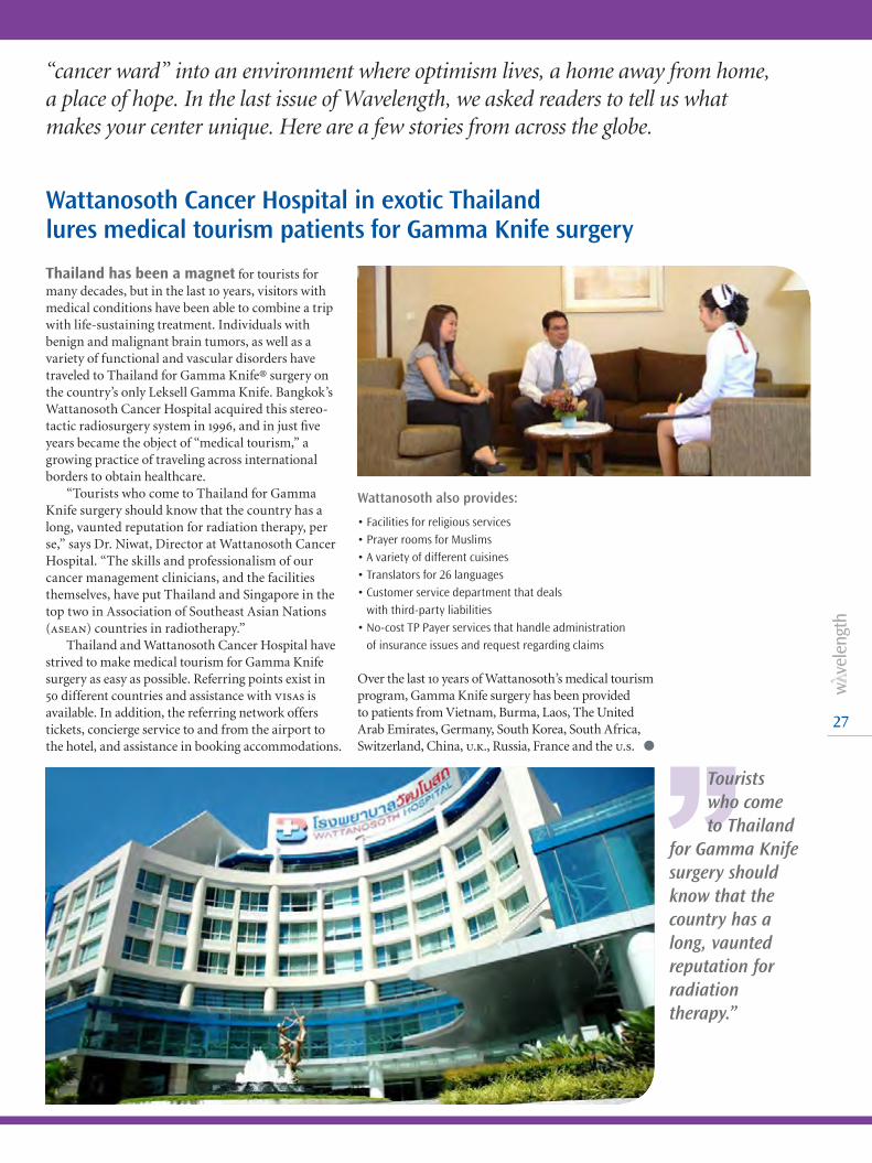
27
Thailand has been a magnet for tourists for many decades, but in the last 10 years, visitors with medical conditions have been able to combine a trip with life-sustaining treatment. Individuals with benign and malignant brain tumors, as well as a variety of functional and vascular disorders have traveled to Thailand for Gamma Knife® surgery on the country’s only Leksell Gamma Knife. Bangkok’s Wattanosoth Cancer Hospital acquired this stereo-tactic radiosurgery system in 1996, and in just five years became the object of “medical tourism,” a growing practice of traveling across international borders to obtain healthcare.
“Tourists who come to Thailand for Gamma Knife surgery should know that the country has a long, vaunted reputation for radiation therapy, per se,” says Dr. Niwat, Director at Wattanosoth Cancer Hospital. “The skills and professionalism of our cancer management clinicians, and the facilities themselves, have put Thailand and Singapore in the top two in Association of Southeast Asian Nations (asean) countries in radiotherapy.”
Thailand and Wattanosoth Cancer Hospital have strived to make medical tourism for Gamma Knife surgery as easy as possible. Referring points exist in 50 different countries and assistance with visas is available. In addition, the referring network offers tickets, concierge service to and from the airport to the hotel, and assistance in booking accommodations.
Wattanosoth also provides:
• Facilities for religious services • Prayer rooms for Muslims • A variety of different cuisines • Translators for 26 languages • Customer service department that deals
with third-party liabilities• No-cost TP Payer services that handle administration
of insurance issues and request regarding claims
Over the last 10 years of Wattanosoth’s medical tourism program, Gamma Knife surgery has been provided to patients from Vietnam, Burma, Laos, The United Arab Emirates, Germany, South Korea, South Africa, Switzerland, China, u.k., Russia, France and the u.s. l
“cancer ward” into an environment where optimism lives, a home away from home, a place of hope. In the last issue of Wavelength, we asked readers to tell us what makes your center unique. Here are a few stories from across the globe.
Wattanosoth Cancer Hospital in exotic Thailand lures medical tourism patients for Gamma Knife surgery
’’Tourists who come to Thailand
for Gamma Knife surgery should know that the country has a long, vaunted reputation for radiation therapy.”

28
What makes your center unique?
Does your clinic have a compelling patient story? Did you receive press from a particular treatment at your hospital or center? Do you have some special procedure or details that would be interesting to share? Your challenge: Tell us your story and we may feature your clinic in the August 2012 issue of Wavelength.
Write a brief description of your story and send it, marked “Unique Center”, to [email protected]. Include your name, clinic or hospital name and email address. Send it by June 15, 2012. Photographs (high-resolution jpgs) are welcome and encouraged if they help tell the story. We will contact you for more details. We look forward to hearing from you!
Show us what is happening where you are!
A video display on the door of Lake Constance Radiation Oncology Centre (Singen, Germany) includes the message “Let the Sun Shine,” exhorting patients to think positively and have sunny thoughts despite the reason for their visit to this Southern Germany clinic. The message might as well also signify a plea for cloudless skies, as the radiotherapy center’s lights, treatment systems and other machines and systems get a major percentage of their power from the sun.
In August 2011, the Centre installed an array of 232 solar panel modules on its roof, creating a 400 m2 energy collector that converts sunlight into hundreds of kilowatt-hours of electricity daily.
During the summer, the array’s output will be more than the Centre needs to run its two Elekta Synergy® systems, a large bore ct system and the clinic’s IT technology, lighting and air-conditioning.
In the winter months, the clinic will need to supplement its solar power generation with electric-ity from the power grid, resulting in Lake Constance Radiation Oncology Centre purchasing more electricity than it will produce when averaged over 365 days. However, it is the concept of decentralized power production that is critical, according to Holger Wirtz, the clinic’s Technical Director/Chief of Medical Physics, and brainchild of the solar power project.
“This is a brand new idea. We are shifting the paradigm from centralized to decentralized energy production. We are the first in Germany to follow this model in healthcare and the environment and generate our own energy to drive our ‘industrial processes,’” Mr. Wirtz pronounces. “This decreases the financial investment and effort that utilities expend in creating electricity at a central production point – such as an atomic, coal or hydroelectric plant – and distributing it to every energy consumer. Imagine if every home produced energy from its own solar array independent of the power grid; the current needed to be carried on the grid would be much lower.” l
Germany’s Lake Constance Radiation Oncology Centre is world’s only solar-powered radiation therapy clinic
From the left: Holger Wirtz, Technical Director/Medical Physicist, Mari Björnsgard, m.d., Site Management “Satellite Friedrichshafen,” and Prof. Johannes Lutterbach, m.d , m.b.a., Medical Director
ELEKTA CUSTOMERS IN FOCUS
’’We are the first in Germany to
follow this model in healthcare and the environment and generate our own energy to drive our industrial processes.”

29
The Farber Center has partnered with Donna Karan’s Urban Zen to provide Integrative therapy to all their patients on treatment for free. These sessions include specific yoga therapies, reiki therapy, and oil therapy that deal with different aspects of the symptoms of all diseases. They are designed to assist with the symptoms of pain, anxiety, nausea, insomnia, constipation and exhaustion.
Once treatment is over, The Farber Center for Radiation Oncology has joined forces with Urban Zen’s integrative therapy program (uzit) and developed ohe (optimal healing environment) classes. The four-week integrative program is targeted to empower the cancer patent to get the best treatment of mind, body, and sprit. l
The first and only freestanding radiation oncology facility of its kind in Manhattan, The Farber Center for Radiation Oncology (New York City) represents a warm alternative to a hospital environment, without compromising quality of medical care.
In fact, when you walk into The Farber Center, the first question you’re asked is what you’d like to drink – not what insurance you have. Amenities include the ability to rest by the fireside in a cozy chair before slipping into a plush robe in a private dressing room and a warm, welcoming treatment room. Their exam rooms have spa tables with real fitted sheets instead of paper. You will encounter a multilingual staff, aquariums, and state-of-the-art Elekta equipment. They even accept most insurance.
“No matter what kind of treatment a cancer patient receives, the fight against cancer is more than a physical challenge. It impacts everything from emotional well-being to financial stability,” says Leonard Farber, m.d., radiation oncologist and founder of The Farber Center for Radiation Oncology. “We realize that people exist within a matrix of family, friends, jobs, homes, neighbor-hoods, geographical areas, and psychological and cultural environments, all of which can influence health and disease. Our mission is to develop a treatment plan that is right for our patient and their loved ones in an environment that supports and nurtures them.”
The Farber Center for Radiation Oncology caters to patients’ lives at the center and beyond
Leonard Farber, m.d., radiation oncologist and founder of The Farber Center for Radiation Oncology
’’Our mission is to develop a treatment
plan that is right for our patient and their loved ones in an environment that supports and nurtures them.”

Virtual community encourages collaboration
30
Log on to Elekta’s new Oncology Community website and share informa-tion, contribute to discussion forums, and collaborate with colleagues and Elekta.
This virtual community encourages oncology custom-ers to share their experiences and challenges related to modern radiation therapy practices. A single site brings together several discussion forums, covering a range of radiotherapy techniques.
In this user-driven environment, Elekta customers moderate discussion forums, and visitors can provide unbiased input and feedback. You can discuss clinical research or multi-center trials and engage in cross-site collaboration. The Oncology Community lets you develop partnerships with Elekta and other customers, and even offers the ability to set up private chat rooms for customer collaboration.
Visit:
www.onco-community.com
SAVE THE DATE – GLOBAL ELEKTA CUSTOMERS!
Elekta Oncology Users MeetingOpen to all Elekta customers including:
l Radiation & Medical Oncology Information Systems
l Radiation Oncology Delivery Systems
l Treatment Planning Systems
l Neurosurgery
When? Saturday October 27, 2012 Where? Boston, MA, USAwww.elekta.com/usersmeeting
Calendar of EventsMARCH 20-231st International Congress on Minimally Invasive Neurosurgery Florence, Italy
MARCH 25-29The 16th International Meeting of the Leksell Gamma Knife SocietySydney, Australia
MARCH 31 – APRIL 5CLAN 2012Rio De Janeiro, Brazil
APRIL 14-18AANSMiami, USA
MAY 9-13ESTRO 31Barcelona, Spain
MAY 10-12World Congress of BrachytherapyBarcelona, Spain
MAY 14-1951st PTCOGSeoul, Korea
MAY 16-196th International Congress of the World Federation of Skull Base SocietiesBrighton, UK
JUNE 3-6ASSFNSan Francisco, USA
JUNE 7-10DEGRO 2012Wiesbaden, Germany
JUNE 10-1418th Annual OHBMBeijing, China
JULY 29 – AUGUST 2AAPM 54th Annual MeetingCharlotte, USA
AUGUST 26-3018th International Conference on BiomagnetismParis, France
SEPTEMBER 26-29ESSFNCaiscais, Lisbon, Portugal
OCTOBER 6-10CNS 2012Chicago, USA
OCTOBER 24-27EANS Annual Meeting 2012Bratislava, Slovakia
OCT 28 – NOV 1ASTROBoston, USA

Combined Clarity® and CT ImageCT Image
...clearly visualizing the lumpectomy cavity
With Clarity®, it’s realityCapable of integrating with all linac platforms, Elekta’s Clarity enhances contouring and setup to support PTV margin reductions – all without added ionizing radiation or invasive fiducial markers. With imaging that has proven sub-millimetric spatial accuracy, Clarity takes visualization of soft tissue to new, unsurpassed levels.
Experience the Elekta DifferenceMore at elekta.com/imagine
4513
371
096
8 01
:12
imagine

Corporate Head Office:
Elekta AB (publ) Box 7593, SE-103 93 Stockholm, Sweden
Tel +46 8 587 254 00 Fax +46 8 587 255 00
Regional Sales, Marketing and Service:
North America
Tel +1 770 300 9725 Fax +1 770 448 6338
Europe, Latin America, Africa, Middle East & India
Tel +46 8 587 254 00 Fax +46 8 587 255 00
Asia Pacific
Tel +852 2891 2208 Fax +852 2575 7133
www.elekta.com