W. JAME GARDNERS M.D, - mdedge-files-live.s3.us-east-2 ... filecyst, symptom osf increasin...
6
ENCYSTED SUBDURAL HEMATOMA W. JAMES GARDNER, M.D. Although hematomas of the "dura mater" were described by Virchow in 1857, the relative frequency of this lesion has been fully realized only during the past ten years. The lesion consists of an encysted collection of old blood attached to the inner surface of the dura over the convexity of one or both cerebral hemispheres. In this communica- tion, I shall consider only the encysted form of hematoma and shall not include traumatic subdural effusion (fluid hematoma of Munro 1 ) or the acute subdural hematoma which produces immediate symptoms and is generally accompanied by a laceration of the brain. The latter two lesions are so different in their morphological and clinical aspects as to deserve separate consideration. The accident responsible for the encysted subdural hematoma is a minor one, as a rule, and the force of the blow usually is exerted in an anteroposterior direction. The resulting dislocation of the brain within the cranial cavity stretches and tears one or more of the short cerebral veins communicating with the immobile sagittal sinus. There occurs at once a large extravasation of blood over the convexity of the hemi- sphere in the subdural space. The bleeding ceases when the intra- cranial pressure reaches a level at which no more blood can leave the torn vein. This degree of intracranial pressure is not incompatible with life and the immediate symptoms may be mild and transient. The subsequent behavior of the subdural clot is unique. The usual regressive changes and absorption do not occur with a clot in this loca- tion. Within a few days or a week the clot is surrounded by a mesothelial-like membrane after which the contents liquefy, forming a hemorrhagic cyst. During this stage symptoms may be absent or trivial. The contents of the cyst, however, undergo a slow progressive augmentation 2 due to the accession of cerebrospinal fluid which is drawn through the semipermeable lining by the osmotic tension of the contained blood proteins. Accompanying this increase in size of the cyst, symptoms of increasing intracranial pressure supervene and most of these patients probably die without the true nature of the condition having been recognized (Fig. 1). A summation of our experiences with these lesions produces the following composite picture: A man, aged 50 years, slips on the ice and falls, striking the occiput. He gets to his feet, slightly dazed for a minute, following which he has pain in the head for a few hours, but this is not severe enough to warrant his going to bed. Thereafter he feels quite well and dismisses the accident from his mind. Three weeks later he begins to have headache which increases in severity. The relatives then notice changes in his behavior, accompanied by mental 201
Transcript of W. JAME GARDNERS M.D, - mdedge-files-live.s3.us-east-2 ... filecyst, symptom osf increasin...

ENCYSTED SUBDURAL HEMATOMA W . JAMES GARDNER, M . D .
Although hematomas of the "dura mater" were described by Virchow in 1857, the relative frequency of this lesion has been fully realized only during the past ten years. The lesion consists of an encysted collection of old blood attached to the inner surface of the dura over the convexity of one or both cerebral hemispheres. In this communica-tion, I shall consider only the encysted form of hematoma and shall not include traumatic subdural effusion (fluid hematoma of Munro1) or the acute subdural hematoma which produces immediate symptoms and is generally accompanied by a laceration of the brain. The latter two lesions are so different in their morphological and clinical aspects as to deserve separate consideration.
The accident responsible for the encysted subdural hematoma is a minor one, as a rule, and the force of the blow usually is exerted in an anteroposterior direction. The resulting dislocation of the brain within the cranial cavity stretches and tears one or more of the short cerebral veins communicating with the immobile sagittal sinus. There occurs at once a large extravasation of blood over the convexity of the hemi-sphere in the subdural space. The bleeding ceases when the intra-cranial pressure reaches a level at which no more blood can leave the torn vein. This degree of intracranial pressure is not incompatible with life and the immediate symptoms may be mild and transient.
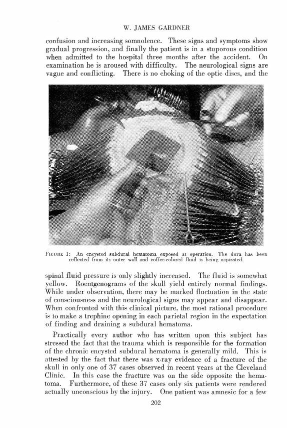
The subsequent behavior of the subdural clot is unique. The usual regressive changes and absorption do not occur with a clot in this loca-tion. Within a few days or a week the clot is surrounded by a mesothelial-like membrane after which the contents liquefy, forming a hemorrhagic cyst. During this stage symptoms may be absent or trivial. The contents of the cyst, however, undergo a slow progressive augmentation2 due to the accession of cerebrospinal fluid which is drawn through the semipermeable lining by the osmotic tension of the contained blood proteins. Accompanying this increase in size of the cyst, symptoms of increasing intracranial pressure supervene and most of these patients probably die without the true nature of the condition having been recognized (Fig. 1) .
A summation of our experiences with these lesions produces the following composite picture: A man, aged 50 years, slips on the ice and falls, striking the occiput. He gets to his feet, slightly dazed for a minute, following which he has pain in the head for a few hours, but this is not severe enough to warrant his going to bed. Thereafter he feels quite well and dismisses the accident from his mind. Three weeks later he begins to have headache which increases in severity. The relatives then notice changes in his behavior, accompanied by mental
201
W. JAMES GARDNER
confusion and increasing somnolence. These signs and symptoms show gradual progression, and finally the patient is in a stuporous condition when admitted to the hospital three months after the accident. On examination he is aroused with difficulty. The neurological signs are vague and conflicting. There is no choking of die optic discs, and the
FIGURE 1: An encysted subdural hematoma exposed at operation. The dura has been reflected from its outer wall and coffee-colored fluid is being aspirated.
spinal fluid pressure is only slightly increased. The fluid is somewhat yellow. Roentgenograms of the skull yield entirely normal findings. While under observation, there may be marked fluctuation in the state of consciousness and the neurological signs may appear and disappear. When confronted with this clinical picture, the most rational procedure is to make a trephine opening in each parietal region in the expectation of finding and draining a subdural hematoma.
Practically every author who has written upon this subject has stressed the fact that the trauma which is responsible for the formation of the chronic encysted subdural hematoma is generally mild. This is attested by the fact that there was x-ray evidence of a fracture of the skull in only one of 37 cases observed in recent years at the Cleveland Clinic. In this case the fracture was on the side opposite the hema-toma. Furthermore, of these 37 cases only six patients were rendered actually unconscious by the injury. One patient was amnesic for a few
202

ENCYSTED SUBDURAL HEMATOMA
days after the injury, although not actually unconscious; several were dazed for a few minutes by the force of the blow. In the remaining cases there was no alteration at all in the state of consciousness produced by the blow. In only four of the 37 CciSGSj a history of the responsible trauma could not be obtained. Of these, one patient had been subject to frequent attacks of epilepsy for 25 years. Another was a boy of 11 years who was quite ataxic due to the presence of a cerebellar tumor. The third patient had been in an automobile accident but stated that he had not injured his head. With this information it seems very likely that the hematoma was the result of an unappreciated or forgotten head injury in each of these three cases. The fourth case in which no history of trauma could be obtained was that of a colored preacher of 62 years with definite slowing of his mental processes due to arteriosclerosis. Because of the memory deficit I do not believe that we can accept his word that head trauma had not occurred.
My experience, therefore, indicates that these lesions are invariably produced by trauma and the burden of proof is upon him who would assume otherwise.
" S P O N T A N E O U S " SUBDURAL HEMATOMA
Since the days of Virchow various authors have tried to differentiate between the "traumatic" and the "spontaneous" or "reactive" types of subdural hematoma. While a spontaneous type may exist, I doubt very much that it ever assumes the typical encysted form of the traumatic variety. The "spontaneous" form is supposed to occur more frequently in the insane, in the decrepit, and in states of acute and chronic alco-holism. For those who maintain that spontaneous hematomas form in these conditions I would like to ask this question. Are there any states other than these in which head trauma is more likely to occur and in which it is less likely to be proven? The answer is no! Many trau-matic hematomas have been described as spontaneous simply due to the failure of the examiner to obtain an adequate history.
For instance, Kaump and Love3 describe the postmortem findings in 30 cases of subdural hematomas. They state that 13 of these hema-tomas were of the traumatic type and 17 were spontaneous. Such a high incidence of the spontaneous lesion is at variance with all published statistics. The difficulty of obtaining the history of trauma has been stressed by many careful observers. Repeated questioning of the rela-tives is frequently necessary in order to establish the fact that the patient has injured his head. In some instances the history can be obtained only after the patient's memory has returned as a result of drainage of the hematoma. Naturally, therefore, when the lesion is identified only at necropsy, the difficulty of obtaining the history of trauma is intensi-fied. Furthermore, in 14 of the 17 cases of so-called spontaneous
203

W. JAMES GARDNER
hematomas reported by Kaump and Love, the sex and age incidence and the gross and microscopic appearance of the lesions differed in no way from those in which a definite history of trauma could be established. Their three spontaneous cases in which there were associated blood dyscrasias did present some gross and microscopic differences and I am prepared to admit that these hematomas were probably genuinely spontaneous.
Recently a theory has been advanced that the subdural hematoma is really an intradural hematoma; that the hemorrhage occurs into the middle, vascular layer of the dura so that the membrane constituting the inner wall of the hematoma is really the inner layer of the dura 4. In my opinion this theory is far from established by the work of these investigators. The difficulty of proving by histological study whether a hematoma is subdural or between the layers of the dura when the lesion is one to four months old is very obvious.
ANALYSIS OF CASES
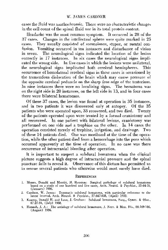
In this series of 37 cases, 33 of the patients were males and four females. The lesion occurred most commonly in the sixth decade of life as shown by the accompanying chart (Fig. 2 ) . In analyzing the nature of the accidents in these 37 cases, we find that automobiles figured in only 10 cases. This is a very low incidence when one con-siders present day craniocerebral injuries as a whole. In ten cases the patient struck his head in falling from a standing position to the floor or ground. Six of these falls were due to icy pavements. In five instances the patient had merely struck his head against a door or low-hanging beam.
In one of the cases the hematoma occurred following a right sub-temporal decompression for a condition which has been called "cerebral pseudotumor." This patient was a man of sixty-seven whose only com-plaint was failing vision which was found to be due to choking of the optic discs. At encephalography, 152 cc. of cerebrospinal fluid were recovered. The resulting films showed the ventricles to be normal with a very marked dilatation of the cerebral sulci on both sides of the brain. There was not the slightest evidence of an expanding intracranial lesion. Immediately after encephalography, a right subtemporal decompression was performed and there was no evidence of a subdural hematoma at that time. The patient's convalescence was uneventful, the intracranial pressure was controlled for a time, and then it recurred. Nine weeks after the encephalogram and decompression, a bilateral parietal trephine was carried out in order to permit the supposedly excessive amount of cerebrospinal fluid to drain into the layers of the scalp. On the right side, much to our surprise, a typical subdural hematoma was encoun-
204

ENCYSTED SUBDURAL HEMATOMA
ENCYSTED SUBDURAL HEMATOMA
F I R S T SECOND T H I R D FOURTH F I F T H S I X T H SEVENTH
D E C A D E S OF L I F E FIGURE 2: Age incidence in 37 cases of encysted subdural hematoma.
tered, and a large quantity of old, dark, fluid blood was removed. We believe that the bleeding in this case occurred as the patient struggled in coming out of his anesthesia, that the hemorrhage was favored by ihe fact that his blood vessels were brittle with age, and the mobility of the brain within the cranial cavity was increased because the cerebro-spinal fluid was replaced with air.
In analyzing these 37 cases as to the site of the trauma, we find that the injury was exerted in an anteroposterior direction in 22, on the side of the head in five, in three cases on the vertex, while in seven instances the exact location of the head injury could not be ascertained. The latent interval between the time of the injury and the onset of definite cerebral signs averages about three weeks, while the lapse of time from the accident to operation is approximately twelve weeks. Where the lapse of time is very much longer than this, there is increasing doubt as to whether the implicated trauma was really the responsible one.
Spinal puncture was performed in 31 of these 37 cases. The pressure of the spinal fluid was definitely increased in 16 instances, while in 19
205
W. JAMES GARDNER
cases the fluid was xanthochromic. There were no characteristic changes in the cell count of the spinal fluid nor in its total protein content.
Headache was the most common symptom. It occurred in 28 of the 37 cases. Changes in the intellectual sphere were quite marked in 25 cases. They usually consisted of somnolence, stupor, or mental con-fusion. Vomiting occurred in ten instances and disturbance of vision in seven. The neurological signs indicated the location of the lesion correctly in 17 instances. In six cases the neurological signs impli-cated the wrong side. In five cases in which the lesions were unilateral, the neurological signs implicated both cerebral hemispheres. This occurrence of homolateral cerebral signs in these cases is occasioned by the tremendous dislocation of the brain which may cause pressure of the opposite cerebral peduncle on the sharp free edge of the tentorium. In nine instances there were no localizing signs. The hematoma was on the right side in 20 instances, on the left side in 13, and in four cases there were bilateral hematomas.
Of these 37 cases, the lesion was found at operation in 35 instances, and in two patients it was discovered only at autopsy. Of the 35 patients who were operated upon, 33 recovered, and two died. Twenty of the patients operated upon were treated by a formal craniotomy and all recovered. In one patient with bilateral lesions, craniotomy was performed on one side and a trephine on the other. In 14 cases the operation consisted merely of trephine, irrigation, and drainage. Two of these 14 patients died. One was moribund at the time of the opera-tion, while the other patient died from a hemorrhage into the pons which occurred apparently at the time of operation. In no case was there recurrence of intracranial bleeding after operation.
It is important to suspect a subdural hematoma when the clinical picture suggests a high degree of intracranial pressure and the spinal puncture fails to reveal it. Observance of this dictum has permitted us to rescue several patients who otherwise would most surely have died.
REFERENCES
1. Munro, Donald and Merritt, H. Houston: Surgical pathology of subdural hematoma based on a study of one hundred and five cases, Arch. Neurol. & Psychiat., 35:64-78, (January) 1936.
2. Gardner, W . James: Traumatic subdural hematoma, with particular reference to the latent interval, Arch. Neurol. & Psychiat., 27:847-858, (April) 1932.
3. Kaump, Donald H. and Love, J. Grafton: Subdural hematoma, Surg., Gynec. & Obst., 67:87-93, (July) 1938.
4. Hannah, J. A . : The aetiology of subdural hematoma, J . Nerv. & Men. Dis., 84:169-186, (August) 1936.
206