Kingdom Protozoa 1 The Protozoans Ciliates Amoeboid Protozoans Flagellated Protozoans.
Upload
chad-gainesCategory
view
217download
2
Volvocine Line
Evolution of Multicellularity in flagellated green algae
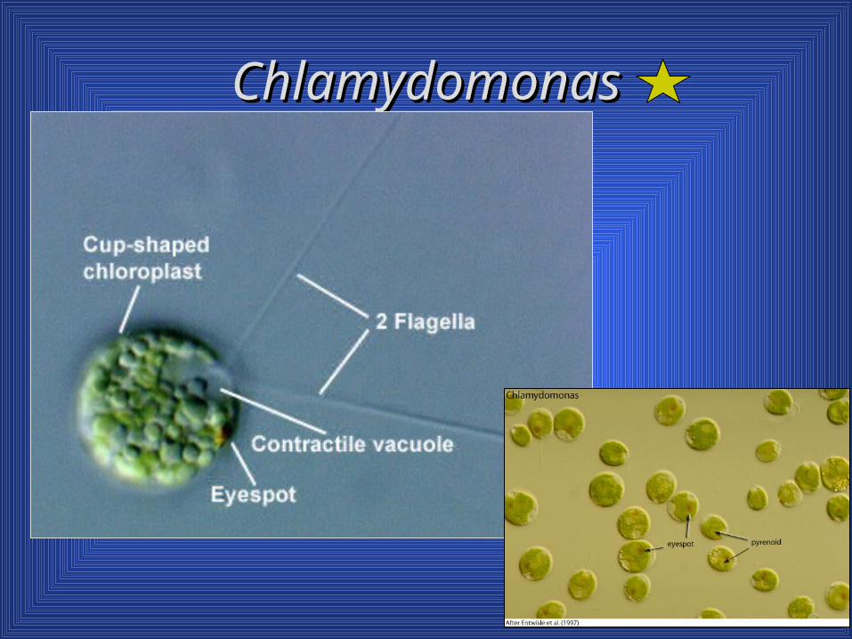
ChlamydomonasChlamydomonas


VolvoxVolvoxsee also see also Volvocine Evolution notes

Protozoan Biodiversity
A guide to the major groupsSpecies seen in lab are marked with a


Bauplan for protozoa
Small size (high SA:V) because of limitations imposed on by diffusion; allows for efficient nutrient assimilation (in photoautrotrophs and mixotrophs), excretion, and gas exchange
Locomotion provided by pseudopodia, cilia or flagella

Bauplan for protozoa Feeding/nutrition:
Photoautotrophs Heterotrophs
Pinocytosis (cell drinking dissolved nutrients) Osmotroph - rely on uptake of small organic molecules Phagocytosis (cells eating solid particles) Use food vacuoles for intracellular digestion Some such as those with fixed shapes, may have
Cytostome (“cell mouth”) and Cytoproct (“cell anus”)
Mixotrophs - heterotrophs or photoautotrophs

Bauplan for protozoa
Very sensitive to external stimuli, but do not have a nervous system; some may have sensory cilia
May respond to Light Mechanical stimuli Chemical gradients Temperature gradients;
Reproduction varies: sexual or asexual (binary fission, multiple fission, budding)

Supergroups
Excavata: feeding groove, phagotrophy Euglenozoa: flagella, Euglena mitochondria Archaeplastida: algae Alveolata: alveoli Stramenopila: very diverse, straw-like hairs on
flagella Rhizaria: plankton, pseudopodia Amoebozoa: pseudopodia, slime molds Opisthokonta: single posterior flagellum
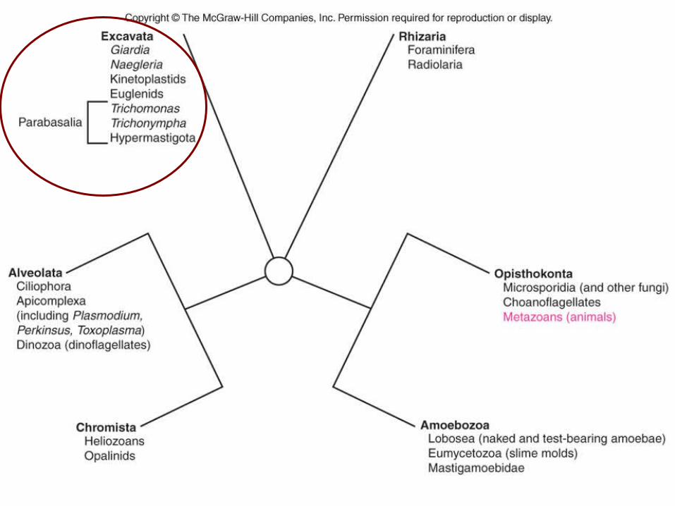

Phylogeny

Supergroup Excavata
Related to some of Earth’s earliest eukaryotes
Named for a feeding groove “excavated” into the cells of many representatives
Food particles are taken into cells by phagotrophy Endocytosis and evolutionary basis for
endosymbiosis

Supergroup Excavata
Jakoba libera
Flagellated protozoans •Single-celled heterotrophs with flagella•Unwalled cells, pellicle retains shape

Zooflagellates
Excavata (clade?) Primitive flagellates
with multiple flagella & feeding groove
Lack Golgi apparatus, mitochondria lacking in some, highly modefied in others
Some important human parasites Giardia Trichomonas Trypanosoma

Excavata, continued Giardia and some other excavates lack
mitochondria. This condition may be primitive or derived. Two separate haploid nuclei – look like big eyes!
Other excavates (e.g. Jakoba) have the most complete mitochondrial genome known - closest to bacterial genome - therefore primitive. Mitochondrial genome of other eukaryotes is
greatly reduced by gene transfer to cell nucleus

Excavata - (excavate - feeding groove that terminates in a cytostome on the cell surface, usually associated with a posteriorly-directed flagellum; not present in all excavate taxa)
Diplomonadida Giardia
Parabasala Trichomonads Trichomonas
vaginalis Trichonymphs Trichonympha spp
Euglenozoa Trypanosomes Trypanosoma spp
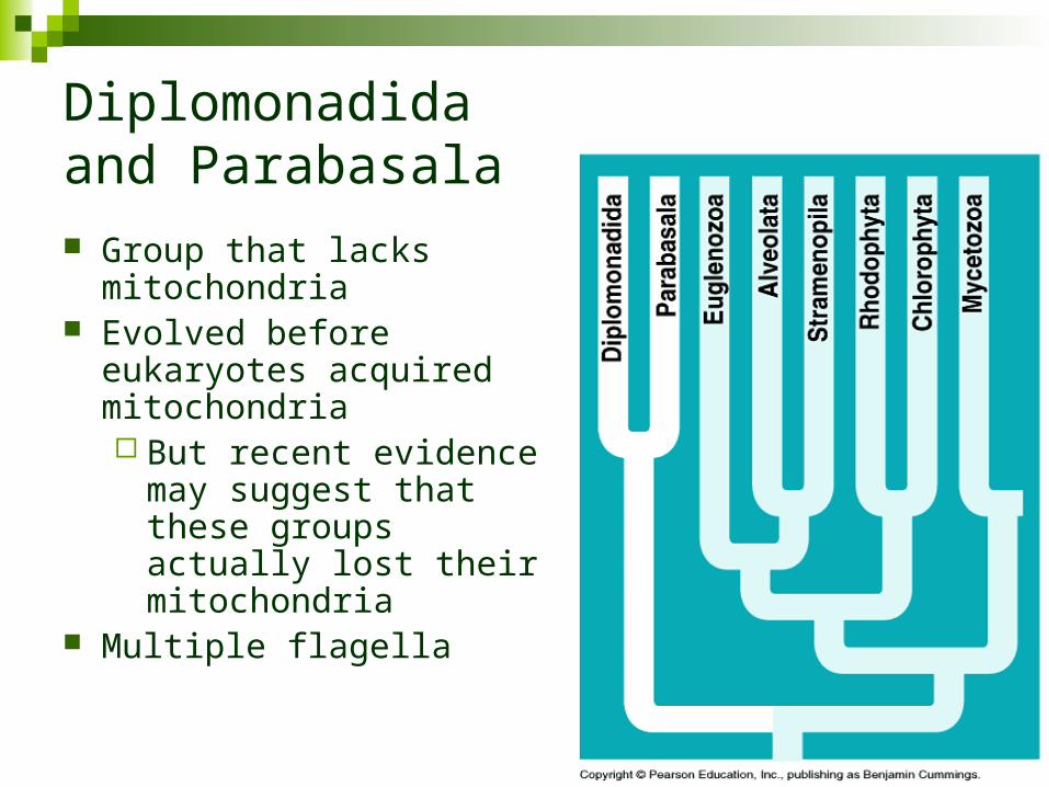
Diplomonadida and Parabasala Group that lacks
mitochondria Evolved before eukaryotes
acquired mitochondria But recent evidence
may suggest that these groups actually lost their mitochondria
Multiple flagella

Diplomonadida* and Parabasala** Giardia lamblia* Trichonympha* Trichomonas
vaginalis**


Giardia intestinalis (lamblia)

Giardia intestinalis

• Water in wilderness areas is often contaminated with cysts from animal feces.
• Cysts hatch in intestines and release trophozoites.
• Always boil and/or treat water before drinking.
• Print out full size slide of life cycle of parasite.
Giardiasis

Giardiasis
Clinical Features: Disease varies from asymptomatic to severe diarrhea
and malabsorption.
Acute giardiasis develops after an incubation period of 1 to 14 days (average of 7 days) and usually lasts 1 to 3 weeks.
Symptoms include diarrhea, abdominal pain, bloating, nausea, and vomiting.
In chronic giardiasis the symptoms are recurrent and malabsorption and debilitation may occur.

Flagellates
Parabasalids Multiple flagella
(hypermastigote) Parabasal bodies-
modified Golgi apparatus Lack mitochondria All are symbiotes in animals Examples:
Trichonympha, Trichomonas
Trichonympha

Trichonymphs
Trichonymphs (phylum Axostylata) are excavates with hundreds of flagella They live in the guts of wood-eating termites and
cockroaches Feed on wood particles consumed by the host
insect They rely on endosymbiotic bacteria to digest
cellulose So insect gets the energy and carbon from
bacterial metabolism – a long trip from wood!

ParabasalidsTermite symbiotes
Guts of termites, wood roaches, other wood-eating insects.
Digestion of cellulose- ecologically critical function.
Acquisition at hatching and molting.
Mutualism Mutualistic bacteria inside
Trichonympha et al. digest the wood. Trichonympha

Eastern termite: Reticulitermes flavipes12 symbiotic flagellates found in the gut(Yamin, M. A. 1979. Sociobiology, 4: 3-119)
1. Dinenympha fimbriata 2. Dinenympha gracilis 3. Holomastigotes elongatum 4. Microjoenia fallax 5. Monocercomonas sp. 6. Personympha major 7. Personympha vertens 8. Spironympha kofoidi 9. Spirotrichonympha flagellata 10. Spirotrichonympha sp. 11. Trichomonas trypanoides 12. Trichonympha agilis
12.7. 2.

Joseph Leidy 1823-1891
Prominent American biologist- Univ. of Pennsylvania. Vertebrate paleontology, parasitology, other fields

TrichonymphaTrichonympha

TrichonymphaTrichonympha
Termite gut symbiontsTermite gut symbionts
StreblomastixStreblomastix


Parabasalids
Trichomonads
• Commensal or parasitic flagellates with axostyle and parabasal body
• Trichomonas vaginalis (far right) in human urogenital tract.

Trichomonas vaginalis Trichomoniasis = STD Clinical Features:
Trichomonas vaginalis infection in women is frequently symptomatic.
Vaginitis with a purulent discharge is the prominent symptom, and can be accompanied by vulvar and cervical lesions, abdominal pain, dysuria and dyspareunia.
The incubation period is 5 to 28 days. In men, the infection is frequently asymptomatic;
occasionally, urethritis, epididymitis, and prostatitis can occur.

Trichomonas vaginalis


Euglenids
Phylum EuglenozoaToday’s euglenids are a modern rep-resentative of an ancient line of life, so different from other protists that some biologists have suggested placing them in a kingdom of their own.


Supergroup Euglenozoa
Supergroup of flagellates named for Euglena
Disk-shaped mitochondrial cristae Euglenoids have unique interlocking
protein strips beneath plasma membranePellicle
Can crawl through mud – euglenoid movement or metaboly

Euglenozoa
Discicristates
Discicristates

Euglenozoa
Most euglenoids live in freshwaterSome have chloroplasts that arose by
secondary endosymbiosis from a green alga
Contractile vacuoles expel excess water


Euglenozoa
Kinetoplastids have an unusually large mass of DNA (kinetoplast) Trypansosoma brucei Most are parasites Have a single giant
mitochondrion Biting insects are vectors Example: Trypanosomes


Flagellates
Kinetoplastida Possess “kinetoplast” region of mitochondrion
Flagellum adheres to cell via “membrane” Includes important parasites of man and
domestic animals: Trypanosoma, Leishmania Digenetic life cycle (two hosts)
vertebrate Gut of blood-feeding insect (vector)


Trypanosomes (Kinetoplastids) Trypanosomes (also in phylum Euglenozoa) are
colorless, mostly pathogenic parasites
They reproduce asexually by mitosis
Kinetoplast – contains extranuclear DNA
Trypanosoma brucei (gambiense) causes African sleeping sickness
T. cruzi causes Chagas’ disease of Central and South America and now potentially in the U.S.

Tsetse fly
Vectors of trypanosome diseases
Trypanosomes in blood smear
Kissing bugKissing bug
Tsetse Fly

25 µm
Flagellum
Red bloodcells
Trypanosomewith undulatingmembrane

Figure 5.27
Trypanosoma cruzi Chagas disease


Trypanosomiasis
T. cruzi life cycle Transmitted to man via
feces of bug Intracellular-
reproduces asexually, forming pseudocysts.
Return to bloodstream and picked up by bug with blood meal.
Do not show antigenic variation.
Triatomine refers to the subfamily Triatominae ofthe family Reduviidae

Chagas’ Disease Clinical Features:
A local lesion can appear at the site of inoculation. The acute phase is usually asymptomatic, but can present with manifestations that include fever, anorexia, lymphadenopathy, mild hepatosplenomegaly, neurological disorders, & myocarditis.
Most acute cases resolve over a period of 2 to 3 months into an asymptomatic chronic stage. The symptomatic chronic stage may not occur for years or even decades after initial infection. Its manifestations include cardiomyopathy; pathologies of the digestive tract such as megaesophagus and megacolon; and weight loss. Chronic Chagas disease and its complications can be fatal.
Charles Darwin may have contracted this disease in Chile during his voyage on the H.M.S. Beagle, which resulted in his infirmity in later life.

Trypanosomiasis
American trypanosomiasis (Chagas disease) Trypanosoma cruzi 16-18 million persons
are infected 100 million are at risk 50,000 deaths
annually leading cause of heart
disease in South and Central America

T. cruzi insect host: Triatoma (Order Hemiptera)

Kinetoplastida
Trypanosoma T. rhodesiense and T.
gambiense cause African sleeping sickness in man.
T. brucei causes nagana in ungulates (hoofed mammals).
Insect host: tse-tse flies, Glossina


TrypanosomiasisAfrican sleeping sickness
Often fatal if untreated. Anemia, fever, edema due to
swollen lymph nodes. Neurological symptoms-
dementia, lethargy, paralysis. Controlled in past decades by
anti-tse-tse programs- now increasing again.
300,000 cases/year

African Sleeping Sickness
Bite reaction Parasitemia
Attacks of fever which starts 2-3 weeks after the bite CNS stage
Changes in character and personality Terminal stage is marked by wasting and
emaciation Death results from coma, intercurrent infection
or cardiac failure


African trypanosomes
Quick-change artists - antigenic variation Fluctuating parasite
number- antigenically distinct forms.
VSG (variant surface glycoprotein) coats cell surface.
Cells switch between different versions of VSG.
Shed VSG causes problems
VSG


Kinetoplastida
Leishmania
• vector: biting sandflies, Phlebotomus (O. Diptera)
• Zoonosis= mainly parasite of animals that also infects man
• reservoir hosts‑ various mammals‑ rodents, carnivores (including dogs)

Leishmaniasis• Intracellular - infects
macrophages in mammal hosts.
• Visceral leishmaniasis suppresses immune response by destroying macrophages in lymph nodes, liver and spleen
• Cutaneous leishmaniasis causes skin ulcers.• 1.5-2 million clinical cases/year- estimate 12
million infected.

Euglenids Cell surface covered by pellicle
Protein strips Unique among eukaryotes Consists of the plasma membrane, a series of
proteinaceous strips underneath the plasma membrane, and groups of microtubules associated with each strip.
Species with longitudinal strips have rigid cells; no metaboly
Species with spirally placed strips are flexible; have metaboly

Euglenids
Photosynthetic species Store paramylon starch in single pyrenoid Have photosensitive eyespot or stigma, that helps orient
towards light Heterotrophic species can either absorb nutrients
from water or may feed on bacteria Two flagella
Long one has mastigonemes and is used for motility Second short flagella is not used for swimming
No sexual reproduction ever reported

Euglena
Paramylon starch Paramylon starch around pyrenoidaround pyrenoid

paramylon
Contractile vacuole

Euglena gracilisEuglena gracilis

Click here for movie of metaboly in Euglena

Euglena acusEuglena acus On slideOn slide

PeranemaPeranema
Anatomy similar to Euglena Anatomy similar to Euglena withoutwithout the chloroplasts and the chloroplasts and pyrenoids.pyrenoids.

Peranema - notice the pellicle

Peranema
Peranema is a predator, capturing and engulfing smaller euglenids. Two rods, located in the mouth area, are used to hold prey during engulfment.
In the euglenid line, as in most other flagellated protists, individuals divide longitudinally.
Division begins with duplication of the basal body at the base of the flagellum creating a cell with two flagella that then splits right down the middle.


Alveolata
Dinoflagellates, apicomplexans, and ciliates
All contain membrane bounded cavities (alveoli)

Supergroup Alveolata
All alveolates have tiny sacs (alveoli) beneath the plasma membraneAll single-celled
Examples: Ciliates, dinoflagellates, and apicomplexans

Alveolates Group characteristics
Sacs (alveoli) lie immediately below the cell surface.
Have tubular inner membranes (cristae) in their mitochondria (Tubies).
Three major taxa with very different adaptive strategies Dinoflagellates (Mostly) parasitic apicomplexa; non-motile Ciliates.

Supergroup Alveolata Ciliophora
Ciliates – conjugation Dinozoa
Dinoflagellates – some photosynthetic, others not Important in nearshore oceans
Apicomplexa Medically important parasites Plasmodium
*Named for saclike membranous vesicle (alveoli) present in cell periphery*

Dinoflagellates
Subphylum Dinozoa = Dinoflagellata


Dinoflagellates Often classed with algae Cell complexity
Single cells or chains of cells. How are their cells organized?
Mesokaryotes – permanently condensed chromosomes
Mitotic spindle located outside of the nucleus (which remains intact during mitosis)
What pigments do they possess? Chlorophyll a, Chlorophyll c and Peridinin.
What storage product is made? Starch and oils.


Dinoflagellates
Cell wall features? Most dinoflagellates are encased in plates of
armor. Thick cellulose plates encased in vesicles beneath the
cell membrane
Some are “naked” and lack these plates
2 flagella present. One trails behind One lies in groove around center of cell Cell spins slowly like a top as it swims

Ceratium
Gonyaulax: Gonyaulax: an aan armoured dinoflagellate. Cell wall is subdivided into multiple polygonal vesicles filled with relatively thick cellulose plates

A “naked” dinoflagellate. Cell wall does not have thickened cellulose armour plates.

Note: Armored dinoflagellate. Know: cingulum, sulcus, hypotheca, epitheca, flagella

Dinoflagellates

Dinoflagellates
Some have elaborate eyespots called ocelli, which have a pigmented portion and a lens-like refractive portion.
Some have trichocysts, which are ejectile organelles similar to the nematocysts in Cnidarians. What other group of protists has these?

CeratiumCeratium

CeratiumCeratium
Note: nuclei with permanently condensed chromosomes

Dinoflagellates Mature dinoflagellates are haploid (1n)
Dikaryotic nuclei – 2 haploid nucleipermanently condensed chromosomes
ReproductionMostly asexualReproduce by fissionA few can reproduce sexually
Gametes formed by mitosis (not meiosis) because the cells are already haploid.
Gametes (1n) are motile Zygotes (2n) formed by fusion of gametes also motile
mei
osis

Dinoflagellates Ecology
90% are marine 10% freshwater About 50% are photosynthetic; the rest are
heterotrophs (parasites) Photosynthetic dinoflagellates are second only to
diatoms as primary producers in coastal waters. May be free-living or symbiotic
Zooxanthellae - symbionts of cnidarians and others
vital to the growth and survival of coral reefs

Zooxanthellae
Dinoflagellate endosymbionts of animals and protozoa
Coral reef builders

Zooxanthellae Symbiotic dinoflagellates found in many
marine invertebrates Genus Symbiodinium Sponges, corals, jellyfish, Tridacnid clams and
flatworms Also found within protists, such as ciliates,
foraminiferans, and colonial radiolarians.

ZooxanthellaeZooxanthellae

Zooxanthellae
Endosymbionts of animals and protozoa In coral polyps zooxanthellae are found in the
second layer of cells below the epidermis; one algal cell per animal cell.
Important components of reef building corals* Provide them with nutrients Remove waste Contribute to the production of calcium carbonate
skeletons
* More about this when we study Cnidarians

Zooxanthellae
Mutualism Host organism ingests the dinoflagellate and
incorporate it into its own tissues without harming it.
Dinoflagellate divides repeatedly, and begins to manufacture carbohydrates which are provided to the host.
Many corals get all their food from the zooxanthellae; build reefs much faster with the dinoflagellates present in their tissues.

ZooxanthellaeZooxanthellae

Zooxanthellae
Recall observations on zooxanthellae in tissues of Aiptasia anemones from S219 aquarium
Cassiopeia jellyfish (aquarium) also have zooxanthellae and typically rest upside down in shallow mangrove beds. This provides maximum sun exposure for symbionts Jellyfish also feeds on passing zooplankton Blue structures are vesicular appendages that hold
zooxanthellae

Aiptasia anemone with zooxanthellae
The upper layer of the Acropora sp. is the epidermis. The lower layer is the gastrodermis. Within the cells are round to oval golden spheres. These are the zooxanthellae.

Cassiopeia, the Upside-Down Jelly or Mangrove Jelly (Figure 7), generally lies on upside-down on the substrate where it tends its internal garden of zooxanthellae, which give it a greenish color. While there, the bell margins pulsate creating a current across the oral surface where plankton and other particles are subdued by nematocysts and caught in a gelatinous coating. The captured particles are carried to the mouth or to other secondary mouths that occur on the oral arms. These are animals of warm, shallow water of the West Indies, the Pacific, and the Indian Oceans.

Coral Bleaching = loss of zooxanthellaeCauses – discussed with Cnidarians

Bioluminescent Dinoflagellates

Bioluminescence
Some dinoflagellates are capable of producing light - bioluminescenceMolecules made by the organism
produce light in a chemical reaction.
Luciferin and luciferase Same reaction that occurs in fireflies


Health Issues
Many dinoflagellates produce neurotoxins Poisons that injure the nerves of marine life that feed on
the dinoflagellates May cause massive kills of fish and shellfish, as well as
other forms of marine life. If animals containing these toxins are eaten by humans,
the result may be illness or even death. Neurotoxins affect muscle function, preventing normal
transmission of electrochemical messages from the nerves to the muscles by interfering with the movement of sodium ions through the cellular membranes

Health Issues
These toxins in the water can blow inland in sea spray and cause temporary health problems for people who live near the coast.
The toxin from Gonyaulax catenella is so toxic that an aspirin sized tablet of the poison could kill 35 people; it is one of the strongest known poisons

Neurotoxins Saxitoxin - most common dinoflagellate toxin
100,000 times more potent than cocaine Found in North American shellfish from Alaska to Mexico,
and from Newfoundland to Florida Brevitoxin
Causes fish kills May also cause poisoning in humans when it accumulates
in the tissues of shellfish Red Tides
Population explosions of dinoflagellates that can color the water red.
Shellfish contain high levels of toxins during these times

Gonyaulax and views of red tides
A red tide results from a population explosion of dinoflagellates (an algal bloom). Cell densities are so high that they turn the water a red color.
Boat

Bioluminescent Red Tide
Noctiluca

Neurotoxins Humans may be poisoned:
By eating contaminated fish - Ciguatera Or by eating shellfish, such as clams or mussels -
paralytic shellfish poisoning or PSP. Poisoning is serious but not usually fatal.
Lethal concentrations lead to death from respiratory failure and cardiac arrest within twelve hours of consumption
Old rule of thumb was that shellfish should only be eaten during months with an "R" in them, and not during May to August. Summer brings runoff of nutrients and blooms of dinoflagellates. NOT VERY RELIABLE!

Noctiluca - a bioluminescent marine dinoflagellate; also causes red tides. Can feed heterotrophically by using its longer posterior flagellum to capture prey.


Pfiesteria piscicida Ulcers on fish caused (?) by PfiesteriaNote the long flagella
Pfiesteria and some of its relatives cause death in fish and respiratory and neurological complications in humans

![[BIO] 02 - Origin of Multicellularity (Calsado)](https://static.fdocuments.in/doc/165x107/54686e2bb4af9f3a3f8b5cfa/bio-02-origin-of-multicellularity-calsado.jpg)