Voluntary out-of-body experience: an fMRI study · Smith and Messier Voluntary out-of-body...
10
HUMAN NEUROSCIENCE ORIGINAL RESEARCH ARTICLE published: 10 February 2014 doi: 10.3389/fnhum.2014.00070 Voluntary out-of-body experience: an fMRI study Andra M. Smith and Claude Messier * School of Psychology, University of Ottawa, Ottawa, ON, Canada Edited by: John J. Foxe, Albert Einstein College of Medicine, USA Reviewed by: Aymeric Guillot, University Claude Bernard Lyon 1, France Andre J. Szameitat, Brunel University London, UK *Correspondence: Claude Messier, School of Psychology, University of Ottawa, 136 Jean-Jacques Lussier, Room 2079A, Ottawa, ON K1N 6N5, Canada e-mail: [email protected] The present single-case study examined functional brain imaging patterns in a participant that reported being able, at will, to produce somatosensory sensations that are experienced as her body moving outside the boundaries of her physical body all the while remaining aware of her unmoving physical body.We found that the brain functional changes associ- ated with the reported extra-corporeal experience (ECE) were different than those observed in motor imagery. Activations were mainly left-sided and involved the left supplementary motor area and supramarginal and posterior superior temporal gyri, the last two overlapping with the temporal parietal junction that has been associated with out-of-body experiences. The cerebellum also showed activation that is consistent with the participant’s report of the impression of movement during the ECE. There was also left middle and superior orbital frontal gyri activity, regions often associated with action monitoring. The results suggest that the ECE reported here represents an unusual type of kinesthetic imagery. Keywords: body representation, cerebellum, kinesthetic imagery, motor imagery, out-of-body experiences, somatosensory systems, temporal parietal junction INTRODUCTION The experience of one’s body is a central process to allow us to interact with the outside world. Body experience is based on the integration of visual, vestibular, and somatosensory infor- mation (Giummarra et al., 2008; Berlucchi and Aglioti, 2010; de Vignemont, 2011; Blanke, 2012; Moseley et al., 2012). This infor- mation allows the tracking of the body in space and in relation with other objects and beings in our environment. Tracking of our body in turn, guides our movements (Goodale et al., 2008). The conscious experience of our body is generally congruent across sensory modalities so that, what we see of our body is also what we feel from somatosensory and vestibular sensations (Tsakiris, 2010). The sensations and percept associated with our body in movement can also be elicited in our imagination albeit most of the time in an attenuated form. Motor imagery corresponds to the cognitive version of motor actions without actual motor movements (Guillot et al., 2012; Moran et al., 2012). This motor “imagery” encompass visual components when we imagine move- ments as we would see them from our own perspective or from a third-person perspective (imagine someone else moving – or imagine ourselves moving but from a third-person perspective) and proprioceptive and vestibular components often referred to kinesthetic “imagery” (Guillot et al., 2009, p. 698). Motor imagery is intertwined within the brain’s preparatory processes preceding action and, up to a certain point, the brain’s processes subserving actual movement (Guillot and Collet, 2005). The strongest support for this view has come from functional imaging that demon- strated strong but incomplete overlap between imagery, action preparation, and action (Porro et al., 1996; Guillot et al., 2008, 2009; Szameitat et al., 2012a,b). These studies show that motor imagery is dependent both on brain regions associated with the performance of motor action but also on the somatosensory brain regions associated with body perception. Voluntary and involun- tary motor imagery is also present in amputated individuals with an associated phantom limb often together with somatosensory perception (Melzack, 1989, p. 657; Ramachandran and Hirstein, 1998, p. 493). Some amputees can also train themselves to expe- rience an anatomically impossible movement with their phantom limb suggesting the plasticity of sensorimotor systems (Moseley and Brugger, 2009, p. 1069). The multi-component nature of body representation is also revealed in perceptual illusions such as the rubber hand illusion (Botvinick and Cohen, 1998). In the rubber hand illusion, the vision-based belief that the rubber hand is not part of the par- ticipant’s body is countered by the simultaneous touching of the rubber hand and the real hand and leads to a shift in the attri- bution of the localization of sensory stimulation from the real hand to the rubber hand (Hohwy and Paton, 2010). During the process of establishing the illusion, from completely separate to unity with the rubber hand, several intermediate illusory experi- ences can take place (Valenzuela Moguillansky et al.,2013, p. 1001). In one experiment using a moveable hand model, conditions could be manipulated so that participants reported a dissociation of the sense of ownership (impression that the fake hand is their own) or the sense of agency (impression that participants controlled the movements of the fake hand) (Kalckert and Ehrsson, 2012). Mismatch between the observed position of the hand model and the sensed position of the real hand reduced sense of ownership but did not disrupt the impression of agency. Conversely, passive movement reduced agency but left ownership intact (Kalckert and Ehrsson, 2012). These observations suggest that agency and own- ership may depend on different but overlapping brain networks (Jackson et al., 2006, p. 703). Another experiment demonstrated that concurrent limb and full-body orientation illusions elicited by virtual reality visual displacement were undissociated and not dependent on action (Olive and Berthoz, 2012, p. 1050). During these illusions, the participants do not doubt that the shifted body perception is illusory (Blanke and Metzinger, 2009). Frontiers in Human Neuroscience www.frontiersin.org February 2014 |Volume 8 | Article 70 | 1
Transcript of Voluntary out-of-body experience: an fMRI study · Smith and Messier Voluntary out-of-body...

HUMAN NEUROSCIENCEORIGINAL RESEARCH ARTICLE
published: 10 February 2014doi: 10.3389/fnhum.2014.00070
Voluntary out-of-body experience: an fMRI studyAndra M. Smith and Claude Messier*
School of Psychology, University of Ottawa, Ottawa, ON, Canada
Edited by:John J. Foxe, Albert Einstein Collegeof Medicine, USA
Reviewed by:Aymeric Guillot, University ClaudeBernard Lyon 1, FranceAndre J. Szameitat, Brunel UniversityLondon, UK
*Correspondence:Claude Messier , School ofPsychology, University of Ottawa, 136Jean-Jacques Lussier, Room 2079A,Ottawa, ON K1N 6N5, Canadae-mail: [email protected]
The present single-case study examined functional brain imaging patterns in a participantthat reported being able, at will, to produce somatosensory sensations that are experiencedas her body moving outside the boundaries of her physical body all the while remainingaware of her unmoving physical body. We found that the brain functional changes associ-ated with the reported extra-corporeal experience (ECE) were different than those observedin motor imagery. Activations were mainly left-sided and involved the left supplementarymotor area and supramarginal and posterior superior temporal gyri, the last two overlappingwith the temporal parietal junction that has been associated with out-of-body experiences.The cerebellum also showed activation that is consistent with the participant’s report of theimpression of movement during the ECE. There was also left middle and superior orbitalfrontal gyri activity, regions often associated with action monitoring. The results suggestthat the ECE reported here represents an unusual type of kinesthetic imagery.
Keywords: body representation, cerebellum, kinesthetic imagery, motor imagery, out-of-body experiences,somatosensory systems, temporal parietal junction
INTRODUCTIONThe experience of one’s body is a central process to allow usto interact with the outside world. Body experience is based onthe integration of visual, vestibular, and somatosensory infor-mation (Giummarra et al., 2008; Berlucchi and Aglioti, 2010; deVignemont, 2011; Blanke, 2012; Moseley et al., 2012). This infor-mation allows the tracking of the body in space and in relationwith other objects and beings in our environment. Tracking of ourbody in turn, guides our movements (Goodale et al., 2008). Theconscious experience of our body is generally congruent acrosssensory modalities so that, what we see of our body is also whatwe feel from somatosensory and vestibular sensations (Tsakiris,2010). The sensations and percept associated with our body inmovement can also be elicited in our imagination albeit mostof the time in an attenuated form. Motor imagery correspondsto the cognitive version of motor actions without actual motormovements (Guillot et al., 2012; Moran et al., 2012). This motor“imagery” encompass visual components when we imagine move-ments as we would see them from our own perspective or froma third-person perspective (imagine someone else moving – orimagine ourselves moving but from a third-person perspective)and proprioceptive and vestibular components often referred tokinesthetic “imagery” (Guillot et al., 2009, p. 698). Motor imageryis intertwined within the brain’s preparatory processes precedingaction and, up to a certain point, the brain’s processes subservingactual movement (Guillot and Collet, 2005). The strongest supportfor this view has come from functional imaging that demon-strated strong but incomplete overlap between imagery, actionpreparation, and action (Porro et al., 1996; Guillot et al., 2008,2009; Szameitat et al., 2012a,b). These studies show that motorimagery is dependent both on brain regions associated with theperformance of motor action but also on the somatosensory brainregions associated with body perception. Voluntary and involun-tary motor imagery is also present in amputated individuals with
an associated phantom limb often together with somatosensoryperception (Melzack, 1989, p. 657; Ramachandran and Hirstein,1998, p. 493). Some amputees can also train themselves to expe-rience an anatomically impossible movement with their phantomlimb suggesting the plasticity of sensorimotor systems (Moseleyand Brugger, 2009, p. 1069).
The multi-component nature of body representation is alsorevealed in perceptual illusions such as the rubber hand illusion(Botvinick and Cohen, 1998). In the rubber hand illusion, thevision-based belief that the rubber hand is not part of the par-ticipant’s body is countered by the simultaneous touching of therubber hand and the real hand and leads to a shift in the attri-bution of the localization of sensory stimulation from the realhand to the rubber hand (Hohwy and Paton, 2010). During theprocess of establishing the illusion, from completely separate tounity with the rubber hand, several intermediate illusory experi-ences can take place (Valenzuela Moguillansky et al., 2013, p. 1001).In one experiment using a moveable hand model, conditions couldbe manipulated so that participants reported a dissociation of thesense of ownership (impression that the fake hand is their own)or the sense of agency (impression that participants controlledthe movements of the fake hand) (Kalckert and Ehrsson, 2012).Mismatch between the observed position of the hand model andthe sensed position of the real hand reduced sense of ownershipbut did not disrupt the impression of agency. Conversely, passivemovement reduced agency but left ownership intact (Kalckert andEhrsson, 2012). These observations suggest that agency and own-ership may depend on different but overlapping brain networks(Jackson et al., 2006, p. 703). Another experiment demonstratedthat concurrent limb and full-body orientation illusions elicitedby virtual reality visual displacement were undissociated and notdependent on action (Olive and Berthoz, 2012, p. 1050).
During these illusions, the participants do not doubt that theshifted body perception is illusory (Blanke and Metzinger, 2009).
Frontiers in Human Neuroscience www.frontiersin.org February 2014 | Volume 8 | Article 70 | 1
Smith and Messier Voluntary out-of-body experience
In contrast, shifted body perception of neurological origin (Blankeand Mohr, 2005) or pharmacologically induced (Morgan et al.,2011; Wilkins et al., 2011) can lead to ambiguous embodimentwhereas people report that the illusory body or body part ismore realistic or corresponds to a “double” of their body. In thedescriptions below, the “double” refers to the illusory body (orparts thereof). There seems to be a general consensus in adoptingthe classification proposed by Brugger to describe these illusions(Brugger and Regard, 1997). Autoscopic hallucination is a visualhallucination of the upper part of a double of the body. Heau-toscopy is a visual and somesthetic hallucination. The double,which appears as through a veil, can mirror the person’s move-ments. Heautoscopy hallucination is also accompanied by a vaguefeeling of detachment and depersonalization. The double is feltvaguely as another self. Feeling of a presence is a mostly somes-thetic hallucination that a double is present usually close by or eventouching but not seen. Feeling of a presence is also called sensed-presence experience when the presence is identified as anotherperson (Cheyne and Girard, 2007, p. 1065). Out-of-body experi-ence is a visual and somesthetic experience in which the doubleis seen from a different perspective, often motionless. Because thebody in this experience is “seen” from a third-person perspective(i.e., from above), the body seen is illusory even if it is congruentwith the body’s position during the illusion (e.g., lying down). Theexperience is accompanied by a profound feeling of being outsideof the body and with feelings of meaningfulness of the experience.
Three studies of self-reported anomalous body experiences inunremarkable normal people (Braithwaite et al., 2011, p. 876;Braithwaite et al., 2011, p. 1063; Braithwaite et al., 2013, p. 1064).In the first one, it was noted that most instances of spontaneousanomalous body experiences occurred during a relaxed or bor-derline sleeping state and one-third reported (seeing) their bodyfrom a different perspective while the rest reported a visual orsomatosensory shift in perspective. The participants who reportedout-of-body experience also self-reported more perceptual anom-alies (Braithwaite et al., 2011, p. 876). In two subsequent exper-iments, participants self-reporting anomalous body experiences(mostly of visual nature) were more likely to respond strongly toaversive visual patterns suggesting that the visual system of the par-ticipants are somehow different, at least functionally (Braithwaiteet al., 2013, p. 1064; Braithwaite et al., 2013, p. 1063). The authorsalso derived the hypothesis that these anomalous body experiencesdepended on temporal lobe anomalies as measured by perceptualtasks and questionnaires (Braithwaite et al., 2011, p. 876).
There also have been imaging enquiries into the brain areasinvolved in body representation illusions in neurologically intactparticipants (Blanke, 2012). Brain imaging studies have suggestedthat activity in sensory integration areas such as the intrapari-etal sulcus and the ventral premotor cortex are associated withthe establishment of the rubber hand illusion (Ehrsson et al.,2004, 2005, 2007; Tsakiris et al., 2007). One experiment has usedrepeated transcranial magnetic stimulation to gain informationon the brain areas involved in the rubber hand illusion (Tsakiriset al., 2008). They found that, when the activity of the tempo-ral parietal junction (TPJ) was perturbed by repeated transcra-nial magnetic stimulation, the processing of body representationmental imagery was impaired. However, in another transcranial
magnetic stimulation study, mental rotation of letter stimuli wasnot affected suggesting a specific effect for body representation(Blanke et al., 2005). Another experiment showed that, the tem-poral parietal junction, which is involved in self processing andmultisensory integration of body-related information; and theextrastriate body area (EBA), which responds selectively to humanbodies and body parts mental imagery is performed with mentallyembodied (EBA) or disembodied (TPJ) self location (Arzy et al.,2006). The more intense hallucinations or illusions are usuallyassociated with brain lesions, abnormal brain function such asepilepsy, major psychiatric syndromes, dissociative drugs such asketamine, or in micro-gravity conditions (Kornilova, 1997).
The study of the lesioned or abnormal brain areas is often usedto gain insight into the brain areas involved in normal body repre-sentation phenomena. However, there is also anecdotal evidencethat these intense hallucinations can occur in non-neurologicalcases but they have a low occurrence and, apart from micro-gravityillusions, are unpredictable. In the present report, we used func-tional MRI to examine an otherwise “normal,” healthy individualthat reported the ability to, at will, vividly experience her bodymoving outside her physical body while lying down at rest. Thesubjective description of the participant led us to use the termextra-corporeal experience (ECE) throughout this manuscript tounderline the difference between the phenomenon studied hereand the more common definition of out-of-body experiences. Weincluded a number of guided imagery tasks to specify the ECE-related brain activity. One control task was motor imagery fora different movement (jumping jacks). A second control condi-tion was alternating between actual finger movements and motorimagery of the same movement. Finally, we were interested indetermining if there was a difference between imagining her-self performing the ECE (but not experiencing the ECE) differedfrom the imagining of another person performing the same ECEmovement.
MATERIALS AND METHODSPARTICIPANTThe participant was a right-handed woman, age 24, who was apsychology graduate student at the time of testing. She signed aninformed consent approved by the University of Ottawa ResearchEthics Board. The participant was in an undergraduate class thatpresented data on body representation hallucinations in patientsthat report experiences of their body outside their physical body(Blanke and Arzy, 2005). The participant spontaneously reportedafter class that she could have a similar “out of body” experience.She appeared surprised that not everyone could experience this.The participant described her experience as one she began per-forming as a child when bored with “sleep time” at preschool.She discovered she could elicit the experience of moving aboveher body and used this as a distraction during the time kids wereasked to nap. She continued to perform this experience as she grewup assuming, as mentioned, that “everyone could do it.” This wasoften done before sleep onset as an aid to enter sleep. She describedthe experience as variable depending on her frame of mind. Shewas able to see herself rotating in the air above her body, lying flat,and rolling along with the horizontal plane. She reported some-times watching herself move from above but remained aware of
Frontiers in Human Neuroscience www.frontiersin.org February 2014 | Volume 8 | Article 70 | 2
Smith and Messier Voluntary out-of-body experience
her unmoving “real” body. The participant reported no particularemotions linked to the experience. As an adult, the participantonly infrequently “practiced” the experience; the experience doesnot occur spontaneously but is induced wilfully. The participantdescribes the experience in the following terms:“I feel myself mov-ing, or, more accurately, can make myself feel as if I am moving.I know perfectly well that I am not actually moving. There isno duality of body and mind when this happens, not really. Infact, I am hyper-sensitive to my body at that point, because I amconcentrating so hard on the sensation of moving. I am the onemoving – me – my body. For example, if I ‘spin’ for long enough,I get dizzy. I do not see myself above my body. Rather, my wholebody has moved up. I feel it as being above where I know it actu-ally is. I usually also picture myself as moving up in my mind’seye, but the mind is not substantive. It does not move unless thebody does.”
PROCEDUREFour questionnaires were administered. The Pittsburgh SleepQuality Index (Buysse et al., 1989) was used to detect possible sleepdisturbances because sleep onset disturbances have been associ-ated with altered somatosensory or vestibular perceptions (Braith-waite et al., 2011). In order to estimate visual and kinestheticimagery, the participant was asked to complete the 8-item Move-ment Imagery Questionnaire-Revised (MIQ-R; Hall and Martin,1997) and the 20-item Kinesthetic and Visual Imagery Question-naire (KVIQ; Malouin et al., 2007). Finally, the PAS perceptualaberration scale (Arzy et al., 2007) was administered.
DATA ACQUISITIONThe experimenter provided instructions to the participantthrough MRI earphones. The data was collected in one imag-ing session during which time both anatomical and functionalMR images were obtained. All imaging was performed using a1.5-T Siemens Magnetom Symphony MRI scanner. The partici-pant lay supine with her head secured in a custom head holder.A conventional T1-weighted spin echo localizer was acquired andused to prescribe a subsequent 3D FLASH (TR/TE 11.2/21 ms, flipangle 60°, field of view (FOV) 26 cm× 26 cm, 256× 256 matrix,slice thickness 1.5 mm) volume acquisition used for further struc-tural analyses. A T2 FLAIR scan was also performed and inspectedby a neuroradiologist following the scanning session to ensurethat there was no structural anomaly. Whole brain fMRI was per-formed using a T2*-weighted echo planar pulse sequence (TR/TE3000/40 ms, flip angle 90°, FOV 24 cm× 24 cm, 64× 64 matrix,slice thickness 5 mm, 27 axial slices, bandwidth 62.5 kHz).
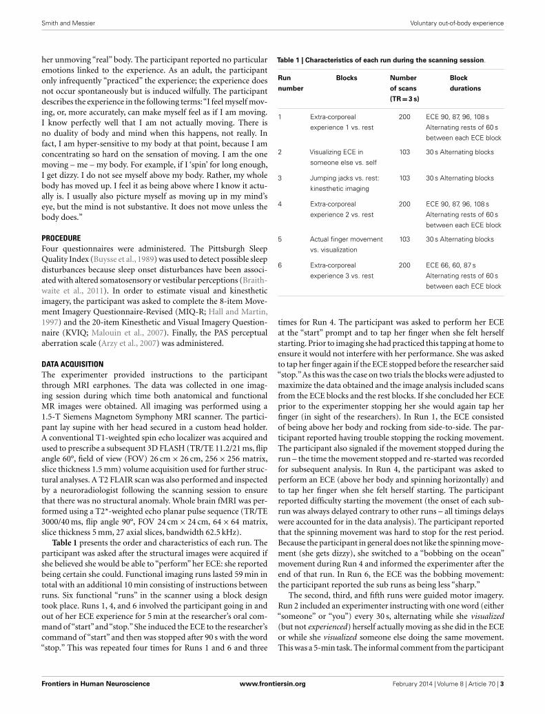
Table 1 presents the order and characteristics of each run. Theparticipant was asked after the structural images were acquired ifshe believed she would be able to “perform” her ECE: she reportedbeing certain she could. Functional imaging runs lasted 59 min intotal with an additional 10 min consisting of instructions betweenruns. Six functional “runs” in the scanner using a block designtook place. Runs 1, 4, and 6 involved the participant going in andout of her ECE experience for 5 min at the researcher’s oral com-mand of “start”and“stop.”She induced the ECE to the researcher’scommand of “start” and then was stopped after 90 s with the word“stop.” This was repeated four times for Runs 1 and 6 and three
Table 1 | Characteristics of each run during the scanning session.
Run
number
Blocks Number
of scans
(TR = 3 s)
Block
durations
1 Extra-corporeal
experience 1 vs. rest
200 ECE 90, 87, 96, 108 s
Alternating rests of 60 s
between each ECE block
2 Visualizing ECE in
someone else vs. self
103 30 s Alternating blocks
3 Jumping jacks vs. rest:
kinesthetic imaging
103 30 s Alternating blocks
4 Extra-corporeal
experience 2 vs. rest
200 ECE 90, 87, 96, 108 s
Alternating rests of 60 s
between each ECE block
5 Actual finger movement
vs. visualization
103 30 s Alternating blocks
6 Extra-corporeal
experience 3 vs. rest
200 ECE 66, 60, 87 s
Alternating rests of 60 s
between each ECE block
times for Run 4. The participant was asked to perform her ECEat the “start” prompt and to tap her finger when she felt herselfstarting. Prior to imaging she had practiced this tapping at home toensure it would not interfere with her performance. She was askedto tap her finger again if the ECE stopped before the researcher said“stop.”As this was the case on two trials the blocks were adjusted tomaximize the data obtained and the image analysis included scansfrom the ECE blocks and the rest blocks. If she concluded her ECEprior to the experimenter stopping her she would again tap herfinger (in sight of the researchers). In Run 1, the ECE consistedof being above her body and rocking from side-to-side. The par-ticipant reported having trouble stopping the rocking movement.The participant also signaled if the movement stopped during therun – the time the movement stopped and re-started was recordedfor subsequent analysis. In Run 4, the participant was asked toperform an ECE (above her body and spinning horizontally) andto tap her finger when she felt herself starting. The participantreported difficulty starting the movement (the onset of each sub-run was always delayed contrary to other runs – all timings delayswere accounted for in the data analysis). The participant reportedthat the spinning movement was hard to stop for the rest period.Because the participant in general does not like the spinning move-ment (she gets dizzy), she switched to a “bobbing on the ocean”movement during Run 4 and informed the experimenter after theend of that run. In Run 6, the ECE was the bobbing movement:the participant reported the sub runs as being less “sharp.”
The second, third, and fifth runs were guided motor imagery.Run 2 included an experimenter instructing with one word (either“someone” or “you”) every 30 s, alternating while she visualized(but not experienced) herself actually moving as she did in the ECEor while she visualized someone else doing the same movement.This was a 5-min task. The informal comment from the participant
Frontiers in Human Neuroscience www.frontiersin.org February 2014 | Volume 8 | Article 70 | 3

Smith and Messier Voluntary out-of-body experience
was that she did not “feel herself moving” when “visualizing” herexperience during run 2. We were interested in determining ifthere was a difference between imagining herself performing theECE (but not experiencing the ECE) differed from the imagingof another person performing the same ECE movement. Run 3included the same alternating block design whereby the partici-pant imagined herself performing jumping jacks or resting : this wasa control task to determine which structures were involved in non-ECE motor imagery. The participant practiced the instructions forRun 3 prior to starting the run to ensure that she was able to visu-alize herself. From the participant’s comments, it was inferred thatvisualizing herself doing jumping jacks did not involve the move-ment sensations associated with her extra corporeal experience.Run 5 involved the participant moving her right hand fingers (oneat a time) to her thumb at a frequency of 2 Hz and then visualizingherself perform the same movement. Again, the participant didnot report a sensation of movement. This control task was addedto determine the brain areas involved in a simple motor actionand its imagined version. Again, each block was 30 s and the Runwas 5 min. Our conversations with the participant suggested thather extra corporeal experience involved the sensation of move-ment while other imagery tasks she performed did not involve thissensation.
IMAGE POST-PROCESSINGThe functional images were reconstructed and whole brain imageswere realigned to correct for motion by employing the proce-dure of Friston et al. (1995), using Statistical Parametric Mapping(SPM8) software. The motion correction did not exceed 1 mm.Images were spatially normalized to match the echo planar imag-ing (EPI) template provided in SPM8 with 2 mm× 2 mm× 2 mmvoxel sizes. Images were then smoothed with a 10 mm full-widthat half-maximum Gaussian filter.
STATISTICAL ANALYSESA fixed effects analysis was performed with data from each Runseparately. The blocks of ECE were compared with the rest blocksfrom the same Run. The Runs with motor imagery and/or visu-alizations were analyzed by contrasting the two types of blocks,for example in Run 3 scans from the rest blocks were subtractedfrom the visualization of jumping jack blocks (Jumping Jacksminus Rest).
RESULTSQUESTIONNAIRESThe MIQ-R results indicated that the participant had kinestheticimagery comparable to that observed in competitive sport ath-letes (M = 5.5) but higher visual imagery (M = 7) (Roberts et al.,2008). In the KVIQ, the participant scored an average of 4.1 on thevisual imagery scale (comparable to healthy but older controls)and 4.3 on the kinesthetic imagery scale, which is higher thanthe same controls. The Pittsburgh Sleep Quality Index (PSQI= 5)was slightly higher than would be expected in healthy partici-pants (PSQI= 2.67): this was essentially due to longer sleep latency(90 min). In the PAS perceptual aberration scale, the participantresponded“false”to most statements except for the following items(her answers in italics): (T.12) Now and then, when I look in the
mirror, my face seems quite different than usual. (Only when con-templating my own mortality); (T.15) Sometimes when I look atthings like tables and chairs, they seem strange. (Occasionally butvoluntary. Sometimes late at night, I can play with perspective i.e.,make things appear closer/farther away. Also, sometimes, ordinaryobjects seem bizarre in the sense that all existence is bizarre); (T.23)It has seemed at times as if my body was melting into my sur-roundings. (Always voluntary. I can make it feel like my body isgoing down into my bed); (T.31) Sometimes I feel like everythingaround me is tilting. (Almost always this is voluntary . . . usuallywhen I am bored in class).
ECE RESULTSThe participant reported being successful at beginning and endingher ECE on demand of the experimenter. The experience for Run1 began immediately and she began to see herself above her bodyrocking with her feet moving down and up as her head movedup and down as in bobbing in ocean waves. The second ECE Runwas the most intense and involved the participant watching herselfabove her own body, spinning along the horizontal axis. The finalECE involved the participant spinning as in the second ECE.
Neural activation patterns for each of these ECE Runs wereanalyzed separately with rest subtracted from the experience.Given the lack of significant difference between the results ofeach of the three Runs, all ECE Runs were combined into oneanalysis to increase power and observe brain regions that wereconcomitantly activated for each Run. Results are reported witha family wise error (FWE) very stringent correction for multi-ple comparisons at 0.001. Results are presented in Figure 1. Themost significantly and consistently activated areas during the ECEcompared to the non-ECE blocks were left lateralized in the sup-plementary motor area (SMA) (x, y, z =−2, −18, 62, cluster247, T = 6.66, p= 0.001), supramarginal gyrus/posterior supe-rior temporal gyrus (x, y, z =−64, −46, 24, cluster 60, T = 6.04,p= 0.001), inferior temporal gyrus (x, y, z =−48,−54,−20, clus-ter 72, T = 5.89, p= 0.001), middle and superior orbital frontalgyri (x, y, z,=−26, 56,−10, T = 5.05, p= 0.001), and the cerebel-lum (x, y, z =−50, −48, −30, T = 5.76, p= 0.001). The parietaland superior temporal activation taken together correspond to thetemporal parietal junction. There was significantly less activationduring the ECE blocks compared to non-ECE blocks (Figure 2)in bilateral posterior visual regions: the lingual gyrus (x, y, z = 14,−64, 4, cluster 19205, T = 13.23, p= 0.001) and the cuneus (x, y,z = 0,−92, 18, cluster 19205, T = 12.71, p= 0.001).
VISUALIZATION RESULTSDuring imagining herself moving as she did in the first ECE (Run1), without inducing an ECE, controlling for multiple compar-isons at a p < 0.001, the participant activated more left cerebellum(x, y, z =−46, −48, −44, cluster 406, T = 5.66, p= 0.001) andbilateral lingual gyrus (x, y, z =−14,−62, 6, cluster 980, T = 5.00,p= 0.001; x, y, z = 6,−58, 8, cluster 790, T = 4.82, p= 0.001) thanwhen imagining someone else moving in the same way (Figure 3).Similarly, she showed significantly less activity during self-imagining than imagining someone else in the bilateral superiororbital frontal gyrus (x, y, z =−18, 66,−2, cluster 148, T = 4.40,p= 0.025; x, y, z = 14, 68,−2, cluster 146, T = 4.38, p= 0.026).
Frontiers in Human Neuroscience www.frontiersin.org February 2014 | Volume 8 | Article 70 | 4

Smith and Messier Voluntary out-of-body experience
FIGURE 1 | Rendered image of significantly activated regions of thebrain while the participant was having extra-corporeal experiences.Most significantly activated regions are lateralized to the left side andinclude the supplementary motor area (F), the cerebellum (B,D,E), thesupramarginal gyrus (D,F), the inferior temporal gyrus (B,D,F), the middleand superior orbitofrontal gyri (A,C,D,E). The p-value was set at 0.001uncorrected for this image with the cluster threshold at 200 significantvoxels.
FIGURE 2 | Areas of reduced activity during the ECEs compared to rest.The visual cortex is particularly impacted. (A) Representation of the rightside; (B) activity on the left. The p-value for this image was set at 0.05 FWEcorrected.
The second control task involved the participant imagining her-self performing jumping jacks and then not imagining anythingand just keeping her eyes closed waiting for the next start cue forthe jumping jacks. Results are presented in Figure 4. The imaginingof herself performing the jumping jacks, controlling for multiple
comparisons at p < 0.001, revealed significantly more activity inthe posterior SMA (x, y, z =−2, −10, 60, cluster 1424, T = 7.95,p= 0.001), paracentral lobule (x, y, z = 0, −12, 68, cluster 1424,T = 6.72, p= 0.001), middle temporal gyrus (BA22) (x, y, z = 68,−48,8, cluster 132,T = 5.72,p= 0.04),precentral gyrus (BA44) (x,y, z =−60, 6, 22, cluster 136, T = 5.11, p= 0.035), inferior parietallobule (x, y, z =−40, −64, 58, cluster 265, T = 4.64, p= 0.001),and superior temporal gyrus (BA22) (x, y, z = 68,−34, 12, cluster156, T = 4.78, p= 0.019). The TPJ activity was more bilateral thanduring the ECE runs (Figure 4). There was also less activity in bilat-eral cuneus (x, y, z = 6,−76, 4, cluster 22067,T = 10.16, p= 0.001)and bilateral superior orbital frontal gyrus (x, y, z =−28, 26,−28,cluster 617, T = 6.50, p= 0.001; x, y, z = 4, 48, −28, cluster 455,T = 5.69, p= 0.001) during the jumping jack imagery comparedto rest.
Another contrast of interest was the actual movement of the fin-gers to the thumb compared with imagining the same movement(Figure 5). There was significantly more activation during theimagining vs. the actual movement in several areas that were simi-larly (but not identically) activated during the ECE. These includedthe bilateral inferior frontal triangularis (x, y, z = 50, 40, −14,cluster 326, T = 5.27, p= 0.001; x, y, z =−42, 58, 0, cluster 1132,T = 5.18, p= 0.001), left middle temporal gyrus (x, y, z =−62,−58, −2, cluster 371, T = 6.31, p= 0.001), left cerebellum (x, y,z =−22, −88, −46, cluster 270, T = 5.97, p= 0.002), left supe-rior parietal lobule (x, y, z =−36, −60, 50, cluster 581, T = 5.56,p= 0.001), and a more anterior part of the SMA (bilateral) (x,y, z = 0, 14, 58, cluster 711, T = 5.56, p= 0.001). Finally, therewas significantly less activity during imagining than movement(Figure 6) in the left postcentral and precentral gyri (x, y, z =−32,−30, 70, cluster 1756, T = 12.85, p= 0.001; x, y, z =−36,−30, 62,cluster 1756, T = 12.05, p= 0.001, respectively), and right cere-bellum (x, y, z = 10, −56, −22, cluster 997, T = 9.95, p= 0.001),areas similar to those activated during the jumping jack condition.
DISCUSSIONThe present experiment examined functional brain imaging pat-terns in a participant that reported being able, at will, to producesomatosensory sensations that are experienced as her body movingoutside the boundaries of her physical body while remaining awareof her unmoving physical body. It is interesting that the develop-ment of the participant’s ability was associated with delayed sleeponset in childhood (which persisted in adulthood) because theoccurrence of out-of-body experiences has been frequently asso-ciated with hypnagogic phenomena (Cheyne et al., 1999; Terhune,2009). The reported experience is similar to what is defined byBrugger as an out-of-body experience but without the feeling ofbeing only outside of her body and without any of the emotionalcontent typically reported in out-of-body experiences (Bruggerand Regard, 1997). The subjective description of the participantled us to use the term ECE throughout this manuscript to under-line the difference between the phenomenon studied here andthe more common definition of out-of-body experiences. Also,because the ECE was private to the participant, we have to rely onthe participant’s descriptions to interpret the results. With thesecaveats in mind, we find that the brain functional changes associ-ated with the reported ECE were different than those observed in
Frontiers in Human Neuroscience www.frontiersin.org February 2014 | Volume 8 | Article 70 | 5

Smith and Messier Voluntary out-of-body experience
FIGURE 3 | Results from visualizing herself doing the same action she performed in the first ECE vs. visualizing another person performing the samemovement. (A) Bilateral lingual gyrus differences in activity and (B) the left cerebellar differences. The p-value for this image was set at 0.001 uncorrected.
FIGURE 4 | Results from visualizing herself performing jumping jacks compared to rest. (A) Right hemisphere; (B) dorsal view of the SMA activity; and(C) left hemisphere activation. The p-value for this image was set to 0.001 uncorrected with the cluster threshold at 100 significant voxels.
motor imagery. The results suggest that the ECE reported hererepresents an unusual type of kinesthetic imagery that sharessome features of previously described out-of-body experiencesand some features of more typical motor imagery.
The ECE was reported as a mixture of visual imagery and kines-thetic imagery but the kinesthetic component was prominent asevidenced by the report of feeling dizzy when performing a rota-tional movement. The prominence of kinesthetic experience overthe visual experience is consistent with a strong bilateral deacti-vation of the lingual gyrus and cuneus encompassing the primaryvisual cortex. Activations are mainly left-sided and involve the leftSMA, supramarginal and posterior superior temporal gyri (the lasttwo overlap with the temporal parietal junction, which has beenassociated with out-of-body experiences). The cerebellum alsoshows strong activation that is consistent with the participant’sreport of the impression of movement during the ECE. Thereare also left middle and superior orbital frontal gyri activations,structures often associated with action monitoring.
The TPJ activation that was observed during the ECE isconsistent with patient cases that report autoscopy and out-of-body experiences when the functional integrity of that area isaltered (Blanke et al., 2004; Blanke and Mohr, 2005; Blanke,2012). Studies of experimentally induced altered body imageryhave demonstrated that transcranial magnetic stimulation of theTPJ area can interfere with the ability of healthy individuals toimagine themselves in body orientations similar to out-of-body
experiences (Blanke et al., 2005). Electrical stimulation of the TPJin epileptic patients also produces various sensations associatedwith out-of-body experience (Blanke et al., 2002). Interestingly,several of the active clusters found in the present experiment dur-ing the ECE (left supramarginal gyrus, left inferior temporal gyrus,left cerebellum) correspond closely to clusters with mirror prop-erties associated with action observation and execution that wereidentified by a recent meta-analysis (Molenberghs et al., 2012).
The middle orbital frontal gyrus is a highly multimodal areathat has been associated with performance monitoring and pro-vides flexibility in response to selection based on ongoing feedback(Elliott et al., 2000). The cluster that we observed in the left orbitalfrontal gyrus corresponds to cluster 6 of the K-6 solution describedby (Kahnt et al., 2012) in their parcelation of the orbitofrontal cor-tex (Kahnt et al., 2012). They reported functional connectivity withadjacent regions in the lateral prefrontal cortex as well as regionsin the inferior parietal cortex and the lateral inferior temporal cor-tex; the latter two structures correspond to activations we observedduring the ECE.
We also instructed the participant to alternate between visu-alizing herself performing her ECE and visualizing someone elseperforming the same movement with the specific instruction thatshe should not experience the ECE but only “see” it. The goal wasto guide the participant toward taking a first-person perspective ofher own experience and transposing it to a third-person perspec-tive. The first-person perspective was associated with a bilateral
Frontiers in Human Neuroscience www.frontiersin.org February 2014 | Volume 8 | Article 70 | 6

Smith and Messier Voluntary out-of-body experience
increase in the lingual gyrus and another one in the left cerebellum:this may indicate that imagining herself included both a visualcomponent and possibly a kinesthetic component (even following
FIGURE 5 |There was significantly more activation during thevisualization of finger movement compared to the actual movement.Each letter represents a different view of the brain (A) anterior view, (B)posterior view, (C) right lateral view, (D) left lateral view, (E) ventral view,and (F) dorsal view. The p-value for this image was set to 0.001 uncorrectedwith the cluster threshold at 100 significant voxels.
a specific instruction to avoid this) that was absent when visu-alizing using the third-person view. The self-visualization wasaccompanied by a reduction in orbitofrontal activation that mayindicate that visualizing herself was easier than taking the third-person view and required less monitoring of activity. Jacksonet al. (2006) studied activations in participants observing handor foot movements seen either from a first-person perspective ora third-person perspective. They found significantly more activityin the left sensory-motor cortex for first-person, during observa-tion alone, and in the lingual gyrus for third-person perspectivesuggesting that perspective taking is associated with a differentpattern of activation (Jackson et al., 2006). It is difficult to recon-cile the higher lingual cortex activity observed with our participanttaking the first-person view and the higher activity with the third-person perspective in Jackson et al. (2006). However, in that study,participants were only shown pictures corresponding to first- orthird-person view of static limbs whereas our participant wasinstructed to visualize a whole body movement. A similar pro-cedure contrasting first and third-person view was used in a studyin which participants viewed hand movements from the two per-spectives (Lorey et al., 2009) and in a study where participants wereinstructed to imagine using a tool presented to them on a pictureor imagine someone else using the same tool (Ruby and Decety,2001). Both these studies reported activation differences whencontrasting first- and third-person views. Our results obtainedcomparing first- and third-person perspective for the ECE experi-ence is similar in that activation differences were observed betweenthe two conditions when the participant “only imagined” the ECE.The pattern of differences that we observed was unsurprisinglyquite different than in previous studies likely owing to the taskdifferences and the number of participants (Ruby and Decety,2001; Lorey et al., 2009).
In the third condition, we examined the brain areas involved ina whole body motor imagery to examine if the ECE was similar tomotor imagery in this participant. The first general observation isthat in this condition, activations tended to be bilateral as opposedto mainly left-sided activations observed in the ECE. The secondobservation is that the activations when the participant was told
FIGURE 6 | Motor areas significantly activated more duringmovement of her fingers to thumb compared with visualizing thesame movement. (A) Representation of the left primary motor cortex;
(B) representation of the right cerebellum. The p-value for this imagewas set to 0.001 uncorrected with the cluster threshold at 100 significantvoxels.
Frontiers in Human Neuroscience www.frontiersin.org February 2014 | Volume 8 | Article 70 | 7

Smith and Messier Voluntary out-of-body experience
to imagine doing jumping jacks were less extensive than for theECE. They included bilateral SMA extending into the paracentrallobule, bilateral inferior parietal lobule, right middle and superiortemporal gyri, and left precentral gyrus. There was reduced activityin the cuneus bilaterally and in the superior orbital frontal gyrusalso bilaterally. Activations of the SMA, inferior parietal lobule,and precentral gyrus have been reported in two previous studiesof kinesthetic imagery using hand movements (Guillot et al., 2009;Szameitat et al., 2012b). ECE and whole body motor imagery wereboth associated with a reduction in cuneus activation (but lessso for motor imagery) suggesting that visual imagery was inhib-ited during both conditions. During motor imagery, there was lessactivity in the superior orbital frontal cortex whereas there wasmore activity in the middle and superior orbital frontal cortexduring ECE. This is suggestive of more motor monitoring duringECE than motor imagery.
The last condition was an attempt to compare the activationsassociated with actual hand movements to imagining the samemovement in this participant (Guillot et al., 2009; Szameitat et al.,2012b). In one of these studies, there were 13 participants selectedon the basis of excellent motor imagery (Guillot et al., 2009)whereas the other included 21 unselected participants (Szameitatet al., 2012b). The number of participants in both these stud-ies achieved a greater statistical power and reported many moreactivations than in the present single-case study. The finger move-ments used in the Guillot et al. study was a learned and practicedsequence, more complex than the one we used, which could beconsidered more of an automatic nature. The movement used inthe Szameitat et al. study consisted of a simple wrist movementtimed with a tone. Although it is not clear how comparable thesestudies are with the present observations, there are a number ofconcordant findings. First, real and imagined movements produceactivations in the SMA. The activations reported by Szameitat et al.(2012a,b) in the contrast imagery-rest include premotor areas inthe precentral gyrus, superior frontal gyrus, and bilateral infe-rior frontal gyri that were also observed in the “jumping jacks”condition of our participant.
It has been shown that visual imagery is reliant on the occipitallobe and the superior parietal lobule, as well as lateral premotorcortex, while kinesthetic imagery is more associated with motorareas and inferior parietal activity (Guillot et al., 2009, p. 698). TheECE in the present study activated the left side of several areas asso-ciated with kinesthetic imagery and was associated with a strongdeactivation of the visual cortex. This suggests that her experi-ence really was a novel one, with a strong kinesthetic component.This was a healthy young woman with no brain abnormalities,thus providing a window into the brain during non-pathological,self-elicited ECE.
There are a number of limitations to the present study. Thefirst obvious one is that we relied on the participant’s report ofher experience. Given that the participant spontaneously reportedher experience assuming that it was a common occurrence andthe detailed (and unusual) description of how she developed thisability, we are inclined to take her report at face value. The privatenature of imagery is common to most research in imagery (includ-ing other imagery conditions in the present report) although a
number of control measures have been devised but they were notused here. One example of such measures is the increase in heartrate and pulmonary ventilation during imagined actions (Decetyet al., 1993; Wuyam et al., 1995). The description of the imagerytasks could have been more clearly specified including the “jump-ing jacks” condition and the third-person ECE task (Moran et al.,2012). Statistical power was obviously limited in this single-casestudy, which means that potentially several activations escapeddetection. Limited statistical power could also have preventedus from finding activation differences when the participant per-formed “variations” of her ECE experience (spinning vs. “bobbingon the ocean”).
This is the first study with a non-pathological participant who isable to elicit an ECE upon demand. Clearly, replication is requiredto ascertain if this pattern of activation is similar in other peoplewho can have self-initiated ECE. The existence of such a case andits presentation raises the possibility that this phenomenon mayhave a significant incidence but unreported because people do notthink this is exceptional. Alternatively, the ability might be presentin infancy but is lost without regular practice. This would be rem-iniscent of the discovery and eventual study of synesthesia thatsome researchers now hypothesized is more prevalent in youngpeople or can be developed (Deroy and Spence, 2013; Simner,2013).
AUTHOR CONTRIBUTIONSClaude Messier and Andra M. Smith designed the experiment,collected the data, and wrote the manuscript. Andra M. Smithanalyzed the MRI data and prepared the figures.
ACKNOWLEDGMENTSThis project was funded by a grant from the Natural Sciences andEngineering Council of Canada to Andra M. Smith. We wouldlike to thank Drs. Francine Malouin and Julien Doyon for helpingwith the visual and kinesthetic imagery questionnaire and Dr. OlafBlanke for the perceptual aberration scale.
REFERENCESArzy, S., Mohr, C., Michel, C. M., and Blanke, O. (2007). Duration and not strength
of activation in temporo-parietal cortex positively correlates with schizotypy.Neuroimage 35, 326–333. doi:10.1016/j.neuroimage.2006.11.027
Arzy, S., Thut, G., Mohr, C., Michel, C. M., and Blanke, O. (2006). Neural basis ofembodiment: distinct contributions of temporoparietal junction and extrastriatebody area. J. Neurosci. 26, 8074–8081. doi:10.1523/JNEUROSCI.0745-06.2006
Berlucchi, G., and Aglioti, S. M. (2010). The body in the brain revisited. Exp. BrainRes. 200, 25–35. doi:10.1007/s00221-009-1970-7
Blanke, O. (2012). Multisensory brain mechanisms of bodily self-consciousness.Nat. Rev. Neurosci. 13, 556–571. doi:10.1038/nrn3292
Blanke, O., and Arzy, S. (2005). The out-of-body experience: disturbed self-processing at the temporo-parietal junction. Neuroscientist 11, 16–24. doi:10.1177/1073858404270885
Blanke, O., Landis, T., Spinelli, L., and Seeck, M. (2004). Out-of-body experienceand autoscopy of neurological origin. Brain 127(Pt 2), 243–258. doi:10.1093/brain/awh040
Blanke, O., and Metzinger, T. (2009). Full-body illusions and minimal phenomenalselfhood. Trends Cogn. Sci. 13, 7–13. doi:10.1016/j.tics.2008.10.003
Blanke, O., and Mohr, C. (2005). Out-of-body experience, heautoscopy, and auto-scopic hallucination of neurological origin implications for neurocognitivemechanisms of corporeal awareness and self-consciousness. Brain Res. Brain Res.Rev. 50, 184–199. doi:10.1016/j.brainresrev.2005.05.008
Frontiers in Human Neuroscience www.frontiersin.org February 2014 | Volume 8 | Article 70 | 8

Smith and Messier Voluntary out-of-body experience
Blanke, O., Mohr, C., Michel, C. M., Pascual-Leone, A., Brugger, P., Seeck, M.,et al. (2005). Linking out-of-body experience and self processing to mentalown-body imagery at the temporoparietal junction. J. Neurosci. 25, 550–557.doi:10.1523/JNEUROSCI.2612-04.2005
Blanke, O., Ortigue, S., Landis, T., and Seeck, M. (2002). Stimulating illusory own-body perceptions. Nature 419, 269–270. doi:10.1038/419269a
Botvinick, M., and Cohen, J. (1998). Rubber hands ‘feel’ touch that eyes see. Nature391, 756. doi:10.1038/35784
Braithwaite, J. J., Broglia, E., Bagshaw, A. P., and Wilkins, A. J. (2013). Evidence forelevated cortical hyperexcitability and its association with out-of-body experi-ences in the non-clinical population: new findings from a pattern-glare task.Cortex 49, 793–805. doi:10.1016/j.cortex.2011.11.013
Braithwaite, J. J., Samson, D., Apperly, I., Broglia, E., and Hulleman, J. (2011). Cog-nitive correlates of the spontaneous out-of-body experience (OBE) in the psy-chologically normal population: evidence for an increased role of temporal-lobeinstability, body-distortion processing, and impairments in own-body transfor-mations. Cortex 47, 839–853. doi:10.1016/j.cortex.2010.05.002
Brugger, P., and Regard, M. (1997). Illusory reduplication of one’s own body: phe-nomenology and classification of autoscopic phenomena. Cogn. Neuropsychiatry2, 19–38. doi:10.1080/135468097396397
Buysse, D. J., Reynolds, C. F. III, Monk, T. H., Berman, S. R., and Kupfer, D. J. (1989).The Pittsburgh sleep quality index: a new instrument for psychiatric practice andresearch. Psychiatry Res. 28, 193–213. doi:10.1016/0165-1781(89)90047-4
Cheyne, J. A., and Girard, T. A. (2007). The nature and varieties of felt presence expe-riences: a reply to Nielsen. Conscious. Cogn. 16, 984–991. doi:10.1016/j.concog.2007.02.003
Cheyne, J. A., Rueffer, S. D., and Newby-Clark, I. R. (1999). Hypnagogic andhypnopompic hallucinations during sleep paralysis: neurological and culturalconstruction of the night-mare. Conscious. Cogn. 8, 319–337. doi:10.1006/ccog.1999.0404
de Vignemont, F. (2011). Embodiment, ownership and disownership. Conscious.Cogn. 20, 82–93. doi:10.1016/j.concog.2010.09.004
Decety, J., Jeannerod, M., Durozard, D., and Baverel, G. (1993). Central activationsof autonomic effectors during mental simulation of motor actions in humans.J. Physiol. 461, 549–563.
Deroy, O., and Spence, C. (2013). Are we all born synaesthetic? Examiningthe neonatal synaesthesia hypothesis. Neurosci. Biobehav. Rev. 37, 1240–1253.doi:10.1016/j.neubiorev.2013.04.001
Ehrsson, H. H., Holmes, N. P., and Passingham, R. E. (2005). Touching a rubberhand: feeling of body ownership is associated with activity in multisensory brainareas. J. Neurosci. 25, 10564–10573. doi:10.1523/JNEUROSCI.0800-05.2005
Ehrsson, H. H., Spence, C., and Passingham, R. E. (2004). That’s my hand! activityin premotor cortex reflects feeling of ownership of a limb. Science 305, 875–877.doi:10.1126/science.1097011
Ehrsson, H. H., Wiech, K., Weiskopf, N., Dolan, R. J., and Passingham, R. E. (2007).Threatening a rubber hand that you feel is yours elicits a cortical anxiety response.Proc. Natl. Acad. Sci. U.S.A. 104, 9828–9833. doi:10.1073/pnas.0610011104
Elliott, R., Dolan, R. J., and Frith, C. D. (2000). Dissociable functions in the medialand lateral orbitofrontal cortex: evidence from human neuroimaging studies.Cereb. Cortex 10, 308–317. doi:10.1093/cercor/10.3.308
Friston, K. J., Ashburner, J., Frith, C. D., Poline, J. B., Heather, J. D., and Frackowiak,R. S. J. (1995). Spatial registration and normalization of images. Hum. BrainMapp. 3, 165–189. doi:10.1002/hbm.460030303
Giummarra, M. J., Gibson, S. J., Georgiou-Karistianis, N., and Bradshaw, J. L. (2008).Mechanisms underlying embodiment, disembodiment and loss of embodiment.Neurosci. Biobehav. Rev. 32, 143–160. doi:10.1016/j.neubiorev.2007.07.001
Goodale, M. A., Gonzalez, C. L., and Kroliczak, G. (2008). Action rules: why thevisual control of reaching and grasping is not always influenced by perceptualillusions. Perception 37, 355–366. doi:10.1068/p5876
Guillot, A., and Collet, C. (2005). Contribution from neurophysiological and psy-chological methods to the study of motor imagery. Brain Res. Brain Res. Rev. 50,387–397. doi:10.1016/j.brainresrev.2005.09.004
Guillot, A., Collet, C., Nguyen, V. A., Malouin, F., Richards, C., and Doyon, J.(2008). Functional neuroanatomical networks associated with expertise in motorimagery. Neuroimage 41, 1471–1483. doi:10.1016/j.neuroimage.2008.03.042
Guillot, A., Collet, C., Nguyen, V. A., Malouin, F., Richards, C., and Doyon, J. (2009).Brain activity during visual versus kinesthetic imagery: an fMRI study. Hum.Brain Mapp. 30, 2157–2172. doi:10.1002/hbm.20658
Guillot, A., Di Rienzo, F., Macintyre, T., Moran, A., and Collet, C. (2012). Imaginingis not doing but involves specific motor commands: a review of experimen-tal data related to motor inhibition. Front. Hum. Neurosci. 6:247. doi:10.3389/fnhum.2012.00247
Hall, C. R., and Martin, K. A. (1997). Measuring movement imagery abilities:a revision of the movement imagery questionnaire. J. Ment. Imagery 21,143–154.
Hohwy, J., and Paton, B. (2010). Explaining away the body: experiences of super-naturally caused touch and touch on non-hand objects within the rubber handillusion. PLoS ONE 5:e9416. doi:10.1371/journal.pone.0009416
Jackson, P. L., Meltzoff,A. N., and Decety, J. (2006). Neural circuits involved in imita-tion and perspective-taking. Neuroimage 31,429–439. doi:10.1016/j.neuroimage.2005.11.026
Kahnt, T., Chang, L. J., Park, S. Q., Heinzle, J., and Haynes, J. D. (2012). Connectivity-based parcellation of the human orbitofrontal cortex. J. Neurosci. 32, 6240–6250.doi:10.1523/JNEUROSCI.0257-12.2012
Kalckert, A., and Ehrsson, H. H. (2012). Moving a rubber hand that feels likeyour own: a dissociation of ownership and agency. Front. Hum. Neurosci. 6:40.doi:10.3389/fnhum.2012.00040
Kornilova, L. N. (1997). Orientation illusions in spaceflight. J. Vestib. Res. 7, 429–439.doi:10.1016/S0957-4271(96)00184-X
Lorey, B., Bischoff, M., Pilgramm, S., Stark, R., Munzert, J., and Zentgraf, K. (2009).The embodied nature of motor imagery: the influence of posture and perspec-tive. Exp. Brain Res. 194, 233–243. doi:10.1007/s00221-008-1693-1
Malouin, F., Richards, C. L., Jackson, P. L., Lafleur, M. F., Durand, A., and Doyon, J.(2007). The kinesthetic and visual imagery questionnaire (KVIQ) for assessingmotor imagery in persons with physical disabilities: a reliability and constructvalidity study. J. Neurol. Phys. Ther. 31, 20–29. doi:10.1097/01.NPT.0000260567.24122.64
Melzack, R. (1989). Phantom limbs, the self and the brain. Canadian Psychol. 30,1–16. doi:10.1037/h0079793
Molenberghs, P., Cunnington, R., and Mattingley, J. B. (2012). Brain regions withmirror properties: a meta-analysis of 125 human fMRI studies. Neurosci. Biobe-hav. Rev. 36, 341–349. doi:10.1016/j.neubiorev.2011.07.004
Moran, A., Guillot, A., MacIntyre, T., and Collet, C. (2012). Re-imagining motorimagery: building bridges between cognitive neuroscience and sport psychology.Br. J. Psychol. 103, 224–247. doi:10.1111/j.2044-8295.2011.02068.x
Morgan, H. L., Turner, D. C., Corlett, P. R., Absalom, A. R., Adapa, R., Arana, F.S., et al. (2011). Exploring the impact of ketamine on the experience of illusorybody ownership. Biol. Psychiatry 69, 35–41. doi:10.1016/j.biopsych.2010.07.032
Moseley, G. L., Brugger, P. (2009). Interdependence of movement and anatomypersists when amputees learn a physiologically impossible movement of theirphantom limb. Proc. Natl. Acad. Sci. U.S.A. 106, 18798–18802. doi:10.1073/pnas.0907151106
Moseley, G. L., Gallace, A., and Spence, C. (2012). Bodily illusions in health and dis-ease: physiological and clinical perspectives and the concept of a cortical ‘bodymatrix’. Neurosci. Biobehav. Rev. 36, 34–46. doi:10.1016/j.neubiorev.2011.03.013
Olive, I., and Berthoz, A. (2012). Combined induction of rubber-hand illusion andout-of-body experiences. Front. Psychol. 3:128. doi:10.3389/fpsyg.2012.00128
Porro, C. A., Francescato, M. P., Cettolo, V., Diamond, M. E., Baraldi, P., Zuiani, C.,et al. (1996). Primary motor and sensory cortex activation during motor per-formance and motor imagery: a functional magnetic resonance imaging study.J. Neurosci. 16, 7688–7698.
Ramachandran, V. S., and Hirstein, W. (1998). The perception of phantom limbs.The D. O. Hebb lecture. Brain 121, 1603–1630. doi:10.1093/brain/121.9.1603
Roberts, R., Callow, N., Hardy, L., Markland, D., and Bringer, J. (2008). Movementimagery ability: development and assessment of a revised version of the vividnessof movement imagery questionnaire. J. Sport Exerc. Psychol. 30, 200–221.
Ruby, P., and Decety, J. (2001). Effect of subjective perspective taking during sim-ulation of action: a PET investigation of agency. Nat. Neurosci. 4, 546–550.doi:10.1038/87510
Simner, J. (2013). Why are there different types of synesthete? Front. Psychol. 4:558.doi:10.3389/fpsyg.2013.00558
Szameitat, A. J., McNamara, A., Shen, S., and Sterr, A. (2012a). Neural activation andfunctional connectivity during motor imagery of bimanual everyday actions.PLoS ONE 7:e38506. doi:10.1371/journal.pone.0038506
Szameitat, A. J., Shen, S., Conforto, A., and Sterr, A. (2012b). Cortical activa-tion during executed, imagined, observed, and passive wrist movements in
Frontiers in Human Neuroscience www.frontiersin.org February 2014 | Volume 8 | Article 70 | 9

Smith and Messier Voluntary out-of-body experience
healthy volunteers and stroke patients. Neuroimage 62, 266–280. doi:10.1016/j.neuroimage.2012.05.009
Terhune, D. B. (2009). The incidence and determinants of visual phenomenologyduring out-of-body experiences. Cortex 45, 236–242. doi:10.1016/j.cortex.2007.06.007
Tsakiris, M. (2010). My body in the brain: a neurocognitive model of body-ownership. Neuropsychologia 48, 703–712. doi:10.1016/j.neuropsychologia.2009.09.034
Tsakiris, M., Costantini, M., and Haggard, P. (2008). The role of the right temporo-parietal junction in maintaining a coherent sense of one’s body. Neuropsychologia46, 3014–3018. doi:10.1016/j.neuropsychologia.2008.06.004
Tsakiris, M., Hesse, M. D., Boy, C., Haggard, P., and Fink, G. R. (2007). Neural signa-tures of body ownership: a sensory network for bodily self-consciousness. Cereb.Cortex 17, 2235–2244. doi:10.1093/cercor/bhl131
Valenzuela Moguillansky, C., O’Regan, J. K., and Petitmengin, C. (2013). Exploringthe subjective experience of the “rubber hand” illusion. Front. Hum. Neurosci.7:659. doi:10.3389/fnhum.2013.00659
Wilkins, L. K., Girard, T. A., and Cheyne, J. A. (2011). Ketamine as a primary predic-tor of out-of-body experiences associated with multiple substance use. Conscious.Cogn. 20, 943–950. doi:10.1016/j.concog.2011.01.005
Wuyam, B., Moosavi, S. H., Decety, J., Adams, L., Lansing, R. W., and Guz, A. (1995).Imagination of dynamic exercise produced ventilatory responses which weremore apparent in competitive sportsmen. J. Physiol. 482(Pt 3), 713–724.
Conflict of Interest Statement: The authors declare that the research was conductedin the absence of any commercial or financial relationships that could be construedas a potential conflict of interest.
Received: 24 October 2013; accepted: 28 January 2014; published online: 10 February2014.Citation: Smith AM and Messier C (2014) Voluntary out-of-body experience: an fMRIstudy. Front. Hum. Neurosci. 8:70. doi: 10.3389/fnhum.2014.00070This article was submitted to the journal Frontiers in Human Neuroscience.Copyright © 2014 Smith and Messier . This is an open-access article distributed underthe terms of the Creative Commons Attribution License (CC BY). The use, distributionor reproduction in other forums is permitted, provided the original author(s) or licensorare credited and that the original publication in this journal is cited, in accordance withaccepted academic practice. No use, distribution or reproduction is permitted whichdoes not comply with these terms.
Frontiers in Human Neuroscience www.frontiersin.org February 2014 | Volume 8 | Article 70 | 10