Volumetric object modeling for surgical simulation
12
Medical Image Analysis (1998) volume 2, number 2, pp 121–132 c Oxford University Press Volumetric object modeling for surgical simulation Sarah Gibson 1* , Christina Fyock 1 , Eric Grimson 2 , Takeo Kanade 3 , Ron Kikinis 4 , Hugh Lauer 1 , Neil McKenzie 1 , Andrew Mor 1,3 , Shin Nakajima 4 , Hide Ohkami 1 , Randy Osborne 1 , Joseph Samosky 2, 4 and Akira Sawada 1, 5 1 MERL, 201 Broadway, Cambridge, MA 02139, USA 2 Massachusetts Institute of Technology, Cambridge, MA, USA 3 Carnegie Mellon University, Pittsburgh, PA, USA 4 Brigham and Women’s Hospital, Boston, MA, USA 5 Mitsubishi Electric Corporation, Hyogo, Japan Abstract Surgical simulation has many applications in medical education, surgical training, surgical plan- ning and intra-operative assistance. However, extending current surface-based computer graphics methods to model phenomena such as the deformation, cutting, tearing or repairing of soft tissues poses significant challenges for real-time interactions. This paper discusses the use of volumetric methods for modeling complex anatomy and tissue interactions. New techniques are introduced that use volumetric methods for modeling soft-tissue deformation and tissue cutting at interactive rates. An initial prototype for simulating arthroscopic knee surgery is described which uses volumetric models of the knee derived from 3-D magnetic resonance imaging, visual feedback via real-time volume and polygon rendering, and haptic feedback provided by a force-feedback device. Keywords: surgical simulation, volume graphics, volumetric modeling Received March 3, 1997; revised July 7, 1997; accepted September 11, 1997 1. INTRODUCTION Computer-based surgical simulation has many applications in medical education, surgical training, surgical planning and intra-operative assistance. In education and training, surgical simulation can reduce costs associated with cadaver speci- mens, provide experience with a greater variety of pathologies and complications, and provide the ability to repeat or replay training procedures. In surgical planning, simulators can enable rehearsal of difficult procedures or planning on patient- specific anatomy and can enhance communication among medical professionals or between doctors and their patients. Intra-operatively, computer modeling can aid in navigation by augmenting the limited surgical field with a more global view of a patient’s anatomy. For example, computer modeling could provide guidance by preventing the surgical instrument from entering sensitive regions. * Corresponding author (e-mail: [email protected]) In order to provide useful feedback to the user, surgical simulators must provide adequate realism. Tissue models should respond in a realistic way when they are manipulated. Rendered images of the surgical field must be realistic enough to be compelling. Haptic or force feedback must mimic forces experienced in real-life because the sense of touch provides important cues in surgery. These requirements impose sig- nificant demands on the surgical simulator. These challenges include a need for physically realistic modeling (such as soft- tissue deformation and tissue cutting or tearing) and tradeoffs among physical and visual realism, the need for real-time interaction and cost. In computer graphics, objects are commonly represented by surface-based polygonal models. Because graphics workstations have special-purpose hardware for fast polygon rendering and because algorithms and systems have been developed for modeling physical interactions between (rigid) polygonal objects, currently there are advantages to using polygonal models in a surgical simulator. However, because
-
Upload
sarah-gibson -
Category
Documents
-
view
212 -
download
0
Transcript of Volumetric object modeling for surgical simulation
Medical Image Analysis (1998) volume 2, number 2, pp 121–132c© Oxford University Press
Volumetric object modeling for surgical simulation
Sarah Gibson1∗, Christina Fyock1, Eric Grimson2, Takeo Kanade3, Ron Kikinis4,Hugh Lauer1, Neil McKenzie1, Andrew Mor1,3, Shin Nakajima4, Hide Ohkami1,Randy Osborne1, Joseph Samosky2,4 and Akira Sawada1,5
1MERL, 201 Broadway, Cambridge, MA 02139, USA2Massachusetts Institute of Technology, Cambridge, MA, USA3Carnegie Mellon University, Pittsburgh, PA, USA4Brigham and Women’s Hospital, Boston, MA, USA5Mitsubishi Electric Corporation, Hyogo, Japan
AbstractSurgical simulation has many applications in medical education, surgical training, surgical plan-ning and intra-operative assistance. However, extending current surface-based computer graphicsmethods to model phenomena such as the deformation, cutting, tearing or repairing of soft tissuesposes significant challenges for real-time interactions. This paper discusses the use of volumetricmethods for modeling complex anatomy and tissue interactions. New techniques are introducedthat use volumetric methods for modeling soft-tissue deformation and tissue cutting at interactiverates. An initial prototype for simulating arthroscopic knee surgery is described which usesvolumetric models of the knee derived from 3-D magnetic resonance imaging, visual feedbackvia real-time volume and polygon rendering, and haptic feedback provided by a force-feedbackdevice.
Keywords: surgical simulation, volume graphics, volumetric modeling
Received March 3, 1997; revised July 7, 1997; accepted September 11, 1997
1. INTRODUCTION
Computer-based surgical simulation has many applications inmedical education, surgical training, surgical planning andintra-operative assistance. In education and training, surgicalsimulation can reduce costs associated with cadaver speci-mens, provide experience with a greater variety of pathologiesand complications, and provide the ability to repeat or replaytraining procedures. In surgical planning, simulators canenable rehearsal of difficult procedures or planning on patient-specific anatomy and can enhance communication amongmedical professionals or between doctors and their patients.Intra-operatively, computer modeling can aid in navigationby augmenting the limited surgical field with a more globalview of a patient’s anatomy. For example, computer modelingcould provide guidance by preventing the surgical instrumentfrom entering sensitive regions.
∗Corresponding author(e-mail: [email protected])
In order to provide useful feedback to the user, surgicalsimulators must provide adequate realism. Tissue modelsshould respond in a realistic way when they are manipulated.Rendered images of the surgical field must be realistic enoughto be compelling. Haptic or force feedback must mimic forcesexperienced in real-life because the sense of touch providesimportant cues in surgery. These requirements impose sig-nificant demands on the surgical simulator. These challengesinclude a need for physically realistic modeling (such as soft-tissue deformation and tissue cutting or tearing) and tradeoffsamong physical and visual realism, the need for real-timeinteraction and cost.
In computer graphics, objects are commonly representedby surface-based polygonal models. Because graphicsworkstations have special-purpose hardware for fast polygonrendering and because algorithms and systems have beendeveloped for modeling physical interactions between (rigid)polygonal objects, currently there are advantages to usingpolygonal models in a surgical simulator. However, because
122 S. Gibson et al.
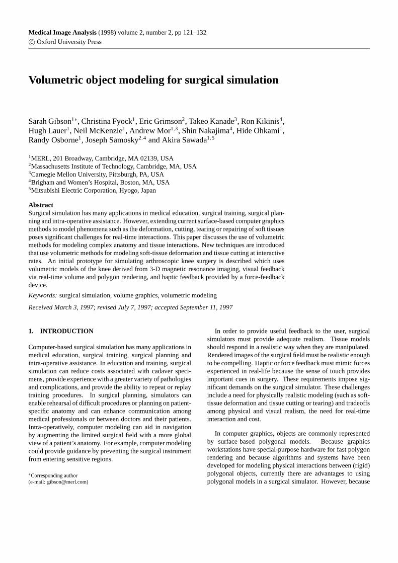
Figure 1. Surgical simulation system components. This systemprovides a closed feedback loop between the user and the simulator.The user can manipulate object models and observe the results bothvisually and haptically.
they cannot represent interior structure, surface-basedmodels are inadequate for modeling objects and tissueswith complex interiors, for modeling the deformation ofarbitrary or heterogeneous volumes and for simulatingcutting through tissues. In addition, surface-based methodscannot model fluid flow, tissue tearing or bone fracturing.In contrast, volumetric models can incorporate a great dealof information about interior structure and the mechanicalproperties of heterogeneous tissues. By adjusting forcesthat hold volumetric elements together, it may be possibleto model fluids, tearing and fracturing using volumetricmodels. In this paper, we discuss techniques for deformingvolumetric object models, for detecting collisions betweenvolumetric objects and for modeling tissue cutting. Inaddition, we describe an initial prototype system forsimulating arthroscopic knee surgery.
The long-term goal of this project is a computer-basedsurgical simulation system that uses models generated frompatient specific data. As illustrated in Figure 1, the systemwill provide both visual and haptic feedback to the user viareal-time rendering and a force-feedback device. Physicallyrealistic interactions between anatomical models and user-controlled surgical instruments will provide a closed feedbackloop between the user and the simulator. Important tech-nologies required by the simulator include: image acquisitionand segmentation of patient-specific 3-D data to generateobject models; visual and haptic feedback through real-timerendering and an electromechanical force-feedback device;manipulation of visual parameters and physically realisticinteractions with the model through a haptic interface.
2. PRIOR WORK
There are several surgical simulation systems underdevelopment both commercially and in research laboratories.
For example, a number of endoscopy simulators enablenavigation through stationary object models (Vining et al.,1993, 1994; Geiger and Kikinis, 1995; Hong et al., 1995;Lorensen et al., 1995; Ziegler et al., 1995). In somenavigation or virtual fly-throughs, path planning and collisionavoidance between the virtual endoscope tip and staticobject surfaces are incorporated into the models (Geigerand Kikinis, 1995; Hong et al., 1995; Lorensen et al.,1995). Deformation of surface models is used to modelsoft-tissues by some surgical simulations systems (e.g. Coveret al., 1993; Kuhnapfel et al., 1997). These systems useeither mass–spring models or control points on spline-basedsurfaces for interactive deformation of surface-based objectmodels.
A number of groups have used volumetric methods formodeling deformation and cutting of tissue volumes. Finite-element analysis has been applied to facial and muscle mod-eling (Waters, 1987; Terzopoulos and Waters, 1990; Chen,1991), and in surgical simulation (e.g. Pieper, 1992; Hunteret al., 1993; Cotin et al., 1996). Manipulation of voxel-basedobjects (Kaufman, 1996) has been used in object modeling(Gibson, 1995) and combined with force feedback for hapticexploration of voxel-based objects (Avila and Sobierajski,1996). Sculpting of volumetric objects is described in Wangand Kaufman (1995) and Galyean and Hughes (1991).
3. VOLUMETRIC METHODS FOR SURGICALSIMULATION
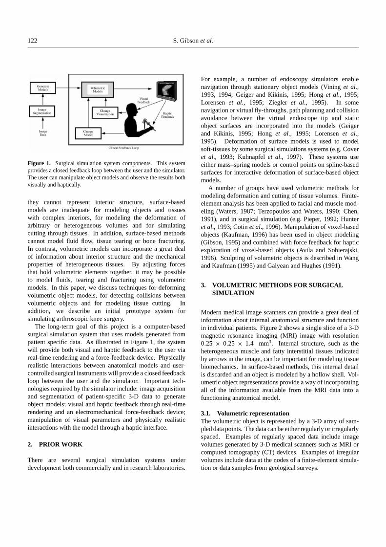
Modern medical image scanners can provide a great deal ofinformation about internal anatomical structure and functionin individual patients. Figure 2 shows a single slice of a 3-Dmagnetic resonance imaging (MRI) image with resolution0.25 × 0.25 × 1.4 mm3. Internal structure, such as theheterogeneous muscle and fatty interstitial tissues indicatedby arrows in the image, can be important for modeling tissuebiomechanics. In surface-based methods, this internal detailis discarded and an object is modeled by a hollow shell. Vol-umetric object representations provide a way of incorporatingall of the information available from the MRI data into afunctioning anatomical model.
3.1. Volumetric representationThe volumetric object is represented by a 3-D array of sam-pled data points. The data can be either regularly or irregularlyspaced. Examples of regularly spaced data include imagevolumes generated by 3-D medical scanners such as MRI orcomputed tomography (CT) devices. Examples of irregularvolumes include data at the nodes of a finite-element simula-tion or data samples from geological surveys.
Volumetric object modeling for surgical simulation 123
Figure 2. Single plane from a high-resolution MRI image volumewith voxel resolution: 0.25 × 0.25 × 1.4 mm3. Arrows indicatestructure in muscle and fatty tissue that are important in modelingtissue behavior but that are hard to incorporate into surface-basedmodels.
Elements in the volume encode information about visualand/or physical properties of the object. Each element canbe represented by a single value, such as the sampled imageintensity, or a more complex data structure that encodes infor-mation such as color, transparency, edge strength, elasticity,tissue type and connections to neighboring elements. Theability to encode many different attributes for each elementenables the modeling of complex materials, structures andbehaviors. However, since volumetric objects can consistof thousands to millions of elements (an MRI image of size256×256×256 contains 16 million voxels) this representationposes significant challenges for rendering, data storage andretrieval and tissue modeling. Some approaches for dealingwith these large volumes in rendering and tissue modelingare presented here. Future research into hierarchical datarepresentations and data compression will make volumetricmethods even more practical for real-time applications.
3.2. Soft-tissue deformation with volumetric models3.2.1. Mass–spring systems and linear finite-element
methodsThe two most common methods used for modeling volumet-ric deformation are mass–spring systems and linear finite-element methods (FEM). Though mass–spring systems and
linear FEM methods are different techniques, in a static sim-ulation both result in a large system of linear equations of theform
Ku = F,
where K is the object stiffness matrix (a function of materialelastic properties which depend on the method), u is thevector representing element displacements and F is a vectorrepresenting internal and external forces in the system. In adynamic simulation, where changes in the shape are observedas the object moves towards equilibrium, the effect of inertialand damping forces are modeled using a system of second-order differential equations:
Mu+ Cu+ Ku = F
where M and C are mass and damping matrices respectively,and u and u are the first- and second-order derivatives of uwith respect to time. In a dynamic computer simulation, thissystem is evolved through time using numerical techniques.
In both static and dynamic simulations, solving for thedisplacements, u, is computationally demanding for a largesystem. The interaction speed can be increased by a sig-nificant reduction in the number of elements in the system(Hunter et al., 1993) and/or pre-processing of the data. Bro-Nielsen simplifies the system by solving for displacementsof only surface elements and by assuming a small numberof externally applied forces (Bro-Nielsen, 1997). Pentlandand Williams (1989) pre-calculate the deformation modes of agiven object and calculate deformations for an arbitrary forceas a superposition of these deformation modes. Bro-Nielsenand Cotin (Bro-Nielsen, 1995; Bro-Nielsen and Cotin, 1996;Cotin et al., 1996) use a similar method, pre-calculating re-sponses to infinitesimal forces and deformations for each nodein the element and then approximating the global deformationas a linear superposition of these pre-calculated responses.
These methods have a number of limitations. First, evenwith significant preprocessing, the size of the deformation islimited and the number of elements must remain relativelysmall for real-time modeling. Cotin et al. use the abovemethod in a system that can deform a liver model with 8000elements at 15 frames per seconda. This is still a relativelysmall number of elements, especially if the complex structureof internal vessels and ducts are modeled. In addition, thesystem requires hours of preprocessing which prohibits inter-active changes in topology due to tissue cutting, tearing orsuturing.
Second, the assumptions of linearity and superposition arenot valid for human tissue. Figure 3 shows a typical stretchversus applied force curve for human tissue [see Fung (1993)
aPersonal communication from Stephan Cotin, April, 1997.
124 S. Gibson et al.
increasingload
decreasingload
Stre
ss /
Forc
e / L
oad
Strain / Stretch / Deformation
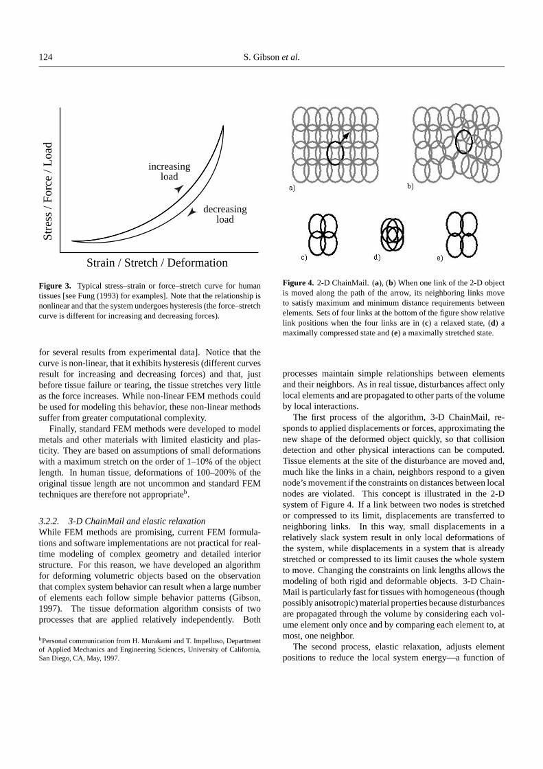
Figure 3. Typical stress–strain or force–stretch curve for humantissues [see Fung (1993) for examples]. Note that the relationship isnonlinear and that the system undergoes hysteresis (the force–stretchcurve is different for increasing and decreasing forces).
for several results from experimental data]. Notice that thecurve is non-linear, that it exhibits hysteresis (different curvesresult for increasing and decreasing forces) and that, justbefore tissue failure or tearing, the tissue stretches very littleas the force increases. While non-linear FEM methods couldbe used for modeling this behavior, these non-linear methodssuffer from greater computational complexity.
Finally, standard FEM methods were developed to modelmetals and other materials with limited elasticity and plas-ticity. They are based on assumptions of small deformationswith a maximum stretch on the order of 1–10% of the objectlength. In human tissue, deformations of 100–200% of theoriginal tissue length are not uncommon and standard FEMtechniques are therefore not appropriateb.
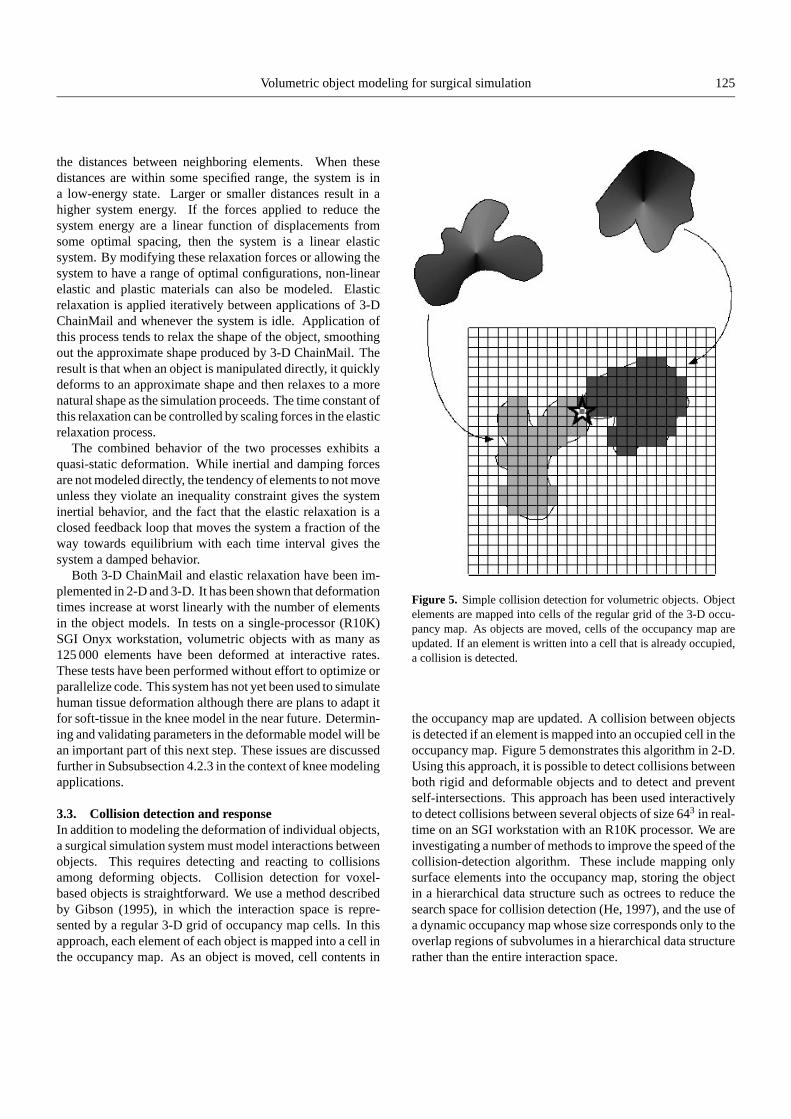
3.2.2. 3-D ChainMail and elastic relaxationWhile FEM methods are promising, current FEM formula-tions and software implementations are not practical for real-time modeling of complex geometry and detailed interiorstructure. For this reason, we have developed an algorithmfor deforming volumetric objects based on the observationthat complex system behavior can result when a large numberof elements each follow simple behavior patterns (Gibson,1997). The tissue deformation algorithm consists of twoprocesses that are applied relatively independently. Both
bPersonal communication from H. Murakami and T. Impelluso, Departmentof Applied Mechanics and Engineering Sciences, University of California,San Diego, CA, May, 1997.
Figure 4. 2-D ChainMail. (a), (b) When one link of the 2-D objectis moved along the path of the arrow, its neighboring links moveto satisfy maximum and minimum distance requirements betweenelements. Sets of four links at the bottom of the figure show relativelink positions when the four links are in (c) a relaxed state, (d) amaximally compressed state and (e) a maximally stretched state.
processes maintain simple relationships between elementsand their neighbors. As in real tissue, disturbances affect onlylocal elements and are propagated to other parts of the volumeby local interactions.
The first process of the algorithm, 3-D ChainMail, re-sponds to applied displacements or forces, approximating thenew shape of the deformed object quickly, so that collisiondetection and other physical interactions can be computed.Tissue elements at the site of the disturbance are moved and,much like the links in a chain, neighbors respond to a givennode’s movement if the constraints on distances between localnodes are violated. This concept is illustrated in the 2-Dsystem of Figure 4. If a link between two nodes is stretchedor compressed to its limit, displacements are transferred toneighboring links. In this way, small displacements in arelatively slack system result in only local deformations ofthe system, while displacements in a system that is alreadystretched or compressed to its limit causes the whole systemto move. Changing the constraints on link lengths allows themodeling of both rigid and deformable objects. 3-D Chain-Mail is particularly fast for tissues with homogeneous (thoughpossibly anisotropic) material properties because disturbancesare propagated through the volume by considering each vol-ume element only once and by comparing each element to, atmost, one neighbor.
The second process, elastic relaxation, adjusts elementpositions to reduce the local system energy—a function of
Volumetric object modeling for surgical simulation 125
the distances between neighboring elements. When thesedistances are within some specified range, the system is ina low-energy state. Larger or smaller distances result in ahigher system energy. If the forces applied to reduce thesystem energy are a linear function of displacements fromsome optimal spacing, then the system is a linear elasticsystem. By modifying these relaxation forces or allowing thesystem to have a range of optimal configurations, non-linearelastic and plastic materials can also be modeled. Elasticrelaxation is applied iteratively between applications of 3-DChainMail and whenever the system is idle. Application ofthis process tends to relax the shape of the object, smoothingout the approximate shape produced by 3-D ChainMail. Theresult is that when an object is manipulated directly, it quicklydeforms to an approximate shape and then relaxes to a morenatural shape as the simulation proceeds. The time constant ofthis relaxation can be controlled by scaling forces in the elasticrelaxation process.
The combined behavior of the two processes exhibits aquasi-static deformation. While inertial and damping forcesare not modeled directly, the tendency of elements to not moveunless they violate an inequality constraint gives the systeminertial behavior, and the fact that the elastic relaxation is aclosed feedback loop that moves the system a fraction of theway towards equilibrium with each time interval gives thesystem a damped behavior.
Both 3-D ChainMail and elastic relaxation have been im-plemented in 2-D and 3-D. It has been shown that deformationtimes increase at worst linearly with the number of elementsin the object models. In tests on a single-processor (R10K)SGI Onyx workstation, volumetric objects with as many as125 000 elements have been deformed at interactive rates.These tests have been performed without effort to optimize orparallelize code. This system has not yet been used to simulatehuman tissue deformation although there are plans to adapt itfor soft-tissue in the knee model in the near future. Determin-ing and validating parameters in the deformable model will bean important part of this next step. These issues are discussedfurther in Subsubsection 4.2.3 in the context of knee modelingapplications.
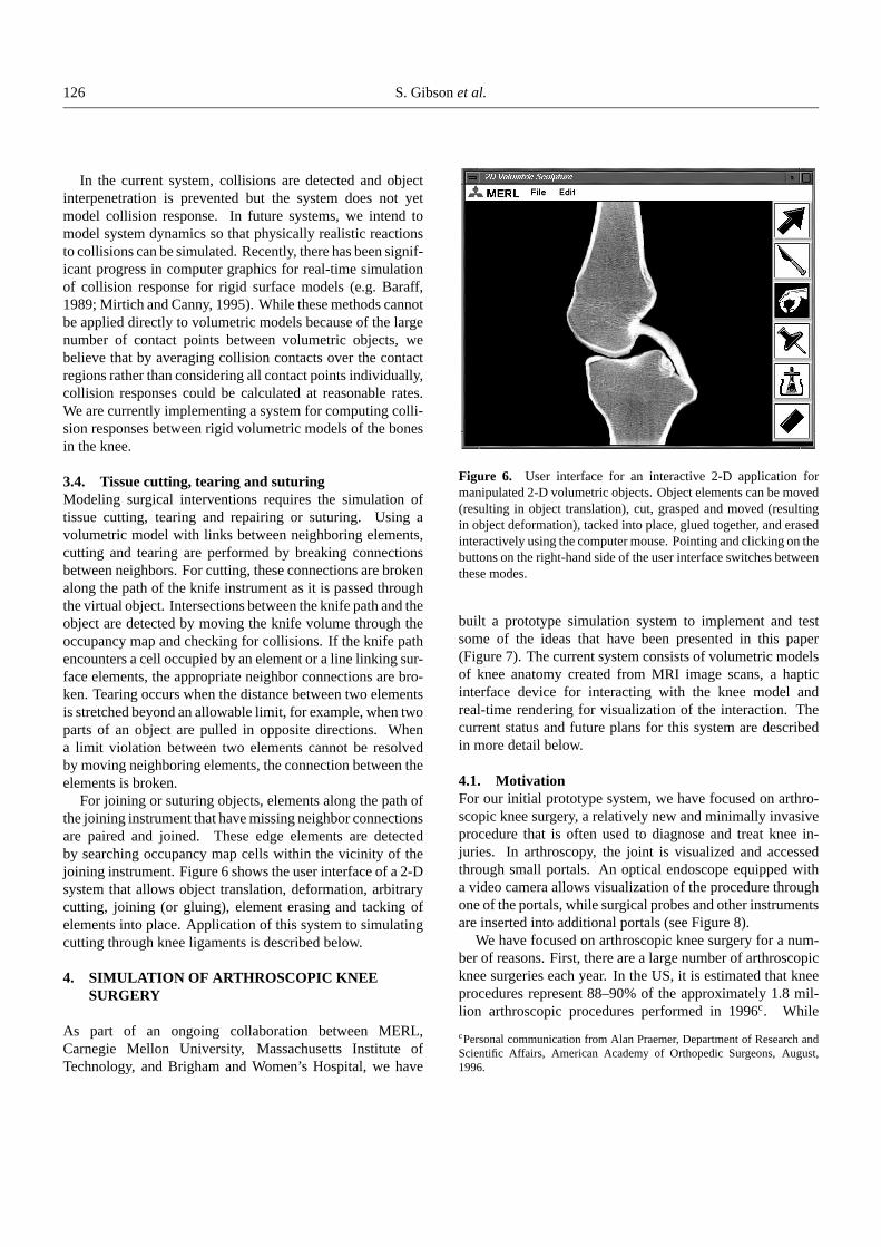
3.3. Collision detection and responseIn addition to modeling the deformation of individual objects,a surgical simulation system must model interactions betweenobjects. This requires detecting and reacting to collisionsamong deforming objects. Collision detection for voxel-based objects is straightforward. We use a method describedby Gibson (1995), in which the interaction space is repre-sented by a regular 3-D grid of occupancy map cells. In thisapproach, each element of each object is mapped into a cell inthe occupancy map. As an object is moved, cell contents in
Figure 5. Simple collision detection for volumetric objects. Objectelements are mapped into cells of the regular grid of the 3-D occu-pancy map. As objects are moved, cells of the occupancy map areupdated. If an element is written into a cell that is already occupied,a collision is detected.
the occupancy map are updated. A collision between objectsis detected if an element is mapped into an occupied cell in theoccupancy map. Figure 5 demonstrates this algorithm in 2-D.Using this approach, it is possible to detect collisions betweenboth rigid and deformable objects and to detect and preventself-intersections. This approach has been used interactivelyto detect collisions between several objects of size 643 in real-time on an SGI workstation with an R10K processor. We areinvestigating a number of methods to improve the speed of thecollision-detection algorithm. These include mapping onlysurface elements into the occupancy map, storing the objectin a hierarchical data structure such as octrees to reduce thesearch space for collision detection (He, 1997), and the use ofa dynamic occupancy map whose size corresponds only to theoverlap regions of subvolumes in a hierarchical data structurerather than the entire interaction space.
126 S. Gibson et al.
In the current system, collisions are detected and objectinterpenetration is prevented but the system does not yetmodel collision response. In future systems, we intend tomodel system dynamics so that physically realistic reactionsto collisions can be simulated. Recently, there has been signif-icant progress in computer graphics for real-time simulationof collision response for rigid surface models (e.g. Baraff,1989; Mirtich and Canny, 1995). While these methods cannotbe applied directly to volumetric models because of the largenumber of contact points between volumetric objects, webelieve that by averaging collision contacts over the contactregions rather than considering all contact points individually,collision responses could be calculated at reasonable rates.We are currently implementing a system for computing colli-sion responses between rigid volumetric models of the bonesin the knee.
3.4. Tissue cutting, tearing and suturingModeling surgical interventions requires the simulation oftissue cutting, tearing and repairing or suturing. Using avolumetric model with links between neighboring elements,cutting and tearing are performed by breaking connectionsbetween neighbors. For cutting, these connections are brokenalong the path of the knife instrument as it is passed throughthe virtual object. Intersections between the knife path and theobject are detected by moving the knife volume through theoccupancy map and checking for collisions. If the knife pathencounters a cell occupied by an element or a line linking sur-face elements, the appropriate neighbor connections are bro-ken. Tearing occurs when the distance between two elementsis stretched beyond an allowable limit, for example, when twoparts of an object are pulled in opposite directions. Whena limit violation between two elements cannot be resolvedby moving neighboring elements, the connection between theelements is broken.
For joining or suturing objects, elements along the path ofthe joining instrument that have missing neighbor connectionsare paired and joined. These edge elements are detectedby searching occupancy map cells within the vicinity of thejoining instrument. Figure 6 shows the user interface of a 2-Dsystem that allows object translation, deformation, arbitrarycutting, joining (or gluing), element erasing and tacking ofelements into place. Application of this system to simulatingcutting through knee ligaments is described below.
4. SIMULATION OF ARTHROSCOPIC KNEESURGERY
As part of an ongoing collaboration between MERL,Carnegie Mellon University, Massachusetts Institute ofTechnology, and Brigham and Women’s Hospital, we have
Figure 6. User interface for an interactive 2-D application formanipulated 2-D volumetric objects. Object elements can be moved(resulting in object translation), cut, grasped and moved (resultingin object deformation), tacked into place, glued together, and erasedinteractively using the computer mouse. Pointing and clicking on thebuttons on the right-hand side of the user interface switches betweenthese modes.
built a prototype simulation system to implement and testsome of the ideas that have been presented in this paper(Figure 7). The current system consists of volumetric modelsof knee anatomy created from MRI image scans, a hapticinterface device for interacting with the knee model andreal-time rendering for visualization of the interaction. Thecurrent status and future plans for this system are describedin more detail below.
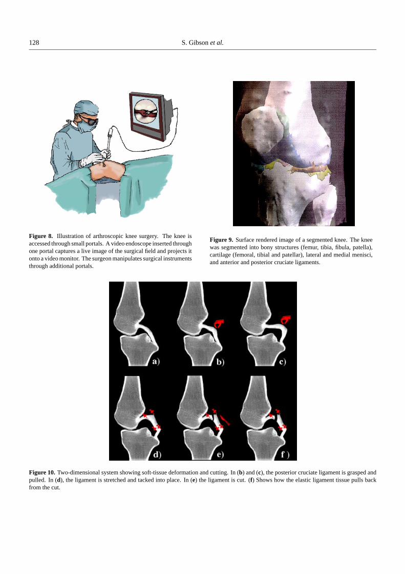
4.1. MotivationFor our initial prototype system, we have focused on arthro-scopic knee surgery, a relatively new and minimally invasiveprocedure that is often used to diagnose and treat knee in-juries. In arthroscopy, the joint is visualized and accessedthrough small portals. An optical endoscope equipped witha video camera allows visualization of the procedure throughone of the portals, while surgical probes and other instrumentsare inserted into additional portals (see Figure 8).
We have focused on arthroscopic knee surgery for a num-ber of reasons. First, there are a large number of arthroscopicknee surgeries each year. In the US, it is estimated that kneeprocedures represent 88–90% of the approximately 1.8 mil-lion arthroscopic procedures performed in 1996c. While
cPersonal communication from Alan Praemer, Department of Research andScientific Affairs, American Academy of Orthopedic Surgeons, August,1996.
Volumetric object modeling for surgical simulation 127
Figure 7. Prototype surgical simulation system. A 3-D volumetricknee model has been generated from MRI images. A force-feedbackdevice allows the user to haptically explore the bony surfaces ofthe knee model while real-time polygon and/or volume renderingprovide visual feedback to the user.
arthroscopic procedures have been shown to reduce costsand increase patient recovery rates, they suffer from specifictechnical limitations, namely, limited visibility through thearthroscope; difficulty in orienting the camera to the surgeon’sviewpoint and restricted motion of surgical tools. Computersimulation will enhance the education and training of sur-geons, and help them deal with these technical challenges.
Second, the knee offers an ideal initial platform for thisproject. Important structures lie in a limited volume so thatthe size of a volumetric knee model is reasonable. Manyof the important structures in the knee are rigid so initialhaptic and rendering implementations can be performed onstatic data sets. In addition, the soft-tissues that are of pri-mary importance—cartilage layers, the menisci and cruciateligaments—are small enough for reasonable testing of tissuedeformation and cutting algorithms and for performing real-time rendering. As faster hardware, more efficient algorithmsand better data representations are developed, techniques de-veloped for the arthroscopic knee simulator will be extendedto other systems.
Finally, the knee has been modeled in a number of studiesfocusing on biomechanics and bio-dynamics modeling (e.g.Kaufman, 1988; Fijan, 1990; Blankevoort et al., 1991) exper-imental measurements (e.g. Blankevoort et al., 1984; Fung,1993) and surgical simulations (e.g. Logan et al., 1996; Bajajet al., 1997). This provides a body of literature for testing
dynamic predictions of our model, obtaining measured tissueparameters and comparing our results with other surgical sim-ulation systems.
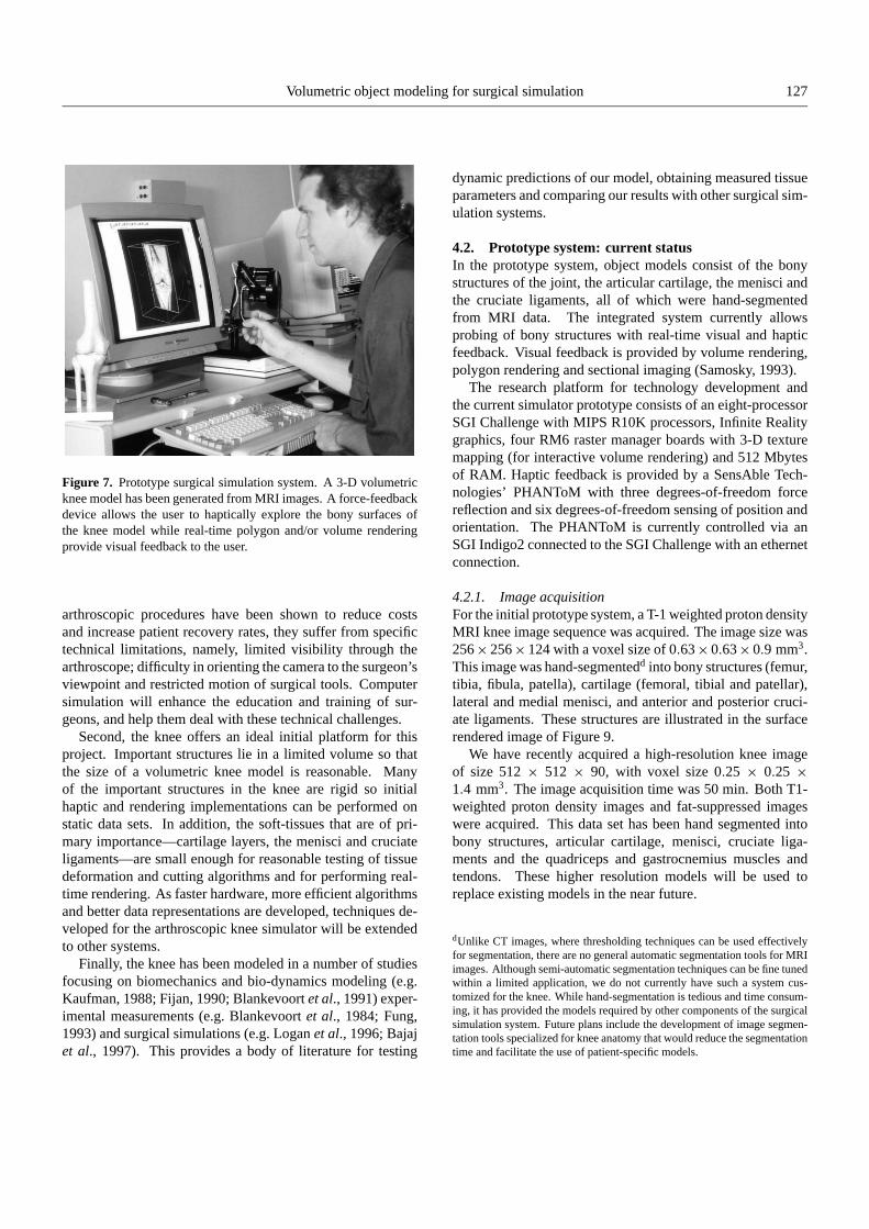
4.2. Prototype system: current statusIn the prototype system, object models consist of the bonystructures of the joint, the articular cartilage, the menisci andthe cruciate ligaments, all of which were hand-segmentedfrom MRI data. The integrated system currently allowsprobing of bony structures with real-time visual and hapticfeedback. Visual feedback is provided by volume rendering,polygon rendering and sectional imaging (Samosky, 1993).
The research platform for technology development andthe current simulator prototype consists of an eight-processorSGI Challenge with MIPS R10K processors, Infinite Realitygraphics, four RM6 raster manager boards with 3-D texturemapping (for interactive volume rendering) and 512 Mbytesof RAM. Haptic feedback is provided by a SensAble Tech-nologies’ PHANToM with three degrees-of-freedom forcereflection and six degrees-of-freedom sensing of position andorientation. The PHANToM is currently controlled via anSGI Indigo2 connected to the SGI Challenge with an ethernetconnection.
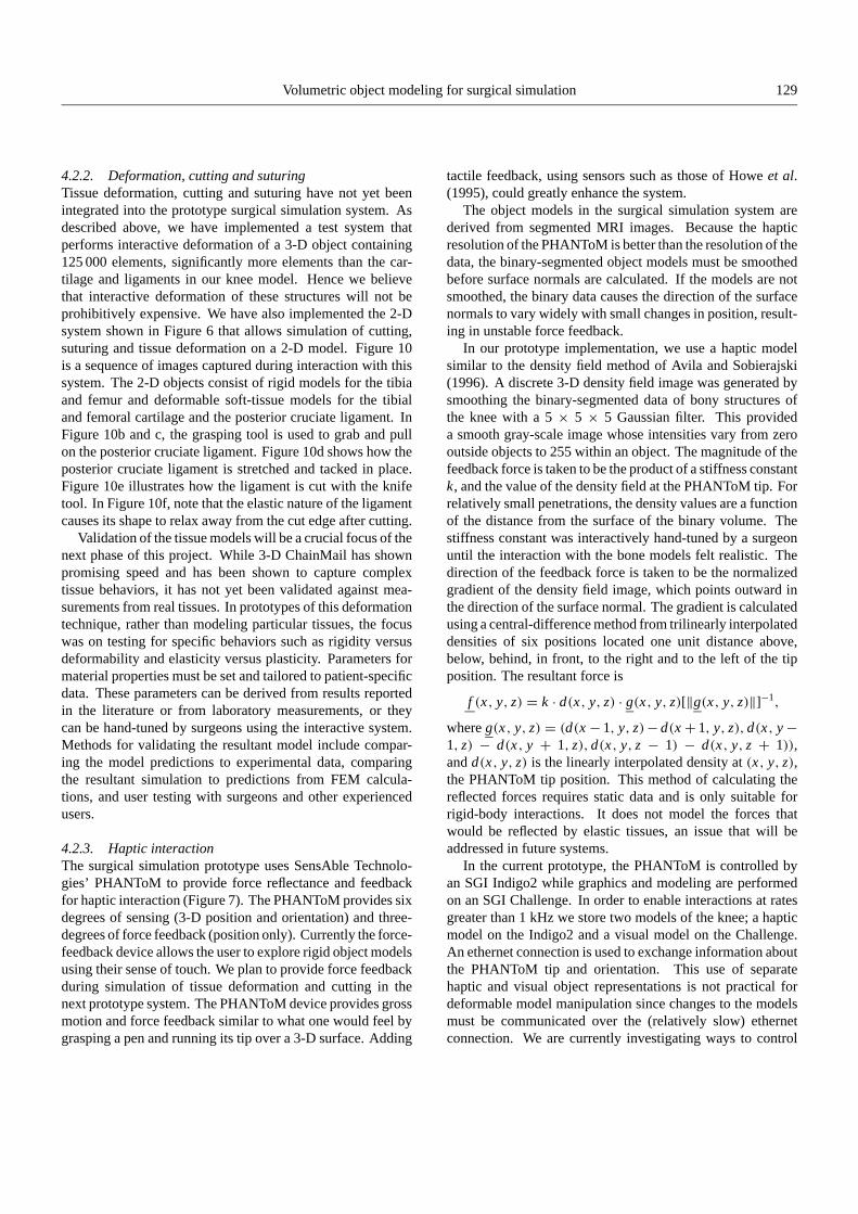
4.2.1. Image acquisitionFor the initial prototype system, a T-1 weighted proton densityMRI knee image sequence was acquired. The image size was256× 256× 124 with a voxel size of 0.63× 0.63× 0.9 mm3.This image was hand-segmentedd into bony structures (femur,tibia, fibula, patella), cartilage (femoral, tibial and patellar),lateral and medial menisci, and anterior and posterior cruci-ate ligaments. These structures are illustrated in the surfacerendered image of Figure 9.
We have recently acquired a high-resolution knee imageof size 512 × 512 × 90, with voxel size 0.25 × 0.25 ×1.4 mm3. The image acquisition time was 50 min. Both T1-weighted proton density images and fat-suppressed imageswere acquired. This data set has been hand segmented intobony structures, articular cartilage, menisci, cruciate liga-ments and the quadriceps and gastrocnemius muscles andtendons. These higher resolution models will be used toreplace existing models in the near future.
dUnlike CT images, where thresholding techniques can be used effectivelyfor segmentation, there are no general automatic segmentation tools for MRIimages. Although semi-automatic segmentation techniques can be fine tunedwithin a limited application, we do not currently have such a system cus-tomized for the knee. While hand-segmentation is tedious and time consum-ing, it has provided the models required by other components of the surgicalsimulation system. Future plans include the development of image segmen-tation tools specialized for knee anatomy that would reduce the segmentationtime and facilitate the use of patient-specific models.
128 S. Gibson et al.
Figure 8. Illustration of arthroscopic knee surgery. The knee isaccessed through small portals. A video endoscope inserted throughone portal captures a live image of the surgical field and projects itonto a video monitor. The surgeon manipulates surgical instrumentsthrough additional portals.
Figure 9. Surface rendered image of a segmented knee. The kneewas segmented into bony structures (femur, tibia, fibula, patella),cartilage (femoral, tibial and patellar), lateral and medial menisci,and anterior and posterior cruciate ligaments.
Figure 10. Two-dimensional system showing soft-tissue deformation and cutting. In (b) and (c), the posterior cruciate ligament is grasped andpulled. In (d), the ligament is stretched and tacked into place. In (e) the ligament is cut. (f) Shows how the elastic ligament tissue pulls backfrom the cut.
Volumetric object modeling for surgical simulation 129
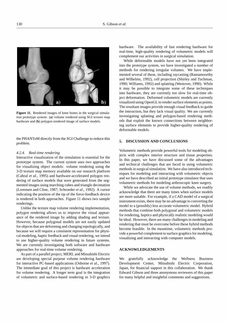
4.2.2. Deformation, cutting and suturingTissue deformation, cutting and suturing have not yet beenintegrated into the prototype surgical simulation system. Asdescribed above, we have implemented a test system thatperforms interactive deformation of a 3-D object containing125 000 elements, significantly more elements than the car-tilage and ligaments in our knee model. Hence we believethat interactive deformation of these structures will not beprohibitively expensive. We have also implemented the 2-Dsystem shown in Figure 6 that allows simulation of cutting,suturing and tissue deformation on a 2-D model. Figure 10is a sequence of images captured during interaction with thissystem. The 2-D objects consist of rigid models for the tibiaand femur and deformable soft-tissue models for the tibialand femoral cartilage and the posterior cruciate ligament. InFigure 10b and c, the grasping tool is used to grab and pullon the posterior cruciate ligament. Figure 10d shows how theposterior cruciate ligament is stretched and tacked in place.Figure 10e illustrates how the ligament is cut with the knifetool. In Figure 10f, note that the elastic nature of the ligamentcauses its shape to relax away from the cut edge after cutting.
Validation of the tissue models will be a crucial focus of thenext phase of this project. While 3-D ChainMail has shownpromising speed and has been shown to capture complextissue behaviors, it has not yet been validated against mea-surements from real tissues. In prototypes of this deformationtechnique, rather than modeling particular tissues, the focuswas on testing for specific behaviors such as rigidity versusdeformability and elasticity versus plasticity. Parameters formaterial properties must be set and tailored to patient-specificdata. These parameters can be derived from results reportedin the literature or from laboratory measurements, or theycan be hand-tuned by surgeons using the interactive system.Methods for validating the resultant model include compar-ing the model predictions to experimental data, comparingthe resultant simulation to predictions from FEM calcula-tions, and user testing with surgeons and other experiencedusers.
4.2.3. Haptic interactionThe surgical simulation prototype uses SensAble Technolo-gies’ PHANToM to provide force reflectance and feedbackfor haptic interaction (Figure 7). The PHANToM provides sixdegrees of sensing (3-D position and orientation) and three-degrees of force feedback (position only). Currently the force-feedback device allows the user to explore rigid object modelsusing their sense of touch. We plan to provide force feedbackduring simulation of tissue deformation and cutting in thenext prototype system. The PHANToM device provides grossmotion and force feedback similar to what one would feel bygrasping a pen and running its tip over a 3-D surface. Adding
tactile feedback, using sensors such as those of Howe et al.(1995), could greatly enhance the system.
The object models in the surgical simulation system arederived from segmented MRI images. Because the hapticresolution of the PHANToM is better than the resolution of thedata, the binary-segmented object models must be smoothedbefore surface normals are calculated. If the models are notsmoothed, the binary data causes the direction of the surfacenormals to vary widely with small changes in position, result-ing in unstable force feedback.
In our prototype implementation, we use a haptic modelsimilar to the density field method of Avila and Sobierajski(1996). A discrete 3-D density field image was generated bysmoothing the binary-segmented data of bony structures ofthe knee with a 5 × 5 × 5 Gaussian filter. This provideda smooth gray-scale image whose intensities vary from zerooutside objects to 255 within an object. The magnitude of thefeedback force is taken to be the product of a stiffness constantk, and the value of the density field at the PHANToM tip. Forrelatively small penetrations, the density values are a functionof the distance from the surface of the binary volume. Thestiffness constant was interactively hand-tuned by a surgeonuntil the interaction with the bone models felt realistic. Thedirection of the feedback force is taken to be the normalizedgradient of the density field image, which points outward inthe direction of the surface normal. The gradient is calculatedusing a central-difference method from trilinearly interpolateddensities of six positions located one unit distance above,below, behind, in front, to the right and to the left of the tipposition. The resultant force is
f (x, y, z) = k · d(x, y, z) · g(x, y, z)[‖g(x, y, z)‖]−1,
where g(x, y, z) = (d(x− 1, y, z)− d(x+ 1, y, z), d(x, y−1, z) − d(x, y + 1, z), d(x, y, z − 1) − d(x, y, z + 1)),and d(x, y, z) is the linearly interpolated density at (x, y, z),the PHANToM tip position. This method of calculating thereflected forces requires static data and is only suitable forrigid-body interactions. It does not model the forces thatwould be reflected by elastic tissues, an issue that will beaddressed in future systems.
In the current prototype, the PHANToM is controlled byan SGI Indigo2 while graphics and modeling are performedon an SGI Challenge. In order to enable interactions at ratesgreater than 1 kHz we store two models of the knee; a hapticmodel on the Indigo2 and a visual model on the Challenge.An ethernet connection is used to exchange information aboutthe PHANToM tip and orientation. This use of separatehaptic and visual object representations is not practical fordeformable model manipulation since changes to the modelsmust be communicated over the (relatively slow) ethernetconnection. We are currently investigating ways to control
130 S. Gibson et al.
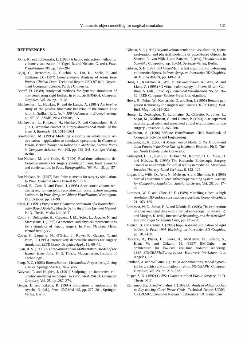
Figure 11. Rendered images of knee bones in the surgical simula-tion prototype system: (a) volume rendered using SGI texture maphardware and (b) polygon rendered image of surface models.
the PHANToM directly from the SGI Challenge to reduce thisproblem.
4.2.4. Real-time renderingInteractive visualization of the simulation is essential for theprototype system. The current system uses two approachesfor visualizing object models: volume rendering using the3-D texture map memory available on our research platform(Cabral et al., 1995) and hardware-accelerated polygon ren-dering of surface models that were generated from the seg-mented images using marching cubes and triangle decimation(Lorensen and Cline, 1987; Schroeder et al., 1992). A cursorindicating the position of the tip of the force-feedback deviceis rendered in both approaches. Figure 11 shows two samplerenderings.
Unlike the texture map volume rendering implementation,polygon rendering allows us to improve the visual appear-ance of the rendered image by adding shading and texture.However, because polygonal models are not easily updatedfor objects that are deforming and changing topologically, andbecause we will require a consistent representation for physi-cal modeling, haptic feedback and visual rendering, we intendto use higher-quality volume rendering in future systems.We are currently investigating both software and hardwareapproaches for real-time volume rendering.
As part of a parallel project, MERL and Mitsubishi Electricare developing special purpose volume rendering hardwarefor interactive PC-based applications (Osborne et al., 1997).The immediate goal of this project is hardware accelerationfor volume rendering. A longer term goal is the integrationof volumetric and surface-based rendering in 3-D graphics
hardware. The availability of fast rendering hardware forreal-time, high-quality rendering of volumetric models willcomplement our activities in surgical simulation.
While deformable models have not yet been integratedinto the prototype system, we have investigated a number ofmethods for rendering irregular volumes. We have imple-mented several of these, including raycasting (Ramamoorthyand Wilhelms, 1992), cell projection (Shirley and Tuchman,1990; Williams, 1992) and splatting (Westover, 1990). Whileit may be possible to integrate some of these techniquesinto hardware, they are currently too slow for real-time ob-ject deformation. Deformed volumetric models are currentlyvisualized using OpenGL to render surface elements as points.The resultant images provide enough visual feedback to guidethe interaction, but they lack visual quality. We are currentlyinvestigating splatting and polygon-based rendering meth-ods that exploit the known connections between neighbor-ing surface elements to provide higher-quality rendering ofdeformable models.
5. DISCUSSION AND CONCLUSIONS
Volumetric methods provide powerful tools for modeling ob-jects with complex interior structure and tissue properties.In this paper, we have discussed some of the advantagesand technical challenges that are faced in using volumetricmethods in surgical simulation. We have also introduced tech-niques for modeling and interacting with volumetric objectsand we have described an initial prototype simulator that usesvolumetric methods for modeling arthroscopic knee surgery.
While we advocate the use of volume methods, we readilyacknowledge that there are many times when surface modelsare more suitable. For example, if a CAD model of a surgicalinstrument exists, there may be no advantage to converting themodel to a (possibly) less accurate volumetric model. Hybridmethods that combine both polygonal and volumetric modelsfor rendering, haptics and physically realistic modeling wouldbe ideal. However, there are many challenges in modeling andrendering that must be overcome before these hybrid methodsbecome feasible. In the meantime, volumetric methods pro-vide a powerful complement to surface graphics for modeling,visualizing and interacting with computer models.
ACKNOWLEDGEMENTS
We gratefully acknowledge the Wellness BusinessDevelopment Center, Mitsubishi Electric Corporation,Japan, for financial support in this collaboration. We thankEdward Gibson and three anonymous reviewers of this paperfor many helpful and insightful comments and suggestions.
Volumetric object modeling for surgical simulation 131
REFERENCES
Avila, R. and Sobierajski, L. (1996) A haptic interaction method forvolume visualization. In Yagel, R. and Nielson, G. (ed.), Proc.Visualization ’96, pp. 197–204.
Bajaj, C., Bernardini, F., Cutchin, S., Lin, K., Sacks, E. andSchikore, D. (1997) Comprehensive Analysis of Joints fromPatient Clinical Data. Technical Report CSD-97-019, Depart-ment Computer Science, Purdue University.
Baraff, D. (1989) Analytical methods for dynamic simulation ofnon-penetrating rigid bodies. In Proc. SIGGRAPH, ComputerGraphics, Vol. 24, pp. 19–28.
Blankevoort, L., Huiskes, R. and de Lange, A. (1984) An in-vitrostudy of the passive kinematic behavior of the human kneejoint. In Spilker, R. L. (ed.), 1984 Advances in Bioengineering,pp. 57–58. ASME, New Orleans, LA.
Blankevoort, L., Kuiper, J. H., Huiskes, R. and Grootenboer, H. J.(1991) Articular contact in a three-dimensional model of theknee. J. Biomech., 24, 1019–1031.
Bro-Nielsen, M. (1995) Modeling elasticity in solids using ac-tive cubes—application to simulated operations. In ComputerVision, Virtual Reality and Robotics in Medicine, Lecture Notesin Computer Science, Vol. 905, pp. 535–541. Springer-Verlag,Berlin.
Bro-Nielsen, M. and Cotin, S. (1996) Real-time volumetric de-formable models for surgery simulation using finite elementsand condensation. In Proc. Eurographics, ’96, Vol. 15, pp. 57–66.
Bro-Nielsen, M. (1997) Fast finite elements for surgery simulation.In Proc. Medicine Meets Virtual Reality V.
Cabral, B., Cam, N. and Foran, J. (1995) Accelerated volume ren-dering and tomographic reconstruction using texture mappinghardware. In Proc. Symp. on Volume Visualization, Washington,DC, October, pp. 91–98.
Chen, D. (1991) Pump it up: Computer Animation of a Biomechani-cally Based Model of Muscle Using the Finite Element Method.Ph.D. Thesis, Media Lab, MIT.
Cotin, S., Delingette, H., Clement, J. M., Soler, L., Ayache, N. andMarescaux, J. (1996) Geometrical and physical representationsfor a simulator of hepatic surgery. In Proc. Medicine MeetsVirtual Reality IV.
Cover, S., Ezquerra, N., O’Brian, J., Rowe, R., Gadacz, T. andPalm, E. (1993) Interactively deformable models for surgerysimulation. IEEE Comp. Graphics Appl., 13, 68–75.
Fijan, R. S. (1990) A Three-dimensional Mathematical Model of theHuman Knee Joint. Ph.D. Thesis, Massachusetts Institute ofTechnology.
Fung, Y. C. (1993) Biomechanics: Mechanical Properties of LivingTissues. Springer-Verlag, New York.
Galyean, T. and Hughes, J. (1991) Sculpting: an interactive vol-umetric modeling technique. In Proc. SIGGRAPH, ComputerGraphics, Vol. 25, pp. 267–274.
Geiger, B. and Kikinis, R. (1995) Simulation of endoscopy. InAyache, N. (ed.), Proc. CVRMed ’95, pp. 277–281. Springer-Verlag, Berlin.
Gibson, S. F. (1995) Beyond volume rendering: visualization, hapticexploration, and physical modeling of voxel-based objects. InScateni, R., van Wijk, J. and Zanarini, P. (eds), Visualization inScientific Computing, pp. 10–24. Springer-Verlag, Berlin.
Gibson, S. F. (1997) 3D ChainMail: a fast algorithm for deformingvolumetric objects. In Proc. Symp. on Interactive 3D Graphics,ACM SIGGRAPH, pp. 149–154.
Hong, L., Kaufman, A., Wei, Y., Viswambharen, A., Wax, M. andLiang, Z. (1995) 3D virtual colonoscopy. In Loew, M. and Ger-shon, N. (eds.), Proc. of Biomedical Visualization ’95, pp. 26–32. IEEE Computer Society Press, Los Alamitos.
Howe, R., Peine, W., Kontarinis, D. and Son, J. (1995) Remote pal-pation technology for surgical applications. IEEE Engng Med.Biol. Mag., 14, 318–323.
Hunter, I., Doukoglou, T., Lafontaine, S., Charette, P., Jones, L.,Sagar, M., Mallinson, G. and Hunter, P. (1993) A teleoperatedmicrosurgical robot and associated virtual environment for eyesurgery. Presence, 2, 265–280.
Kaufmann, A. (1996) Volume Visualization. CRC Handbook ofComputer Science and Engineering.
Kaufman, K. R. (1988) A Mathematical Model of the Muscle andJoint Forces in the Knee During Isokinetic Exercise. Ph.D. The-sis, North Dakota State University.
Kuhnapfel, U. G., Kuhn, C., Hubner, M., Krumm, H. G., Mass, H.and Neisius, B. (1997) The Karlsruhe Endoscopic SurgeryTrainer as an example for virtual reality in medicine. MinimallyInvasive Therapy Allied Technol., 6, 122–125.
Logan, I. P., Wills, D., Avis, N., Mohsen, A. and Sherman, K. (1996)Virtual environment knee arthroscopy training system. Societyfor Computing Simulation, Simulation Series, Vol. 28, pp. 17–22.
Lorensen, W. E. and Cline, H. E. (1989) Marching cubes: a highresolution 3D surface construction algorithm. Comp. Graphics,21, 163–169.
Lorensen, W. E., Jolesz, F. A. and Kikinis, R. (1995) The explorationof cross-sectional data with a virtual endoscope. In Satava, R.and Morgan, K. (eds), Interactive Technology and the New Med-ical Paradigm for Health Care, pp. 221–230.
Mirtich, B. and Canny, J. (1995) Impulse-based simulation of rigidbodies. In Proc. 1995 Workshop on Interactive 3D Graphics,pp. 181–188.
Osborne, R., Pfister, H., Lauer, H., McKenzie, N., Gibson, S.,Hiatt, W. and Ohkami, H. (1997) EM-Cube: anarchitecture for low-cost real-time volume rendering.1997 SIGGRAPH/Eurographics Hardware Workshop, LosAngeles, CA.
Pentland, A. and Williams, J. (1989) Good vibrations: modal dynam-ics for graphics and animation. In Proc. SIGGRAPH, ComputerGraphics, Vol. 23, pp. 215–222.
Pieper, S. D. (1992) CAPS: Computer-aided Plastic Surgery. Ph.D.Thesis, MIT.
Ramamoorthy, S. and Wilhelms, J. (1992) An Analysis of Approachesto Ray-tracing Curvi-linear Grids. Technical Report UCSC-CRL-92-07, Computer Research Laboratory, UC Santa Cruz.

132 S. Gibson et al.
Samosky, J. (1993) SectionView—a System for Interactively Specify-ing and Visualizing Sections Through Three-dimensional Med-ical Image Data. MS Thesis, MIT.
Shirley, P. and Tuchman, A. (1990) A polygonal approximation todirect scalar volume rendering. In Proc. San Diego Workshopon Volume Visualization, pp. 63–70.
Schroeder, W. J., Zarge, J. A. and Lorensen, W. E. (1992) Decimationof triangle meshes. Comp. Graphics, 26, 65–70.
Terzopoulos, D. and Waters, K. (1990) Physically-based facial mod-eling, analysis, and animation. J. Visualization Comp. Anima-tion, 1, 73–80.
Vining, D., Liu, K., Choplin, R. and Hapnoik, E. (1996) Virtual Bron-choscopy: Relationships of Virtual Reality Endobronchial Sim-ulations to Actual Bronchoscopic Findings. Chest, 109, 549–553.
Vining, D., Stelts, D., Ahn, D., Hemler, P., Ge, Y., Hunt, G.,Siege, C., McCorquodale, D., Sarojak, M. and Ferretti, G.(1997) FreeFlight: A Virtual Endoscope System. Proc.CVRMed–MRCAS’97 (Joint conference on Computer Visionand Virtual Reality and Medical Robotics and ComputerAssisted Surgery), Grenoble, pp. 413–417.
Wang, S. and Kaufman, A. (1995) Volume sculpting. In ACM Symp.on Interactive 3D Graphics, Monterey, CA, April, pp. 151–156.
Waters, K. (1987) A muscle model for animating three-dimensionalfacial expression. Comp. Graphics, 21, 17–24.
Westover, L. (1990) Footprint evaluation for volume rendering. InProc. SIGGRAPH, Computer Graphics, Vol. 24, pp. 144–153.
Williams, P. L. (1992) Interactive splatting of non-rectilinear vol-umes. In Proc. IEEE Visualization ’92, pp. 37–44.
Ziegler, R., Fischer, G., Muller, W. and Gobel, M. (1995) A vir-tual reality medical training system. In Ayache, N. (ed.), Proc.CVRMed ’95, pp. 282–286. Springer-Verlag, Berlin.