VOLUME - XI ISSUE - LXIV JUL / AUG 2014 - tulipgroup.com · 1 VOLUME - XI ISSUE - LXIV JUL / AUG...
12
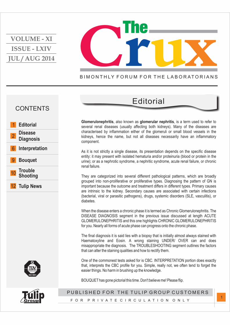
1 VOLUME - XI ISSUE - LXIV JUL / AUG 2014 Editorial Disease Diagnosis Bouquet Interpretation Tulip News 9 10 2 1 12 Trouble Shooting 6 Glomerulonephritis, also known as glomerular nephritis, is a term used to refer to several renal diseases (usually affecting both kidneys). Many of the diseases are characterised by inflammation either of the glomeruli or small blood vessels in the kidneys, hence the name, but not all diseases necessarily have an inflammatory component. As it is not strictly a single disease, its presentation depends on the specific disease entity: it may present with isolated hematuria and/or proteinuria (blood or protein in the urine); or as a nephrotic syndrome, a nephritic syndrome, acute renal failure, or chronic renal failure. They are categorized into several different pathological patterns, which are broadly grouped into non-proliferative or proliferative types. Diagnosing the pattern of GN is important because the outcome and treatment differs in different types. Primary causes are intrinsic to the kidney. Secondary causes are associated with certain infections (bacterial, viral or parasitic pathogens), drugs, systemic disorders (SLE, vasculitis), or diabetes. When the disease enters a chronic phase it is termed as Chronic Glomerulonephritis. The DISEASE DIAGNOSIS segment in the previous issue discussed at length ACUTE GLOMERULONEPHRITIS and this one highlights CHRONIC GLOMERULONEPHRITIS for you. Nearly all forms of acute phase can progress onto the chronic phase. The final diagnosis it is said lies with a biopsy that is initially almost always stained with Haematoxyline and Eosin. A wrong staining UNDER/ OVER can and does misappropriate the diagnosis. The TROUBLESHOOTING segment outlines the factors that can alter the staining qualities and how to rectify them. One of the commonest tests asked for is CBC. INTERPRETATION portion does exactly that, interprets the CBC profile for you. Simple, really not, we often tend to forget the easier things. No harm in brushing up the knowledge. BOUQUET has gone pictorial this time. Don't believe me! Please flip.
Transcript of VOLUME - XI ISSUE - LXIV JUL / AUG 2014 - tulipgroup.com · 1 VOLUME - XI ISSUE - LXIV JUL / AUG...
1
VOLUME - XI
ISSUE - LXIV
JUL / AUG 2014
Editorial
DiseaseDiagnosis
Bouquet
Interpretation
Tulip News
9
10
2
1
12
TroubleShooting
6
Glomerulonephritis, also known as glomerular nephritis, is a term used to refer to several renal diseases (usually affecting both kidneys). Many of the diseases are characterised by inflammation either of the glomeruli or small blood vessels in the kidneys, hence the name, but not all diseases necessarily have an inflammatory component.
As it is not strictly a single disease, its presentation depends on the specific disease entity: it may present with isolated hematuria and/or proteinuria (blood or protein in the urine); or as a nephrotic syndrome, a nephritic syndrome, acute renal failure, or chronic renal failure.
They are categorized into several different pathological patterns, which are broadly grouped into non-proliferative or proliferative types. Diagnosing the pattern of GN is important because the outcome and treatment differs in different types. Primary causes are intrinsic to the kidney. Secondary causes are associated with certain infections (bacterial, viral or parasitic pathogens), drugs, systemic disorders (SLE, vasculitis), or diabetes.
When the disease enters a chronic phase it is termed as Chronic Glomerulonephritis. The DISEASE DIAGNOSIS segment in the previous issue discussed at length ACUTE GLOMERULONEPHRITIS and this one highlights CHRONIC GLOMERULONEPHRITIS for you. Nearly all forms of acute phase can progress onto the chronic phase.
The final diagnosis it is said lies with a biopsy that is initially almost always stained with Haematoxyline and Eosin. A wrong staining UNDER/ OVER can and does misappropriate the diagnosis. The TROUBLESHOOTING segment outlines the factors that can alter the staining qualities and how to rectify them.
One of the commonest tests asked for is CBC. INTERPRETATION portion does exactly that, interprets the CBC profile for you. Simple, really not, we often tend to forget the easier things. No harm in brushing up the knowledge.
BOUQUET has gone pictorial this time. Don't believe me! Please flip.

JUL/AUG
2
and further nephron loss. (stages 1-3), a substantial decline in the GFR may lead to only slight increases in serum creatinine levels. Azotemia (ie, a rise in blood urea nitrogen [BUN] and serum creatinine levels) is apparent when the GFR decreases to less than 60-70 mL/min. In addition to a rise in BUN and creatinine levels, the substantial reduction in the GFR results in the following: thus resulting in anemia. resulting in hypocalcemia, secondary hyperparathyro id ism, hyperphosphatemia, and renal osteodystrophy.
potassium, salt, and water excretion, resulting in acidosis, hyperkalemia, hypertension, and edema. leading to increased bleeding tendencies.
products (uremic toxins) affects virtually all organ systems. Azotemia occurring with the signs and symptoms listed above is known as uremia. Uremia occurs at a GFR of approximately 10 mL/min. Some of these toxins (eg, BUN, creatinine, phenols, and guanidines) have been identified, but none has been found to be responsible for all the symptoms.
The progression from acute glomerulonephritis to chronic glomerulonephritis is variable, depending to a considerable extent on the cause of the condition. Whereas complete recovery of renal function is the rule for patients with poststreptococcal glomerulonephritis, several other glomerulonephritides, such as immunoglobulin A (IgA) nephropathy, often have a relatively benign course, and many do not progress to ESRD. Progression patterns may be summarized as follows:
About 90% of patients progress to ESRD within weeks or months.
About 80% of patients progress to ESRD in 10 years; patients with the collapsing variant (malignant focal segmental glomerulosclerosis) have a more rapid progression; this form may be idiopathic or related to HIV infection. About 20-30% of patients with membranous nephropathy progress to chronic renal failure (CRF) and ESRD in 10 years.
About 40% of pat ients wi th membranoproliferative glomerulonephritis progress to CRF and ESRD in 10 years. About 10% of patients with IgA nephropathy progress to CRF and ESRD in 10 years.
About 1-2% of patients with poststreptococcal glomerulonephritis progress to CRF and ESRD; older children who present with crescentic glomerulonephritis are at greatest risk. Overall, about 20% of patients with lupus nephritis progress to CRF and ESRD in 10 years; however, patients with certain histologic variants (eg, class IV) may have a more rapid decline.
In the United States, chronic glomerulonephritis is the third leading cause of ESRD and accounts for 10% of patients on dialysis. chronic glomerulonephritis has accounted for as many as 40% of patients on dialysis; however, subsequent data suggest that in Japan, for
In early renal disease
Accumulation of toxic waste
Rapidly progressive glomerulonephritis or crescentic glomerulonephritis -
Focal segmental glomerulosclerosis -
Membranous nephropathy -
Membranoproliferative glomerulonephr i t is -
Lupus nephritis -
In Japan and some Asian countries,
Decreased production of erythropoietin,Decreased production of vitamin D,
Reduction in acid,
Platelet dysfunction,
IgA nephropathy -
Poststreptococcal glomerulonephritis -
Etiology
Epidemiology
CHRONIC GLOMERULONEPHRITISBackground
Pathophysiology
Nearly all forms of acute glomerulonephritis have a tendency to progress to chronic glomerulonephritis. The condition is characterized by irreversible and progressive glomerular and tubulointerstitial fibrosis, ultimately leading to a reduction in the glomerular filtration rate (GFR) and retention of uremic toxins. If disease progression is not halted with therapy, the net results are chronic kidney disease (CKD), end-stage renal disease (ESRD), and cardiovascular disease. The diagnosis of CKD can be made
without knowledge of the specific cause. defines CKD on the basis of either of the
following: based on abnormal urinalysis results (eg, proteinuria or hematuria) or structural abnormalities observed on ultrasound images.
for 3 or more months. the NKF developed guidelines that classify the
progression of renal disease into 5 stages, from kidney disease with a preserved GFR to end-stage kidney failure. This classification includes treatment strategies for each progressive level, as follows: This stage is characterized by kidney
damage with a normal GFR (≥ 90 mL/min); the action plan consists of diagnosis and treatment, treatment of comorbid conditions, slowing of the progressing of kidney disease, and reduction of cardiovascular disease risks. This stage is characterized by kidney damage with a mild decrease in the GFR (60-90 mL/min); the action plan is estimation of the progression of kidney disease. This stage is characterized by a moderately decreased GFR (to 30-59 mL/min); the action plan consists of evaluation and treatment of complications. This stage is characterized by a severe decrease in the GFR (to 15-29 mL/min); the action plan is preparation for renal replacement therapy. This stage is characterized by kidney failure; the action plan is kidney replacement if the patient is uremic. biopsy results cannot help distinguish the primary disease. Histology and clues to the etiology are often derived from other systemic diseases (if present). Considerable cause-specific variability is observed in the rate at which acute glomerulonephritis progresses to chronic glomerulonephritis. on the type of chronic glomerulonephritis (see Etiology). ESRD and death are common outcomes unless renal replacement therapy is instituted.
Reduction in nephron mass from the initial injury reduces the GFR. This reduction leads to hypertrophy and hyperfiltration of the remaining nephrons and to the initiation of intraglomerular hypertension. These changes occur in order to increase the GFR of the remaining nephrons, thus minimizing the functional consequences of nephron loss. The changes, however, are ultimately detrimental because they lead to glomerulosclerosis
The National Kidney Foundation (NKF)
In accordance with this definition,
At the later stages of glomerular injury,
The prognosis depends
Evidence of kidney damage
A GFR of less than 60 mL/min
Stage 1 –
Stage 2 –
Stage 3 –
Stage 4 –
Stage 5 –
DISEASE DIAGNOSIS
JUL/AUG

JUL/AUG
3
instance, the rate of chronic glomerulonephritis in patients on dialysis is 28%. The cause of this declining rate is not known.
Concurrent with the decline in chronic glomerulonephritis in these countries is an increase in diabetic nephropathy in up to 40% of patients on dialysis.
Dietary education is paramount in managing patients with CKD. The typical dietary restriction is 2 g of sodium, 2 g of potassium, and 40-60 g of protein a day. Additional restrictions may apply for diabetes, hyperlipidemia, and fluid overload.
regarding the types of ESRD therapy. The specific choices of ESRD therapy include hemodialysis, peritoneal dialysis, and renal transplantation. For further information, see Mayo Clinic - Kidney Transplant Information.
should be educated on home hemodialysis (in which patients are trained to do their dialysis at home) and center hemodialysis (in which patients come to a center 3 times a week for 3.5- to 4-hour dialysis sessions). They should also be educated on the types of vascular access. Arteriovenous fistulae should be created when the GFR falls below 25 mL/min or the serum creatinine level is greater than 4 mg/dL to allow maturation of the access before the initiation of dialysis. and most developed countries, patients on dialysis can travel. In fact, there are even dialysis cruises. However, patients should inform their social workers to make the necessary arrangements beforehand to ensure that the destination has the right resources to continue dialysis. and loss of libido are common in patients with kidney disease, especially men. Patients should be instructed to seek medical therapy if they experience these symptoms. worsen during pregnancy in patients with CKD, particularly when the serum creatinine level exceeds 2 mg/dL, and this leads to decreased fetal viability and increased maternal morbidity in pregnant women with CKD. Therefore, women with CKD should consult their doctors before becoming pregnant.
The history should begin by focusing on cause-specific symptoms to determine the source of the chronic kidney disease (CKD) if this is unknown. Recognition of such symptoms facilitates the planning of further workup and management of the disease (if systemic). related to uremia to determine if renal replacement therapy is needed. The following symptoms suggest uremia: and fatigue.
appetite, and weight. . nausea and vomiting. in taste sensation. in sleep pattern (ie, sleepiness in daytime and wakefulness at night). neuropathy. . . and hypertension suggests volume retention. Dyspnea or chest pain that varies with position suggests fluid overload and pericarditis, respectively. Leg cramps may suggest hypocalcemia or other electrolyte abnormalities. Weakness, lethargy, and fatigue may be due to anemia.
Cause-specific physical examination findings are discussed
Patient Education
History
Physical Examination
Patients should be educated
Patients opting for hemodialysis
In the United States
Sexual dysfunction
Renal failure and hypertension
The next step is to look for symptoms
The presence of edema
Weakness Loss of energy, Pruritus Early morning
Change ReversalPeripheral
Seizures Tremors
elsewhere, in articles describing the specific causes (see Etiology). Uremia-specific physical findings include the following:
. (if severe volume overload is present). rales (if pulmonary edema is present). rub in pericarditis. in the epigastric region or blood in the stool (possible indicators of uremic gastritis or enteropathy). and asterixis (indicators of advanced uremia).
The presence of the following complications generally indicates a need for urgent dialysis: acidosis. edema.
. encephalopathy. gastrointestinal bleeding. neuropathy. and hypocalcemia.
.
. Renal Failure. Acute. Crescentic. Diffuse
Proliferative. Membranoproliferative. Membranous.
Nonstreptococcal Associated With Infection. Poststreptococcal. Rapidly Progressive.
.
The presence of dysmorphic red blood cells (RBCs), albumin, or RBC casts suggests glomerulonephritis as the cause of renal failure. Waxy or broad casts are observed in all forms of chronic k idney d isease (CKD), inc luding chronic glomerulonephritis. Low urine-specific gravity indicates loss of tubular concentrating ability, an early finding in persons with CKD. See Urinalysis.
Urinary protein excretion can be estimated by calculating the protein-to-creatinine ratio on a spot morning urine sample. The ratio of urinary protein concentration (in mg/dL) to urinary creatinine (in mg/dL) reflects 24-hour protein excretion in grams. For instance, if the spot urine protein value is 300 mg/dL and the creatinine value is 150 mg/dL, the protein-to-creatinine ratio is 2. Thus, in this example, the 24-hour urine
protein excretion is 2 g. is used to assess and monitor the glomerular filtration rate (GFR). The 2 formulas available for calculation of the GFR are the Cockcroft-Gault formula, which estimates creatinine clearance, and the Modification of Diet in Renal Disease (MDRD) Study formula, which is used to calculate the GFR directly.
is simple to use but overestimates the GFR by 10-15% because creatinine is both filtered and secreted. The MDRD formula is much more complex but can be determined with a smartphone and tablet application available from the National Kidney Foundation or can be calculated online through the Hypertension, Dialysis, and Clinical Nephrology Web site.
is also used to monitor response to therapy and to initiate an early transition to renal replacement therapy (eg, dialysis access placement and transplantation evaluation). The degree of proteinuria, especially albuminuria, helps predict the renal prognosis in patients with
Hypertension Jugular venous distentionPulmonary
Pericardial friction Tenderness
Decreased sensation
Metabolic PulmonaryPericarditis Uremic Uremic
Uremic Severe anemiaHyperkalemia
Azotemia Chronic Glomerulonephritis,Glomerulonephritis, Glomerulonephritis,
Glomerulonephritis,Glomerulonephritis, Glomerulonephritis,
Glomerulonephritis,Glomerulonephritis,
Uremia
Urinalysis:
Urinary protein excretion:
Complications
Differential Diagnoses
Laboratory Studies
The estimated creatinine clearance rate
The Cockcroft-Gault formula
The estimated creatinine clearance rate
JUL/AUG
4
JUL/AUG
significant impact on retarding disease progression. for some glomerular diseases (eg, lupus) should be
implemented in appropriate settings. Aggressively manage anemia and renal osteodystrophy (eg, hyperphosphatemia, hypocalcemia, or hyperparathyroidism) before initiating renal replacement therapy. Also, aggressively manage comorbid conditions, such as heart disease and diabetes.
(urinary protein excretion >3.5 g/day) may have hyperlipidemia. As a part of cardiovascular health care, the lipid profile should be checked, and lipid-lowering therapy should be started for patients with hyperlipidemia. may induce or exacerbate diabetes, hypertension, weight gain, fat redistribution in the trunk (buffalo hump) and face (moon facies), cosmetic problems (eg, hirsutism and acne), and osteoporosis.
levels and blood pressure. Obtain baseline bone densitometry values. Repeat bone densitometry for bone pain. Oral calcium supplements (1 g/day) and vitamin D (400-800 IU/day) are recommended for prophylaxis against osteoporosis.
for patients with proteinuria in excess of 1 g/day is less than 125/75 mm Hg; for patients with proteinuria of less than 1 g/day, the target pressure is less than 130/80 mm Hg.
(ACEIs) are commonly used and are usually the first choice for treatment of hypertension in patients with chronic renal failure (CRF). ACEIs are renoprotective agents that have additional benefits beyond lowering pressure. They effectively reduce proteinuria, in part by reducing the efferent arteriolar vascular tone, thereby decreasing intraglomerular hypertension.
ACEIs have been shown to be superior to conventional therapy in slowing the decline of the glomerular filtration rate (GFR) in patients with diabetic and nondiabetic proteinuric nephropathies. Therefore, ACEIs should be considered for treatment of even normotensive patients with
significant proteinuria. (ARBs) in renal protection is increasingly being established, and these medications have been found to retard the progression of CKD in patients with diabetic or nondiabetic nephropathy, much as ACEIs do. and ARB therapy has been shown to achieve better pressure control and preservation of renal function than either therapy alone could. Therefore, in patients without hyperkalemia or an acute rise in serum creatinine levels after the use of either therapy, combination therapy should
be attempted. However, in patients with vascular disease or diabetes, combination ACEI and ARB therapy has been associated with increased adverse effects, including hyperkalemia, worsening renal function, and mortality. As such, combination ACEI and ARB therapy should not be used to treat
hypertension in these groups of patients with CKD. because of decreased free-water clearance, and
high doses may be required to control edema and hypertension when the GFR falls below 25 mL/min. Diuretics are also useful in counteracting the hyperkalemic potential of ACEIs and ARBs.
Specific therapies
Nephrotic patients
Steroid therapy
Monitor fasting blood glucose
Pressure management: Pharmacologic Therapy
The target pressure
Angiotensin-converting enzyme inhibitors
In particular,
The role of angiotensin II receptor blockers
A combination of ACEI therapy
Diuretics are often required
chronic glomerulonephritis. Patients with proteinuria exceeding 1 g/day have an increased risk of progression to end-stage renal failure. with chronic glomerulonephritis, Hayakawa et al examined the relations between (1) plasma adiponectin, leptin, and proteinuria levels; (2) GFR; and (3)
metabolic risk factors. They found that plasma adiponectin levels were much higher in patients with heavy proteinuria (38.8 ± 27.8 ìg/mL) than they were in patients who had mild proteinuria (13.3 ± 5.1 ìg/mL) or moderate proteinuria (18.1 ± 8.0 ìg/mL). Serum leptin levels, however, did not differ according to the degree of proteinuria.
Anemia is a significant finding in patients with some decline in the GFR. Physicians must be aware that anemia can occur even in patients with serum creatinine levels lower than 2 mg/dL. Even severe anemia can occur at low serum creatinine levels. Anemia is the result of marked impairment of erythropoietin production.
Serum creatinine and urea nitrogen levels are elevated. Impaired excretion of potassium, free water, and acid results in hyperkalemia, hyponatremia, and low serum bicarbonate levels, respectively. Impaired vitamin D-3 production results in hypocalcemia, hyperphosphatemia, and high levels of parathyroid hormone. Low serum albumin levels may be present if uremia interferes with nutrition or if the patient is nephrotic.
Obtain a renal ultrasonogram to determine renal size, to assess for the presence of both kidneys, and to exclude structural lesions that may be responsible for azotemia. Small kidneys often indicate an irreversible process.
If the kidney is small, kidney biopsy is usually unnecessary; no specific pattern of disease can be discerned at this point. A kidney biopsy may be considered in the minority of patients who exhibit an acute exacerbation of their chronic disease. This may be particularly pertinent to patients with preserved kidney size and in those with lupus nephritis.
the glomeruli may still show some histologic evidence of the primary disease. In advanced stages, the glomeruli are hyalinized and obsolescent. The tubules are disrupted and atrophic, and marked interstitial fibrosis and arterial and arteriolar sclerosis occur.
Patients with chronic kidney disease (CKD) who are admitted to the hospital should receive careful monitoring of weight, intake, output, and renal function so that acute renal failure (ARF) can be diagnosed and treated early if it occurs,. All potentially nephrotoxic agents must be adjusted for the degree of CKD. Furthermore, agents such as nonsteroidal anti-inflammatory drugs (NSAIDs), aminoglycosides, and intravenous (IV) contrast media must be avoided unless the benefits clearly outweigh the risks; they are strongly associated with ARF.
to end-stage renal disease (ESRD) can be slowed by a variety of measures, including aggressive control of diabetes, hypertension, and proteinuria. Dietary protein restriction, phosphate restriction, and hyperlipidemia control may have
In a study of 38 patients
In early stages,
Complete blood count:
Serum chemistry:
Renal ultrasonography:
Kidney biopsy:
Progression from CKD
Other Studies
Approach Considerations
JUL/AUG

5
JUL/AUG
JUL/AUG
However, they should be used with caution when given together with ACEIs or ARBs because the decline in intraglomerular pressure induced by ACEIs or ARBs may be exacerbated by the volume depletion induced by diuretics, potentially precipitating
ARF. calcium channel blockers, central alpha 2
agonists (eg, clonidine), alpha antagonists, and direct 1
vasodilators (eg, minoxidil and nitrates) may be used to achieve the target pressure. Because progressive fibrosis is the hallmark of chronic glomerulonephritis, some investigators have focused on finding inhibitors of fibrosis in an attempt to slow progression. Of the many compounds that have been considered, pirfenidone, an inhibitor of transforming growth factor beta and hence of collagen synthesis, has emerged as the best candidate. in an open-label study involving 21 patients with idiopathic and postadaptive focal segmental glomerulosclerosis, found that pirfenidone yielded a median 25% improvement in the rate of decline of the estimated GFR; the drug
did not affect proteinuria or blood pressure. Among the adverse events attributed to therapy were dyspepsia, sedation, and photosensitive dermatitis. It is hoped that pirfenidone therapy will prove an effective means of slowing progressive fibrosis; however, more studies are needed.
Cells have the ability to produce antioxidants, anti-inflammatory and detoxifying enzymes that are useful for cell viability, but this pathway is constantly being inhibited by an enzyme called KEAP. Inhibition of KEAP may therefore improve the antioxidant activity of cells and promote cell viability. Bardoxolone, an oleanolic acid derivative, blocks Keap and has been postulated as a potential mechanism to retard progression of CKD. In one study, patients receiving bardoxolone had significant increases in the mean (± standard deviation) estimated GFR, as compared with placebo, at 24 weeks. The improvement persisted at 52 weeks, suggesting that bardoxolone may have promise for the treatment of CKD, but more studies are
needed. Larger trials are ongoing to address the effect of bardoxolone on CKD. Sodium bicarbonate has been shown to reduce tubulointerstitial injury and endothelin production with substantial benefits in slowing progressive kidney damage. In one study on patients with advanced kidney failure, the administration of sodium
bicarbonate preserved GFR decline. Even in patients with relatively preserved GFR in stage 2 CKD, the administration of sodium bicarbonate was shown to preserve kidney function over
5years. Preliminary studies using aliskiren, a direct renin inhibitor, show reductions in proteinuria
over 6 months, but further studies are needed to determine the role of direct renin inhibitors in retarding progression of CKD.
Renal osteodystrophy can be managed early by replacing vitamin D and by administering phosphate binders. Seek and treat nonuremic causes of anemia, such as iron deficiency, before instituting therapy with
Beta blockers,
Cho et al,
Fibrosis inhibition:
Role of antioxidants (bardoxolone):
Role of sodium bicarbonate:
Role of direct renin inhibitors:
Management of other problems:
erythropoietin. (if present) to reduce overall cardiovascular comorbidity, even though evidence for renal protection is lacking. Discuss options for renal replacement therapy (eg, hemodialysis, peritoneal dialysis, and renal transplantation).
vascular access when the GFR falls below 20-25 mL/min, when the serum creatinine level exceeds 4 mg/dL, or when the rate of increase in the serum creatinine level indicates the need for dialysis within 1 year. Arteriovenous fistulas are preferred to arteriovenous grafts because of their long-term high-patency rates and should be placed whenever possible. Place peritoneal dialysis catheters 2-3 weeks before anticipated dialysis therapy. before the initiation of dialysis results in better survival than transplantation after the initiation of dialysis. Therefore, preemptive transplantation should be explored from live donors. Patients without live donors can be placed on the deceased donor wait list when the GFR falls below 20 mL/min to accrue time. Patients who opt for no treatment when it is indicated should be informed of imminent renal failure in a shorter time. to educational programs for early rehabilitation from dialysis or transplantation.
Patients with any evidence of kidney disease should be referred to a kidney specialist (ie, a nephrologist). Early referral of patients with CRF (serum creatinine, 1.5-2 mg/dL) to a nephrologist is important for managing complications and organizing the transition to renal replacement therapy. Some evidence indicates that early referral of CRF patients to a nephrologist improves the short-term outcome. The nephrologist will usually determine the frequency of visits on the basis of the degree of CKD.
a surgical consultation should be sought for creation of an arteriovenous fistula or graft to allow insertion of a peritoneal dialysis catheter. Consultation with a transplant surgeon is appropriate for evaluation for kidney transplantation.
Protein-restricted diets (0.4-0.6 g/kg/day) are controversial but may be beneficial in slowing the decline in the GFR and reducing hyperphosphatemia (serum phosphate > 5.5 mg/dL) in patients with serum creatinine levels higher than 4 mg/dL. Monitor these patients for signs of malnutrition, which may contraindicate protein restriction. Educate patients about how potassium-rich diets help control hyperkalemia. Many dietary restrictions are no longer necessary with the initiation of renal replacement therapy.
to increase their activity level as tolerated. Increased activity may aid in blood pressure control.
The goals of pharmacotherapy are to reduce morbidity and to prevent complications. Medications used to treat chronic glomerulonephritis include angiotensin-converting enzyme (ACE) inhibitors (ACEIs), diuretics, calcium channel blockers, beta-adrenergic blockers, and alpha-adrenergic agonists.
Treat hyperlipidemia
Arrange permanent
Preemptive transplantation
Expose patients
Renal Replacement Therapy:
When dialysis is imminent,
Encourage patients
Consultations
Diet and Activity
Medication Summary

INTERPRETATION
6
JUL/AUG
COMPLETE BLOOD COUNTSA complete blood count (CBC) gives important information about the kinds and numbers of cells in the blood, especially red blood cells, white blood cells and platelets. A CBC helps your doctor check any symptoms, such as weakness, fatigue, or bruising, you may have. A CBC also helps him or her diagnose conditions, such as anemia, infection, and many other disorders.
White blood cells protect the body against infection. If an infection develops, white blood cells attack and destroy the bacteria, virus, or other organism causing it. White blood cells are bigger than red blood cells but fewer in number. When a person has a bacterial infection, the number of white cells rises very quickly. The number of white blood cells is sometimes used to find an infection or to see how the body is dealing with cancer treatment.
The major types of white blood cells are neutrophils, lymphocytes, monocytes, eosinophils, and basophils. Immature neutrophils, called band neutrophils, are also part of this test. Each type of cell plays a different role in protecting the body. The numbers of each one of these types of white blood cells give important information about the immune system. Too many or too few of the different types of white blood cells can help find an infection, an allergic or toxic reaction to medicines or chemicals, and many conditions, such as leukemia.
Red blood cells carry oxygen from the lungs to the rest of the body. They also carry carbon dioxide back to the lungs so it can be exhaled. If the RBC count is low (anemia), the body may not be getting the oxygen it needs. If the count is too high (a condition called polycythemia), there is a chance that the red blood cells will clump together and block tiny blood vessels (capillaries). This also makes it hard for your red blood cells to carry oxygen.
This test measures the amount of space (volume) red blood cells take up in the blood. The value is given as a percentage of red blood cells in a volume of blood. For example, a hematocrit of 38 means that 38% of the blood's volume is made of red blood cells. Hematocrit and hemoglobin values are the two major tests that show if anemia or polycythemia is present.
The hemoglobin molecule fills up the red blood cells. It carries oxygen and gives the blood cell its red color. The hemoglobin test measures the amount of hemoglobin in blood and is a good measure of the blood's ability to carry oxygen throughout the body. There are three red blood cell indices: mean corpuscular volume (MCV), mean corpuscular hemoglobin (MCH), and mean corpuscular hemoglobin concentration (MCHC). They are measured by a machine, and their values come from other measurements in a CBC. The MCV shows the size of the red blood cells. The MCH value is the amount of hemoglobin in an average red blood cell. The MCHC measures the concentration of hemoglobin in an average red blood cell. These numbers help in the diagnosis of
A CBC test usually includes: White blood cell (WBC, leukocyte) count.
White blood cell types (WBC differential).
Red blood cell (RBC) count.
Hematocrit (HCT, packed cell volume, PCV).
Hemoglobin (Hgb).
Red blood cell indices.
different types of anemia. Red cell distribution width (RDW) can also be measured which shows if the cells are all the same or different sizes or shapes. Platelets (thrombocytes) are the smallest type of blood cell. They are important in blood clotting. When bleeding occurs, the platelets swell, clump together, and form a sticky plug that helps stop the bleeding. If there are too few platelets, uncontrolled bleeding may be a problem. If there are too many platelets, there is a chance of a blood clot forming in a blood vessel. Also, platelets may be involved in hardening of the arteries (atherosclerosis).
Mean platelet volume measures the average amount (volume) of platelets. Mean platelet volume is used along with platelet count to diagnose some diseases. If the platelet count is normal, the mean platelet volume can still be too high or too low. to be done at the same time as a CBC but it is not part of the regular CBC test. In this test, a drop of blood is spread (smeared) on a slide and stained with a special dye. The slide is looked at under a microscope. The number, size, and shape of red blood cells, white blood cells, and platelets are recorded. Blood cells with different shapes or sizes can help diagnose many blood diseases, such as leukemia, malaria, or sickle cell disease.
A complete blood count may be done to: such as fatigue, weakness, fever, bruising, or weight
loss. for anemia. has been lost if there is bleeding. polycythemia. for an infection.
of the blood, such as leukemia. with some types of drug or radiation treatment.
is affecting the blood cells and counts. for high and low values before a surgery. if there are too many or too few of certain types of cells. This may help find other conditions, such as too many eosinophils may mean an allergy or asthma is present. may be done as part of a regular physical examination. A blood count can give valuable information about the general state of your health.
You do not need to do anything before having this test.
Your health professional drawing blood will: around your upper arm to stop the flow of blood. This makes the veins below the band larger so it is easier to put a needle into the vein. with alcohol.
More than one needle stick may be needed. to the needle to fill it with blood. from your arm when enough blood is collected. or cotton ball over the needle site as the needle is removed. to the site and then a bandage. a heel stick will be done instead of a blood draw from a vein.
The blood sample is taken from a vein in your arm. An elastic band is wrapped around your upper arm. It may feel tight. You may feel nothing at all from the needle, or you may feel a quick
Platelet (thrombocyte) count.
Mean platelet volume (MPV).
Your doctor may order a blood smear test
Find the cause of symptoms
Check See how much bloodDiagnose Check
Diagnose diseases Check how the body is dealingCheck how abnormal bleeding
Screen See
A complete blood count
Wrap an elastic band
Clean the needle site Put the needle into the vein. Attach a tube
Remove the band Put a gauze pad
Put pressureIf this blood test is done on a baby,
Why It Is Done
How To Prepare
How It Is Done
How It Feels
JUL/AUG

7
JUL/AUG
White blood cell types (WBC differential)
Red blood cell (RBC) count
1 Hematocrit (HCT)
Hemoglobin (Hgb)
Red blood cell indices
Red cell distribution width (RDW)
Platelet (thrombocyte) count
Neutrophils: 50%–62%Band neutrophils: 3%–6%Lymphocytes: 25%–40%Monocytes: 3%–7%Eosinophils: 0%–3%Basophils: 0%–1%
Men: 4.5–5.5 million RBCs per microliter (mcL) or 124.5–5.5 x 10 /liter (L)
12Women: 4.0–5.0 million RBCs per mcL or 4.0–5.0 x 10 /L12Children: 3.8–6.0 million RBCs per mcL or 3.8–6.0 x 10 /L12Newborn: 4.1–6.1 million RBCs per mcL or 4.1–6.1 x 10 /L
Men: 42%–52% or 0.42–0.52 volume fraction
Women: 36%–48% or 0.36–0.48 volume fraction
Children: 29%–59% or 0.29–0.59 volume fraction
Newborn: 44%–64% or 0.44–0.64 volume fraction
Men: 14–17.4 grams per deciliter (g/dL) or 140–174 grams per liter (g/L)
Women: 12–16 g/dL or 120–160 g/L
Children: 9.5–20.5 g/dL or 95–205 g/L
Newborn: 14.5–24.5 g/dL or 145–245 g/L
In general, a normal hemoglobin level is about one-third the value of the hematocrit.
Mean corpuscular volume 84–96 femtoliters (fL)(MCV)—Adults:
Mean corpuscular hemoglobin 28–34 picograms (pg) (MCH)—Adults: per cell
Mean corpuscular hemoglobin 32–36 grams per concentration (MCHC)—Adults: deciliter (g/dL)
Normal: 11.5%–14.5%
3Adults: 140,000–400,000 platelets per mm or 140–400 x 910 /L
3Children: 150,000–450,000 platelets per mm or 150–450 x 910/L
sting or pinch.
There is very little chance of a problem from having a blood sample taken from a vein. at the site. You can lower the chance of bruising by keeping pressure on the site for several minutes. the vein may become swollen after the blood sample is taken. This problem is called phlebitis. A warm compress can be used several times a day to treat this. can be a problem for people with bleeding disorders. Aspirin, warfarin (such as Coumadin), and other blood-thinning medicines can make bleeding more likely. If you have bleeding or clotting problems, or if you take blood-thinning medicine, tell your doctor before your blood sample is taken.
A complete blood count (CBC) gives important information about the kinds and numbers of cells in the blood, especially red blood cells, white blood cells, and platelets. A CBC helps your doctor check any symptoms, such as weakness, fatigue, or bruising, you may have. A CBC also helps him or her diagnose conditions, such as anemia, infection, and many other disorders. The normal values listed here—called a reference range—are just a guide. These ranges vary from lab to lab, and your lab may have a different range for what's normal. Your lab report should contain the range your lab uses. Also, your doctor will evaluate your results based on your health and other factors. This means that a value that falls outside the normal values listed here may still be normal for you or your lab.
depend on age, sex, how high above sea level you live, and the type of blood sample. Your doctor may use all the CBC values to check for a condition. For example, the red blood cell (RBC) count, hemoglobin (Hgb), and hematocrit (HCT) are the most important values needed to tell whether a person has anemia, but the red blood cell indices and the blood smear also help with the diagnosis and may show a possible cause for the anemia.
is good and how the cells look on the smear, your doctor will look at both the number (WBC count) and the WBC differential. To see whether there are too many or too few of a certain type of cell, your doctor will look at the total count and the percentage of that particular cell. There are normal values for the total number of each type of white cell. can change these blood values. Your doctor will talk with you about normal values during each trimester of your pregnancy.
Men and nonpregnant women: 5,000–10,000 WBCs per 3cubic millimeter (mm ) or
95.0–10.0 x 10 WBCs per
liter (L)
Risks
Results
You may get a small bruise
In rare cases,
Ongoing bleeding
Normal:
White blood cell (WBC, leukocyte) count
Normal values for the complete blood count (CBC) tests
To see if the white blood cell (WBC, leukocyte) count
Pregnancy
JUL/AUG
8
JUL/AUG
Mean platelet volume (MPV)
Blood smear
Red blood cell (RBC):
White blood cell (WBC, leukocyte):
Platelets:
Red blood cell (RBC):
3Adults: 7.4–10.4 mcm or 7.4–10.4 fL3Children: 7.4–10.4 mcm or 7.4–10.4 fL
Normal: Blood cells are normal in shape, size, color, and number.Leucocytes show no morphologic abnormality and their total and differential numbers are within normal range.No immature cells are seen.No hemoparasites are observed.Platelets are adequate in number and morphology.
include smoking, exposure to carbon monoxide, long-term lung disease, kidney disease, some cancers, certain forms of heart disease, alcoholism, liver disease, a rare disorder of the bone marrow (polycythemia vera), or a rare disorder of hemoglobin that binds oxygen tightly.
can also cause high RBC values. These conditions include dehydration, diarrhea or vomiting, excessive sweating, and the use of diuretics. The lack of fluid in the body makes the RBC volume look high. This is sometimes called spurious polycythemia.
include infection, inflammation, damage to body tissues (such as a heart attack), severe physical or emotional stress (such as a fever, injury, or surgery), kidney failure, lupus, tuberculosis (TB), rheumatoid arthritis, malnutrition, leukemia, and diseases such as cancer. underactive adrenal glands, thyroid gland problems, certain medicines, or removal of the spleen can also cause high WBC values.
may be seen with bleeding, iron deficiency, some diseases like cancer, or problems with the bone marrow.
Anemia can be caused by heavy menstrual bleeding, stomach ulcers, colon cancer, inflammatory bowel disease, some tumors, Addison's disease, thalassemia, lead poisoning, sickle cell disease, or reactions to some chemicals and medicines. A low RBC value may also be seen if the spleen has been taken out.
or vitamin B12 can also cause anemia, such as pernicious anemia, which is a problem with absorbing vitamin B12.
and a blood smear may help find the cause of anemia.
High values
Low values
Conditions that cause high RBC values
Conditions that affect the body's water content
Conditions that cause high WBC values
The use of corticosteroids,
High platelet values
Anemia lowers RBC values.
A lack of folic acid
The RBC indices value
White blood cell (WBC, leukocyte):
Platelets:
If the elastic bandTaking medicines
A very high white blood cell count
Having an enlarged spleen,
Pregnancy,
The white blood countChildren
normally have higher WBC Other red blood cell tests
Conditions that can lower WBC values
A large spleen
Low platelet values
A large spleen
Erythrocyte sedimentation rate (ESR).
Reticulocyte count.
Hematocrit measurements
include chemotherapy and reactions to other medicines, aplastic anemia, viral infections, malaria, alcoholism, AIDS, lupus, and Cushing's syndrome. can lower the WBC count.
can occur in pregnancy or idiopathic thrombocytopenic purpura (ITP) and other conditions that affect how platelets are made or that destroy platelets.
can lower the platelet count.
Reasons you may not be able to have the test or why the results
may not be helpful include: was on your arm a long time while the blood sample was taken. that can cause low platelet levels. Some examples of the many medicines that cause low platelet levels include steroids, some antibiotics, thiazide diuretics, chemotherapy medicines, quinidine, and meprobamate. or high levels of a type of fat (triglycerides). These can cause falsely high hemoglobin values. which may cause a low platelet count (thrombocytopenia) or a low white blood cell count. An enlarged spleen may be caused by certain types of cancer. which normally causes a low RBC value and less often a high WBC value.
can change by as much as 2,000 WBCs per microliter (mcL) from exercise, stress, or smoking.
(leukocyte) counts than adults. that may be done include:
An ESR test measures how quickly red blood cells (erythrocytes) settle in a test tube. When inflammation in the body is present (such as from an infection or cancer), red blood cells may settle more slowly than normal. An ESR may help find certain inflammatory diseases when CBC results are normal. To learn more, see the topic Sedimentation Rate.
This test counts the number of immature red blood cells (reticulocytes) in a blood sample. Generally, only a few reticulocytes are present in the blood in relation to mature red blood cells. But recent bleeding or mature red blood cells being destroyed can cause a lot of new reticulocytes made. This test can help find some types of anemia and check how well treatment is working. To learn more, see the topic Reticulocyte Count.
can be very different depending on the method and type of machine used to do the test.
What Affects the Test
What To Think About
JUL/AUG
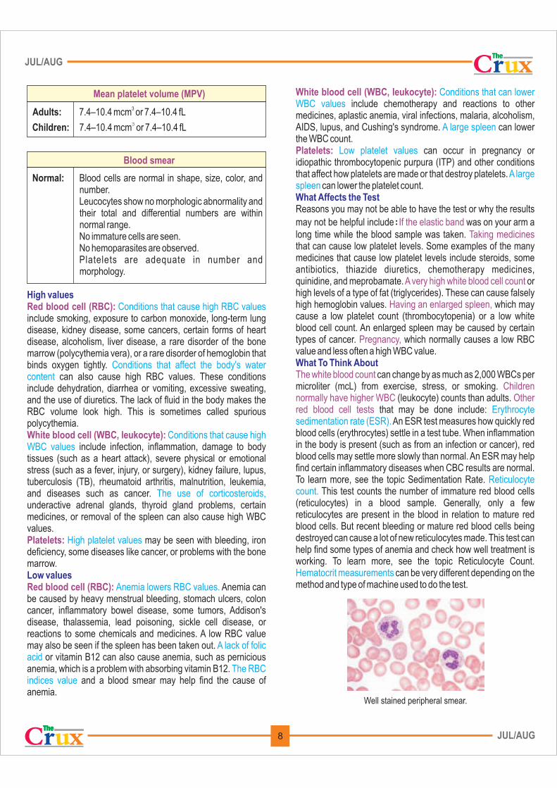
Well stained peripheral smear.

9
JUL/AUG
BOUQUET
In Lighter Vein
Wisdom Whispers
A man and his ever-nagging wife went on vacation to Jerusalem. While they were there, the wife passed away. The undertaker told the husband, "You can have her shipped home for $5,000, or you can bury her here, in the Holy Land, for $150." The man thought about it and told him he would just have her shipped home. The undertaker asked, "Why would you spend $5,000 to ship your wife home, when it would be wonderful to be buried here and you would spend only $150?" The man replied, "Long ago a man called Jesus Christ died here, was buried here, and three days later he rose from the dead. I just can't take that chance.
GOD HAS A SENSE OF HUMOR
A woman received a call that her daughter was sick.
She stopped by the pharmacy to get medication, got back to her car and found that she had locked her keys inside.
The woman found an old rusty coat hanger left on the ground.
She looked at it and said "I don't know how to use this."
She bowed her head and asked God to send her HELP.
Within 5 minutes a beat up old motorcycle pulled up.
A bearded man who was wearing an old biker skull rag.
The man got off of his cycle and asked if he could help.
She said: "Yes, my daughter is sick. I've locked my keys in my car. I must get home. Please, can you use this hanger to unlock my car?"
He said "Sure." He walked over to the car, and in less than a minute the car was open.
She hugged the man and through tears said "Thank You SO Much! You are a very nice man."
The man replied "Lady, I am NOT a nice man. I just got out of PRISON yesterday, I was in prison for car theft."
The woman hugged the man again sobbing, "Oh, thank you God! You even sent me a Professional!"
Is GOD Good or What!?
Brain Teasers
Answers: 1. A, 2. B, 3. C., 4. D
JUL/AUG
A. Emphysema Lung
B. Pulmonary abscess
C. Aspergilloma Lung
D. Carcinoma Lung
Match the following Pulmonary Pathologies
1
2
3
4

10 JUL/AUG
JUL/AUG
TROUBLESHOOTING
White spots are seen in the section after deparaffinization step. If they are not recognized at this point, spotty or irregular staining wil l be seen microscopically on the stained section.
The nuclei are too pale (the hematoxylin is too light).
The nuclei are overstained (the hematoxylin is too dark), or diffuse hematoxylin staining of the cytoplasm has occurred.
Red or red-brown nuclei.
Pale staining with eosin.
Cytoplasm is overstained, and the differentiation is poor.
A. The section was not dried properly before beginning deparaffinization.
B. The slide did not remain in xylene long enough for complete removal of the paraffin.
A. The sections were not stained long enough in hematoxylin.
B. The hematoxylin was overoxidized and should not have been used.
C. The differentiation step was too long. D. Pale nuclei in bone sections may be the
result of over decalcification.
A. The sections were stained too long in hematoxylin.
B. The sections are too thick.C. The differentiation step was too short.
A. The hematoxylin is breaking down.B. The sections were not blued sufficiently.
A. The pH of the eosin solution may be above 5.0, possibly caused by carryover of the bluing reagent.
B. The sections may be too thin. C. Slides may have been left too long in the
dehydrating solutions.
A. The eosin solution may be too concentrated, especially if phloxine is present.
B. The section may have been stained for too long.
C. The sections may have been passed through the dehydrating alcohols too rapidly for good differentiation of the eosin to occur.
A. The slides must be treated with absolute alcohol to remove the water and then retreated with xylene to remove the paraffin. If incomplete drying is severe, the sections may loosen from the slides.
B. The slides should be returned to xylene for a longer time.
A. The section must be restained. When sections have been placed in an extremely acidic fixative such as Zenker solution, the ability to stain the nucleus may be impaired and the time in the hematoxylin may have to be increased, or a method to increase tissue basophilia may be needed.
B. Discard hematoxylin and replace with fresh.C. Run back and restainD. No solution
A. Decolorize the section and restain, making appropriate adjustments in the staining time of hematoxylin.
B. Recut the section. C. Decolorize the section and restain, making
a p p r o p r i a t e a d j u s t m e n t s i n t h e differentiation times.
A. Check the oxidation status of the hematoxylin.
B. Allow a longer time for bluing of the sections; it is impossible to over blue the sections.
A. Check the pH of the eosin solution, and adjust it to a pH of 4.6 to 5.0 with acetic acid if necessary. Be sure the bluing reagent is completely removed before transferring the slides to the eosin.
B. Check the thickness of the section.C. Restain with eosin and do not allow the
stained slides to stand in the lower concentrations of alcohols.
A. Dilute the eosin solution.B. Decrease the staining time. C. Allow more time in each of the dehydrating
solutions for adequate differentiation of the eosin.
(Also, check the section thickness.)
H & E STAINING
PROBLEM CAUSE SOLUTION

11 JUL/AUG
JUL/AUG
Blue—black precipitate on top of the sections.
Water bubbles are seen microscopically in the stained sections.
Difficulty bringing some areas of the tissue in focus with light microscopy.
The mounting medium has retracted from the edge of the cover glass.
The water and the slides turn milky when the slides are placed in the water following the rehydrating alcohols.
The slides are hazy or milky in the last xylene rinse prior to coverslipping.
The mounted stained sections do not show the usual transparency and crispness when viewed by light microscopy.
The metallic sheen that develops on most hematoxylin solutions has been picked up on the slide.
The sections were not completely dehydrated, and water is present in the mounted section.
Mounting medium may be present on top of the cover glass.
A. The cover glass is warped.B. The mounting medium has been thinned too
much with xylene.
Xylene has not been removed completely by the alcohols.
Water has not been completely removed from the sections before being placed in the xylene.
The mounting medium may be too thick, causing the cover glass to be held too far above the tissue.
Filter the hematoxylin solution daily before staining slides.
Remove the cover glass and mounting medium with xylene. Return the slide to fresh absolute alcohol (several changes). After the sections are dehydrated, clear with fresh xylene and mount with synthetic resin. All dehydrating and clearing solutions should be changed before staining any more sections.
Remove the cover glass and remount with a clean cover glass. Review the method used for mounting sections, and modify if needed.
A. Remove the cover glass and apply a new cover glass.
B. Apply a new cover glass with fresh mounting medium. Keep the mounting medium container tightly capped when not in use. Use a small container for the mounting medium and discard when it becomes too thick.
Change the alcohols, back the slides up to absolute alcohol, and rehydrate the sections.
Change the alcohol solutions, especially the anhydrous or absolute reagents. Redehydrate the sections and clear in fresh xylene.
Remove the cover glass and mounting medium with xylene. Remount the section with fresh mounting medium.
H & E STAINING
PROBLEM CAUSE SOLUTION
Good H&E staining Under stained H&E stain

JUL/AUG
Printed and published by D.G. Tripathi, Edited by Dr. Ramnik Sood, M.D. (Path.) for and on behalf of Tulip Diagnostics Private Ltd., Gitanjali, Tulip Block, Dr. Antonio Do Rego Bagh, Alto Santacruz, Bambolim Complex Post Office, Goa - 403 202, INDIA.Fax: (0832) 2458544. E-mail: [email protected] Website: www.tulipgroup.com
12
JUL/AUG
STOP CALCULATING, MEASURE OSMOLALITY DIRECTLY
Temp
TempPlateau
Pure Water
Aqueous Solution
Supercooling
Freezing Initiation Point
Turning Point
1 min. 2 min. Time
0°C
Measuring Value(Freezing point
depression)
Curves describing Freezing Point Depression
Group
Cryobasic-1
Freezing Point Osmometer
Cryobasic-1Cryobasic-1
Freezing Point Osmometry Principle
Freezing point of a solution is lower than that of pure
solvent. Addition of solute to the solution causes further
decrease in Freezing Point. By precise measurement of
the solution the Osmolality can be measured.
Emergency/ Internal medicine, Urology/ Renal Function,
Endrocrinology, Obstetrics, Gynecology, Surgery/Critical
Care, IVF Centre, Pharmacy and General Quality Control.
Use of Freezing Point Osmometer
Cryobasic 1 Features
Benefits of Freezing Point Osmometry
•
•
•
•
•
•
•
•
•
•
Automated Single Channel Measurement with high accuracy & precision.
Fastest Start up time.
Small sample (Serum, Plasma, Urine or any Biological fluid) volume of 30 µl.
Wide Measurement range 0 to 3000 mOsm/Kg H2O.
Automatic Calibration.
Unparalleled repeatability & reproducibility.
Performs rapid & inexpensive measurement.
Requires small sample size.
Optimized for most biological & aqueous applications.
Samples can be recovered for retesting/ another test.