Common carotid artery CCA External Carotid artery ECA Internal ...
Upload
truongtuongCategory
view
217download
1
SpenceMariya Kuk, Thapat Wannarong, Vadim Beletsky, Grace Parraga, Aaron Fenster and J. David
Volume of Carotid Artery Ulceration as a Predictor of Cardiovascular Events
Print ISSN: 0039-2499. Online ISSN: 1524-4628 Copyright © 2014 American Heart Association, Inc. All rights reserved.
is published by the American Heart Association, 7272 Greenville Avenue, Dallas, TX 75231Stroke published online April 8, 2014;Stroke.
http://stroke.ahajournals.org/content/early/2014/04/08/STROKEAHA.114.005163World Wide Web at:
The online version of this article, along with updated information and services, is located on the
http://stroke.ahajournals.org/content/suppl/2014/04/08/STROKEAHA.114.005163.DC1.htmlData Supplement (unedited) at:
http://stroke.ahajournals.org//subscriptions/
is online at: Stroke Information about subscribing to Subscriptions:
http://www.lww.com/reprints Information about reprints can be found online at: Reprints:
document. Permissions and Rights Question and Answer process is available in the
Request Permissions in the middle column of the Web page under Services. Further information about thisOnce the online version of the published article for which permission is being requested is located, click
can be obtained via RightsLink, a service of the Copyright Clearance Center, not the Editorial Office.Strokein Requests for permissions to reproduce figures, tables, or portions of articles originally publishedPermissions:
by guest on April 9, 2014http://stroke.ahajournals.org/Downloaded from by guest on April 9, 2014http://stroke.ahajournals.org/Downloaded from by guest on April 9, 2014http://stroke.ahajournals.org/Downloaded from by guest on April 9, 2014http://stroke.ahajournals.org/Downloaded from by guest on April 9, 2014http://stroke.ahajournals.org/Downloaded from by guest on April 9, 2014http://stroke.ahajournals.org/Downloaded from by guest on April 9, 2014http://stroke.ahajournals.org/Downloaded from

1
Ulceration of carotid atherosclerotic plaques has been associated with plaque rupture, intraplaque hemorrhage,1
larger lipid core, and decreased amounts of fibrous tissue1 and decreased stability.1,2 Ulceration tends to be associated with greater plaque thickness and plaque volume.2,3
Plaque morphology has been previously assessed with the use of ultrasound imaging. Previous studies have categorized plaque as having an irregular or smooth morphology and found that irregularity of plaque significantly increased the risk of stroke.4 Other studies, specifically focusing on ulcer-ations, also noted the increased risk in stroke development with ulcerated plaques.5–7
Our group reported previously that ulcer number (≥3 ulcers when both left and right carotid arteries are combined) pre-dicted stroke, transient ischemic attack (TIA), or death.7 The aim of the current study is to determine whether total ulcer vol-ume (TUV) identifies high-risk patients with atherosclerosis.
MethodsStudy PopulationAs described previously,8 participants were patients attending the Stroke Prevention and Atherosclerosis Center clinic at London Ontario, Canada, who volunteered for a carotid imaging study fund-ed by the Heart and Stroke Foundation of Canada. They had been referred to the Hypertension Clinic, the Premature Atherosclerosis Clinic, or the Stroke Prevention Clinic. All had been monitored annu-ally with carotid duplex ultrasound, including measurement of total plaque area. Patients were eligible if their total plaque area was in the top tertile among the patient populations (40–600 mm2), representing moderate-to-severe atherosclerosis. Hypertension was defined as on antihypertensive medication or a baseline blood pressure <140 sys-tolic or 90 diastolic; diabetes mellitus was defined as on medication for diabetes mellitus; dyslipidemic was defined as on lipid-lowering medication. In total, 349 patients were enrolled in the study and consented to a protocol approved by the Western University Human Ethics Research board, approval number 12401E. The participants were followed up for ≤5 years.
Background and Purpose—Previous studies have shown the presence of ulceration in atherosclerotic plaque either by categorizing the plaque as complex (irregular morphology with ulcers) or smooth or by quantifying the number of ulcers observed in a specific region of interest. The aim of this study was to quantify carotid total ulcer volume by 3-dimensional ultrasound to investigate the relationship of total ulcer volume to vascular events (strokes, transient ischemic attack, myocardial infarction, revascularization, or death because of cardiovascular reasons).
Methods—In total, 349 at-risk subjects provided written informed consent to carotid 3-dimensional ultrasound and were analyzed for ulcerations. Ulcer volume was defined as a distinct discontinuity in an atherosclerotic plaque, with a volume ≥1.00 mm3 as measured using manual segmentation. The sum of the volumes of all ulcers seen in both carotids was the total ulcer volume. Participants were monitored for ≤5 years for outcomes, including cardiovascular events and death.
Results—Kaplan–Meier survival analysis showed that subjects with total ulcer volume ≥5 mm3 experienced a significantly higher risk of developing stroke, transient ischemic attack, or death (P=0.009) and of developing stroke/transient ischemic attack/death/myocardial infarction/revascularization (P=0.017). Lower ulcer volumes did not predict events nor did ulcer depth.
Conclusions—Volume of carotid ulceration on 3-dimensional ultrasound predicts cardiovascular events. In addition to improving risk stratification, ulceration is a potential therapeutic target. (Stroke. 2014;45:00-00.)
Key Words: carotid arteries ◼ ischemic attack, transient ◼ stroke ◼ ulcer
Volume of Carotid Artery Ulceration as a Predictor of Cardiovascular Events
Mariya Kuk; Thapat Wannarong, MD; Vadim Beletsky, MD; Grace Parraga, PhD; Aaron Fenster, PhD; J. David Spence, MD
Received February 12, 2014; accepted March 5, 2014.From the Department of Pathology and Toxicology, Schulich School of Medicine and Dentistry (M.K.), Department of Psychiatry (V.B.), Imaging
Research Group, Robarts Research Institute (G.P., A.F., J.D.S.), and Stroke Prevention and Atherosclerosis Research Centre, Robarts Research Institute (J.D.S.), Western University, London, Ontario, Canada; and Department of Internal Medicine, Faculty of Medicine, Siriraj Hospital, Mahidol University, Bangkok, Thailand (T.W.).
The online-only Data Supplement is available with this article at http://stroke.ahajournals.org/lookup/suppl/doi:10.1161/STROKEAHA. 114.005163/-/DC1.
Correspondence to J. David Spence, MD, Stroke Prevention and Atherosclerosis Research Centre, Robarts Research Institute, University of Western Ontario, 1400 Western Rd, London, Ontario, Canada N6G 2V2. E-mail [email protected]
© 2014 American Heart Association, Inc.
Stroke is available at http://stroke.ahajournals.org DOI: 10.1161/STROKEAHA.114.005163
Original Contribution
by guest on April 9, 2014http://stroke.ahajournals.org/Downloaded from

2 Stroke May 2014
Medical TherapyAll the participants received standard intensive medical therapy that is routine in our clinic, as previously described.9 This included advice on smoking cessation, maintenance of a healthy weight, exercise, moderate alcohol consumption, blood pressure control, lipid-lower-ing therapy, and antiplatelet therapy or anticoagulation, as appropri-ate. There was no alteration of therapy based on ulcer volumes.
Total Plaque AreaCarotid plaque burden, a strong predictor of cardiovascular risk,10–14 was measured as previously described10 by tracing with a cursor the area of every plaque seen in the internal, external, and common ca-rotid arteries on both sides in the plane of the longitudinal view in which the plaque was biggest. The sum of all plaque areas was the total plaque area.
Ascertainment of OutcomesAt each annual visit, the participants self-reported any events that occurred during the previous year; these were verified in hospital records. Events included stroke, TIA, revascularization (stenting, by-pass, or endarterectomy of any artery), or death. Cause of death was confirmed by the primary care physician.
A TIA was defined as an episode of focal neurological symp-toms diagnosed by a stroke neurologist as being caused by cerebral ischemia, with symptoms persisting <24 hours and no correspond-ing lesion seen on brain imaging. A stroke was an episode of focal neurological dysfunction caused by cerebral ischemia, with symp-toms and signs persisting >24 hours, or symptoms and signs persist-ing <24 hours, and a corresponding lesion seen on brain imaging. Vascular death was defined as death because of stroke, myocardial infarction (MI), aortic dissection, or vascular surgery/intervention. Revascularization was a carotid endarterectomy or stenting, coro-nary bypass or stenting, or bypass or stenting of the aorta or a pe-ripheral artery.
The primary outcome was a combination of stroke, TIA, or vascu-lar death; for the survival analyses, the first event in each patient was used to determine event-free survival. A secondary outcome was a combination of stroke, TIA, vascular death, MI, or revascularization (carotid, coronary, or peripheral); again, the first event in each patient was used in survival analyses.
Ulcer Identification and Measurement of Ulcer Depth and Ulcer VolumeA 3-dimensional (3D) scanning system was used to generate the 3D ultrasound images of the left and right carotid arteries of patients re-cruited for the study as described previously.5,10 Both carotid arter-ies were scanned for a length of 4 cm, centered at the bifurcation. As previously described,5 we defined an ulcer as being a continuous and distinct depression into the atherosclerotic plaque with diameter ≥1.00 mm and depth ≥1.00 mm, with a volume ≥1.00 mm3.
Ulcer depth was measured from a contour matching the endothelial surface on either side of the ulcer to the depth of the ulcer, at points along the width of the ulcer (Figure 1). Maximum ulcer depth was the greatest depth for the deepest ulcer in each case with ulceration.
Manual segmentation was used to measure the volume of each ulcer. On identification of a distinct, continuous depression in the plaque, the observer identified the point at which the depression be-gan and started the initial volume measurement by tracing the contour of the depression in a cross-sectional slice. The contours were traced in magnified views at intervals of 0.1 mm between the frames until the depression was no longer visible in the plaque. The slices had a thickness of 1 mm, and the volume was computed by the sum of area×thickness for all slices of each ulcer traced (Figure 2). A partici-pant was considered to have no ulcerations when there was no depres-sion in the carotid artery plaque ≥1.00 mm3. The sum of the volumes of all ulcers seen in both carotids was TUV.
Reliability of Measurement of TUVIntraobserver and interobserver reliability of repeat measurement of ulcer volume was computed on 50 randomized images before the beginning of the study using IBM SPSS version 20 software. Intraobserver reliability was assessed for 1 blinded observer (M.K.), and the interobserver reliability was determined by comparing mea-surements made independently by 2 blinded observers (M.K. and T.W.). The intraclass correlation coefficient was 0.72 for the intraob-server reliability, and the intraclass correlation coefficient for interob-server reliability was 0.84 using average measures.
Statistical AnalysisKaplan–Meier survival analysis with log-rank pooled over strata function was used to analyze differences in event-free survival time
Figure 1. Measurement of ulcer volume and ulcer depth. Contours of ulcers were traced and depth of ulcers measured in cross-sectional views. Each slice had a thickness of 1 mm; ulcer volume was computed from the sum of the volumes of all slices in which ulceration was traced (Figure I in the online-only Data Supplement).
by guest on April 9, 2014http://stroke.ahajournals.org/Downloaded from

Kuk et al Carotid Plaque Volume Predicts Events 3
between participants with a TUV <5.00 versus ≥5.00 mm3. When multiple events occurred in a single participant, the time to the first event was counted in the Kaplan–Meier survival analysis.
ResultsOf the 349 patients initially recruited to the study, 313 had 3D ultrasound scans that were technically adequate for measure-ment of total plaque volume. Results are presented for those cases. Mean age was 70.05±8.77 (SD) years, 57.5% were men, 80% were hypertensive, and 76% had dyslipidemia. There were 26 patients with stenosis of 1 carotid between 70% and 90% and 1 patient with 1 occluded carotid artery. Patients with carotid stenosis ≥70% did not have signifi-cantly higher TUV (P=0.13) or total ulcer number (P=0.12) Previous history of the participants before enrollment in the study included stroke in 22.7%, TIA in 44.1%, coronary bypass in 10.6%, and MI in 16.1%; 21.8% had diabetes mel-litus, 10.2% still smoked, 53.1% quit smoking, and 36.6% had never smoked. Carotid stenosis of 50% to 69% was pres-ent in 29.9% of cases.
One or more carotid ulcers were present in 124 participants. Of those with ulceration, TUV was 1.00 to 1.99 mm3 in 46, 2.00 to 2.99 mm3 in 34, 3.00 to 3.99 mm3 in 18, 4.00 to 4.99 mm3 in 7, and > 5.0 mm3 in 19. Mean TUV was 3.03 (range, 1.01–12.35) mm3. Figure 2 shows the distribution of TUV by number of ulcers in a given participant. Baseline variables for the 2 groups of patients (ulcer volume <5 versus ≥5 mm3) and medications being taken at baseline and at the first follow-up visit are shown in Table 1. Patients with >5 mm3 of ulcer vol-ume had a higher number of ulcers, had higher total plaque area, were more likely to be smokers, and were more likely to be taking anticoagulants; no other variable was significant.
The median duration of patient follow-up was 3.17 years (range, 0.07–5 years; mean, 1024 days; SD, 313 days). There were complete data available on ulcer volume and survival in 313 of the 322 participants with measurement of ulcer vol-ume. Among them, 10 had stroke, 13 had TIA, 5 had MI, 15 had revascularization procedure, and 13 died (4 deaths were
vascular, 3 died from cancer, and 6 died from other causes). Overall, there were 59 events that occurred in 49 patients.
First event included the following: 13 revascularization events, 11 TIA, 5 MI, 10 strokes, 4 cardiovascular deaths, and 6 noncardiovascular deaths (2 because of cancer, 4 because of other causes). Secondary events occurred in 6 patients; these included 2 TIA, 2 strokes, 2 revascularization events, 2 deaths: 1 because of cancer and 1 of unknown cause. One person had a second recurrent TIA after having a stroke followed by the sec-ondary TIA (thus stroke was the primary event and there were 2 TIAs afterward). Another patient, after having an MI as the ini-tial event, died after revascularization from an unknown cause.
Table 2 shows the distribution of events by the 2 cat-egories of ulcer volume. Figure II in the online-only Data Supplement shows the relationship of total plaque area to total plaque volume.
Event-free survival free of stroke, TIA, or death was sig-nificantly worse in patients with TUV ≥5 mm3 (log-rank
Figure 2. Distribution of total ulcer volume by ulcer number. The number of ulcers ranged from 0 to 4; total ulcer volume ranged from 1.01 to 12.35 mm3; total ulcer volume and ulcer number overlapped considerably.
Table 1. Baseline Variables by Ulcer Volume Categories
Baseline Variable
Ulcer Volume <5 mm3
(n=303)
Ulcer Volume ≥5 mm3
(n=19)P
Value*
Continuous variables: mean±SD
Age, y 69.88±8.65 72.68±10.55 0.18
Ulcer volume, mm3 0.81±1.25 7.15±2.04 0.0001
Ulcer number 0.42±0.63 2.42±0.77 0.0001
Systolic blood pressure, mm Hg 134±20 138±21 0.40
Diastolic blood pressure, mm Hg 76±13 75±9 0.73
Total cholesterol, mmol/L 4.12±1.01 4.23±1.35 0.64
Triglycerides, mmol/L 1.39±0.78 1.39±0.79 0.98
HDL cholesterol, mmol/L 1.40±0.44 1.43±0.42 0.83
LDL cholesterol, mmol/L 2.13±0.95 2.22±1.08 0.69
Homocysteine, μmol/L 9.60±4.11 10.25±4.11 0.76
Smoking, pack-years 14.82±20.54 17.82±16.03 0.54
Total plaque area, mm2 166.73±80.97 266.21±125.93 0.0001
Maximal stenosis, %† 44.81±14.31 45.26±17.11 0.90
Categorical variables, %
Sex (male) 57.1% 63.2% 0.39
Diabetic 21.8% 10.5% 0.194
Still smoking 9.6% 21.1% 0.025
Medication at baseline
ACE inhibitor 49.3% 52.6% 0.48
Antiplatelet agent 79.3% 63.2% 0.09
Statin 85.7% 89.5% 0.48
Anticoagulant 10.5% 35.6% 0.016
Medication at first follow-up visit
ACE inhibitor 50.3% 57.9% 0.35
Antiplatelet agent 79.2% 68.4% 0.20
Statin 89.9% 94.7% 0.43
Anticoagulant 10.1% 26.3% 0.046
ACE indicates angiotensin-converting enzyme; HDL, high-density lipoprotein; and LDL, low-density lipoprotein.
*ANOVA for continuous variables, exact χ2 for categorical variables.†Internal carotid stenosis on the side with the higher percent stenosis.
by guest on April 9, 2014http://stroke.ahajournals.org/Downloaded from

4 Stroke May 2014
P=0.009; Figure 3); for survival free of stroke, TIA, death, MI, or revascularization, the log-rank P value was 0.017. In sen-sitivity analyses, ulcer volumes <5 mm3 did not significantly predict events. At a cutoff of 3 mm3, the log-rank P value for stroke, TIA, or death was 0.80 (Figure III in the online-only Data Supplement).
Maximum ulcer depth ranged from 0.7 to 2.7 mm (mean, 0.53+0.70 mm). Ulcer depth did not predict events (Figure IV in the online-only Data Supplement).
DiscussionIn the North American Symptomatic Carotid Endarterectomy Trial (NASCET), the presence of angiographically identified ulcers was associated with a clinically important increase in risk: “The risk of ipsilateral stroke at 24 months for medi-cally treated patients with ulcerated plaques increased incre-mentally from 26.3% to 73.2% as the degree of stenosis increased from 75% to 95%. For patients with no ulcer, the
risk of stroke remained constant at 21.3% for all degrees of stenosis. The net result yielded relative risks of stroke (ulcer versus no ulcer) ranging from 1.24 (95% confidence interval, 0.61–2.52) to 3.43 (95% confidence interval, 1.49–7.88).”6 However, angiography was not sensitive or specific for the detection of ulceration identified in surgical specimens: “Sensitivity and specificity of detecting ulcerated plaques were 45.9% and 74.1%, respectively. The positive predictive value of identifying an ulcer was 71.8%.”15 Fisher et al16 found that histologically validated ulceration was more common in endarterectomy specimens from patients with symptomatic carotid stenosis versus asymptomatic stenosis. De Bray et al17 found that the reproducibility of detection of ulceration by 2D ultrasound was only 0.41. Schminke et al5 reported that they could reliably follow progression or regression of ulceration by 3D ultrasound.
Our findings also indicate that the volume of ulceration can be measured reliably and that ulcer volume identifies patients at risk of cardiovascular events. We found that the volume of carotid plaque ulceration measured by 3D carotid ultrasound imaging predicted risk of stroke, TIA, or death and the risk of stroke/TIA/death/MI/revascularization. These findings are in agreement with previously reported studies showing that irreg-ular and ulcerated plaques were associated with an increased risk of stroke.5,8,9 This study is unique in that it assessed the volume of ulceration as a predictor of vascular events.
Some readers may be surprised to see an ulcer dimension as high as 5 mm.3 However, this did not represent ulcer depth. Figure 2 shows that among patients with only 1 ulcer, the largest ulcer was <8 mm3 (ie, ≈2 mm wide by 2 mm long×2 mm deep), and most were <4 mm3 (ie, ≈2 mm wide by 2 mm long×1 mm deep). As shown in Figure II in the online-only Data Supplement, patients with TUV ≥5 mm3 and a mean total plaque area of 266 mm2 (2.66 cm2) as shown in Table 1 would have total plaque volumes of <400 mm3, so they would have ample plaque to accommodate ulcers of that size.
In view of the small size of our study, validation will be required by other groups and in larger numbers of patients. It would be useful to know whether large ulcers on 1 side predict ipsilateral stroke or TIA.
It seems likely that the presence of a large volume ulcer-ation may be useful in identifying which patients with asymp-tomatic carotid stenosis and which patients with symptomatic stenosis <60% to 70% might benefit from endarterectomy or stenting. It is also possible that ulceration might represent a therapeutic target in the management of high-risk patients and in the evaluation of new therapies for atherosclerosis.
ConclusionsCarotid TUV predicts higher risk of cardiovascular events. In addition to identifying high-risk patients who would warrant more intensive medical therapy, a high ulcer volume may help in identifying which patients with carotid stenosis might ben-efit from endarterectomy or stenting.
Sources of FundingThe main study was funded by the Heart and Stroke Foundation of Canada (Ontario), grant number NA5912. M. Kuk was supported by the Canadian Stroke Network Summer Studentship Program and the
Figure 3. Kaplan–Meier survival analysis curves for participants with total ulcer volume (TUV) ≥5.00 mm3 and those with no ulcerations or TUV <5 mm3. Time is shown in days until the first occurrence of any of the following events: stroke, transient isch-emic attack (TIA), or cardiovascular death during the duration of follow-up; log-rank P=0.017.
Table 2. Distribution of First Events by the 2 Categories of Total Ulcer Volume
Total Ulcer Volume
<5 mm3
(n=303)≥5 mm3
(n=19)
Transient ischemic attack 9 (3%) 2 (10.5%)
Stroke 9 (3%) 1 (5.3%)
Myocardial infarction 5 (1.7%) 0
Revascularization 11 (3.6%) 2 (10.5%)
Vascular death 4 (1.3%) 0
Total vascular events 38 (12.5%) 5 (26.3%)
Nonvascular death 5 (1.7%) 1 (5.3%)
Total events 43 (14.2%) 6 (31.6%)
by guest on April 9, 2014http://stroke.ahajournals.org/Downloaded from

Kuk et al Carotid Plaque Volume Predicts Events 5
Scholars Elective Program of the Schulich School of Medicine and Dentistry, Western University, London, Canada.
DisclosuresNone.
References 1. Lovett JK, Gallagher PJ, Hands LJ, Walton J, Rothwell PM. Histological
correlates of carotid plaque surface morphology on lumen contrast imag-ing. Circulation. 2004;110:2190–2197.
2. Homburg PJ, Rozie S, van Gils MJ, van den Bouwhuijsen QJ, Niessen WJ, Dippel DW, et al. Association between carotid artery plaque ulcer-ation and plaque composition evaluated with multidetector CT angiogra-phy. Stroke. 2011;42:367–372.
3. Riccio SA, House AA, Spence JD, Fenster A, Parraga G. Carotid ultra-sound phenotypes in vulnerable populations. Cardiovasc Ultrasound. 2006;4:44.
4. Rothwell PM, Gibson R, Warlow CP. Interrelation between plaque sur-face morphology and degree of stenosis on carotid angiograms and the risk of ischemic stroke in patients with symptomatic carotid stenosis. On behalf of the European Carotid Surgery Trialists’ Collaborative Group. Stroke. 2000;31:615–621.
5. Schminke U, Motsch L, Hilker L, Kessler C. Three-dimensional ultrasound observation of carotid artery plaque ulceration. Stroke. 2000;31:1651–1655.
6. Eliasziw M, Streifler JY, Fox AJ, Hachinski VC, Ferguson GG, Barnett HJ. Significance of plaque ulceration in symptomatic patients with high-grade carotid stenosis. North American Symptomatic Carotid Endarterectomy Trial. Stroke. 1994;25:304–308.
7. Madani A, Beletsky V, Tamayo A, Munoz C, Spence JD. High-risk asymptomatic carotid stenosis: ulceration on 3D ultrasound vs TCD microemboli. Neurology. 2011;77:744–750.
8. Wannarong T, Parraga G, Buchanan D, Fenster A, House AA, Hackam DG, et al. Progression of carotid plaque volume predicts cardiovascular events. Stroke. 2013;44:1859–1865.
9. Spence JD, Hackam DG. Treating arteries instead of risk factors: a paradigm change in management of atherosclerosis. Stroke. 2010;41:1193–1199.
10. Spence JD, Eliasziw M, DiCicco M, Hackam DG, Galil R, Lohmann T. Carotid plaque area: a tool for targeting and evaluating vascular preven-tive therapy. Stroke. 2002;33:2916–2922.
11. Inaba Y, Chen JA, Bergmann SR. Carotid plaque, compared with carotid intima-media thickness, more accurately predicts coronary artery disease events: a meta-analysis. Atherosclerosis. 2012;220:128–133.
12. Spence JD. Carotid plaque measurement is superior to IMT Invited edi-torial comment on: carotid plaque, compared with carotid intima-media thickness, more accurately predicts coronary artery disease events: a meta-analysis—Yoichi Inaba, M.D., Jennifer A. Chen M.D., Steven R. Bergmann M.D., Ph.D. Atherosclerosis. 2012;220:34–35.
13. Johnsen SH, Mathiesen EB, Joakimsen O, Stensland E, Wilsgaard T, Løchen ML, et al. Carotid atherosclerosis is a stronger predictor of myo-cardial infarction in women than in men: a 6-year follow-up study of 6226 persons: the Tromsø Study. Stroke. 2007;38:2873–2880.
14. Mathiesen EB, Johnsen SH, Wilsgaard T, Bønaa KH, Løchen ML, Njølstad I. Carotid plaque area and intima-media thickness in predic-tion of first-ever ischemic stroke: a 10-year follow-up of 6584 men and women: the Tromsø Study. Stroke. 2011;42:972–978.
15. Streifler JY, Eliasziw M, Fox AJ, Benavente OR, Hachinski VC, Ferguson GG, et al. Angiographic detection of carotid plaque ulceration. Comparison with surgical observations in a multicenter study. North American Symptomatic Carotid Endarterectomy Trial. Stroke. 1994;25:1130–1132.
16. Fisher M, Paganini-Hill A, Martin A, Cosgrove M, Toole JF, Barnett HJ, et al. Carotid plaque pathology: thrombosis, ulceration, and stroke patho-genesis. Stroke. 2005;36:253–257.
17. de Bray JM, Baud JM, Delanoy P, Camuzat JP, Dehans V, Descamp-Le Chevoir J, et al. Reproducibility in ultrasonic characterization of carotid plaques. Cerebrovasc Dis. 1998;8:273–277.
by guest on April 9, 2014http://stroke.ahajournals.org/Downloaded from

SUPPLEMENTAL MATERIAL
Volume of Carotid Artery Ulceration as a predictor of Cardiovascular Events
Mariya Kuk, Thapat Wannarong M.D., Vadim Beletsky M.D., Grace Parraga Ph.D., Aaron Fenster Ph.D.,
J. David Spence M.D.
Supplemental Figure I. Measurement of ulcer volume
Ulcer Volume is calculated as (Area1* ½ d1) + (Area2* ½ d1)+ (Area2* ½ d2)+(Area3*½ d2) Note that Area of each contour is calculated as an area of an irregular polygon Also d1 = d2 = 1 mm

Supplemental Figure II. Relation of total plaque area to total plaque volume
Total plaque area is related to total plaque volume by the formula -8.554 + (236 x plaque area in cm2).
This gave an R of 0.93, based on measurements performed simultaneously in 272 Oji-Cree subjects1. A
plaque area of 0.5 cm2 (50 mm2), with a 5-year risk of stroke, death or myocardial infarction of ~ 10%,
corresponds to a plaque volume of 100mm3, and carries a 10-year risk of approximately 20%2.
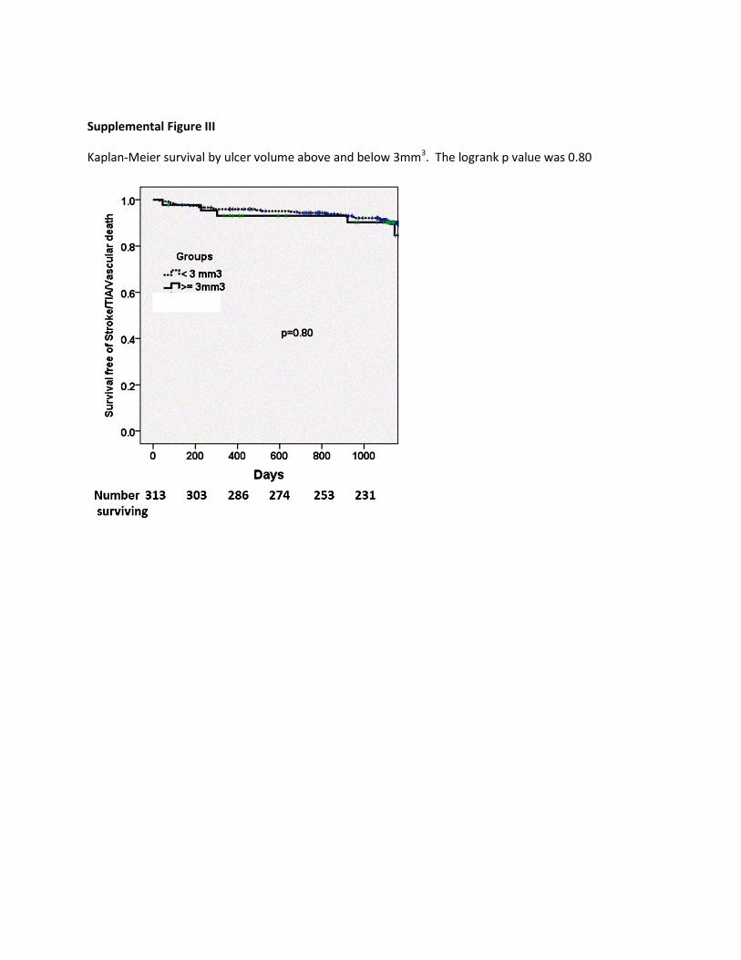
Supplemental Figure III
Kaplan-Meier survival by ulcer volume above and below 3mm3. The logrank p value was 0.80

Supplemental Figure IV. Survival free of Stroke/Death/TIA by maximum ulcer depth.
The upper panel shows Kaplan-Meier survival by maximum ulcer depth above and below 1mm, the
lower panel by maximum ulcer depth above and below 2 mm. Neither was significant by logrank test.

Reference List
(1) Al Shali K, House AA, Hanley AJ, Khan HM, Harris SB, Mamakeesick M et al. Differences between carotid wall morphological phenotypes measured by ultrasound in one, two and three dimensions. Atherosclerosis. 2005;178:319-25.
(2) Spence JD, Eliasziw M, DiCicco M, Hackam DG, Galil R, Lohmann T. Carotid Plaque Area: A Tool for Targeting and Evaluating Vascular Preventive Therapy. Stroke. 2002;33:2916-22.