VOLUME II ISAKOS NEWSLETTER 2014 Current Concepts on … · David Sadigursky, MD, BRAZIL Gonzalo...
56
ISAKOS NEWSLETTER 2014 • VOLUME II Current Concepts on Arthroscopy, Knee Surgery & Orthopaedic Sports Medicine CT arthrogram axial view demonstrating a Bennett lesion on the posterior glenoid attached, but possibly becoming fragmented. INSIDE 18 MANAGEMENT OF THE PATELLA ON TOTAL KNEE 20 ACL TEARS IN ATHLETES WITH OPEN PHYSES 24 TKR COMPONENT MALROTATION 29 GROWTH FACTORS & PLATELET RICH PLASMA 42 WORST CASE SCENARIO
Transcript of VOLUME II ISAKOS NEWSLETTER 2014 Current Concepts on … · David Sadigursky, MD, BRAZIL Gonzalo...

ISA
KO
S N
EW
SL
ET
TE
R 2
01
4 •
VO
LU
ME
II
Cur
rent
Con
cept
s on
Art
hros
copy
, Kne
e Su
rger
y &
Ort
hopa
edic
Spo
rts
Med
icin
e
CT arthrogram axial view demonstrating a Bennett lesion on the posterior glenoid attached, but possibly becoming fragmented.
INSIDE 18 MANAGEMENT OF THE PATELLA ON TOTAL KNEE
20 ACL TEARS IN ATHLETES WITH OPEN PHYSES
24 TKR COMPONENT MALROTATION
29 GROWTH FACTORS & PLATELET RICH PLASMA
42 WORST CASE SCENARIO

3
EDITOROmer Mei-Dan, MD, USA
EDITORIAL BOARDNadim Aslam, FRCS Orth, UNITED KINGDOMMandeep S. Dhillon, Prof., MS, FAMS, INDIALucio S. R. Ernlund, MD, MSc, BRAZILNorimasa Nakamura, MD, PhD, JAPANDavid Sadigursky, MD, BRAZILGonzalo Samitier, MD, PhD, USADaniel A. Slullitel, MD, Prof., ARGENTINALuis Alberto Vargas, MD, USAJames H. Lubowitz, MD, USA, Past Editor
inthis issueEditor’s Message . . . . . . . . . . . . . . . . . . . . .1President’s Message . . . . . . . . . . . . . . . . . .210th Biennial ISAKOS Congress . . . . . . . . . .4ISAKOS Awards . . . . . . . . . . . . . . . . . . . . . .6Lifestyle. . . . . . . . . . . . . . . . . . . . . . . . . . .10ISAKOS Asks . . . . . . . . . . . . . . . . . . . . . . .17Pearls & Pitfalls – Surgical Technique. . . . .18Current Concepts. . . . . . . . . . . . . . . . . . . .20In a Nutshell . . . . . . . . . . . . . . . . . . . . . . .36Fellowship Reports . . . . . . . . . . . . . . . . . .37Worst Case Scenario . . . . . . . . . . . . . . . . .42Bring it On . . . . . . . . . . . . . . . . . . . . . . . . .44Approved Course Reports . . . . . . . . . . . . .48Upcoming Approved Courses . . . . . . . . . . .52
ISAKOS takes pride in our mission to “advance the worldwide exchange and dissemination of education, research and patient care in arthroscopy, knee surgery and orthopaedic sports medicine”. Our pride in this mission is evident in our recent activities, including our international collaborative courses, our online education expansion, and our upcoming Biennial Congress.
ISAKOS has been busy thus far in 2014, completing three collaborative courses in Brazil, India and China. These courses included collaboration with international soccer/football powerhouse FIFA, the Indian Arthroscopy Society, and three Chinese specialty societies, including the Chinese Medical Association, the Chinese Orthopaedic Association, and the Chinese Society for Sports Medicine. More information on these courses can be found on page 48 of this Newsletter. Do you have a course you would like to partner with ISAKOS on? Email [email protected] – we would love to hear from you!
ISAKOS continues to expand our online educational offerings. We understand that not everyone has the financial resources or time to commit to attending an ISAKOS Congress or international collaborative course, so we have put special effort into our online education. Have you been to the ISAKOS Global Link? Thirty surgical demonstration videos by the world’s leading experts are waiting for you! Videos featuring Freddie Fu, Guillermo Arce, Stephen Burkhart, Marc Philippon and more are available for viewing at your leisure. Also included in Global Link are Congress handouts, abstracts and more.
ISAKOS is also pleased to partner with OrthoEvidence. The ISAKOS & OrthoEvidence portal includes unique content specially selected for ISAKOS members. Recent topics include total knee arthroplasty, use of platelet rich plasma in reducing pain, use of injections, shoulder instability and ACL repair. We encourage all ISAKOS Members to make use of this valuable resource.
Finally, ISAKOS is preparing for our hallmark educational event – the 10th Biennial ISAKOS Congress! This event marks the 20th anniversary of ISAKOS. More than 300 international faculty look forward to sharing their diverse knowledge with ISAKOS Congress attendees. Three pre-courses will be offered, as well as the concurrent Sports Rehabilitation Course. More information on the ISAKOS Congress can be found on Page 3. We encourage all members to share your knowledge and participate in the Congress by submitting a Scientific Abstract. Abstract Submission deadline is September 1, 2014.
We thank you for investing in your education and sharing your knowledge with ISAKOS!
International Society of Arthroscopy, Knee Surgery and Orthopaedic Sports Medicine
2410 Camino Ramon, Suite 215San Ramon, CA 94583 – 4318 USA
Telephone: +1 925 807 – 1197Fax: +1 925 807 – 1199Email: [email protected]
ISAKOS is investing in building education!
“An investment in knowledge pays the best interest.” – Benjamin Franklin

ISAKOS NEWSLETTER 2014: Volume II 1
EDITOR’S MESSAGE
Blink of an eye, and the first half of 2014 is behind us. We are now only a year away from the 10th Biennial ISAKOS Congress in Lyon. Summer in France – what a treat!
The food, the Alps, the wine. What a great destination for a (family) vacation…Are we all thinking of it that way? This is one of few interesting questions we had asked some of the world’s most famous surgeons. The answers, as it seems, were not so straightforward.
Furthermore, being an international organization, which represents so many different countries, ethnicities, experiences and cultures, we also wanted to discuss the gender ratio in our field, and understand how this issue is being perceived by our global leaders.
This Newsletter features an increased Current Concept section, including topics related to Hip Arthroscopy, ACL Tears, Bennets Lesions, Femoroacetabular Impingement, PRP, and topics related to Total Knee Arthroplasty.
We are also back to touch on the basic terms of our field, to educate ourselves on the ‘bread and butter’ that we usually tend to overlook, like how we determined the viability of the cartilage we are treating on a daily basis.
We will learn from, and give special recognition to one of the most innovative surgeons of our time, Dr. Laurent Lafosse, of France. His students and fellows share with us their feelings and the experience gained under his guiding hands. Out-of-the-box thinking at its best.
Finally, Dr. Slullitel reminds us in his “Worst Case Scenario” that we are not just orthopaedic surgeons – we are doctors. Therefore, we need to review our patient’s global care, to not miss things which can negatively effect the long term management, and add extra stress to a field which, to begin with, is far from being stress free.
Wishing you well for this second half of 2014! Enjoy.
Omer Mei-Dan, MD, USAISAKOS Newsletter Editor
Members Only Banner
OrthoEvidenceGo to www.isakos.com and log in to myISAKOS 2Click on
myPublications in the myISAKOS Dashboard 3
Click “Access” the ISAKOS/OrthoEvidence Portal
myPublications
myPublications
AccessmyISAKOS
ISAKOS / OrthoEvidence Portal
ISAKOS Members Now Receive Full Access to
1

ISAKOS NEWSLETTER 2014: Volume II 2
Dear Friends of ISAKOS,
Greetings from the ISAKOS Executive Team! On behalf of our leadership group, thank you for your continued membership with ISAKOS. As ISAKOS prepares to celebrate our 20th Anniversary at the ISAKOS Congress in Lyon, I am especially grateful for our diversity of members – whether you have been a member for all 20 years, or only recently joined! ISAKOS is a unique melting pot…. we have more than 4,000 members from 92 different countries. This diversity is a hallmark of our society and we appreciate every one of you!
ISAKOS recently met at the American Academy of Orthopaedic Surgeons meeting in New Orleans. The ISAKOS Executive Committee arrived early to hold a strategic conference to discuss some initiatives important to the future growth and maintenance of our society. We are very excited about the new projects that will be released in the coming months, including the awarding of the first ISAKOS Teaching Center Scholarships, and the inaugural Masaki Watanabe Arthroscopy Traveling Fellowship. These are just two of the initiatives developed by YOUR committees for the benefit of our membership.
ISAKOS is also pleased to formally announce the creation of the Journal of ISAKOS. It is important to note that ISAKOS will continue our ongoing relationship with the Journal of Arthroscopy: Arthroscopic and Related Surgery. The Journal of ISAKOS will engage and advance the knowledge and treatment of musculoskeletal diseases and disorders among specialists and other interested health professionals across the globe. The Journal of ISAKOS will be composed of a mix of peer-reviewed mini – and systematic review articles invited from the international author community of orthopaedic surgeons, sports medicine physicians, and knee surgeons and emanating from the ISAKOS Congress’s instructional course lectures and other important topics identified by the editors. Our goal is to develop a Journal that addresses all specialties practiced by our diverse members. A Journal Development Task Force is hard at work selecting the Journal Editor, and we look forward to sharing more information about the Journal of ISAKOS over the coming months.
Finally, the 10th Biennial ISAKOS Congress is promising to be another smashing success. We have already invited more than 350 unique speakers from 40 different countries. More information on the ISAKOS Congress can be found on pages 4 – 9 of this Newsletter. I also encourage you to visit the Congress website at www.isakos.com/2015congress. The Interactive Agenda is a live look at the Congress Scientific Program and contains up to the moment information on speakers and presentation topics. The beautiful city of Lyon is a classic backdrop for an ISAKOS Congress, with more than 2000 years of colorful, tumultuous and thriving history celebrating the art of living, luxury and fine cuisine.
As you can see, ISAKOS has been busy, and we continue to work hard for our membership. As we approach the milestone 20th anniversary, we are taking time to reflect on where we have been, and also looking forward to the future of where we can and will go. The future is bright for ISAKOS, and we look forward to continuing to grow with all of our members!
Masahiro Kurosaka, MD, JAPAN ISAKOS President 2013 – 2015
PRESIDENT’S MESSAGE
View the Interactive
Agenda
Have you submitted your Congress scientific abstract? More than 250 scientific papers are anticipated to be accepted. The Deadline for Submission is September 1, 2014 – submit your abstract today!

ISAKOS NEWSLETTER 2014: Volume II 3
JUNE 7–11, 2015LYON, FRANCE
isAkoscongress
10TH Biennial
2015

ISAKOS NEWSLETTER 2014: Volume II 4
The 10th Biennial ISAKOS Congress will be held on June 7 – 11, 2015 at the Lyon Convention Centre at the Cité Internationale.
More than 300 faculty have been invited to celebrate the 20th anniversary of ISAKOS at our hallmark educational event. The ISAKOS Congress program is unique in the diversity of subjects that are covered. Instructional Course Lectures, Symposia, Lectures and Debates will focus on topics such as injuries in the athlete, osteotomy, operative complications, cartilage repair, use of grafts, and osteoarthritis.
The ISAKOS Congress offers a unique opportunity to learn from, and interact with a diverse international faculty. Drawing the best and the brightest faculty, ISAKOS is pleased to offer our attendees the chance to interact with international experts in didactic sessions. Congress participants are encouraged to ask questions, and our faculty look forward to meeting you!
The ISAKOS Congress Scientific Program is available online for your viewing, and is current to the moment with all program changes.
For those interested in a more specific educational experience, the ISAKOS Congress will include three pre-courses focusing on “Advances in the Management of Knee Pathology,” “International Update on Surgical Controversies of the Shoulder” and “ISAKOS & FIFA: Treatment of Soccer Players”. The Pre-Courses will be held on Saturday, June 6th, and the Knee and Shoulder courses will include surgical demonstrations in addition to didactic sessions.
Sports medicine specialists should consider attending the ISAKOS Congress Sports Rehabilitation Concurrent Course. Intended for physical therapists, athletic trainers, coaches and physicians, the Sports Rehabilitation Course will focus on management or prevention of injuries to the athlete. Faculty will include both physiotherapists and orthopaedic surgeons for a diversity of perspective. Past topics of presentation have included Biomechanics of Exercise, Effects of Graft Type and Technique on Post-Operative Rehabilitation, Functional Training and Return to Activity, New Treatments for Muscle Injuries, Treatment Options for Tendinopathy, Electrical Stimulation for Rehabilitation of Muscle Injuries, and Anatomy of Major Joints.
ISAKOS is pleased to announce that Abstract Submission for the 2015 ISAKOS Congress is open! Abstracts are currently being accepted on all topics related to the practice of arthroscopy, knee surgery and orthopaedic sports medicine. Presenting authors are invited to visit the ISAKOS website (www.isakos.com/2015Congress) to complete the online abstract submission process. Abstracts will only be accepted through the online submission process – please do not email abstracts directly to the office. Please note – submission of an Abstract is required to apply for some of the ISAKOS Awards. For more information on ISAKOS Awards, please refer to pages 6 – 7. Abstracts must be submitted by midnight on September 1, 2014.
You are a vital part of the ISAKOS Congress! We encourage you to experience the unparalleled education, and be part of the international community that is the ISAKOS Congress – we look forward to seeing you in Lyon!
ISAKOS looks forward to welcoming the international arthroscopy, orthopaedic and sports medicine community to Lyon, France for the 10th Biennial ISAKOS Congress!
Anastasios GeorgoulisISAKOS Congress Program Chair 2015
2015 ISAKOS CONGRESS
Julian FellerISAKOS Congress Program Deputy Chair 2015

ISAKOS NEWSLETTER 2014: Volume II 5
DEADLINE: September 1, 2014 www.isakos.com /2015congress
2015 Congress Content Pre-Course: Advances in the Management of Knee Pathology: ACL, Meniscus, Patellofemoral, Osteotomy, and Chondral Pathology
Pre-Course: International Update on Surgical Controversies of the Shoulder
Pre-Course: ISAKOS and FIFA: Treatment of Soccer Players
Sports Rehabilitation Concurrent Course Five meeting days with 300 Scientific Papers
Panel Discussions and Debates Symposia Lunchtime Lectures and Workshops Surgical Demonstrations Instructional Course Lectures Paper and ePoster Presentations Technical Exhibits CME Certification Spouse and Guest Morning Café Welcome Reception
JUNE 7–11, 2015
isAkoscongress
10TH Biennial
2015LYON, FRANCE
call for abstracts
Presenting authors are allowed one submission per award. Additionally, each abstract may only be applied to one award.
Apply Today www.isakos.com/2015congress
John J. Joyce Award Richard B. Caspari Award Scientific Research Award
Preliminary Program Available Online
JUNE 7–11, 2015
isAkoscongress
10TH Biennial
2015LYON, FRANCE
Preliminary Program
www.isakos.com/2015Congress
Albert Trillat Young Investigator’s Award Achilles Orthopaedic Sports Medicine Research Award Patellofemoral Research Excellence Award
Awards
#ISAKOS2015

ISAKOS NEWSLETTER 2014: Volume II 6
ISAKOS AwardsThe ISAKOS Fellowship and Awards Program is committed to recognizing and honoring researchers whose work has contributed to better understanding and communication within the fields of Arthroscopy, Knee Surgery and Orthopaedic Sports Medicine.
Applicants, please note: Presenting authors are allowed one submission per award. Additionally, each abstract may only be applied to one of the following awards. To be considered for an Award or Fellowship, abstracts and award applications must be submitted before September 1st, 2014, unless otherwise noted.
John J. Joyce AwardSponsored by Smith & Nephew, Inc.John Joyce created the award with the intention to stimulate and reward younger members who contribute high-quality data and presentations. Thus, orthopaedic residents and fellows, with a study related to arthroscopic treatment are encouraged to apply for this award.
Richard B. Caspari AwardSponsored by Mitek Sports MedicineRichard B. Caspari was an innovator, teacher and leader in the field of Arthroscopy. The Richard B. Caspari Award was established with the intention of st imulat ing and rewarding abstracts and presentations in the subject of the upper extremity.
Scientific Research AwardSponsored by OssürThe Scientif ic Research Award is intended to stimulate and reward abstracts and presentations related to the subject of Scientific Research.
01 Lyon Convention Center02 Views of the city of Lyon03 Lyon Bridge
01
2015 ISAKOS CONGRESS
Apply for an Award

ISAKOS NEWSLETTER 2014: Volume II 7
Albert Trillat Young Investigator’s AwardSponsored by StrykerPast president and founder of the International Society of the Knee, Professor Albert Trillat was one of the pioneers in knee surgery and sports traumatology. This award provides recognition for a young researcher who has done outstanding clinical laboratory research contributing to the understanding, care or prevention of injuries to the knee.
Achilles Orthopaedic Sports Medicine Research AwardSponsored by DJO, Inc.The Achi l les Or thopaedic Spor ts Medic ine Research Award recognizes researcher(s) who have performed the most outstanding clinical or laboratory research in the field of sports medicine, such as the care and prevention of injuries.
Patellofemoral Research Excellence AwardSponsored by The Patellofemoral Foundation, Inc.The Patellofemoral Research Excellence award intends to encourage research and expertise in the f ie ld of patel lofemoral disorders. The Patellofemoral Research Excellence Award seeks to encourage outstanding research leading to improved understanding, prevention and treatment of patellofemoral pain or instability.
ISAKOS FellowshipsThe Patellofemoral Traveling FellowshipSponsored by the Patellofemoral Foundation, Inc. and DJO Inc.The Patellofemoral Traveling Fellowship is available on a competitive basis to an orthopaedic surgeon interested in the study and advancement of understanding of the patellofemoral joint. The purpose of the fellowship is to promote better understanding and communication around the world regarding patellofemoral pain.
The Upper Extremity Traveling FellowshipThis fellowship was developed by the ISAKOS Upper Extremity Committee to promote better understanding and communication regarding injuries or conditions involving the structures of the Upper Extremity. This Traveling Fellowship is available on a competitive basis to an orthopaedic surgeon interested in the study and advancement of understanding of the Upper Extremity.
New for 2015!The Masaki Watanabe Arthroscopy Traveling FellowshipDr. Masaki Watanabe developed the first device for minimally invasive surgery. In honor of Dr. Watanabe’s ideas and accomplishments, the Masaki Watanabe Arthroscopy Traveling Fellowship Award is a new traveling fellowship sponsored by the Arthroscopy Committee of ISAKOS that will provide funding for two young arthroscopic surgeons to learn more about the current practice of arthroscopic surgery from well-respected experts in the field. The knowledge learned by the traveling fellows can then be taken back to their respective countries to improve patient care and advance the local teaching of arthroscopic surgery.
JUNE 7–11, 2015
isAkoscongress
10TH Biennial
2015LYON, FRANCE
03
02
2015 ISAKOS CONGRESS

ISAKOS NEWSLETTER 2014: Volume II 8
2015 ISAKOS CONGRESS
A Tale of One ISAKOS Member: From Florida to Lyon
Kivanc Atesok, MD, MSc ISAKOS Membership Committee
It was year 2005, I was in training when I attended my first ISAKOS Biannual Meeting in Florida. It was such an exciting time in my career! Just before the meeting, a Tissue Engineering Symposium was organized by Dr. Gary Poehling in Winston Salem, where I first had my eyes opened to see what the cross-roads in medicine will be in the near future: Regenerative Medicine and Stem Cells. I was so impressed to meet many distinguished leaders of Orthopaedic Surgery and Sports Medicine that I felt a strong desire to be more involved with the activities of ISAKOS. Who would not feel the same way after having an opportunity to get closer to icons like Dr. Doral, Dr. Fu, Dr. Karlsson, Dr. Lubowitz, Dr. Ochi, Dr. Myers and many more names that are written in my “Look-up-to” list...?
It did not take more than two years for me to decide to study for Master of Science degree in Regenerative Medicine and Stem Cells. A dream that started with ISAKOS was becoming the reality: I was accepted to The University of Toronto as an MSc graduate student to study in the field of Tissue Engineering. I was so enthusiastic that the project eventually received awards and honours, and I found myself being invited to Stem Cell forums as one of the recognized scientists in the field. Furthermore, my desire to be involved more with ISAKOS was also being fulfilled. After the Editorial Board, I also had the opportunity to work in the Membership and Program Committees. But it was not all; ISAKOS gave me way more opportunities such as organizing symposia, having fellowship programs recognized, and above all, improving myself as a physician and a person.
Today, when I think about 2015 ISAKOS Biannual Meeting in Lyon, I can feel the same excitement I felt just before my first ISAKOS Meeting in Florida years ago. By the way, I have never been to France...! I drink French wine, I use French perfume, I dine in French restaurants and I read French literature, but never been to France...! Another reason to be thankful for being an ISAKOS member: now I have the chance to visit France...! And Lyon, the city between two rivers (Rhône and Saône)...! I know that Lyon is the second richest city in France after Paris, a UNESCO World Heritage Site due to its historic significance, renowned Lyonnaise cuisine, and our valuable French members would know better – many other qualities unique to Lyon that I will be aware of during the ISAKOS meeting.
Thinking about the past 9 years as an ISAKOS member and looking towards the future, Eleanor Roosevelt’s words become even more meaningful to me: “The future belongs to those who believe in the beauty of their dreams.” ISAKOS opens the doors to its members to walk towards their dreams. Non-material rewards that ISAKOS membership could bring us along the way are l imited only by our imagination and motivation.

ISAKOS NEWSLETTER 2014: Volume II 9
2015 ISAKOS CONGRESS
“ I know of one thing that you can do well in Lyon, and that’s eat,”
the 19th-century French novelist Stendhal remarked. Two centuries later, the image of France’s third-largest metropolis is still buried under a heap of food.
Long considered France’s capital of gastronomy, Lyon is blessed with culinary history as rich as its extraordinary produce. When the French economy crashed after World War I, the city’s formidable female chefs shifted their talents from wealthy mansions to the city’s restaurants and bouchons, using the region’s fine ingredients to prepare simple yet perfect meals. The ‘’machons’’ (translated as an informal meal) and the ‘’bouchons,’’ (translated to home-cooking) fed the city’s silk workers and developed into Lyon’s most adored style of cuisine.
Lyon is home to twenty true bouchons, most of which are located in the well-traveled sections of the city. All are distinctly casual, very popular (reservations are a must) and filled with Lyonnais. While execution may vary, the food is the essence of homey-ness. Diners will find a variety of bistro staples – from mackerel with white wine to Lyon’s celebrated charcuterie to tete de veau and pike quenelles (the delicate dumplings are a Lyonnais specialty) – as well as dishes peculiar to bouchons.
These include Lyonnais salads composed of dandelion greens or frisee lettuce tossed with bacon, croutons and poached egg; salads of sheep’s feet seasoned with a tart remoulade sauce; gateau de foie de volaille (a steamed pudding-like dish made of chicken livers) served in a bechamel sauce and garnished with quenelles or ravioli; sabodet, a sausage made of pig’s head; and tripe – the most bouchon-esque of all dishes: a large wedge of tripe marinated in wine, breaded and fried and served with a piquant sauce.
It is often said that Lyon is crossed by three rivers, the Rhone, the Saone and the Beaujolais, the wine most closely associated with the city. Beaujolais is characterized with gentle tannins, juicy fruit and a light to medium body. Beaujolais wines have a bold minerality and the thin-skinned Gamay grapes create a fresh, tart acidity that stands up to hearty meals and offers big flavors that won’t overpower subtle dishes. Beaujolais is delicious with roast chicken, sausage and beef; as well as an ideal choice for a spicy meal. The sweetness and relatively low alcohol content makes Beaujolais perfect for hot dishes that feature peppers or curries.
We hope Congress attendees have the opportunity to enjoy the diverse culinary culture of Lyon!

ISAKOS NEWSLETTER 2014: Volume II 10
Shoulder Surgery with a Renowned Expert
Gonzalo Samitier, MD, PhDFellow at Alps Surgery Institute (ASI), Clinique Generale – Annecy, FRANCE
Contributors: (current and recent former Fellows at ASI): Thomas J. Christensen, MD2
Ruth A. Delaney, MD3
Simon J. Fogerty Dip SEM (UK), FRCS (Tr&Orth)4
Ashis Gupta, MD1
Kalojan Petkin, MD1
Stephen A. Parada, MD3
Claudio Rosso, MD, MSc5
1 Alps Surgery Institute (ASI), Clinique Generale – Annecy, France2 Reno Orthopaedic Clinic. Reno, NV, USA3 Harvard Shoulder Service/Brigham and Women’s Hospital,
Boston, MA, USA4 Calderdale and Huddersfield NHS Foundation Trust,
West Yorkshire (UK)5 University Hospital of Basel (Switzerland)
IntroductionThe next ISAKOS meeting will be held in Lyon (France), a vibrant metropolis which makes the most out of its unique architectural, cultural and gastronomic heritage. Nothing else has to be said, France is one of the most attractive destinations in the world, and it is easy to get lost in its pride, mastery and sophistications.
It is well known that France has made great contributions in our field and specifically, the French school has a solid reputation in shoulder surgery; most of us are familiar with terms such as Latarjet, Grammont reverse shoulder arthroplasty, Goutallier classification among many others terms, and those involved in shoulder surgery have numerous examples in past and current scientific literature.
In this article we want to focus on one of those considered a spearhead in shoulder surgery, Dr. Laurent Lafosse; his surgical skills and his ideas leave no one indifferent and his experience and innovative thinking contribute to pushing shoulder surgery always a little further. He currently practices in
“Clinique Generale”, an efficient private hospital in the city of Annecy, a scenic jewel of the French-Alps; he partners
with two other shoulder surgeons, Drs. Bruno Toussant and Jerome Bahurel. At any time, the Alps Surgery Institute has 3 to 6 selected fellows that they incorporate to the team for periods of 3 months to 1 year; they are attracted to the high volume and complexity of the procedures to which they are exposed; surgeons from all over the world come to visit or assist in any of the different courses throughout the year at the Institute, making it a constant cultural and scientific exchange core.
Dr. Lafosse´s scientific work in shoulder surgery is extensive and well represented in the literature. Some of his most well-known contributions are: his approach to shoulder instability as the pioneer in all-arthroscopic Latarjet, his management of complex cuff tears using the lasso loop stitch, arthroscopic brachial plexus releases and his multiple approaches for shoulder arthroplasty.
The aim of this article is to focus on two common surgical conditions of the shoulder and to offer different treatment options frequently performed at our institution. The text is sprinkled with former ASI fellows´ comments about their training at the Shoulder Institute; Dr. Gupta and Dr. Petkin, current fellows also contributed to reviewing and optimizing the scientific content of this report. Special thanks to Stephen Parada who has described the “y” shape biceps tenotomy in detail and former fellows in general as some of his previous work was used to build this article.
1. Treatment Options for the Long Head of the Biceps Tendo
Shoulder pain is often of a multifactorial etiology; it can include pathology of the long head of the biceps tendon. This tendon inserts as an intra-articular structure. Biceps pathology is commonly associated with pathology of the rotator cuff and/or superior labrum. The mainstay of operative treatment is either a tenotomy or some type of tenodesis. Cosmesis and potential changes in strength or biceps muscle cramps is a concern when biceps tenotomy is performed; biceps tenodesis minimize this problems but implies a technically more demanding procedure with potential implant related problems and longer rehabilitation.
Next we describe two surgical options to solve efficiently proximal long head of the biceps pathology; surgeons could include them on their therapeutic arsenal.
LIFESTYLE
01
“Innovative shoulder surgery at its best”
– Claudio Rosso
Laurent Lafosse, MD, FRANCE

ISAKOS NEWSLETTER 2014: Volume II 11
LIFESTYLE
1.1. Arthroscopic Proximal Biceps Tenodesis to Supraspinatus Tendon
In most of our arthroscopic procedures, the patient is placed in the beach-chair position with the arm in a longitudinal traction device weighing approximately 3 kg. A standard 30° scope is used for the entire procedure, and a pressure-sensitive fluid pump set to 60 mm Hg is commonly used. A traditional posterior portal is made, and a diagnostic arthroscopy is performed. Our indications for biceps tenodesis include relatively young and active population with biceps tendonitis (hyperemia, synovitis), tendinopathy, partial or full-thickness tears, and biceps instability secondary to pulley disruption; in addition, the subscapularis or any cuff pathology is always thoroughly examined.
Once the decision is made to perform a biceps tenodesis, it is performed in the following fashion:
1. An additional portal is made through the rotator interval just anterior to the long head of the biceps tendon or the supraspinatus (“D” portal in Figure 1).
2. In cases of anterosuperior cuff tears, this area can be accessed easily from the subacromial space through the supraspinatus tear and is often used to prepare the tuberosity for cuff repairs before moving the scope to the subacromial space.
3. We obtain this or any portal with needle localization and use a blunt trocar to create a path for instruments and/or the scope. If no cuff tear is evident, the portal can still be used, but care must be taken not to damage the intact cuff.
4. Next the lateral portal (“C”) is created and becomes the viewing portal, while “D” becomes the primary working portal (Figure 1). With this portal, the surgeon can view the supraspinatus, biceps, and subscapularis as well as anchor placement.
5. When intra-articular visualization is adequate, the greater tuberosity is prepared, and a double-loaded suture anchor 4.5 or 5.5 mm is placed. With small tears, the anchor is placed just posterior to the biceps groove; in medium-sized tears, the anchor is placed more posteriorly. If no cuff tear is evident, the anchor is placed in the groove.
6. Next, the biceps is tenodesed to the supraspinatus and humeral head using the lasso-loop technique described by Lafosse and colleagues using a piercing suture retriever device (1) (Figure 2).
7. Before sutures are passed, the biceps is partially transected near its insertion into the superior labrum.
8. Once this is complete, the supraspinatus tendon is pierced, and the corresponding suture limb is grasped and pulled completely through the cuff tendon.
9. The suture has now essentially locked the biceps and has been incorporated into the cuff tendon. The other suture limb is then passed in similar fashion through the supraspinatus and biceps tendon. The lasso loop can be switched so the supraspinatus tendon is locked when the sutures are tied.
10. The biceps tendon is than detached from the labrum using the lateral and anterolateral portals and is debrided to 1 cm of the tenodesis site and our first suture is tied; the limb passing freely through both tendons is the post such that when tension is placed, biceps and cuff are secured to the bone. Both limbs are then tied using a series of half-hitch knots creating a “pulver taft”-like tenodesis.
11. Due to the lasso-loop configuration, sliding knots cannot be used; but it is important to note that the post must be the the non lasso stitch; in order to pull the biceps towards the anchor.
12. In the event of an absent rotator cuff tear the whole procedure will be performed intra articularly, scope in the posterior portal and our instruments through “D” portal; the suture anchor is placed in the biceps groove and the lasso loop stitch will only include the biceps tendon.
This method theoretically accomplish two goals: one, eliminates a potential shoulder pain generator with proximal biceps tenodesis and two, it restores the glenohumeral joint stabilizer function of the biceps. It is believed that, when the biceps tendon is secured to the supraspinatus, opposing vectors of both muscle tendon units are responsible for humeral head depression and compression into the glenoid.
02
“A true innovator and talented surgeon who is shaping the future of arthroscopic shoulder surgery” – Ruth Delaney
ISAKOS NEWSLETTER 2014: Volume II 12
Shoulder Surgery with:
1.2. Y Shape Biceps Tenotomy (physiologic tenodesis)
The decision to perform Y-shaped tenotomy is based on patient factors and associated pathology and doesn´t differ from indications for classic biceps tenotomy. Once the decision is made, the biceps anchor is prepared by using an arthroscopic radiofrequency device to dissect a portion of the anterior-superior and posterior-superior labrum off of the superior glenoid tubercle in continuity with the biceps tendon. This is performed by starting with the anterior-superior labrum, using the radiofrequency device to dissect both under the labrum where a superior labrum, anterior to posterior (SLAP) tear would occur and then posterior to the biceps anchor as well. The dissection is continued laterally until a “Y” is formed between the LHB and the two limbs of the labrum. This essentially creates a complete dissection of the superior labrum that stays connected with the LHB. This “Y” morphology locks the stump of the biceps in the entry of the biceps groove and prevents it from displacing distally in the bicipital groove.
This obviously is not meant to recreate a true tenodesis, but in our follow-up, it creates a cosmetic appearance which is improved to that of the simple tenotomy, usually resulting in a one centimeter or less asymmetry in the proximal position of the biceps when compared to the contralateral side.
We have not found that debriding this superior aspect of the labrum is detrimental in any way to the patient clinically and there have been no reported cased of post-operative instability noted; this looks logical as there is no ligament attachement at the upper part of the glenoid. This new technique is an alternative to either biceps tenotomy or tenodesis in the appropriate setting and offers the benefit of a cosmetic appearance with minimal operative time and without the need of additional implants there by reducing operating costs; the quality of care delivered is maintained and there is no delay in the rehabilitation process secondary to this technical gesture.
2. Management of the Subscapularis tendon during Anatomic Shoulder Replacement
Total shoulder arthroplasty [TSA] in selected patients with an intact rotator cuff, remains the gold standard for treatment of osteoarthritis, rheumatoid arthritis, fractures sequelae and avascular necrosis. It provides predictable pain relief with proven good to excellent long term functional outcomes; with classic deltopectoral approach subscapularis tenotomy or osteotomy is generally performed; as a result we have to be cautious in the postoperative care for the first 6 weeks to protect the repair. Non healing of the subscapularis tendon has long been recognised as a potential source of poor patient outcomes and often, when it occurs, is an indication for early repair or revision surgery.
To avoid this complication associated with failure, several techniques have been developed, including lesser tuberosity osteotomy rather than tenotomy; trans-acromial, trans-supraspinatus approaches; Below we described in detail our variants of two other surgical options focused in maximize subscapularis tendon healing or preservation (Figure 3).
2.1. Minimally invasive, rotator cuff sparing technique (2)
The mainstay of this technique is basically to accomplish the entire procedure through the rotator interval without violate any of the rotator cuff tendons and to have unrestricted postoperative rehabilitation. This technique can be performed via superior (transdeltoid) approach or using the classic deltopectoral.
In the case of a large inferior osteophyte, we favor the deltopectoral approach in order to have a better access to the inferior part of the humeral head. In the case of posterior glenoid tilt in type B1 and B2 glenoids, we prefer the superior approach in order to be facing the glenoid.
The ideal candidates are:• Mild to moderate primary glenohumeral osteoarthritis• Intact rotator cuff• Mobile glenohumeral joint
The technique is generally contraindicated in patients with gross glenohumeral deformity or immobile joints, as frequent position changes of the arm are needed to facilitate access via the superior and inferior “windows”. Revisions, obese and/or very muscular patients are relative contraindications. If needed, at any stage, the procedure can be easily converted to the traditional technique via subscapularis detachment.
LIFESTYLE
“... now, when I operate I have in my mind, ‘what would Laurent do next ?’, and this always helps me through difficult surgical situations” – Simon Fogerty
03

ISAKOS NEWSLETTER 2014: Volume II 13
LIFESTYLE
Surgical technique (deltopectoral approach)After combined general and interscalene regional anaesthesia the patient is positioned in the beach chair position with approximately 45 degrees of tilt of the backrest; a 10 cm skin incision is made, approximately 2-3cm lateral and parallel to the deltopectoral interval, beginning at the level of the coracoid and extending distally towards the mid-humerus.
A more lateralized approach to classic deltopectoral skin incision offers the following advantages:1. It ensures that the Cephalic vein will always be found
medial to our skin incision and so can be quickly identified. 2. A lateralized incision improves visualisation of the glenoid.3. It creates an overlap of normal skin over the deltopectoral
interval ensuring natural tissue planes to be restored post operatively.
A standardised dissection of the deltopectoral interval is performed till the subscapularis is reached. The superior and inferior borders of the subscapularis tendon are identified, using the anterior circumflex humeral vessels as a guide to the inferior aspect of the tendon.
Inferior WindowThe inferior window is the first arthrotomy made and is used to expose the inferior aspect of the joint for removal of osteophytes. The inferior window can also be used to check component positioning during trialling and insertion. The window is opened below the subscapularis tendon, medially as far as the glenoid and laterally to the tendon insertion of subscapularis.
To open the window, a 1cm partial tenotomy of the superior part of the pectoralis major tendon is performed, exposing the underlying latissimus dorsi tendon. The anterior circumflex humeral vessels are ligated and cauterized.
The arm is then flexed to approximately 60 degrees and externally rotated approximately 20 degrees. The plane between the subscapularis tendon and the joint capsule is developed and the tendon is retracted superiorly, exposing the antero-inferior joint capsule. The axilliary nerve is located and exposed adjacent to the joint capsule inferior to the glenoid neck and a small Hohmann retractor is placed to protect the nerve.
The inferior arthrotomy is then performed into the axillary pouch. The capsulotomy is extended laterally along the capsular attachment to the humerus and medially along the glenoid neck, and the capsule is fully excised. Once completed, a second retractor is placed at the inferior aspect of the humeral head exposing the osteophytes anteriorly and inferiorly. The osteophytes are resected using curved osteotomes and rongeurs. Complete removal of the osteophytes can be confirmed by palpation. It is important to ensure that the posterior osteophytes, which are difficult to access, have been removed. After removal of the osteophytes, the inferior aspect of the glenoid can be visualised and an inferior soft tissue release performed. The position of the axillary nerve should be considered at all times during the capsulotomy and removal of the osteophytes.
Superior WindowAttention is then turned to the superior window. This window opens the joint via the rotator interval and is used for further joint preparation, instrumentation, trialling and insertion of the prosthesis. To improve visualisation, the shoulder is extended approximately 40 degrees in relation to the patient’s body and in neutral rotation. The rotator interval tissue is completely excised including the coracohumeral ligament, superior glenohumeral ligament, proximal portion of the biceps tendon and joint capsule. The rotator interval is trapezoidal in shape and extends from the anterior border of the supraspinatus tendon to the glenoid medially and then laterally along the superior border of the subscapularis tendon. The lateral border is formed by the bicipital groove.
To open the interval the tissue is incised along the superior border of the subscapularis tendon. The biceps tendon can then be identified intraarticularly and the bicipital groove can opened. In cases where the superior border of the subscapularis tendon and CHL is not easy to identify, the bicipital groove at the upper part of the epiphysis will guide the surgeon to the rotator interval. A biceps soft tissue tenodesis is performed within the groove and the proximal portion of the biceps tendon is excised, along with all remaining interval tissue.
“Outstanding instructor and arthroscopist. He teaches his fellows
to open their minds to a thorough understanding of the shoulder,
providing for endless possibilities”– Tom Christensen
04

ISAKOS NEWSLETTER 2014: Volume II 14
LIFESTYLE
Shoulder Surgery with:
After complete excision, two modified small Hohmann retractors are placed through the interval at the anterior and posterior aspect of the humeral head. An excellent view of the superior aspect of the glenohumeral joint is obtained with minimal retraction.
A full soft tissue release is performed, further elevating the anterior joint capsule from the glenoid and mobilising the subscapularis tendon underneath the coracoid process. Blind dissection of the anterior aspect of the subscapularis muscle, behind the coracoid and conjoint tendon, is avoided as the nerves to subscapularis can enter the muscle quite laterally; however, careful dissection above and behind the muscle is continued until the shoulder can be freely externally rotated and elevated. The range of motion is checked prior to bony resection to maximise postoperative range of motion.
Instrumentation, Trialling and Insertion of ProsthesisIn order to manage the osteotomy of the head through the superior window, as the posterior side of the resection is not easy to expose, the resection is managed in 2 steps: the anterior part of the head is resected first followed by the posterior part. The osteotome is splitting the head in a vertical fashion followed by the antero-posterior osteotomy according the anatomy of the humeral neck. Once the anterior part is removed, the posterior part becomes easier to expose and remove. The humeral head resection is then performed with an oscillating tip saw or flat osteotome. This allows the saw blade or the osteotome to slide though the rotator interval without damaging the supraspinatus or subscapularis tendons. Most of the resection is performed with the saw; however, the final resection is completed with an osteotome to avoid damage to underlying structures. The resected head is reconstructed on the back table and used to determine the appropriate size of the prosthetic head. Residual humeral head osteophytes are removed.
Once the head has been removed, the humerus is subluxed inferiorly with the use of the inferior glenoid retractor and the modified Hohmann retractors are used on the anterior and posterior aspect of the glenoid. Once again, the force on the retractors is minimal. The technique avoids the need to fully dislocate the humeral head anteriorly or posteriorly minimizing any potential stretch injury to the axillary and brachial plexus nerves. An excellent view of the glenoid is obtained and the posterior soft tissue releases of the capsule from the glenoid and removal of glenoid osteophytes can be completed to prepare the glenoid in a standard manner. Once the glenoid component is implanted, then the humeral canal preparation is completed with no specific instrumentation needed. The definitive prosthesis is inserted via the rotator interval initially in an anteverted position. As the prosthesis is pushed into the shaft, the humeral head is rotated under the supraspinatus tendon with the aid of a customised spanner attached to the metaphyseal component. Correct version of the humeral component is facilitated by the intact infraspinatus and subscapularis tendons as the intact footprint of the tendon prevents over rotation in either direction.
Using superior and inferior windows, correct version and size of the prosthesis is confirmed prior to final impaction. Occasionally a single stitch is used to close the interval tissue over the bicipital groove.
2.2. V shape subscapularis tenotomyDespite that some clinical studies have shown that osteotomy have improved functional outcomes and healing rate, subscapularis tenotomy in TSA has been shown to be biomechanically and clinically safe in order to restore the anatomy after TSA. Nevertheless there is inherent risk of re-rupture or non healing associated to subscapularis repair. Below we describe a new V-shaped subscapularis tenotomy in order to provide accurate location for anatomic restoration, better mechanical forces for reattachement and more surface area for tendon healing while performing the tenotomy.
After standard deltopectoral approach, the first step in subscapularis tenotomy is to identify the borders of the tendon as well as its musculotendinous junction. About 1 cm medial to the subscapularis insertion, we first mark the superior and inferior aspect of the tenotomy using two stitches at the upper part and the lower part of the subscapularis respectively. The subscapularis tendon incisions, superior and inferior are directed towards the medial aspect of the tendon in a 45º angle converging in the center and forming a triangle of tissue with base over the lesser tuberosity. The subscapularis and underlying anterior capsule are usually incised together. At the end of the procedure, the pre-located stitches when aligned will facilitate the repair.
The advantage of the triangle shaped cuff of subscapularis tendon left on the lesser tuberosity is that it offers a broad surface to perform adequate anatomic soft-tissue repair (Figure 4). Traction is done from our most medial previously placed stitches marking the tenotomy site and repair of the subscapularis tendon is usually performed in figure-of-8 fashion using four to five heavy nonabsorbable sutures. The type of suture and suture configuration is dependent on surgeon preference. All repairs should be anatomic, and shortening of the musculotendinous unit should be avoided. After the subscapularis repair is complete, the lateral aspect of the rotator interval is closed, often with the arm in slight external rotation to prevent loss of external rotation caused by rotator interval tightening.
Bibliography1. Lafosse L, Shah AA, Butler RB, Fowler RL. Arthroscopic Biceps
Tenodesis to Supraspinatus Tendon: Technical Note. Am J Orthop (Belle Mead NJ). 2011 Jul;40(7):345 – 7
2. Lafosse L, Schnaser E, Haag M, Gobezie R. Primary total shoulder arthroplasty performed entirely through the rotator interval: technique and minimum two-year outcomes. J Shoulder Elbow Surg. 2009 Nov-Dec;18(6):864 – 73
01 Drawing portals for biceps tendon repair02 Biceps is tenodesed to the supraspinatus and humeral head 03 Subscapularis sparing technique through anterolateral approach04 V shape tenotomy of the subscapularis tendon

ISAKOS NEWSLETTER 2014: Volume II 15
LIFESTYLE
Professor Gary G. Poehling, Long-Time Editor-in-Chief of Arthroscopy: The Journal of Arthroscopic and Related Surgery, RetiredOur friend and ISAKOS President (1997 – 99), Gary Poehling is retiring as Editor–in-Chief of Arthroscopy: The Journal of Arthroscopic and Related Surgery after a long and very successful career. Gary has gained lots of experience and wisdom. He has worked as an orthopaedic surgeon for 42 years and has been academically very successful with many scientific publications.
Gary was born and raised in La Crosse, Wisconsin. He received his Bachelor’s degree in Biology in 1964 and his MD degree from Marquette University in Wisconsin in 1968. Dr. Poehling’s internship and residency were completed at Duke University Medical Center. When serving in the United States Air force and located in Japan, Gary had the opportunity to train with the founding fathers of arthroscopy. He joined the faculty at Bowman Gray School of Medicine at Wake Forest University in 1976 and was appointed Chairman of the Department of Orthopaedic Surgery in 1989. In 1989, Gary obtained a certificate of added qualification of surgery of the hand.
Gary´s main interest has been in the development of new arthroscopy techniques and surgical instrumentation as well as broadening the development of minimally invasive surgery, including the hip, wrist, elbow and shoulder and has served on a design team for the development of a unicompartmental knee prosthesis. Most recently he has worked on the development of tissue engineering, including a tissue-engineered anterior cruciate ligament. Gary is considered to be one of the leading experts on arthroscopy.
Gary has been very active academically with 142 scientific publications. He has been involved in several outcome studies such as evaluating techniques of high tibial osteotomy and postsurgical outcomes of patients with ACL injury. He also has an interest in minimally invasive surgery in the treatment of distal radial fractures and traumatic wrist and elbow disorders. Gary has more recently been involved in research for transplantation of articular cartilage. He has also assisted in the development of computer software programs to track arthroscopic surgical procedures. These programs are the foundation of a nationwide database of information pertaining to arthroscopic surgery.
Gary has served since 1991 as the Editor-in-Chief of Arthroscopy: The Journal of Arthroscopic and Related Surgery. This journal was founded by the Arthroscopy Association of North America. When ISAKOS was founded 1995, Arthroscopy was established as our official journal and made available to all members.
Together with his team, Gary has developed Arthroscopy to be one of the leading journals in our field in the world. They have made the journal much more diversified and been open to include many new areas. Today this journal is a must for most orthopaedic surgeons around the world.
Gary played a huge role in the foundation of ISAKOS in 1995 and during the following years when ISAKOS struggled to find its form and heart, which required lots of work and discussion. He served as the second president of our society, 1997-1999. At the end of his presidency, Gary organized the ISAKOS Biennial Congress in Washington DC in 1999, which set the stage for future events. He has been instrumental in the success of ISAKOS following his term as President, serving as co-chair for the Strategic Planning committee with Ken DeHaven. They initiated discussions of the mission and vision for ISAKOS and assisted in the growth of our young society. Gary Poehling has been very instrumental in making ISAKOS a recognized world-wide international society with over 4,000 members and a very good economy and structure. In appreciation of his great work Gary Poehling was made honorary member of ISAKOS in 2007.
Gary is an outstanding educator and mentor. We are grateful for his friendship and support. We also appreciate all the good times we have spent with Gary and his wonderful wife of many years, Sandy. Gary has been strongly supported by his wife and large family.
We would like to thank Gary, not only for his extraordinary work in developing our discipline, but also for continuing to be such a strong supporter of ISAKOS. Gary must also be congratulated for his great work in developing Arthroscopy: The Journal of Arthroscopic and Related Surgery to be what it is today. We all owe Gary Poehling our gratitude for his vision and support. Thank you, Gary!
Sincerely
Peter J. Fowler, FRCS, CANADA ISAKOS President, 1995 – 1997
Gary G. Poehling, MD, USA ISAKOS President, 1997 – 1999
Roland P. Jakob, MD, SWITZERLAND ISAKOS President, 1999 – 2001
Barry R. Tietiens, FRACS, NEW ZEALAND ISAKOS President, 2001 – 2003
Per A. Renström, MD PhD, SWEDEN ISAKOS President, 2003 – 2005
John A. Bergfeld, MD, USA ISAKOS President, 2005 – 2007
Paolo Aglietti, MD, ITALY ISAKOS President, 2007 – 2009
Freddie H. Fu, MD, USA ISAKOS President, 2009 – 2011
Moises Cohen, MD, PhD, BRAZIL ISAKOS President, 2011 – 2013

16
Join the ISAKOS Global Conversation!
Facebook.com/myISAKOS
Twitter.com/ISAKOS Join the ISAKOS Group on LinkedIn
Tweeting about the Congress? Starting now, and during the Congress, include hashtag #ISAKOS2015 in your Tweets!

ISAKOS ASKSTell us about yourself, and we will tell you where you sit in the crowd.
The ISAKOS Newsletter Poll questions are available on the ISAKOS homepage – www.isakos.com. Results and additional comments will be published in the next ISAKOS Newsletter.
1 Does hip arthroscopy require separate fellowship training program?
1. No, sports fellowship with exposure to 10 – 20 hip scopes is enough
2. Sports fellowship plus a cadaver course will suffice
3. Yes, it is a stand alone subspecialty which requires high volume exposure and experience
4. Arthroscopy, in all joints, doesn’t require fellowship training, residency experience is enough
2 What’s the best way to stay current within your field?
1. Annual scientific meetings 2. Reading scientific papers on a
monthly basis 3. Work with residents and fellows 4. Attend / Teach cadaver labs and
hands on courses 5. Conduct high level research
3 What is the best retirement age for Ortho surgeon…?
1. Never 2. 55 yo 3. 65 yo 4. 75 yo
Visit Page 30 for the results of the ISAKOS Newsletter 2014: Volume I Poll Deadline: September 1, 2014
Time is Running Out!
To submit an abstract, visit www.isakos.com/2015congress
To be considered for an Award or Fellowship, abstracts and award applications must be submitted before September 1st, 2014.

ISAKOS NEWSLETTER 2014: Volume II 18
Management of the Patella on Total Knee Arthroplasty
David Sadigursky, MDSalvador, Bahia, BRAZIL
One of the subjects of greatest controversy in total knee arthroplasty (TKA), refers to resurfacing (RS) or not the patella (NRS). Arguments both for and against the procedure have been individually justified and reported in the literature.
The resurfacing technique began to be performed due to a greater incidence of anterior knee pain, with non-resurfacing, undergoing modifications in the components used over the years, increasing from 30% to 68% between 1970 and 1985 (Ranawat 2002). More recently, this popularity started to decline, and patellar non-resurfacing started to gain popularity among surgeons around the world. Some authors recommend not resurfacing the patella (NRS), some recommend selectively resurfacing the patella and others recommend always resurfacing the patella (RS). Data published in the literature show that, in the short term, the outcome scores are similar when it comes to pain and function, in addition to the fact that the two groups of patients present postoperative complications, adding further debate on the subject.
Historical studies between 1986 and 2003 about non-resurfacing showed a higher incidence of anterior knee pain, between 10% and 29%, in comparison to those who did the patellar component (Soudry & Insall 1986; Picetti 1990; Levitzky& Scott 1993; Boyd 1993; Water & Bentley 2003). In meta-analysis studies between 2005 and 2009, results with greater anterior knee pain and higher re-operation rates are demonstrated. Rheumatoid arthritis patients, however, present the best results, with resurfacing featuring as a consensus in these studies.
Resurfacing has been quest ioned due to f requent complication rates, between 4% and 50%. The most common reasons for patellar complications are patellar fracture, instability, loosening, tendon breakage and soft tissue impingement. These complications have led surgeons to prefer non-resurfacing in order to avoid such disastrous complications. However, these complications could be attributed to implant types used with lower desigs and the surgical technique employed (Boyd 1993). Consequently, new implants were developed with more anatomic formats to support the patella during the range of motion, known as “patella-friendly” TKA (Matsuda 2000). However, the studies were unable to demonstrate differences in results when compared with the rest of these implants not deemed
“patella-friendly”.
Over 25 years, the proportion of reviews attributed to patella resurfacing have been dropping from almost 50% in 1980 to about 12% presently. The prevalence of patellofemoral complications have also declined significantly, rating around 4% – 5% currently (Schindler 2011).
Pavlou et al., in a meta-analysis performed with 18 randomized controlled trials compared resurfacing during TKA (n = 3463) with NRS patients (n = 3612), finding no significant difference between the groups regarding the prevalence of anterior knee pain and functional outcome. The authors demonstrated the weaknesses of the study, which depended on the quality of the randomized trials included, with different follow-ups and different implant designs that were analysed together. Even if non-resurfacing patients have the option of a new resurfacing procedure in second time, the literature has been demonstrating lower results in anterior knee pain relief in these reoperation cases. Muoneke et al. in 2003, in a study with 20 patients who underwent a resurfacing procedure in second time, with a follow-up of 6 months, demonstrated that only 44% of the patients improved, while 30% had complications such as fracture, instability and decreased range of motion, which denote a procedure with inferior results when performed in second time.
PEARLS & PITFALLS – SURGICAL TECHNIQUE

ISAKOS NEWSLETTER 2014: Volume II 19
In 2012, Pilling et al. in a meta-analysis published in JBJS Am, while analyzing randomized and controlled trials, found similar results in anterior knee pain and satisfaction, however, resurfacing patients underwent fewer additional procedures. The possibility of being subjected to new surgical procedure due to previous knee pain was 1% if the patella was resurfaced compared with 6% if it had not been subjected to the procedure. This difference probably occurred due to the likely temptation to attribute anterior knee pain to not resurfacing the patella.
In general, Total Knee Arthropathy, when it comes to function and longevity, has been investigated for years, demonstrating a rate of 15-20% of dissatisfaction with the function and relief of pain. This unsatisfactory outcome for a group of patients may not be related to known complications such as infection, instability, loosening, misalignment and implant failure. Therefore, the function and the preoperative pain must be defined, along with psychological and emotional factors that can consistently interfere with long-term results (Noiseux et al. 2014). Other etiological factors might be related to anterior knee pain and decrease of outcome scores, such as discomfort on incision, decreased sensitivity, neuromas, bursitis, tendonitis, patellar instability and fractures (Burnett & Bourne Inst Course Lect 2004). Dennis et al. (2010), showed the incidence of painful patellar creptos after TKA, with implants that replace the posterior cruciate ligament, in the range of 0% to 14%. The causes found are related to several factors including the decrease of patellar tendon length, the use of a smaller patellar component, the increase of anterior femoral condyle offset, which can increase the intensity of quadriceps tendon contact against the superior aspect of the inter-condylar box, hence increasing the risk of fibro-sinovial proliferation with the entrapment in the inter-condylar region of the femoral component. It is recommended to pay attention to these factors in order to avoid the increase in prior post-Arthroplasty incidence. These can be added to the set of factors that can contribute to the decision between the patellar resurfacing or not.
The most recently developed implant types and the assessment tools specifically validated to assess post-TKA patellofemoral pain and function must be included in future studies to reach a consensus regarding this subject, including regarding a group of selective patellar resurfacing patients. More precise assessment criteria must be developed in order to define this procedure. Therefore, we remain uncertain concerning which procedure would be the most appropriate one between performing patellar resurfacing or not (Bourne 2011).
For surgeons who do not perform patellar resurfacing, some authors present the possibility of decreasing the incidence of anterior knee pain with the patellar denervation procedure, which has been performed by many surgeons all over the world, including surgeons in Brazil. The thermo-coagulation around the patella margin with electro-cautery was first written by Keblish in 1991. In the Netherlands, 56% of the surgeons defend the procedure (Van Jorbergen et al. 2010). The term used is itself considered controversial, due to the anatomy of the innervation of the patella. It is innerved by multiple superficial sensory nerves, and the presence of Substance-P fibers, Ruffini and Pacini Corpuscles were documented. However, there is no evidence of their exact role in the patella (Maralcan et al. 2005).
We usually perform circumferential thermocoagulation of the patella rim with electrocautery (ECP) for patellar non-resurfacing. Pulavarti et all (2013), in a randomized controlled trial with 126 patients separated in two groups (63=EPC and 58 = no EPC) with a 2-year follow-up, demonstrated that the circumpatellar electrocautery seems to be a safe procedure that produces an increase in the satisfaction rate and flexion gain in patients after 2 years from the surgery. On the other hand, no clinical or statistical differences were found in the validated standardized assessment rating scores. This outcome contrasts with the one reached by Yim at al (2012), who evaluated the clinical effects in reducing anterior knee pain performing ECP in bilateral Arthroplasty with patellar non-resurfacing during TKA, not finding any statistical differences in the improvement of function, range of motion and assessment clinical scores.
However, it is not surprising that Arthroplasty national records data show a very wide gap in the proportion of patellar resurfacing in different countries. This is a fact that cannot be attributed simply to cultural differences, making it necessary to analyse the multifactorial aspect.
Many studies are yet to be carried out so that we can define the best procedure to be performed, with the development of appropriate tools aim to assess the outcome more precisely, as well as the development of more specific implants. Taking the evidence found in the present literature into consideration, the performance of patellar resurfacing should still be considered as a choice of the surgeon and it should be reached in agreement with the patient, who must be aware of the possible complications associated with each person’s individual factors.
The appropriate surgical technique with the correction of alignment and positioning of the implant remains the best method in order to avoid anterior knee pain and its associated complications, achieving more satisfactory results in the long term. As pointed out by Schindler in his review, with the phrase of the Roman poet Ovid (43BC-18AD) “in medio tutissimus ibis”, considering the compromise of the selective resurfacing, that is, the decision between the extremes should be defined by more precise criteria.
PEARLS & PITFALLS – SURGICAL TECHNIQUE
ISAKOS NEWSLETTER 2014: Volume II 20
CURRENT CONCEPTS
ACL Tears in Athletes with Open Physes
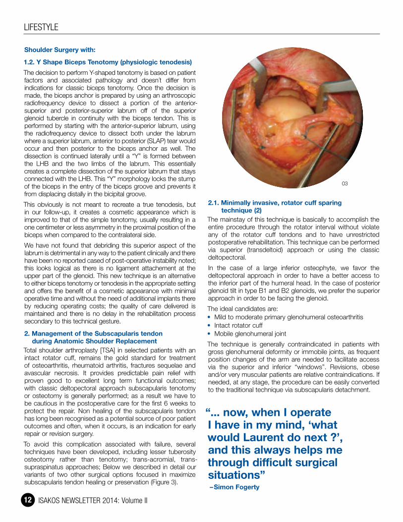
Francesco Giron, MD, PhDSOD Traumatologia e Ortopedia Generale, Azienda Ospedaliera Universitaria Careggi, Firenze, ITALY
Traditionally, intrasubstance tears of the anterior cruciate ligament (ACL) were once thought to be rare among pediatric patients. This belief was motivated by the fact that midsubstance tears were assumed to occur only in adults, and the tibial eminence fracture was considered to be the pediatric equivalent of the ACL tear in adults. However in the past twenty years, complete ruptures of the ACL in adolescent patients have been rising in prevalence because of increased participation and higher levels of competition among young athletes especially in sports that involve cutting, pivoting, twisting and collision such as soccer, basketball, volleyball and football.
Nowadays a midsubstance tear of the anterior cruciate ligament in children is no longer considered a rare injury. In a recent study of high school athletes, female soccer players were found to have the highest rate of ACL injury, with an incidence of 14.08 per 100,000 exposures; male football players had the second highest rate, with 13.87 injuries per 100,000 exposures. Stanitski et al. noted that 47% of preadolescent and 65% of adolescent athletes presenting with an acute hemarthrosis of the knee were ultimately diagnosed with acute ACL rupture.
Management of ACL injuries in the skeletally immature patient is still controversial. While in adults the traditional arthroscopically assisted ACL reconstruction with bony tunnels allows excellent results in terms of knee stability recovery, this technique was usually avoided in younger patients with significant growth remaining because drilling across the growth plate carries a risk of future physeal malfunction and resultant growth disturbance and angular deformity. Thus traditional care of the skeletally immature patient with an ACL tear has relied on bracing and activity modification until the young athlete is close enough to skeletal maturity to undergo transphyseal reconstruction. However, nonsurgical management gradually leads to recurrent instability, resulting in secondary injuries to the surrounding articular cartilage and menisci, and in early degenerative changes of the joint.
Many surgical treatment options have been introduced, including primary repair and reconstruction, using both extra-articular as well as intraarticular procedures. Reports on primary repair and extraarticular reconstruction procedures have shown only little success at providing long-term knee stability. On the other hand, intraarticular anatomical reconstruction of the ACL may result to physeal injury and lower extremity deformities. Others suggest that surgical correction with the use of techniques similar to those used in adult reconstruction produces satisfactory mechanical results without physeal damage, varus or valgus angulation or leg-length discrepancy.
In this article we will discuss the various aspects of this pathology, reviewing the most recent literature, in order to attempt to give some useful guidelines for surgeons involved in the management of this often difficult group of patients.
From an anatomical point of view, the ACL develops in utero, appearing at twenty-four weeks of gestation as a confluence of collagen ligament fibers that blend with the periosteum. It comprises two functional and anatomic bundles, the anteromedial and posterolateral bundles. The anteromedial bundle originates on the femur at the transition between the intercondylar line and the cartilage margin, and it inserts along the medial aspect of the intercondylar eminence. The posterolateral bundle originates at the anteroinferior aspect of the femoral ACL origin, and it inserts just lateral to the central aspect of the intercondylar eminence. The sizes of the anteromedial and posterolateral bundles can vary according to patient height, weight, and body mass index (BMI). Additional considerations include the anatomy of the tibial and femoral physes. The distal femoral physis contributes 70% of the total femoral length and 37% of the total limb length over the course of skeletal development, at an average rate of 10 mm per year. The distance between the femoral physis and the femoral origin of the ACL remains unchanged from gestation through skeletal maturity and averages approximately 3 mm. The proximal tibial physis contributes approximately 55% of the total tibial length and 25% of the total limb length over the course of skeletal development, at a rate of 6.4 mm per year.
When dealing with a knee injury in a child a proper physical evaluation, an accurate interview in the presence of the parents, and maturity assessment are paramount in diagnosing an ACL tear. A focused history investigating on chronicity, mechanism of injury, and symptoms is critical. Any event of swelling or effusion in the knee requires further investigation. The presenting signs and symptoms of an ACL tear in pediatric patients are similar to those experienced by adults. The athlete may experience a pop at the time of injury. Initial examination of the patient should rule out concomitant musculoskeletal injuries. Examination of the knee should includeinspection for acute hemarthrosis, which is helpful in determining the severity of the injury. ACL injuries can be present in up to 65% of adolescents presenting with acute traumatic hemarthrosis.

ISAKOS NEWSLETTER 2014: Volume II 21
CURRENT CONCEPTS
However due to a incomplete development of proprioception most of these patients do not complain of any knee instability but they are frequently referred to the physician by the parents reporting recurrent episodes of pain and swelling after sports activities. A gentle palpation is used to detect any underlying effusion. Stability tests should be performed first in the uninjured knee and then in the injured knee in order to reassure the young patient. When a restriction in the range of motion is recorded, a concomitant injury to the meniscus and/or cartilage should be suspected. A proper Lachman test is critical to assess an abnormal anterior tibial translation and the lack of a firm end point. Imaging of the injured knee is essential in devising a treatment plan. Initial AP, lateral, notch, and sunrise radiographs of the injured knee are helpful in assessing osseous structures and skeletal maturity. Magnetic resonance imaging (MRI) is indicated to evaluate for partial versus complete ACL injuries, define associated ligamentous pathology, and assess for suspected meniscal derangement. MRI is 95% sensitive and 88% specific for detecting ACL tears in children. In addition to the standard radiographic evaluation performed in adults, some authors suggest to additionally obtain fifty-one-inch standing (130-cm) anteroposterior hip-to-ankle radiographs. This allows for accurate preoperative assessment of subtle limb-length discrepancy and angular deformity.
Skeletal maturity is traditionally assessed using the Tanner staging system and with the use of a posteroanterior radiograph of the left hand, with reference to the Greulich and Pyle atlas. Child athletes who are being considered for ACL reconstruction can be classified as either prepubescent or pubescent. The prepubescent patient has physiologic findings consistent with Tanner stages 1 and 2 and a bone age of <12 years in boys and <11 years in girls. The pubescent patient has physiologic findings consistent with Tanner stages 3 and 4 and a bone age of 13 to 16 years in boys and 12 to 14 years in girls.
Historically, nonsurgical treatment options for skeletally immature patients with ACL tears consisted of activity modification, functional bracing, and physical rehabilitation. The rationale for this approach was to allow the patient to achieve skeletal maturity before performing a transphyseal ACL reconstruction, thereby minimizing the risk of physeal violation and potential growth deformity. This nonsurgical approach should be best used in the highly compliant, low-demand patient who has no additional intra-articular pathologies or who has a partial ACL tear. Nevertheless nonoperative management is an appealing option given the increased healing potential of children and the risk of physeal damage with surgical reconstruction, clinical results following nonoperative management have not been favorable. Partial ACL injuries represent one-quarter to one-half of the midsubstance ACL tears that occur in children. Although children tend to have better healing capacity than adults, animal studies have demonstrated mixed results regarding the precise healing potential after partial ACL transection. Kocher et al. showed that approximately one-third of children with a partial ACL tear who were treated nonoperatively with a hinged knee brace, partial weight-bearing for six to eight weeks, and a progressive ACL rehabilitation protocol ultimately required surgical reconstruction for persistent instability. The authors noted several risk factors for failure of nonoperative management and developed an algorithm for acute treatment. Overall, they recommended surgical management for patients with a tear greater than one-half of the thickness of the ACL, a tear of the posterolateral bundle, a pivot-shift examination result of grade B or greater, or a skeletal age of more than fourteen years. Recent papers on nonoperative management of a complete ACL rupture generally report a poor outcome. Additionally, it is associated with a high rate of sport dropout because of recurrent instability, as studies have demonstrated that up to 50% of children treated nonoperatively do not return to athletic activity. Progressive instability can result in progressive meniscal and articular cartilage damage as well as Fairbanks changes (e.g., condylar squaring and joint space narrowing on an anteroposterior radiograph) in 61% of knees. Instability and cartilage degeneration are typically observed in patients who do not modify their post-injury activity level, as is often the case in active children and adolescents.
There has been considerable debate in the literature regarding the optimal time to perform ACL reconstruction in skeletally immature patients. Concerns regarding physeal damage, growth arrest, and subsequent sequelae including angular deformity and limb-length discrepancy have led some surgeons to delay surgical management until skeletal maturity. Delayed reconstruction, however, has its own important drawbacks including the possible development of progressive intraarticular pathology. These poor results have resulted in the development of modern operative techniques for pediatric ACL reconstruction.
01

ISAKOS NEWSLETTER 2014: Volume II 22
CURRENT CONCEPTS
ACL Tears in Athletes with Open Physes
Traditional surgical techniques for adult ACL reconstruction include the creation of transphyseal tunnels followed by graft tensioning and fixation. In the skeletally immature patient, conventional tunnel placement and graft tensioning risks iatrogenic growth disturbance caused by physeal violation. Multiple animal and human clinical series have documented such growth disturbances. More recent animal studies have investigated the effect of small tunnels and soft-tissue grafts on physeal bar formation and subsequent growth arrest. They confirmed that the presence of soft-tissue grafts in the transphyseal tunnels prevented physeal bar formation. Furthermore they also found that transphyseal tunnels that occupy <5% of the physeal cross-sectional area do not appear to cause growth disturbance. However, when >7% to 9% of the cross-sectional area of the physis is violated, growth disturbance is possible even in the presence of a soft-tissue graft. Clinical reports of growth disturbance are rare. Kocher et al. surveyed members of the Herodicus Society and ACL Study Group regarding growth disturbance after skeletally immature ACL reconstruction and noted 15 cases of growth disturbance out of more than 500 ACL reconstructions. They concluded that associated risk factors for growth disturbance included fixation hardware across the physis, large tunnels (≥12 mm), lateral extra-articular tenodesis, dissection in proximity to the perichondrial ring of LaCroix, and suturing near the tibial tubercle. More recent clinical studies have examined the use of MRI to characterize physeal injury after transphyseal ACL reconstruction in skeletally immature patients. Using a custom three-dimensional modeling following transphyseal ACL reconstruction, it has been found that, with 8-mm tunnels, less than 3% of the cross-sectional area of the physis is violated. Moreover, using a custom computerized model of the physis, it has been documented that tunnel diameter is potentially more offensive to the physis than drill angle. Also the effect of an anatomic positioning of the femoral tunnel with the use of single – or double-bundle techniques has been investigated. The anatomic placement of the femoral tunnel is drilled at a more oblique angle that, if performed in a skeletally immature patient, places the lateral physis at an increased risk of injury. Using a computer-aided three-dimensional model to incorporate the drilling of an anatomic double-bundle ACL femoral tunnel with a variety of drill-hole diameters it has been found that the average physeal volume removed, as a percentage of the total physeal volume, was between 3.7% and 6.5%. Based on these data, anatomic double-bundle drill-hole placement during ACL reconstruction seems to produce a zone of physeal injury that appears to increase the risk of abnormal growth in the distal femoral physis postoperatively. Several other studies with MRI investigated the postoperative effects a transphyseal ACL reconstruction on the growth plate in young athletes. It was confirmed that the average bone tunnel–to–growth plate cross-sectional area ratio was less than 3%.
Some patients developed MRI documented focal physeal disruption without a perceived clinical growth disturbance. Moreover compar ing the physes of the surg ica l ly reconstructed knee and the contralateral knee 6 months postoperatively, MR imaging showed narrowing of the growth plates in the reconstructed knee in comparison with that in the contralateral knee. Radiographic analyses showed corticalization around the drill holes in all cases. Although ACL reconstruction caused narrowing or the early closure of the open physes in adolescents, the patients did not experience limb-length discrepancies and angular deformities. All the authors concluded that, although transphyseal reconstruction of the ACL may not be a benign procedure in younger children with substantial growth potential, it is possible to safely perform this procedure in adolescents with limited growth remaining.
Independently from the surgical technique adopted to reconstruct the ACL, an unanimous agreement has been found concerning the graft choice. In case of a prepubescent patient, a tendon with bone plugs (e.g., bone-patellar tendon bone graft) may induce local growth arrest if placed across the physis, so a soft tissue graft must be used. In case of pubescent patients with closing physes the graft selection follows the same indications as the adult patients and the surgeon preference. Conversely, there has been considerable controversy regarding the use of autograft versus allograft tissue. In a recent study, the Multicenter Orthopaedic Outcomes Network patient database was analyzed to evaluate graft choice and the risks associated with failure and graft rupture. Patients between ages 10 and 19 years were found to have the highest percentage of graft failures regardless of graft type. Moreover, the odds of graft rupture were four times higher with allograft reconstruction than with autograft reconstruction.
Modern surgical techniques can be classified as physeal sparing, partial transphyseal and complete transphyseal. A variety of physeal-sparing techniques has been described. In one technique, the hamstring tendons were kept intact at their distal attachment, passed through a transepiphyseal tibial tunnel, looped over a staple at the femoral ACL origin, brought back through the tibial tunnel, and sutured onto themselves. In a second technique an autograft hamstring tendon was passed through the distal femoral epiphysis and fixed using a suspension fixation device over the lateral femoral condyle.
02

ISAKOS NEWSLETTER 2014: Volume II 23
CURRENT CONCEPTS
The remaining portion of the graft was then passed through an all-epiphyseal tibial tunnel and fixed with a post distal to the proximal tibial physis. Finally, in a third surgical technique combined intra – and extra-articular reconstruction. This procedure is a modification of the combined intra – and extra-articular reconstruction described by MacIntosh and Darby. All these techniques showed excellent clinical results and no reported evidence of growth disturbance. A biomechanical evaluation of these techniques was also performed to determine which most closely restored native knee kinematics. In a controlled laboratory setting, displacement and rotation of the tibia with respect to the femur were measured in the intact knee after ACL disruption and after ACL reconstruction using all epiphyseal, transtibial over-the-top, and iliotibial band physeal-sparing techniques. The results showed that, although all physeal-sparing reconstruction techniques restored some stability to the knee, the iliotibial band reconstruction best restored AP stability and rotation control; however, it appeared to slightly overconstrain the knee to rotational forces at some flexion angles. The partial transphyseal techniques combine a hybrid of physeal-sparing reconstruction and traditional transphyseal procedures performed in adults. Usually the distal femoral physis is left undisturbed, thereby minimizing the risk of growth arrest. Smaller bone tunnels (6 to 8 mm) and more vertical tunnels are used to limit to <5% the overall cross-sectional area of tibial physis that is interrupted. Also these techniques showed excellent clinical results and only one patient was reported to suffer a 2-cm limb-length discrepancy. Complete transphyseal reconstruction strongly resembles adult-type ACL reconstruction, with some differences, including smaller, more vertical tunnels, no hardware or bone blocks at the level of the closing physis, and metaphyseal f ixation. This procedure is typically performed on adolescents with little to no growth remaining. Outcomes from transphyseal reconstruction have been generally successful, with a mean IKDC score of 91.5, a mean Lysholm score of 93.5, and a return to the preinjury activity level in 88.8% of patients. The limb-length discrepancy is typically minimal, averaging 1.2 mm, but the operatively treated limb may range from 7 mm short to 7 mm long. Recently several clinical outcome studies tout the success of complete transphyseal ACL reconstruction also in prepubescent athletes. In these studies the ACL reconstruction was performed following the guidelines for ACL reconstruction initially proposed by Meller et al. to avoid growth disturbances in skeletally immature animals. The tibial tuberosity was spared to prevent a genu recurvatum, the thermal damage to the growth plates was avoided, a small-diameter drill was used in the center of the growth plate, a soft tissue graft was used, the graft fixation was achieved far from the growth plates, the perforated growth plates were filled by the soft tissue graft, and the graft was moderately pretensioned before fixation. Following these principles several authors were able to achieve excellent clinical results in terms of knee stability and sports activity recovery without any reported evidence of growth disturbance.
Postoperatively a proper rehabilitation is critical to ensure the success in clinical and functional outcome. Child and adolescent athletes are different from adult patients. Compliance can be problematic in this age group, and patients must be carefully monitored at school and home by teachers, parents, and caretakers. Activity restrictions and compliance with graduated activity levels are critical to successful outcomes. In the first four or six weeks postoperatively the use of a brace should be strongly encouraged in this type of patients who are notoriously more hazardous and careless than adult patients. Complete weight-bearing is allowed at the end of the first postoperative month. Progressive rehabilitation consisting of ROM recovery, closed kinetic chain quadriceps and hamstring strengthening, patellar mobilization, and swimming pool exercises are used for the first months postoperatively. Initiation of straight-line jogging and plyometrics begins at 4 months after reconstruction. A full return to cutting and pivoting activities and sports is allowed at approximately 7-9 months postoperatively.
In conclusion ACL ruptures in skeleta l ly immature patients are becoming an emerging orthopaedic problem with increased single-sport concentration, year-round participation, and less time spent in free play. It is important to properly diagnose and manage these injuries. There is now considerable evidence in the literature that conservative management of ACL tears in children produces poor results with subsequent instability. Nonsurgical management, including activity modification, bracing, and physical therapy, shoud best indicated only in patients with partial tears involving <50% of the ACL diameter. In patients with complete ruptures, chronologic, physiologic, and skeletal maturity must be assessed to appropriately address the injury. Treatment options are defined on assessment of the patient’s skeletal age and include physealsparing, partial or complete transphyseal, and adult-type anatomic ACL reconstruction. Postoperative management includes weight-bearing and activity modifications, bracing, and a progressive physical therapy protocol emphasizing ROM, closed kinetic chain strengthening, and a gradual and measured return to sport-specific maneuvers. Surgical complications are rare.
01 MRI sagittal view of an ACL tear in an 11-year girl02 Intraoperative X-ray view of a femoral tunnel for ACL reconstruction
drilled through the distal femoral epiphysis and a tibia tunnel drilled through the growth plate in a 13-year old boy
03 X-ray view of an ibrid transphyseal sparing over the top technique of ACL reconstruction
03

ISAKOS NEWSLETTER 2014: Volume II 24
TKR Component Malrotation: A Common Unrecognized Cause of Pain & Stiffness
Dr. Christopher Vertullo MBBS FRACS Orthopaedic Surgery & Sports Medicine Centre, Bond University, Griffith University
BackgroundWhile Total Knee Replacement (TKR) is a highly successful surgical intervention for end stage knee osteoarthritis, a significant proportion of patients unfortunately remain dissatisfied after TKR. Younger active patients are more likely to be dissatisfied and undergo subsequent revision as a result.
Pain and stiffness with consequently reduced function is the most common cause of patient dissatisfaction. Increasing evidence suggests that femoral and tibial component internal rotation is a very potent cause of both pain and stiffness after TKR. Berger et al. in 1998 reported on patellofemoral complications in internally rotated components requiring revision. Barrack et al. in 2001 linked anterior knee pain after TKR to component mal-rotation despite normal alignment, with a five times greater risk of anterior knee pain in the internally rotated group. Nicoll and Rowley reported in 2010, 56% of 36 painful TKRs having rotational errors in a series of 740, compared to pain free TKRs. Bèdard et al. in 2011 reported improved range of motion after revision TKR for stiffness of a series of internally rotated tibial components with a pre-operative range of 10.1º – 71.5º to a improved post – operative range 0.8º – 100º.
All stiff TKRs in this series had internal rotation of either one component or both. Bhattee et al. in 2013 reported a series of 23 TKR cases with unresurfaced patella’s and anterior knee pain. All 23 had some degree of internal rotation on CT scan. The 61% who remained dissatisfied after patella resurfacing had a mean of 2.88º femoral internal rotation compared to 0.92º internal rotation in the satisfied group.
Marked component internal rotation can also result in extensor mechanism mal-tracking, with patella dislocation, fracture or avascular necrosis as a result. Higher rates of lateral release in patella avascular necrosis cases may not be only due to vascular issues but also mal-tracking issues due to malrotation. In the Australian National Joint Replacement Registry (ANJRR) 26.4% of revisions were performed for either pain, patellofemoral pain or arthrofibrosis, suggesting the majority of these patients had internally rotated components. Only component loosening or lysis results in more revisions in the ANJRR. There is evidence that posterior stabilized components are more prone to pain with malrotation.
Interestingly, while in the last decade a large amount of effort has gone towards devising methods to improve TKR coronal plane alignment, such as navigation and patient specific instrumentation, the role of coronal plane mal-alignment as an isolated cause of pain, stiffness and prosthesis failure remains unclear in modern prostheses. By 2013, only 2.2% patients underwent revision for mal-alignment in the ANJRR.
No series has reported component external rotation of having a detrimental effect, with pain-free well – functioning comparator TKRs having a mean of a few degrees of external rotation of both tibial and femur.
CURRENT CONCEPTS
01
02

ISAKOS NEWSLETTER 2014: Volume II 25
Anatomic Rotational LandmarksUnlike coronal plane alignment landmarks, femoral and tibia rotational landmarks are difficult to define, inaccurate and variable. Small surgical errors can result in relatively large malrotations. As a result no single perfect foolproof method of component rotation exists and hence using a combination of landmarks is the gold standard.
Three anatomic landmarks exist for femoral rotation. Berger et al. described the Surgical Epicondylar Axis (SEA) in 1993. The SEA is a line from the lateral epicondyle to the medial collateral sulcus. It is the most accurate of the three landmarks, and is the only landmark that remains of value during revision. Whiteside’s line was described in 1995 as a
“line through the deepest part of the patella groove anteriorly and the centre of the intercondylar notch posteriorly”. It is perpendicular to the SEA and is slightly less accurate. The final landmark is the Posterior Condylar Axis (PCA), which is 3 degrees internally rotated to the SEA. It is the most easily instrumented and hence remains the most commonly utilized. Unfortunately it is also the most inaccurate landmark, particularly in valgus knees with lateral femoral hypoplasia. Using the PCA in isolation is ill-advised, often resulting in inadvertent femoral component internal rotation.
Siston et al. in 2005 compared multiple different methods to achieve correct femoral rotation, finding that navigated methods in isolation were no better than anatomic methods. Combined methods had the greatest accuracy, particularly either the SEA and Whiteside’s line or the SEA and navigated patella tracking. The author uses all three rotational axis, with the SEA and Whiteside’s Line drawn onto the resected distal femur with a surgical marker.
Tibial Component rotational landmarks are even more inaccurate, with no general consensus and a variety of described landmarks. Lawrie et al. performed a cadaveric tibial landmarks study in 2011, reporting that no anatomic tibial landmark was parallel to their newly described dynamic Knee Motion Axis (KMA). They found that the commonly used Medial 1/3 Tubercle to PCL (Insall’s) Axis resulted in slight external rotation of 3.5% during squatting. The Medial – Lateral Axis of the resected tibial surface, resulted in excessive internal rotation, as did the Posterior Tibia Axis (tangent to the posterior aspect of the tibial plateau). The Central Tubercle Axis (a line from geometric centre of the resected tibia to the central tubercle) and Femoral Epicondylar Axis resulted in excessive internal rotation.
The author uses the Medial 1/3 Tubercle Axis, but does reference the Medial – Lateral Axis as well. It should also be realized that tibial component medio-lateral translation also effects rotation, with 1 – 1.5º external rotation per mm of medial translation. Deeper resection and severe varus makes all landmarks less accurate, with tibial component placement 10º externally rotated to the Posterior Tibial Axis the best option. Other non-tibial landmark methods, such as the
“self-aligning – free floating trial” technique or using the 2nd metatarsal axis are more error prone.
Surgeons using minimization of the surgical incision, or minimally invasive surgery, should be aware that tibial component internal rotation is made more l ikely by diminishing proximal tibial visualization. Posterior tibial retractors should be used with care, as they tend to drive tibial trials and components into internal rotation when posterior access is tight. Trying to maximize tibial component coverage with larger implants will inadvertently internally rotate the tibia. Asymmetric tibial components can make rotational placement difficult to judge. Most inadvertent surgical events, such as poor visualization, posterior retractors, or the lateral femoral condyle in a contracted tight joint tend to push tibial trials into internal rotation.
If the above anatomic landmarks are indistinct or the surgeon remains intra-operatively uncertain about the correct rotation, choosing the position with slight external rotation is the safest option. As Bell et al. noted in 2012, external rotation of either tibia or femur appears to be asymptomatic, while slight internal rotation typically results in pain.
CURRENT CONCEPTS
03

ISAKOS NEWSLETTER 2014: Volume II 26
CURRENT CONCEPTS
TKR Component Malrotation: A Common Unrecognized Cause of Pain & Stiffness
Investigation and ManagementThe most important diagnostic tool is a high degree of suspicion and a CT scanning protocol when confronted with a stiff, painful or dissatisfied TKR. Patients will have been painful and stiff since implantation, differing from other common causes of pain such as loosening. Multiple prior manipulations and second or third opinions will be typical. Often diagnosis such as Chronic Regional Pain Syndrome, Arthrofibrosis or metal sensitivity will have been made previously.
Other causes of pain and stiffness always require exclusion, such as infection, loosening or referred pain.
Malrotated patients often have no clinical signs, but a reduced range of motion is common. With the patient seated and the knee at ninety degrees, hanging over the bed edge, greater external rotation of the foot on the affected side can be evident. Patella instability or mal-tracking may also be present. Plain radiographs typically appear normal, however, in cases with severe tibial internal rotation, the fibular head will appear more covered by the tibia. In Figure 3, plain radiographs of a TKR with internal rotation of both components reveal subtle increased fibula head coverage. Figure 4 and 5 are CT Scans of the same malrotated TKR. This patient complained of pain, stiffness and patella instability, and had an externally rotated foot at 90º.
CT Scan composite images of landmarks and components are vital to investigate rotation. Composite CT images of the SEA and Posterior Condyles allow rotational angle calculation. Berger has described a CT technique for tibial component assessment (Figure 5). Asymmetric tibial components and posterior femoral condyles need to be interpreted with caution.
Revision should be only undertaken i f the patient’s dissatisfaction warrants a major surgical procedure and its associated risks. Patients must be aware that revision may not result in total satisfaction. Revision of both components is usually recommended as both are usually mal-rotated, but a case can be made for isolated tibial or femoral component revision if the remaining component is aligned, appropriately sized and stable. It is vital not to compound the errors of the original procedure. Patella resurfacing or stabilization procedures in a mal-rotated TKR are unlikely to result in a satisfied patient.
SummaryPain and stiffness after TKR is commonly due to component internal rotation of the femur, tibia or both.
Mild component external rotation does not appear to produce detrimental effects.
A combination of anatomic landmarks is the most accurate method of rotational assessment.
If in doubt, select a slightly externally rotated position for both tibial & femoral components.
CT Scan is the only method to investigate for malrotation.
Revision of both femoral and tibial components is usually indicated.
01 Femoral Anatomic Landmarks02 Various Tibial Rotational Axes03 Plain XR of Internally Rotated Femoral and Tibial Components04 Internally Rotated Femur on CT Scan05 Internally Rotated Tibia on CT Scan
04
05

ISAKOS NEWSLETTER 2014: Volume II 27
CURRENT CONCEPTS
Femoroacetabular Impingement
Leandro Ejnisman, MD
Additional Author: Robert F. LaPrade, MD, PhD, USAISAKOS Sports Medicine Committee
IntroductionFemoroacetabular impingement (FAI) is a common cause of hip pain in the athlete. It is important for a sports medicine physician to be able to diagnose and treat this condition because a delay in the diagnosis can lead to a longer period of recovery and return to sport. Moreover, FAI is a cause for early hip osteoarthritis; and early intervention is indicated in order to avoid severe chondral damage.
Clinical PresentationPatients typically present with inguinal pain, which might present with traumatic or insidious onset. The pain may also be referred to the lateral thigh, inner thigh and buttocks. Activities involved in this condition are sports with a rotational demand on the hip, such as soccer, ice hockey, football, rugby and skiing. Patients frequently complain about pain during prolonged sitting.
The physical exam usually demonstrates decreased internal rotation. The most important finding is the anterior impingement sign (Fig 1). It is considered positive when it elicits anterior hip pain during flexion, adduction and internal rotation. It is also important to clear spine pathology and inguinal hernia, because these conditions may mimic FAI.
ClassificationFAI is classified in 3 types: cam, pincer and mixed-type. Cam impingement refers to the femoral side. The femoral head-neck junction has a bony “bump”, and this bump impinges on the acetabular rim as the hip is flexed and internally rotated. The labral tissue can be detached at the chondrolabral junction. An acetabular cartilage flap can be seen adjacent to the labral tear.
Pincer impingement refers to bony impingement on the acetabular side. It can be caused by global or focal overcoverage of the femoral head. Global overcoverage occurs in cases of coxa profunda and protrusio acetabuli. Normally, the acetabular floor is lateral to the ilioischial line. In cases of coxa profunda, the acetabular floor is medial to the ilioischial line. In cases of protrusio acetabuli, the femoral head is medial to the ilioischial line. Focal overcoverage is seen in cases of acetabular retroversion. In pincer impingement, the labrum is usually degenerated and labral cysts can be found.
The most common type of FAI is the mixed-type, representing 75% of patients. Pincer and cam characteristics co-exist in the same patient. On the femoral side, a high alpha angle is found, and on the acetabular side, overcoverage signs are present. Surgical findings can demonstrate a bruised or degenerated labrum, which might be detached from the acetabular rim, suggesting a mixed pattern of labral injury.
ImagingRadiographs are of paramount importance in FAI investigation. Many physicians neglect their use, and order only a MRI. The x-ray will often evaluate the hip bony morphology better than the MRI. Proper position during radiographs is essential because minor pelvic inclination may alter FAI radiographic findings.
The anteroposterior pelvic x-ray is evaluated. One must look for signs of pincer impingement. The cross-over sign is diagnostic of focal overcoverage. The acetabular walls should cross in the lateral aspect of the acetabulum. When the cross-over sign is present, the posterior and anterior wall cross before the lateral aspect of the acetabulum. The acetabular center-edge angle is measured, as well as the joint space.
01

ISAKOS NEWSLETTER 2014: Volume II 28
CURRENT CONCEPTS
Femoroacetabular Impingement
A profile of the femoral head should be also obtained. There are multiple profiles described and the most commonly used are the Dunn and cross-table views. The alpha angle should be measured on these views (Fig 2). There is no consensus on the normal value of the alpha angle, but we and most others consider values above 55º as abnormal.
MRI is used to look for labral tears and chondral damage. Acetabular and femoral cysts (herniation pits are a typical FAI finding in the femoral head-neck junction), ligamentum teres pathology, muscle injuries and tendonitis are also evaluated. MRI has a role in the differential diagnosis of FAI (i.e. avascular necrosis, loose bodies and synovial disease).
Treatment Initially, conservative treatment can be instituted. It is based on rest or activity modification, anti-inflammatory drugs and physical therapy. However, conservative treatment often does not yield good clinical results, and surgical treatment is often necessary.
Surgical treatment can be performed both by open and arthroscopic approaches. Recent reports suggest a faster recovery with fewer complications in the arthroscopic approach. Albeit the approach chosen, the main goal is to restore normal bony and soft tissue anatomy.
On the acetabular side, the pincer lesion is treated by an acetabuloplasty, where the prominent anterior acetabular wall is trimmed with an arthroscopic burr. On the femoral side, the femoral contour is reshaped to restore the normal femoral head-neck offset. Labral tears should be repaired with suture anchors or reconstructed, usually with an iliotibial band graft, for a deficient labrum.
ConclusionIt is important for the sports medicine physician to get acquainted with FAI. When proper diagnosis is obtained in a timely fashion, treatment can be effective, and high rates of patient satisfaction and return to sport are expected.
01 Anterior impingement sign02 Alpha angle measurement. In this example the alpha angle measures
70º. A herniation pit is also seen (arrow).
02
Comprehensive Approaches to Articular Cartilage Repair and Hip Arthroscopy
January 23 – 25, 2015 Carlsbad, California
15INTERNATIONALSPORTS MEDICINEFELLOWS CONFERENCE
th
ISAKOS is sending two international sports medicine fellows to attend the annual ISMF Conference held in January in Carlsbad, California.
Apply today at www.isakos.com
Deadline: September 1, 2014

ISAKOS NEWSLETTER 2014: Volume II 29
CURRENT CONCEPTS
Growth Factors (GF) and Platelet Rich Plasma (PRP) All PRP Are Not Alike
Ramon Cugat, MD, PhDFundación Garcia Cugat Mutualidad de Futbolistas Españoles –Federación Española de Fútbol Hospital Quirón Barcelona, SPAIN
Additional Author: Francois Kelberine, MDClinique Provencale Parc Rambot Aix en Provence, France
IntroductionGreater understanding of the cell structure and function have opened new fields of investigation for possible health benefits of living beings.
To act, living cells must find a support or three-dimensional matrix and should receive stimuli from signaling molecules and growth factors.
This has led to a new specialty, Regenerative Medicine, which according to authors such as Greenwood (1) Anitua and Sanchez (2) among many others is an emerging field of interdisciplinary research and clinical applications focused on the repair, replacement or regeneration of cells, tissues or organs to restore impaired function from any cause, including congenital defects, disease and trauma. It uses a combination of technological approaches that go beyond traditional transplantation and replacement therapies. These approaches may include, but are not limited to, the use of stem cells, progenitor cells, soluble molecules, recombinant GF, biomaterials, nanotechnology, genetic engineering, tissue engineering and advanced cell therapy.
This article focusses on PRP and GF: what they are, how they are obtained, how they work and what benefits they can bring to the field of Orthopedic Surgery.
What are Growth Factors (GF)?They are proteins, key mediators that regulate activities especially hæmostasis and tissue repair. They are involved in almost all physiological healing processes. Some authors call them proteins, Differentiation Factors and Growth Factors.
Where are they?In plasma they are mainly produced by the platelets and stored in the α – granules within platelets. Their amount is correlated with the number of platelets. They are also synthesized by some tissues such as liver or fat tissue. Some similar proteins can be found outside the plasma in pituitary, kidneys, submandibular gland, lacrimal gland and Brunner gland.
How do they act?They are released from the cell that produces them or from their storage granules under thrombine action first then a cascade is initiated. They interact with a corresponding cell membrane receptor. They act as signals sent between cells to complete a specific activity extracellular or intracellular amplifying the signal.
It is of great interest that the same package of GF acts differently depending on the cellular environment: target tissue (muscle, tendon, cartilage,…) and timeframe of the healing process.
What are they called and what do they do?The names of the growth factors indicate their activity and/or origin. Here are some examples:• PDGF (Platelet Derived GF) is chemotactic for fibroblasts,
smooth muscle cells and leucocytes (3). It stimulates neutrophil phagocytosis, leads to the release of proteins from neutrophils and monocytes. It initiates collagen synthesis and connective tissue replication.
• VEGF (Vascular Endothelial GF) stimulates angiogenesis by endothelial cells replication.
• TGF-β1 (Transforming GF) belongs to the family of BMP (Bone Morphogenetic Proteins) among many others (4). Its action is cell-dependant in relation with the environment. It limits (regulates) cell proliferation. It increases extracellular matrix synthesis and inhibits its degradation. And it has an immunosuppressive effect (5).
• EGF (Epidermal growth factor) attracts fibroblast and stimulates its mitosis as well as keratinocyte, increases fibronectin and collagen synthesis. So it accelerates wound closure.
• FGF a & b (acid and basic Fibroblastic GF) stimulates proliferation of most cells involved in repair, is produced by numerous types of cells so their amount is not only correlated to the number of platelets.
• IGF (Insulin like GF) type I or II, HGF (Hepatocyte GF). They are not synthesized by platelets so they are not correlated to their amount. They induce cell proliferation, differentiation and synthesis of type I collagen.
Many experimental, animal and clinical studies support that GFs promote hemostasis, cell proliferation, migration (chemotaxis) and differentiation. So they lead to angiogenesis, regeneration and extracellular matrix synthesis especially in acute injuries.

ISAKOS NEWSLETTER 2014: Volume II 30
Growth Factors (GF) and Platelet Rich Plasma (PRP) All PRP Are Not Alike
Growth factors act synergistically. In plasma and PRP, their concentrations are physiologically balanced.
It differs from recombinant GF which are artificially produced. They are used in tissue research. And when one alone is studied, biological responses can be clearly obviated.
How PRP is obtained?PRP is a plasma concentrate with an amount of platelets above the serum baseline.
From a fresh blood harvesting, centrifugation will insulate PRP. Its use is mandatory bound to the patient itself, within a short time in sterile conditions.
BUT there are many companies currently claiming to have kits capable of obtaining PRP or GF with and without cells. The Medicine Agency/Administration for each country regulates its use and makes it mandatory to use an approved kit and to follow an approved method.
However they di ffer dramatical ly by the amount of harvested blood, the number, speed, time and intensity of centrifugation. So the final product is fully different from one technique to another (6-11).
The disparity is major with regards to:• Amount of platelets (with different concentrations).• Leucocytes (with or without, concentrated or baseline).• Erythrocytes (with or without, concentrated or baseline).• To release GF: Not activated, activated, if activated
what with? • Types of Presentation: Liquid, clot, membrane.
Clinical relevance The review of the current literature about the efficacy of PRP is controversial.
Randomized Controlled Trials: Some having reported positive results to shorten healing processes, while others found them to be useless. But beyond the efficacy of PRPs, the difference in content, the way to apply and the tissue in concern are THE major issues.
PRP are not alike and PRP is not concentrated blood. The authors believe that a concentrated amount of GF, physiologically balanced, is a toolbox for injured tissue. It should lead to an accelerated healing process although one should be very critical about commercial claims.
Conclusion:GF can be useful in ligaments, tendons, muscles, bone and cartilage injuries in Orthopaedics and Orthopaedic Sports Medicine, shortening the repair-regeneration healing processes or helping in case of bad evolution.
The best substances to reproduce a physiological repair are those that respect the chemical and cellular composition of the plasma.
CURRENT CONCEPTS
1 How long is a typical work day?
1. 8 hours
2. 9 hours
3. 10 hours
4. 12+ hours
2 What is the common turn over between cases in your outpatient OR?
1. < 15m
2. 15 – 30m
3. 30 – 45m
4. 45 – 60m
5. > 60m
3 How many weeks of vacation do you take annually?
1. 2w
2. 3w
3. 4w
4. 5w
5. > 6w
4 At what age do you plan to retire?
1. < 55
2. 60
3. 65
4. 70
5. > 70
4. never
19%
6%
39%
36%
23%
29%
48%
13%
19%
32%
10%
13%
0%
13%
ISAKOS ASKEDYOU ANSWERED
Results of the ISAKOS Newsletter 2014: Volume I Poll
13%
13%
23%
35%
16%
0%

ISAKOS NEWSLETTER 2014: Volume II 31
CURRENT CONCEPTS
Hip Arthroscopy: Current Advances and Scientific Evidence
Aaron J. Krych, MDDepartment of Orthopedic Surgery, Mayo Clinic and Mayo Foundation, Rochester, MN, USA
Additional Authors: Volker Musahl MD2
Bruce A Levy MD1
1 Department of Orthopedic Surgery, Mayo Clinic and Mayo Foundation, Rochester, Minnesota
2 Department of Orthopedic Surgery, University of Pittsburg, Pittsburg, Pennsylvania
Hip arthroscopy is a rapidly evolving field, and the number of cases performed each year in the US has increased dramatically. From 2006 to 2010 alone, there was a 600% increase cited in one registry. Symptomatic femoral acetabular impingement with labral tear remains the most common indication for hip arthroscopy, but as our instrumentation and surgical techniques become more refined, the ability to treat patients with a wide variety of hip disorders has made hip arthroscopy one of the most exciting areas in orthopedic surgery.
From a surgical standpoint, we typically divide the hip into three areas: the central compartment, the per iphera l compartment , and the per i -trochanteric space. In general, access to the central compartment allows treatment of acetabular rim (Pincer) lesions, labral tears, loose bodies, chondral injuries, l igamentum teres tears, and internal snapping from the iliopsoas tendon. The peripheral compartment allows treatment of loose bodies, and femoral neck (CAM) lesions and the peri-trochanteric space allows treatment of trochanteric bursitis, external snapping hip and gluteus medius and minimus (so-called “rotator cuff of the hip”) tears.
Clinical outcomes after arthroscopic hip surgery have demonstrated excellent clinical success. Patients with femoral acetabular impingement and labral tear in particular have been able to return to high levels of function and even professional sport with this operation. The decision to recommend hip arthroscopy is based on patient factors, symptomatology, imaging f indings, physical examination, and patient expectations.
As with any evolving surgical field scientific, evidence is lacking. Currently there is a paucity of data in the literature documenting long-term outcomes after hip arthroscopy, and few studies have reported outcomes on these newer surgical techniques. As well, l imited studies have been performed comparing arthroscopic to more traditional open surgical techniques. Having said that, in a recent Hip Arthroscopy Theme issue in the Journal Knee Surgery Sports Traumatology and Arthroscopy (KSSTA), a plethora of new scientific data has just been published.
In this newsletter we will review the Theme issue manuscripts to give the reader a more thorough understanding of most “up-to-date” scientific evidence pertaining to hip arthroscopy.
While randomized controlled trials are still lacking in this field, the KSSTA theme issue highlights 39 original articles with increased knowledge in the areas of diagnosis of hip pathology, labral function, outcome of hip arthroscopy, areas of novel treatment, and complications of hip arthroscopy. As hip surgeons, we must carefully assess the scientific literature to review indications and advance surgical techniques to ultimately improve the outcomes for our patients.
01
02

ISAKOS NEWSLETTER 2014: Volume II 32
CURRENT CONCEPTS
Hip Arthroscopy: Current Advances and Scientific Evidence
The correct diagnosis of hip pathology remains challenging. This is highlighted by a survey study that showed the majority of surgeons were unsure of evidence supporting the best clinical test for FAI. Ayeni et al. showed that maximal squat test had only limited diagnostic ability, and may not be clinically helpful to distinguish whether CAM-type FAI is symptomatic in a patient. A radiographic clue reported by Ji and colleagues may be herniation pits on the femoral head-neck junction, as the prevalence was higher in symptomatic patients with pincer-type FAI. In some cases, diagnostic hip injections are routinely used in these challenging cases to differentiate pain sources. However, Ayeni et al. report that pain relief from a local anesthetic injection may not be a good predictor of arthroscopic management of FAI, but no pain relief from this injection predicts a high likelihood of a negative result from surgery. For therapeutic purposes, Krych et al. report that cortisone injection had only limited value as the average pain relief duration was only 10 days. However, hyaluronic acid demonstrated more optimistic results in a prospective trial of twenty patients with decreased pain scores and improved function at 12 months.
The acetabular labrum is theorized to have the roles of intra-articular fluid pressurization and hip stability. In a cadaveric model, Philippon et al. found that partial labral resection decreased intra-articular fluid pressurization, and labral repair (better with through suture techniques over looped suture techniques) or labral reconstruction was able to restore pressurizations to the intact state.
Similarly, the same authors demonstrated that the labrum was the primary hip stabilizer to distraction forces at small displacements, which may explain hip microinstability with loss of labral function. Clinically, the importance of labral preservation was emphasized in several studies. Dippman et al. showed that improvements in pain and function after labral repair was most significant from 3 to 6 months postoperatively. In a systematic review, Ayeni et al. demonstrated better clinical outcomes with labral repair compared to labral debridement. In a retrospective review of early hip arthroscopy procedures with labral débridement, Krych et al. reported only modest outcomes with a 45% failure rate. In cases where the labrum has been excised or is not repairable, a systematic review by Ayeni et al. demonstrated that labral reconstruction can have short-term improvement in patient pain and function.
As surgeons we strive to improve outcomes in our patients. A Scandinavian expert group described the development of a hip registry with its baseline data, which will provide for future scientific work. In addition, it is important to use valid, reliable, and responsive instruments to objectively assess patient outcome. Levy et al. highlight multimodal analgesia as a safe and effective method of providing a high satisfaction rate of patients undergoing outpatient hip arthroscopy. Intra-operatively, Sa and colleagues demonstrated that the alpha angle is a good predictor of outcome and represents a simple, reproducible and inexpensive guide that can be used. Postoperatively, comprehensive rehabilitation is a key component to successful outcome in any of these procedures. In failure of primary hip arthroscopy, the results of revision hip arthroscopy at medium-term follow-up reported by Aprato et al. demonstrated that outcomes can improve, but may not be as successful as in primary hip arthroscopy.
03
04

ISAKOS NEWSLETTER 2014: Volume II 33
CURRENT CONCEPTS
The scope of treatment for hip arthroscopy continues to expand with many novel techniques introduced in the KSSTA special issue. In case reports, the hip arthroscope has been shown to be valuable with arthroscopic reduction and internal fixation of selected acetabular fractures and femoral head fractures. Hip arthroscopy to address intra-articular pathology has also been shown effective as a combined approach with periacetabular osteotomy for dysplastic hips. In addition, arthroscopic treatment can be helpful in treating acute septic arthritis of the hip joint in adults and recurrent acetabular osteoid osteoma. Lastly, Safran et al. have demonstrated an endoscopic technique to address ischiofemoral impingement in the hip by increasing the space between the ischium and femur.
While indications continue to expand, complications are not absent from hip arthroscopy. Dietrich et al. emphasize the steep learning curve in hip arthroscopy and demonstrate the importance of training with an expert surgeon. Zingg and colleagues report a 1.9% rate of femoral neck insufficiency fractures following cam resection and femoral neck osteochondroplasty, and offer guidelines for postoperative rehabilitation. On the soft tissue side, Smith et al. demonstrate that the capsular restraints in the hip are essential to maintaining normal hip biomechanics. In revision hip arthroscopy, McCormick et al. report that capsular defects are relatively common, which may lead to worse outcomes. In addition to capsular defects, Willimon et al. show that adhesions can develop following hip arthroscopy, with a higher likelihood in patients less than 30 and rehabilitation without circumduction.
As the field of hip arthroscopy and hip preservation is still young, there is ample opportunity for future research. Surgeons and scientists, who critically evaluate scientific literature must review and refine indications and surgical techniques. Similarly, we must improve our understanding of rehabilitation procedures and for measures of ( re-) in jury prevention. Future research is required for the enhancement of surgical repair procedures with biologics, such as growth factor delivery, stem cells, or tissue engineered materials. Rigorous future research, critical appraisal of indications and techniques, as well as peer-review will then assure advancing of hip arthroscopy and ultimately help improving outcome for our patients.
Although several recent systematic reviews and metaanalyses have increased the strength of evidence-based research, there is still a paucity of randomized controlled trials in hip arthroscopy surgery. Through future research we therefore need to develop evidence-based guidelines that inform surgeons about surgical indications and outcomes of hip arthroscopy. Furthermore, there is a need to provide results from well-designed investigations addressing areas such as diagnostic algorithms and surgical outcomes. An ideal course to advance hip arthroscopy research would be to gather information about the latest surgical techniques from expert clinicians, obtain methodological guidance from researchers, and obtain validated outcomes measures from patients.
As the knowledge and understanding of hip disease and arthroscopy continues to grow, we hope this ISAKOS newsletter article serves a summary of current literature and stimulates further investigation into hip arthroscopy with the goal of improving outcomes for athletes and patients with hip conditions. Ultimately, high-level research will be crucial for improving hip arthroscopy as a successful treatment for patients with disorders of the hip.
01 Intraoperative arthroscopy photo demonstrating exposure of a large cam lesion through a T-cut capsulotomy.
02 Intraoperative arthroscopy photo demonstrating cam lesion resection and recontouring of the femoral neck through a T-cut capsulotomy.
03 Intraoperative arthroscopy photo demonstrating closure of the T-cut capsulotomy.
04 Preoperative frog leg lateral view of distal cam bump with loss of femoral neck offset.
05 Postoperative frog leg lateral view of cam bump resection and femoral neck recontouring.
05
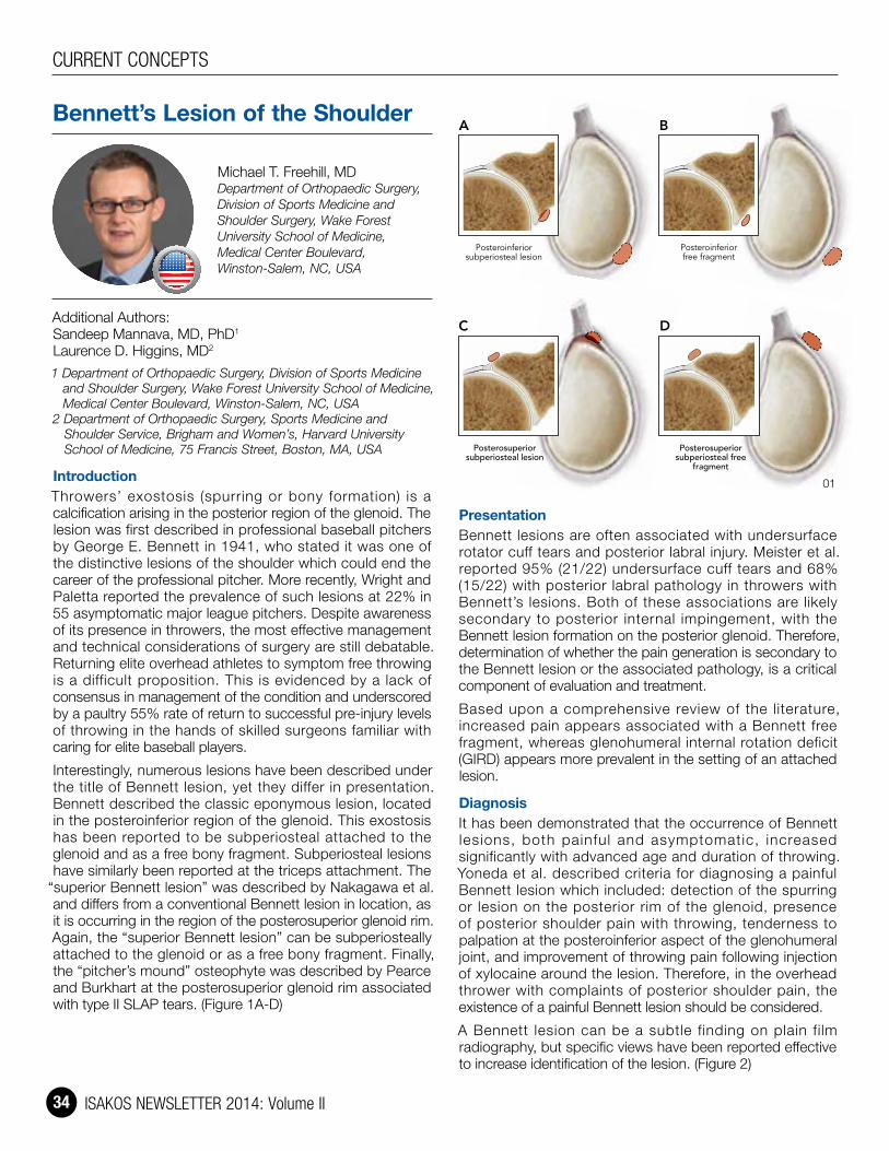
ISAKOS NEWSLETTER 2014: Volume II 34
CURRENT CONCEPTS
Bennett’s Lesion of the Shoulder
Michael T. Freehill, MDDepartment of Orthopaedic Surgery, Division of Sports Medicine and Shoulder Surgery, Wake Forest University School of Medicine, Medical Center Boulevard, Winston-Salem, NC, USA
Additional Authors: Sandeep Mannava, MD, PhD1
Laurence D. Higgins, MD2
1 Department of Orthopaedic Surgery, Division of Sports Medicine and Shoulder Surgery, Wake Forest University School of Medicine, Medical Center Boulevard, Winston-Salem, NC, USA
2 Department of Orthopaedic Surgery, Sports Medicine and Shoulder Service, Brigham and Women’s, Harvard University School of Medicine, 75 Francis Street, Boston, MA, USA
IntroductionThrowers’ exostosis (spurring or bony formation) is a calcification arising in the posterior region of the glenoid. The lesion was first described in professional baseball pitchers by George E. Bennett in 1941, who stated it was one of the distinctive lesions of the shoulder which could end the career of the professional pitcher. More recently, Wright and Paletta reported the prevalence of such lesions at 22% in 55 asymptomatic major league pitchers. Despite awareness of its presence in throwers, the most effective management and technical considerations of surgery are still debatable. Returning elite overhead athletes to symptom free throwing is a difficult proposition. This is evidenced by a lack of consensus in management of the condition and underscored by a paultry 55% rate of return to successful pre-injury levels of throwing in the hands of skilled surgeons familiar with caring for elite baseball players.
Interestingly, numerous lesions have been described under the title of Bennett lesion, yet they differ in presentation. Bennett described the classic eponymous lesion, located in the posteroinferior region of the glenoid. This exostosis has been reported to be subperiosteal attached to the glenoid and as a free bony fragment. Subperiosteal lesions have similarly been reported at the triceps attachment. The
“superior Bennett lesion” was described by Nakagawa et al. and differs from a conventional Bennett lesion in location, as it is occurring in the region of the posterosuperior glenoid rim. Again, the “superior Bennett lesion” can be subperiosteally attached to the glenoid or as a free bony fragment. Finally, the “pitcher’s mound” osteophyte was described by Pearce and Burkhart at the posterosuperior glenoid rim associated with type II SLAP tears. (Figure 1A-D)
PresentationBennett lesions are often associated with undersurface rotator cuff tears and posterior labral injury. Meister et al. reported 95% (21/22) undersurface cuff tears and 68% (15/22) with posterior labral pathology in throwers with Bennett’s lesions. Both of these associations are likely secondary to posterior internal impingement, with the Bennett lesion formation on the posterior glenoid. Therefore, determination of whether the pain generation is secondary to the Bennett lesion or the associated pathology, is a critical component of evaluation and treatment.
Based upon a comprehensive review of the literature, increased pain appears associated with a Bennett free fragment, whereas glenohumeral internal rotation deficit (GIRD) appears more prevalent in the setting of an attached lesion.
DiagnosisIt has been demonstrated that the occurrence of Bennett lesions, both painful and asymptomatic, increased significantly with advanced age and duration of throwing. Yoneda et al. described criteria for diagnosing a painful Bennett lesion which included: detection of the spurring or lesion on the posterior rim of the glenoid, presence of posterior shoulder pain with throwing, tenderness to palpation at the posteroinferior aspect of the glenohumeral joint, and improvement of throwing pain following injection of xylocaine around the lesion. Therefore, in the overhead thrower with complaints of posterior shoulder pain, the existence of a painful Bennett lesion should be considered.
A Bennett lesion can be a subtle finding on plain film radiography, but specific views have been reported effective to increase identification of the lesion. (Figure 2)
Freehill Fig 1
Posteroinferiorsubperiosteal lesion
Posteroinferiorfree fragment
BA
C D
Posterosuperiorsubperiosteal free
fragment
Posterosuperiorsubperiosteal lesion
01

ISAKOS NEWSLETTER 2014: Volume II 35
CURRENT CONCEPTS
Advanced imaging modal i t ies inc luding computed tomography (CT) (Figure 3, 4, 5) and magnetic resonance imaging (MRI) have been reported to demonstrate accurate localization of the lesions. These studies can more readily identify a calcification adjacent to the posterior glenoid or in the adjacent capsular tissues. Theoretically, MRI offers the advantage of evalauation of the shoulder joint, although a small bony lesion could be interpreted as labrum in this region.
TreatmentThe treatment of a Bennett lesion remains debatable. Determining a course of action is further complicated by reviewing the results of classic (inferior) lesions versus superior Bennett lesions, and free fragments versus subperiosteal or attached osteophytic lesions. Treatment is initially conservative with stretching of the posterior capsule and strengthening of the external rotators. None of the twelve pitchers identified in the Wright and Paletta study required surgical intervention for the lesion during their time with the respective baseball organization. Two of the twelve (17%) did require time on the disabled list; however, neither individual had symptoms or complaints of posterior shoulder pain.
Early accounts for management encouraged open resection; however, conservative management was later advocated as early surgical results were poor. There are many reports detailing both open and arthroscopic resection of the Bennett’s lesion with varying results. Currently, a failure of conservative management, with an inability to return to asymptomatic throwing, warrants arthroscopic intervention. Addressing the associated pathology and not the Bennett lesion proper has been proposed, however, there have been reports of arthroscopic removal of the isolated Bennett lesions in patients with pain while throwing which resulted in complete relief of symptoms. Review of these particular studies is beyond the scope of this review; however, upon successful resection of the lesion, additional technical considerations remain. These include: leaving the capsule in situ, repairing the capsule to the labrum, repairing the capsule side to side, shifting excess capsule superiorly, or repairing the capsule and the labrum to the glenoid with suture anchors. Further concern exists when dealing with throwing athletes, chiefly baseball players. Over-tightening of the posterior capsule could inhibit full external rotation and be detrimental to velocity compromising their ability to compete.
ConclusionDifferentiation between classic (inferior) versus superior Bennett lesions and whether the lesion is attached or a free fragment is important when treating the overhead thrower. Furthermore, “pitcher’s mound” lesions have also been reported in the posterosuperior region, but are arguably a misnomer, as no patient described was under 52 years, nor were any baseball players. There are enough differences in the current Bennett’s lesion literature with regards to location, presentation, and treatment options that careful consideration of the patients’ functional level, concomitant shoulder pathology, and etiology of pain should be considered prior to counseling a patient regarding their conservative and surgical options.
01 Images of the various types of Bennett lesions described in the literature: avulsed posteroinferior lesion (1A) subperiosteal posteroinferior lesion (1B) avulsed posterosuperior lesion (1C) attached posterosuperior lesion (“Pitcher’s Mound”) (1D).
02 Plain film axillary view demonsrating possible osseous change in the posterior glenoid.
03 CT arthrogram sagittal view demonstrating osseous change on posterior glenoid appearing attached.
04 CT arthrogram axial view demonstrating a Bennett lesion on the posterior glenoid attached, but possibly becoming fragmented.
05 CT bony reconstructions with posteroinferior attached Bennett lesion.
03 04
05
02

ISAKOS NEWSLETTER 2014: Volume II 36
Assessment of Quality and Viability of Cartilage Tissue
Cecilia Pascual-Garrido, MDUniversity of Colorado, Denver, CO, USA
Intra-operative assessment of cartilage quality and viability is crucial to decide the best surgical treatment option. Reparative procedures such as: drilling, ORIF etc attempt to restore the native articular cartilage. On the contrast, restorative treatments (microfracture, allograft, OATS, ACI, De Novo etc) attempt to replace damaged cartilage with hyaline or hyaline type tissue. Deciding on performing reparative or restorative treatment will depend on multiple factors including quality and viability of the tissue.
Macroscopically, hyaline cartilage is normally characterized with a pearly bluish in color with firm consistency. On the contrast, fibrocartilage must be suspected when a white fibrous color is observed. Presence of cracks, fissures, defects, blisters are also important to assess cartilage damage. Definitive diagnosis of hyaline or fibrocartilage tissue is performed histologically. Safranin O is the most common stain used to assess cartilage tissue. The intensity of Safranin O is directly proportional to the proteoglycan content.
Macroscopically, cartilage viability is not possible to assess. Optical coherence tomography (OCT) has been developed to assess cross-sectional images of articular cartilage arthroscopically in near real time at resolution (10-20um) that are comparable to low power histology. Clinical data have shown that OCT can be used clinically to provide qualitative and quantitative assessments of early articular cartilage degeneration strongly correlating with arthroscopy.
Histologically, the 2 most common reagents to assess viability are calcein-AM and ethidium homodimer (EtHd). The combination of calcein – AM and EtHd is commercially sold as a staining kit (Live-Dead assay). Live cells emit a green fluorescence while dead cells emit a red stain. Recently the specificity of this staining system has been questioned, specially when cartilage is stored at 4C for fresh cartilage allograft. More specific, consistent and accurate means of analyzing viability is still necessary.
What’s the difference between X-ray and fluoroscopyAn X-ray machine produces a controlled beam of radiation, which is used to create an image of the inside body. This beam is directed at the area being examined. After passing through the body, the beam falls on a piece of film or a special plate where it casts a type of shadow. Different tissues in the body block or absorb the radiation differently.
The images obtained during X-ray exams may be viewed on film or put through a process called “digitizing” so that they can be viewed on a computer screen.
Fluoroscopy is an imaging technique that uses X-rays to obtain real time moving images. In its simplest form a fluoroscope consist of an X-ray source and the fluorescent screen between which the patient is placed. There are two main configurations of permanently installed fluoroscopic systems. One class commonly utilizes a radiolucent patient examination table with an under-table mounted tube and an imaging system mounted over the table, while the other is commonly referred to as a C-arm system used where greater flexibility in the examination process is needed such as in orthopedic procedures. Currently, fluoroscopy is the main imaging modality in orthopaedic surgery despite advancements in navigation surgery. It is crucial that both the orthopedic surgeon and radiology technologyst are familiar with it use. Recently a study performed by Pally et al. revealed that there is lack of standard technology employed by surgeons and techs with regards to direction of the fluoroscopy. This lead to delays, more radiation exposure and frustration. A potential system to facilitate communication between surgeon and tech should be developed to increase efficiency.
What is RF plasma?RF stands for radiofrequency. It means “controlled ablation” and refers to the process of surgically dissociating tissue using plasma-based radiofrequency device. This technology has applications across several surgical fields. The primary operating method for this technology are: tissue ablation (tissue removal), tissue coagulation (shrinkage) and hemostasis.
The RF could be used in two settings: non plasma and plasma settings. The difference among them is the amount of voltage that is apply. At lower settings, the heated fluid interacts with nearby tissue which produces the tissue to shrink. At higher settings, the electrical field interacts with surrounding fluid, such as saline, to excite electrolytes and molecules in the solution and create a high-density energy field called plasma.
What is the difference between Bipolar & unipolar Diathermia?Diathermia is the use of high frequency to produce heat. The effects of diathermy depend on the current intensity and wave – form used. Coagulation is produced by interrupted pulses of current. Cutting is produced by continues current.
Monopolar uses an electrical plate that is placed on patient and acts as indifferent electrode. The current passes between the instrument and the indifferent electrode. Localized heating is produced at the tip of instrument. Bipolar consist of a forceps (instrument). The two electrodes are combined in the forceps. Current passes between tips and not through the patient.
IN A NUTSHELL

ISAKOS NEWSLETTER 2014: Volume II 37
Report on the United Kingdom Trip of the ISAKOS Upper Extremity Traveling Fellowship 2013November – December 2013
Dr. Sedeek Mohamed SedeekMBBCH, MCH Ortho, FRCS Ireland Orthopedic Surgery Consultant, Al Ahly hospital, Alexandria, EGYPT
Introduction:As I wanted to see more surgery in the upper extremity sports subspeciality. I was selected by ISAKOS to do the Upper Extremity Traveling Fellowship. I chose to go to Pilgrim Hospital in Boston in the United Kingdom.
General information:Boston is a town and small port in Lincolnshire, on the east coast of England. It is the largest town of the wider Borough of Boston local government district. The borough had a total population of 64,637 at the 2011 census, whilst the town itself had a population of 35,124 at the 2001 census. It is due north of Greenwich on the Prime Meridian.
Boston’s most notable landmark is St Botolph’s Church (The Stump), with one of the highest towers in England visible in the flat lands of Lincolnshire for miles.
Pilgrim HospitalPilgrim Hospital was opened in 1976 to replace a number of small hospitals. It has a 24-hour major Accident and Emergency Department and all main specialties. The hospital has recently undergone extensive redevelopment. The Department of Trauma and Orthopaedics is a busy unit with consultants specializing in all areas of the body, The orthopedic sports medicine surgery program includes the entire spectrum of sport and arthroplasty surgery problems with a variety of experiences in this branch, there is also a busy sports medicine clinic especially for shoulder and knee cases.
Hosts:Mr. Prabhakar MotkurConsultant Orthopaedic Surgeon Interests: Shoulder and upper limb surgery, sports-related injuries, shoulder, elbow and wrist arthroplasty, hip and knee.
Mr. Dipak RajConsultant Orthopaedic Surgeon Interests: Knee and hip replacement, ACL reconstruction, soft tissue knee problems, patello-femoral joint instability, upper limb trauma.
Mr. Rajiv DeshmukhConsultant (Trauma and Orthopaedics) Interests: Upper limb, knee, intra-articular fractures
Fellowship Planning:I started to plan for the fellowship after I have been selected by the ISAKOS for this fellowship. I contacted many centers in the United Kingdom and finally I chose Pilgrim hospital as it was the most convenient choice for my fellowship program and for the whole circumstances as well. Afterwards, I confirmed the fellowship planning details with Pilgrim Hospital HR department, the consultants at the hospital and the ISAKOS office. Subsequently, I managed to finalize the itinerary and I informed everyone of it. Lastly, I booked plane tickets and accommodations.
FELLOWSHIP REPORTS
01
02

ISAKOS NEWSLETTER 2014: Volume II 38
FELLOWSHIP REPORTS
Report on the United Kingdom Trip of the ISAKOS Upper Extremity Traveling Fellowship 2013
Fellowship programMy visit started first by flying to London then I took the train to Boston. I enjoyed the journey by train as I was able to see the green fields. Firstly, I met Mrs. Lynn Russel, the human resources administrator at the hospital. I gave her the requested documents and she took me to orientation tour in the hospital. Later I met Mr. Raj and Mr. Motkur in the clinic. They gave me more details about the hospital clinics and operative slots in the hospital. I had a quite busy schedule in the hospital, however I enjoyed it much.
My schedule included attending the clinic on a regular basis. I was attending shoulder, sports knee and fracture clinics. The department was a major referral center for all sports, arthroplasty and trauma cases in the Lincolnshire area. Therefore the clinics were busy and this gave me a chance to come across different varieties of interesting cases.
I attended the operative theatre lists of upper limb cases with Mr. Motkur. I saw and learned different surgical procedure such as arthroscopic Bankart repair, arthroscopic rotator cuff repair, arthroscopic slap lesion repair, mini open and open rotator cuff repair, arthroscopic subacromial decompression, arthroscopic capsular release for adhesive capsulitis, Mumford Procedure (Open or Arthroscopic), arthroscopic and open acromioplasty, Arthroscopic biceps tenodesis, total shoulder arthroplasty, reverse shoulder prosthesis shoulder hemiarthroplasty and elbow arthroscopy. I saw trauma fracture cases of upper limb, such open reduction and internal fixation of fracture clavicle, surgical fixation of fracture proximal humerus, and humeral shaft fractures. Open reduction and internal fixation of distal humerus fractures, olecranon and surgical fixation of distal radius fractures. Additionally, I had a good chance to see some shoulder surgeries with Mr. Deshmukh.
Furthermore, I attended the operative lists by Mr. Raj on a regular basis. He did a lot of interesting surgeries such as arthroscopic anterior cruciate ligament reconstruction, revision anterior cruciate ligament reconstruction, arthroscopic partial meniscectomy and meniscal repair, arthroscopic lateral release, arthroscopic medial patellofemoral ligament reconstruction, arthroscopic microfracture of the knee, total knee arthroplasty and total hip arthroplasty.
Additionally, I had been exposed to lower limb trauma cases such as bipolar hemiarthroplasty hip, surgical fixation of fracture femur, patella and tibia and open reduction and internal fixation of ankle fractures.
I had attended the daily trauma round regularly and this gave me a good opportunity to have a scientific friendly discussion with my colleagues on the interesting trauma presented cases. Similarly, I had attended the weekly Monday teaching meetings presenting different orthopedic scientific subjects.
Thankfully, Mr. Raj gave me a good chance to do research work with him on different interesting scientific topics and I am still working on these research studies.03
04
05

ISAKOS NEWSLETTER 2014: Volume II 39
FELLOWSHIP REPORTS
Conclusion and gain from this fellowshipI thoroughly enjoyed my Upper Limb Travelling Fellowship in Boston in United Kingdom.
I felt the faculty was warm in their welcome and appreciative of the difficulties faced by international visitors and made every effort to make me feel comfortable and ensured maximal educational exposure. I consider myself lucky as I had almost a dual fellowship in upper limb and lower limb with very good surgeons such as Mr. Motkur and Mr. Raj.
I figured out how to professionally assess the difficult case of shoulder and knee problem. I had more conclusive insights on the different protocols for management; surgical versus conservative, the surgical techniques preferred by the experts and the results they got. The fellowship will greatly affect my overall performance as a shoulder and sport surgeon and will have a positive influence on my future career.
Acknowledgement• I would like to express my special
appreciation and thanks to Mr. Motkur, Mr. Raj and Mr. Deshmukh for all their help and encouragement, they have been tremendous mentors for me.
• I would also like to thank all the nursing staff in operative theatre and clinic at Pilgrim Hospital for their kind support.
• I also want to thank deeply All members of the ISAKOS committee who selected me for this fellowship.
• A special thanks to Mr. Taimur Mian (the registrar of Mr. Raj), Mr. A K Singh and Mr. Rayan (the registrar of Mr. Motkur) for their friendly manners and their kind support throughout the period of my fellowship, they have been as new precious friends.
• At the end, I would like express my appreciation to Mrs. Lynn Russel (Human Resources Administrator) she gave me all her effort to solve all my difficulties and to make me feel comfortable in my fellowship.
01 Mr. Motkur (left) and me (right) after a long operative list in the operating theatre
02 Mr. Motkur (left), me (middle) and Mr. Rayan in the operating theatre
03 I am with Mr. Raj in the clinic04 I am with Mr. Raj and Mr. Singh in the operating
theatre05 Mr. Singh (left) Mr. Taimur Mian (middle) and me after
finishing of Monday teaching meeting
The ISAKOS Young Investigator’s Program is a two part program:Part I: Eight (8) scholarship recipients will receive a $2,000US stipend to support travel to attend the 10th Biennial ISAKOS Congress in Lyon. While in Lyon, these researchers will receive instruction from ISAKOS Scientific Committee members on research methods, proposal writing, and more. Registration fees for the 10th Biennial ISAKOS Congress will be waived for scholarship recipients.
Part II: Scholarship recipients will be invited to submit a research proposal. Up to three researchers will receive a grant of $10,000 each to complete a research project under the guidance of a mentor-researcher determined by the researcher and ISAKOS Scientific Committee. Research must be completed within the assigned term, and regular reports will be required as research progresses.
Application RequirementsThis opportunity will be available on a competitive basis to an orthopaedic surgeon between the ages of 30 and 45 years, interested in the study and advancement of Arthroscopy, Knee Surgery and Orthopaedic Sports Medicine.
Applicants are required to submit two short essays (250 words or less), two letters of recommendation and their curriculum vitae. The first short essay must indicate their interest in research and reasons for applying for the mentorship program, and the second short essay should indicate their academic goals and teaching opportunities. Please ensure submitted CVs include orthopaedic training, including fellowships, list of publications and awards.
All applications must be submitted by November 1, 2014.
ISAKOS Announces Young Investigator Mentorship Program!The ISAKOS Scientific Committee is pleased to announce the formation of the ISAKOS Yo u n g I n v e s t i g a t o r Mentorsh ip Program. This unique program will pair young investigators f rom count r i es w i th developing economies w i t h e x p e r i e n c e d research leaders fo r mentoring and promotion of scientific research. The Young Investigator Program seeks to stimulate research and educat ion in developing c o u n t r i e s , f o s t e r i n t e r n a t i o n a l collaboration, and promote academic excellence in arthroscopy, knee surgery and orthopaedic sports medicine.

ISAKOS NEWSLETTER 2014: Volume II 40
FELLOWSHIP REPORTS
2014 International Sports Medicine Fellows Conference (ISMF): Comprehensive Approaches to Articular Cartilage Repair & Hip ArthroscopyCourse Chairs: Bert R. Mandelbaum, MDRalph A. Gambardella, MD
Program Directors: Jason Scopp, MDMichael Gerhardt, MD
Fellows: Dr. Georgios Karnatzikos, Thessaloniki, GREECEDr. Dnyanesh Lad, Mumbai, INDIA
First of all, we would like to take the opportunity to express our deep gratitude to the International Society of Arthroscopy, Knee Surgery and Orthopaedic Sports Medicine (ISAKOS) for selecting us to participate in the “2014 International Sports Medicine Fellows Conference (ISMF): Comprehensive Approaches to Articular Cartilage Repair & Hip Arthroscopy” as International Fellows, with a particular mention to
Michele C. Johnson, Executive Director of ISAKOS and the ISAKOS Office staff for their support.
Also, we would like to thank our hosts, the Course Chairs (Bert Mandelbaum and Ralph Gambardella), the Program Directors (Jason Scopp and Michael Gerardt) along with the invited faculty and trainers for the great organization and their efforts to teach us all the advanced procedures in cartilage regeneration.
We would also like to make a special mention and thank Alberto Gobbi, Secretary General of International Cartilage Repair Society (ICRS) and invited faculty for many years now to the ISMF Conference, who gave us the opportunity to attend this workshop by recommending us for the scholarship. We first had a chance to interact with Dr. Gobbi as fellows at Orthopaedic Arthroscopic Surgery International (OASI) Bioresearch Foundation in Milan, Italy, where we completed a Fellowship in Sports Medicine and Arthroscopy and Emerging Technologies in Cartilage Regeneration Strategies. Under his guidance we learnt about various cartilage repair techniques particularly treatment of cartilage defects with stem cells and various orthopaedic applications of platelet rich plasma (PRP) and growth factors.
Fellowship planningPlanning started after receiving the good news regarding our selection on October 2013; ISAKOS did approve a grant to cover our flight costs and accommodation as international fellows, while registration was free. At that moment, I was working as a consultant Orthopaedic, Sports Medicine and Trauma surgeon at the military hospital “Hôpital d’ Instruction des Armées, Omar Bongo Ondimba”, in Libreville, Gabon and Dr. Dnyanesh Lad, from Deenanath Mangeshkar Hospital in Pune, India, was completing his fellowship at OASI Bioresearch Foundation in Milan, Italy. Therefore we were excited with the opportunity to improve our knowledge and skills in the field of Sports Medicine which would be of a great value for our work in our countries.
The ConferenceThe 14th International Sports Medicine Fellows Conference was held in Carlsbad, California on January 17-19, 2014. The course provided a unique opportunity to learn and participate in the largest gathering of 2013-2014 sports medicine fellows. The two day course included a variety of lectures and surgical skills workshops. The faculty taught toward the fellows’ potential, while giving advice aimed toward passing the Orthopaedic Board Exam.
Welcome Reception: On Friday, January 17, all meeting attendees we were invited to enjoy cocktails and hors d’oeuvres at the Sheraton Carlsbad Resort and Spa, while meeting new colleagues.
Faculty: The conference had a large faculty, including some of the biggest names in the field of Sports Medicine and it was an honour to hear lectures from these giants, many of whom were pioneers in research and cartilage treatment modalities and we hope that in the future we will again be able to meet and work with some more of these surgeons.

ISAKOS NEWSLETTER 2014: Volume II 41
FELLOWSHIP REPORTS
Course ContentThe two day program with an optional half-day session included: Scientific Papers, Panel Discussions, Hands-On Workshops, Small Group Demonstrations.
Cadaver Workshops & Demonstrations: The cadaver workshops included techniques in cartilage restoration and repair, Carticel®, osteochondral autografts and allografts.
Optional Half-Day Hip Arthroscopy Session: The Sunday session featured a Hip Arthroscopy session including lectures as well as cadaver demonstrations on hip arthroscopy by expert surgeons.
The wet and dry cadaver workshops were extremely well conducted. The number of persons allotted to the cadaver was appropriate and allowed each person enough time to arthroscope the joints and perform the various techniques being taught. It was organized keeping in mind the attending members were young orthopaedic surgeons at the start of their career and was hence extremely useful.
Special Offer for Fellows in Attendance at the 2014 ISMF ConferenceAnother great surprise was settled for all the Fellows in attendance of the 2014 ISMF Conference: an ISAKOS Membership for the calendar year of 2014 (through 12/31/14) to at NO CHARGE!!
California “The Golden State”California itself is a lovely state on the West Coast of the United States and we would like to extend a warm thank you to all those friends who we made there for showing us a wonderful time around the city of San Diego. Carlsbad, the city that hosted the course, is a wealthy seaside resort city occupying a 7-mile stretch of Pacific coastline in North San Diego County. Carlsbad is home to many major attractions; we were able to enjoy the mild weather and stunning beaches and visit LEGOLAND, a theme park dedicated to exploring all things related to Legos! We had some memorable moments there and we both know we will be friends for a lifetime!
Again we would like to extend our gratitude to ISAKOS and the hosts for giving us such opportunity to learn the latest advancements in articular cartilage treatment, restoration and repair.
Dr. Georgios Karnatzikos
Dr. Dnynanesh Lad

ISAKOS NEWSLETTER 2014: Volume II 42
A Really Serious Complication?!
Prof Adj Dr. Daniel Slullitel Instituto Slullitel Ortopedia y TraumaUniversidad Abierta InteamericanaRosario, ARGENTINA
In 2007 a woman police officer came to my office with a long history of previous knee surgeries following a motorcycle accident in 2004. Her first surgery was PCL reconstruction surgery (Image 1), second was ACL plus PCL revision surgery a year later.
At present she still has symptoms of instability during daily activities.
Examination revealed posterolateral deficient corner and anteroposterior laxity. The MRI showed signs of lateral collateral ligament tear plus abnormal signs of the reconstructed ACL and the re reconstructed PCL (Image 2).
We decided to proceed with a Posterolateral Corner Reconstruction with allograft in an Ellyson type reconstruction plus an ACL Revision with Achilles allograft, leaving the twice-reconstructed PCL as is, because it seems stable. Post surgery she was 45 days with splints and crutches, non weight bearing. After formal rehab, at three months, she reached normal gait and good flexion-extension.
At that point she returned to my office with her husband very worried. The reason was unexpected: she was pregnant and conception date was calculated to be around her surgery. She was concerned about the risks of the drugs used following the procedure. She reported on taking contraceptives regularly and did not know what was the cause of it’s failure.
So we went through the full list of drugs she has taken. One of the drugs administered was rifampicin used as prophylactic agent, as suggested by the our infectious surveillance hospital team. I have learned, albeit late… that it interferes with the contraceptives being a probable cause of the unexpected pregnancy.
I spent the next six months worrying, charging the weight of that medicine use, and the probability of potential intra uterine defects.
Fortunately that was not the case. Her surgery result remained satisfactory till this very date, and so mother and son (Image 3 and 4).
If I can make one recommendation to my fellow surgeons….
Do you know the interactions of all the drugs you use?
Do you know the interactions of the drugs
that others make you use?
Do you know the interactions of the drugs that your patients use in a daily basis?
WORST CASE SCENARIO
01 Rx First Pcl surgery02 MRI showing deficient ACL and doubtful PCL with artifacts because of
the quantity of fixation devices03 Standing Xr at 7 years04 Mother and Son
01
02
03 04

ISAKOS NEWSLETTER 2014: Volume II 43
Partnered sinceSeptember 2012
290 publishedACE reports by
ISAKOS members
of ALL reportson OE
60% of which areauthor verified*
35 of which contain exclusive interviews
the top 3 reports read byISAKOS members are
*ACE reports receive more views and
achieve global reach more easily when they
are author verified
OE and ISAKOSa match made in evidence
213 contributingauthors
from ISAKOS
>10%+
ACE report
#5865ACE report
#4910ACE report
#4940Get access to OE for FREE
via the MYISAKOS portal
Go to www.isakos.com and log in to myISAKOS 2
Click on myPublications in the myISAKOS Dashboard 3
Click “Access” the ISAKOS/OrthoEvidence Portal
myPublications
myPublications
AccessmyISAKOS
ISAKOS / OrthoEvidence Portal
1

ISAKOS NEWSLETTER 2014: Volume II 44
BRING IT ON
Asking Questions of Orthopaedic Leaders
John Bergfeld, MD Cleveland, Ohio, USA
Robert G. Marx, MD, MSc, FRCSC New York, New York, USA
Freddie H. Fu, MD Pittsburgh, Pennsylvania, USA
Nicola Maffulli, MD, PhD, MS, FRCS (Orth) London, England, UNITED KINGDOM
James H. Lubowitz, MD Taos, New Mexico, USA
Guillermo R. Arce, MD Buenos Aires, ARGENTINA
Joao Espregueira-Mendes, MD, PhD Porto, PORTUGAL
Why there are so very few female Ortho surgeons and how we can improve this?
J. Bergfeld: Here in the United States females still have the major burden of child care. Certainly less than in previous generations but it is still there. I remember, 40-50 years ago in the United States, females made up only 5-6% of the doctors in the medical schools. Now, presently we have 50% female students. With our cultural changes of the male playing more and more of a role in the management of the household, I think we will see more and more female orthopaedic surgeons. The formation of the USA female orthopaedic surgeon society, ie the Ruth Jackson Society (a society where female orthopaedic surgeons can come together to discuss similar problems) will also help in the US.
R. Marx Orthopaedics has traditionally been a male-dominated specialty in North America. This probably relates to stereotypes as well as the physical demands of the job and the time commitment. This has been changing over the last few decades and I believe it will continue to change. Currently, the president of AOSSM is my colleague Jo Hannafin, the first female to hold this position. As women’s rights advance around the world, I believe there will be more female orthopedic surgeons everywhere – which will benefit both patients and the specialty in general.
F. FuTraditionally this is true, but the number of women in our field is certainly increasing over the past 5 years or so. In Pittsburgh, we have one of the most diverse programs in the world, which includes 20% women. Many women (and men too!) are family oriented and want to spend time at home with their kids, in addition to being dedicated to their profession. As leaders in our field, we need to create and provide opportunity for talented people, regardless of gender and race, while also taking into account their obligations and commitment to family.

ISAKOS NEWSLETTER 2014: Volume II 45
BRING IT ON
Asking Questions of Orthopaedic Leaders
Why there are so very few female Ortho surgeons and how we can improve this?
N. MaffulliThe traditional model of training surgeons was established at a time when the expression ‘equality of the sexes’ was not invented yet. Hence, the apprenticeship model, with long hours long years of training and little time for anything but orthopaedics. Now things have changed: the number of women entering medical school has increased, and at least in the UK some medical schools have a majority of female entrants. This has not translated yet in a corresponding increased number of female trainees in the traditionally male dominated specialties. However, things are changing. When I was a Senior Registrar, there were probably less than 10 women in higher surgical orthopaedic training. Now, each program has a bunch. In Europe, with the implementation of the European Working Time Directive, from more than 100 hours of work per week we have gone down to a maximum of 48. Structured teaching and assessment have come to the forefront, and lady orthopaedic surgeons seem to prosper and flourish.
Although there has not been a centrally concerted drive to recruit more women in our specialty, this seems to have happened by default. The more humane working conditions favor the entry of women in Orthopaedics, and career breaks, though still difficult to plan and obtain, are at least contemplated. Many of these women will have doctors as partners, and will have families, to have nurseries at work would help. There are now several prominent female Orthopaedic Surgeons in leading positions, and the British Orthopaedic Association has had its first female President. They will act as mentors and catalysts for the junior members of the profession, and help their recruitment.
J. LubowitzFemale medical students must be encouraged and actively recruited by orthopaedic surgeons and trainees with whom they interact.
G. ArceGeneral Orthopaedics was often seen as a tough specialty where you need to be strong enough to reduce a dislocated hip or a femoral fracture. Nowadays the number of female orthopaedic surgeons is increasing. Foot and Hand surgery, sports and arthroscopy are sub-specialties where they can compete with men with even conditions. Research is another topic where they can do a great job, much better than men. Promoting these issues as an option for residence and fellowship programs are the way to include more women in orthopaedics
J. Espregueira-MendesTraditionally, females do not choose orthopedics. The reason was that our specialty was correlated with the need of physical strength. This was changed by sports and hand surgery. Nevertheless, the number of female orthopaedic surgeons is increasing every year and it reflects the increased number of female doctors that graduate. In my university, in the last years, we had 90% of female surgeons! So we do not need to do anything. Time will do it for us.
Why there are so very few female Ortho
surgeons and how we can improve this?
To take or not take the family to a scientific meeting?
What is the importance of academic day in a weekly working schedule?
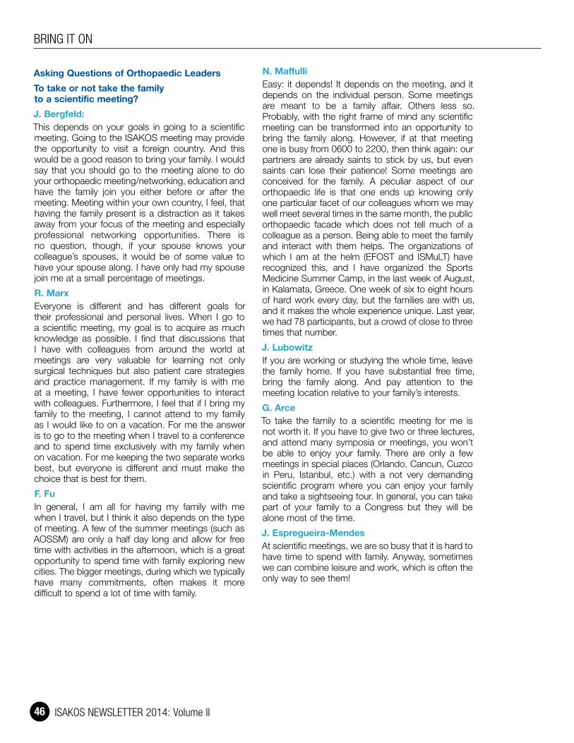
ISAKOS NEWSLETTER 2014: Volume II 46
BRING IT ON
Asking Questions of Orthopaedic Leaders
To take or not take the family to a scientific meeting?
J. Bergfeld: This depends on your goals in going to a scientific meeting. Going to the ISAKOS meeting may provide the opportunity to visit a foreign country. And this would be a good reason to bring your family. I would say that you should go to the meeting alone to do your orthopaedic meeting/networking, education and have the family join you either before or after the meeting. Meeting within your own country, I feel, that having the family present is a distraction as it takes away from your focus of the meeting and especially professional networking opportunities. There is no question, though, if your spouse knows your colleague’s spouses, it would be of some value to have your spouse along. I have only had my spouse join me at a small percentage of meetings.
R. MarxEveryone is different and has different goals for their professional and personal lives. When I go to a scientific meeting, my goal is to acquire as much knowledge as possible. I find that discussions that I have with colleagues from around the world at meetings are very valuable for learning not only surgical techniques but also patient care strategies and practice management. If my family is with me at a meeting, I have fewer opportunities to interact with colleagues. Furthermore, I feel that if I bring my family to the meeting, I cannot attend to my family as I would like to on a vacation. For me the answer is to go to the meeting when I travel to a conference and to spend time exclusively with my family when on vacation. For me keeping the two separate works best, but everyone is different and must make the choice that is best for them.
F. FuIn general, I am all for having my family with me when I travel, but I think it also depends on the type of meeting. A few of the summer meetings (such as AOSSM) are only a half day long and allow for free time with activities in the afternoon, which is a great opportunity to spend time with family exploring new cities. The bigger meetings, during which we typically have many commitments, often makes it more difficult to spend a lot of time with family.
N. MaffulliEasy: it depends! It depends on the meeting, and it depends on the individual person. Some meetings are meant to be a family affair. Others less so. Probably, with the right frame of mind any scientific meeting can be transformed into an opportunity to bring the family along. However, if at that meeting one is busy from 0600 to 2200, then think again: our partners are already saints to stick by us, but even saints can lose their patience! Some meetings are conceived for the family. A peculiar aspect of our orthopaedic life is that one ends up knowing only one particular facet of our colleagues whom we may well meet several times in the same month, the public orthopaedic facade which does not tell much of a colleague as a person. Being able to meet the family and interact with them helps. The organizations of which I am at the helm (EFOST and ISMuLT) have recognized this, and I have organized the Sports Medicine Summer Camp, in the last week of August, in Kalamata, Greece. One week of six to eight hours of hard work every day, but the families are with us, and it makes the whole experience unique. Last year, we had 78 participants, but a crowd of close to three times that number.
J. LubowitzIf you are working or studying the whole time, leave the family home. If you have substantial free time, bring the family along. And pay attention to the meeting location relative to your family’s interests.
G. ArceTo take the family to a scientific meeting for me is not worth it. If you have to give two or three lectures, and attend many symposia or meetings, you won’t be able to enjoy your family. There are only a few meetings in special places (Orlando, Cancun, Cuzco in Peru, Istanbul, etc.) with a not very demanding scientific program where you can enjoy your family and take a sightseeing tour. In general, you can take part of your family to a Congress but they will be alone most of the time.
J. Espregueira-MendesAt scientific meetings, we are so busy that it is hard to have time to spend with family. Anyway, sometimes we can combine leisure and work, which is often the only way to see them!

ISAKOS NEWSLETTER 2014: Volume II 47
BRING IT ON
Asking Questions of Orthopaedic Leaders
What is the importance of academic day in a weekly working schedule?
J. Bergfeld: Academic day again depends on your practice format. If you are a “clinical scientist”, you need this day for your research effort. Trying to squeeze in your nonclinical work between patients, and at the end of the day, just doesn’t work. If you are not involved in clinical science, then you may need a day to update your orthopaedic knowledge or sometimes if you have a hectic practice, a day of just getting rest from physical and mental stress of your orthopaedic practice.
R. MarxAcademic time is very important during the week to accomplish my academic goals including research and teaching. However, time is always at a premium in a busy clinical practice. In addition to working during my academic time as well as evenings and weekends, I find airplane travel a very efficient time for reading and writing orthopedics. In the airport and on the airplane I am generally free of distractions and find I can get a lot done, whether it is personal or professional travel.
F. FuAcademic days are very important. In addition to research, it allows for discussion and teaching time with colleagues, residents and fellows in an informal setting. Most importantly, it provides us with an opportunity to learn and think critically about our work. Every week, our Anatomic ACL Research Group meets for 3 hours and during this time, we review every case for that particular week in detail and learn a lot. We need to hold ourselves and our profession to a high standard, and this provides us with an important mechanism to doing the best we can for our patients.
N. MaffulliI am a clinical academic; I am biased. To me, one day is probably not enough; but, even if you are a jobbing surgeon, I believe that the academic part of your practice will give a breath of freshness to the way in which one interprets Orthopaedics We are still stuck in an world where orthopods are famous for not embracing the science behind our art, and being averse to changing our practice if the scientific evidence produces does not match what we believe to be right. The academic time not only allows to catch up with the latest scientific news in our field, but should permit us to mount little but locally relevant studies to show how good (or not!, as it may be) our practice is. It will help us to mature and change, and to appreciate that at times we are unreasonably stuck in practices that are no longer relevant.
J. LubowitzStress and burn-out result from too many obligations and not enough time, so professional balance requires time set aside for administrative or academic responsibilities, CME, exercise, family, travel and hobbies. For surgeons during their most clinically productive years, it is essential to carve out a half-day for administration and/or academics.
G. ArceQuite important! To stay academic is a key part of our daily job. The academic day is very encouraging for the ones who love our profession. To feel good, keep your mind open, have self confidence in what you are doing and transfer these positive feelings to your patients, is superb for them and for us!
J. Espregueira-MendesA department without an academic day is weak and fails the mission. We are dedicated to treating patients and improve their quality of life, educate and conduct research. An academic day is the time where we can discuss cases and share opinions. We can teach, learn and have a second medical opinion. Today, with an academic day, our residents and faculty thank us. Tomorrow will be our patients!
Asking Questions of Orthopaedic Leaders

ISAKOS NEWSLETTER 2014: Volume II 48
ISAKOS & FIFA Collaborative Course on Football MedicineFebruary 15-16, 2014Sao Paulo, BRAZIL
ISAKOS was pleased to partner with F-MARC and FIFA to host the Football Medicine Conference 2014 in São Paulo, Brazil on 15 and 16 February. This course was the first pre-World Cup medical event, and included more than 500 participants.
Organized by the FIFA Medical Centers of Excellence in Brazil (UNIFESP – USP – Home ), the purpose of the event was to share experiences in various segments of Sports Medicine as it relates to football. The course included professionals in the sports medicine field from around the world, including Singapore, Malaysia, Qatar, South Africa, Portugal, Spain, Belgium, Switzerland, England, France, Cameroon, Argentina, and Paraguay.
The scientific program included speakers from the Confederation of South American Football (CONMEBOL), Brazilian Football Confederation (CBF), and Paulista Football Federation (FPF). International presentations were also given by representatives of FIFA Medical Centers of Excellence such as France, Malaysia, Belgium, Switzerland, and England, among others. We had the opportunity to see the presentations describing the preparations of 12 physicians responsible for each one of the venues of the matches of the World Cup.
Some very well-known physicians participated in the event including Collin Fuller (United Kingdom), Efraim Kramer (South Africa), Gurcharan Singh (Malaysia), Jiri Dvorak (Switzerland), Joao Espregueira-Mendes (Portugal), Michel D’Hooghe (Belgium), Osvaldo Pangrazio (Paraguay), Pieter D’Hooge (Qatar) and ISAKOS First Vice President, Philippe Neyret (France).
José Luiz Runco, Flamengo and the Brazilian national football team doctor, Joaquim Grava, Corinthians doctor, Michel D’Hooge, President of the FIFA Medical Committee, and Jiri Dvorak, FIFA Medican Director, were honored during the event with the Golden Ball of Soccer Medicine.
APPROVED COURSE REPORTS
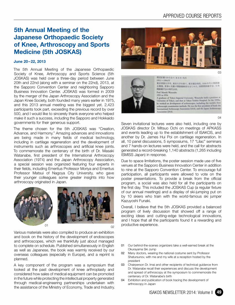
ISAKOS NEWSLETTER 2014: Volume II 49
APPROVED COURSE REPORTS
5th Annual Meeting of the Japanese Orthopaedic Society of Knee, Arthroscopy and Sports Medicine (5th JOSKAS)June 20 – 22, 2013
The 5th Annual Meeting of the Japanese Orthopaedic Society of Knee, Arthroscopy and Sports Science (5th JOSKAS) was held over a three-day period between June 20th and 22nd (along with a seminar on the 22nd), 2013, at the Sapporo Convention Center and neighboring Sapporo Business Innovation Center. JOSKAS was formed in 2009 by the merger of the Japan Arthroscopy Association and the Japan Knee Society, both founded many years earlier in 1975, and this 2013 annual meeting was the biggest yet. 2,423 participants took part, exceeding the previous record by over 500, and I would like to sincerely thank everyone who helped make it such a success, including the Sapporo and Hokkaido governments for their generous support.
The theme chosen for the 5th JOSKAS was “Creation, Advance, and Harmony.” Amazing advances and innovations are being made in many fields of medical technology, including in cartilage regeneration and the development of instruments such as arthroscopes and artificial knee joints. To commemorate the centenary of the birth of Dr. Masaki Watanabe, first president of the International Arthroscopy Association (1974) and the Japan Arthroscopy Association, a special session was organized featuring four experts in their fields, including Emeritus Professor Moriya and Emeritus Professor Matsui of Nagoya City University, who gave their younger colleagues some greater insights into how arthroscopy originated in Japan.
Various materials were also compiled to produce an exhibition and book on the history of the development of endoscopes and arthroscopes, which we thankfully just about managed to complete on schedule. Published simultaneously in English as well as Japanese, the book was warmly received by our overseas colleagues (especially in Europe), and a reprint is planned.
A key component of the program was a symposium that looked at the past development of knee arthroplasty and considered how sales of medical equipment can be promoted in the future while protecting the intellectual property generated through medical-engineering partnerships undertaken with the assistance of the Ministry of Economy, Trade and Industry.
Seven invitational lectures were also held, including one by JOSKAS director Dr. Mitsuo Ochi on meetings of APKASS and events leading up to the establishment of ISAKOS, and another by Dr. James Hui Poi on cartilage regeneration. In all, 10 panel discussions, 5 symposiums, 17 “Lilac” seminars and 7 hands-on lectures were held, and the call for abstracts generated a record-breaking 1,140 abstracts (1,265 including ISMISS Japan) in response.
Due to space limitations, the poster session made use of five venues at the Sapporo Business Innovation Center in addition to nine at the Sapporo Convention Center. To encourage full participation, all participants were allowed to vote on the poster presentations. To provide a break from the official program, a social was also held for all the participants on the first day. This included the JOSKAS Cup (a regular fixture of our annual meetings) and a display of ski-jumping put on by 10 skiers who train with the world-famous ski jumper Kazuyoshi Funaki.
Overall, I believe that the 5th JOSKAS provided a balanced program of lively discussion that showed off a range of exciting ideas and cutting-edge technological innovations, and I hope that all the participants found it a rewarding and productive experience.
01 Our behind-the-scenes organizers take a well-earned break at the Okurayama Ski Jump
02 Many doctors, wearing the national costume sent by Professor Shatursunov, with me and my wife at a reception hosted by the president
03 Chairperson Dr. Imai and other recipients of technical guidance from Dr. Watanabe recall their experiences and discuss the development and spread of arthroscopy at the symposium to commemorate the centenary of Dr. Watanabe’s birth
04 Exhibition and publication of book tracing the development of arthroscopy in Japan
03
04
01 02

ISAKOS NEWSLETTER 2014: Volume II 50
APPROVED COURSE REPORTS
Report for the 4th Pune Knee CourseApril 26-27, 2014
The 4th Pune Knee Course was held on 26th and 27th April, 2014 at Hotel J W Marriott, SB Road, Pune. The event was a great success with 501 orthopaedic surgeons as delegates, three International and 16 National faculties. The format of the meeting was that of Live Surgeries, Panel Discussions and Patient Evaluation sessions.
The international faculties were:
1. Prof. Dr. Freddie Fu, Pittsburgh, USA2. Dr. Jonathan Herald, Sydney, AUSTRALIA3. Dr. Lieve Vanden Berghe, BELGIUM
The Indian Faculties were:
The opening day started with Dr. Jonathan Herald demonstrating a single bundle anatomic ACL Reconstruction surgery. There was two way audio-video communication and a lot of discussion went on during the surgical procedure. This was followed by a video technique session on graft harvesting techniques and later there was a panel discussion on ACL injuries. In the panel discussions, cases were presented and the panelists were invited to give their opinions on the same. Lot of discussions took place with the delegates asking questions from the floor as well.
Prof. Dr. Freddie Fu examined a patient with an ACL rupture on stage and discussed the line of management and reasoning for decision making. This was something new for the delegates and they all felt this was something very useful to them in their clinical practice.
There was an interesting session on management of cartilage injuries conducted by Dr. Deepak Goyal – President of Indian Cartilage Society and member of the Asia Pacific Cartilage Society. This was followed by a live surgical demonstration of ACI implantation surgery using gel based technique.
The post lunch session covered multi ligament injuries in great detail. There was a live surgical demonstration on PCL Reconstruction by Dr. Anant Joshi. The course chairman, Dr. Sachin Tapasvi demonstrated a combined anatomic BTB ACL Reconstruction + PLC reconstruction surgery as well as a combined anatomic MCL reconstruction + ACL augmentation surgery. There were panel discussions and patient examination sessions highlighting multi ligament injuries as well.
The delegates were absolutely glued to their seats and it was indeed heartening to see that even at 6.30 in the evening the hall was completely packed and the delegates were extremely attentive.
The course dinner was held at the same venue. It was indeed an excellent gathering for all the delegates since they had a unique opportunity to interact with the faculty members informally.
26th April started off with a big bang with Prof. Freddie Fu doing a live surgical demonstration of anatomic Double Bundle ACL Reconstruction at 8.00 in the morning and the venue hall was packed to the capacity to watch the master.
1. Dr. David Rajan2. Dr. Anant Joshi3. Dr. Abhay Narvekar4. Dr. Clement Joseph5. Dr. Dinshaw Pardiwala6. Dr. Parag Sancheti7. Dr. Deepak Goyal8. Dr. Arumugam
9. Dr. Sundararajan10. Dr. Pankaj Amite11. Dr. Vivek Pandey12. Dr. Anupama Patil13. Dr. Roshan Wade14. Dr. Sachin Tapasvi15. Dr. Nilesh Kamat16. Dr. Shirish Pathak

ISAKOS NEWSLETTER 2014: Volume II 51
Prof. Dr. Fu explained to the delegates in great detail about the anatomical aspects of ACL reconstruction during the Live Surgery. This was followed by Dr. Clement Joseph doing a live surgical demonstration of MPFL reconstruction for recurrent patellar instability.
Failed ACL Reconstruction is an emerging problem in this present decade. This was adequately tackled through panel discussions and Prof. Dr. Freddie Fu also examined a patient with failed ACL and discussed the role of osteotomies for coronal and sagital plane correction, tackling bone tunnel defects and graft choice options in such situations.
The highlight of this meeting was the thought provoking oration that was delivered by Dr. Freddie Fu. It was titled “My Journey of 30 Years in ACL Reconstruction Surgery.” It was fascinating to hear Dr. Freddie Fu speak about how the ACL technique has evolved over the past three decades. It also gave a lot of insight into the amazing amount of research that has taken place in this field. Prof. Dr. Fu concluded his oration by giving us an insight into the future of ACL Reconstruction. The oration was chaired by Dr. Naresh Goyal, Vice-President of Indian Orthopaedic Association, Dr. J. Talesara, President-Poona Orthopaedic Society and Dr. Parag Sancheti, Secretary-Asia Pacific Knee Society.
Prof. Dr. Fu was felicitated in the traditional manner by honouring him with a “Puneri Pagdi”, ‘Shawl” and a Citation. He was given a standing ovation by over 500 delegates.
The latter half of the session tackled the different problems of patella-femoral instability via patient evaluation sessions and panel discussions. The grand finale was a Live Surgical demonstrationof a Revision ACL Reconstruction with Micro fracture with Open Wedge High Tibial Osteotomy, performed by Dr. Dinshaw Pardiwala.
The whole event was an outstanding success and the delegates as well as the faculty were all praises for the course for the course content, academic quality, excellent audiovisual presentation and fine arrangements and hospitality.
The organizers of the Pune Knee Course wish to say a BIG THANK YOU to the Indian Arthroscopy Society, ISAKOS, Maharashtra Orthopaedic Association & the Poona Orthopaedic Society for their support our program.
Severance Arthroscopy Fresh Cadaver WorkshopsApril 2014The 57th knee and 58th shoulder cadaver workshops were held at Surgical Anatomy Education Center of Severance Hospital, Yonsei University Health System, Seoul on 5th and 13th April respectively.
The workshop that is one of the traditional arthroscopy cadaver workshops, have been held at Yonsei University Health System in Seoul, Korea for 58 times since July 7th, 2001, and have been ISAKOS Approved Courses since 11th workshop on November 1st, 2003.
Lecture for anatomic structure and portals preceded the demonstration and practice. The highlight of the program was specialist knee arthroscopy surgeons were selected as instructors for knee workshop and specialist shoulder arthroscopy surgeons were selected for the shoulder workshop. The emphasis was to have the “hands on” experience for the candidates after observing the demonstration by Prof. Sung-Jae Kim and instructors (Prof. Nam-Hong Choi, Prof. Sung-Hwan Kim, Prof. Yon-Sik Yoo and Prof. Sang-Jin Shin). Because fresh cadaver offered participants operating field like a routine operating field, participants were able to train themselves for various operative techniques under the special guidance of instructors.
Additionally ankle and elbow arthroscopy were also demonstrated by Prof. Sung-Jae Kim.
The workshops were very useful educational programs for the participants interested in the shoulder and knee arthroscopy. Each participant would get the one step forward in arthroscopic experience and capacity through these workshops.
Course Chairman:Prof. Sung-Jae Kim MD, PhD.
Surgical Anatomy Education Center of Severance Hospital
APPROVED COURSE REPORTS
Lab Instructors:57th Knee workshop
Sung-Jae Kim, MDNam-Hong Choi, MDShin-Kang Cho, MDJong-Min Kim, MDYoung-Jin Seo, MDSung-Hwan Kim, MDDae-Heup Song, MDJae-Hun Jeong, MD
58th Shoulder workshop
Sung-Jae Kim, MDYon-Sik Yoo, MDSang-Jin Shin, MDJong-Hun Ji, MDHyoung-Sik Kim, MDSang-Wook Ryu, MDDoo-Hyung Lee, MDYong-Min Chun, MD

ISAKOS NEWSLETTER 2014: Volume II 52
Advanced Instructional Course on Arthroscopy of Shoulder, Elbow, Wrist and Knee; Arthroplasty of Shoulder and ElbowUniversity Medical Centre UtrechtUtrecht, NETHERLANDS June 30 – July 4, 2014Chair(s): Dr. W. Jaap WillemsFor further information, please contact:
Arthroscopy and Arthroplasty 2014Tel: +31-30-276 9174Fax: +31-30-276 9251www.shoulder-elbow-knee.nl
The ICRS Focus Meeting – The KneeFIFA Auditorium SonnenbergZurich, SWITZERLAND July 3 – 4, 2014Chair(s): Christoph ErggeletFor further information, please contact:
Lei JiTel: +41-44 503 73 73Fax: +41-44 503 73 72www.cartilage.org/index.php?pid=203
Vail International Complex Knee Symposium (VICKS)Vail Cascade-Vail, ColoradoUSA July 17 –19, 2014Chair(s): Robert F. LaPrade MD, PhDFor further information, please contact:
Robert F. LaPrade MD, PhDTel: 970-479-5881Fax: 970-479-5813www.drlaprade.com
2014 The Ligament Reconstruction Seminar & Live Surgery in SapporoFaculty House “Trillium (Enreiso)” in Hokkaido UniversitySapporo, JAPAN July 31–August 1, 2014Chair(s): Kazunori YasudaFor further information, please contact:
Yasuyuki KawaguchiTel: +81-11-706-7211Fax: +81-11-706-7822hokudai-med-sports.org/en/index.html
1st Iranian Live Arthroscopic FestivalAkhtar HospitalTehran, IRAN August 14 –15, 2014Chair(s): Dr. Sohrab KeyhaniFor further information, please contact:
Raheleh KalantariTel: +98-214-4484274Fax: +98-214-4484299www.iranianlivesurgery2014.com
ISKSAA 2014 (Knee / Shoulder Arthroscopy & Arthroplasty)The Leela Ambience Hotel, Gurgaon, New DelhiNew Delhi, INDIA September 4 –7, 2014Chair(s): Dr. Pushpinder Singh BajajFor further information, please contact:
Dr. Pushpinder Singh BajajTel: +91-98-11056525Fax: +91-11-41057558www.isksaa.com/isksaa2014/
Orthopedic Surgery Controversies 2014The Surf and Sand HotelLaguna Beach, CA USA September 11–13, 2014Chair(s): Wesley M. Nottage, MDFor further information, please contact:
Kathryn SnellTel: 657-218-8432Fax: 949-581-6687www.orthopedicsurgerycontroversies.net
Knee Dislocation and Multi-Lligament Knee ReconstructionMayo ClinicRochester, MNUSA September 18 –19, 2014Chair(s): Bruce LevyFor further information, please contact:
Jenna PedersonTel: 800-323-2688Fax: 507-538-7234www.mayo.edu/cme
Birmingham Patellofemoral Master Class 2014Queen Elizabeth University Hospital, Birmingham UKBurmingham UK, UNITED KINGDOM September 19 –20, 2014Chair(s): S. Tanweer AshrafFor further information, please contact:
Tanweer Ashraf, David PenfordTel: +44-1476-860759Fax: +44-1476-860759www.birminghampatfem.org
12th Congress of Turkish Society of Sports Traumatology Arthroscopy and Knee Surgery (TUSYAD 2014 Congress)Kaya Convention CenterIzmir, TURKEY September 23 –27, 2014Chair(s): Halit Pinar, MDFor further information, please contact:
Emin Taskiran, MDTel: +90-532-416-55-46Fax: +90-232-463-95-15www.tusyad.org
19th International Shoulder Course in MunichUniversity Hospital Klinikum rechts der IsarMunich, GERMANY September 29 – October 1, 2014Chair(s): Prof. Dr. Andreas B. ImhoffFor further information, please contact:
Christian HolweinTel: +49-89-4140-7821Fax: +49-89-4140-7841www.sportortho.med.tu-muenchen.de/home/
13th Indian Arthroscopy Society CongressHotel Taj KrishnaHyderabad, INDIA October 10 –12, 2014Chair(s): Dr. Raghuveer Reddy KFor further information, please contact:
Dr. Raghuveer Reddy KTel: +91-98-4902-9934Fax: +91-40-2335-0696www.ias2014.org
16èmes Journées Lyonnaises de Chirurgie du Genou – The Young Arthritic KneeConvention CenterLyon, FRANCE October 16 –18, 2014Chair(s): Michel Bonnin, MDFor further information, please contact:
MCO CongrèsTel: +33-495-093800Fax: +33-495-093801www.lyon-genou.com
6th International Hip Arthroscopy MeetingAlte KongresshalleMunich, GERMANY November 21–22, 2014Chair(s): Michael Dienst MD, Richard Villar FRCSFor further information, please contact:
Stefanie MattTel: +49-761 69699-243Fax: +49-89-206082-333www.hipmeeting.de
Orthopaedic Summit 2014: Evolving TechniquesMGM Grand HotelLas Vegas, NVUSA December 4 – 6, 2014Chair(s): Kevin Plancher, MDFor further information, please contact:
Ortho Summit Course OfficeTel: 925-807-1190Fax: 925-807-1199www.orthosummit.com
XXIV International Conference on Sports Rehabilitation and Traumatology – Football Medicine Strategies for Player CareQueen Elizabeth II Conference Centre – WembleyLondon, UNITED KINGDOM April 11– 13, 2015Chair(s): Peter Brukner, Stefano Della VillaFor further information, please contact:
Giulia IndelicatoTel: +39-0512-986878Fax: +39-0511-9902200www.FootballMedicineStrategies.com
UPCOMING ISAKOS APPROVED COURSES
Is your Course an…
Approved-CourseISAKOSISAKOS
www.isakos.com/meetings

Smith & Nephew, Inc.150 Minuteman RoadAndover, MA 01810 USA
www.smith-nephew.comT +1 978 749 1000 US Customer Service: +1 800 343 5717International Customer Service: +1 978 749 1140
™ Trademark of Smith & Nephew. Registered US Patent and Trademark Office. All Trademarks acknowledged ©2014 Smith & Nephew. All rights reserved. Printed in USA. 00348 V2 03/14
ENDOBUTTON™ CL ULTRAFixation Devices
CLANCYTM ANATOMIC CRUCIATE GUIDEFlexible Drill System
ACUFEX™ PINPOINT Anatomic ACL Guide System
BIOSURE™ SYNCTibial Fixation Device
Helping to put patients back together again.Nobody knows anatomic ACL reconstruction like Smith & Nephew. We’ve been restoring the ACL using anatomic techniques from the very beginning. So whether your reconstruction preference is soft tissue or bone-tendon-bone, using an inside-out or outside-in technique, we have everything you need – including the product training to make it your own. Smith & Nephew. The Anatomic ACL Company.
Learn more at smith-nephew.com/anatomicACL
KNE14_00348_V2_AnaACL_ISAKOSNewsltr_8.5x11_F.indd 1 7/21/14 4:37 PM

International Society of Arthroscopy, Knee Surgery and Orthopaedic Sports Medicine2410 Camino Ramon, Suite 215San Ramon, CA 94583 – 4318 USA
Telephone: +1 925 807 – 1197Fax: +1 925 807 – [email protected]
JoinISAKOSJoin
ISAKOSJoin
ISAKOSJoin
ISAKOS
To be considered for an Award or Fellowship, abstracts and award applications must be submitted before September 1st, 2014.
The ISAKOS Fellowship and Awards Program is committed to recognizing and honoring researchers whose work has contributed to better understanding and communication within the fields of Arthroscopy, Knee Surgery and Orthopaedic Sports Medicine.
Presenting authors are allowed one submission per award. Additionally, each abstract may only be applied to one award.
Award Applications for the 2015 Congress are due!
JUNE 7–11, 2015
isAkoscongress
10TH Biennial
2015LYON, FRANCEcall for
abstractsView the Interactive Agenda
DEADLINE: September 1, 2014 www.isakos.com /2015congress