Volume 9 Number 1 7 January 2018 Pages 1–268 Chemical …
9
Chemical Science ISSN 2041-6539 rsc.li/chemical-science Volume 9 Number 1 7 January 2018 Pages 1–268 EDGE ARTICLE Xinjing Tang et al. Caged circular siRNAs for photomodulation of gene expression in cells and mice
Transcript of Volume 9 Number 1 7 January 2018 Pages 1–268 Chemical …

ChemicalScience
ISSN 2041-6539
rsc.li/chemical-science
Volume 9 Number 1 7 January 2018 Pages 1–268
EDGE ARTICLEXinjing Tang et al.Caged circular siRNAs for photomodulation of gene expression in cells and mice
ChemicalScience
EDGE ARTICLE
Ope
n A
cces
s A
rtic
le. P
ublis
hed
on 1
9 O
ctob
er 2
017.
Dow
nloa
ded
on 1
2/8/
2021
4:0
9:27
PM
. T
his
artic
le is
lice
nsed
und
er a
Cre
ativ
e C
omm
ons
Attr
ibut
ion-
Non
Com
mer
cial
3.0
Unp
orte
d L
icen
ce.
View Article OnlineView Journal | View Issue
Caged circular siR
aState Key Laboratory of Natural and Biom
Sciences, Peking University, No. 38, Xueyu
[email protected] and Health Analytical Center, Pekin
100191, China
† Electronic supplementary informa10.1039/c7sc03842a
‡ These authors contributed equally to th
Cite this: Chem. Sci., 2018, 9, 44
Received 2nd September 2017Accepted 18th October 2017
DOI: 10.1039/c7sc03842a
rsc.li/chemical-science
44 | Chem. Sci., 2018, 9, 44–51
NAs for photomodulation of geneexpression in cells and mice†
Liangliang Zhang,‡a Duanwei Liang,‡a YuanWang,‡a Dong Li,‡a Jinhao Zhang,a Li Wu,a
Mengke Feng,a Fan Yi,a Luzheng Xu,b Liandi Lei,b Quan Dua and XinJing Tang *a
By means of RNA interference (RNAi), small interfering RNAs (siRNAs) play important roles in gene function
study and drug development. Recently, photolabile siRNAs were developed to elucidate the process of
gene silencing in terms of space, time and degree through chemical modification of siRNAs. We report
herein a novel type of photolabile siRNA that was synthesized through cyclizing two ends of a single
stranded RNA with a photocleavable linker. These circular siRNAs became more resistant to serum
degradation. Using reporter assays of firefly/Renilla luciferase and GFP/RFP, the gene silencing activities
of caged circular siRNAs for both genes were evaluated in HEK293 cells. The results indicated that the
target genes were successfully photomodulated using these caged circular siRNAs that were formed by
caged circular antisense guide RNAs and their linear complementary sense RNAs. Using the caged
circular siRNA targeting GFP, we also successfully achieved photomodulation of GFP expression in mice.
Upon further optimization, this new type of caged circular siRNA is expected to be a promising tool for
studying gene therapy.
Introduction
RNA interference is a potent and specic gene silencingapproach1,2 which usually requires short double-stranded RNAand results in RISC formation and Dicer processing.3,4 Gener-ally, two strategies are used to produce double-stranded RNA:(1) precursor shRNAs are transcribed endogenously from plas-mids into shRNAs by RNA polymerases, followed by Dicer pro-cessing to produce active siRNAs within cells; (2) chemicallysynthetic siRNAs are formed by two complementary single-stranded RNAs. Although shRNA is more biologically relevant,synthetic siRNAs are currently still the most widely used forboth laboratory and clinical applications.5–9
Despite the potency and specicity of siRNAs in genesilencing, spatial and/or temporal regulation of siRNA activity isstill difficult due to the constitutive transcription of shRNAs ordelivery obstacles of siRNAs. Similar to other reported photo-labile oligonucleotides,10–29 photolabile siRNAs have beendeveloped to achieve dedicated regulation of gene expres-sion.30–43 To date several approaches have been developed basedon the unique structural properties of siRNAs and the
imetic Drugs, School of Pharmaceutical
an Rd, Beijing 100191, China. E-mail:
g University, No. 38, Xueyuan Rd, Beijing
tion (ESI) available. See DOI:
is work.
processing of RNAi (Fig. 1). Disturbing a siRNA duplex confor-mation might interfere with the formation of siRNA/RISCcomplexes and/or further siRNA processing. Heckel et al.incorporated a 2-(2-nitrophenyl)propyl (NPP)-caged deoxy-guanosine nucleotide in a siRNA antisense guide strand at the9th to the 11th position. This modication approach didinterfere with the formation of the siRNA/RISC complex,leading to compromised siRNA activity.37 In another study,Deiters et al. synthesized 6-nitropiperonyloxymethyl (NPOM)-caged guanosine and uridine phosphoramidites and site-specically incorporated these caged nucleosides into theantisense guide strand of siRNAs at the cutting site and/or seed
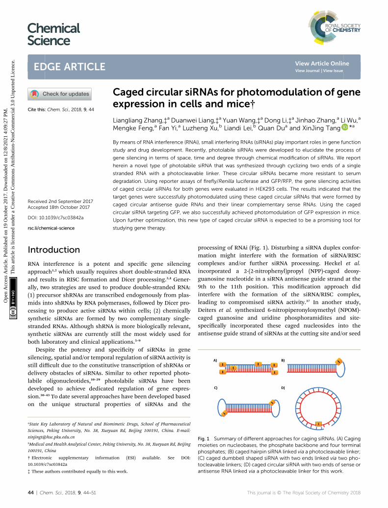
Fig. 1 Summary of different approaches for caging siRNAs. (A) Cagingmoieties on nucleobases, the phosphate backbone and four terminalphosphates; (B) caged hairpin siRNA linked via a photocleavable linker;(C) caged dumbbell shaped siRNA with two ends linked via two pho-tocleavable linkers; (D) caged circular siRNA with two ends of sense orantisense RNA linked via a photocleavable linker for this work.
This journal is © The Royal Society of Chemistry 2018

Table 1 The sequences of oligonucleotides used in the study and thelinker structure
Edge Article Chemical Science
Ope
n A
cces
s A
rtic
le. P
ublis
hed
on 1
9 O
ctob
er 2
017.
Dow
nloa
ded
on 1
2/8/
2021
4:0
9:27
PM
. T
his
artic
le is
lice
nsed
und
er a
Cre
ativ
e C
omm
ons
Attr
ibut
ion-
Non
Com
mer
cial
3.0
Unp
orte
d L
icen
ce.
View Article Online
region.30 Stochastic labelling of the phosphate backbone ofsiRNA duplexes is another approach for the synthesis of pho-tolabile siRNAs through coupling chemistry of the photolabilediazo moiety with phosphate groups.
Friedman et al. reported DNMPE-caged double-strandedsiRNAs to photomodulate the silencing of GFP expressionwithout interrupting RFP expression in the cells.31–33 Usinga similar approach, 20-F substituted siRNAs were caged andtheir RNAi activities on transient GFP repression were photo-modulated in zebrash embryos with spatial resolution.41 Thenumber of caging groups per siRNA increased with higherDMNPE diazo concentration. Heavily caged siRNA led to theelimination of its gene silencing activity, but such repressioncould not be fully recovered with cell-affordable light irradia-tion.33,37,41 By attaching a large caging group (cyclododecyl 4,5-dimethoxy-2-nitrophenyethyl moiety) at all four terminal phos-phate groups, caged sdRNAs were developed for preventing RNAprocessing and further gene silencing activity.34 Site-speciclabelling of the terminal phosphate group was also achievedthrough the attachment of DMNPE or a biotinylated photo-cleavable linker.36 However, this modication approach ispartially tolerated due to the intact 50-phosphoester moiety.Previously in our lab, we caged each phosphate group of siRNAthrough site-specic incorporation of a photolabile nucleotidephosphoramidite and screened all possible caging phosphatepositions for efficient photomodulation of siRNA activity.44 Werecently reported caged siRNA modied with a single vitamin Eat the 50 terminal phosphate of antisense RNA and successfullyachieved the photomodulation of RNAi-induced gene silencing,possibly due to the binding of vitamin E receptor protein.45
Another kind of caged siRNA was developed by linking anantisense guide strand with a complementary sense strand RNAvia a photocleavable linker. Unfortunately, no improvement ofthe photomodulation of siRNA activity was observed.36
Based on our and others’ previous achievements on cagedcircular antisense oligonucleotides,19–21,46 we further intendedto develop a new generation of caged siRNAs with a circularstructure. In the literature, usually sense and antisense strandsare cyclized to form siRNAs or shRNAs with a dumbbell struc-ture.40,47,48 Xi et al. reported circular dumbbell sdRNAs with analkyl linkage. They found that this was more potent in RNAigene silencing than their open-ended counterpart, likely due totheir enhanced stability.48 Dmochowski et al. also reporteda caged circular siRNA duplex with the dumbbell structure,where two photocleavable linkers were used to link the 50 and 30
ends of the sense and antisense siRNA strands (Fig. 1C).40
Different from previously reported circular RNAs, we proposedto develop a novel type of caged circular siRNA (Fig. 1D).Cyclization of the 50 and 30 ends of the sense or antisense single-stranded RNA was achieved to form a 21-nucleobase ringthrough both chemical and enzymatic cyclization methods,then a complementary RNA strand was hybridized to the cagedcircular RNA strand to form a caged circular siRNA duplex(Table 1). Upon exposure to light, the circular siRNAs wererestored to the corresponding linear ones with a 50 terminalphosphate group of RNA oligonucleotides. Using a dual lucif-erase assay49 and GFP expression assay, photoregulation of
This journal is © The Royal Society of Chemistry 2018
rey luciferase reporter gene expression and GFP expressionwas achieved by this new type of caged circular siRNA. Furtherin vivo studies conrmed that it was possible to achieve pho-tomodulation of siRNA activity in a mouse tumor model usingthe caged circular siRNA.
ExperimentalGeneral experimental procedures
All chemical reactions in the synthesis of the photocleavablelinker were performed under an inert atmosphere using dryreagents and solvents. Column chromatography was performedwith silica gel 60 (200–300 mesh). 1H-NMR (400 MHz) and 13C-NMR (100 MHz) spectra were taken on Bruker AVANCE III-400spectrometers and standardized to the solvent NMR peak.Mass spectra of small molecules were obtained on a WatersXevo TQD Mass Spectrometer using electrospray ionization(ESI). The reactions were conducted in a dark room whennecessary.
Purication of caged circular RNAs
Crude caged circular RNAs obtained from chemical synthesis orenzymatic synthesis were dissolved in PBS buffer and mixedwith RNA loading buffer (0.25% bromophenol blue, 30% glyc-erol in DEPC-treated water). The solutions (6 mL per well) wereloaded to 20% native PAGE (8.6 � 6.8 cm and 1 mm thick) gels.The gels were then electrophoresed at 80 V for 3 h, using Tris–borate–EDTA (TBE) buffer (pH 8.2). For each preparative gel, thetwo side lanes of the gel were cut and stained with SYBR Gold(Invitrogen) for gel imaging. The images were printed outaccording to the size of the gel in order to mark the location of
Chem. Sci., 2018, 9, 44–51 | 45
Chemical Science Edge Article
Ope
n A
cces
s A
rtic
le. P
ublis
hed
on 1
9 O
ctob
er 2
017.
Dow
nloa
ded
on 1
2/8/
2021
4:0
9:27
PM
. T
his
artic
le is
lice
nsed
und
er a
Cre
ativ
e C
omm
ons
Attr
ibut
ion-
Non
Com
mer
cial
3.0
Unp
orte
d L
icen
ce.
View Article Online
the circular RNA without SYBR Gold stain. The gel at themarked location was cut, crumbled into tiny particles andimmersed into TBE buffer at 37 �C overnight. Aer removingthe solid gel particles through ltration, RNA solutions weredesalted and concentrated using Millipore-amicon ultra-0.5 mLcentrifugal lters (cutoff ¼ 3000). The collected products werefreeze-dried to the nal caged circular single-stranded RNA.
The caged circular single-stranded RNA was dissolved in PBSbuffer to make a 20 mmol L�1 stock solution and was thenmixedwith an equal amount of the linear complementary RNA to formthe caged circular siRNA duplex. This duplex was annealed byheating to 80 �C for 5 minutes and subsequent cooling to roomtemperature for at least 1 hour for further use. Tms of linear andcircular siRNAs were measured with a DU800 UV-Vis spec-trometer with 1 mM concentration of the correspondingduplexes.
Characterization of RNA oligonucleotides
Single-stranded oligonucleotides (�0.2 nmol) were dissolved inwater/acetonitrile (50 : 50, 20 mL) containing 1% triethylamineto make a nal concentration of 10 mM. The solutions were thenanalyzed with a Waters Xevo G2 Q-Tof spectrometer with elec-trospray ionization (ESI) in the negative ion mode.
The double-stranded siRNA was characterized with MALDI-TOF-MS. The matrix solution was made by dissolving 2,5-dihy-droxybenzoic acid (DHB, 10 mg) in 50% acetonitrile/watersolution (1 mL). The sample solution (10 mM, 0.8 mL) and thematrix solution (0.8 mL) were mixed and spotted on the surfaceof the sample plate. Aer the sample was dried, siRNA was thenanalysed with MALDI-TOF-MS according to the manufacturer’sprotocol.
Photocleavage ability of caged circular single-stranded RNA
Caged circular single-stranded RNA was diluted with PBS buffer(pH 7.2) to make a nal concentration of 2 mM (30 mL). Thesolution (5 mL per well) was irradiated using a UV-LED lamp(365 nm, 7 mW cm�2) from the bottom of the plate for 0, 1, 2, 3,4, 5 and 6 minutes. Aer mixing with 1 mL RNA loading buffer,the samples were subject to gel analysis for photocleavage studyusing 20% native PAGE under similar gel running conditions ofRNA purication.
Serum stability of a caged circular siRNA
Caged circular siRNA or control linear siRNA (10 mM, 2 mL) wasincubated at 37 �C in enzyme buffer solution to make a nalconcentration of 2 mM (20 mL). 30% fetal bovine serum (FBS) wasused for studying siRNA stability. 5 mL of solution was aliquotedat different time points, and immediately frozen in liquidnitrogen and then stored at �80 �C until assaying. 1 mL of 6�RNA loading buffer was added to the aliquots. The samples wererun on 20% native polyacrylamide gels in 1� TBE bufferaccording to the procedure mentioned before.
46 | Chem. Sci., 2018, 9, 44–51
Cell culture and RNAi assay (luciferase)
HEK293 cells were grown in Dulbecco’s modied Eagle’smedium supplemented with 10% fetal bovine serum, 2 mM L-glutamine, 100 units per mL penicillin and 100 mg mL�1
streptomycin (Life Technologies, Gibco). The cells were seededinto 24-well plates at �1 � 105 cells per well one day beforetransfection (lipofectamine 2000). siQuant vectors (100 ng perwell) carrying the target site of the siRNA were transfected intoHEK293 cells at �50% conuence, together with the pRL-TKcontrol vector (50 ng per well), with or without the controlsiRNAs or caged circular siRNAs (1 or 5 nM). The activities ofboth luciferases were determined by a Synergy HT uorometer(BioTek, USA) and then rey luciferase activity was normalizedto Renilla luciferase for each well. The silencing efficacy of eachsiRNA was calculated by comparison with samples transfectedwith only vectors without siRNA treatment. For light-irradiatedcellular experiments, aer transfection, the cells were culturedfor another 4 h, washed with PBS, and then irradiated usinga UV-LED lamp (365 nm, 7 mW cm�2) from the bottom of theplate for 3 minutes.
Cell culture and RNAi assay (GFP expression assay)
HEK293 cells were trypsinized and seeded in 12 well plates with1 mL 2� 105 cells mL�1 for each well. According to the standardtransfection protocol, the reporter plasmids (pEGFP-N1,pDsReds-N2) and corresponding siRNAs were cotransfected tocells using Lipofectamine 2000 in optiMEM. Aer 4 h, cells inone set of experiments were irradiated to induce uncaging ofphotolabile circular siRNAs. Then all the cells were subject totheir mediums being replaced with 1 mL fresh DMEM con-taining 10% FBS for each well. Aer another 20 h culture in theincubator, the images of each well were collected by automaticinverted uorescence microscopy (Olympus, IX83) under thesame condition. The excitation and emission wavelengths are488 nm and 509 nm for GFP and 560 nm and 585 nm for RFP,respectively.
Aer plasmid and siRNA transfection and incubation underthe same condition, the transfected cells were washed using 1�PBS buffer (pH 7.2) and trypsinized, and their uorescenceintensity was further quantied by ow cytometry (BD FACSAriaII) with RFP gene expression as an internal control. All experi-ments were repeated at least three times.
Half-well patterning experiment
HEK293 cells were trypsinized and then seeded in 6 well plateswith 2 mL 2 � 105 cells mL�1 for each well. Aer the cells werecotransfected with GFP (400 ng per well) and RFP (400 ng perwell) plasmids as well as SG/c-AG (10 nM) for 4 h, black adhesivetape was aligned to cover half of each well to avoid light irra-diation. Then the wells were irradiated for 3 minutes and themedium was replaced with 2 mL fresh DMEM containing 10%FBS. Aer incubation for another 20 h, the images were taken byautomatic inverted uorescence microscopy and were analyzedusing Volocity Demo 6.1.1.
This journal is © The Royal Society of Chemistry 2018
Edge Article Chemical Science
Ope
n A
cces
s A
rtic
le. P
ublis
hed
on 1
9 O
ctob
er 2
017.
Dow
nloa
ded
on 1
2/8/
2021
4:0
9:27
PM
. T
his
artic
le is
lice
nsed
und
er a
Cre
ativ
e C
omm
ons
Attr
ibut
ion-
Non
Com
mer
cial
3.0
Unp
orte
d L
icen
ce.
View Article Online
Animal experiment
Six-week-old BALB/c nude mice (Department of LaboratoryAnimal Science of the Peking University Health Science, Beijing,China) were subcutaneously injected in the inner le and rightthighs with U87-GFP cells (6 � 105 cells per site in a volume of60 mL). When the tumors grew to around 0.2 cm in diameter(about 4 days), the mice were randomly divided into two groupsfor intratumoral injection. One group was injected with PBS onthe le and with linear siRNAs (SG/AG) on the right side,respectively. Another group was injected with caged circularsiRNAs (SG/c-AG) on both sides. For each injection, 3 nmollinear siRNAs (SG/AG) or caged circular siRNAs (SG/c-AG) wasmixed with 20 mL transfection reagent (Entranster-in vivo;Engreen, Beijing, China) in vivo and then incubated for 15minutes at room temperature. The volume of each injection was60 mL. All injections were in accordance with themanufacturer’sinstructions. 4 h aer injection, the mice injected with thecaged circular siRNA were xed on a table and their le tumorswere irradiated using a UV-LED lamp (365 nm, 7 mW cm�2) for3 minutes, whereas other parts of the body were covered bytinfoil to avoid light exposure. The irradiated mice were thenmarked and returned to live with other mice in the group. Thesemice were then imaged at different time points (0 h, 12 h, 24 hand 48 h) using Maestro Automated In Vivo Imaging system.Then tumor uorescence was quantied under the sameparameters, and the uorescence intensity of tumors atdifferent time points was normalized to that of each mouse atjust before the siRNA injection. The facility is in keeping withnational standard “Laboratory Animal—Requirements of Envi-ronment and Housing Facilities” (GB 14925—2001). The care oflaboratory animals and animal experimental operation conformto “Beijing Administration Rule of Laboratory Animal” and wasapproved by the Animal Care and Use Committee of PekingUniversity.
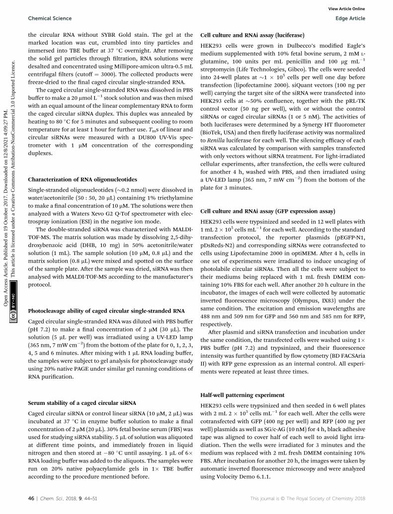
Fig. 2 PAGE (20%) analysis of the photocleavage ability of a cagedcircular single-stranded RNA (2 mM, c-AL) in 1� PBS buffer (pH 7.2). Thecaged RNA was irradiated using a UV-LED lamp (365 nm, 7 mW cm�2)for 0, 1, 2, 3, 4, 5 and 6 minutes. The charts are percentages of thecircular and cleaved RNAs upon light irradiation at different timeintervals after gel was quantified with ImageQuant.
Results and discussionSynthesis and purication of caged circular siRNAs
In our previous study, caged antisense oligonucleotides weresynthesized by rst coupling with photolabile o-nitrophenylglycol phosphoramidite, followed by coupling of MMT–NH–C6phosphoramidite.21,30 The MMT group was then removed torelease free amine for the coupling reaction with succinicanhydride to introduce carboxylic acid for the cyclization ofoligonucleotides. Here we simplied the procedure througha single step coupling reaction, in which a new photolabilephosphoramidite linker (PL, Table 1) was designed to introducecarboxylic acid at the 50 terminal of single-stranded RNA forcyclization. A 50 phosphate group was generated in the activatedsiRNA aer PL was removed upon light activation. As shown inScheme S1,† PL was readily synthesized in ve steps with 37.6%yield through Grignard reagent exchange and coupling,50,51 o-Cl-trityl protection of carboxylic acid, ketone reduction andphosphoramidition.
To introduce an amine group at the 30 terminal of the RNAstrand, 50-DMT-T (C6 amino)-succinyl-CPG was used as the rst
This journal is © The Royal Society of Chemistry 2018
nucleotide of the sequence, which contains a 30 terminal aminewith no effect on the 30 terminal hydroxyl group. Aer RNAsynthesis, the photocleavable phosphoramidite linker (PL) wascoupled to the RNA sequence as the last coupling monomeraccording to the standard RNA synthesis procedure in DMT-offmode. Raw oligonucleotides containing the 30 amine and 50 acidfunctional groups were obtained aer cleavage and depro-tection with concentrated ammonium hydroxide, followed byremoval of TBDMS on RNA nucleotide monomers with TEA.3HFsolution and RP-HPLC purication. The obtained single-stranded RNA was then cyclized with the formation of anamide bond (Fig. S1†) and the yield of isolated circular RNA isaround 20%.
The caged circular RNA oligonucleotides were puried by RP-HPLC, as shown in Fig. S2.† However, when the collected peakswere further analyzed by PAGE, the presence of a tiny amount ofsingle stranded starting RNA was still observed. Due to the factthat the pathway of siRNA is a catalytic process, the existence ofeven a small amount of linear siRNA may result in an obviousRNAi effect. We then chose to purify these caged circular RNAsthrough PAGE gels as shown in Fig. S3.† Compared to linearRNAs, the circular RNAs ran faster in PAGE gel, thereforefacilitating further purication of the caged circular RNAs. Thecaged circular single-stranded RNA was then collected andanalyzed by ESI-MS in negative mode [1% TEA in H2O/CH3CN(50 : 50)] as listed in Tables S1 and S2.† With the addition of anequal amount of complementary RNA, the caged circular siRNAwas annealed and evaluated in this study.
Photocleavage of caged circular single-stranded RNAs
In order to nd an appropriate irradiation time to efficientlycleave the photolabile linker, the single stranded caged circularRNA strand was treated by light irradiation to recover the cor-responding linear RNA (Scheme S2†). Upon light irradiation (0,1, 2, 3, 4, 5 and 6 min), the irradiated samples were then subject
Chem. Sci., 2018, 9, 44–51 | 47
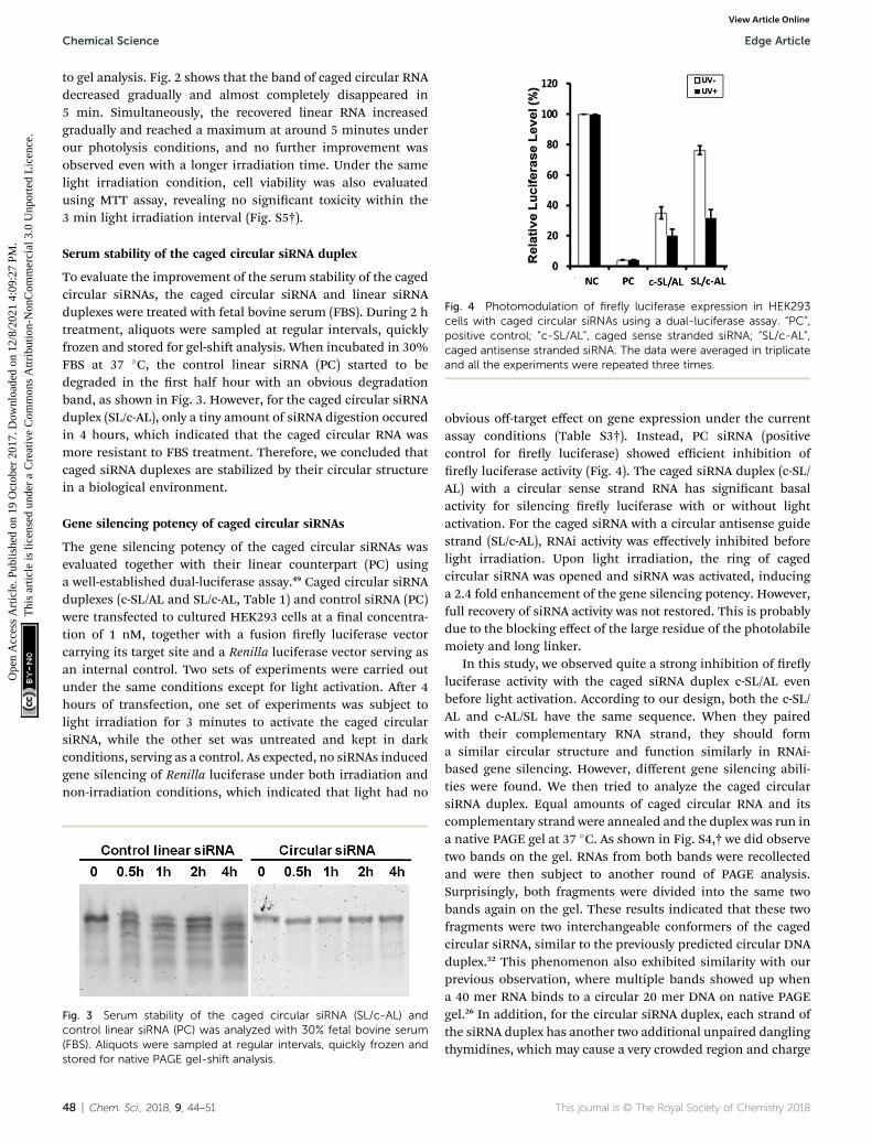
Fig. 4 Photomodulation of firefly luciferase expression in HEK293cells with caged circular siRNAs using a dual-luciferase assay. “PC”,positive control; “c-SL/AL”, caged sense stranded siRNA; “SL/c-AL”,caged antisense stranded siRNA. The data were averaged in triplicateand all the experiments were repeated three times.
Chemical Science Edge Article
Ope
n A
cces
s A
rtic
le. P
ublis
hed
on 1
9 O
ctob
er 2
017.
Dow
nloa
ded
on 1
2/8/
2021
4:0
9:27
PM
. T
his
artic
le is
lice
nsed
und
er a
Cre
ativ
e C
omm
ons
Attr
ibut
ion-
Non
Com
mer
cial
3.0
Unp
orte
d L
icen
ce.
View Article Online
to gel analysis. Fig. 2 shows that the band of caged circular RNAdecreased gradually and almost completely disappeared in5 min. Simultaneously, the recovered linear RNA increasedgradually and reached a maximum at around 5 minutes underour photolysis conditions, and no further improvement wasobserved even with a longer irradiation time. Under the samelight irradiation condition, cell viability was also evaluatedusing MTT assay, revealing no signicant toxicity within the3 min light irradiation interval (Fig. S5†).
Serum stability of the caged circular siRNA duplex
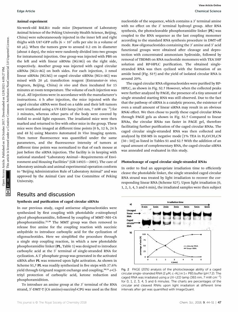
To evaluate the improvement of the serum stability of the cagedcircular siRNAs, the caged circular siRNA and linear siRNAduplexes were treated with fetal bovine serum (FBS). During 2 htreatment, aliquots were sampled at regular intervals, quicklyfrozen and stored for gel-shi analysis. When incubated in 30%FBS at 37 �C, the control linear siRNA (PC) started to bedegraded in the rst half hour with an obvious degradationband, as shown in Fig. 3. However, for the caged circular siRNAduplex (SL/c-AL), only a tiny amount of siRNA digestion occuredin 4 hours, which indicated that the caged circular RNA wasmore resistant to FBS treatment. Therefore, we concluded thatcaged siRNA duplexes are stabilized by their circular structurein a biological environment.
Gene silencing potency of caged circular siRNAs
The gene silencing potency of the caged circular siRNAs wasevaluated together with their linear counterpart (PC) usinga well-established dual-luciferase assay.49 Caged circular siRNAduplexes (c-SL/AL and SL/c-AL, Table 1) and control siRNA (PC)were transfected to cultured HEK293 cells at a nal concentra-tion of 1 nM, together with a fusion rey luciferase vectorcarrying its target site and a Renilla luciferase vector serving asan internal control. Two sets of experiments were carried outunder the same conditions except for light activation. Aer 4hours of transfection, one set of experiments was subject tolight irradiation for 3 minutes to activate the caged circularsiRNA, while the other set was untreated and kept in darkconditions, serving as a control. As expected, no siRNAs inducedgene silencing of Renilla luciferase under both irradiation andnon-irradiation conditions, which indicated that light had no
Fig. 3 Serum stability of the caged circular siRNA (SL/c-AL) andcontrol linear siRNA (PC) was analyzed with 30% fetal bovine serum(FBS). Aliquots were sampled at regular intervals, quickly frozen andstored for native PAGE gel-shift analysis.
48 | Chem. Sci., 2018, 9, 44–51
obvious off-target effect on gene expression under the currentassay conditions (Table S3†). Instead, PC siRNA (positivecontrol for rey luciferase) showed efficient inhibition ofrey luciferase activity (Fig. 4). The caged siRNA duplex (c-SL/AL) with a circular sense strand RNA has signicant basalactivity for silencing rey luciferase with or without lightactivation. For the caged siRNA with a circular antisense guidestrand (SL/c-AL), RNAi activity was effectively inhibited beforelight irradiation. Upon light irradiation, the ring of cagedcircular siRNA was opened and siRNA was activated, inducinga 2.4 fold enhancement of the gene silencing potency. However,full recovery of siRNA activity was not restored. This is probablydue to the blocking effect of the large residue of the photolabilemoiety and long linker.
In this study, we observed quite a strong inhibition of reyluciferase activity with the caged siRNA duplex c-SL/AL evenbefore light activation. According to our design, both the c-SL/AL and c-AL/SL have the same sequence. When they pairedwith their complementary RNA strand, they should forma similar circular structure and function similarly in RNAi-based gene silencing. However, different gene silencing abili-ties were found. We then tried to analyze the caged circularsiRNA duplex. Equal amounts of caged circular RNA and itscomplementary strand were annealed and the duplex was run ina native PAGE gel at 37 �C. As shown in Fig. S4,† we did observetwo bands on the gel. RNAs from both bands were recollectedand were then subject to another round of PAGE analysis.Surprisingly, both fragments were divided into the same twobands again on the gel. These results indicated that these twofragments were two interchangeable conformers of the cagedcircular siRNA, similar to the previously predicted circular DNAduplex.52 This phenomenon also exhibited similarity with ourprevious observation, where multiple bands showed up whena 40 mer RNA binds to a circular 20 mer DNA on native PAGEgel.26 In addition, for the circular siRNA duplex, each strand ofthe siRNA duplex has another two additional unpaired danglingthymidines, which may cause a very crowded region and charge
This journal is © The Royal Society of Chemistry 2018
Edge Article Chemical Science
Ope
n A
cces
s A
rtic
le. P
ublis
hed
on 1
9 O
ctob
er 2
017.
Dow
nloa
ded
on 1
2/8/
2021
4:0
9:27
PM
. T
his
artic
le is
lice
nsed
und
er a
Cre
ativ
e C
omm
ons
Attr
ibut
ion-
Non
Com
mer
cial
3.0
Unp
orte
d L
icen
ce.
View Article Online
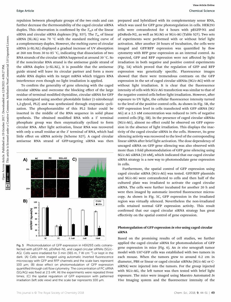
repulsion between phosphate groups of the two ends and canfurther decrease the thermostability of the caged circular siRNAduplex. This observation is conrmed by the Tms of the linearsiRNA and circular siRNA duplexes (Fig. S5†). The Tm of linearsiRNA (SL/AL) was 70 �C with the standard melting curve ofa complementary duplex. However, the melting curve of circularsiRNA (c-SL/AL) displayed a gradual increase of UV absorptionat 260 nm from 30 to 85 �C, indicating that dissociation of twoRNA strands of the circular siRNA happened at around 30 �C. Soif the noncircular RNA strand is the antisense guide strand ofthe siRNA duplex (c-SL/AL), it is possible that the antisenseguide strand will leave its circular partner and form a morestable RNA duplex with its target mRNA which triggers RNAinterference even though no light irradiation is applied.
To conrm the generality of gene silencing with the cagedcircular siRNAs and overcome the blocking effect of the largeresidue of terminal modied thymidine, circular siRNA for GFPwas redesigned using another photolabile linker (1-nitrobenzyl1,2-glycol, PL2) and was synthesized through enzymatic cycli-zation. The phosphoramidite of this PL2 linker could beinserted in the middle of the RNA sequence in solid phasesynthesis. The obtained modied RNA with a 50 terminalphosphate group was then enzymatically cyclized to formcircular RNA. Aer light activation, linear RNA was recoveredwith only a small residue at the 30 terminal of RNA, which hadlittle effect on siRNA activity (Scheme S2†). A caged circularantisense RNA strand of GFP-targeting siRNA was then
Fig. 5 Photomodulation of GFP expression in HEK293 cells cotrans-fected with pEGFP-N1, pDsRed-N1, and caged circular siRNAs (SG/c-AG). Cells were irradiated for 3 min (365 m, 7 W cm�2) or kept in thedark. (A) Cells were imaged using automatic inverted fluorescencemicroscopy with GFP and RFP channels and the scale bars represent100 mm; (B) dose effect on photomodulation of GFP expressionquantified through cell flow cytometry. The concentration of PC siRNA(SG/AG) was fixed at 2.5 nM. All the experiments were repeated threetimes; (C) the spatial regulation of GFP expression with patternedirradiation (left side view) and the scale bar represents 100 mm.
This journal is © The Royal Society of Chemistry 2018
prepared and hybridized with its complementary sense RNA,which was used for GFP gene photoregulation in cells. HEK293cells were cotransfected for 4 hours with pEGFP-N1 andpDsReds-N2, as well as SG/AG or SG/c-AG (Table S2†). Two setsof experiments were performed with or without brief lightactivation. Aer another 20 hours of incubation, the cells wereimaged and GFP/RFP expression was quantied by owcytometry with RFP gene expression as an internal control. Asexpected, GFP and RFP expression were not affected by lightirradiation in both negative and positive control experiments(Fig. 5A) which proved that the regulation of GFP and RFPexpression was genetically specic. Fluorescence imagesshowed that there were tremendous contrasts on the GFPexpression in the set of caged circular siRNAs (SG/c-AG) with orwithout light irradiation. It is clear that the uorescenceintensity of cells with SG/c-AG transfection was similar to that ofthe negative control cells before light irradiation. However, aerexposure to UV light, the cellular uorescence intensity turnedto the level of the positive control cells. As shown in Fig. 5B, theGFP expression level in cells transfected with GFP siRNA (SG/AG) at a 2.5 nM concentration was reduced to 34% of negativecontrol cells (Fig. 5B). In the presence of caged circular siRNAs(SG/c-AG), almost no effect could be observed on GFP expres-sion in the absence of light irradiation. This displays the inac-tivity of the caged circular siRNA in the cells. However, its genesilencing activity was recovered to the level of the correspondinglinear siRNA aer brief light activation. The dose dependency ofuncaged siRNA on GFP gene silencing was also observed withmore than 3 fold photomodulation of GFP gene silencing usingSG/c-AG siRNA (10 nM), which indicated that our caged circularsiRNA strategy is a new way to photomodulate gene expressionin cells.
Furthermore, the spatial control of GFP expression usingcaged circular siRNA (SG/c-AG) was tested. GFP/RFP plasmidsand SG/c-AG were cotransfected to cells and then half of thecultured plate was irradiated to activate the caged circularsiRNA. The cells were further incubated for another 20 h andwere then imaged by automatic inverted uorescence micros-copy. As shown in Fig. 5C, GFP expression in the irradiatedregion was virtually silenced. Nevertheless the non-irradiatedcells retained normal GFP expression activity. This resultconrmed that our caged circular siRNA strategy has greateffectivity on the spatial control of gene expression.
Photoregulation of GFP expression in vivo using caged circularsiRNA
Based on the promising results of cell studies, we furtherapplied the caged circular siRNA for photomodulation of GFPgene expression in mice (Fig. 6). An in vivo xenogra tumormodel with U87-GFP cells was established with two tumors ineach mouse. When the tumors grew to around 0.2 cm indiameter, PBS or linear or caged circular siRNAs (SG/c-AG or C-siRNA) were injected into the tumors. For the group injectedwith SG/c-AG, the le tumor was then tested with brief lightexposure. The mice were imaged using Maestro Automated InVivo Imaging system and the uorescence intensity of the
Chem. Sci., 2018, 9, 44–51 | 49
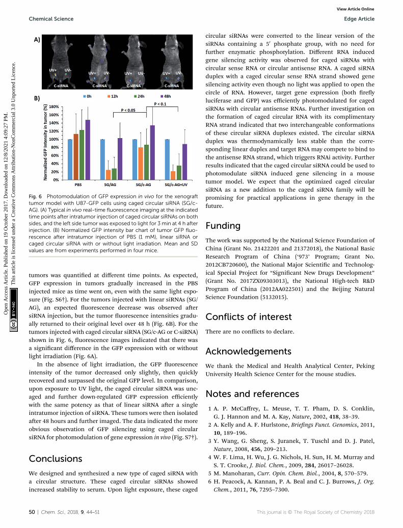
Fig. 6 Photomodulation of GFP expression in vivo for the xenografttumor model with U87-GFP cells using caged circular siRNA (SG/c-AG). (A) Typical in vivo real-time fluorescence imaging at the indicatedtime points after intratumor injection of caged circular siRNAs on bothsides, and the left side tumor was exposed to light for 3 min at 4 h afterinjection. (B) Normalized GFP intensity bar chart of tumor GFP fluo-rescence after intratumor injection of PBS (1 mM), linear siRNA orcaged circular siRNA with or without light irradiation. Mean and SDvalues are from experiments performed in four mice.
Chemical Science Edge Article
Ope
n A
cces
s A
rtic
le. P
ublis
hed
on 1
9 O
ctob
er 2
017.
Dow
nloa
ded
on 1
2/8/
2021
4:0
9:27
PM
. T
his
artic
le is
lice
nsed
und
er a
Cre
ativ
e C
omm
ons
Attr
ibut
ion-
Non
Com
mer
cial
3.0
Unp
orte
d L
icen
ce.
View Article Online
tumors was quantied at different time points. As expected,GFP expression in tumors gradually increased in the PBSinjected mice as time went on, even with the same light expo-sure (Fig. S6†). For the tumors injected with linear siRNAs (SG/AG), an expected uorescence decrease was observed aersiRNA injection, but the tumor uorescence intensities gradu-ally returned to their original level over 48 h (Fig. 6B). For thetumors injected with caged circular siRNA (SG/c-AG or C-siRNA)shown in Fig. 6, uorescence images indicated that there wasa signicant difference in the GFP expression with or withoutlight irradiation (Fig. 6A).
In the absence of light irradiation, the GFP uorescenceintensity of the tumor decreased only slightly, then quicklyrecovered and surpassed the original GFP level. In comparison,upon exposure to UV light, the caged circular siRNA was unc-aged and further down-regulated GFP expression efficientlywith the same potency as that of linear siRNA aer a singleintratumor injection of siRNA. These tumors were then isolatedaer 48 hours and further imaged. The data indicated the moreobvious observation of GFP silencing using caged circularsiRNA for photomodulation of gene expression in vivo (Fig. S7†).
Conclusions
We designed and synthesized a new type of caged siRNA witha circular structure. These caged circular siRNAs showedincreased stability to serum. Upon light exposure, these caged
50 | Chem. Sci., 2018, 9, 44–51
circular siRNAs were converted to the linear version of thesiRNAs containing a 50 phosphate group, with no need forfurther enzymatic phosphorylation. Different RNA inducedgene silencing activity was observed for caged siRNAs withcircular sense RNA or circular antisense RNA. A caged siRNAduplex with a caged circular sense RNA strand showed genesilencing activity even though no light was applied to open thecircle of RNA. However, target gene expression (both reyluciferase and GFP) was efficiently photomodulated for cagedsiRNAs with circular antisense RNAs. Further investigation onthe formation of caged circular RNA with its complimentaryRNA strand indicated that two interchangeable conformationsof these circular siRNA duplexes existed. The circular siRNAduplex was thermodynamically less stable than the corre-sponding linear duplex and target RNA may compete to bind tothe antisense RNA strand, which triggers RNAi activity. Furtherresults indicated that the caged circular siRNA could be used tophotomodulate siRNA induced gene silencing in a mousetumor model. We expect that the optimized caged circularsiRNA as a new addition to the caged siRNA family will bepromising for practical applications in gene therapy in thefuture.
Funding
The work was supported by the National Science Foundation ofChina (Grant No. 21422201 and 21372018), the National BasicResearch Program of China (‘973’ Program; Grant No.2012CB720600), the National Major Scientic and Technolog-ical Special Project for “Signicant New Drugs Development”(Grant No. 2017ZX09303013), the National High-tech R&DProgram of China (2012AA022501) and the Beijing NaturalScience Foundation (5132015).
Conflicts of interest
There are no conicts to declare.
Acknowledgements
We thank the Medical and Health Analytical Center, PekingUniversity Health Science Center for the mouse studies.
Notes and references
1 A. P. McCaffrey, L. Meuse, T. T. Pham, D. S. Conklin,G. J. Hannon and M. A. Kay, Nature, 2002, 418, 38–39.
2 A. Kelly and A. F. Hurlstone, Briengs Funct. Genomics, 2011,10, 189–196.
3 Y. Wang, G. Sheng, S. Juranek, T. Tuschl and D. J. Patel,Nature, 2008, 456, 209–213.
4 W. F. Lima, H. Wu, J. G. Nichols, H. Sun, H. M. Murray andS. T. Crooke, J. Biol. Chem., 2009, 284, 26017–26028.
5 M. Manoharan, Curr. Opin. Chem. Biol., 2004, 8, 570–579.6 H. Peacock, A. Kannan, P. A. Beal and C. J. Burrows, J. Org.Chem., 2011, 76, 7295–7300.
This journal is © The Royal Society of Chemistry 2018

Edge Article Chemical Science
Ope
n A
cces
s A
rtic
le. P
ublis
hed
on 1
9 O
ctob
er 2
017.
Dow
nloa
ded
on 1
2/8/
2021
4:0
9:27
PM
. T
his
artic
le is
lice
nsed
und
er a
Cre
ativ
e C
omm
ons
Attr
ibut
ion-
Non
Com
mer
cial
3.0
Unp
orte
d L
icen
ce.
View Article Online
7 W. F. Lima, T. P. Prakash, H. M. Murray, G. A. Kinberger,W. Li, A. E. Chappell, C. S. Li, S. F. Murray, H. Gaus,P. P. Seth, E. E. Swayze and S. T. Crooke, Cell, 2012, 150,883–894.
8 D. Yu, H. Pendergraff, J. Liu, H. B. Kordasiewicz,D. W. Cleveland, E. E. Swayze, W. F. Lima, S. T. Crooke,T. P. Prakash and D. R. Corey, Cell, 2012, 150, 895–908.
9 D. Cejka, D. Losert and V. Wacheck, Clin. Sci., 2006, 110, 47–58.
10 D. D. Young, H. Lusic, M. O. Lively, J. A. Yoder and A. Deiters,ChemBioChem, 2008, 9, 2937–2940.
11 A. Deiters, Curr. Opin. Chem. Biol., 2009, 13, 678–686.12 X. Ouyang, I. A. Shestopalov, S. Sinha, G. Zheng, C. L. Pitt,
W. H. Li, A. J. Olson and J. K. Chen, J. Am. Chem. Soc.,2009, 131, 13255–13269.
13 A. Deiters, ChemBioChem, 2010, 11, 47–53.14 A. Deiters, R. A. Garne, H. Lusic, J. M. Govan, M. Dush,
N. M. Nascone-Yoder and J. A. Yoder, J. Am. Chem. Soc.,2010, 132, 15644–15650.
15 C. Brieke, F. Rohrbach, A. Gottschalk, G. Mayer andA. Heckel, Angew. Chem., Int. Ed., 2012, 51, 8446–8476.
16 J. M. Govan, R. Uprety, J. Hemphill, M. O. Lively andA. Deiters, ACS Chem. Biol., 2012, 7, 1247–1256.
17 I. Ahmed and L. Fruk, Mol. BioSyst., 2013, 9, 565–570.18 C. M. Connelly, R. Uprety, J. Hemphill and A. Deiters, Mol.
BioSyst., 2012, 8, 2987–2993.19 J. L. Richards, G. K. Seward, Y. H. Wang and
I. J. Dmochowski, ChemBioChem, 2010, 11, 320–324.20 L. Wu, Y. Wang, J. Wu, C. Lv, J. Wang and X. Tang, Nucleic
Acids Res., 2013, 41, 677–686.21 Y. Wang, L. Wu, P. Wang, C. Lv, Z. Yang and X. Tang, Nucleic
Acids Res., 2012, 40, 11155–11162.22 X. Tang, Photochemistry, ed. A. Albini and E. Fasani, 2013,
vol. 41, pp. 319–341.23 M. Su, F. Yang, C. Lv, L. Yu, X. Gu, J. Wang, Z. Li and X. Tang,
J. Chin. Pharm. Sci., 2010, 19, 5–14.24 X. Tang, S. Maegawa, E. S. Weinberg and I. J. Dmochowski, J.
Am. Chem. Soc., 2007, 129, 11000–11001.25 X. Tang and I. J. Dmochowski, Nat. Protoc., 2007, 1, 3041–
3048.26 X. Tang and I. J. Dmochowski, Angew. Chem., Int. Ed., 2006,
45, 3523–3526.27 G. Zheng, L. Cochella, J. Liu, O. Hobert and W.-h. Li, ACS
Chem. Biol., 2011, 6, 1332–1338.28 P. K. Jain, V. Ramanan, A. G. Schepers, N. S. Dalvie, A. Panda,
H. E. Fleming and S. N. Bhatia, Angew. Chem., Int. Ed., 2016,55, 12628–12632.
29 W. Zhou and A. Deiters, Angew. Chem., Int. Ed., 2016, 55,5394–5399.
This journal is © The Royal Society of Chemistry 2018
30 J. M. Govan, D. D. Young, H. Lusic, Q. Liu, M. O. Lively andA. Deiters, Nucleic Acids Res., 2013, 41, 10518–10528.
31 A. Kala and S. H. Friedman, Pharm. Res., 2011, 28, 3050–3057.
32 S. Shah, P. K. Jain, A. Kala, D. Karunakaran andS. H. Friedman, Nucleic Acids Res., 2009, 37, 4508–4517.
33 S. Shah, S. Rangarajan and S. H. Friedman, Angew. Chem.,Int. Ed., 2005, 44, 1328–1332.
34 P. K. Jain, S. Shah and S. H. Friedman, J. Am. Chem. Soc.,2010, 133, 440–446.
35 J. P. Casey, R. A. Blidner and W. T. Monroe, Mol. Pharm.,2009, 6, 669–685.
36 S. Shah and S. H. Friedman, Oligonucleotides, 2007, 17, 35–43.
37 V. Mikat and A. Heckel, RNA, 2007, 13, 2341–2347.38 Q. N. Nguyen, R. V. Chavli, J. T. Marques, P. G. Conrad II,
D. Wang, W. He, B. E. Belisle, A. Zhang, L. M. Pastor andF. R. Witney, Biochim. Biophys. Acta, Biomembr., 2006, 1758,394–403.
39 Y. Matsushita-Ishiodori and T. Ohtsuki, Acc. Chem. Res.,2012, 45, 1039–1047.
40 J. C. Griepenburg, B. K. Ruble and I. J. Dmochowski, Bioorg.Med. Chem., 2013, 21, 6198–6204.
41 R. A. Blidner, K. R. Svoboda, R. P. Hammer andW. T. Monroe, Mol. BioSyst., 2008, 4, 431–440.
42 J. Hemphill, E. K. Borchardt, K. Brown, A. Asokan andA. Deiters, J. Am. Chem. Soc., 2015, 137, 5642–5645.
43 T. Lucas, F. Schafer, P. Muller, S. A. Eming, A. Heckel andS. Dimmeler, Nat. Commun., 2017, 8, 15162.
44 L. Wu, F. Pei, J. Zhang, J. Wu, M. Feng, Y. Wang, H. Jin,L. Zhang and X. Tang, Chem. –Eur. J., 2014, 20, 12114–12122.
45 Y. Ji, J. Yang, L. Wu, L. Yu and X. Tang, Angew. Chem., Int. Ed.,2016, 55, 2152–2156.
46 S. Yamazoe, I. A. Shestopalov, E. Provost, S. D. Leach andJ. K. Chen, Angew. Chem., Int. Ed., 2012, 51, 6908–6911.
47 N. Abe, H. Abe and Y. Ito, J. Am. Chem. Soc., 2007, 129, 15108–15109.
48 L. Wei, L. Cao and Z. Xi, Angew. Chem., Int. Ed., 2013, 52,6501–6503.
49 M. Chen, L. Zhang, H.-Y. Zhang, X. Xiong, B. Wang, Q. Du,B. Lu, C. Wahlestedt and Z. Liang, Proc. Natl. Acad. Sci. U.S. A., 2005, 102, 2356–2361.
50 L. F. Fieser and W. H. Daudt, J. Am. Chem. Soc., 1941, 63,782–788.
51 I. Sapountzis and P. Knochel, Angew. Chem., Int. Ed., 2002,41, 1610–1611.
52 C. Kim, O. C. Lee, J. Y. Kim, W. Sung and N. K. Lee, Angew.Chem., Int. Ed., 2015, 54, 8943–8947.
Chem. Sci., 2018, 9, 44–51 | 51