Volume 22 January/March 2016 Number 1 - kcus.ba journal 9 web.pdf · CHA2DS2-VASc skor za procjenu...
56
Number 1 January/March 2016 Volume 22, Number 1, pp 01 - 54 January/March 2016 Volume 22
-
Upload
trinhnguyet -
Category
Documents
-
view
213 -
download
0
Transcript of Volume 22 January/March 2016 Number 1 - kcus.ba journal 9 web.pdf · CHA2DS2-VASc skor za procjenu...

Number 1January/March 2016
Volume 22, Num
ber 1, pp 01 - 54 January/M
arch 2016
Volume 22


New ICU - Central Medical Building - Clinical Center University of SarajevoNova Intenzivna njega - Klinički Centar Univerziteta u Sarajevu
02
New Central Medical Building - University Clinical Center SarajevoNovi Centralni Medicinski Blok - Univerzitetski klinički centar u Sarajevu
03

New Central Medical Building - University Clinical Center SarajevoNovi Centralni Medicinski Blok - Univerzitetski klinički centar u Sarajevu
03
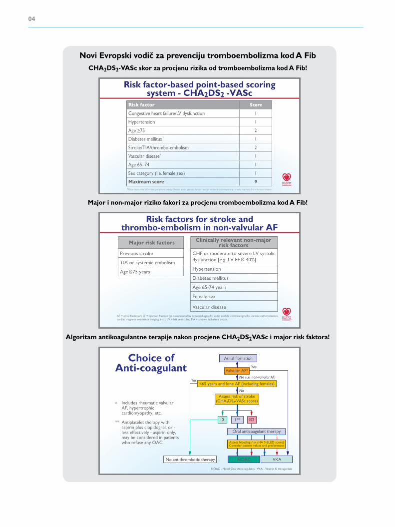
Risk factor Score
Congestive heart failure/LV dysfunction 1
Hypertension 1
Age >75 2
Diabetes mellitus 1
Stroke/TIA/thrombo-embolism 2
Vascular disease* 1
147–56 egA
1)xes elamef .e.i( yrogetac xeS
9erocs mumixaM
Risk factor-based point-based scoringsystem - CHA2DS2 -VASc
*Prior myocardial infarction, peripheral artery disease, aortic plaque. Actual rates of stroke in contemporary cohorts may vary from these estimates.
AF = atrial fibrilation; EF = ejection fraction (as documented by echocardiography, radio nuclide ventriculography, cardiac catheterization, cardiac magnetic resonance imaging, etc.); LV = left venticular; TIA = trasient ischaemic attack.
Risk factors for stroke andthrombo-embolism in non-valvular AF
CHF or moderate to severe LV systolicdysfunction [e.g. LV EF � 40%]
Hypertension
Clinically relevant non-majorrisk factors
Diabetes mellitus
Age 65-74 years
Female sex
Vascular disease
Major risk factors
Previous stroke
TIA or systemic embolism
Age �75 years
Atrial fibrilation
Yes
Yes
No (i.e. non-valvular AF)
No
<65 years and lone AF (including females)
Valvular AF*
Oral anticoagulant therapy
Assess bleeding risk (HA S-BLED score)Consider patient values and preferences
Choice ofAnti-coagulant
Includes rheumatic valvularAF, hypertrophiccardiomyopathy, etc.
Antiplatelet therapy withaspirin plus clopidogrel, or -less effectively - aspirin only,may be considered in patientswho refuse any OAC
**
*
Assess risk of stroke(CHA2DS2-VASc score)
0 1** �2
No antithrombotic therapy NOAC VKA
NOAC - Novel Oral Anticoagulants, VKA - Vitamin K Antagonists
CHA2DS2-VASc skor za procjenu rizika od tromboembolizma kod A Fib!
Novi Evropski vodič za prevenciju tromboembolizma kod A Fib
Major i non-major riziko fakori za procjenu tromboembolizma kod A Fib!
Algoritam antikoagulantne terapije nakon procjene CHA2DS2VASc i major risk faktora!
04

Medical Journal www.ukcs.ba
Editor-in-ChiefMirza Dilić
Editorial Board
International Advisory Board
Kenan Arnautović (USA), Raffaele Bugiardini (Italy), Erol Ćetin (Turkey), Maria Dorobantu (Romania), Oktay Ergene (Turkey), Zlatko Fras (Slovenia), Dan Gaita (Romania), Mario Ivanuša (Croatia), Steen Dalby Kristensen (Denmark), Mimoza Lezhe (Albania), Mario Marzilli (Italy), Milica Medić-Stojanovska (Serbia), Davor Miličić (Croatia), Fausto Pinto (Portugal), Mihailo Popovici (Moldova), Marcella Rietschel (Germany), Nadan Rustemović (Croatia), Georges Saade (Lebanon), Petar Seferović (Serbia), Dragan Stanisavljević (Slovenia), Panos Vardas (Greece), GordanVujanić (UK), Jose Zamorano (Spain)
English language revisionSvjetlana Baroševčić
Medical Journal is Indexed in
EBSCO publishing USAwww.ebscohost.com
Member of National Journals Networks of the European Society of Cardiology
PUBLISHER:
General ManagerUCCS
AIMS AND SCOPE
EDITORIAL OFFICEAddress: Medical Journal, Institute for Research and Development, University Clinical Center Sarajevo71000 Sarajevo, Bolnička 25, Bosnia and Herzegovina, Phone: +387 33 668 415; +387 33 297 264. Email: [email protected] Web. www.ukcs.ba Technical secretariat: [email protected]: [email protected]
SUBSCRIPTIONAnnual subscription rates: Bosnia and Herzegovina € 50; Europe € 80; and other € 100.
SUPPLEMENTS, REPRINTS AND CORPORATE SALESFor requests from industry and companies regarding supplements, bulk articles reprints, spon-sored subscriptions, translation opportunities for previously published material, and corporate online opportunities, please contact;Email: [email protected]
PRINTEurografika ZvornikPrinted on acid-free paper.
TECHNICAL EDITOREurografika
CIRCULATION500 copies
The Medical Journal is the official quarterly journal of the Institute for Research and Develop-ment of the University Clinical Center Sarajevo and has been published regularly since 1994. It is published in the languages of the people of Bosnia and Herzegovina i.e. Bosnian, Croatian and Serbian as well as in English.
The Medical Journal aims to publish the highest quality materials, both clinical and scientific, on all aspects of clinical medicine. It offers the reader a collection of contemporary, original, peer-re-viewed papers, professional articles, review articles, editorials, along with special articles and case reports.
Copyright: the full text of the articles published in the Medical Journal can be used for education-al and personal aims i.e. references cited upon the authors’ permission. If the basic aim is commercial no parts of the published materials may be used or reproduced without the permis-sion of the publisher. Special permission is available for educational and non-profit educational classroom use. Electronic storage or usage: except as outlined above, no parts of this publication may be reproduced, stored in a retrieval system or transmitted in any form or by any means without prior written permission from the Publisher.All rights reserved©2015. Institute for Research and Development UCCS.
Notice: the authors, editor and publisher do not accept responsibility for any loss or damage arising from actions or decisions based on information contained in this publication; ultimate responsibility for the treatment of patients and interpretation of published materials lies with the medical practitioner. The opinions expressed are those of the authors and the inclusion in this publication of materials relating to a specific product, method or technique does not amount to an endorsement of its value or quality, or of the claims made by its manufacturer.
Institute for Research and DevelopmentUniversity Clinical Center Sarajevo71000 Sarajevo, Bolnička 25Bosnia and Herzegovina
Sebija Izetbegović, MD, PhDSebija Izetbegović, Enra Suljić-Mehmedika,Ismet Gavrankapetanović, Safet Guska, Almira Hadžović-Džuvo, Adnan Kapidžić, Abdulah Kučukalić, Mehmed Kulić, Benjamin Kulovac, Bakir Mehić, Nermina Obralić, Lilijana Oruč, Amer Ovčina, Asja Prohić, Svjetlana Radović, Senija Rašić, Mustafa Hiroš, Secretary
For publisher:

Content Medical Journal (2016) Vol. 22, No. 1
Original article
Influence of various factors on the occurrence of postoperative seroma following breast cancer surgery . . . . . 07 Sadat Pušina, Mirhan Salibašić, Emir Bičakčić, Amela Salibašić-Drnda
Absence of the national network for the primary treatment of acute coronary syndrome jeopardizes results of the surgical myocardial revascularization . . . . . . . . . . . . . . . . . . . . . . . . . . . . . . . . . . . . . . . . . . . . . . . . . . . . . . . . . . . . . . . . . . . . . . . . . . . . . . . . . . . . . . . . . . . . . . . . . . . . . . . . . 11 Sanko Pandur, Omer Perva, Faruk Čustović, Edin Omerbašić, Enela Đonlić, Amina Mehić
Serum nitric oxide level in end-stage renal disease patients with different duration of dialysis therapy . . . . . . . . . 16Nesina Avdagić, Nermina Babić, Asija Začiragić, Anela Šubo, Amela Dervišević, Adnan Hadžimuratović, Sadeta Begić-Kapetanović,
Amela Bečiragić; Izeta Aganović-Mušinović
The incidence and some characteristics of patients with hemophilia in Bosnia and Herzegovina . . . . . . . . . . . . . . . . . . . . 20Edo Hasanbegović, Jelica Predojević-Samardžić, Nermana Čengić
Evaluation of surgical treatment of Achilles tendon rupture . . . . . . . . . . . . . . . . . . . . . . . . . . . . . . . . . . . . . . . . . . . . . . . . . . . . . . . . . . . . . . . . . . . . . . . . . . . . . . . . . . . . . . . . 23Adnana Talić-Tanović, Fuad Džanković, Adnan Papović, Mehmed Zahirović
Benefits of cochlear implant speech processor upgrade . . . . . . . . . . . . . . . . . . . . . . . . . . . . . . . . . . . . . . . . . . . . . . . . . . . . . . . . . . . . . . . . . . . . . . . . . . . . . . . . . . . . . . . . . . . . . . . . 27Sanja Špirić, Dmitar Travar, Predrag Špirić, Slobodan Spremo, Mirjana Gnjatić
Serum nitric oxide concentration in rheumatoid arthritis patients: the association with disease activity . . . . . . . . 30Amela Dervišević, Nermina Babić, Asija Začiragić, Nesina Avdagić, Šekib Sokolović, Almir Fajkić, Orhan Lepara, Anela Šubo, Jasminko Huskić
Defining the vascular skin territories of the septocutaneous blood vessels of the forearm with a special overview on their use in the fasciocutaneous flaps surgery . . . . . . . . . . . . . . . . . . . . . . . . . . . . . . . . . . . . . . . . . . . . . . . . . . . . . . . . . . . . . . . . . . . . . . . . . . . . . . . . . . . . . . . . . . . . . 34Darko Jović, Aleksandar Jakovljević, Jovan Ćulum, Branislava Jakovljević, Ljiljana Latinović, Olivera Kosovac, Darko Lukić
Kinesiotherapy in early rehabilitation of patients after surgery of herniated cervical intervertebral disc . . . . . . . 37Ksenija Miladinović, Narcisa Vavra-Hadžiahmetović, Mirsad Muftić, Damir Čelik
Advantages of locking compresion plates in treatment of proximal humerus fracture . . . . . . . . . . . . . . . . . . . . . . . . . . . . . . . . . . . . . . . . . 41Faruk Lazović, Ismet Gavrankapetanović, Adnana Talić-Tanović, Đemil Omerović, Mehmed Jamakosmanović
Comparative advantages of VATS in relation to standard thoracic drainage in primary pleural empyema treatment . . . . . . . . . . . . . . . . . . . . . . . . . . . . . . . . . . . . . . . . . . . . . . . . . . . . . . . . . . . . . . . . . . . . . . . . . . . . . . . . . . . . . . . . . . . . . . . . . . . . . . . . . . . . . . . . . . . . . . . . . . . . . . . . . . . . . . . . . . . . . . . . . . . . . . . . . . . . . . . . . . . . . . . . . . . . . . . . . . . . . 45Ilijaz Pilav, Safet Guska, Safet Mušanović
Review article
Exposure to ionizing radiation for medical purposes: effects on population, monitoring, management and reporting of radiation doses . . . . . . . . . . . . . . . . . . . . . . . . . . . . . . . . . . . . . . . . . . . . . . . . . . . . . . . . . . . . . . . . . . . . . . . . . . . . . . . . . . . . . . . . . . . . . . . . . . . . . . . . . . . . . . . . . . . . . . . . . . . . . . . . . . . . . . . . . . . . . . . 49Sandra Vegar-Zubović, Spomenka Kristić, Irmina Sefić-Pašić
Instructions to authors . . . . . . . . . . . . . . . . . . . . . . . . . . . . . . . . . . . . . . . . . . . . . . . . . . . . . . . . . . . . . . . . . . . . . . . . . . . . . . . . . . . . . . . . . . . . . . . . . . . . . . . . . . . . . . . . . . . . . . . . . . . . . . . . . . . . . . . . . . . . . . . . . . . . . . . . . . . . . 52
Uputstva autorima . . . . . . . . . . . . . . . . . . . . . . . . . . . . . . . . . . . . . . . . . . . . . . . . . . . . . . . . . . . . . . . . . . . . . . . . . . . . . . . . . . . . . . . . . . . . . . . . . . . . . . . . . . . . . . . . . . . . . . . . . . . . . . . . . . . . . . . . . . . . . . . . . . . . . . . . . . . . . . . . . . . . . 54

Influence of various factors on the occurrence of postoperative seroma following breast cancer surgery
Uticaj različitih faktora na nastanak postoperativnog seroma nakon operativnih zahvata kod karcinoma dojke
Sadat Pušina1*, Mirhan Salibašić1, Emir Bičakčić1, Amela Salibašić-Drnda2
1Surgical Oncology Clinic, University Clinical Centre Sarajevo, Bolnička 25, 71000 Sarajevo, Bosnia and Herzegovina; 2 Faculty of Pharmacy, University of Sarajevo, Zmaja od Bosne 8, 71000 Sarajevo, Bosnia and Herzegovina
*Corresponding author
ABSTRACT
Introduction: breast surgery and axilla are among the operational procedures of low morbidity and mortality unless associated with the simultaneous reconstructive breast surgery. Complications of breast cancer surgery may be non-specific (seroma, wound infec-tion, bleeding, etc.) or specific for surgical procedures on the breast (fibrosis breast cancer lymphedema, cellulite, etc.) or in the axilla (seroma, lymphedema, neurosensory problems, limited mobility of upper extremity, etc). The objective was to evaluate the impact of certain factors (size and histological type of tumor, state of axillary lymph nodes, hormone receptor status, type of surgery, number of drains, preoperative systemic adjuvant therapy) in the occurrence of postoperative seroma. Patients: a total of 168 patients with proven primary invasive breast cancer and defined-stage disease (TNM clas-sification/AJCC), was included in the study, with or without applied neoadjuvant systemic therapy, aging between 18-75 years. Materials and methods: the study was retrospective, clinical-manipulative and descriptive-analytic. Clinical, radiological and histopathological find-ings were defined by the appropriate type of surgical treatment in the form of conservative breast surgery (wide local excision, quadrantec-tomy ) or radical breast surgery (modified radical mastectomy) with dissection of axillary lymph II level nodes (CALND) biopsy or axillary sentinel lymph node (SLNB). The postoperative seroma was defined as any clinically diagnosed accumulation of fluid in the axilla or under the skin incision treated with multiple needle aspirations. The appear-ance of seroma was analyzed in respect to the age of patients, pre-operative chemotherapy, type of surgery, number of vacuum drains, type of hormone receptors, grade and tumor size and number of dis-sected positive axillary lymph nodes. Results: the results showed that there was a statistically significant difference in respect to the occur-rence of postoperative seroma noted in patients who went through preoperative neoadjuvant chemotherapy (χ2=17.818, p=0.0001) and in respect to tumor size (χ2=15.972, p=0.0001). Results of the mul-tivariate logistic regression analysis showed a statistically significant effect on postoperative seroma in patients who had preoperative neoadjuvant chemotherapy (OR=6.000 (2.370 to 15.19), p=0.0001, 95% CI 1.213 (1.069- 1.660) and on tumor size [in patients with tumor ranging from 2 to 5 cm (stage T2) OR=2.115 (0.932 to 4.8); p=0.000; 95% CI 3.218 (1.84 to 7.17), while for tumors over 5 cm (stage T3 and
T4) OR=11.8 (3.657 to 37.8) p=0.000, 95% CI 2.26 (2.26 to 2.26)]. Conclusion: the research showed that there was a statistically signifi-cant difference in the occurrence of postoperative seroma in relation to conducted preoperative neoadjuvant therapy and tumor size. It was also revealed that factors such as the age of patients, histological type and degree of tumor differentiation, state of the axillary lymph nodes, hormone receptor status, type of surgery and number of drains were not associated with the postoperative seroma develop-ment.
Key words: breast cancer surgery, complications, postoperative se-roma
SAŽETAK
Uvod: hirurgija dojke i aksile spada u grupu operativnih proce-dura niskog morbiditeta i mortaliteta izuzev ako nisu udružene sa istovremenim rekonstruktivnim zahvatima na dojci. Komplikacije hirurgije karcinoma dojke mogu biti nespecifične (serom, infekcija rane, krvarenje itd.) ili specifične za operativne procedure na dojci (fibroza dojke, limfedem dojke, celulitis itd.) ili u aksili (serom, lim-fedem, neurosenzorni problemi, ograničena pokretljivost ruke itd.). Cilj: evaluacija uticaja pojedinih faktora (veličina, histološki tip i step-en diferenciranosti tumora, stanje aksilarnih limfnih čvorova, status hormonskih receptora, vrsta operativnog zahvata, broj drenova, sistemska adjuvantna terapija) na nastanak postoperativnog seroma. Pacijenti: analizirano je 168 pacijentica sa dokazanim primarnim in-vazivnim karcinomom dojke i definisanim stadijem oboljenja (TNM klasifikacija/AJCC), sa ili bez primjenjene neoadjuvatne sistemske terapije, dobi između 18-75 godina, operisanih u periodu od janu-ara 2014. do januara 2015. godine. Materijali i metode: istraživanje je bilo retrospektivno, kliničko-manipulativno i deskriptivno-analitičko. Prema kliničkom, radiološkom i patohistološkom nalazu definisan je odgovarajući tip operativnog tretmana u vidu poštednih operativnih procedura (segmentektomija, kvadrantektomija ili hemimastektomi-ja) ili radikalnog operativnog zahvata (modificirane radikalne mastek-tomije) sa disekcijom II nivoa aksilarnih limfnih čvorova (CALND) ili biopsijom aksilarnog sentinel limfnog čvora (SLNB). Postoperativni serom je definisan kao bilo koje klinički dijagnosticirano nakupljan-je tečnosti u aksili ili ispod kože operativnog reza tretirano sa mul-
Medical Journal (2016) Vol. 22, No. 1, 07 - 10 Original article
INTRODUCTION
Breast cancer is the most common malignancy in women and as a cause of death associated with malignant disease is the second among the most common, immediately after lung cancer (1).
Thanks to a widespread screening program for early detection of breast cancer and the use of non-invasive (ultrasound, mammography and magnetic resonance breast) and invasive radiological methods of the breast and axilla examination (tissue biopsy of the breast and axillary controlled by ultrasound) an increasing number of patients having breast cancer reveals the early stages of the disease when surgical treatment of breast cancer is the method of choice (2).
Breast and axillar surgery are operational procedures of low morbidity and mortality given that they are not associated with the simultaneous breast reconstructive surgery. Due to the increasing number of people affected by breast cancer at old age, we can still expect a variety of postoperative complications that can lead to increased morbidity producing additional costs of treatment and delaying the application of postoperative adjuvant therapy.
Complications of breast cancer surgery may be nonspecific (seroma, wound infection, bleeding, etc.) or specific operational procedures on the breast (fibrosis breast cancer lymphedema, cellulitis, etc.) or in the axilla (seroma, lymphedema, impaired neurosensitivity, limited mobility of upper extremity, etc.) (3).
The occurrence of complications could depend on various factors such as the patient age, type of surgical treatment, the extent of axillary lymph node dissection, the use of neoadjuvant chemotherapy, comorbidities, etc. The objective of the research was to evaluate the impact of certain factors (size and grade of tumor, the condition of axillary lymph nodes, hormone receptor status, type of surgery, number of drains, systemic adjuvant therapy) to the occurrence of postoperative seroma.
MATERIALS AND METHODS
A total of 168 patients of the Clinic of Oncology Surgery, University Clinical Center Sarajevo (UCCS), diagnosed with primary invasive breast cancer and defined-stage disease (TNM classification / AJCC) with or without applied neoadjuvant systemic therapy, age between 18-75, surgically treated during the period from January
2014 to January 2015.The study was retrospective, clinical-manipulative and
descriptive-analytic. According to the clinical, radiological and histopathological findings, the appropriate type of surgical treatment was defined in the form of conservative breast surgery (wide local excision, quadrantectomy ) or radical breast surgery (modified radical mastectomy) with dissection of axillary lymph II level nodes (CALND) biopsy or axillary sentinel lymph node (SLNB). After surgical removal of breast cancer simultaneous reconstructive procedures were not performed. Demographic and clinical information were obtained from the patients‘ histories.
The postoperative seroma was defined as any clinically diagnosed accumulation of fluid in the axilla or under the skin incision treated with multiple needle aspirations.
The emergence of seroma was analyzed in relation to the age of patients, administering preoperative chemotherapy, type of surgery, number of vacuum drains, type of hormone receptors, grade and tumor size and number of dissected positive axillary lymph nodes.
Histopathological analysis of breast tumors and lymph nodes was carried out at the Institute of Pathology and Cytology of the UCCS based on the corresponding protocol.
From the obtained data given variables we calculated individual relative risk (odds ratio) using univariate regression analysis, and statistical significance by using chi-square test with a significance level, p <0.05. Multivariate logistic regression analyzes was performed to assess the independent risk factors for seroma. All variables in the course of study were considered as independent predictive factors and the formation of seroma was observed as a dependent variable in the multivariate analysis. Results of the analysis were presented in tables, and compared to the results of modern relevant researches in this field.
RESULTS
The study which was conducted in the period from January 2014 to January 2015 included 168 patients diagnosed with primary invasive breast cancer, average age of 58.87 ± SD (20-83), and surgically treated at the Clinic for Oncology and Glandular Surgery of the UCCS. Preoperative neoadjuvant chemotherapy was performed on 14.3% (24/168) of patients.
08 S. PuŠina et al.
tiplim iglenim aspiracijama. Nastanak seroma je analiziran u relaciji sa starošću pacijenata, ordiniranjem preoperativne kemoterapije, vrstom operativnog zahvata, brojem vakum-drenova, vrstom hor-monskih receptora, gradusom i veličinom tumora i brojem diseciranih pozitivnih aksilarnih limfnih čvorova. Rezultati: statistički značajna ra-zlika u odnosu na nastanak postoperativnog seroma je ustanovljena kod pacijentica kod kojih je provedena preoperativna neoadjuvatna kemoterapija (χ2=17.818; p=0.0001) i u odnosu na veličinu tumora (χ2=15.972; p=0.0001). Rezultati multivarijante logističke regresione analize uticaja nezavisnih faktora na nastanak postoperativnog ser-oma pokazuju da statistički signifikantan uticaj na pojavu postoper-ativnog seroma imaju preoperativna neoadjuvatna kemoterapija (OR=6.000 (2.370-15.19); p=0.0001; 95% CI 1.213 (1.069-1.660) i
veličina tumora [kod pacijenata sa veličinom tumora od 2 do 5cm (stadij T2) OR=2.115 (0.932-4.8); p=0.000; 95% CI 3.218 (1.84-7.17), dok je za tumore preko 5cm (stadij T3 i T4) OR=11.8 (3.657-37.8); p=0.000; 95% CI 2.26 (2.26-2.26)]. Zaključak: provedeno istraživanje je pokazalo da postoji statistički značajna razlika u nastanku postoper-ativnog seroma u odnosu na provedenu preoperativnu neoadjuvantu terapiju i veličinu tumora.Takođe je ustanovljeno da faktori kao što su starosna dob, vrsta operativnog zahvata, broj drenova, hormonski receptori, stepen diferenciranosti tumora i broj pozitivnih limfnih čv-orova nisu povezani sa nastankom postoperativnog seroma.
Ključne riječi: hirurgija karcinoma dojke, komplikacije, postopera-tivni serom

Radical breast surgery (RBS) was performed on 71.4% (120/168) of patients, while conservative breast surgery (CBS) was performed on 28.5% (48/168) of patients. Conservative breast surgery patients were drained with one active drain while two drains were used in radical breast surgery patients.
Regarding the lymph nodes, five positive lymph nodes were found in 80.09% (139/168) of patients, 6 to 10 positive lymph nodes in 6.5% (11/168) of the patients, whereas 10.7 % (18/168) of patients had over 10 positive lymph nodes. The postoperative seroma was observed in 30.9% (52/168) of patients.
In 70.2% (118/168) of patients, breast cancer was estrogen and progesterone positive, in 13.1% (22/168) of patients breast cancer was Her-2 positive, and in 16.7% (28/168) of patients it was „triple negative”. In relation to tumor differentiation, grade II was recorded in 43.5% (73/168), grade III in 32.7% (55/168) and grade I in 13.1% (22/168) of patients. „Carcinoma in situ“ was recorded in 10.7% (18/168) of patients. With respect to T-stage tumors, the most frequent was T2 stage which occurred in 51.8% (87/168) of patients, followed by TCis and T1 stage tumors registered in 33.9% (57/168) of patients, whereas the minimum percentage of tumors related to stages T3 and T4, recorded in 14.3% (24/168) of patients.
Results of univariate analysis compared to the variables studied in relation to the occurrence of postoperative seroma are shown in Table 1.
A statistically significant difference in relation to the occurrence of postoperative seroma was recorded in patients who went through preoperative neoadjuvant chemotherapy (χ2=17.818, p=0.0001) and in relation to the size of the tumor (χ2=15.972, p=0.0001).
Results of multivariate logistic regression analyzing the influence of independent factors on the occurrence of postoperative seroma are shown in Table 2.
Results of multivariate logistic regression analyzing the influence of independent factors on the occurrence of postoperative sero-ma showed that the preoperative neoadjuvant chemotherapy had a statistically significant effect on postoperative seroma (OR = 6.000 (2.370 to 15.19), p = 0.0001, 95% CI 1,213 (1,069- 1.660) and tumor size [in patients with tumor ranging from 2 to 5 cm (stage T2) OR = 2.115 (0.932 to 4.8); p = 0.000; 95% CI 3.218 (1.84 to 7.17), while for tumors over 5 cm (stage T3 and T4) OR = 11.8 (3.657 to 37.8) p = 0.000, 95% CI 2.26 (2.26 to 2.26)].
The occurrence of postoperative seroma did not show statistical significance related to any other tested variable (age, type of surgery, number of drains, hormone receptors, the grade and size of tumor and number of the positive axillary lymph nodes).
DISCUSSION
Breast cancer is the most common malignancy in women where the radical or partial surgical treatment with CALDN or SLNB is per-formed as the most common treatments for breast cancer. A large number of post surgical complications can occur after breast can-cer surgery being mostly caused by the occurrence of postoperative seroma which incidence after breast and axillary surgery ranges be-tween 2.5 to 60% (4).
It describes several factors that can be involved in the formation of seroma such as extensiveness of dissection of lymph nodes, num-ber of positive lymph nodes, the use of post-operative radiation ther-apy or ligating blood thermocauterization of lymphatic vessels during the surgery, etc. Some opinions regarding the possible causes for seroma consider it still individual. Pathophysiology of seroma is rela-tively poorly explained, controversial, and the optimal way to reduce the occurrence of seroma is still unclear. It is mostly believed that it results from a rich network of lymphatic mammary lymph ducts to
Influence of various factors on the occurrence of postoperative seroma following breast cancer surgery 09
Group withseroma
(n=52) (%)
Group withoutseroma
(n=116) (%)OR (95% CI)* P**
Number of drains (n): One Two Preoperative chemotherapy: Yes No Hormon receptor: “Triple” negative Estrogen and progesterone posit. HER2 positiveAge (years): < 45 45-65 >65 Surgical procedures: Conservative: Radical:Grade of tumor: „Carcinoma in situ“ G I G II G IIISize of tumor (cm): < 2 cm 2 – 5 cm > 5 cmNumber of positive lymph nodes: 0-5 6-10 >10
10 (19.2)42 (80.8)
36 (69.2) 16 (30.8)
7 (13.5)41 (78.8)
4 (7.7)
5 (9.6)28 (53.8)19 (36.5)
11 (21.2)41 (78.8)
2 (3.8)8 (15.4)25 (48.1)17 (32.7)
10 (19.2)27 (51.9)15 (28.8)
43 (82.7)3 (5.8)6 (11.5)
0,097
0,0001
0,230
0,394
0,106
0,263
0,0001
0,707
35 (30.2)81 (69.8)
108 (93.1)8 (6.9)
21 (18.1)77 (66.4)18 (15.5)
16 (13.8)69 (59.5)32 (26.7)
37 (31.9)79 (68.1)
16 (13.8)14 (12.1)48 (41.4)38 (32.8)
47 (40.5)60 (51.7)
9 (7.8)
96 (82.8)8 (6.9)
12 (10.3)
1,00 (ref.)1.814 (0.819-4.021)
1.00 (ref.)6.000 (2.370-15.19)
1.00 (ref.)1.597 (0.627-4.07)0.667 (0.168-2.65)
1.00 (ref.)1.298 (0.434-3.88)1.900 (0.599-6.02)
1.00 (ref.)7.958(3.678-17.22)
1.00 (ref.)4.571(0.828-25.2)
4.166 (0.887-19.58)3.579 (0.793-17.3)
1.00 (ref.)2.115 (0.932-4.8)11.8 (3.657-37.8)
1.00 (ref.)0.837 (0.212-3.31)1.116 (0.393-3.171)
* OR - based on univariate logistic regression analysis** P - based on chi-square test
Table 1 Results of univariate analysis, investigated vari-ables odds ratio and chi-square test in relation to postop-erative seroma occurrence.
â (SE) Wald p OR (95% CI)
Number of drains (n): One Two Preoperative chemotherapy: Yes No Hormon receptor: “Triple” negative Estrogen and progesterone posit. HER2 positiveAge (years): < 45 45-65 >65 Surgical procedures: Conservative: Radical:Grade of tumor: „Carcinoma in situ“ G I G II G IIISize of tumor (cm): < 2 cm 2 – 5 cm > 5 cmNumber of positive lymph nodes: 0-5 6-10 >10
-0.024 (0.25)
-3.408 (0.577)
-0.282 (0.669)1.449 (0.889)
-0.231 (0.685)0.243 (0.452)
-3.353 (14.93)
-0.072 (0.438)0.373 (0.626)1.535 (1.243)
-9.109 (0.508)9.700 (0.001)
-0.119 (0.60)
0.796 (0.939)
1,00 (ref.)0,452 (0,000-2,354)
1,00 (ref.)1,213 (1,069-1,660)
1,00 (ref.)1,326 (0,357-4,922)4,259 (0,746-14,32)
1,00 (ref.)0,793 (0,207-3,034)0,784 (0,324-1,901)
1,00 (ref.)0,461 (0,000-3,254)
1,00 (ref.)0,930 (0,394-2,197)0,698 (0,202-2,349)
4,640 (0,406-13,053)
1,00 (ref.)3,218 (1,84-7,17)2,26 (2,26-2,26)
1,00 (ref.)0,888 (0,273-2,883)
2,217 (0,352-13,953)
-0,001
-2,590
-0,1772,657
-0,1140,289
-0,000
-0,0270,3551,524
-3,7572,524
-0,0390,720
-0,993
-0,0001
-0,6740,103
-0,7350,591
-0,993
-0,8690,5510,217
-0,0000,000
-0,8430,396
Table 2 Results of multivariate logistic regression analysis show the influence of independent factors on the occur-rence of postoperative seroma.
the axillary, supraclavicular and internal mammary lymph nodes, tend-ing to create seroma formation in any enclosed space after surgical interventions on the breast (5,6,7,8,9,10).
In this research, the percentage of postoperative seroma was 30.9% (52/168). According to certain authors, the percentage of postoperative seroma varies from 2 to 51%, which corresponds to the results of our study (4,5,6). Other authors quoted lower inci-dence of seroma collection after breast and axilla surgery, ranging from 15 to 18%. (7,8)
All these authors stated that the postoperative seroma was mostly an early postoperative complication of breast cancer surgery (4,5,6,7,8).
According to the prospective randomized study conducted by Petrek et al. the most striking factors for seroma are the extensive-ness and the number of affected dissection of axillary lymph nodes (11). Gonzalez and Hashemi et al. in two separate studies disclosed that the only statistically significant factor influencing development of seroma is type of surgery (radical opposite conserving), explaining that extensive dissection with radical mastectomy and modified dis-section of axillary lymph nodes leads to the damage of a number of blood and lymph vessels, and consequent “leak” of blood or lymph fluid with large areas of surgical wound leading to the occurrence of seroma (12,13,14).
In our survey, the appearance of postoperative seroma did not show significant association with the type of surgery and the number of dissected lymph nodes.
In the previously mentioned studies, factors such as the patient age, thickness, size of the tumor and neoadjuvant therapy, have no effect on the formation of seroma. However, our study revealed that tumor size showed statistical significance in connection to develop-ment of postoperative seroma (χ2=15.972, p=0.0001). Specifically, the ratio of tumor size and the size of the breast was one of the main parameters in choosing surgery type (radical or conserving). This is supported by a recent study conducted by Lumachia et al. who showed that tumor size and the total amount of fluid drained are main factors for seroma formation after axillary dissection in patients who have undergone breast cancer surgery (15).
In addition to tumor size, preoperative neoadjuvant chemother-apy had significant influence on the postoperative seroma develop-ment in our research (χ2=17.818, p=0.0001), which corresponds to the results of studies conducted by Woodworth et al. who described the association between adjuvant chemotherapy and seroma forma-tion (4).
This research has not established statistical significance between the number of placed drains, age of patients, tumor size, histological grade and type of hormone receptors in the appearance of postop-erative seroma.
CONCLUSION
The various complications of breast cancer surgery depend on various factors (age of the patient, the type of surgical treatment, the extent of dissection of axillary lymph nodes, the use of neoadjuvant chemotherapy, comorbidity, etc.) which importance varies in the genesis of postoperative complications. The occurrence of postoperative seroma is the most common complication which does
not endanger breast cancer operated patients‘ life, but may affect the cost of treatment, length of hospitalization and the start of systemic adjuvant therapy. The research has shown that there is a statistically significant difference in the occurrence of postoperative seroma in relation to conducted preoperative neoadjuvant therapy and tumor size. It was also noted that factors such as the age of patients, size and grade of tumor, number of the positive axillary lymph nodes, hormone receptor status, type of surgery, number of drains, are not associated with the development of postoperative seroma.
Conflict of interest: none declared.
REFERENCES
1. American Cancer Society, Breast Cancer Facts & Figures 2013-2014. Atlanta: American Can-cer Society, Inc. Surveillance Research, 2013:3.
2. National Comprehensive Cancer Network (NCCN) (2013) NCCN Guidelinies, Breast Can-cer, version 3.2013; Senior Adult Oncology.
3. Vitug AF, Newman LA. Complications in breast surgery. Surg Clin N Am. 2007;87:431-451. 4. Woodworth PA, McBoyle MF, Helmer SD, Beamer RL. Seroma formation after breast cancer
surgery: incidence and predicting factors. Am Surg. 2000;66(5):444-50. 5. Barwell J, Campbell L, Watkins RM, Teasdale C. How long should suction drains stay in after
breast surgery with axillary dissection? Ann R Coll Surg Engl. 1997;79:435-437.6. Brayant M, Baum M. Postoperative seroma following mastectomy and axillary dissection. Br
J Surg. 1987;74:1187.7. Akinci M, Cetin B, Aslan S, Kulacoglu H. Factors affecting seroma formation after mastecto-
my with full axillary dissection. Acta Chir Belg. 2009;109:481-483.8. Kuroi K, Shimozuma K, Taguchi T, Imai H, Yamashiro H, Ohsumi S, et al. Evidence-based risk
factors for seroma formation in breast surgery. Jpn J Clin Oncol. 2006;36(4):197-206. 9. Pogson CJ, Adwani A, Ebbs SR. Seroma following breast cancer surgery. Eur J Surg Oncol.
2003;29:711-717. 10. Kumar S, Lal B, Misra MC. Post-mastectomy seroma: a new look into the aetiology of an old
problem. J R Coll Surg Edinb. 1995;40:292-294. 11. Petrek JA, Peters MM, Nori S, Knauer C, Kinne DW, Rogatko A. Axillary lymphadenectomy.
A prospective randomized trial of 13 factors influencing drainage including early or delayed arm mobilization. Arch Surg. 1990;125:378-382.
12. Gonzalez EA, Saltzstein EC, Riedner CS, Nelson BK. Seroma formation following breast cancer surgery. Breast J. 2003;9:385-388.
13. Hashemi E, Kaviani A, Najafi M, Ebrahimi M, Hooshmand H, Montazeri A. Seroma formation after surgery for breast cancer. World J Surg Oncol. 2004;2:44.
14. Loo WT, Chow LW. Factors predicting seroma formation after mastectomy for Chinese breast cancer patients. Indian J Cancer. 2007;44:99-103.
15. Lumachi F, Brandes AA, Burelli P, Basso SM, Iacobone M, Ermani M. Seroma prevention following axillary dissection in patients with breast cancer by using ultrasound scissors: a prospective clinical study. Eur J Surg Oncol. 2004;30:526-530.
Reprint requests and correspondence:Pušina Sadat, MD, MSc,Surgical Oncology Clinic University Clinical Centre SarajevoBolnička 25, 71000 Sarajevo Bosnia and HerzegovinaPhone: +387 62 340 388Email: [email protected]
10 S. PuŠina et al.

Absence of the national network for the primary treatment of acute coronary syndrome jeopardizes results of the surgical myocardial revascularization
Odsustvo nacionalne mreže primarnog zbrinjavanjaakutnog koronarnog sindroma ugrožava rezultate hirurške revaskularizacije miokarda
Sanko Pandur1*, Omer Perva1, Faruk Čustović2, Edin Omerbašić1, Enela Đonlić1, Amina Mehić1
1Clinic of Cardiosurgery, University Clinical Center Sarajevo, Bolnička 25, 71000 Sarajevo, Bosnia and Herzegovina; 2Department of Internal Medicine/Cardiology,
General Hospital “Prim. Dr. Abdulah Nakaš” Sarajevo, Kranjčevićeva 12, 71000 Sarajevo, Bosnia and Herzegovina
*Corresponding author
ABSTRACT
Left ventricular dysfunction with reduced ejection fraction and heart failure is the most common result of ischemic heart disease and a reflection of the devastating effects of myocardial infarction on the viability of the cardiac muscle mass. The aim of the study was to show that there is a large part of cardio surgery patients with impaired ejection fraction in our clinical practice, to compare the results with advanced European countries and establish the basis for monitoring the performance of percutaneous coronary interventions in terms of candidates for coronary revascularization. Materials and methods: we analyzed 120 coronary surgical patients divided into two groups. Out of the total number of patients 60 had stenosis of the left main coronary artery and affected other coronary systems, whereas 60 patients had changes in coronary arteries of all three systems except on the main stem of the left coronary artery. All patients under-went an echocardiography for preoperative estimation of the left ventricle ejection fraction. Afterwards, we compared the group of 120 patients with 466 isolated coronary surgery patients in four-year casuistry (2012-2015). Results: there was no statistically significant dif-ference between the groups with a reduced ejection fraction (below 50 %) where χ2 test was 5.175 and p=0.0752. In the group of 120 pa-tients with EF 35% and less there were 23.33% patients. In a four year casuistry of 466 patients, we found out that in 22.53% of patients ejection fraction of the left ventricle was 35% and less. There was no statistically significant difference between the two group of patients (120:466) whose EF LV was 35% and less (p=0.948). Conclusion: we found that there was a large number of patients with impaired ejec-tion fraction. Comparison of the group of 120 patients with a four year casuistry group showed that there was no significant difference, which suggests an inadequacy of the primary care of acute myocar-dial infarction and urges the establishment of a national network of primary care for acute coronary syndrome.
Key words: coronary surgery, left ventricle dysfunction, PPCI network
SAŽETAK
Disfunkcija lijeve komore sa sniženom ejekcionom frak-cijom i srčano popuštanje najčešća je posljedica ishemijske bolesti srca i odraz je devastirajućih efekata miokardnog in-farkta na vijabilnu mišićnu masu. Cilj rada je pokazati veliki udio kardiohirurških koronarnih bolesnika sa oslabljenom ejekcionom frakcijom u našoj kliničkoj praksi, komparirati rezultate sa naprednim Evropskim zemljama i utvrditi os-novu za praćenje uspješnosti perkutanih koronarnih inter-vencija sa aspekta kandidata za hiruršku revaskularizaciju miokarda. Materijali i metode: analizirali smo 120 koronarnih hirurških bolesnika od kojih je 60 sa stenozom glavnog sta-bla lijeve koronarne ar terije (G1) i 60 elektivnih koronarnih bolesnika (G2). Bolesnicima je preoperativno procjenjivana ehokardiografski ejekciona frakcija . Zatim smo uporedili ejekcione frakcije ispitivane grupe od 120 bolesnika sa čet-verogodišnjom kazuistikom 466 izoliranih koronarnih hiru-rških bolesnika operiranih u našoj ustanovi. Rezultati: nije utvrđena statistički signif ikantna razlika među grupama G1: G2 sa sniženom ejekcionom frakcijom ispod 50% gdje je χ2 test 5.175, a p=0.0752. U grupi od 120 bolesnika ejakcionu frakciju 35% i manje imalo je 23.33% bolesnika. U četverogo-dišnjoj kazuistici od 466 bolesnika EF 35% i manju našli smo u 22.53% bolesnika. Poređenjem grupa sa ejekcionom frakci-jom 35 % i manje nije utvrđena signif ikantna razlika, p=0.948. Zaključak: našli smo izrazito veliki broj bolesnika sa oštećen-om ejekcionom frakcijom. Upoređujući grupu od 120 bolesni-ka sa četverogodišnjom kazuistikom nismo našli signif ikantnu razliku, što upućuje na nedostatnost primarnog zbrinjavanja akutnog infarkta miokarda i urgira mrežu primarnog zbrinja-vanja akutnog koronarnog sindroma.
Ključne riječi: koronarna hirurgija, disfunkcija lijeve komore, PPCI mreža
Medical Journal (2016) Vol. 22, No. 1, 11 - 15 Original article

INTRODUCTION
Left ventricular dysfunction after acute myocardial infarction is the most serious long-term complication due to the devastating effects on the viability of myocardial muscle mass (1).
Left ventricular dysfunction (DLV) is defined as a reduced myocardial contractility with a consequent reduction in ejection fraction (EF) of the heart. Arbitrary, lowering EF of 40-50% is considered to be moderately reduced EF and below 40% of severely depressed EF. It depends on the severity of coronary atherosclerosis, atherosclerotic lesion location, extent of infarction, re-infarction, time between acute myocardial infarction and surgery.
Pre-existing risk factors such as diabetes, hypertension, smoking, peripheral vascular disease, chronic renal failure, obstructive pulmonary disease burden further course of the disease and complications of treatment.
Patients with left ventricular dysfunction, triple-vessel coronary artery disease with or without involvement of the left main coronary artery and patients with diabetes are preferred candidates for coronary bypass grafting according to the current guidelines for revascularization (2). Three important randomized multi-centre trials: STICH (3), SYNTAX and FREEDOM led to these conclusions.
Methods of surgical treatment is coronary artery bypass grafting with the use of extracorporeal circulation, or off-pump beating heart techniques and techniques of creating a bypass on the beating heart but with the use of extracorporeal circulation. Usage of the internal mammary artery is a golden standard for LAD territory revascularization. For other territories other arterial or venous grafts are used (4).
Despite the promising results of coronary surgery, patients with reduced EF of the LV remain the subject of intense medical care (5) because of heart failure, malignant arrhythmias and sudden cardiac death. Long-term survival of these patients is significantly less than in patients undergoing coronary artery surgery with preserved EF (6).
The role of percutaneous coronary intervention in acute coronary syndrome was revolutionary, because it changed the course of coronary patients by preserving the viability of myocardial mass.
A developed national network of interventional cardiology in advanced countries changed the rate of patients with preoperative impaired left ventricular function and improved long-term results of surgical myocardial revascularization (7).
MATERIALS AND METHODS
We analyzed 120 operated coronary patients of whom 60 pa-tients (group G1) with left main stenosis and inclusion of other cor-onary arteries evaluated in the period from 1 June 2011 to 30 Sep-tember 2013, and 60 patients (group G2) without signs of left main stenosis but with advanced coronary artery disease of other vessels with indication for operative treatment, evaluated in the period from 1 December 2012 to 31 March 2013. We designed a prospective study. We put this group of patients in a four year casuistic context of coronary surgery in the period from 1 January 2012 to 31 December 2015.
Criteria for inclusion in the study included patients of both sexes,
regardless of age, patients with indication for coronary artery bypass due to primary ischemic heart disease and patients with a defined heart failure and previous interventional procedures.
Criteria for exclusion from the study included valvar patients with coronary artery disease, patients with affected ascending aorta and coronary artery disease and valvar patients with the need for creating one coronary bypass other than the left anterior descending artery and the right coronary artery.
Methods of research
This was a clinical, prospective, descriptive-analytical and manipu-lative study. The results are shown in text and graphics. Demographic information was drawn from personal documents and history of the disease. Forms of clinical manifestations among defined groups were done on the basis of medical records. Preoperative ejection fraction of patients was estimated by echocardiography – Simpson’s method and by defining the wall kinetics. The diagnostic ECHO check-up was performed on the same device, GE Vivid 7, by two echocardiogra-phers with almost uniform criteria.
Statistics
Statistical analysis was performed using MedCalc for Windows, version 12.6, MedCalc Software, Ostend, Belgium. All variables were examined using standard statistical analysis methods. Parametrical data were tested with Student T test, Chi square test and the com-parison between the groups was also performed throughout correla-tion tests. The level of significance alfa, p value of less than 0.05 was considered as statistically significant.
RESULTS
Analysis of demographic parameters and risk factors
The study included a total of 120 patient, of whom 60 patients with left main stenosis (Group 1-G1) and 60 electively selected pa-tients (Group 2-G2) who did not require urgent surgical treatment (Table 1).
The average age of all subjects was 59.79 ± 9.30 years (ranging from 31 to 80 years). The average age in Group 1 (G1) was 61.55 ± 8.98 years (ranging from 36 to 80 years), and 58.03 ± 9.34 years (ranging from 31 to 77 years) in Group 2 (G2) (Table 1). There was a statistically significant difference in age between the groups of sub-jects (t (118)=3.5167, p=0.0377 (mutually).
12 S. Pandur et al.
Gender
Parameters P*
NS**
0.0377*
M
F
46 (77%)
14 (23%)
61.55 ± 8.98Age (years)
Number of respondents (%)
* - comparison between G1 i G2** NS – not significant
G1
52 (87%)
8 (13%)
58.03 ± 9.34
Number of respondents (%)
G2
98 (82%)
22 (18%)
59.79 ± 9.30
Number of respondents (%)
ALL
Table 1 Baseline characteristics of patients.

Absence of the national network for the primary treatment of acute coronary syndrome jeopardizes results of the surgical myocardial revascularization 13
Comparison of patients with preoperative ejection fraction (pre-operative ejection fraction was estimated by echocardiography – Simpson’s method)
Using the student T test for independent samples the results of ejection fraction of both groups (preoperatively and postoperatively) were compared. There was no significant difference in the results be-tween the groups before surgery in G1 (M=45.12, SD=9.61) and G2 (M=44.13, SD=9.04), t (120 =-.378, p=0.1708 (mutually) or postop-eratively in G1 (M=42.83, SD=10.89) and G2 (M=41.75, SD=9.56), t (120) = - 1.403, p=0.1631 (mutually) (Figure 1).
The analysis done through Chi-square test of independence showed no significant difference in the distribution of the frequency of patients with lower preoperative values of ejection fraction (EF% <50%) between the groups, χ2 (2, N=120) = 5.175, p=0.0752 (Figure 2).
Overview of ejection fractions in patients who were treated surgically in the 2012 -2015 period and results of comparison between the groups of patients who had EF 35% or less (120 patients vs. 4-year casuistry of 466 patients) showed no significant statistical difference (Table 2).
DISCUSSION
As part of the preoperative preparation all patients underwent echocardiographic assessment of ejection fraction (EF) of the left ventricle using the Simpson method. In the group with LM stenosis average EF was 45.41% ranging from 20% to 60%. In the elective group the average value was 44.13% ranging from 20% to 55%. There were no statistically significant differences in the mean value of EF between the groups and p = 0.1708. However, there was a significant difference between the groups in the number of patients with impaired EF below 35%.
The group of patients with LM stenosis patients with EF below 35% consisted of 9 patients, or 15%, and in the elective group, the number of patients with an EF below 35% was 18 or 30%. The total number of patients with impaired EF was 77 or 64.17%. In the group with LM disease the number of patients with impaired EF was 35 or 58.33%, and in the elective group the number was 42 or 70%. These high values motivated us to look at them in the context of four-year casuistry in the period 2012 - 2015, where we concluded that there was a little difference in the comparison, and that the number of patients with a reduced EF was extremely high. A particular problem was the quota of patients with an EF of 35% or less. Preoperative dysfunction of the left ventricle is a known risk factor for early and late mortality after myocardial revascularization (8). In patients with low EF CABG surgery has proved to be a superior therapeutic option in comparison to other types of treatment, and it is proved that those patients have better significant clinical improvement and long-term survival. In patients with low EF - CABG is also associated with higher postoperative morbidity and mortality compared to patients with near-normal EF. In a large Dutch study (2010) conducted by Hamada et al, (9) based on 10285 patients treated with CABG surgery followed patients who were divided into three groups according to the EF /to 35%, 36-49% and 50% and more/ and compared them to general population in terms of Dutch estimates of survival. The study found that the ejection fraction below 50% was associated with relatively poor outcome for all monitored patients in comparison to those which EF was above 50% in which the results were better than expected. The group with EF below 35% consisted of 364 patients or 3.53%. The total number of patients with impaired EF was 2,081 or 20.22%. These values indicate the outstanding early care of acute coronary syndrome and myocardial mass rescue from definitive destruction.
Early mortality in the group with poor fraction was 10.5%, and late mortality after 10 years was 22.4%. Finland study conducted
(oEF ≥
50%
EF
36-49%
EF
≤35%
Total
2012 – 2015 (466 pts) G1+G2 (120 pts)Comparison results
G1+G2 vs 4 year casuistry(only EF 35% or less)
M
155
128
88
371 (79.62%)
F
48
30
17
95(20.38)
Total
203
158
105
466
43.56%
33.9%
22.53%
100%
Total
42
50
28
120
35%
41.67
23.33%
100%
P = 0.9487
Table 2 The 2012-2015 casuistry of isolated CABG and comparison results bewteen the two groups of patiens with EF 35% or less.
Figure 1 Ejection fraction of the LV in both groups of pa-tients preoperatively and postoperatively.
G1! G2!Groups !
19; 32% !9; 15%
24; 40% !26; 43% !
17; 28% !25; 42% !
0!
10!
20!
30!
40!
50!
60!
70!
From 50% or more !
From 36 % -49% !to 35% !
p=0.0752 !
Figure 2 Ejection fraction – distribution frequency accord-ing to group values.

by Hakonnen et al. (10) and published in 2014 demonstrated an incredible 18% of patients-candidates for surgical revascularization with reduced EF (below 50%). An Italian study by Gatti & Associates (2015) showed that the amount of patients with reduced EF (35% and less) was 6.85%. If we compare these facts with ours we will see that our operating casuistry with low EF is 6-8 times higher than in developed European countries with consequent complications, early and late mortality. Care of ACS in our institution has been made for the last 8 years, but unsystematically. We can see the results of the trial care from the 2012-2015 period, but an established network of care for ACS on national level surely can provide better results in the preservation of myocardial function, a smaller number of surgical candidates for revascularization who have reduced EF, and better early and late survival. Study reports about patients with ejection fraction of the left ventricle of 35% or less.
Credo - Kyoto study (2) showed better five-year outcomes of patients with LVD treated with coronary surgery in comparison to PCI. The improvement in the ejection fraction of 10 % compared to the pre-operative in the first 6 months is considered a predictor of patient functional improvement. Hospital mortality of patients with EF below 35% varies from 5.3-10% according to reports of Hammad and Gatti, (11) and operative mortality in the last 5 years has a dramatic fall in the early postoperative period (30 days) and ranges from 2-4%. These results can be incorporated in our operating results. Late mortality, five and ten year survival is much worse in these patients compared to those with good EF. Report from Velasquez et al. (2015) (12,13) talks about five and ten year survival of patients with LVD and reduced EF. A five year survival was recorded in 61% of patients, and a ten year survival in 42% of operated patients (14). Average survival in coronary surgery is between 15 and 17.5 years. This data show the PCI contribution on preservation of the viability of muscle mass after AMI A, improvement of perioperative complications and survival. The most common causes of death in these patients were heart failure (15) and sudden cardiac death due to malignant arrhythmias. According to electrophysiologists all patients with reduced LV EF (less than 35%) and NYHA functional class II and III were recommended for ICD implantation (Class 1, Level A). The ICD implantation was also recommended to patients with LVD after AMI who had an EF of less than 30% and belonged to NYHA class I and to patients who survived cardiac arrest due to VF (16). Only under these criteria 50% of patients are candidates for the implantation of an internal cardioverter defibrillator. Our economic situation and the economical participation of patients in the acquisition of the ICD do not allow widespread use of ICD in these patients.
Assessment of viability and postoperative impact on the viability of the anterior wall is of great importance, because an improvement of the viability of the anterior wall is a powerful predictor of improvement of ejection fraction after CABG. We often forget the
role of the right ventricular function in the estimation of the coronary patients. One of the important factors in early and late readmission to hospital is also right ventricular dysfunction (17).
CONCLUSION
In the above study we presented a large number of coronary surgical patients with moderately and severely reduced ejection fraction of the left ventricle. These patients represented a therapeutic challenge. Compared to the advanced European countries (Italy, Netherlands, Finland), we found that there was a 3-7 times higher incidence of these patients in our clinical material. Improving care for acute myocardial infarction with percutaneous Coronary intervention in the „golden hour“ can save the viability of the myocardial muscle mass, reduce future operating risk, improve quality of life and early and late survival of these patients. It is an urgent requirement to establish a national network for PCI for timely help to larger number of patients, and to reduce a number of those with impaired left ventricular function as candidates for surgical intervention. This research can be the basis for future comparisons of progress in managing ACS and reducing the number of patients with left ventricular dysfunction.
Conflict of interest: none declared.
REFERENCES
1. Nagendran J, Norris CM, Graham MM, Ross DB, MacArthur RG, Keiser TM, et al.
Coronary revascularisation for patients with severe left ventricular disfunction. Ann
Thorac Surg. 2013;96:2038-2044.
2. Marui A, Kimura T, Nishiwaki N, Mitsudo K, Komiya T, Hanyu M, et al. –CREDO-Kyoto
PCI/CABG Registry Cohort 2. Comparison of five years outcomes of coronary artery
bypass graft versus percutaneous coronary interventions in patients with left ventricular
ejection fraction. Am J Cardiol. 2014;114(7):988-996.
3. Sharma S, Raman S, Sun B, Sai-Sudhakar C, Firstenberg M, Sirak J, Crestanello JA.
Anterior wall viability and low ejection fraction predict functional improvement after
CABG. J Surg Res. 2011;171(2):416-21.
4. McGee EC Jr, McCarthy PM. Do patients with heart failure benefit from coronary ar-
tery bypass graft? Curr Opin Cardiol. 2012;27(6):629-633.
5. Fang JC. Underestimating medical therapy for coronary disease...again. N Engl J of Med.
2011;364:1671-3.
6. Mark DB, Knight JD, Velasquez EJ, Wasilewski J, Howlett JG, Smith PK, et al. Quality of
life outcomes with coronary artery bypass surgery in ischaemic left ventricular dysfunc-
tion: a randomized trial . Ann Intern Med. 2014;161(6):392-399.
7. Deb S, Wijeysmedera AC, Ko DT, Tsubota A, Hill S, Fremes SE. Coronary artery by-
pass surgery versus percutaneous coronary interventions in coronary revascularization
: a systematic review. JAMA. 2013;310(19):2086-95.
8. Hannan EL, Zhong Y, Lahey SJ, Culliford AT, Gold JP, Smith CR, et al. 30-day readmis-
sions after coronary artery bypass graft surgery in New York State. JACC Cardiovasc
Interv. 2011;4(5):569-76.
9. Hamad MAS, van Straten A HM, Shonberger J PAM, ter Woorst JF, de Wolf AM,
Martens EJ, et al. Preoperative ejection fraction as predictor of survival after coronary
artery bypass grafting: comparison with a matched general population. J Cardiothorac
Surg. 2010;5:29.
10. Hokkanen M, Jarvinen O, Huhtala H, Tarkka M. A 12 year follow-up on the changes in
health-related quality of life after coronary artery bypass surgery. Eur J Cardio-Thorac
Surg. 2014;45:329-334.
11. Gatti G, Maschietto L, Dell Angelo L, Benussi B, Forti G, Dreas L, et al. Predictors of
intermediate and long-term outcomes of coronary bypass surgery in patients with left
ventricular dysfunction. Heart Vessels 2015. DOI 10.1007/s 00380-015-0714-9.
Hammad et al. (Netherland, 2010)
Hakkonen et al. (Finland, 2014)
Gatti et al. (Italia, 2015)
Pandur et al. (BiH, 2015)
Number of patients EF between 36–50% EF ≤ 35%
10285
508
4383
466
20.22 %
18%
56.4%
3.5 %
2.5%
6.8%
22.53%
14 S. Pandur et al.

12. Velasquez RJ, Bonow RO. Reavscularization in severe left ventricular dysfunction. J Am
Coll Cardiol. 2015;65(6):615-624.
13. Velazquez EJ, Lee KL, Deja MA, Jain A, Sopko G, Marchenko A, et al. Coronary artery
bypass in patients with left ventricular disfunction. N Engl J Med. 2011;364:1607-16.
14. Pinto N, Haluska B, Mundy J, Griffin R, Wood A, Shah P. Ischaemic cardiomyopathy:
midterm survival and its predictors. Asian Cardiovasc Thorac Ann. 2012;20(6):669-74.
15. Moreyra AE, Deng Y, Wilson AC, Cosgrove NM, Kostis WJ, Kostis JB. Incidence and
trends of heart failure admission after coronary artery bypass surgery. Eur J Heart Fail.
2013;15(1):46-53.
16. Kasumoto FM, Calkins H, Boehmer J, Buxton AE, Chung MK, Gold Mr, et al. HRS/
ACC/AHA expert consensus statement on the use of implantable cardioverter-de-
fibrillator therapy inpatients who are not included or not well represented in clinical
trials. Circulation. 2014;130:94-125.
17. Lella LK, Sales VL, Goldsmith Y, Chan J, Iskandir M, Gulkarov I, et al. Reduced Right
Ventricular Function Predicts Long-Term Cardiac Re-Hospitalization after Cardiac Sur-
gery. PLoS One. 2015;10(7).
Reprint requests and correspondence:Sanko Pandur, MD, PhDClinic of Cardiosurgery University Clinical Center Sarajevo Bolnička 25, 71000 SarajevoBosnia and HerzegovinaPhone: + 387 61 796 791Email: [email protected]
Absence of the national network for the primary treatment of acute coronary syndrome jeopardizes results of the surgical myocardial revascularization 15
Our contribution to the reduction of cardiovascular diseases in Bosnia and Herzegovina!Naš prilog redukciji kardiovaskularnih bolesti u Bosni i Hercegovini!

Serum nitric oxide level in end-stage renal disease patients with different duration of dialysis therapy
Serumski nivo azotnog oksida kod pacijenata u završnoj fazi bubrežne bolesti sa različitom dužinom trajanja dijalizne terapijeNesina Avdagić1*, Nermina Babić1, Asija Začiragić1, Anela Šubo2, Amela Dervišević1, Adnan Hadžimuratović3, Sadeta Begić-Kapetanović3, Amela Bečiragić4, Izeta Aganović-Mušinović5
1Department of Human Physiology, Medical Faculty, University Sarajevo, Čekaluša 90, 71000 Sarajevo, Bosnia and Herzegovina; 2Department of Internal Medicine,
General Hospital “Prim. dr. Abdulah Nakaš”, Kranjčevićeva 12, 71000 Sarajevo, Bosnia and Herzegovina; 3Clinic of Pediatric Surgery, University Clinical Center
Sarajevo, Bolnička 25, 71000 Sarajevo, Bosnia and Herzegovina; 4Clinic of Hemodialysis, University Clinical Center Sarajevo, Bolnička 25, 71000 Sarajevo, Bosnia
and Herzegovina; 5Department of Genetics, Medical Faculty, University of Sarajevo, Čekaluša 90, 71000 Sarajevo, Bosnia and Herzegovina
*Corresponding author
ABSTRACT
A deficiency of endogenous vasodilator nitric oxide (NO) has been implicated as a potential cause of increased blood pressure in end-stage renal disease (ESRD) patients. The aim of this study was to investigate the changes in serum NO concentrations in hemodi-alysis (HD) patients related to hemodialysis duration. The 60 ESRD patients of both genders were divided in two equal groups (n=30) based on the hemodialysis therapy duration: HD group < 5 years and HD group ≥ 5 years. Control group consisted of 30 age and gender matched apparently healthy individuals. The serum NO level was determined by classic colorimetrical Griess reaction. The serum NO concentration was statistically different (p=0.001) between the HD group < 5 years and HD group ≥ 5 years, and healthy controls. No significant difference (p=0.17) in serum NO level was observed between HD patients with different duration of dialysis therapy. Se-rum NO level had fair diagnostic accuracy (AUC=0,716; p=0.001) in distinguishing HD patients from healthy controls, while in differentiat-ing HD patients with different duration of dialysis therapy serum NO level had a poor diagnostic accuracy (AUC 0.603; p=0.17). Obtained results demonstrated increased serum concentration NO in ESRD patients on hemodialysis therapy. Considering the complexity of dif-ferent mechanisms which modulate the hemodynamic processes in this group of patients, we can conclude that increased systemic NO production in hemodialysis patient plays an important role in the dis-order of these relations, which increases the risk for cardiovascular morbidity and mortality.
Key words: nitric oxide, end-stage renal disease, hemodialysis
SAŽETAK
Nedostatak endogenog vazodilatator azotnog oksida (NO) smatra se potencijalnim uzrokom povišenog krvnog pritiska kod pacijenata u završnoj fazi bubrežne bolesti (ESRD). Cilj ovog istraživanja bio je da se ispitaju promjene u serumskoj koncentraciji NO kod hemodijaliznih (HD) pacijenata sa različitom dužinom trajanja hemodijalizne terapije. 60 ESRD pacijenata, oba spola, su na osnovu dužine trajanja dijalizne terapije bili podjeljeni na dvije jednakobrojne grupe (n=30): HD gru-pa < 5 godina i HD grupa ≥ 5 godina. Kontrolnu grupu je činilo 30 zdravih ispitanika odgovarajuće dobi i spola. Serumska koncentracija NO određivana je klasičnom kolorimetrijskom Griess reakcijom. Se-rumska koncentracija NO se statistički razlikovala (p=0.001) između HD grupe < 5 godina, HD grupe ≥ 5 godina i kontrolne grupe. Nije utvrđena značajna razlika (p=0.17) u serumskoj koncentraciji NO između HD pacijenata sa različitom dužinom trajanja dijalizne terapije. Serumska koncentracija NO je imala prosječnu dijagnostičku tačnost (AUC=0.716; p=0.001) u razlikovanju HD pacijenata od zdravih ispitan-ika, dok je u razlikovanju između HD pacijenata sa različitom dužinom trajanja dijalizne terapije serumska koncentracija NO imala slabu di-jagnostičku tačnost (AUC 0.603; p=0.17). Rezultati ovog istraživanja pokazuju da je kod pacijenata sa ESRD koji su na hemodijaliznoj terapiji povišena koncentracija NO u serumu. Uzimajući u obzir kompleksnost različitih mehanizama koji moduliraju hemodinamske procese kod ove skupine bolesnika, možemo zaključiti da pojačano sistemsko stvaranje NO kod hemodijaliznih pacijenta ima važnu ulogu u poremećaju ovih odnosa što povećava rizik za kardiovaskularni morbiditet i mortlitet.
Ključne riječi: azotni oksid, završna faza bubrežne bolesti, hemodijaliza
INTRODUCTION
It is already well known that nitric oxide (NO), free radical gas, is an important cellular signaling molecule involved in many physiological and pathological processes (1). NO is synthesized
enzymatically from amino acid L-arginine by three distinct isoforms of the enzyme, nitric oxide synthase (NOS). Two of these isoform are expressed in constitutive manner, predominantly in the vascular endothelium (eNOS) and in the nervous system (nNOS). Under normal physiological condition these isoforms of NOS generate
Medical Journal (2016) Vol. 22, No. 1, 16 - 19 Original article
low, transient levels of NO in response to increases in calcium concentrations. These low levels of NO contribute to blood pressure regulation, platelet aggregation and adhesion, gastrointestinal motility and neurotransmission (2,3,4). However, third isoform inducible NOS has a special role. When it is induced by endotoxine and/or cytokines, it generates high sustained levels of NO. In certain condition, NO would produce peroxynitrite contributing to cell cytotoxicity and tissue damage and up-regulation of the inflammatory response. In fact an excess of NO is involved in pathophysiology of numerous diseases, including cardiovascular and metabolic diseases (5), kidney disease (6), inflammatory bowel disease (7), neurological diseases (8), many tumors (9) etc.
In the resting state, basal release of NO by endothelial cells contribute to the vasodilatation and maintenance of normal blood pressure. Conversely, relative NO deficiency leads to a rise in blood pressure (10,11). It has already been demonstrated by experimental evidence that NO plays an important role in the pathophysiology of hypertension (12,13). Furthermore, there is evidence that vascular endothelial NO production may be defective in some patients with primary and secondary hypertension (14). Hypertension is prevalent in patients with end-stage renal disease (ESRD), irrespective of the etiology of their renal failure. Previous studies have reported that hypertension is associated with endothelial dysfunction characterized by impaired NO synthesis, angiogenesis and reduced activation of coagulations factors, cell proliferations with increased adhesion on the vessel wall (12).
In the past decades there is clinical and experimental evidence that chronic renal disease (CRD) and end-stage renal disease (ESRD) are associated with NO deficiency (15,16,17). This could result from arginine deficiency caused by a loss of functional renal mass, increased endogenous NO synthase inhibitors that accumulate in renal failure, and/or other causes, such as increased oxidative stress (17). The aim of this study was to determine whether there were changes in serum NO levels in the ESRD patients with different duration of hemodialysis therapy.
MATERIALS AND METHODS
Subjects
The cross-sectional study included 60 patients with end-stage renal disease (ESRD), of both sexes. Based on the duration of hemodialysis therapy the study groups were divided into two equal subgroups (n=30): a group of subjects who were on hemodialysis therapy between three months and five years (HD group < 5 years), and a group of subjects who were on hemodialysis therapy for five or more years (HD group ≥ 5). Control group (n=30) consisted of age and gender-matched, apparently healthy individuals. All subjects involved in the study went through detailed anamnestic questionnaire, medical history, physical examination, standard laboratory analyses and blood pressure measuring. Recommended value for blood pressure in ESRD population - consistently <140 mm Hg systolic and <90 mm Hg diastolic (18). Written informed consent was obtained from all of the study participants. The study was carried out at the Hemodialysis Clinic of the University Clinical Center Sarajevo, with the approval of the Local Ethics Committee.
Investigations were carried out in accordance with the Declaration of Helsinki as revised in 2000.
NO measurement
All blood samples were collected before the start of dialysis from the cubital vein using vacutainer technique, and allowed to coagulate. After coagulation the samples were centrifuged at 2000g for 5 min, and the serum samples were deproteinized by adding 750 µl of distilled water and 50 µl of 30% ZnSO4 to 250 µl to each serum sample. Samples were further centrifuged at 2000g for 10 min and frozen at -200C until the analysis was performed. The serum NO concentration was determined by measuring nitrite concentration, a stable metabolic product of NO with oxygen. Conversion of nitrates (NO3-) to nitrites (NO2-) was done with elementary zinc. The nitrite concentration was determined by classic colorimetrical Griess reaction (19). Absorbance was measured at 546 nm. The results were expressed in μmol/L.
Determination of serum NO concentration of patients and the control group was performed at the Department of Human Physiology, Medical Faculty, University of Sarajevo.
Statistical analysis
The Shapiro-Wilk test of normality was used to test the normality and variance homogeneity of data. Data was presented as mean ± SEM for normally distributed variables and median and interquartile ranges for skewed variables. Categorical variables were shown as frequencies. The difference in normally distributed data was tested by independent t-test. The difference in values of parameters that showed skewed distribution was assessed by Kruskal-Wallis test, followed by Mann-Whitney U test. To determinate the accuracy and respective best cut-off values of serum NO level for differentiating hemodialysis patients with different duration of dialysis therapy from healthy controls, as well as, for differentiating HD group < 5 years from HD group ≥ 5 years, the Receiver Operating Characteristic (ROC) curves and their corresponding areas under the curve (AUC) were used. Accuracy rate diagnosing measures were calculated with 95 % Confidence Interval (95% CI). A p value < 0.05 was considered statistically significant for all comparisons. The SPSS for Windows (version 13.0; SPSS, Chicago, IL, USA) software was used.
RESULTS Table 1 summarizes baseline characteristics of HD patients with different duration of dialysis therapy and the control subjects. A significant difference in age, gender and pre-dialysis systolic blood pressure was found when HD patients with different duration of dialysis therapy were compared to the control group (p<0.05). Patients from HD group ≥ 5 years had significantly lower pre-dialysis diastolic blood pressure compared to the control group (p=0.04). No significant difference in age, gender, pre-dialysis systolic and diastolic blood pressure was observed between HD patients with different dialysis therapy duration. There was no statistically significant difference between BMI and smoking between the study participants.
Serum nitric oxide level in end-stage renal disease patients with different duration of dialysis therapy 17
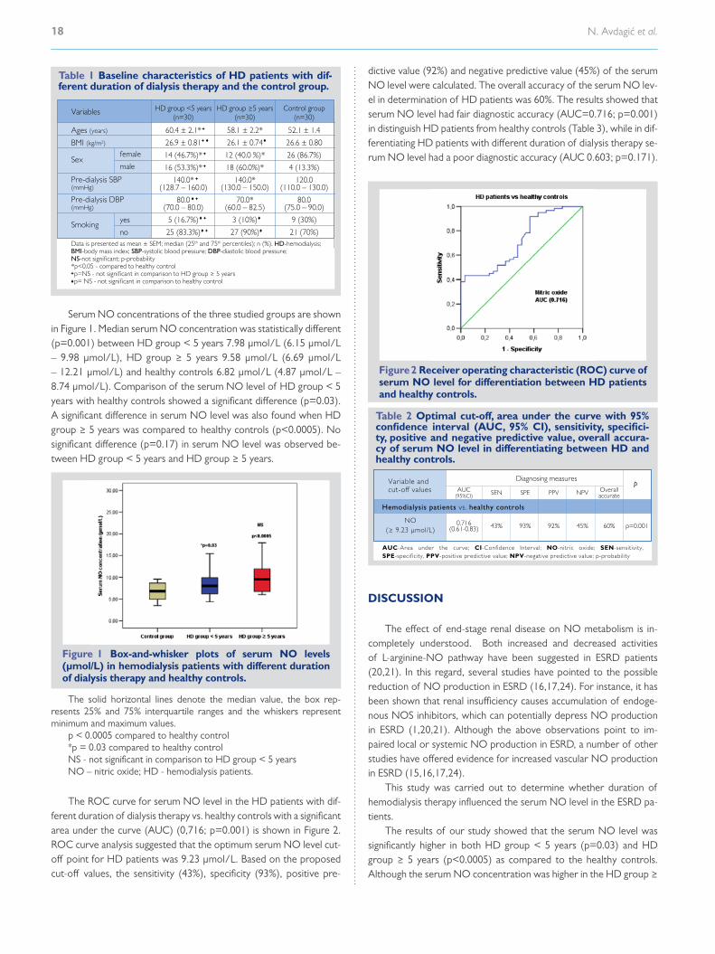
Serum NO concentrations of the three studied groups are shown in Figure 1. Median serum NO concentration was statistically different (p=0.001) between HD group < 5 years 7.98 µmol/L (6.15 µmol/L – 9.98 µmol/L), HD group ≥ 5 years 9.58 µmol/L (6.69 µmol/L – 12.21 µmol/L) and healthy controls 6.82 µmol/L (4.87 µmol/L – 8.74 µmol/L). Comparison of the serum NO level of HD group < 5 years with healthy controls showed a significant difference (p=0.03). A significant difference in serum NO level was also found when HD group ≥ 5 years was compared to healthy controls (p<0.0005). No significant difference (p=0.17) in serum NO level was observed be-tween HD group < 5 years and HD group ≥ 5 years.
The solid horizontal lines denote the median value, the box rep-resents 25% and 75% interquartile ranges and the whiskers represent minimum and maximum values.
p < 0.0005 compared to healthy control*p = 0.03 compared to healthy controlNS - not significant in comparison to HD group < 5 years NO – nitric oxide; HD - hemodialysis patients.
The ROC curve for serum NO level in the HD patients with dif-ferent duration of dialysis therapy vs. healthy controls with a significant area under the curve (AUC) (0,716; p=0.001) is shown in Figure 2. ROC curve analysis suggested that the optimum serum NO level cut-off point for HD patients was 9.23 µmol/L. Based on the proposed cut-off values, the sensitivity (43%), specificity (93%), positive pre-
dictive value (92%) and negative predictive value (45%) of the serum NO level were calculated. The overall accuracy of the serum NO lev-el in determination of HD patients was 60%. The results showed that serum NO level had fair diagnostic accuracy (AUC=0.716; p=0.001) in distinguish HD patients from healthy controls (Table 3), while in dif-ferentiating HD patients with different duration of dialysis therapy se-rum NO level had a poor diagnostic accuracy (AUC 0.603; p=0.171).
DISCUSSION
The effect of end-stage renal disease on NO metabolism is in-completely understood. Both increased and decreased activities of L-arginine-NO pathway have been suggested in ESRD patients (20,21). In this regard, several studies have pointed to the possible reduction of NO production in ESRD (16,17,24). For instance, it has been shown that renal insufficiency causes accumulation of endoge-nous NOS inhibitors, which can potentially depress NO production in ESRD (1,20,21). Although the above observations point to im-paired local or systemic NO production in ESRD, a number of other studies have offered evidence for increased vascular NO production in ESRD (15,16,17,24).
This study was carried out to determine whether duration of hemodialysis therapy influenced the serum NO level in the ESRD pa-tients.
The results of our study showed that the serum NO level was significantly higher in both HD group < 5 years (p=0.03) and HD group ≥ 5 years (p<0.0005) as compared to the healthy controls. Although the serum NO concentration was higher in the HD group ≥
Variables HD group <5 years
(n=30)
Ages (years)
BMI (kg/m2)
Sex
Pre-dialysis SBP (mmHg)
Pre-dialysis DBP(mmHg)
Smoking
60.4 ± 2.1*
26.9 ± 0.81
14 (46.7%)*
16 (53.3%)*
140.0*(128.7 – 160.0)
80.0(70.0 – 80.0)
5 (16.7%)
25 (83.3%)
52.1 ± 1.4
26.6 ± 0.80
26 (86.7%)
4 (13.3%)
120.0(110.0 – 130.0)
80.0(75.0 – 90.0)
9 (30%)
21 (70%)
58.1 ± 2.2*
26.1 ± 0.74
12 (40.0 %)*
18 (60.0%)*
140.0*(130.0 – 150.0)
70.0*(60.0 – 82.5)
3 (10%)
27 (90%)
HD group ≥5 years(n=30)
Control group(n=30)
female
male
yes
noData is presented as mean ± SEM; median (25th and 75th percentiles); n (%). HD-hemodialysis; BMI-body mass index; SBP-systolic blood pressure; DBP-diastolic blood pressure; NS-not significant; p-probability*p<0.05 - compared to healthy control p=NS - not significant in comparison to HD group ≥ 5 years p= NS - not significant in comparison to healthy control
Table 1 Baseline characteristics of HD patients with dif-ferent duration of dialysis therapy and the control group.
Figure 1 Box-and-whisker plots of serum NO levels (µmol/L) in hemodialysis patients with different duration of dialysis therapy and healthy controls.
Figure 2 Receiver operating characteristic (ROC) curve of serum NO level for differentiation between HD patients and healthy controls.
AUC(95%CI)
Hemodialysis patients vs. healthy controls
SEN SPE PPV NPV
0.716(0.61-0.83) 43% 93% 92% 45% 60% p=0.001
Overallaccurate
Diagnosing measuresVariable and cut-o� values
NO(≥ 9.23 µmol/L)
p
AUC-Area under the curve; CI-Confidence Interval; NO-nitric oxide; SEN-sensitivity, SPE-specificity, PPV-positive predictive value; NPV-negative predictive value; p-probability
Table 2 Optimal cut-off, area under the curve with 95% confidence interval (AUC, 95% CI), sensitivity, specifici-ty, positive and negative predictive value, overall accura-cy of serum NO level in differentiating between HD and healthy controls.
18 N. Avdagić et al.

5 years as compared to HD group < 5 years, there was no statistically significant difference between the compared groups (p=0.17). Our results are completely in accordance with the results of Schmidt et al. (16, 17) who also found statistically significant higher serum NO con-centration in ESRD patients as compared to the control group. How-ever, these authors have demonstrated that total NO production is low in ESRD, and concluded that this NO deficiency may contribute to hypertension and disease progression in ESRD. The same authors conducted a third study (24) to determine whether 24-hour NOX (NO2 and NO3) excretion (a qualitative index of total NO produc-tion) was reduced in patients with chronic renal disease. They con-ducted a study with controlled low-NOX intake and found that low 24-hour NOX output in ESRD patients suggested that total systemic NO production was low in humans with impaired renal function, as well as inadequate plasma clearance.
Meenakshi et al. (15) conducted a study to determine whether there were any changes in the levels of NO in chronic renal failure (CRF), as the disease progresses. These authors have demonstrat-ed that CRF patients on maintenance hemodialysis have markedly increased NO concentration compared to the controls, and suggest that measurement of NO level in the peripheral blood may be used as an indicator of prognostic follow-up in the CRF patients on dialy-sis. This finding was in accordance with the report of Dejanova et al. (25) who also observed significantly higher serum NO level in HD patients compared to controls. However, with regard to hemodialysis duration these authors did not find statistically significant difference in serum NO concentration, which is in accordance with our results. In their opinion, the increased nitric oxide values observed in HD patients is probably due to the induction of hemodialysis membrane and/or a lack of renal excretion.
We further investigated the ability of serum NO concentration in differentiating HD patients from healthy controls as well as HD patients with different duration of dialysis therapy. In our study, based on the selected optimal cut-off value by ROC curve analysis, serum NO values showed a low sensitivity (43%) and excellent specificity (93%) in distinguishing HD patients from healthy controls. The results of our study demonstrate that the serum NO level displays a fair di-agnostic accuracy in distinguishing hemodialysis patients from healthy controls (AUC=0,716; p=0.001) and poor diagnostic accuracy in distinguishing hemodialysis patients with different duration of dialysis therapy (AUC 0.603; p=0.17).
CONCLUSION
Up-regulation of systemic nitric oxide synthesis may play a key role in the modulation of complex hemodynamic disorders in ESRD patients. The present study demonstrates that serum NO produc-tion is high in ESRD patients on hemodialysis therapy as compared to the controls. The obtained results support the hypothesis that serum NO level measurement in ESRD patients can be used as independent marker only to distinguish hemodialysis patients from healthy control, but not in differentiating hemodialysis patients with different duration of dialysis therapy. However, due to small sample size, our findings are warranted for further confirmation in larger studies.
Conflict of interest: none declared.
REFERENCES
1. Moncada S, Palmer RM, Higgs EA. Nitric oxide: physiology, pathophysiology and phar-
macology. Pharmacol Rev. 1991;43(2):109-42.
2. Bredt DS. Endogenous nitric oxide synthesis: biological functions and pathophysiology.
Free Radic Res. 1999;31(6):577-596.
3. Roberts W, Riba R, Homer-Vanniasinkam S, Farndale RW, Naseem KM. Nitric oxide
specifically inhibits integrin-mediated platelet adhesion and spreading on collagen. J
Thromb Haemost. 2008;6(12):2175-85.
4. Guix FX, Uribesalgo I, Coma M, Muñoz FJ. The physiology and pathophysiology of nitric
oxide in the brain. Prog Neurobiol. 2005;76(2):126-52.
5. Omar SA, Webb AJ, Lundberg JO, Weitzberg E. Therapeutic effects of inorganic nitrate
and nitrite in cardiovascular and metabolic diseases. J Intern Med. 2015; doi: 10.1111/
joim.12441.
6. Orida NK, Lai ChS. Nitric oxide and renal patient. Dialysis and transplantation.
2000;29(4):174-185.
7. Avdagić N, Zaćiragić A, Babić N, Hukić M, Seremet M, Lepara O, Nakaš-Ićindić E. Nitric
oxide as a potential biomarker in inflammatory bowel disease. Bosn J Basic Med Sci.
2013;13(1):5-9.
8. Ibragić S, Sofić E, Suljić E, Avdagić N, Bajraktarević A, Tahirović I. Serum nitric oxide con-
centrations in patients with multiple sclerosis and patients with epilepsy. J Neural Transm
(Vienna). 2012;119(1):7-11.
9. Vannini F, Kashfi K, Nath N. The dual role of iNOS in cancer. Redox Biol. 2015;6:334-343.
10. Van Hove CE, Van der Donckt C, Herman AG, Bult H, Fransen P. Vasodilator efficacy
of nitric oxide depends on mechanisms of intracellular calcium mobilization in mouse
aortic smooth muscle cells. Br J Pharmacol. 2009;158(3):920-930.
11. Sandoo A, Jet JCS Veldhuijzen van Zanten, Metsios GS, Carroll D, Kitas GD. The endo-
thelium and its role in regulating vascular tone. Open Cardiovasc Med J. 2010;4:302-312.
12. Neves MF, Kasal DA, Cunha AR, Medeiros F. Vascular Dysfunction as Target Organ
Damage in Animal Models of Hypertension. Int J Hypertens. 2012;2012:187526.
13. Kopkan L, Cervenka L. Renal interactions of renin-angiotensin system, nitric oxide and
superoxide anion: implications in the pathophysiology of salt-sensitivity and hyperten-
sion. Physiol Res. 2009;58(2):S55-S67.
14. Sinha N, Dabla PK. Oxidative stress and antioxidants in hypertension-a current review.
Curr Hypertens Rev. 2015;11(2):132-42.
15. Meenakshi SR, Agarwal R. Nitric oxide levels in patients with chronic renal disease. J Clin
Diagn Res. 2013;7(7):1288-90.
16. Schmidt RJ, Yokota S, Tracy TS, Sorkin MI, Baylis C. Nitric oxide production is low in end-
stage renal disease patients on peritoneal dialysis. Am J Physiol. 1999;276(5Pt 2):F794-7.
17. Schmidt RJ, Domico J, Samsell L, Yokota S, Tracy TS, Sorkin MI et al. Indices of activity of
the nitric oxide system in patients on hemodialysis. Am J Kidney Dis. 1999;34:228-234.
18. Clinical Practice Guideline for the Evaluation and Management of Chronic Kidney Dis-
ease. KDIGO 2012.
19. Green LC, Wagner DA, Glogowski J, Skipper PL, Wishnok JS, Tannenbaum SR. Analysis
of nitrate, nitrite and [15N]nitrate in biological fluids. Anal Biochem 1982;126(1):131-8.
20. MacAllister RJ, Whitley GSJ, Vallance P. Effect of guanidino and uremic toxins on nitric
oxide systems. Kidney Int. 1994;45:737-742.
21. Vallance P, Leone A, Calver A, Collier J, Moncada S. Accumulation of an endogenous
inhibitor of nitric oxide synthesis in chronic renal failure. Lancet. 1992;339:572-575.
22. Eggers PW. Effect of transplantation on the Medicare end-stage renal disease program.
N Engl J Med. 1988;318(4):223-9.
23. Raymond T. Krediet RT, Balafa O. Cardiovascular risk in patient the peritoneal dialysis.
Nat Rev Nephrol. 2010;6:451- 460.
24. Schmidt RJ, Baylis C. Total nitric oxide production is low in patients with chronic renal
disease. Kidney Int. 2000;58:1261-1266.
25. Dejanova B, Dejanov P, Petrovska S, Jakovljevic V. Nitric oxide in hemodialysis patients.
Ser J Exp Clin Res. 2011;12 (2):75-78.
Reprint requests and correspondence:Nesina Avdagić, MD, PhDDepartment of Physiology Medical Faculty, University of SarajevoČekaluša 90, 71000 Sarajevo, Bosnia and HerzegovinaPhone/fax: + 387 33 226 472Email: [email protected]
Serum nitric oxide level in end-stage renal disease patients with different duration of dialysis therapy 19

The incidence and some characteristics of patients with hemophilia in Bosnia and Herzegovina
Incidenca i neke karakteristike pacijenata sa hemofilijom u Bosni i Hercegovini
Edo Hasanbegović1*, Jelica Predojević-Samardžić2, Nermana Čengić1
1Pediatric Clinic, University Clinical Center Sarajevo, Patriotske lige 81, 71 000 Sarajevo, Bosnia and Herzegovina; 2Children’s Hospital, University Clinical Center Banja Luka, Zdrave Korde 1, 78000, Banja Luka, Bosnia and Herzegovina
*Corresponding author
ABSTRACT
Introduction: hemophilia is a rare coagulation disorder, characterized by a tendency to bleed. The transmission of the disease is linked to a sex chromosome X, and it usually affects only males, while females are carriers. Objective: the aim of this study is to show the structure of patients with blood coagulation disorders in B&H, distribution of hemophilia A to the severity of the disease, age and developed inhibitors in patients. Materials and methods: we retrospectively analyzed the medical records of patients with congenital bleeding disorders in the largest children‘s hospitals in the two entities of Bosnia and Herzegovina: Pediatric Clinic of the University Clinical Center Sarajevo, in the Federation of Bosnia and Herzegovina (FB&H) and the University Children‘s Hospital Banja Luka, in the Republic of Srpska (RS). We also analyzed data from the Health Insurance Funds in both entities, in terms of annual consumption of coagulation factors in 2015. Results: the study showed that most people registered with coagulation disorders were with hemophilia A, 146 (68%), hemophilia B was in the second place with 35 (16%) patients, and there were 28 (13%) patients in the third place with Von Willebrand disease (VWD). There were 6 (3%) patients with other blood coagulation disorders. Majority of patients suffered from severe hemophilia A, 61 (42%). Moderate hemophilia A was associated with 48 (33%) patients and 37 (25%) patients suffered from mild form of hemophilia. There were 52 (36%) patients with hemophilia A under the age of 18 and 94 (64%) patients over 18 years of age. Low titre inhibitors <5 BU were developed in 6 (4%) patients with hemophilia A and high titre inhibitors ≥ 5 BU in 3 (2%) patients. Hemophilia B patients did not develop inhibitors. Conclusion: according to the World Federation of Hemophilia a comprehensive treatment of patients with hemophilia is realized at tertiary level centers. The introduction of home treatment and regular prophylaxis is a priority for all children with severe hemophilia, and for most adult patients.
Key words: hemophilia, inhibitors, prophylaxis
SAŽETAK
Uvod: hemofilija je rijedak poremećaj koagulacije, koju karakteriše sklonost krvarenju. Ovaj poremećaj se prenosi vezano za polni hromozom X, te uglavnom oboljevaju samo muškarci, dok su žene prenosioci. Cilj rada: da se prikaže struktura oboljelih od poremećaja koagulacije krvi u BiH, rasprostranjenost hemofilije A prema težini bolesti, starosnu dob i razvijene inhibitore kod oboljelih. Ispitanici i metode: retrospektivno su analizirane historije bolesti pacijenata s urođenim poremećajima krvarenja u najvećim dječijim bolnicama u dva entiteta Bosne i Hercegovine: Pedijatrijska klinika Univerzitetskog kliničkog centra u Sarajevu (UKCS), u Federaciji Bosne i Hercegovine (FBiH) i Univerzitetska Dječija bolnica Banja Luka u Republici Srpskoj (RS). Također su analizirani i podatci iz Fonda zdravstvenog osiguranja oba entiteta u pogledu godišnje potrošnje faktora koagulacije u 2015. Rezultati: istraživanje je pokazalo da najviše registrovanih osoba sa poremećajem koagulacije otpada na hemofiliju A 146 (68%), hemofilija B je na drugom mjestu sa 35 (16%) oboljelih, a na trećem mjestu sa 28 (13%) bilo je oboljelih od Von Willebrandove bolesti (VWD). Na ostale poremećaje krvarenja otpada 6 (3%) oboljelih. Najviše pacijenata je obolilo od teške forme hemofilije A, 61(42%). Na umjereni oblik hemofilije A otpada 48 (33%), a na blage forme 37 (25%). Broj oboljelih od hemofilije A ispod 18 godina je 52 (36%), a broj oboljelih iznad 18 godina je 94 (64%) pacijenata. Blagi oblik razvijenih inhibitora sa titrom < 5BU imalo je 6 (4%) pacijenata sa hemofilijom A, a sa visokim titrom ≥ 5BU, 3 (2%). Kod hemofilije B nije bilo pacijenata sa razvijenim inhibitorima. Zaključak: sveobuhvatan tretman osoba oboljelih od hemofilije prema standardu Svjetske federacije za hemofiliju, obavljaju centri na tercijarnoj razini. Uvođenje kućnog liječenja i redovna profilaksa predstavljaju prioritet za svu djecu s teškom hemofilijom i za većinu odraslih pacijenata.
Ključne riječi: hemofilija, inhibitori, profilaksa
Medical Journal (2016) Vol. 22, No. 1, 20 - 22 Original article

INTRODUCTION
Hemophilia is a rare recessive hereditary disorder of blood clotting, which is characterized by a tendency to bleed. The word hemophilia has Greek roots haima (blood), philein (love) and represents the earliest described hereditary disease.
The transmission of the disease is linked to a sex chromosome X, and it usually affects only males, while females are carriers. In about 30% of patients with hemophilia, family history was negative, indicating a spontaneous mutation of recessive gene for FVIII or transfer of the mutated genes for several generations. These are sporadic hemophilias. Hemophilia is characterized by a lack of specific coagulation factors, and classified into hemophilia A, or classic hemophilia related to the lack of factor VIII, hemophilia B (Christmas disease) related to the lack of factor IX, and hemophilia C related to the lack of factor XI. Hemophilia A occurs in 80% of all cases and is five times more common than hemophilia B, while hemophilia C is even less likely to occur (1,2).
It is assumed that the history of hemophilia is old as the history of man. The Talmud, sacred book of the Jews, written 1.500 years ago, stated that young Jew should not be circumcised, because the two sons of the same mother and three sons of her sister bled and died after circumcision.
Hemophilia is sometimes called ‚‘the royal disease“ because the Queen Victoria transferred the gene for this disease to courts across Europe through her daughters (2,3,4).
The diagnosis of hemophilia is set based on personal and family medical history, clinical and laboratory findings. Hemophilia should be suspected if there is a history of hemophilia in the family, bruising and bleeding in the first years of life, spontaneous bleeding in joints and muscles, bleeding that is difficult to stop after injuries, tooth extraction or surgery. In hemophilia is lower F VIII, prolonged the coagulation time and active partial thromboplastin time (APTT). The platelet count is normal and the bleeding time is regular (4,5,6).
The clinical presentation mainly depends on the degree of deficit factor; F VIII level less than 1% indicates a serious condition, where occurs spontaneous bleeding not related to trauma. In moderate hemophilia with the values of F VIII 1-5%, bleeding occurs with a blow or fall, and patients with F VIII activity of 10-15%, bleed only after strong trauma (mild form). Most newborns do not show signs of the disease in the newborn period. With the development of motor functions (crawling, rising to the feet, walking) bleeding in joints and muscles appears in the form of large hematomas. Dentition or circumcision in the infant period may sometimes be the first bleeding in children suffering from hemophilia. The disease has a tendency to repeated bleeding into the same joint, which often results in degenerative changes, ankylosis of joints, osteoporosis and muscle atrophy. Rarely, there is bleeding in the CNS after head trauma or in the gastrointestinal tract (7,8,9).
The aim of this study is to show the structure of patients with blood coagulation disorders in B&H, distribution of hemophilia A to the severity of the disease, age and developed inhibitors in patients.
MATERIALS AND METHODS
We retrospectively analyzed the medical records of patients
with congenital bleeding disorders in the largest children‘s hospitals in the two entities of Bosnia and Herzegovina: Pediatric Clinic of the UCCS, in the Federation of Bosnia and Herzegovina (FB&H) and the University Children‘s Hospital Banja Luka, in the Republic of Srpska (RS). We also analyzed data from the Health Insurance Funds in both entities, in terms of annual consumption of coagulation factors in 2015.
The analysis was done according to the type of blood coagulation disorder, distribution of hemophilia, age structure, level of inhibitors and therapeutic options.
RESULTS
Table 1 shows the most common congenital blood coagulation disorders in Bosnia and Herzegovina. The majority of patients were registered with hemophilia A, 146 (68%), hemophilia B was in the second place with 35 (16%) patients, and Von Willebrand disease (VWD) was in the third place with 28 (13%) patients. 6 (3%) patients had other blood coagulation disorders.
Table 2 shows the distribution of hemophilia A to the severity of the disease. Majority of patients suffered from severe hemophilia A, 61 (42%). Moderate hemophilia A was associated with 48 (33%) patients and 37 (25%) patients suffered from mild form of hemophilia.
Table 3 shows the age structure of patients with hemophilia A. There were 52 (36%) patients with hemophilia A under the age of 18, and 94 (64%) patients over 18 years of age.
Table 4 shows the level of developed inhibitors in hemophiliacs. A total of 9 (6%) patients developed inhibitors. Low titre inhibitors
The incidence and some characteristics of patients with hemophilia in Bosnia and Herzegovina 21
Hemophilia A
Hemophilia B
Von Willebrand disease (VWD)
Other blood coagulation disorders
Total
Number of patientsDisease Percentage (%)
146
35
28
6
215
68
16
13
3
100
Table 1 The structure of patients with blood coagulation disorders in Bosnia and Herzegovina.
Mild
Moderate
Severe
Total
Number of reg. patientsHemophilia A Percentage (%)
37
48
61
146
25
33
42
100
Table 2 The distribution of hemophilia A to the severity of the disease in Bosnia and Herzegovina.
Number of patients > 18 yearsNumber of patients < 18 years
52 (36%) 94 (64%)
Table 3 The age structure of patients with hemophilia A.
Titre < 5 BU
Titre ≥ 5 BU
TOTAL
Hemophilia ALevel of inhibitors (BU) Hemophilia B
6
3
9
(%)
4
2
6
/
/
0
Table 4 The developed inhibitors in patients with hemophilia.
<5 BU were developed in 6 (4%) patients with hemophilia A and high titre inhibitors ≥ 5 BU in 3 (2%) patients. Hemophilia B patients did not develop inhibitors.
DISCUSSION
Hemophilia is a rare coagulation disorder characterized by a tendency to bleed. It is estimated that according to the last census Bosnia and Herzegovina has 3.791.662 inhabitants. The number of patients with hemophilia A and B is 181. Since Bosnia and Herzegovina is a European country, the priority health care of people with hemophilia is to achieve European protection principles for hemophilia. The introduction of home treatment and delivery of coagulation factors in the local pharmacies are the most important tasks to be achieved along with the introduction of regular prophylaxis for all children with severe hemophilia, and for most adult patients. With regard to congenital blood coagulation disorders in Bosnia and Herzegovina, majority of patients were registered with hemophilia A, 146 (68%), hemophilia B was in second place with 35 (16%) patients, and Von Willebrand disease (VWD with 28 (13%) patients was in the third place. 6 (3%) patients had other blood coagulation disorders (Table 1). A similar proportion of patients according to the severity of the disease, was also presented by the World Federation of Hemophilia (WFH) (10,11).
The distribution of hemophilia A to the severity of the disease in B&H show that the majority of patients suffer from severe hemophilia A, 61 (42%). 48 (33%) patients suffer from moderate hemophilia A, while 37 (25%) of patients has a mild form of the disease. Similar results have been described by other authors (9, 10).
The age structure of patients with hemophilia A showed that there was 52 (36%) patients with hemophilia A under the age of 18, and 94 (64%) patients over the age of 18.
Out of all patients with hemophilia, 9 (6%) of them developed inhibitors. Low titre inhibitors < 5 BU were developed in 6 (4%) patients with hemophilia A and high titre inhibitors ≥ 5 BU in 3 (2%) patients. Hemophilia B patients did not develop inhibitors (12,13,14).
The data show a much smaller percentage of developed inhibitors compared to other countries in the world.
CONCLUSION
According to the World Federation of Hemophilia a comprehensive treatment of patients with hemophilia is realized in tertiary level centers. In developed countries, patients with hemophilia live a normal life with proper treatment. The problem relates to unregistered patients. The most important priorities would be the establishment of the National Center for Hemophilia, the National Registry of patients with hemo-philia and development of orthopedic surgery for patients with joints disability.
Conflict of interest: none declared.
REFERENCES
1. Ingram GIC. The history of haemophilia. Haemophilia. 1997;3:5-13.
2. Otto JC. The Medical Repository. 1803:Vol VI(1):1-4. Genmedhist info. Retrieved 2013-11-
21.
3. Peyvandi F. Epidemiology and management of rare bleeding disorders. Haemophilia.
2012;18(Suppl.1):3-51.
4. Christine A. Lee, Erik E. Berntorp, W. Keith Hoots. Textbook of Hemophilia, 2nd Edition,
2010, Wiley-Blackwell.
5. White GC, Rosendaal F, Alderot LM, Lusher JM, Rothchild C, Ingeslev J. Definitions in hae-
mophilia. Recommendations of the Scientific Subcommittee of F VIII and F IX of the SSC of
ISTH. Thromb Haemost. 2001;85:560.
6. Montgomery RR, Gill JC Scott JP. Haemophilia and von Wilebrand disease. In: Nathan DG,
Oski FA. Haematology of Infancy and Childhood. 5 th. ed. Philadelphia: WB Saunders
1998;1644 - 59.
7. Giannelli F, Green PM. The molecular basis of haemophilia A and B. Baileres Clin Haematol.
1996;9:211-28.
8. Llinás A. Haemophilia arthropathy. Haemophilia. 2010;16:121. PubMed PMID: 20590868.
9. Peyvandi F, Jayandharan G, Chandy M, Srivastava A, Nakaya SM, Johnson MJ, et al. Genetic
diagnosis of haemophilia and other inherited bleeding disorders. Haemophilia. 2006;12(Sup-
pl.3):82-9.
10. Bolton-Maggs PH, Favaloro EJ, Hillarp A, Jennings I, Kohler HP. Difficulties and pitfalls in the
laboratory diagnosis of bleeding disorders. Haemophilia. 2012;18(Suppl.4):66-72.
11. Ingerslev J. Laboratory assays in hemophilia. In: Lee CA, Berntorp EE, Hoots WK, editors.
Textbook of hemophilia. London: Wilet Blackwell Company. 2010;14-47.
12. Guidelines for the management of hemophilia. World Federation of Hemophilia, 2005.
13. Colvin BT, Astermark J, Fischer K, Gringeri A, Lassila R, Schramm W, Thomas A, Ingerslev
J. Inter Disciplinary Working Group. European principles of haemophilia care. Haemophilia.
2008;14(2):361-74.
14. Hay CR, Brown S, Collins PW, Keeling DM, Liesner R. The diagnosis and management
of factor VIII and IX inhibitors: a guideline from the United Kingdom Haemophilia Centre
Doctors Organization. Br J Haematol. 2006;133(6):591-605.
15. Bolton-Maggs PH, Favaloro EJ, Hillarp A, Jennings I, Kohler HP. Difficulties and pitfalls in the
laboratory diagnosis of bleeding disorders. Haemophilia. 2012;18(Suppl.4):66-72.
16. Ingerslev J. Laboratory assays in hemophilia. In: Lee CA, Berntorp EE, Hoots WK, editors.
Textbook of hemophilia. London: Wilet Blackwell Company. 2010;14-47.
Reprint requests and correspondence:Edo Hasanbegović, MD, PhDPediatric ClinicUniversity Clinical Center SarajevoPatriotske lige 81, 71000 SarajevoBosnia and HerzegovinaPhone: + 387 33 566 448Email: [email protected]
22 E. Hasanbegović et al.

Evaluation of surgical treatment of Achilles tendon rupture
Evaluacija operativnog liječenja rupture ahilove tetive
Adnana Talić-Tanović*, Fuad Džanković, Adnan Papović, Mehmed Zahirović
Clinic of Orthopedics and Traumatology, University Clinical Center Sarajevo, Bolnička 25, 71000 Sarajevo, Bosnia and Herzegovina
*Corresponding author
ABSTRACT
Achilles tendon or heel tendon is the strongest tendon in human body, and is around 15 cm long. The tendon is an extension of muscle threads of the three-headed muscle consisting of gastrocnemius muscle (m.gastrocnemius), soleus muscle (m.soleus) and plantar muscles (m.plantaris), and attaches to the heel bone. Name tendon comes from Greek mythology, the Greek hero Achilles (Achilles‘ heel). Rupture of the Achilles tendon usually occurs during recreational and sports activities, and the highest rate is reported in former athletes aged 40 years (football players, skiers, athletes). Ruptured tendon damage the surrounding tissue - usually occurs muscle contusion, hematoma develops that infiltrates the muscle tissue and damage contractility. Traumatized healed tendon rarely achieves the strength of a healthy tendon and is subject to new injuries of less effort. Diagnosis of rupture is made by the clinical examination, positive Thompson‘s sign and mandatory ultrasound examination. The aim of the work is to show the principles of surgical treatment and priority for emergency surgical treatment of fresh Achilles tendon rupture in relation to the delayed surgery. With surgical treatment the ends of the tendon should be smooth, thus the ends of the tendons that are torn or crushed get refreshed. Regardless of the type of stitches used, the tendon must not be under tension so that the suture does not dehiscence, and again achieves the physiological length. The study included total of 58 patients, of which 17 patients (45.71%) had an emergency surgery, and 31 (54.29%) delayed operative treatment. The average age of patients was 39 years. All patients were male and were surgically treated by open and percutaneous sutures method. The obtained results showed that in the group of urgently operated patients there was no case of rerupture and splicing, and no signs of infection, whereas there was one case of pulmonary thromboembolism. In the group of patients with delayed surgery there was one case of rupture, 3 cases of superficial infections, and 2 cases of wound dehiscence. The recovery of tendon muscle strength and tendon reaching the full range of motion required a shorter time in the group of emergency treated patients. With regard to fresh Achilles tendon rupture, the preference should be given to the emergency surgery - the treatment up to 12 hours from the time of injury.
Key words: Achilles tendon rupture, emergency operative treat-ment, delayed operative treatment
SAŽETAK
Petna tetiva ili Ahilova tetiva je najsnažnija tetiva kod čovjeka koja je duga oko 15 cm. Tetiva je produžetak mišićnih niti troglavog gnjatnog mišića (kojeg čine trbušasti mišić lista (musculus gastrocnemius), široki listoliki mišić (musculus soleus) i tabanski mišića (musculus plantaris), a hvata se za petnu kost. Naziv Ahilova tetiva dolazi iz grčke mitologije, od grčkog junaka Ahileja (Ahilova peta). Ruptura Ahilove tetive se najčešče događa u rekreativnoj i spotskoj aktivnosti, a najviše kod bivših sportista u dobi oko 40 godina (fudbaleri, skijaši, atletičari). Rupturom tetive oštećuje se i okolno tkivo - obično nastaje kontuzija mišića, razvija se hematom koji inf iltrira mišićno tkivo i oštećuje kontraktibilnost. Traumatizirana zacijeljena tetiva rijetko postiže čvrstoću zdrave tetive i podložna je novim ozljedama na manji napor. Dijagnoza rupture se postavlja kliničkim pregledom, Thompson-ov znak pozitivan i obavezno ultrazvučni pregled. Cilj rada je da prikaže principe operativnog liječenja i prednost hitnog operativnog liječenja svježe rupture Ahilove tetive u odnosu na odgođeni operativni zahvat. Kod operativnog tretmana krajevi tetive trebaju biti glatki, pa se stoga krajeve tetiva koji su rastrgani ili zgnječeni osvježe. Bez obzira na vrstu šavova tetiva ne smije biti pod tenzijom da šav ne dehiscira, da ponovo ima f iziološku dužinu. Radom je obuhvaćeno 58 pacijenata, od kojih je 17 pacijenta (45,71%) zbrinuto kao hitni operativni zahvat, a 31 (54,29%) kao odgođeni. Prosječna starost pacijenata je 39 godina. Svi pacijenti su muškog spola i operativno su liječeni otvorenom metodom suturama i perkutanim suturama. Dobijeni rezultati su pokazali da u grupi hitno operisanih nije bilo nesrastanja i reruptura, kao ni znakova infekcije, dok je bila jedna tromboembolija pluća. U grupi pacijenata sa odgođenim operativnim zahvatom imali smo jednu rerupturu, 3 površne infekcije i 2 dehiscencije rane. Oporavak mišićne snage tetive i dostizanje punog obima pokreta zahtjevao je kraće vrijeme kod hitno operisanih pacijenata. Kod svježe rupture Ahilove tetive prednost treba dati hitnom operativnom zahvatu - liječenju do 12 sati od vremena povređivanja .
Ključne riječi: ruptura Ahilove tetive, hitni operativni zahvat, elektivni operativni zahvat
Medical Journal (2016) Vol. 22, No. 1, 23 - 26 Original article
INTRODUCTION
The tendon is named after the Greek hero Achilles, who according to the legend was bathed in the river Styx by his mother Thetis, holding him by the heel. Achilles was vulnerable only in that place, and was killed when Paris‘ poisoned arrow (directed by Apollo) hit his heel. In medical literature the name Achilles tendon (tendo Achillis) was introduced by professor of anatomy from Loewen and the first work related to the Achilles tendon rupture was published by Ambrose Pare in 1633 (1,2). Achilles tendon is part of a three-headed muscle of the lower leg that starts with condyle thigh and head of the fibula and ends with tendon that attaches to the heel bone. The muscle bends the lower leg and foot, taking part in all the movements while walking, standing on tiptoe, squat, run, jump and landing.
The factors leading to increased incidence of rupture are the association between weight and strength of athletes, increased intensity of training, increased use of unnatural substances (eg. creatine), and the use of corticosteroids, growth hormones and testosterone. An increase Achilles tendon rupture is proportional to the increase in recreational sports activity in both young and elderly - even top athletes are not spared. The cause of the rupture is the combination of existence of relative hipovascularized zone and repeated microtraumas which cause the inflammatory reparative process, which due to poor vascularization is not able to oppose the stresses (4,5). Recently, there is a reported trend of an increased interest in sports and recreational activities among the people of mid-life ages (6,7). It is more common among men than women, the ratio being 9:1. The mechanism of Achilles tendon rupture is most often eccentric contraction in the ankle dorsiflexion and knee extension when m.soleus and m.gastroknemius are at their maximum tension (7,8).
Achilles tendon allows standing on toes, running, jumping, walking normally, climbing up and down the stairs. According to the action, m. triceps surae raises the heel and the entire body on your toes. It strongly raises the rear part of the foot, separating it from the ground and throws forward (1,6). When ruptured (torn) tendon clinical picture is typical. On the site of the rupture, two to three fingers wide above the heel, it can be seen clearly and touched the recess length of 2-3 cm, because the tendon is not toned, an broken ends are stretched. The patient feels a strong pain as a „shot with a rock“ in the heel area, and sometimes hears the sound phenomenon called „the bang/the burst“. Clinical signs are palpable, painful „hollow“ in the Achilles tendon and a partial or complete loss of plantar flexion. Positive Thompson („squeeze“) test, or lack of plantar flexion of the foot to manual compression in the quadriceps is a sure sign of the rupture (2,3,9).
Non-operative treatment is indicated in the elderly and in patients who have contraindications from anesthesiological aspect. In that case upper-knee circulatory cast with foot is placed in equines of about 20 degrees and the knee in flexion of 15-20 degrees for a period of 3 weeks, followed by another 3 weeks tibia plaster immobilization gravity equinus foot, and the last 3 weeks of immobilization with the foot in a neutral position and the ability to support (3 + 3 + 3). Partial joint stiffness (contracture) follows this treatment and the healing tendon requires a long period of physical therapy for satisfactory movement in the ankle joint. The scar at the site of ends growing together over the tendon is stiff and rigid, not more resilient as the
rest of the tendon and should avoid efforts with that foot, because there is a re-rupture tendon on earlier place (8,9,10).
Tendon rupture is associated with muscle contusion, and hematoma developing which infiltrates the muscle tissue and impairs contractility. Hematoma and swelling puts pressure on the posterior tibial artery and other neurovascular elements. Any late recognition and delayed surgery can cause complications. If we do not recognize tendon rupture or postpone surgery and treat the injured with conservative methods, a larger scar develops between the ends of broken parts, twice weaker than the rest of the healthy part of the tendon. The action of the lower leg muscles is impaired which is very important when walking, climbing stairs and up the slope and standing on tiptoe. The tendon significantly weakens and develops disability (1,2,11).
The Achilles tendon rupture is most common 2-6 cm from its grips on the calcaneus bone, which is the zone of tendon hipovascularization, thus in larger defect the healing time is largely extended. Rehabilitation after surgery is very gradual and dosed and controlled strictly by physiatrist and surgeon. The result of such treatment is successful return to sport and other activities. Operative treatment has advantages for anatomic restitution of tendons and the preservation of its length, reducing scar tissue surgically (except in patients who for other reasons are unable to operate) given that an open access to tissues enables reconstruction of the tendon.
The aim of this paper is to demonstrate the principles and benefits of emergency surgical treatment of fresh Achilles tendon rupture in comparison to elective surgery.
MATERIALS AND METHODS
The study included a total of 58 patients treated at the Clinic of Orthopedics and Traumatology of the University Clinical Centre Sarajevo in the period from 1 January 2013 to 31 December 2014. Patients were divided into two groups: group I comprising 17 patients (45.71%) operated immediately (emergency surgery treatment) and group II consisting of 31 patients (54.29%) treated with elective surgery treatment. Before the start of treatment both groups of patients were subjected to a thorough clinical examination, X-ray of the lower leg with the ankle, and the ultrasound of Achilles tendon with linear probe of 7 MHz - 10 MHz. Operative treatments were carried out by the open method and percutaneous sutures. The patient is placed in ventral decubitus, with the foot in plantar flexion of about 15 degrees, which is important in order to bring the cut ends of the tendon into direct contact. Absorbable suture is used. When the thread ends are tightened, the ends of the tendon chords come into direct contact. Once the ends of the Achilles tendon were connected, defect on the site of rupture was not palpable and Thompsons‘ sign had to be negative in order to achieve a the foot stability in pronation and supination, and active performance of movements.
While the patient was on the operating table, upper-limb plaster was placed with foot in equines of about 15 degrees and the knee in flexion. After 2-3 weeks the popliteal plaster was set, where certain surgeons retain the position of the foot in equines position for 2 more weeks, then plaster for travel lasting more than 2 weeks. All patients from the time of admission to the Clinic received thromboprophylaxis. Following the plaster removal, physical therapy was carried out.
24 A. Talić-Tanović et al.

Evaluation of surgical treatment of Achilles tendon rupture 25
RESULTS The average age of patients was 38.8 ± 2.79 years (23-57 years
old), whereas the average age in the group of patients treated with emergency surgery was 37.1 ± 3.98 years, and 40.2 ± 2.39 in the group treated with elective surgery with the statistically significant dif-ference (t = 3.59; p = 0.0010, p <0.05) as shown in Figure 1.
The work results showed that there were no cases of rerup-tures and no healing in the group of operated patients. In the group treated with elective surgery, 1 rupture and 1 no healing of rupture was recorded, thus in both cases we performed surgery. In emer-gently operated patients no infection was detected, but 1 pulmonary thromboembolism was registered (2.85%). Thus it can be concluded that most of the patients did not have complications, which was sta-tistically significant (χ2 test, p <0.1) as compared to the total number of study patients (Figure 2).
Emergency surgery of the Achilles tendon rupture was made within 12 hours of the injury, and elective surgeries was done on the fourth day after injury on average. Achilles tendon rupture of pa-tients occurred in 33% of cases during basketball play, in 17% during football plays, in 13% it occurred during slips, in 9% while jogging, tennis 3%, and during other activities 25%.
It is observed that in the examined patients the rupture occurred on the right side in 28 cases (60.86%), while the left side of the rup-ture occurred in 18 (39.14%) of cases (Figure 3).
There were no signs of deep infection, whereas a superficial wound infection occurred in one case, which was repaired by regu-lar wound bandaging and guided antibiotic treatment based on swab wound finding (highly sensitive to Staphylococcus aureus).
Immobilization of the injured limb lasted 47.26 ± 1.69 days on average; 42.68 ± 1.01 days in the group of urgently operated pa-tients, and 46-54 days in the group of patients with delayed opera-tion, with statistically significant difference (p = 9.493; p = 0.0001, p <0.05) as shown in Figure 4.
DISCUSSION
The tendon is a solid elastic structure made of fibrous connective tissue that connects muscle to bone. Under the control of muscle it directs movements, carries and resists the force of muscle contraction, and protects the muscle against external forces. When the load forces are greater than the threshold of tendon endurance, the tendon rupture occurs. The strong force that overloads the tendon can cause rupture. Permanent microtrauma, which are the result of incorrect or excessive load during sports activities, create morphological changes in connective and supportive tissue. In that way, morphologically altered tendon has an excessive burden which leads to its rupture. Similar methods of injury are reported in our study patients (1,2,3).
Injuries may result from more predisposing factors such as: anatomical variations and biomechanical abnormalities, difference in leg length, disorder shaft of the lower extremities in the hip, knee angular deformities (O or X legs), excessive rotation of the lower leg to the outside, and lowered the hollowed foot lead to changes and the tension in the string (3,4,5). Inflammation, and subsequent rupture of the Achilles tendon occurs after a sudden increase in activity of play, including change in playing pads from hard surfaces (concrete) to a softer (land), as well as due to prolonged repetitive microtrauma to the tendon due to inadequate equipment or no efficient training (6,7).
Ruptures are more common in older recreational athletes over the age of forty and occur in sudden acceleration where the incidence reaches 5.5% (12). The incidence of injury is about 0.2% in the general population. In the US the incidence is 18 per 100 000 inhabitants. Ruptures are more common in the male population, which is shown in our research, as well as in research work of other authors. This injury is more common in men than in women who practice recreational sports (1,7). The average age of patients is between 38 and 40 years, corresponding to the results of other authors and research works on this topic (12,14).
Figure 1 Average age.
Figure 4 Duration of treatment with cast immobilization.
Figure 2 Presence of complications.
Figure 3 Side of injury.
rightleft

Main tendon blood supply is carried out through its mesotendon, with the richest supply through the front mesentery. As it turned out, the increase in the patient age that mesenteric supply decreases. With aging there is a loss of viscosity and elasticity as a result of changes in the collagen. This loss of elasticity and increased stiffness of the tissue predispose the injury. Repeated microtrauma in this area prevent reparative processes and degenerative exhaustion like this may be responsible for many of Achilles tendon ruptures (4,6,7).
In our study, all ruptures were located in the area of 2-6 cm proximal to the attachment of the calcaneus bone, relatively avascular zone. The tendon is the thinnest in that place and blood supply scarcest. Angiographic studies of Lagergena and Lindholm carried out in this „sensitive zone“ between the 2 and 6 cm above the Achilles tendon insertions to calcaneus bone, showed a link between the hypovascular zone and tendon rupture (4). During the injury, the injured often continues walking and can no longer run. There is a possibility that the injury to the tendon is mistakenly replaced with the compromising ankle and muscle injury. Such injury requires a thorough clinical examination, X-ray, and lower limb Echo. All patients were examined by ultrasound which was important for the diagnosis confirmation and measurement of the defect length (1,2,3).
The skin in the front part of the lower limb is the only covering the bone which makes it exposed to direct impact of trauma, especially the front-inner part of lower-limb. Vascular internal network of the skin is poor, it quickly becomes ischemic and bulls/lumps are quickly formed. According to literature data, 12% of bulls occurs three hours after injury, and 43% after 24 hours which is consistent with our research results (12,14).
Out of the patients studied in the group of urgently operated, complications occurred in only 2.85% of patients, and they related to thromboembolism resulting from the plaster cast immobilization, not from the operation, as described in the literature (12,13,14).
Wound dehiscence, or divergence of its edge was reported in the group of patients with elective operation treatment. This complication depends on the type of thread used for stitching, local findings of the skin. Furthermore, the persistent presence of subcutaneous hematoma affects the postoperative wound and scar results. Material used in sewing after implantation act as a foreign body which causes the local tissue reaction (10,14). Operative treatment by percutaneous suture is rarely used at our Clinic. In five patients from our study excellent results were reported. According to Wallace RG et al. the percutaneous treatment of Achilles tendon is more successful than open surgery. This method turns a possible treatment in a small operating room, under local infiltrative anesthesia. Complications are rare and less extensive as compared to other methods. The duration of cast immobilization and lack of sports activities are shorter than in those treated with other surgical and nonsurgical methods. That is a simple surgical technique performed under local anesthesia in a small operating room, outpatient surgery or with one-day hospital stay (0.4 days). It involves wearing a short cast immobilization (7.5 weeks on average), early mobilization and physical therapy, a shorter period of injury to full healing, and start of normal sports activities and the establishment of normal functions (mean 4.52 months), no postoperative scars, more improved aesthetic appearance, and minimal complications.
Bartelli R, Gaiani L, Palmonari M have presented a successful treatment with percutaneous sutures even with ‚outdated‘ ruptures of Achilles tendon.
CONCLUSION
This study does not show statistically significant difference between these two types of treatments, but highlights the following conclusions: emergency surgery of Achilles tendon rupture is a standard procedure at the Department of Orthopedics and Traumatology, UCCS; emergency surgery procedure yields a lower risk of infection, reduces the time of cast immobilization and the time of hospitalization, rehabilitation is faster, and thus the return to sports and other daily activities.
Conflict of interest: none declared.
REFERENCES
1. Sharma P, Maffulli N. Biology of tendon injury: healing, modeling and remodeling. J of
Musculoskelet Neuronal Interact. 2006;6(2):181-90.
2. Hadžiahmetović Z, Vavra N. Traumatologija. Sarajevo: Avicena; 2005.
3. Talić-Tanović A, Hadžiahmetović Z. Sportska traumatologija. Sarajevo: Foto art;
2012.
4. Lagergren C, Lindholm A. Vascular distribution in the Achilles tendon: an angiograph-
ic and microangiographic study. Acta Chir Scand. 1959;116(5-6):491-495.
5. Horstmann T, Lukas C, Merk J, Brauner T, Mundermann A. Deficits 10-years after
Achilles tendon repair. Int J Sports Med. 2012;33(6):474-479.
6. Ham P, Maughan KL. Achilles tendinopathy and tendon rupture. http://www.upto-
date.com/contents/
7. Silbernagel KG, Brorsson A, Olsson N, Eriksson BI, Karlsson J, Nilsson-Helander K. Sex
differences in outcome after an acute Achilles tendon rupture. Orthop J Sports Med.
2015;3(6):2325967115586768.
8. Stavrou M, Seraphim A, Al-Hadithy N, Mordecai SC. Review article: Treatment for
Achilles tendon ruptures in athletes. Journal of Orthopaedic Surgery. 2013;21(2):232-
235.
9. Chiodo CP, Glazebrook M, Bluman EM, Choen BE, Femino JE, Giza E, et al. Di-
agnosis and treatment of acute Achilles tendon rupture. J Am Acad Orthop Surg.
2010;18(8):503-510.
10. Pajala A, Kangas J, Siira P, Ohtonen P, Leppilahti J. Augmented compared with non-
augmented surgical repair of a fresh total Achilles tendon rupture. A prospective
randomized study. J Bone Joint Surg Am. 2009;91(5):1092-100.
11. Hess GW. Achilles tendon rupture: a review of etiology, population, anatomy, risk
factors, and injury prevention. Foot Ankle Spec. 2010;3(1):29-32.
12. Roberts WO, ed. Bull’s Handbook of Sports Injuries. 2nd ed. New York: Mc-
Graw-Hill; 2004.
13. Bertelli R, Gaiani L, Palmonari M. Neglected rupture of the Achilles tendon treated
with a percutaneous technique. Foot Ankle Surg. 2009;15(4):169-173.
14. Thompson J, Baravarian B. Acute and chronic Achilles tendon ruptures in athletes.
Clin Pediatr Med Surg. 2011;28(1):117-35.
Reprint requests and correspondence:Adnana Talić-Tanović, MD, PhD Clinic of Orthopedics and TraumatologyUniversity Clinical Centre Sarajevo Bolnička 25, 71000 SarajevoBosnia and Herzegovina Phone: +387 33 297677 Email: [email protected]
26 A. Talić-Tanović et al.
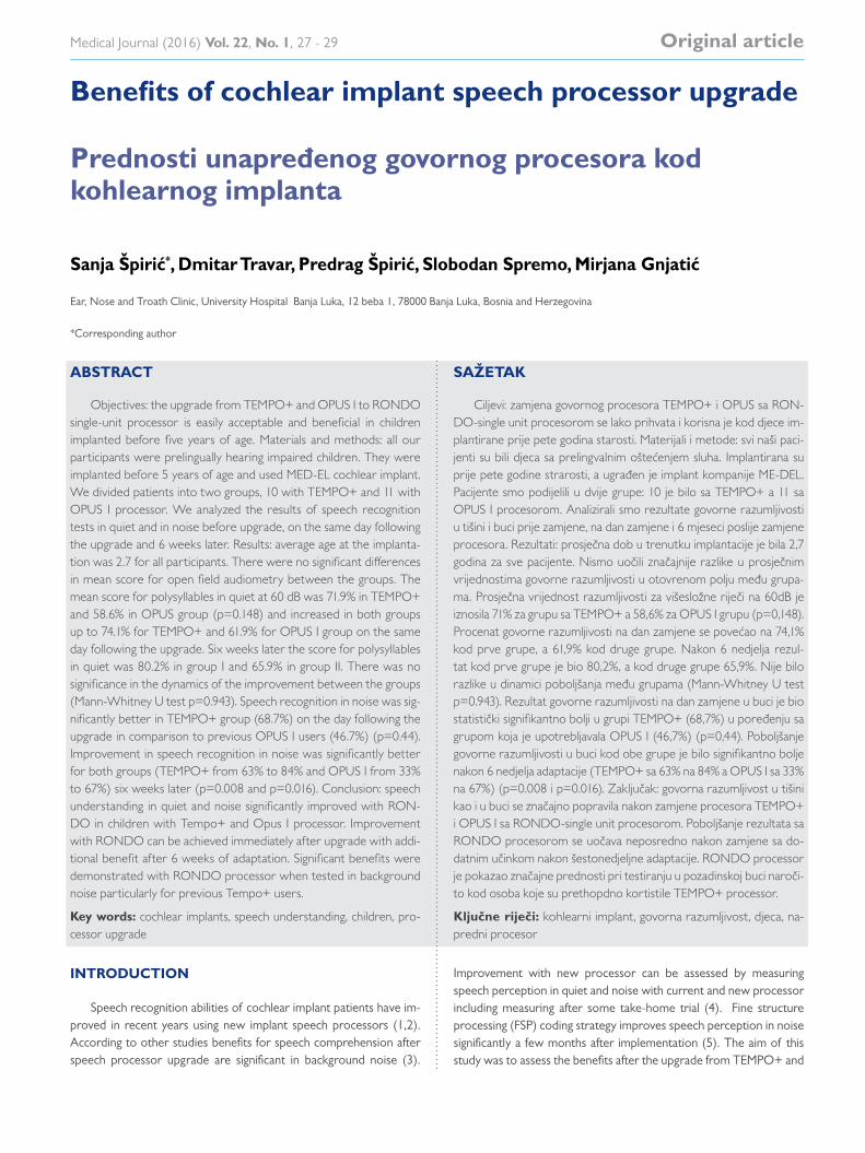
Benefits of cochlear implant speech processor upgrade
Prednosti unapređenog govornog procesora kod kohlearnog implanta
Sanja Špirić*, Dmitar Travar, Predrag Špirić, Slobodan Spremo, Mirjana Gnjatić
Ear, Nose and Troath Clinic, University Hospital Banja Luka, 12 beba 1, 78000 Banja Luka, Bosnia and Herzegovina
*Corresponding author
ABSTRACT
Objectives: the upgrade from TEMPO+ and OPUS I to RONDO single-unit processor is easily acceptable and beneficial in children implanted before five years of age. Materials and methods: all our participants were prelingually hearing impaired children. They were implanted before 5 years of age and used MED-EL cochlear implant. We divided patients into two groups, 10 with TEMPO+ and 11 with OPUS I processor. We analyzed the results of speech recognition tests in quiet and in noise before upgrade, on the same day following the upgrade and 6 weeks later. Results: average age at the implanta-tion was 2.7 for all participants. There were no significant differences in mean score for open field audiometry between the groups. The mean score for polysyllables in quiet at 60 dB was 71.9% in TEMPO+ and 58.6% in OPUS group (p=0.148) and increased in both groups up to 74.1% for TEMPO+ and 61.9% for OPUS I group on the same day following the upgrade. Six weeks later the score for polysyllables in quiet was 80.2% in group I and 65.9% in group II. There was no significance in the dynamics of the improvement between the groups (Mann-Whitney U test p=0.943). Speech recognition in noise was sig-nificantly better in TEMPO+ group (68.7%) on the day following the upgrade in comparison to previous OPUS I users (46.7%) (p=0.44). Improvement in speech recognition in noise was significantly better for both groups (TEMPO+ from 63% to 84% and OPUS I from 33% to 67%) six weeks later (p=0.008 and p=0.016). Conclusion: speech understanding in quiet and noise significantly improved with RON-DO in children with Tempo+ and Opus I processor. Improvement with RONDO can be achieved immediately after upgrade with addi-tional benefit after 6 weeks of adaptation. Significant benefits were demonstrated with RONDO processor when tested in background noise particularly for previous Tempo+ users.
Key words: cochlear implants, speech understanding, children, pro-cessor upgrade
SAŽETAK
Ciljevi: zamjena govornog procesora TEMPO+ i OPUS sa RON-DO-single unit procesorom se lako prihvata i korisna je kod djece im-plantirane prije pete godina starosti. Materijali i metode: svi naši paci-jenti su bili djeca sa prelingvalnim oštećenjem sluha. Implantirana su prije pete godine strarosti, a ugrađen je implant kompanije ME-DEL. Pacijente smo podijelili u dvije grupe: 10 je bilo sa TEMPO+ a 11 sa OPUS I procesorom. Analizirali smo rezultate govorne razumljivosti u tišini i buci prije zamjene, na dan zamjene i 6 mjeseci poslije zamjene procesora. Rezultati: prosječna dob u trenutku implantacije je bila 2,7 godina za sve pacijente. Nismo uočili značajnije razlike u prosječnim vrijednostima govorne razumljivosti u otovrenom polju među grupa-ma. Prosječna vrijednost razumljivosti za višesložne riječi na 60dB je iznosila 71% za grupu sa TEMPO+ a 58,6% za OPUS I grupu (p=0,148). Procenat govorne razumljivosti na dan zamjene se povećao na 74,1% kod prve grupe, a 61,9% kod druge grupe. Nakon 6 nedjelja rezul-tat kod prve grupe je bio 80,2%, a kod druge grupe 65,9%. Nije bilo razlike u dinamici poboljšanja među grupama (Mann-Whitney U test p=0.943). Rezultat govorne razumljivosti na dan zamjene u buci je bio statistički signifikantno bolji u grupi TEMPO+ (68,7%) u poređenju sa grupom koja je upotrebljavala OPUS I (46,7%) (p=0,44). Poboljšanje govorne razumljivosti u buci kod obe grupe je bilo signifikantno bolje nakon 6 nedjelja adaptacije (TEMPO+ sa 63% na 84% a OPUS I sa 33% na 67%) (p=0.008 i p=0.016). Zaključak: govorna razumljivost u tišini kao i u buci se značajno popravila nakon zamjene procesora TEMPO+ i OPUS I sa RONDO-single unit procesorom. Poboljšanje rezultata sa RONDO procesorom se uočava neposredno nakon zamjene sa do-datnim učinkom nakon šestonedjeljne adaptacije. RONDO processor je pokazao značajne prednosti pri testiranju u pozadinskoj buci naroči-to kod osoba koje su prethopdno kortistile TEMPO+ processor.
Ključne riječi: kohlearni implant, govorna razumljivost, djeca, na-predni procesor
INTRODUCTION
Speech recognition abilities of cochlear implant patients have im-proved in recent years using new implant speech processors (1,2). According to other studies benefits for speech comprehension after speech processor upgrade are significant in background noise (3).
Improvement with new processor can be assessed by measuring speech perception in quiet and noise with current and new processor including measuring after some take-home trial (4). Fine structure processing (FSP) coding strategy improves speech perception in noise significantly a few months after implementation (5). The aim of this study was to assess the benefits after the upgrade from TEMPO+ and
Medical Journal (2016) Vol. 22, No. 1, 27 - 29 Original article
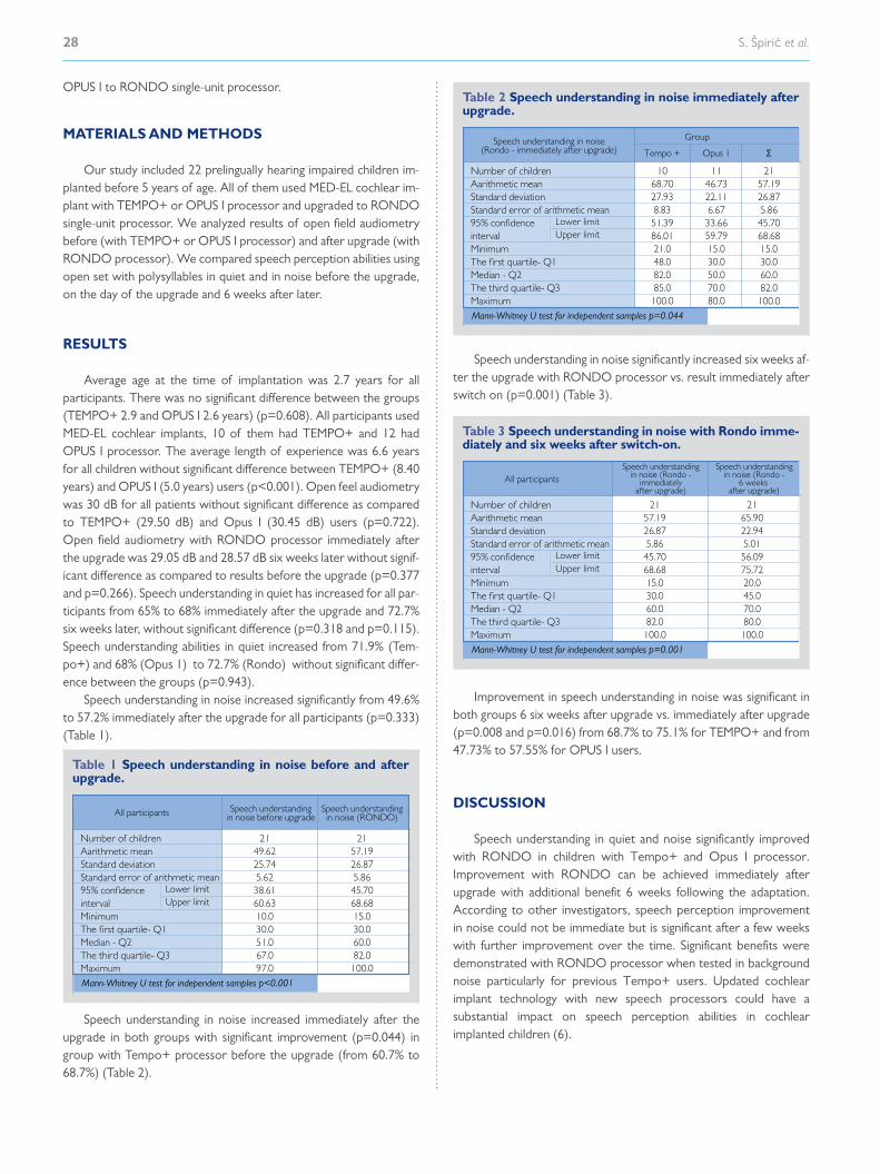
OPUS I to RONDO single-unit processor.
MATERIALS AND METHODS
Our study included 22 prelingually hearing impaired children im-planted before 5 years of age. All of them used MED-EL cochlear im-plant with TEMPO+ or OPUS I processor and upgraded to RONDO single-unit processor. We analyzed results of open field audiometry before (with TEMPO+ or OPUS I processor) and after upgrade (with RONDO processor). We compared speech perception abilities using open set with polysyllables in quiet and in noise before the upgrade, on the day of the upgrade and 6 weeks after later.
RESULTS
Average age at the time of implantation was 2.7 years for all participants. There was no significant difference between the groups (TEMPO+ 2.9 and OPUS I 2.6 years) (p=0.608). All participants used MED-EL cochlear implants, 10 of them had TEMPO+ and 12 had OPUS I processor. The average length of experience was 6.6 years for all children without significant difference between TEMPO+ (8.40 years) and OPUS I (5.0 years) users (p<0.001). Open feel audiometry was 30 dB for all patients without significant difference as compared to TEMPO+ (29.50 dB) and Opus I (30.45 dB) users (p=0.722). Open field audiometry with RONDO processor immediately after the upgrade was 29.05 dB and 28.57 dB six weeks later without signif-icant difference as compared to results before the upgrade (p=0.377 and p=0.266). Speech understanding in quiet has increased for all par-ticipants from 65% to 68% immediately after the upgrade and 72.7% six weeks later, without significant difference (p=0.318 and p=0.115). Speech understanding abilities in quiet increased from 71.9% (Tem-po+) and 68% (Opus 1) to 72.7% (Rondo) without significant differ-ence between the groups (p=0.943).
Speech understanding in noise increased significantly from 49.6% to 57.2% immediately after the upgrade for all participants (p=0.333) (Table 1).
Speech understanding in noise increased immediately after the upgrade in both groups with significant improvement (p=0.044) in group with Tempo+ processor before the upgrade (from 60.7% to 68.7%) (Table 2).
Speech understanding in noise significantly increased six weeks af-ter the upgrade with RONDO processor vs. result immediately after switch on (p=0.001) (Table 3).
Improvement in speech understanding in noise was significant in both groups 6 six weeks after upgrade vs. immediately after upgrade (p=0.008 and p=0.016) from 68.7% to 75.1% for TEMPO+ and from 47.73% to 57.55% for OPUS I users.
DISCUSSION
Speech understanding in quiet and noise significantly improved with RONDO in children with Tempo+ and Opus I processor. Improvement with RONDO can be achieved immediately after upgrade with additional benefit 6 weeks following the adaptation. According to other investigators, speech perception improvement in noise could not be immediate but is significant after a few weeks with further improvement over the time. Significant benefits were demonstrated with RONDO processor when tested in background noise particularly for previous Tempo+ users. Updated cochlear implant technology with new speech processors could have a substantial impact on speech perception abilities in cochlear implanted children (6).
28 S. ŠpiriĆ et al.
All participants Speech understandingin noise before upgrade
Speech understandingin noise (RONDO)
Number of childrenAarithmetic meanStandard deviationStandard error of arithmetic mean95% confidence interval MinimumThe first quartile- Q1Median - Q2The third quartile- Q3Maximum
2149.6225.745.6238.6160.6310.030.051.067.097.0
2157.1926.875.8645.7068.6815.030.060.082.0100.0
Mann-Whitney U test for independent samples p<0.001
Lower limitUpper limit
Table 1 Speech understanding in noise before and after upgrade.
Speech understanding in noise(Rondo - immediately after upgrade)
Group
Tempo + Opus 1 Σ
Number of childrenAarithmetic meanStandard deviationStandard error of arithmetic mean95% confidence interval MinimumThe first quartile- Q1Median - Q2The third quartile- Q3Maximum
1068.7027.938.83
51.3986.0121.048.082.085.0
100.0
1146.7322.116.67
33.6659.7915.030.050.070.080.0
2157.1926.875.8645.7068.6815.030.060.082.0100.0
Mann-Whitney U test for independent samples p=0.044
Lower limitUpper limit
Table 2 Speech understanding in noise immediately after upgrade.
All participantsSpeech understanding
in noise (Rondo -immediately
after upgrade)
Speech understandingin noise (Rondo -
6 weeks after upgrade)
Number of childrenAarithmetic meanStandard deviationStandard error of arithmetic mean95% confidence interval MinimumThe first quartile- Q1Median - Q2The third quartile- Q3Maximum
2157.1926.875.86
45.7068.6815.030.060.082.0
100.0
2165.9022.945.01
56.0975.7220.045.070.080.0
100.0Mann-Whitney U test for independent samples p=0.001
Lower limitUpper limit
Table 3 Speech understanding in noise with Rondo imme-diately and six weeks after switch-on.
Benefits of cochlear implant speech processor upgrade 29
CONCLUSION
RONDO speech processor upgrade supports the expectations of increased benefit in speech understanding abilities in cochlear implanted children. Improvement is significant in condition of background noise with additional benefit after certain adaptation time.
Conflict of interest: none declared.
REFERENCES
1. Fu QJ. Temporal processing and speech recognition in cochlear implant users. Neu-
roreport. 2002;13(13):1635-9.
2. Magalhães AT, Goffi-Gomez MV, Hoshino AC, Tsuji RK, Bento RF, Brito R. Con-
verted and upgraded maps programmed in the newer speech processor for the first
generation of multichannel cochlear implant. Otol Neurotol. 2013;34(7):1193-200.
3. Müller-Deile J, Kortmann T, Hoppe U, Hessel H, Morsnowski A. Improving
speech comprehension using a new cochlear implant speech processor. HNO.
2009;57(6):567-74.
4. Mosnier I, Marx M, Venail F, Loundon N, Roux-Vaillard S, Sterkers O. Benefits from
upgarde to the CP810 sound processor for Nucleus 24 cochlear implant recipients.
Eur Arch Otorhinolaryngol. 2014;271(1):49-57.
5. Kleine Punte A, De Bodt M, Van de Heyning P. Long-term improvement of speech
perception with the fine structure processing coding strategy in cochlear implants.
ORL J Otorhinolaryngol Relat Spec. 2014;76(1):36-43.
6. Davidson L, Geers A, Brenner C. Cochlear implant characteristics and speech
perception skills of adolescents with long-term device use. Otol Neurotol.
2010;31(8):1310-4.
Reprint requests and correspondence:Sanja Špirić, MD, PhDEar, Nose and Troath Clinic University Hospital Banja Luka12 beba 1, 78000 Banja LukaBosnia and HerzegovinaPhone: + 387 65 517 379Email: [email protected]

Serum nitric oxide concentration in rheumatoid arthritis patients: the association with disease activity
Serumska koncentracija nitričnog oksida kod pacijenata s reumatoidnim artritisom: povezanost s aktivnošću bolesti Amela DerviševićI*, Nermina Babić1, Asija Začiragić1, Nesina Avdagić1, Šekib Sokolović2, Almir Fajkić3, Orhan Lepara1, Anela Šubo4, Jasminko Huskić1
1Department of Human Physiology, Medical Faculty, University of Sarajevo, Čekaluša 90, 71000 Sarajevo, Bosnia and Herzegovina; 2Cardiology Clinic, University Clinical Center Sarajevo, Bolnička 25, 71000 Sarajevo, Bosnia and Herzegovina; 3Department of Pathophysiology, Medical Faculty, University of Sarajevo, Čekaluša 90, 71000 Sarajevo, Bosnia and Herzegovina; 4Department of Internal Medicine, General Hospital „Prim. Dr Abdulah Nakaš“, Kranjčevićeva 12, 71000 Sarajevo, Bosnia and Herzegovina
*Corresponding author
ABSTRACT
The aim of this cross sectional pilot study was to determine and compare serum nitric oxide (NO) concentration in 63 pa-tients with rheumatoid arthritis (RA) and in 31 apparently healthy individuals. Based on the Disease Activity Score (DAS28 score), RA patients were divided into three subgroups: low stage of dis-ease activity (DAS28 ≤3.2; n=19); moderate stage of disease ac-tivity (3.2<DAS28≤5.1; n=19) and high stage of disease activity (DAS28>5.1; n=25). Serum NO concentration was determined by classic colorimetric Griess reaction. Elementary zinc was used for the conversion of nitrate (NO32-) into nitrite (NO22-). Median se-rum NO concentration was not statistically different (p=0.36) be-tween the control group [9.23 µmol/L (8.34 µmol/L-10.39 µmol/L)] and the group of RA patients [8.70 µmol/L (8.16 µmol/L-10.30 µmol/L)]. There was no significant difference (p=0.27) between NO concentration in serum of RA patients with high disease ac-tivity [9.59 µmol/L (8.43 µmol/L-11.02 µmol/L)], moderate disease activity [8.70 µmol/L (7.81 µmol/L-9.59 µmol/L)], low disease activity [8.52 µmol/L (8.16 µmol/L-10.30 µmol/L) and the control group. Re-sults of this study indicate that serum NO concentration measured by classic colorimetric Griess reaction is not associated with RA disease activity. Larger prospective studies are needed in order to access potential use of serum NO concentration in the evaluation of RA disease activity.
Key words: nitric oxide, rheumatoid arthritis, disease activity
SAŽETAK
Cilj ove presječne pilot studije je bio da se utvrdi i uporedi serumska koncentracija nitričnog oksida (NO) kod 63 pacijenta s reumatoidnim artritisom (RA) i 31 naizgled zdrava ispitanika. Na osnovu scora aktivnosti bolesti (DAS28 scor), pacijenti sa RA su po-dijeljeni u tri podskupine: niski stadij aktivnosti bolesti (DAS28≤3.2; n=19); srednji stadij aktivnosti bolesti (3.2<DAS28≤5.1; n=19) i vi-soki stadij aktivnosti bolesti (DAS28>5.1; n=25). Serumska koncen-tracija NO je određena klasičnom kolorimetrijskom reakcijom po Griessu. Pretvorba nitrata (NO32-) u nitrite (NO22-) je izvršena upotrebom elementarnog cinka. Nije utvrđena statistički značaj-na razlika (p=0.36) između serumske koncentracije NO kontrolne skupine [9.23 umol/L (8.34 umol/L-10.39 umol/L)] i skupine pacije-nata s RA [8.70 umol/L (8.16 umol/L-10.30 umol/L)]. Nije utvrđena statistički značajna razlika (p=0.27) između serumskih koncen-tracija NO pacijenata s visokom aktivnošću bolesti [9.59 pmol/L (8.43 umol/L-11.02 umol/L)], srednjom aktivnošću bolesti [8.70 umol/L (7.81 umol/L-9.59 umol/L)], niskom aktivnošću bolesti [8.52 umol/L (8.16 umol/L-10.30 umol/L) i ispitanika kontrolne skupine. Rezultati ovog istraživanja pokazuju da serumska koncentracija NO mjerena klasičnom kolorimetrijskom Griess reakcijom nije povezana sa stadijem aktivnosti RA. Potrebne su veće prospek-tivne studije kako bi se utvrdila potencijalna uloga serumskog NO u procjeni aktivnosti bolesti kod pacijenata s RA.
Ključne riječi: nitrični oksid, reumatoidni artritis, aktivnost bolesti
Medical Journal (2016) Vol. 22, No. 1, 30 - 33 Original article
INTRODUCTION
Nitric oxide (NO) is an unique gaseous signalling molecule with wide variety of functions in numerous physiological and pathological processes (1). Several studies confirm important roles of nitric oxide (NO) in regulation of blood vessels tone, platelet function,
neurotransmission, immune regulation, mitochondrial functions and apoptosis (2,3).
This free-radical with short life-time is generated endogenously from L-arginine by the enzyme NO synthase (NOS). There are three distinct isoforms of NOS: endothelial NOS (eNOS), neuronal NOS (nNOS) and inducibile NOS (iNOS) (4).

Serum nitric oxide concentration in rheumatoid arthritis patients: the association with disease activity 31
Endothelial and neuronal NOS are dependent upon intracellular calcium level and generally produce low levels of NO involved in normal physiologic processes. Inducible NOS is calcium-independent isoform of NOS and is present mainly in monocytes/macrophages, neutrophil granulocytes and other immunology cells. Upon stimulation of these cells by certain pro-inflammatory cytokines in inflammatory reactions or by endotoxin in bacterial infection high amounts of NO are produced. These high amounts of NO produced by iNOS perform important functions for host defense, but excess of NO production is also associated with various malignancies and inflammatory conditions including juvenile diabetes, multiple sclerosis, arthritis and ulcerative colitis (5,6).
Due to the short half-life of NO, usually its end products nitrate (NO32-) or nitrite (NO22-) are measured as an index of NO production.
Rheumatoid arthritis (RA) is a chronic progressive autoimmune disease, mainly manifested by damage, deformity and dysfunction of multiple joints. Besides the joints, the disease also affects multiple other organs and systems. The extra-articular manifestations of RA include damage of exocrine lacrimal and salivary glands, vasculitis, anemia and peripheral neuropathy. RA affects 0.5-1% of the adult population worldwide and is more common in women than men at a ratio of approximately 3:1. It is a multifactorial disease in which a combination of genetic and environmental factors contributes to the disease progression (7,8).
Primary pathological process in RA is happening in the area of synovial membrane of a joint which becomes thicker, edematous and infiltrated with mononuclear cells (9).
Considering the heterogeneous presentation of the disease among different individuals, the ability to predict the progession of disease in patients with RA is of great importance in everyday clinical practice.
Experiments in animal models of arthritis and studies in patients with RA have reported important role of NO in inflammatory joint diseases. NO mediates many different cell functions at the site of synovial inflammation, including cytokine production, signal transduction, mitochondrial functions and apoptosis (10).
Thus, the objective of this study was to explore whether serum NO concentration measured by classic colorimetric Griess reaction is associated with RA and disease activity in laboratory diagnostics.
MATERIALS AND METHODS
This cross-sectional study included all eligible RA patients (n=63), hospitalized at the Cardiology Clinic of the University Clinical Center Sarajevo (UCCS) in the period between 1 January 2011 and 30 June 2011, who met the 1987 American College of Rheumatology (ACR) revised criteria for the RA classification. Based on the stage of disease activity evaluated by Disease Activity Score (DAS28 score), RA patients were divided into three subgroups: low stage of disease activity (DAS28≤3.2; n=19), moderate stage of disease activity (3.2<DAS28≤5.1; n=19) and high stage of disease activity (DAS28>5.1; n=25). The control group consisted of 31 healthy individuals. Upon careful explanation of the study procedure, all subjects gave their informed consent to answering the questionnaire and provided residual blood for analysis. The study was approved by
the Ethical Committee of the UCCS.The exclusion criteria for the studied group of RA patients
included other rheumatic diseases, malignancy, active local or systemic inflammation caused by bacterial and viral infections, and pregnancy. During the study the use of tobacco products was prohibited.
Subjects of the control group had no history of RA or other rheumatic, autoimmune and inflammatory diseases, hyperlipidemia, hypertension or coronary heart diseases. None of the control subjects received any medication and they were not current smokers or alcohol consumers. Detailed history was taken from patients using specially prepared questionnaire that included questions relevant to RA such as disease duration, morning stiffness and drug taking history. All patients underwent thorough clinical examination with special attention to articular examination. The disease activity in RA patients was determined by experienced physician with the use of DAS28 score.
DAS28 is a mathematical formula with four variables:DAS28 = 0.56x√(TEN28) + 0.28x√(SW28) + 0.70xLn(ESR) +
0.014x(GH).(TEN28: 28 joint count for tenderness, SW28: 28 joint count for
swelling, Ln(ESR): the natural logarithm of Westergren’s erythrocyte sedimentation rate (ESR); GH: general health or patient’s global assessment of disease activity on the visual analog scale (VAS) of 100 mm).
After overnight fast, venous blood samples were collected early in the morning and allowed to coagulate. After coagulation, the serum samples were centrifuged for 5 minutes at 2000 g and deproteinized by adding 0.75 ml of distilled water and 0.05 ml of 30% zinc sulfate solution to 0.25 ml of each sample. Several minutes later, the samples were centrifuged for 10 minutes at 2000 g and separated supernatant was stored at -200C.
NO measurement
The NO level in obtained supernatant was determined by measuring nitrite concentrations, a stable metabolic endproduct of NO. Conversion of nitrate (NO32-) into nitrite (NO22-) was done with elementary zinc. Nitrite concentration in serum was determined by classic colorimetric Griess reaction (11).
Briefly, equal volumes of samples and Griess reagent were mixed at room temperature. After 5 minutes, the absorbance was measured at 570 nm using Perkin Elmer 550 S spectrophotometer. The concentration of nitrite was determined by a standard curve prepared with sodium nitrite (1-200). The results were expressed as μmol/L.
Determination of NO concentration in serum of RA patients and control individuals was done at the Department of Human Physiology of the Sarajevo University Medical School.
Statistical analysis
The Kolmogorov-Smirnov and Shapiro-Wilk tests were used to determine normality. Due to the non-normal distribution, non-parametric tests, such as Mann-Whitney U and Kruskal-Wallis, were chosen for reporting significance. Correlation coefficient between variables was assessed by the Spearman’s test.
Statistical significance was set at p<0.05. All statistical analyses
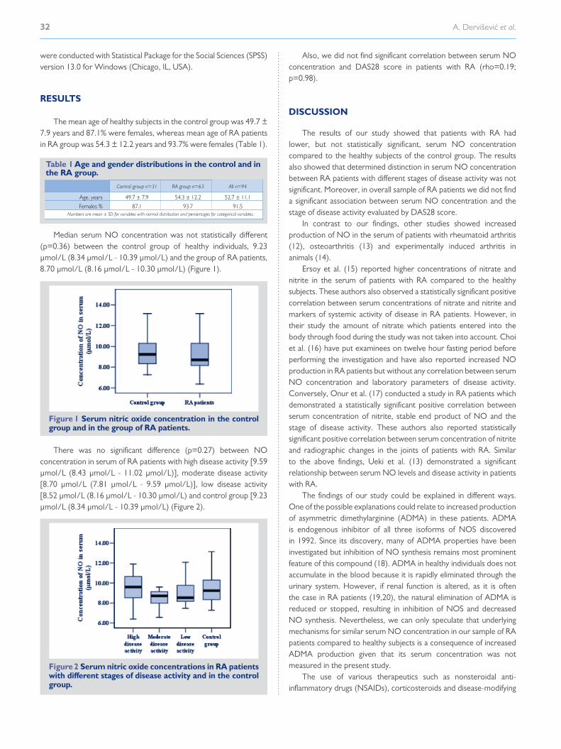
were conducted with Statistical Package for the Social Sciences (SPSS) version 13.0 for Windows (Chicago, IL, USA).
RESULTS The mean age of healthy subjects in the control group was 49.7 ± 7.9 years and 87.1% were females, whereas mean age of RA patients in RA group was 54.3 ± 12.2 years and 93.7% were females (Table 1).
Median serum NO concentration was not statistically different (p=0.36) between the control group of healthy individuals, 9.23 µmol/L (8.34 µmol/L - 10.39 µmol/L) and the group of RA patients, 8.70 µmol/L (8.16 µmol/L - 10.30 µmol/L) (Figure 1).
There was no significant difference (p=0.27) between NO concentration in serum of RA patients with high disease activity [9.59 µmol/L (8.43 µmol/L - 11.02 µmol/L)], moderate disease activity [8.70 µmol/L (7.81 µmol/L - 9.59 µmol/L)], low disease activity [8.52 µmol/L (8.16 µmol/L - 10.30 µmol/L) and control group [9.23 µmol/L (8.34 µmol/L - 10.39 µmol/L) (Figure 2).
Also, we did not find significant correlation between serum NO concentration and DAS28 score in patients with RA (rho=0.19; p=0.98).
DISCUSSION
The results of our study showed that patients with RA had lower, but not statistically significant, serum NO concentration compared to the healthy subjects of the control group. The results also showed that determined distinction in serum NO concentration between RA patients with different stages of disease activity was not significant. Moreover, in overall sample of RA patients we did not find a significant association between serum NO concentration and the stage of disease activity evaluated by DAS28 score.
In contrast to our findings, other studies showed increased production of NO in the serum of patients with rheumatoid arthritis (12), osteoarthritis (13) and experimentally induced arthritis in animals (14).
Ersoy et al. (15) reported higher concentrations of nitrate and nitrite in the serum of patients with RA compared to the healthy subjects. These authors also observed a statistically significant positive correlation between serum concentrations of nitrate and nitrite and markers of systemic activity of disease in RA patients. However, in their study the amount of nitrate which patients entered into the body through food during the study was not taken into account. Choi et al. (16) have put examinees on twelve hour fasting period before performing the investigation and have also reported increased NO production in RA patients but without any correlation between serum NO concentration and laboratory parameters of disease activity. Conversely, Onur et al. (17) conducted a study in RA patients which demonstrated a statistically significant positive correlation between serum concentration of nitrite, stable end product of NO and the stage of disease activity. These authors also reported statistically significant positive correlation between serum concentration of nitrite and radiographic changes in the joints of patients with RA. Similar to the above findings, Ueki et al. (13) demonstrated a significant relationship between serum NO levels and disease activity in patients with RA.
The findings of our study could be explained in different ways. One of the possible explanations could relate to increased production of asymmetric dimethylarginine (ADMA) in these patients. ADMA is endogenous inhibitor of all three isoforms of NOS discovered in 1992. Since its discovery, many of ADMA properties have been investigated but inhibition of NO synthesis remains most prominent feature of this compound (18). ADMA in healthy individuals does not accumulate in the blood because it is rapidly eliminated through the urinary system. However, if renal function is altered, as it is often the case in RA patients (19,20), the natural elimination of ADMA is reduced or stopped, resulting in inhibition of NOS and decreased NO synthesis. Nevertheless, we can only speculate that underlying mechanisms for similar serum NO concentration in our sample of RA patients compared to healthy subjects is a consequence of increased ADMA production given that its serum concentration was not measured in the present study.
The use of various therapeutics such as nonsteroidal anti-inflammatory drugs (NSAIDs), corticosteroids and disease-modifying
32 A. Dervišević et al.
Figure 1 Serum nitric oxide concentration in the control group and in the group of RA patients.
Figure 2 Serum nitric oxide concentrations in RA patients with different stages of disease activity and in the control group.
Age, years
Females %
49.7 ± 7.9
87.1
Control group n=31
54.3 ± 12.2
93.7
RA group n=63 All n=94
52.7 ± 11.1
91.5Numbers are mean ± SD for variables with normal distribution and percentages for categorical variables.
Table 1 Age and gender distributions in the control and in the RA group.

anti-rheumatic drugs (DMARDs) may also reduce the activity of NOS and affect NO production (21). In the present study, out of the total of RA patients, 58.7% were taking methotrexate (MTX) as the most commonly used DMARD for the treatment of RA. Methotrexate blocks the synthesis of tetrahydrobiopterin, one of the essential cofactors of NO synthases, which could be another reason for the reduced serum NO levels (14).
Moreover, observed similar NO levels in RA patients and the control group could also be a result of endothelial dysfunction, a condition primarily characterized by decreased production and bioavailability of NO. In patients with RA according to the research of Vaundo et al. (22), systemic inflammation is a reason of endothelial hyper-reactivity, caused by increased production of pro-inflammatory cytokines, mostly TNF-α with consequent increase of C-reactive protein (CRP) synthesis. Earlier study by Verma et al. (23) reported that increased levels of CRP induce decreased expression of eNOS with consequent decrease of NO production. It has also been demonstrated that some infectious microorganisms including cytomegalovirus, which is considered to be one of the etiological causes for RA, can induce endothelial dysfunction and lead to decrease of NO production in RA patients (24).
CONCLUSION
Results of the present pilot study indicate that serum NO concentration in patients with rheumatoid arthritis measured using Griess reaction is not associated with RA disease activity. Longitudinal studies with larger sample of RA patients are needed in order to fully clarify the role of NO in the pathogenesis of RA.
Conflict of interest: none declared.
REFERENCES
1. Martin W. Nitroxyl anion - the universal signaling partner of endogenously produced
nitric oxide? Br J Pharmacol. 2009;157(4):537-39.
2. Tousoulis D, Kampoli AM, Tentolouris C, Papageorgiou N, Stefanadis C. The role of
nitric oxide on endothelial function. Curr Vasc Pharmacol. 2012;10(1):4-18.
3. Moncada S, Higgs A. The L-arginine nitric oxide pathway. N Engl J Med.
1993;329(27):2002-12.
4. Bredt DS. Endogenous nitrice oxide synthesis: biological functions and pathophysiology.
Free Radic Res. 1999;31(6):577-96.
5. Kanwar JR, Kanwar RK, Burrow H, Baratchi S. Recent advances on the roles of NO in
cancer and chronic inflammatory disorders. Curr Med Chem. 2009;16(19):2373-94.
6. Gonzalez-Gay MA, Garcia-Unzueta MT, Berja A, Vazquez- Rodriguez TR, Miranda-
Filloy JA, Gonzalez-Juanatey C, et al. Short-term effect of anti-TNF-alpha therapy
on nitric oxide production in patients with severe rheumatoid arthritis. Clin Exp
Rheumatol. 2009;27(3):452-8.
7. Firestein GS. Evolving concepts of rheumatoid arthritis. Nature. 2003;423:356-61.
8. Dieude P, Cornelis F. Genetic basis of rheumatoid arthritis. Joint Bone Spine.
2005;72(6):520-26.
9. Perić P, Perić B. Temporomandibularni artritis u sklopu reumatoidnog artritisa -
diferencijalno dijagnostički pristup temporomandibularnoj boli u stomatološkoj praksi.
Acta Stomatol Croat. 2002;3(1):107-12.
10. Nagy G, Koncz A, Telarico T, Fernandez D, Ersek B, Buzás E, et al. Central role of nitric
oxide in the pathogenesis of rheumatoid arthritis and systemic lupus erythematosus.
Arthritis Res Ther. 2010;12(3):210.
11. Green LC, Wagner DA, Glogowski J, Skipper PL, Wishnok JS, Tannenbaum SR. Analysis
of nitrate, nitrite and [15N]nitrate in biological fluids. Anal Biochem.1982;126(1):131-38.
12. Farrell AJ, Blake DR, Palmar RMJ. Increased concentrations of nitrite in synovial fluid
and serum samples suggest increased nitric oxide synthesis in rheumatic diseases. Ann
Rheum Dis. 1992;51(11):1219-22.
13. Ueki Y, Miyake S, Tominaga Y, Eguchi K. Increased nitric oxide levels in patients with
rheumatoid arthritis. J Rheumatol. 1996;23(2):230-6.
14. Stichtenoth DO, Frolich JC. Nitric oxide and inflammatory joint diseases. Br J of
Rheumatol. 1998;37(3):246-57.
15. Ersoy Y, Ozerol E, Baysal O, Temel I, MacWalter RS, Meral U, et al. Serum nitrate
and nitrite levels in patients with rheumatoid arthritis, ankylosing spondylitis, and
osteoarthritis. Ann Rheum Dis. 2002;61(1):76-8.
16. Choi JW. Nitric oxide production is increased in patients with rheumatoid arthritis
but does not correlate with laboratory parameters of disease activity. Clin Chim Acta.
2003;336(1-2):83-7.
17. Onur O, Akinci AS, Akbiyik F, Unsal I. Elevated levels of nitrate in rheumatoid arthritis.
Rheumatol Int. 2001;20(4):154-8.
18. Alpoim PN, Sousa LP, Mota AP, Rios DR, Dusse LM. Asymmetric Dimethylarginine
(ADMA) in cardiovascular and renal disease. Clin Chim Acta. 2014;440:36-9.
19. Antero Anders HJ, Vielhauer V. Renal co-morbidity in patients with rheumatic diseases.
Arthritis Res Ther. 2011;13(3):222.
20. Hickson LJ, Crowson CS, Gabriel SE, McCarthy JT, Matteson EL. Development of
reduced kidney function in rheumatoid arthritis. Am J Kidney Dis. 2014;63(2):206-13.
21. Allanore Y, Borderie D, Hilliquin P, Hernvann A, Levacher M, Lemaréchal H, et al. Low
levels of nitric oxide (NO) in systemic sclerosis: inducible NO synthase production is
decreased in cultured peripheral blood monocyte/macrophage cells. Rheumatology
(Oxford). 2001;40(10):1089-96.
22. Vaudo G, Marchesi S, Gerli R, Allegrucci R, Giordano A, Siepi O, et al. Endothelial
dysfunction in young patients with rheumatoid arthritis and low disease activity. Ann
Rheum Dis. 2004;63(1):31-5.
23. Verma S, Wang CH, Li SH, Dumont AS, Fedak PW, Badiwala MV, et al. A self-
fulfilling prophecy: C-reactive protein attenuates nitric oxide producion and inhibits
angiogenesis. Circulation. 2002;106(8):913-9.
24. Xu Q. Role of heat shock proteins in atherosclerosis. Arterioscler Thromb Vasc Biol.
2002;22(10):1547-59.
Reprint requests and correspondence:Amela Dervišević, MD, MScDepartment of Human PhysiologyMedical Faculty, University of SarajevoČekaluša 90, 71000 SarajevoBosnia and HerzegovinaEmail: [email protected]
Serum nitric oxide concentration in rheumatoid arthritis patients: the association with disease activity 33

Defining the vascular skin territories of the septocutaneous blood vessels of the forearm with a special overview on their use in the fasciocutaneous flaps surgery
Definisanje vaskularnih kožnih teritorija septokutanih krvnih sudova podlaktice sa posebnim osvrtom na njihovu primjenu u hirurgiji fasciokutanih režnjevaDarko Jović1*, Aleksandar Jakovljević2, Jovan Ćulum1, Branislava Jakovljević1, Ljiljana Lati-nović3, Olivera Kosovac4, Darko Lukić5
1Surgical Clinic “S-Tetik“, Arsenija Čarnojevića 2b, 78000 Banja Luka, Bosnia and Herzegovina; 2Department of Surgery, Faculty of Medicine, University of Banja Luka, Save Mrkalja 14 , 78000 Banja Luka, Bosnia and Herzegovina; 3Institute of Pathology, Clinical Centre University of Banja Luka, 12 Beba, 78000 Banja Luka, Bosnia and Herzegovina; 4Specialized Hospital for Aesthetic, Reconstructive and Plastic Surgery “Diona”, Satonova 29, 11000 Belgrade, Republic of Serbia; 5Specialized ORS Hospital, Bokeljska 7, 11000 Belgrade, Republic of Serbia
*Corresponding author
ABSTRACT
The research of the septocutaneous forearm blood vessels created by radial, ulnar and the posterior interosseous arteries, as well as the size of certain skin territories they vascularise, has been performed on fifty cadaver forearms. Thirty forearms were injected with shower gelatine and precise dissection was performed. Remaining part of the research was conducted on twenty selected forearms, four of which were tested with the use of corrosive method. Average skin territory vascularised by the radial artery is 163.57 cm2, while the average skin territory vascularised by the ulnar artery is 190.36 cm2. The posterior in-terosseous artery vascularises the smallest skin territory in the forearm area, the average being 107.48 cm2. These results clear-ly show that vascularised skin territories are directly propor-tional to the size and calibre of the main blood vessels supplying them, as well as with number of septocutaneous perforators they create. This means that the largest number of perforators per artery is created by radial artery (8.1% on average), followed by ulnar artery (5.6% on average) while the posterior interosse-ous artery creates the smallest amount of perforators (4.9% on average). The purpose of this research was to make contribu-tion to the accuracy of the fasciocutaneous forearm flap surgery. Likewise, our aim was to clarify the skin territories which are vascularised by specific septocutaneous perforators making the surgical procedure safer.
Key words: vascular skin territories, reconstruction, septocutane-ous perforators
SAŽETAK
Istraživanje septokutanih krvnih sudova podlaktice, koji nastaju od magistralnih krvnih sudova (arterija radialis, ulnaris i interossea posterior), kao i veličine pojedinih kožnih teritorija koje su vaskularizovane od istih, obavljeno je na 50 kadaveričnih podlaktica. Na 30 podlaktica izvršena su ispitivanja iniciranjem tuš-želatina, te precizna disekcija na lešnom obdukcijskom ma-terijalu. Preostalo istraživanje sprovedeno je na 20 izdvojenih podlaktica, od kojih je na četiri podlaktice sprovedena koro-zivna metoda. Prosječna površina kože, izražena u kvadratnim centimetrima, koju vaskularizuje arterija radialis je 163,57cm2, dok je prosječna vrijednost kožne površine koju vaskularizuje arterija ulnaris nešto veća i iznosi 190,36cm2. Arterija interos-sea posterior vaskularizuje najmanju kožnu površinu u predjelu podlaktice, koja u prosjeku iznosi 107,48cm2. Iz ovih rezultata jasno se vidi da su vaskularizovane kožne površine u direktnoj srazmijeri sa veličinom i kalibrom samih magistralnih krvnih su-dova koji ih ishranjuju, kao i sa brojem septokutanih perforato-ra koji od njih nastaju. Tako najveći broj perforatora po arteriji (u prosjeku 8,1%) nastaje od arterije radialis, nešto manji broj od arterije ulnaris (u prosjeku 5,6%), a najmanje od arteri-je interossea posterior (u prosjeku 4,9%). Ovim ispitivanjem smo željeli doprinijeti preciznosti same hirurgije podlaktičnih fasciokutanih reženjeva, razjasniti kožne teriotrije koje pojed-ini septokutani perforatori vaskularizuju, te učiniti na taj način sam operativni zahvat sigurnijim.
Ključne riječi: vaskularne kožne teritorije, rekonstrukcija, septo-kutani perforatori
Medical Journal (2016) Vol. 22, No. 1, 34 - 36 Original article

INTRODUCTION
When it comes to reconstruction of the forearm and hand defects, fasciocutaneous forearm flaps are of great importance, and based on the Cormack and Lamberty classification the most common are fasciocutaneous flaps type C. The best known are posterior interosseous flap, radial forearm flap (Chinese flap) and ulnar forearm flap. These are fasciocutaneous forearm flaps through which forearm main blood vessels flow, out of which septocutaneous blood vessels branch out and vascularise corresponding forearm skin and hypodermis territories (1-5). Number, size, topography and the places where the septocutaneous blood vessels of the forearm‘s main arteries branch out are of extreme importance during clinical implementation of aforesaid flaps. This means that in the procedure itself, apart from the radial, ulnar and the posterior interosseous arteries, at least one septocutaneous perforator must be used. The exact places where the perforators are separated from the main blood vessels along with their topography (blood flow) make the precise dissection and elevation of the fasciocutaneous flaps easier. Skin territories which are vascularised by specific septocutaneous blood vessels, including their boundaries, are of particular significance since that is the manner in which safe territories of skin for particular flaps are defined. Extensive knowledge of the topography and the size of the skin territories based on the vascularisation of the forearm‘s septocutaneous blood vessels reduce the possibility of making a mistake during the elevation of the fasciocutaneous flaps, thus making the surgical procedure safer. Reverse-Flow Posterior Interosseous Flap was for the first time described in 1986 by Laijin (6) in Chinese literature, in English literature by Penteada in 1986 (7) and by Zancolli in 1988 (8). Chinese flap, or fasciocutaneous forearm flap, was for the first time described in China in 1981 by Yang Guofan et al. (1, 9, 10).
MATERIALS AND METHODS
Research of septocutaneous perforator artery branches of the forearm was performed on fifty cadaver forearms. Thirty forearms were injected with shower gelatine and precise dissection was per-formed. Remaining part of the research was conducted on twenty selected forearms, of which four forearms were used for the cor-rosive method. We used the method of injecting shower gelatine into the artery system, and after colouring, a precise microsurgical dissecting was performed which included all measurements (number, place of occurrence, calibre, as well as the topography of septocuta-neous perforators). Corrosive method was performed by injection of methyl methacrylate into the very posterior interosseous artery, after which the corrosion of the forearm tissue was performed in 40% solution of NaOH, resulting in plastic casts of posterior interosseous artery with its septocutaneous perforators.
Segregation of vascularized cutaneous perforators of the ulnar, radial and posterior interossea arteries was preceded by an elongat-ed cut, 10cm long, in the area of cubital fossa and the dissection of the brachial artery and its ending branches. Through the cut on the brachial artery we were able to place two catheters, one in the ulnar and one in the radial artery, after which we fastened them from the outside with a tightening loop. After rinsing the arteries with warm water, we selectively injected 20ml of 5% melted gelatine coloured
in blue and red ink. The separated forearms were entered through the common interosseous artery with a thin catheter and the area of vascularization was separated with green dyed gelatine. The injection lasted until the area was coloured without the overlap of vascular cutaneous territories. After 10 minutes, which were necessary for the gelatine to solidify, the dissection started. We measured the obtained coloured skin territories with a planimeter.
RESULTS
The vascular skin territory of the septocutaneous forearm blood vessels.Table 1 shows the results of the average value of skin territories
vascularised by septocutaneous perforators of radial, ulnar and posterior interosseous arteries.
Table 2 shows the number of septocutaneous arteries: radial, ulnar and posterior interosseous artery by the thirds of the forearm.
Figure 1 shows skin territory in different colours after the injection of shower gelatine in some of the main blood vessels. Radial artery was injected with red, ulnar artery with blue and posterior interosseous artery with green shower gelatine, based on which the skin territory vascularised by certain arteries could be measured.
Figure 2 shows the posterior side of the forearm where the field injected with green shower gelatine represents the skin territory vascularised by the posterior interosseous artery.
Defining the vascular skin territories of the septocutaneous blood vessels of the forearm with a special overview on their use in the fasciocutaneous flaps surgery 35
Min.
Max.
Middle value
Standard deviation
Total
110
212.5
163.57
45.62
40
125
261.2
190.36
56.71
40
63.1
175
107.48
39.60
40
Skin surface(cm2) of theradial artery
Skin surface(cm2) of thetulnar artery
Skin surface (cm2)of the posterior
interosseous artery
Table 1 Average value of skin territories.
Proximal third
Medial third
Distal third
Total number
2
3,7
2,4
8,1
1,2
2
2,4
5,6
0,7
2,1
2,1
4,9
Radial artery Ulnar artery Posteriorinterosseous artery
Numberof perforators
Table 2 Septocutaneous arteries.
Figure 1 Vascular skin territories of the radial and ulnar arteries.
Vascular skin territory ofthe radial artery
Vascular skin territory ofthe ulnar artery

DISCUSSION
The results of the research show that the skin territory vascula-rised by septocutaneous perforators of radial artery is mainly in the radial part of the forearm and the border which separates it from the vascular skin territories of adjacent blood vessels is unclear.
Average skin territory vascularised by the radial artery is 163.57 cm2, while the average skin territory vascularised by the ulnar artery is 190.36 cm2. The posterior interosseous artery vascularises the smallest skin territory in the forearm area, the average being 107.48 cm2. These results clearly show that vascularised skin territories are directly proportional to the size and calibre of the main blood vessels which supply them, as well as with number of septocutaneous perfo-rators they create.This means that the largest number of perforators per artery is created by radial artery (8.1% in average), followed by ulnar artery (5.6% in average), whereas the posterior interosseous artery creates the smallest amount of perforators (4.9% in average).
Cormack states that the skin territory of radial artery includes the lateral half of the anterior side and the lateral edge of the forearm, but not the area above extensor pollicis brevis muscle and abductor pollicis longus muscle, which are vascularised by the posterior inter-osseous artery (1). The border between radical and ulnar territory varies and depends on the size of the cubital artery. The lateral bor-der between the skin territories of radial and the posterior interosse-ous arteries correspond to the lateral edge of the extensor digitorum muscle (11,12,13). Ulnar artery vascularises the skin territory of the posterior part of the forearm. The furthest border of the ulnar skin territory is located in the level of the posterior part of the humerus, while the closest border is in the area of the anterior forearm’s medial line. Skin territory of the posterior interosseous artery lies mainly in the area of lateral edge of the extensor digitorum muscle and borders with the radial territory to the rear area of ulna, where it borders with the ulnar territory (1). According to Costa the skin territory of the posterior interosseous artery lies is in the posterior part of the forearm, 4 cm beneath the intercondylar line, all the way to the wrist joint (11, 14, 15, 16).
CONCLUSION
On the basis of the aforementioned, we may conclude that the
largest area of the forearm, placed at the anterior and posterior part is vascularised by ulnar artery; somewhat smaller skin territory placed at the posterior side of the forearm is vascularised by radial artery, and the smallest territory of the forearm is vascularised by the posterior interosseous artery which lies in the central part of the posterior part of the forearm. The number of perforators is not correlated with the size of skin territories. Radial artery has the largest number of septocutaneous perforators, even though it vascularises the smaller skin territory than ulnar artery. This means that the size and calibre of the septocutaneous perforators themselves has an impact on the vascularised skin territories of the forearm. The borders between the skin territories are irregular, which implies an unclear border and the existence of numerous anamostosis between some of the septocutaneous blood vessels.
Conflict of interest: none declared.
REFERENCES
1. Cormac G.C.,Lamberty B. G. H. The arterial anatomy of skin flaps. Edinburg: Chur-
chill&Livingsone, 1986.
2. Raspanti A, Delcroix L, Ghezzi S, Innocenti M. Study of the tendinous vascularization
for the compound radial forearm flap plus flexor carpi radialis tendon. Sur Radiol Anat.
2015;11:120.
3. Hwang K, Han JY, Chung IH. Hypothenar flap based on a cutaneous perforator branch of
the ulnar artery: an anatomic study. J Reconstr Microsurg. 2005;21(5):297-301.
4. Guimberteau JC, Goin JL, Panconi B, Schumacher B. The reverse ulnar artery forearm
island flap in hand surgery: 54 cases. Plast Reconstr Surg. 1988;81(6):925-32.
5. Li Z, Liu K, Cao Y. The reverse flow ulnar artery island flap: 42 clinical cases. Br J Plast
Surg. 1989;42(3):256-9.
6. Converse MJ. Reconstructive plastic surgery. Philadelphia: W.B. Saunders Company,
1977.
7. Panteado CV, Masquelet AC, Chevrel JP. The anatomic basis of the fasciocutaneous flap
of the posterior interosseous artery. Surg Radiol Anat. 1986;8(4):209-15.
8. Zancolli EA, Angrigian C. Posterior interosseous island forearm flap. J Hand Surg Br.
1988;13(2):130-5.
9. Daya M, Nair V. Free radial forearm flap lip reconstruction: a clinical series and case
reports of technical refinements. Ann Plast Surg. 2009;62(4):361-7.
10. Lane JC, Swan MC, Cassell OC. Closure of the radial forearm donor site using a local
hatchet flap: analysis of 45 consecutive cases. Ann Plast Surg. 2013;70(3):308-12.
11. Costa H, Soutar DS. The distally based island posterior interosseous flap. Br J Plast Surg.
1988;41(3):221-7.
12. McGregor IA, Morgan G. Axial and random pattern flaps. Br J Plast Surg. 1973;26(3):202-
13.
13. Fujiwara M, Kawakatsu M, Yoshida Y, Sumiya A. Modified posterior interosseous flap in
hand reconstruction. Tech Hand Up Extrem Surg. 2003;7(3):102-9.
14. Penteado CV, Masquelet AC, Cheyrel JP. The anatomic basis of the fasciocutaneous flap
of the posterior interosseous artery. Surg Radiol Anat. 1986;8(4):209-15.
15. Angrigiani C, Grilli D, Dominikow D, Zancolli EA. Posterior interosseous reverse fore-
arm flap: experience with 80 consecutive cases. Plast Reconstr Surg. 1993;92(2):285-93.
16. Bell RA, Schneider DS, Wax MK, Superficial ulnar artery; a contraindication to radial
forearm free tissue transfer. Laryngoscope. 2011;121(5):933-6.
Reprint requests and correspondence:Darko Jović, MD, MScSurgical Clinic „S-Tetik“ Ul. Arsenija Čarnojevića 2b, 78000 Banja LukaBosnia and HerzegovinaPhone: + 387 51 430 880Email: [email protected]
36 D. Jović et al.
Figure 2 Vascular skin territory of the posterior interos-seous artery.
Vascular skin territory of the posterior interosseous artery

Kinesiotherapy in early rehabilitation of patients after surgery of herniated cervical intervertebral disc
Kineziterapija u ranoj rehabilitaciji pacijenata nakon operacije hernije cervikalnog intervertebralnog diskusa
Ksenija Miladinović1*, Narcisa Vavra-Hadžiahmetović1, Mirsad Muftić2, Damir Čelik1
1Clinic of Physical and Rehabilitation Medicine, University Clinical Centre Sarajevo, Bolnička 25, 71000 Sarajevo, Bosnia and Herzegovina; 2Faculty of Health Sciences, University Clinical Centre Sarajevo, Bolnička 25, 71000 Sarajevo, Bosnia and Herzegovina
*Corresponding author
ABSTRACT
Background: surgery of herniated cervical intervertebral disc is not completed if not followed by rehabilitative kinesiotherapeutical program, which should complete the effect of surgical intervention and prevent re-laps of herniation. Objective: to analyze effect of different kinesiotherapeu-tical programs in early postoperative rehabilitation of cervical interverte-bral disc herniation. Materials and methods: a retrospective study included 120 patients rehabilitated after surgical treatment of herniated cervical intervertebral disc, done by anterior discectomy and fusion, during the pe-riod from May 2012 to May 2014. Depending on type of kinesiotherapeu-tical program, made based on severe pain occurring during certain type of exercises, patients were divided in 3 groups: the first group, 40.0% (N=48), was performing isotonic and isometric exercises, the second group, 25.8% (N=31), was performing only isotonic exercises, and the third group, 22.5% (N=27), was performing only isometric exercises. All groups were includ-ed in the occupational therapy program. In 11.7% (N=14) of patients ki-nesiotherapy was not indicated. Effects of kinesiotherapeutical programs were evaluated by Manual Muscle Test and Barthel Index. Results: women were represented with 71.7% (N=86), and men with 28.3% (N=34). The average age of patients was 51.7±10 years, ranging from 28-77. The me-dian time period between the surgery and the beginning of rehabilitation was 20.1 day. The most common level of operated disc was C5/C6 (37.5% N=45). There was significant improvement of Barthel Index in the total sample (18.5±2.9 vs. 17.5±4.2) as well as of Manual Muscle Test (4.27±0.59 vs. 3.93±0.75). The first group of patients had significant increase of Bar-thel Index (19.5±1.0 vs. 18.3±2.1) and improvement of Manual Muscle Test (4.5±0.6 vs. 4.15±0.53). The second group also showed significant improve-ment of Barthel Index (15.7±4.5 vs. 14.3±3.2) as well as of Manual Muscle Test (4.0±0.55 vs. 3.6±0.62). Improvement of Barthel Index in the third group was not statistically significant (19.5±0.9 vs. 18.6±1.2), whereas Man-ual Muscle Test showed significant increase of muscle strength (4.2±0.6 vs. 4.0±0.52). Conclusion: all kinesiotherapeutical programs showed positive effects in early rehabilitation of patients after surgical treatment of cervical intervertebral disc. There was no significant difference in the improvement of the Manual Muscle Test between the groups with different kinesiother-apeutical programs, or in the improvement of Barthel Index between first two groups. The third group of patients, which performed only isometric exercises, did not show significant improvement of Barthel Index score.
Key words: postoperative rehabilitation, cervical disc surgery
SAŽETAK
Uvod: operativni tretman hernije cervikalnog intervertebralnog diskusa je nepotpun ako nije popraćen rehabilitacijskim kineziterapijskim programom koji će kompletirati efekte hiruške intervencije i prevenirati relaps hernijacije. Cilj: analizirati efekat različitih kineziterapeutskih programa u ranoj postoperativnoj rehabilitaciji hernije cervikalnog intervertebralnog diskusa. Materijali i metode: retrospektivna studija uključila je 120 pacijenata stacionarno rehabilitiranih nakon hiruškog tretmana hernijacije cervikalnog intervertebralnog diskusa, uradjenog prednjom discektomijom i fuzijom, u periodu od maja 2012. do maja 2014. godine. Ovisno o tipu kineziterapeutskog programa, određenog pojavom jakog bola za vrijeme testnog izvodjenja pojedinih vrsta vježbi, pacijenti su bili podijeljeni u tri grupe: prva grupa, 40.0% (N=48), je izvodila izotonične i izometrijske vježbe, druga grupa, 25.8% (N=31), je izvodila samo izotonične vježbe, a treća grupa, 22.5% (N=27), je izvodila samo izometrijske vježbe. Sve grupe su bile uključene u program okupacione terapije. Kod 11.7% (N=14) pacijenata kineziterapija nije bila indicirana. Efekti kineziterapeutskih programa su bili evaluirani Manuelnim Mišićnim Testom i Bartel Indeksom. Rezultati: žene su bile zastupljene sa 71.7% (N=86), a muškarci sa 28.3% (N=34). Prosječna starosna dob pacijenata bila je 51.7±10 godina, u rasponu od 28-55 godina. Prosječan vremenski period od operacije do započinjanja rehabilitacije iznosio je 20.1 dan. Najčešći nivo operisanog diskusa bio je C5/C6 (37.1% n=13). U cijelom uzorku je došlo do signifikantnog poboljšanja Bartel Indeksa (18.5±2.9 vs. 17.5±4.2), kao i Manuelnog mišićnog testa (4.27±0.59 vs. 3.93±0.75). Prva grupa pacijenata je imala signifikantno povećanje Bartel Indeksa (19.5±1.0 vs. 18.3±2.1), i poboljšanje Manuelnog mišićnog testa (4.5±0.6 vs. 4.15±0.53). Druga grupa pacijenata takodje je pokazala signifikantno povećanje Bartel Indeksa (15.7±4.5 vs. 14.3±3.2), i poboljšanje Manuelnog Mišićnog Testa (4.0±0.55 vs. 3.6±0.62). Poboljšanje Bartel indeksa u trećoj grupi nije bilo signifikantno (19.5±0.9 vs. 18.6±1.2) dok je Manuelni Mišićni Test pokazao signifikatno povećanje mišićne snage (4.2±0.6 vs. 4.0±0.52). Zaključak: svi kineziterapijski programi pokazali su pozitivne efekte u ranoj rehabilitaciji pacijenata nakon operacije hernije cervikalnog intervertebralnog diskusa. Nije nađena signifikantna razlika u poboljšanju Manuelnog mišićnog testa između sve tri grupe sa različitim kineziterapeutskim programima, kao ni u poboljšanju Bartel Indeksa izmedju prve dvije grupe. Treća grupa pacijenata, koja je izvodila samo izometrijske vježbe, nije pokazala signifikantno poboljšanje skora Bartel Indeksa.
Ključne riječi: postoperativna rehabilitacija, operacija vratnog diskusa
Medical Journal (2016) Vol. 22, No. 1, 37 - 40 Original article
38 K. Miladinović et al.
INTRODUCTION
Cervical spine surgery is a major undertaking, and early rehabilita-tion is an important part of helping patients to get the most possible benefit from their surgery.
Spine fusions which require the placement of plates and screws tend to last much longer than simple discectomies. Patients often re-port improvements in the way they feel immediately after they awake after surgery. However, strengthening the weakened muscles and soft tissue surrounding and supporting the neck requires a longer-term program of exercise and therapy. Although many patients see and feel immediate benefits, they need the benefits of a comprehensive rehabilitation program for several months to get the total benefit (1).
Essentially, rehabilitation program can help patients recover from spine surgery as quickly and completely as possible. Controlling pain is an important first step in allowing patients to regain their strength, as it is very difficult to complete a rehabilitation program if one is in a great deal of pain.
Kinesiotherapy is vital to getting better after cervical spine sur-gery. In addition to pain control, exercises aim to strengthen key mus-cles in charge of preserving proper postural relations and the elimina-tion of pressure on the spinal nerve roots.
It is the key to eliminating fatigue, getting patients back to activity safely, and avoiding re-injury, e.g. reherniation or new disc herniation. Ultimately, exercise is critical both in helping the body heal from the original injury and in preventing (or minimizing) future episodes of cervical pain (2).
Depending on the severity of pain and the type of surgery exer-cises are programmed individually. Exercise therapy focuses on:
− muscles in the incision area;− muscles that may have been weakened by nerve problems before the surgery;− small muscles that work around each vertebra and help stabilize the spine.
Most people (even those without spine problems) do not use small muscles very often. However, if these muscles are trained prop-erly, they can provide excellent stabilization that can protect the spine and newly operated area to prevent future problems.
Early postoperative rehabilitation after anterior cervical discecto-my and fusion starts with posture education for daily activities, which means patient education for conducting protective ergonomic posi-tions for cervical spine in activities of daily living, in order to improve stabilization and reduce pain. That is achieved by occupational thera-py program. Also, the task of occupational therapy is to achieve pa-tient’s adherence to long-term regular performance of exercises and protective ergonomics position in order to avoid or manage chronic pain and other symptoms connected to degenerative diseases of cer-vical spine.
Occupational therapy in early postoperative rehabilitation is fol-lowed by diaphragmatic breathing and relaxation exercises.
Isometric and isotonic exercises are introduced in postopera-tive rehabilitation program as soon as possible, in order to decrease swelling, pain and inflammation, prevent stiffness, encourage wound healing and increase activity tolerance.
Isometric exercises are applied on all cervical muscle groups with progressive resistance, performed first by physiotherapist and than
by patient. Session time progressively increases from 5 to 15 minutes. Resistance is also progressive from mild to stronger. These exercises are applied on both upper and lower extremities.
Isotonic exercises in early postoperative rehabilitation program after anterior cervical discectomy and fusion are applied to shoulder region without overhead movements, and to other joints of upper extremities. These exercises prevent stiffness and increase tolerance to daily activities. Collar use is estimated individually.
It is very important to determine an individual exercise program for each patient in early stage of rehabilitation after operation of cer-vical intervertebral disc, otherwise they may lead to unwanted ad-verse effects.
MATERIALS AND METHODS
A retrospective study included 120 patients rehabilitated at the Clinic for Physical and Rehabilitation Medicine of the University Clinical Centre Sarajevo, in the early stage after surgical treatment of herniated cervical intervertebral disc, done by anterior fusion and discectomy, during the period from May 2012 to May 2014.
Depending on type of kinesiotherapeutical program, which was made based on pain occurrence during certain type of exercises, pa-tients were divided in 3 groups: the first group, 40.0% (N=48), was performing isotonic and isometric exercises. Isotonic exercises were applied to shoulder region without overhead movements, and to oth-er joints of upper extremities. Isometric exercises were applied on all cervical muscle groups with manual resistance given by physiothera-pist. Resistance was progressive from mild to stronger. Isometric ex-ercises were applied on both upper and lower extremities. Exercises from this group were performed in two daily sessions in duration of 20 minutes. The second group, 25.8% (N=31), was performing only isotonic exercises in two daily sessions in duration of 15 minutes. In this group of patients isometric exercises provoked pain. The third group, 22.5% (N=27), was performing only isometric exercises in two daily sessions in duration of 15 minutes. In this group of patients isotonic exercises provoked pain.
All groups were included in the program of occupational therapy which main task was to educate patient how to perform daily ac-tivities conducting protective ergonomic spine positions. All patients were performing exercises of diaphragmatic breathing and relaxation.
In 11.7% (N=14) of patients kinesiotherapy was not indicated at all, given that all types of exercises provoked severe pain. They were rehabilitated through occupational therapy and daily activities.
Effects of kinesiotherapeutical programs were evaluated by the Manual Muscle Test (MMT) and Barthel Index (BI).
RESULTS
Out of the total of 120 patients, 71.4% (N=86) were females and 28.6% (N=34) males (Figure 1).
Mean age of patients in the sample was 51.69±10.6 years, me-dian 53 years. The youngest patient was 28 and the oldest 77 years of age. The most frequent age group was from 45-65 year, including 65.8% (N=79) of patients, whereas the least number of patients, 2.5% (N=3) was at 66-75 age group (Figure 2).

The median time period between the surgical treatment of in-tervertebral discus at the level of cervical spine and the beginning of the physical rehabilitation was 20.1 days with interquartile range from 14.1 to 21.0 days.
Cervical intervertebral disc herniation was most frequently lo-cated at the level C5/C6 in 37.5% (N=45) of patients, followed by level C5/C6 - C6/C7 in 25.8% (N=31), and at level C4/C5 in 20.0% (N=24) of patients. The least percent of affected disc in the tested sample was observed at the level C3/C4 in 2.5% (N=3) of patient (Figure 3).
From the baseline, 40.0% (N=48) of patients were involved in ki-nesiotherapy which included isotonic and isometric exercises, 25.8% (N=31) of patients were performing only isotonic exercises, while 22.5% (N=27) of patients were performing isometric exercises only (Figure 4). In 11.7% (N=14) of patients kinesiotherapy was not indicated.
Mean value of the Manual Muscle Test at discharge was 4.27±0.59 which represents a significant increase compared to the admission with mean value of 3.93±0.75 (p<0.001). Also, the values of the Barthel In-dex was significantly higher (p<0.001) at discharge with mean value of 18.5±2.89 compared to admission with mean of 17.5±4.15 (Table 1).
The first group of patients had significant increase of the Manual Muscle Test (4.5±0.6 vs. 4.15±0.53). The second group also showed significant improvement of the Manual Muscle Test (4.0±0.55 vs. 3.6±0.62), as well as the third group (4.2±0.6 vs. 4.0±0.52) (Table 2).
The first group of patients had significant increase of the Barthel Index (19.5±1.0 vs. 18.3±2.1). The second group also showed signif-icant improvement of the Barthel Index (15.7±4.5 vs. 14.3±3.2). Im-provement of the Barthel Index in the third group was not statistically significant (19.5±0.9 vs. 18.6±1.2) (Table 3).
Figure 1 Gender structure of the observed sample.
Figure 2 Distribution of patients according to age groups.
Figure 3 Level of intervertebral disc herniation in the ob-served sample.
Figure 4 Applied kinesiotherapeutical programs in early postoperative rehabilitation.
Kinesiotherapy in early rehabilitation of patients after surgery of herniated cervical intervertebral disc 39
Admission Discharge p
MMT
Barthel Index
4.27±0.59
18.5±2.89
3.93±0.75
17.5±4.15
p<0.001
p<0.001
Table 1 Mean value of Manual Muscle Test and Barthel In-dex before and after early postoperative rehabilitation in the total sample.
Admission Discharge p
Isotonic and isometric exercises
Isotonic exercises
Isometric exercises
4.5±0.6
4.0±0.55
4.2±0.6
4.15±0.53
3,6±0.62
4.0±0.52
0.035
0.045
0.005
Table 2 Manual Muscle Test average values before and af-ter early postoperative rehabilitation in different kinesio-therapeutical program groups.
Admission Discharge p
Isotonic and isometric exercises
Isotonic exercises
Isometric exercises
19.5±1.0
15.7±4.5
19.5±0.9
18.3±2.1
14.3±3.2
18.6±1.2
0.0001
0.0001
0.23
Table 3 Barthel Index average values before and after ear-ly postoperative rehabilitation in different kinesiothera-peutical program groups.

DISCUSSION
Patients with cervical disc disease (herniation and/or spondylot-ic changes) often present complex symptomatology. The symptoms include disc specific and non-specific neck pain, distinct, intense arm pain, sensory loss, motor loss, and reflex abnormalities. Furthermore, the symptoms are often followed by physical and psychological disabil-ity, illness, long periods of sick-leave, and difficulty returning to work. Surgeries aimed to decompress the spinal nerve root and/or medulla have been established worldwide for managing radiculopathy (annual incidence is about 0.8%) due to cervical disc disease. Surgery on one or a few segmental levels is expected to cure cervical disc-specific pain and reduce neurological deficits, but not non-specific neck pain or related ill-nesses. These patients are often considered to have chronic pain. These circumstances have resulted in the need for a structured physiotherapy program designed to improve physical and psychological function, facil-itate performance of daily activities, and teach pain management (3).
Our study analyzed effects of different rehabilitation programs in early stage after the cervical intervertebral disc surgery, which includ-ed isometric exercises for neck and extremities, isotonic exercises for shoulder region without overhead movements, isotonic exercises for upper extremities, and occupational therapy, which task was to inform the patient about movements to avoid during the first weeks after the surgery and the importance of good posture and ergonomic thinking in daily life. There was no similar study to compare the results. There is ongoing prospective, randomized, controlled multi-centre study with an independent, blinded investigator to compare two alternative re-habilitation programs. After inclusion, the patients will be randomized to receive either (A) customary treatment, including information and advice at the Neurosurgery clinic, or (B) customary treatment plus a standardized, structured, behavioral medicine program. The medical exercise therapy focuses on sensorimotor training, neck stabilization, neck muscle endurance, and strengthening the muscles that stabilize the scapula. Additionally, throughout the program, the patient will work on postural correction and ergonomics. Based on a well-defined frame of exercises that provide a standardized, structured progression, the ex-perienced physiotherapist will adjust the program by selecting exercises and dosages appropriate for each patient (after a clinical examination and functional behavioral analysis) (4).
The third group of patients from our study did not show significant improvement of the Barthel Index score. It could be explained by fact that this group did not have isotonic exercises in their program, which affected the activities of daily life.
The importance of early involvement of kinesiotherapeutic pro-gram after surgery for cervical intervertebral disc has been reviewed in several studies.
Study of Greenfield et al. demonstrated that a 4 week postopera-tive exercise program can improve pain disability and spinal function in patients who undergo microdiscectomy. A brief course of active based therapy provided long-term (up to 1 year) benefits to patients follow-ing microdiscectomy. These exercise induced benefits augmented the outcomes provided by surgery. The authors expound on the long-term reconditioning that likely precedes surgery. Moreover, if properly ap-plied, aggressive spinal strengthening performed presurgically may have not only improved surgical outcomes, but helped many patients avoid surgery altogether (5). Positive effects of isometric exercises are very well known. Study of Highland et al. included ninety patients participat-
ed in an 8 week training study. Diagnostic groups included patients with the following: degenerative disc (n=6), herniated disc (n=14), and cer-vical strain (n=70). Full-range isometric strength tests were performed at eight equidistant positions in a device that constrained all motion with the exception of cervical flexion and extension. Post tests were per-formed following training. Significant gains were seen in strength as well as range of motion. Perceived pain was significantly reduced (6).
Study of McFarland showed that both early physical therapy inter-vention and usual care resulted in the same amount of improvement at 6 weeks after anterior cervical fusion surgery (ACF). Addition of cranio cervical flexion (CCF) exercises (deep flexors) was not more effective than usual care for the first 6 weeks after ACF surgery. Long term fol-low up to examine the carry over effects of CCF training may reveal a different outcome (7).
Exercise program of our study did not include cranio cervical flexion in early rehabilitation after anterior cervical fusion and microdiscectomy.
CONCLUSION
All kinesiotherapeutical programs showed positive effects in early rehabilitation of patients after surgical treatment of cervical intervertebral disc, done by anterior fusion and microdiscectomy. There was no significant difference in the improvement of the Manual Muscle Test between the groups with different kinesiotherapeutical program, or improvement of the Barthel Index between the first two groups. The third group of patients, that performed only isometric exercises, did not show significant improvement of the Barthel Index score.
Conflict of interest: none declared.
REFERENCES
1. What to expect following cervical spine surgery. http://umm.edu/ University of
Maryland, Medical Centre.
2. Kurz Larry. Understanding rehabilitation and care following anterior cervical fu-
sion. www.understandspinesurgery.com/Articles/Read/Understanding-Rehabilita-
tion-Care
3. Fouyas IP, Statham PF, Sandercock PA. Cochrane review on the role of surgery in
cervical spondylotic radiculomyelopathy. Spine (Phila Pa 1976). 2002;27(7):736-47.
4. Peolsson A, Öberg B, Wibault J, Dedering Å, Zsigmond P, Bernfort L, et al. Outcome
of physiotherapy after surgery for cervical disc disease: a prospective randomized
multi-centre trial. BMC Musculoskelet Disord. 2014;15:34.
5. Dolan P, Greenfield K, Nelson RJ, Nelson IW. Can exercise therapy improve the
outcome of microdiscectomy? Spine (Phila Pa 1976). 2000;25(12):1523-32.
6. Highland TR, Dreisinger TE, Vie LL, Russell GS. Changes in isometric strength and
range of motion of the isolated cervical spine after eight weeks of clinical rehabilita-
tion. Spine (Phila Pa 1976). 1992;17(6 Suppl.):77-82.
7. McFarland C, Wang S. Comparison of Clinical Outcomes Between Early Physical
Therapy Intervention and Usual Care in Individuals following Anterior Cervical Fu-
sion. Texas Woman’s University, School of Physical Therapy, Dallas, TX, 2012.
Reprint requests and correspondence:Ksenija Miladinović, MD, PhDClinic of Physical and Rehabilitation Medicine University Clinical Centre SarajevoBolnička 25, 71000 Sarajevo, Bosnia and HerzegovinaEmail: [email protected]
40 K. Miladinović et al.

Advantages of locking compresion plates in treatment of proximal humerus fracture
Prednosti upotrebe ugaono stabilnih ploča u tretmanu proksimalnog okrajka humerusa
Faruk Lazović*, Ismet Gavrankapetanović, Adnana Talić-Tanović, Đemil Omerović, Mehmed Jamakosmanović
Clinic of Orthopedics and Traumatology, University Clinical Center Sarajevo, Bolnička 25, 71000 Sarajevo, Bosnia and Herzegovina
*Corresponding author
ABSTRACT
Introduction: fractures of the proximal humerus account for 5-9% of injuries to the appendicular skeleton and are mostly stable, minimally-displaced osteoporotic fractures in the elderly as the result of low-energy trauma. Most of the fractures are without displacement and therefore operative treatment is not necessary. Open reduction with internal fixation (ORIF) is the most common operative method. Among the ORIF methods, osteosynthesis with plate is the most common treatment at the Clinic of Orthopedics and Traumatology of the University Clinical Center Sarajevo (UCCS). The most common plates are conventional T plates and locking compression plates. Aim: to define and determine types of fractures which require the treatment with ORIF method and among them to define those that need to be treated with conventional plate or with locking compression plate; to determinate complications after treatment with conventional plate, and complications after the locking compression plate treatment. Materials and methods: a retrospective, clinical, epidemiologic, descriptive-analytic analysis was performed. Results: the study included 65 patients divided in two groups [first (LCP plate); control (T-plate)]. Out of the total number of patients 37 were treated with LCP while 28 were treated with conventional T-plate [32 male, 33 female (χ2=2.613; p=0.087); mean age in the first group was 56.59±15.85, and 60.39±15.91 in the second group, F=0,912 p=0,343]. The most common cause of injury in both groups was fall (first group 54.1%; control 71.4%) and there was no significant difference between the groups (χ2=6.546; p=0.088). There was no difference between two groups in the number of fracture fragments (first group 2 parts 16.22%, 3 parts 35.14%, 4 parts 48.65%; control group 2 parts 14.81%, 3 parts 33.33%, 4 parts 51.85%). Analysis of postoperative x-ray showed better results in the first group (χ2=11.990; p=0.002). Analysis of x-ray images 6 weeks after the surgery provided better results in the first group (χ2=7.787; p=0.020), and the same results of the x-ray analysis 3 months after the surgery (χ2=7.583; p=0.043). Based on HSS score, results 3 months after the surgery were better in the first group (76.89) as compared to control group (66.32) [F=9,022; p=0.00]. HSS score in 2 part fractures group was 80.15%, good or excellent: in the control 76.3% and 84% in the first group (p=0,311). In 3 part fractures, HSS score was 68.75% good or excellent: 59.3% in the control, 78.2% in the first group (p=0,041).
In 4 part fractures, HSS score was 52.5% good or excellent: 36.2% in control, 68.8% in the first group (p=0,003). Analysis of x-ray images 3 months after the surgery showed better results in the first group in patients with 4 part fractures, and the results were satisfactory in both groups in 2 and 3 part fractures. Four minor complications were determined. There were 7 mayor complications in the control group and two in the first group. Conclusion: conventional T-plates can be used in the treatment of two part fractures. Locking compression plates are superior in treatment of three and four part fractures.
Key words: proximal humerus fractures, conventional plate, locking compression plate
SAŽETAK
Uvod: većina preloma proksimalnog okrajka humerusa su stabilne, minimalno dislocirane osteoporotske frakture kod starijih osoba i rezulatat su djelovanja slabijih sila. U velikom broju slučajeva ova vrste preloma je bez veće dislokacije i operativni tretman nije potreban. Otvorena redukcija i unutrašnja fiksacija je metoda koja se najčešće primjenjuje. Najčešće upotrebljivane ploče za osteosintezu na Klinici za ortopediju i traumatolgiju Univerzitetskog kliničkog centra (UKCS) su konvencionalna T ploča i ugaono stabilna ploča. Ciljevi istraživanja: definisati i odrediti vrstu preloma proksimalnog okrajka humerusa koje treba liječiti sa metodom otvorene redukcije sa unutrašnjom fiksacijom, te u okviru toga definisati prelome koje treba liječiti sa ugaono stabilnom pločom i sa konvencionalnom pločom. Utvrditi učestalost komplikacija nastalih nakon liječenja sa obje vrste ploča. Materijali i metode: istraživanje je bilo retrospektivno, kliničko, epidemiološko, deskriptivno- analitičko. Rezultati: istraživanje je obuhvatilo ukupno 65 pacijenata koji su podjeljeni u dvije grupe [37 LCP pločom (ispitivana), 28 T pločom (kontrolna)]. Kod največeg broja pacijenata u obje grupe uzrok povrijeđivanja je bio pad sa istog nivoa (LCP- 54.1%; T-71.4%) i nije bilo statistički značajne razlike između dvije grupe (χ2=6.546;p=0.088). Analizom rtg nalaza urađenih post operativno (χ2=11.990; p=0.002), šest sedmica nakon operacije (χ2=7.787; p=0.020) i tri mjeseca nakon operacije ustanovljeno je da je stanje ispitanika ispitivane grupe bolje i da postoji statistički značajna razlika
Medical Journal (2016) Vol. 22, No. 1, 41 - 44 Original article

INTRODUCTION
Fractures of the proximal humerus account for 5-9% of injuries to the appendicular skeleton (1). Most are stable, minimally-displaced osteoporotic fractures in the elderly, and occur as a result of low-energy trauma. There are more common in women than in men and their incidence increase with the age (>50). Fractures occurring in the elderly are usually result of low-energy injury (1,2). Neer’s classification system is based on six groups and four main fracture segments (parts) comprising the head, greater tuberosity, lesser tuberosity and shaft. Displacement is defined as more than 1cm of translation or 45 degrees of angulation (3).
The aims of this study were:- To define and determinate type of proximal humerus fracture
that should be treated with ORIF method and within them to define those that should be treated with locking compression plate and conventional plate;
- To define the rate of complications after the treatment with locking compression plate; and
- To define the rate of complications after the treatment with conventional plate.
MATERIALS AND METHODS
A retrospective study was performed on 65 patients with proximal humerus fracture who were surgically treated at the Clinic of Orthopedics and Traumatology of the UCCS in the period from 1 July 2008 to 1 July 2013. Inclusion criteria involved all adult patients with proximal humerus fracture treated with ORIF (open reduction with internal fixation) method. Exclusion criteria were as follows: pathological fractures, patients with metabolic and endocrinologic disease, impact on bone quality, malignant processes, Neer I and VI types of fractures and bilateral proximal humerus fractures.
We analyzed: demographic data, anamnesis and clinical findings, pre and postoperative x-ray, x-ray and clinical findings 6 weeks and 3 months post operatively, and clinical findings after the physiotherapy. Hospital for special surgery (HSS) score was used for clinical findings.
According to the type of plates patients were divided in two groups:
- patients treated with locking compression plate; and patients treated with conventional plate.
RESULTS
The study included 65 patients divided in two groups. Patients treated with LCP plate were in the first group (37 i.e. 56.7%) and patients treated with conventional plate were in control group (28 i.e. 43.1%).
The study included 32 males and 33 females (χ2=2.613; p=0.087). In the first group 40.5% of patients were males and 59.5% females. In the control group 60.7% of patients were males and 39.3% females. Mean age in the first group was 56.59±15.85 and in a control group it was 60.39±15.91 (F=0,912 p=0,343). The most common cause of injury in both groups were falls (first group 54.1%; control 71.4%) and there were no difference between the groups (χ2=6.546; p=0.088). There was no difference between the groups in the number of frac-ture fragments (first group 2-part fractures 16.22, 3-part fractures 35.14%, 4-part fractures 48.65%; in control group 2-part fractures 14.81%, 3-part fractures 33.33%, 4-part fractures 51.85%).
Analysis of postoperative x-ray showed better results in the first group (χ2=11.990; p=0.002) as compared to the control group. Anal-ysis of x-ray images 6 weeks after the surgery showed better results in the first group (χ2=7.787; p=0.020), and the same results after x-ray
42 F. Lazović et al.
(χ2=7.583; p=0.043). Tri mjeseca nakon operativnog zahvata ustanovljeno je da je funkcionalno stanje ispitanika ispitivane skupine (76.89) statistički značajno bolje nego funkcionalno stanje ispitanika kontrolne skupine (66.32), F=9.022; p=0.004. Funkcionalno stanje (HSS skor) u grupi preloma sa dva ulomka je bilo 80,15% dobro ili odlično i to: u kontrolnoj grupi 76.3% i 84% u ispitivanoj grupi (p=0,311).U grupi preloma sa tri ulomka skor je bio 68.75% dobar ili odličan [59.3% u kontrolnoj grupi a 78.2% u ispitivanoj (p=0.041)].U grupi preloma sa četiri ulomka skor je bio 52.5% dobar ili odličan [36.2% u kontrolnoj grupa a 68.8% u ispitivanoj (p=0.003)]. Analizom rtg nalaza post operativno u odnosu na broj ulomaka nije ustanovljena statistički značajna razlika između dvije skupine kod pacijenata sa dva ulomaka. Kod pacijenata sa tri i četiri ulomaka, ispitanici ispitivane skupine su imali statistički značajno bolji nalaz rtg-a u odnosu na ispitanike
kontrolne skupine sa istim brojem ulomaka (χ2=8.192; p=0.017). Primjenom hi-kvadrat testa ustanovljeno je da nalaz 3 mjeseca nakon operacije nije statistički različit kod ispitanika kontrolne i ispitivane skupine sa dva i tri ulomka, međutim ispitanici ispitivane skupine su imali bolji nalaz u slučaju kada su imali četiri ulomka. Ukupno su bile četiri minor komplikacije, po dvije u obje skupine. Major komplikacija je bilo ukupno devet, u ispitivanoj dvije i u kontrolnoj sedam. Zaključak: istraživanjem je ustanovlujeno da se konvencionalne T ploče mogu koristiti kod preloma sa dva ulomka. Utvrđeno je da su ugaono stabilne ploče superiornije u odnosu na konvencionalne ploče u tretmanu preloma sa tri i četiri ulumka.
Ključne riječi: prelomi proksimalnog okrajka humerusa, konvencionalna ploča, ugaono stabilna ploča
Group
Frac
ture
Two - part
Three - part
Four - part
6
16.22
13
35.14
18
48.65
37
100.00%
First
Number
%
Number
%
Number
%
Number
%
4
14.81
9
33.33
14
51.85
27
100.00%
Controle
10
15.38
22
33.84
32
49.23
65
100.00%
Total
TOTAL
χ2=18.140; p=0.782
Table 1 Types of fractures in both groups.

analysis 3 months after the surgery (χ2=7.583; p=0.043). Based on HSS score, results 3 months after the surgery were better in the first group (76.89) as compared to the control group (66.32) [F=9.022; p=0.00].
HSS score in patients with 2-part fractures was 80.15%, good or excellent: in the control group 76.3% and 84% in the first group (p=0,311). In patients with 3-part fractures, HSS score was 68.75% good or excellent: 59.3% in the control and 78.2% in the first group (p=0,041). In patients with 4-part fractures, HSS score was 52.5% good or excellent: 36.2% in the control and 68.8% in the first group (p=0,003).
There were no differences between the two groups after analysis of x-ray images right after the surgery, but results were better in the first group including patients with 3- and 4-part fractures (χ2=8.192; p=0.017). X-ray analysis conducted 6 weeks after the surgery, showed better results in the first group including patients with 3- and 4-part fractures. Analysis of x-ray images 3 months post operatively, showed better results in the first group in patients with 4- part fractures.
Four minor complications were determined. In the first group we noticed one case of heterotrophic ossification and one superfi-cial infection, whereas two cases of subacromial impingement were
recorded in the control group. There were 7 mayor complications in the control group (two failures of fixation, two AVN and three malunions) and 2 in the first group (one failure of fixation and one malunion).
DISCUSSION
In our study we found no differences in gender structure between the two groups (χ2=2.613; p=0.087). In the study of Kummar et all. conducted on 52 patients, male to female ratio was 25/16 (4). In the study of Hussain et all. (2014) conducted on 25 patients, no difference was found in the gender structure (5). In the study of Jost et all. (2012) conducted on 121 patients, female to male ratio was 67/54 (6).
We found no differences in the age distribution. According to Teppas et all. (2013) proximal humeral fractures increase exponentially in patients after the age of 40, with approximately 70% of the patients over 60 years and female (7). According to Maier et all. (2015) in the United States, the incidence of proximal humeral fractures within the Medicare population (sixty-five years of age or older) was approximately 250 per 100,000 in the period from 1999 to 2005 (8).
In adults, the incidence of proximal humeral fracture was lowest in the third decade of life and increased in both sexes until the age of 50 years. In young patients the most common were sport related injuries while in older patients the most common were falls. The largest number of fractures in adult men occurred during the active ages between 30 and 60 years, whereas in women a dramatic increase was noted after menopause.
We found that functional outcome was better in young patients with less comminuted fractures. According to Browner (2014) although the fractures of proximal humerus may occur in any age group, an increased frequency occurs in older middle-aged and elderly individuals, because of the age-related increase in osteoporosis. In the study of Berkes et all. (2013) one of the conclusions was that regardless of the method of fixation utilized, functional outcomes are dependent on anatomic reduction, restoration of the medial column, and a patient’s ability to participate in their postoperative rehabilitation course (9).
According to Denglu Yan et all. (2012.) study conducted on 91 patients, the HSS score in two-part fractures was 80.8% good or excellent: 77.8% in the T-plate group (16 excellent, five good, four general, and two poor), and 84% in the locking-plate group (14 excellent, seven good, three general, and one poor) (10). In three-part fractures, the HSS score was 66.7% good or excellent: 55% in the T-plate group (four excellent, seven good, six general, and three poor), and 78.9% in the locking-plate group (10 excellent, five good, three general, and one poor). There was no difference between the two methods in two-part fractures (P=0,975); however, in three-part fractures, patients with locking plates tended to have a higher HSS score than patients with T-plates (P=0.034).
Based on interpretation of x-ray images after the surgery we found that patients with 3- and 4-parts fractures treated with LCP had better results as compared to those treated with conventional T-plate. According to the study of Hussain et all. (2014) fixation of 2- and 3-part fractures with conventional plate has good functional outcome but is not recommended in 4-part fractures.
Advantages of locking compresion plates in treatment of proximal humerus fracture 43
Figure 1 HSS score, follow-up 3 months post op.
GroupsFracture
2 - part
3 - parts
4 - parts
6
100.0%
2
15.4%
11
84.6%
8
44.4%
10
55.6%
First
Number
%
Number
%
Number
%
Number
%
Number
%
Good
Bad
Good
Bad
Good
12
100.0%
0
0.0%
10
100.0%
4
66.7%
2
33.3%
Controle
18
100.0%
2
8.7%
21
91.3%
12
50.0%
12
50.0%
Total
χ2=6.072; p=0.037
X ray 3monthspost op.
X ray 3monthspost op.
X ray 3monthspost op.
Table 3 X ray 3 months post operatively.
Controle group First group p
Two - part
Three - part
Four - part
84.0%
78.2%
68.8%
First group
80.15%
68.75%
52.50%
76.3%
59.3%
36.2%
0.311
0.041
0.003
Table 2 HSS score, follow-up and outcomes of the two groups.

Seide et all. (2006) conclude that locking the screws increases the stiffness and the fatigue load and improves fatigue behavior of plate osteosynthesis for the proximal humeral region (11). Therefore locking can possibly contribute to regain early joint function and thus reduce impairment of motion, particularly in difficult cases. In addition, with stable fixation, revascularization of the humerus head may be possible reducing the need for joint replacement.
Jo et all. (2012) conclude that conventional plating may still be used in the case of a young patient with good bone quality, or for the treatment of simple two-part greater tuberosity fractures (12).
In the study of Ricchetti et all. (2014) conducted on 54 patients treated with locking compassion plate, minor complications related to one patient who had a superficial wound hematoma in the early postoperative period and two patients had complaints of subacromial plate impingement (13).
Out of major complications, AVN developed in 1 patient. Five patients healed with varus malunion (one 2-part fracture, three 3-part, one 4-part), but none were symptomatic enough to require additional surgery. One of the patients was also considered as a failure of fixation due to fracture displacement following the plating. This patient developed a varus malunion after the proximal locking screws were partially pulled out of the humeral head in the early postoperative period. The fracture continued to heal in this position and the patient was not symptomatic enough to require additional surgery. There was another case of failure of fixation due to nonunion. No cases of hardware failure occurred. The nonunion underwent revision of ORIF using a longer locking plate and iliac crest bone grafting and the fracture continued to heal.
Berkes et all. (2014) conclude that locking plate technology has improved fixation of these fractures and decreased the risk of screw pull-out from the humeral head. Complications of fracture fixation remain despite the use of locking plates and plate augmentation (9).
CONCLUSION
Proximal humerus fractures are most commonly seen in the elderly population (>50 years old). There were no differences in the incidence of fractures according to age distribution. The most common cause of injury was fall, while the fall from the heights had the lowest incidence. Functional outcome was better in young patients with less comminuted fractures. In 2-part fractures results were good for both plates based analysis of x-ray images, but locking compression plates had much better results in three- and four-part fractures. Functional outcome in three-part and four-part fractures was better in patients treated with locking compression plate, while the result in two- part fractures was the same in both groups. Conventional plates can be used in treatment of two-part fractures. Locking plates were superior in fixation of three-part and four-part fractures. Rigid fixation with locking plates provides sufficient primary stability to allow early functional treatment and rehabilitation which reduces risk of complications. Open reduction with internal fixation should be applied when it is possible to reduce articular surface, greater tuberosity and medial column, and also when patient can participate in adequate and early rehabilitation. Patients treated with locking plates had less complications as compared to those treated with conventional plates. Complications of fracture fixation
remain despite continuous improvement of surgical materials and methods. Appropriate screw and plate placement are crucial. Screw penetration of humeral head and varus malreduction should be avoided. Functional outcome depends on anatomic reduction, reconstruction of soft tissues, reestablishment of the medial column and early post operative physiotherapy.
Conflict of interest: none declared.
REFERENCES
1. Aaron D, Shatsky J, Paredes JC, Jiang C, Parsons BO, Flatow EL. Proximal humeral
fractures: internal fixation. J Bone Joint Surg Am. 2012;94(24):2280-8.
2. Murray IR, Amin AK, White TO, Robinson CM. Proximal humeral fractures: current
concepts in classification, treatment and outcomes. J Bone Joint Surg Br. 2011;93(1):1-
11.
3. Maier D, Jaeger M, Izadpanah K, Strohm PC, Suedkamp NP. Proximal Humeral Frac-
ture Treatment in Adults. J Bone Joint Surg Am. 2014;96(3):251-61.
4. Kumar C, Gupta A, Rohit Nath, Javed Ahmad. Open reduction and locking plate fix-
ation of displaced proximal humerus fractures. Indian J Orthop. 2013;47(2):156-160.
5. Hussain S, Gul M, Dhar S. Open reduction and Internal Fixation of Displaced Proximal
Humerus Fractures with AO Stainless Steel T-Plate. Malays Orthop J. 2014;8(1):8-13.
6. Jost B, Spross C, Grehn H, Gerber C. Locking plate fixation of fractures of the prox-
imal humerus: analysis of complications, revision strategies and outcome. J Shoulder
Elbow Surg. 2013;22(4):542-9.
7. Tepass A, Blumenstock G, Weise K, Rolauffs B, Bahrs C. Current strategies for the
treatment of proximal humeral fractures: an analysis of a survey carried out at 348
hospitals in Germany, Austria, and Switzerland. J Shoulder Elbow Surg, 2013;22:e8-
e14.
8. Maier D, Jäger M, Strohm PC, Südkamp NP. Treatment of proximal humeral fractures
- a review of current concepts enlightened by basic principles. Acta Chir Orthop
Traumatol Cech. 2012;79(4):307-16.
9. Berkes MB, Little MT, Lorich DG. Open reduction internal fixation of proximal hu-
merus fractures. Curr Rev Musculoskelet Med. 2013;6(1):47-56.
10. Yan D, Soon Y. Comparative study of T-plates and locking plates in the management
of displaced proximal humeral fractures. Curr Orthopaedic Practice. 2012;23:351-
355.
11. Seide K, Triebe J, Faschingbauer M, Schulz AP, Püschel K, Mehrtens G, et al. Locked
vs. unlocked plate osteosynthesis of the proximal humerus - A biomechanical study.
Clinical Biomechanics. 2007;22:176-82.
12. Jo M, Gardner M. Proximal humerus fractures. Curr Rev Musculoskelet Med.
2012;5(3):192-198.
13. Ricchetti E, Warredner W, Abboud J. Use of locking plates in the treatment of prox-
imal humerus fractures. J Shoulder Elbow Surg. 2010;19(2 Suppl.):66-75.
Reprint requests and correspondence:Faruk Lazović, MDClinic of Orthopedics and TraumatologyUniversity Clinical Center SarajevoBolnička 25, 71000 SarajevoBosnia and HerzegovinaEmail: [email protected]
44 F. Lazović et al.
INTRODUCTION
Method of active thoracic drainage is the most frequently used procedure in the treatment of pleural empyema. In the re-cent years pleural empyema video assisted thoracoscopic surgery within surgical treatment (VATS) has been increasingly applied. VATS is defined as minimal invasive surgery carried out by co-ordination of surgeon’s hand and eye supported by video link.
Literature shows that the results of this method are much better compared to standard pleural drainage techniques, especially in I and II stage of pleural empyema (1,2,3,4,5,6). Although the liter-ature state procedure of decortication using VATS technique, it is not entirely clear if some authors are using this method in late stages of disease (III stage.) Some papers state the VATS decor-tication procedure, but it cannot be said for sure that it is proved that standard Fowler-Delorme procedure has been carried out
Comparative advantages of VATS in relation to standard thoracic drainage in primary pleural empyema treatment
Komparativne prednosti VATS-a u odnosu na standardnu torakalnu drenažu u liječenju primarnog pleuralnog empijema
Ilijaz Pilav*, Safet Guska, Safet Mušanović
Clinic of Thoracic Surgery, University Clinical Centre Sarajevo, Bolnička 25, 71000 Sarajevo, Bosnia and Herzegovina
*Corresponding author
ABSTRACT
Until recently Video-assisted thoracoscopic surgery (VATS) has an important place in surgical treatment of primary pleural empyema (PPE). Patients with anamnesis shorter than 4 weeks have a good op-portunity to be cured only by the VATS. As it is not easy to precisely define the beginning of the disease, it is difficult to say to which period the VATS method will be strictly successful in the PPE treatment. Mate-rials and methods: The study involved 100 patients with findings show-ing PPE in the period of four years who based on the applied surgical procedure were divided into two groups of 50 patients; control and test group. Results: significant difference in the total length of treatment of the test (13.56 ± 7.98) and the control group (19.37 ± 10.07) was found, as well as difference in the length of hospitalization after surgery for the test (9.90 ± 3.315) and the control group (18,46 ± 9,52). Duration of thoracic drainage in the test group was 8.06 ± 3.005, and 17.3 ± 9.98 days in the control group. Treatment was completed by primary procedure without additional interventions in the test group in 94% of patients, and in the control group in 58% of patients. Based on the final outcome all patients in the test group were discharged from the Clinic as cured. Regarding the control group 88% of patients were discharged from the Clinic as cured and 12% of patients as recovered. Significant difference was found between the final results in both groups. Con-clusion: the best time to indicate surgical treatment by using the VATS method is history of disease in duration of four weeks. Debridement or the VATS decortication method is safe and efficient surgical procedure, especially in the first two stages. This method is recommended as the first surgical option for patients in early stages of the disease.
Key words: VATS, thoracic drainage, pleural empyema, disease stage
SAŽETAK
VATS u novije vrijeme zauzima značajno mjesto u hirurškom li-ječenju primarnog pleuralnog empijema (PPE). Pacijenti čija je anam-neza kraća od 4 sedmice imaju dobru priliku da se izliječe samo VATS-om. Kao što nije lako precizno definisati početak oboljenja, teško je sa striktnom preciznošću kazati do kojeg će to perioda metoda VATS-a u liječenju PPE biti uspješna. Materijali i metode: istraživanjem je obuh-vaćeno 100 pacijenata sa nalazima odgovarajućim za PPE u periodu od četiri godine, koji su u odnosu na primjenjenu hiruršku proceduru u liječenju podijeljeni u dvije grupe od po 50 pacijenata, kontrolna i ispitivana. Rezultati: ustanovljena je značajna razlika u ukupnoj dužini liječenja ispitivane (13,56 ± 7,98) i kontrolne grupe (19,37 ± 10,07), kao i razlika u dužini bolničkog liječenja nakon izvedenog operativnog zahvata za ispitivanu (9,90 ± 3,315) i kontrolnu grupu (18,46 ± 9,52). Dužina trajanja torakalne drenaže u ispitivanoj grupi je iznosila 8,06 ± 3,005, a u kontrolnoj 17,3 ± 9,98 dana. Liječenje je završeno pri-marnom procedurom bez dodatnih intervencija u ispitivanoj grupi kod 94%, a u kontrolnoj kod 58% pacijenata. Na osnovu konačnog ishoda svi pacijenti ispitivane grupe sa Klinike su otpušteni kao izliječeni. U kon-trolnoj grupi 88% pacijenata je otpušteno kao izliječeni, 12% pacijenata su otpušteni kao oporavljeni. Ustanovljena je značajna razlika između konačnih ishoda u obje grupe. Zaključak: idealno vrijeme u kome treba indicirati hirurški tretman VATS metodom jeste povijest oboljenja u dužini trajanja do četiri nedjelje. Debridement ili dekortikacija VATS metodom jeste sigurna i efikasna hirurška procedura, pogotovo u prva dva stadija. Može se dati preporuka da se ova metoda primjenjuje kao prvi hirurški izbor za bolesnike u ranim fazama bolesti.
Ključne riječi: VATS, torakalna drenaža, pleuralni empijem, stadij oboljenja
Medical Journal (2016) Vol. 22, No. 1, 45 - 48 Original article

(7,8,9). Success rate of procedures carried out by VATS tech-nique is from 68% to 93%, and it seems that it is in close cor-relation with careful choice of group of patients. Patients whose history is shorter than 4 weeks have a good opportunity to be cured only by VATS method (10,11,12,13,14) while patients with a history of over 5 weeks (presumed stage III) usually require decortication (15,16,17).
MATERIALS AND METHODS
The study involved 100 patients with clinical, laboratory and ra-diologic findings corresponding to PPE, divided into two groups of 50 patients as follows:
− Group 1 (test group) consisting of patients treated by VATS method, and
− Group 2 (control group) consisting of patients treated by stan-dard thoracic drainage
RESULTS
The study was carried out in the period from 1 January 2008 to 31 December 2011 on the sample of 100 patients with primary pleural empyema treated at the Clinic of Thoracic Surgery of the Uni-versity Clinical Centre Sarajevo (UCCS). The patients were divided into two groups. Average age of patients in the test group was 53.82 ± 14.14 (26 to 76) and 56.06 ± 14.46 (21-82) in the control group. Regarding gender structure, 82% (41/50) of patients in the test group were men, and 18% (9/50) were women, with the male/female ratio 4,55:1, whereas 76% (38/50) of patients in the control group were men and 24% (12/50) were women, with ratio 3,17:1. The length of symptoms duration in the pre-clinic stage of disease established by anamnesis, i.e. until admission to the Clinic was in the range from 3 to 43 days for the test group and from 5 to 40 days for the control group. Average duration of symptoms in the test group was 19,2 ± 7,77, and 15,98 ± 8,79 days in the control group (Table 1, Figure 1).
There were 6% (3/50) of patients in the test group in the first stage of the disease and 12% (6/50) in the control group. In the sec-ond stage of the disease there were 78% (39/50) of patients in the test group and 86% (43/50) of patients in the control group, whereas in the third stage of the disease there were 16% (8/50) of patients in the test group and 2% (1/50) in the control group.
On the basis of microbiological analysis, which was performed in all patients, positive culture of pleural puncture was found in 64% (32/50) of patients in the test group, and in 58% (29/50) of patients in the control group. The average length of total treatment at the Clinic was 13.56 ± 7.98 days in the test group and 19.37 ± 10.07 days in the control group. The average length of staying at the Clinic fol-lowing the admission for the surgical procedure was 2.86 ± 0.82 days for the test group and 0.54 ± 1.32 days for the control group, where-as the length of treatment after the surgery was 9.90 ± 3.315 days for the test group and 18.46 ± 9.52 days for the control group (Table 3).
The length of the underwater thoracic drainage from the mo-ment of the surgical procedure in the test group was 8.06 ± 3.05 days and 17.3 ± 9.98 days in the control group (Table 4).
The treatment completed by primary procedure without any ad-ditional intervention was carried out in 94% (47/50) of patients in the test group and in 58% (29/50) of patients in the control group. In 6% (3/50) of patients in the test group the conversion into thoracotomy and decortication of lungs was carried out. In the control group, addi-
46 I. Pilav et al.
TEST GROUPDuration of symptoms CONTROL GROUP
1 week
2 week
3 week
4 week
over 4 weeks
12%(6/50)
40%(20/50)
28%(14/50)
18%(9/50)
2%(1/50)
6%(3/50)
30%(15/50)
20%(10/50)
28%(14/50)
16%(8/50)
Table 1 Duration of symptoms in the weeks prior to ad-mission to the Clinic.
TEST GROUPSTAGE OF DISEASE CONTROL GROUP
I stage
II stage
III stage, early stage
TOTAL
12% (6/50)
86% (43/50)
2% (1/50)
100% (50)
6% (3/50)
78% (39/50)
16% (8/50)
100% (50)
Table 2 Overview of patients according to the stage of dis-ease on admission to the Clinic.
TOTAL LENGTHOF TREATMENT
TEST GROUP
CONTROLGROUP
13.56±7.98
19.37±10.07
2.86±0.82
0.54±1.32
9.90±3.315
18.46±9.52
FROM ADMISSIONTO SURGERY
LENGTH OF TREAT.AFTER SURGERY
AVERAGE LENGTH OF TREATMENT
Table 3 Overview of average length of treatment.
TEST GROUP
8.06 ± 3.005 17.3 ± 9.98
CONTROL GROUP
Table 4 Duration of thoracic drainage in days following the surgery.
Figure 1 Duration of symptoms in the weeks prior to admission to the Clinic.
Figure 2 Overview of patients according to the stage of disease on admission to the Clinic.
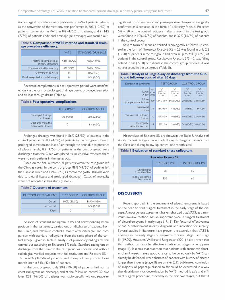
Comparative advantages of VATS in relation to standard thoracic drainage in primary pleural empyema treatment 47
tional surgical procedures were performed in 42% of patients, where-as the conversion to thoracotomy was performed in 20% (10/50) of patients, conversion in VATS in 8% (4/50) of patients, and in 14% (7/50) of patients additional drainage (re-drainage) was carried out.
Recorded complications in post-operative period were manifest-ed only in the form of prolonged drainage due to prolonged secretion and air loss through drains (Table 6).
Prolonged drainage was found in 56% (28/50) of patients in the control group and in 8% (4/50) of patients in the test group. Due to prolonged secretion and loss of air through the drain due to presence of pleural fistula, 8% (4/50) of patients in the control group were discharged from the Clinic with placed Haimlich valve, whereas there were no such patients in the test group.
Based on the final outcome, all patients within the test group left the Clinic as cured. In the control group, 88% (44/50) of patients left the Clinic as cured and 12% (6/50) as recovered (with Haimlich valve due to pleural fistula and prolonged drainage). Cases of mortality were not recorded in this study (Table 7).
Analysis of standard radiogram in PA and corresponding lateral position in the test group, carried out on discharge of patients from the Clinic, and follow-up control a month after discharge, and com-parison with standard radiograms from the same phase of the con-trol group is given in Table 8. Analysis of pulmonary radiograms was carried out according to Rx score 5% scale. Standard radiogram on discharge from the Clinic in the test group was normal and without radiological verified sequelae with full restitution and Rx score 5% = 100 in 68% (34/50) of patients, and during follow-up control one month later in 84% (42/50) of patients.
In the control group only 20% (10/50) of patients had normal chest radiogram on discharge, and at the follow-up control 30 days later 32% (16/50) of patients was radiologically without sequelae.
Significant post-therapeutic and post-operative changes radiologically confirmed as a sequelae in the form of obliterary fc sinus, Rx score 5% = 50 on the control radiogram after a month in the test group were found in 10% (5/50) of patients, and in 32% (16/50) of patients in the control group.
Severe form of sequelae verified radiologically at follow-up con-trol in the form of fibrotorax Rx score 5% = 25 was found in only 2% (1/50) of patients in the test group and even in up to 24% (12/50) of patients in the control group. Rest kavum Rx score 5% = 0, was falling behind in 4% (2/50) of patients in the control group, whereas it was not recorded in the test group (Table 8).
Mean values of Rx score 5% are shown in the Table 9. Analysis of standard chest radiogram was made during discharge of patients from the Clinic and during follow-up control one month later.
DISCUSSION
Recent approach in the treatment of pleural empyema is based on the need to start surgical treatment in the early stage of the dis-ease. Almost general agreement has emphasized that VATS, as a min-imum invasive method, has an important place in surgical treatment of pleural empyema in early stage (17,18). Key factor of effectiveness of VATS debridement is early diagnosis and indication for surgery. Several studies in literature have proven the assertion that VATS is effective in the early stages of empyema thoracic (stage I and stage II) (19,20). However, Waller and Rengarajan (2001) have proven that this method can also be effective in advanced stages of empyema (stage III). It seems that assertion that patients with anamnesis short-er than 4 weeks have a good chance to be cured only by VATS can already be defended, while chances of patients with history of disease longer than 5 weeks (stage III) are smaller (21). Sublimated conclusion of majority of papers published so far could be expressed in a way that debridement or decortication by VATS method is safe and effi-cient surgical procedure, especially in the first two stages, but that it
TEST GROUPDuration of symptoms CONTROL GROUP
LungsX-ray
Clean(complete restitution)
Narrowedfc sinus
Shadowed/Obliteraryfc sinus
Incompletereeksp/Fibrotorax
Ondischargefrom the
Clinic
Ondischargefrom the
Clinic
Rxscore(5%)
100
75
50
25
68%(34/50)
18%(9/50)
12%(6/50)
2%(1/50)
84%(42/50)
4%(2/50)
10%(5/50)
2%(1/50)
Ondischargefrom the
Clinic
Ondischargefrom the
Clinic
20%(10/50)
12%(6/50)
40%(20/50)
24%(12/50)
32%(16/50)
8%(4/50)
32%(16/50)
24%(12/50)
Table 8 Analysis of lungs X-ray on discharge from the Clin-ic and follow-up control after 30 days.
VATS STANDARD DRAINAGE
Treatment completed byprimary procedure
Conversion to thoracotomy
Conversion to VATS
Re-drainage (additional drainage)
94% (47/50)
6% (3/50)
0
0
58% (29/50)
20% (10/50)
8% (4/50)
14% (7/50)
Table 5 Comparison of VATS method and standard drain-age procedure efficiency.
TEST GROUP CONTROL GROUP
Prolonged drainage≥ 2 weeks
Discharge from theClinic with H valve
8% (4/50)
0
56% (28/50)
8% (4/50)
Table 6 Post-operative complications.
TEST GROUP % CONTROL GROUP %
On dischargefrom the Clinic
Follow-up controlafter a month
88
95.5
55
60
Mean values Rx score 5%
Table 9 Evaluation of standard chest radiogram.
TEST GROUP CONTROL GROUP
Cured
Recovered
Died
88% (44/50)
12% (6/50)
0
100% (50/50)
0
0
OUTCOME OF TREATMENT
Table 7 Outcome of treatment.
also has a role in stage III.After research done in this work, it can be accepted without re-
serve that in most cases debridement achieving complete enabling of lungs with full re-expansion is sufficient technical process with concur-rent satisfying result with regard to small pleural trauma and avoiding pleural fistula, which has direct impact on post-operative recovery in terms of shorter duration of thoracic drainage and the length of post-operative treatment.
According to results of this study the average length of total treatment at the Clinic in the test group was significantly shorter than in the control group. As an important indicator of efficiency of the methods applied, in addition to total length of treatment, the duration of thoracic drainage after surgical procedure was analyzed, and it was almost twice shorter when using the VATS method.
Using the VATS method reduces the need for other, more ag-gressive surgical procedures, such as a thoracotomy. Compared to the standard drainage, observed complications are less represented and milder in their scope. The final outcome of treatment of patients treated with the VATS method also show far better results in per-centage of healing, as well as the results based on radiologic analysis performed in the post-operative period Rx score by 5% method.
Analysis of the results gives a clear confirmation of assertion that the VATS method is superior and in every way more efficient than the standard procedure of thoracic drainage in the treatment of pleural empyema in the above mentioned stages of the disease.
Based on this research this method can be introduced as a part of standard protocol in the treatment of primary pleural empyema in patients whose history of disease is up to 5 weeks, and even in case of longer duration of the disease. It can be considered as an ideal surgical method for treatment of pleural empyema in patients whose history of disease is shorter than 4 weeks.
CONCLUSION
Based on the objectives of the research, conducted methodolog-ical treatment and obtained values of the research results, it can be concluded that: the best time to indicate the surgical treatment by the VATS method is the history of disease in duration of four weeks; de-bridement or decortication by the VATS method is safe and efficient surgical procedure, especially in the first two stages, but it also has its role in stage III; it is not necessary to insist on the classic procedure of decortication given that good results in radiological and functional terms can also be achieved by debridement; this method is extremely safe and efficient for patients with low rate of conversion to thoracot-omy, the type and frequency of mild complications; it is recommend-ed that this method is used as the first surgical option for patients in early stages of the disease.
Conflict of interest: none declared.
REFERENCES
1. Drain AJ, Ferguson JI, Sayeed R, Wilkinson S, Ritchie A. Definitive management of
advanced empyema by two-window video-assisted surgery. Asian Cardiovasc Thorac
Ann. 2007;15:238-239.
2. Solaini L, Prusciano F, Bagioni P. Video-assisted thoracic surgery in the treatment of
pleural empyema. Surg Endosc 2007;21:280-284.
3. Eryiğit H, Orki A, Koşar A, Akin O, Kiral H, Urek S, et al. [The role of video-as-
sisted thoracoscopic surgery in the treatment of pleural empyema]. Tuberk Toraks.
2007;55(1):71-6.
4. Schneider CR, Gauderer MW, Blackhurst D, Chandler JC, Abrams RS. Video-as-
sisted thoracoscopic surgery as a primary intervention in pediatric parapneumonic
effusion and empyema. Am Surg. 2010;76:957-961.
5. Drain AJ, Ferguson JI, Sayeed R, Wilkinson S, Ritchie A. Definitive management of
Advanced Empyema by Two-Window Video-Assisted Surgery. Asian Cardiovasc
Thorac Ann. 2007;15(3):238-9.
6. Chan DTL, Sihoe ADL, Chan S, Tsang DSF, Fang B, Lee T-W, Cheng L-C. Surgical
treatment for empyema thoracis: is video-assisted thoracic surgery ‘better’ than tho-
racotomy? Ann Thorac Surg. 2007;84:225-231.
7. Aziz A, Healey JM, Qureshi F, Kane TD, Kurland G, Green M, et al. Comparative
analysis of chest tube thoracostomy and video-assisted thoracoscopic surgery in em-
pyema and parapneumonic effusion associated with empyema in children. Surg Infect
(Larchmt). 2008;9(3):317-23.
8. Bishay M, Short M, Shah K, Nagraj S, Arul S, Parikh D, Jawaheer G. Efficacy of
video-assisted thoracoscopic surgery in managing childhood empyema: a large sin-
gle-centre study. J Pediatr Surg. 2009;44:337-342.
9. Kho P, Karunanantham J, Leung M, Lim E. Debridement alone without decortication
can achieve lung re-expansion in patients with empyema: an observational study. In-
teract CardioVasc Thorac Surg. 2011;12:724-727.
10. Cheng YJ, Wu HH, Chou SH. Video-assisted thoracoscopic surgery in the treatment
of chronic empyema thoracis. Surg Today. 2002;32:19-25.
11. Meier AH, Hess CB, Cilley RE. Complications and treatment failures of video-assist-
ed thoracoscopic debridement for pediatric empyema. Pediatr Surg Int. 2010;26:367-
371.
12. Cristopher B. Komanapalli: ThoracoscopicDecortication. CTSNet 19-Oct-2006
13. Kern L, Robert J, Brutsche M. Management of parapneumonic effusion and empy-
ema: medical thoracoscopy and surgical approach. Respiration. 2011;82(2):193-6.
14. Zahid I, Nagendran M, Routledge T, Scarci M. Comparison of video-assisted thoraco-
scopic surgery and open surgery in the management of primary empyema. Curr Opin
Pulm Med. 2011;17(4):255-9.
15. Luh SP, Hsu GJ, Cheng-Ren C. Complicated parapneumonic effusion and empy-
ema: pleural decortication and video-assisted thoracic surgery. Curr Infect Dis Rep.
2008;10:236-240.
16. Chambers A, Routledge T, Dunning J, Scarci M. Is video-assisted thoracoscopic surgi-
cal decortication superior to open surgery in the management of adults with primary
empyema? Interact Cardiovasc Thorac Surg. 2010;11(2):171-7.
17. Pilav I, Guska S, Mušanović S, Kadrić K. Surgical treatment of pleural empyema ac-
cording to disease stage. MED ARH. 2009;63(5):291-294.
18. Shahin Y, Duffy J, Beggs D, Black E, Majewski A. Surgical management of primary
empyema of the pleural cavity: 5 outcome of 81 patients. Interact CardioVasc Thorac
Surg. 2010;10:565-567.
19. Potaris K, Mihos P, Gakidis I, Chatziantoniou C. Videothoracoscopic and open surgi-
cal management of thoracic empyema. Surg Infect (Larchmt). 2007;8:511-7.
20. Tong BC, Hanna J, Toloza EM, Onaitis MW, D’Amico TA, Harpole DH, et al. Out-
comes of video-assisted thoracoscopic decortication. Ann Thorac Surg. 2010;89:220-
225.
21. Kurt BA, Winterhalter KM, Connors RH, Betz BW, Winters JW. Therapy of parap-
neumonic effusions in children: video-assisted thoracoscopic surgery versus conven-
tional thoracostomy drainage. Pediatrics. 2006;118:e547-53.
Reprint requests and correspondence:Ilijaz Pilav, MDClinic of Thoracic SurgeryUniversity Clinical Centre SarajevoBolnička 25, 71000 SarajevoBosnia and Herzegovina Phone: + 387 61 503-447Email: [email protected]
48 I. Pilav et al.
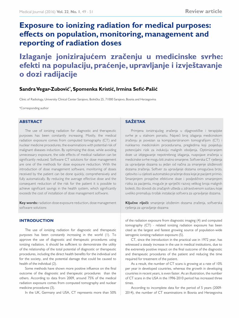
Exposure to ionizing radiation for medical purposes: effects on population, monitoring, management and reporting of radiation doses
Izlaganje jonizirajućem zračenju u medicinske svrhe: efekti na populaciju, praćenje, upravljanje i izvještavanje o dozi radijacije
Sandra Vegar-Zubović*, Spomenka Kristić, Irmina Sefić-Pašić
Clinic of Radiology, University Clinical Center Sarajevo, Bolnička 25, 71000 Sarajevo, Bosnia and Herzegovina
*Corresponding author
ABSTRACT
The use of ionizing radiation for diagnostic and therapeutic purposes has been constantly increasing. Mostly, the medical radiation exposure comes from computed tomography (CT) and nuclear medicine procedures, the examinations with potential risk of malignant diseases induction. By optimizing the dose, while avoiding unnecessary exposure, the side effects of medical radiation can be significantly reduced. Software CT solutions for dose management are one of the methods for dose exposure reduction. With the introduction of dose management software, monitoring of doses received by the patient can be done quickly, comprehensively and fully automatically. By reducing the average effective dose and the consequent reduction of the risk for the patient it is possible to achieve significant savings in the health system, which significantly exceeds the cost of installation of dose management software.
Key words: radiation dose exposure reduction, dose management software solutions
SAŽETAK
Primjena ionizirajućeg zračenja u dijagnostičke i terapijske svrhe je u stalnom porastu. Najveći broj izlaganja medicinskom zračenju je povezan sa kompjuteriziranom tomografijom (CT) i nuklearno medicinskim procedurama, pregledima koji posjeduju potencijalni rizik za indukciju malignih oboljenja. Optimiziranjem doze uz izbjegavanje nepotrebnog izlaganja, nuspojave zračenja u medicinske svrhe mogu biti znatno smanjene. Softverska CT rješenja za upravljanje dozama su jedan od načina za smanjenje izloženosti dozama zračenja. Softver za upravljanje dozama omogućava brzo, cjelovito i u cijelosti automatsko praćenje doza koje je pacijent primio. Smanjenjem prosječne efektivne doze i posljedičnim smanjenjem rizika za pacijenta, moguće je spriječiti razvoj velikog broja malignih bolesti, što dovodi do značajnih ušteda u zdravstvenom sustavu koje uveliko premašuju trošak instalacije softvera za upravljanje dozama.
Ključne riječi: smanjenje izloženim dozama zračenja, softverska rješenja za upravljanje dozama
Medical Journal (2016) Vol. 22, No. 1, 49 - 51 Review article
INTRODUCTION
The use of ionizing radiation for diagnostic and therapeutic purposes has been constantly increasing in the world (1). To approve the use of diagnostic and therapeutic procedures using ionizing radiation, it should be sufficient to demonstrate the utility of the relationship of the total potential of diagnostic or therapeutic procedures, including the direct health benefits for the individual and for the society, and the potential damage that could be caused to health of the individual (2).
Some methods have shown more positive influence on the final outcome of the diagnostic and therapeutic procedures than the others. According to data from 2009, around 75% of the medical radiation exposure comes from computed tomography and nuclear medicine procedures (3).
In the UK, Germany and USA, CT represents more than 50%
of the radiation exposure from diagnostic imaging (4) and computed tomography (CT) - related ionizing radiation exposure has been cited as the largest and fastest growing source of population-wide iatrogenic ionizing radiation exposure (5).
CT, since the introduction in the practical use in 1972 year, has witnessed a steady increase in the use in medical institutions, due to the extremely positive impact on the final outcome of the diagnostic and therapeutic procedures of the patient and reducing the time required for treatment of the patient.
As a result, the number of CT scans is growing at a rate of 10% per year in developed countries, whereas the growth in developing countries in recent years, is even faster. As an illustration, the number of CT scans in the USA in the 1996-2010 period has increased three times.
According to incomplete data for the period of 5 years (2009-2014), the number of CT examinations in Bosnia and Herzegovina

increased by around 75%. At the same time, the number of CT examinations at the Department of Radiology of the UCCS increased from 22,958 examinations to 38,050 - an increase of 67%. A similar trend is expected in the near future.
This fact poses some other challenges to a health system such as population exposure to ionizing radiation. Unfortunately, the impact of this type of radiation is not sufficiently documented. Studies on the biological effects of exposure to low doses of radiation on the human body are mainly based on the monitoring of the radiation effects on the survivors after the 1945 atomic bombings on Japan.
A small number of purely retrospective studies has been recently conducted, following the long-term effects of exposure to x-rays and all of them have shown that there is a correlation between exposure to radiation during CT examinations and an increased risk for the development of malignancies in children and young people. One of the studies has shown small but existing absolute risks of leukaemia and brain cancer after CT scaning (one case of leukaemia and one case of brain tumour per 10000 head CT scans) (6).
Even smaller number of studies have analized the exposure of adults to diagnostic procedures using ionized radiation and related risk from malignant disease development. The reason for that is in the fact that CT-associated cancer risks in adults depend on study type, analised body region, patient age and sex, and also from the fact that it is very difficult to collect data on all conducted radiology procedures during the lifespan (7,8).
Based on that considerations, the International Commission for Radiation Protection (ICRP) coefficients were elaborated to calculate the potential risk of developing malignant tumors after exposure to radiation. The nominal ICRP coefficient of risk, balanced by gender, age and type of examination, is about 5.5% for the received dose of 1 Sievert‘s (Sv). However, an effective dose and with it related potential risk, if any of these parameters are calculated separately, can vary dramatically: for example, the potential risk for the patient to whom an X-ray of the foot was done is less than10-9, and the potential risk for a young girl to whome CT examination of the trunk was performed (thorax, abdomen and pelvis) has increased to significant 10-3 (9).
United States National Academies in their report concerning radiation health effects called the Biologic Effects of Ionizing Radiation (BEIR) VII estimate that 1,050 cases of solid cancer and 85 cases of leukemia are expected to result in 100,000 persons exposed to 100 mSv (10).
Numerous authors have studied the practical application of nominal coefficients risk of malignant diseases development as a result of medical radiation. The study „Projected Cancer Risks From Computed Tomographic Scans Performed in the United States in 2007“ (11), estimated that in the United States 29,000 new malignant diseases will develop as a result of 70 million CT examinations done in 2007 (increased risk compared to general population of about 0.041%). Similar studies in Bosnia and Herzegovina have shown projection of increased risk of about 0.05%, taking into account the dose od radiation to which patients were exposed (12). Therefore, it is realistic to expect that about hundred new cases of malignant diseases will be induced every year in Bosnia and Herzegovina.
The cost of a year of treatment, care and monitoring of a patient with cancer, depending on the type and stage of disease, ranges from several thousand to several hundred thousand BAM.
Based on the increase in the number of CT examinations and the potential risk of induction of malignant diseases there is a great responsibility of health professionals and regulatory authorities in terms of optimizing the radiation dose and implementing the principle od justification of diagnostic medical exposure (13). By optimizing the dose, while avoiding unnecessary exposure, the side effects of medical radiation can be significantly reduced.
In a study published in the New England Journal of Medicine in 2009 (Exposure to Low-Dose Ionizing Radiation from Medical Imaging Procedures in the United States) the author states: „In conclusion, our results suggest that a form of medical imaging in the United States resulting in significant exposure to a large number of younger people to ionizing radiation. It is necessary to encourage the development of new approaches to optimize and ensure the correct use of these procedures“ (3).
Specific solutions in terms of optimization and proper utilization of medical equipment that generates ionizing radiation have already been adopted in the USA, but the new legislation and the EU in 2013 introduced innovations in terms of:
- Obligations to provide written protocols for each type of equipment and time for all relevant categories of patients;
- Mandatory documentation of the radiation dose that patients receive during each time (radiologists and clinical doctors);
- Referral guides for medical radiological examination, which include data on radiation, must be accessible to the reference institutions and individuals;
- The institution is obliged to keep data on individual and cumulative doses of radiation received by the patient (14).
Due to incomplete infrastructure, Member States were given a deadline until 1 January 2018 to fully adapt their systems with the mentioned legal solution.
Bosnia and Herzegovina on its path towards the EU, is obliged to adapt its legislation to the EU law. The existing legislation can be improved, for example, with implementation of more detailed rules regarding regulation of clinical audit, obligation to inform patients about potential risks of radiation etc. (15).
However, the adjustment process is very complex. It was shown on the example of number of institutions in the EU, which have spent significant resources (e.g. time for evaluation and training, funds for new equipment, etc.) without improving the patient‘s safety. It is a time consuming process which have to be done constantly, in order that the team involved in it can periodically analize collected data and suggest further improvements of the system.
With the increasing number of examinations using ionizing radiation, health care institutions, as seen in previous data, are faced with number of challenges:
1. Adjusting the legal regulation of radiation dose;2. The application of the best practices in the management of radiation doses and documentation thereof;3. Reducing costs.
Software solutions, by nature of CT diagnostics as a high-tech process, have been imposed as a possible solution. By the introduction of dose management software, monitoring of doses received by the
50 S. Vegar-Zubović et al.

Exposure to ionizing radiation for medical purposes: effects on population, monitoring, management and reporting of radiation doses 51
patient can be done quickly, comprehensively and fully automatically. International Atomic Energy Agency (IAEA), initiated the
Smart Card /SmartRadTrack project in 2006 with the objective of developing a template for tracking cumulative radiation exposure and whenever possible dose for individual procedure which can be conducted through PACS systems in hospitals (16).
In addition, the analysis of a large number of data can provide radiologists, engineers, physicists and control authorities access to CT dose distribution, productivity and capacity utilization (17, 18), and based on that data to plan the distribution of work and other activities to improve the quality and technological development.
Practice has shown that the institutions that introduced dose management software were able to reduce the average effective dose for CT procedures for 25-30% (19).
CONCLUSION
By reducing the average effective dose and the consequent reduction of the risk for the patient, in terms that the development of a large number of malignant diseases can be prevented, it is possible to achieve significant savings in the health system, which significantly exceed the cost of installation of dose management software.
Conflict of interest: none declared.
REFERENCES
1. United Nations Scientific Committee on the Effects of Atomic Radiation. Sources and
effects of ionizing radiation. Medical radiation exposures, Annex A. 2008 Report to the
General Assembly with annexes. New York, NY: United Nations (in press).
2. McCollough CH. To Scan or not to Scan: Consideration of Medical Benefit in the Justi-
fication of CT Scanning. Health Phys. 2016;110(3):287-90.
3. Fazel R, Krumholz HM, Wang Y, Ross JS, Chen J, Ting HH, et al. Exposure to low-dose
ionizing radiation from medical imaging procedures. N Engl J Med. 2009;361(9):849-57.
4. Tsushima Y, Taketomi-Takahashi A, Takei H, Otake H, Endo K. Radiation exposure
from CT examinations in Japan. BMC Medical Imaging. 2010;10:24.
5. Talati RK, Dunkin J, Parikh S, Moore WH. Current methods of monitoring radiation
exposure from CT. J Am Coll Radiol. 2013;10(9):702-7.
6. Pearce MS, Salotti JA, Little MP, McHugh K, Lee C, Kim KP, et al. Radiation exposure
from CT scans in childhood and subsequent risk of leukaemia and brain tumours: a
retrospective cohort study. Lancet. 2012;380(9840):499-505.
7. Smith-Bindman R, Lipson J, Marcus R, Kim KP, Mahesh M, Gould R, et al. Radiation
dose associated with common computed tomography examinations and the associated
lifetime attributable risk of cancer. Arch Intern Med. 2009;169(22):2078-2086.
8. Lam DL, Larson DB, Eisenberg JD, Forman HP, Lee CI. Communicating potential radia-
tion-induced cancer risks from medical imaging directly to patients. AJR Am J Roentge-
nol. 2015;205(5);962-970.
9. Wall BF, Haylock R, Jansen JTM, Hillier MC, Hart D, Shrimpton PC. Radiation risks
from medical X-ray examinations as a function of the age and sex of the patient. HPA-
CRCE-028. 66 pp. https://www.gov.uk/government/uploads/system/uploads/at-
tachment_data/file/340147/HPA-CRCE-028_for_website.pdf
10. Monson R, Cleaver J, Abrams HL, Bingham E, Buffler PA. Beir VII: health risks from ex-
posure to low levels of ionizing radiation Washington, DC: National Academies Press;
2005.
11. Berrington de González A, Mahesh M, Kim KP, Bhargavan M, Lewis R, Mettler F, Land
C. Projected cancer risks from computed tomographic scans performed in the United
States in 2007. Arch Intern Med. 2009;169(22):2071-7.
12. Ciraj-Bjelac O, Beganovic A, Faj D, Gershan V, Ivanovic S, Videnovic IR, Rehani MM.
Radiation protection of patients in diagnostic radiology: Status of practice in five East-
ern-European countries, based on IAEA project. Eur Journal Radiol. 2011;79(2):e70-3.
13. Malone J, Guleria R, Craven C, Horton P, Järvinen H, Mayo J, et al. Justification of
diagnostic medical exposures: some practical issues. Report of an International Atomic
Energy Agency Consultation. Br J Radiol. 2012;85(1013):523-38.
14. European Society of Radiology (ESR). Summary of the European Directive
2013/59/Euratom: essentials for health professionals in radiology. Insights Imaging.
2015;6(4):411-7.
15. Državna regulatorna/regulativna agencija za radijacijsku i nuklearnu sigurnost. Pravilnik
o zaštiti od jonizirajućeg zračenja kod medicinske ekspozicije. Službeni glasnik BiH” broj
13/11.
16. Rehani MM, Frush DP. Patient exposure tracking: the IAEA smart card project. Radiat
Prot Dosimetry. 2011;147(1-2):314-6.
17. Brink JA. Clinical decision-making tools for exam selection, reporting and dose tracking.
Pediatr Radiol. 2014;44(3):418-21.
18. Seuri R, Rehani MM, Kortesniemi M. How tracking radiologic procedures and dose
helps: experience from Finland. AJR Am J Roentgenol. 2013;200(4):771-5.
19. Cody DD, Fisher TS, Gress DA, Layman RR Jr, McNitt-Gray MF, Pizzutiello RJ Jr, Fairo-
bent LA. AAPM medical physics practice guideline 1.a: CT protocol management and
review practice guideline. J Appl Clin Med Phys. 2013;14(5):3-12.
Reprint requests and correspondence:Sandra Vegar-Zubović, MD, PhDClinic of RadiologyUniversity Clinical Center Sarajevo Bolnička 25 71000 Sarajevo Bosnia and Herzegovina Phone: + 387 33 297 541Email: [email protected]

INSTRUCTIONS TO AUTHORS
Journal “Medical Journal” publishes original research articles, professional, review and educative articles, case reports, criticism, reports,
and Bosnian/Croatian/Serbian language.Authors take responsibility for all the statements and attitudes in their articles. If article was written by several authors, it is necessary to provide full contact details (telephone numbers and email addresses) of the corresponding author for the cooperation during preparation of the text to be published.Authors should indicate whether the procedures carried out on humans were in accordance with the ethical standards of medical deontol-ogy and Declaration of Helsinki.Articles that contain results of animal studies will only be accepted for publication if it is made clear that ethics standard were applied.Measurements should be expressed in units, according to the rules of the SI System.
Manuscript submission should be sent to Editorial Board and addressed to:“MEDICINSKI ŽURNAL”Institut za naučnoistraživački rad i razvoj Kliničkog centra Univerziteta u SarajevuBolnička 2571000 SarajevoBosna i Hercegovinae-mail: [email protected]; [email protected]
COVER LETTERApart from the manuscript, the authors should enclose a cover letter, with the signed statements of all authors, to the Editorial Board of “Medical Journal” stating that:1. the work has not been published or accepted for publication previously in another journal,2. the work is in accordance with the ethical committee standards,3. the work, accepted for publication, becomes ownership of “Medical Journal”.
PREPARATION OF MANUSCRIPT
disk (Word Windows), or e-mail.Spacing: 1,5: left margin: 2,5 cm; right margin: 2,5 cm; top and bottom margin: 2,5 cm.
program in which they are prepared. Articles are written in-extenso in English. The manuscript should be submitted on a good quality CD disc, or by e-mail, together with two printed copies (if it is possible). Sent CD disks will not be returned to the authors.
ARTICLE CONTAINS:
TITLE OF THE ARTICLE IN ENGLISH LANGUAGETITLE OF THE ARTICLE IN BOSNIAN/SERBIAN/CROATIAN (B/S/C) LANGUAGEFirst name and last name of author and co-authors
Name and address of institution in which author/co-authors are employed (same for all authors) in B/S/C and English language as well as the address of corresponding author at the end of the paper.
Summary in B/S/C language with the precise translation in English. Abstract of approximately 200-250 words should concisely describe the contents of the article. Key words
ARTICLE BODYThe main body of the article should be systematically ordered under the following headings:- INTRODUCTION- MATERIALS AND METHODS- RESULTS - DISCUSSION
52 Instructions to authors
INSTRUCTIONS TO AUTHORS
Journal “Medical Journal” publishes original research articles, professional, review and educative articles, case reports, criticism, reports,
and Bosnian/Croatian/Serbian language.Authors take responsibility for all the statements and attitudes in their articles. If article was written by several authors, it is necessary to provide full contact details (telephone numbers and email addresses) of the corresponding author for the cooperation during preparation of the text to be published.Authors should indicate whether the procedures carried out on humans were in accordance with the ethical standards of medical deontol-ogy and Declaration of Helsinki.Articles that contain results of animal studies will only be accepted for publication if it is made clear that ethics standard were applied.Measurements should be expressed in units, according to the rules of the SI System.
Manuscript submission should be sent to Editorial Board and addressed to:“MEDICINSKI ŽURNAL”Institut za naučnoistraživački rad i razvoj Kliničkog centra Univerziteta u SarajevuBolnička 2571000 SarajevoBosna i Hercegovinae-mail: [email protected]; [email protected]
COVER LETTERApart from the manuscript, the authors should enclose a cover letter, with the signed statements of all authors, to the Editorial Board of “Medical Journal” stating that:1. the work has not been published or accepted for publication previously in another journal,2. the work is in accordance with the ethical committee standards,3. the work, accepted for publication, becomes ownership of “Medical Journal”.
PREPARATION OF MANUSCRIPT
disk (Word Windows), or e-mail.Spacing: 1,5: left margin: 2,5 cm; right margin: 2,5 cm; top and bottom margin: 2,5 cm.
program in which they are prepared. Articles are written in-extenso in English. The manuscript should be submitted on a good quality CD disc, or by e-mail, together with two printed copies (if it is possible). Sent CD disks will not be returned to the authors.
ARTICLE CONTAINS:
TITLE OF THE ARTICLE IN ENGLISH LANGUAGETITLE OF THE ARTICLE IN BOSNIAN/SERBIAN/CROATIAN (B/S/C) LANGUAGEFirst name and last name of author and co-authors
Name and address of institution in which author/co-authors are employed (same for all authors) in B/S/C and English language as well as the address of corresponding author at the end of the paper.
Summary in B/S/C language with the precise translation in English. Abstract of approximately 200-250 words should concisely describe the contents of the article. Key words
ARTICLE BODYThe main body of the article should be systematically ordered under the following headings:- INTRODUCTION- MATERIALS AND METHODS- RESULTS - DISCUSSION
Instructions to authors 53
- CONCLUSION- REFERENCES
INTRODUCTIONIntroduction is a concise, short part of the article, and it contains purpose of the article relating to other published articles with the same topic. It is necessary to quote the main problem, aim of investigation, and/or main hypothesis which is investigated.
MATERIALS AND METHODS
protocol and type of clinical investigation, place and period of investigation. Main characteristics of investigation should be described (rand-omization, double-blind test, cross test, placebo test), standard values for tests, time framework (prospective, retrospective study), selection and number of patients – criteria for inclusion and exclusion from the study.
RESULTS
and directly incorporated in the text, at the exact place, with ordinal number and concise heading. Table should have at least two columns
DISCUSSION Discussion is concise and refers to own results, in comparison with the other authors’ results. Citation of references should follow Vancou-
CONCLUSIONConclusion should be concise and should contain most important facts, which were obtained during investigation and its eventual clinical
REFERENCES – Instructions for writing referencesReferences should follow the format of the requirements of Vancouver rules.
number in parenthesis at the end of the sentence according to the order of entering. Every further referring to the same reference, number
numbers in the order of entering in the text (entering reference number). Journal’s title is abbreviated using Index Medicus abbreviations.
It is very important to properly design references according to instructions that may be downloaded from addresses National Library of Medicine Citing Medicine http://www.ncbi.nlm.nih.gov/books/bv.fcg?rid=citmed.TOC&depth=2, or International Committee of Medical Journal Editors Uniform Requirements for Manuscripts Submitted to Biomedical Journals: Sample References http://www.nlm.nih.gov/bsd/uniform_requirements.html.

UPUTSTVA AUTORIMA
Časopis “Medicinski žurnal” objavljuje originalne naučne radove, stručne, pregledne i edukativne, prikaze slučajeva, recenzije, saopćenja, stručne obavijesti i drugo iz područja svih medicinskih disciplina. Rad in-extenso (cjelokupan) piše se na engleskom jeziku, uz sažetak i naslov rada koji uz engleski trebaju biti napisani i na našim jezicima (bosanski, hrvatski i srpski). Autori su odgovorni za sve navode i stavove u nji-hovim radovima. Ukoliko je rad pisalo više autora, potrebno je navesti tačnu adresu (uz telefonski broj i e-mail adresu) onog autora s kojim će uredništvo sarađivati pri uređenju teksta za objavljivanje.Ukoliko su u radu prikazana istraživanja na ljudima, mora se navesti da su provedena u skladu s načelima medicinske deontologije i Deklaracije iz Helsinkija.Ukoliko su u radu prikazana istraživanja na životinjama, mora se navesti da su provedena u skladu s etičkim načelima. Prilikom navođenja mjernih jedinica, treba poštovati pravila navedena u SI sistemu.Radovi se šalju Redakciji na adresu:“MEDICINSKI ŽURNAL”Institut za naučnoistraživački rad i razvoj Kliničkog centra Univerziteta u SarajevuBolnička 2571000 SarajevoBosna i Hercegovinae-mail: [email protected]; [email protected]
POPRATNO PISMOUz svoj rad, autori su dužni Redakciji “Medicinskog žurnala” dostaviti popratno pismo, koje sadržava vlastoručno potpisanu izjavu svih autora:1. da navedeni rad nije objavljen ili primljen za objavljivanje u nekom drugom časopisu,2. da je istraživanje odobrila Etička komisija,3. da prihvaćeni rad postaje vlasništvo “Medicinskog žurnala”.
OPSEG I OBLIK RUKOPISA
Windows), ili e-mail.Prored: 1,5: lijeva margina: 2,5 cm; desna margina: 2,5 cm; gornja i donja margina: 2,5 cm.
obavezno napisati na engleskom jeziku, a sažetak i naslov još i na našem jeziku.Rad se dostavlja na CD-u, i/ili e-mailom, uz dva štampana primjerka (ako je moguće). CD se ne vraća.
RAD SADRŽI:
NASLOV RADA NA ENGLESKOM JEZIKUNASLOV RADA NA NAŠEM JEZIKU
Ime i prezime autora i koautora
Naziv i puna adresa institucije u kojoj je autor-koautor/i zaposlen/i (jednako za sve autore), na engleskom jeziku, te na kraju rada navedena adresa kontakt-autora.
Sažetak na našem jeziku, kao i na engleskom - max. 200–250 riječi, s najznačajnijim činjenicama i podatcima iz kojih se može dobiti uvid u kompletan rad. Ključne riječi - Key words, na našem jeziku i na engleskom, ukupno do pet riječi, navode se ispod Sažetka, odnosno Abstracta.
SADRŽAJSadržaj rada mora biti sistematično i strukturno pripremljen i podijeljen u poglavlja i to:- UVOD- MATERIJAL I METODE- REZULTATI- DISKUSIJA- ZAKLJUČAK- LITERATURA
54 Uputstva autorima
UVODUvod je kratak, koncizan dio rada i u njemu se navodi svrha rada u odnosu na druge objavljene radove sa istom tematikom. Potrebno je navesti glavni problem, cilj istraživanja i/ili glavnu hipotezu koja se provjerava.
MATERIJAL I METODE
literaturi. U kliničko-epidemiološkim studijama opisuju se: uzorak, protokol i tip kliničkog istraživanja, mjesto i vrijeme istraživanja. Potreb-no je opisati glavne karakteristike istraživanja (npr. randomizacija, dvostruko slijepi pokus, unakrsno testiranje, testiranje s placebom itd.), standardne vrijednosti za testove, vremenski odnos (prospektivna, retrospektivna studija), izbor i broj ispitanika – kriterije za uključivanje i isključivanje u istraživanje.
REZULTATI-
ose u tekst gdje im je mjesto, s rednim brojem i konciznim naslovom. Tabela treba imati najmanje dva stupca s obrazloženjem što prikazuje;
DISKUSIJAPiše se koncizno i odnosi se prvenstveno na vlastite rezultate, a potom se nastavlja upoređivanje vlastitih rezultata s rezultatima drugih autora, pri čemu se citiranje literature navodi po važećim Vankuverskim pravilima. Diskusija se završava potvrdom zadatog cilja ili hipoteze, odnosno njihovim negiranjem.
ZAKLJUČAK i oak ,unejmirp ukčinilk unlautneve uvohijn i ajnavižartsi mokot udar u olšod es hijok od ecinejnič ejintibjan ižrdas ad ,katark edub ad aberT
LITERATURA - Upute za citiranje - pisanje literatureLiteratura se obavezno citira po Vankuverskim pravilima.Svaku tvrdnju, saznanje ili misao treba potvrditi referencom. Reference u tekstu treba označiti po redoslijedu unošenja arapskim brojevima u zagradi na kraju rečenice. Ukoliko se kasnije u tekstu pozivamo na istu referencu, navodimo broj koji je referenca dobila prilikom prvog unošenja/pominjanja u tekstu. Literatura se popisuje na kraju rada, rednim brojevima pod kojim su reference unesene u tekst (ulazni broj reference), a naslov časopisa se skraćuje po pravilima koje određuje Index Medicus. Ukoliko je citirani rad napisalo više autora, navodi se prvih šest i doda “et al.”.Vrlo je važno ispravno oblikovati reference prema uputama koje se mogu preuzeti na adresama National Library of Medicine Citing Medi-cine http://www.ncbi.nlm.nih.gov/books/bv.fcgi?rid=citmed.TOC&depth=2 , ili International Committee of Medical Journal Editors Uniform Requirements for Manuscripts Submitted to Biomedical Journals: Sample References http://www.nlm.nih.gov/bsd/uniform_requirements.html.
Uputstva autorima 55