
Dr Nahar - Injury among athletes and obese overweight obese population

Biochemical and Biophysical Research Communications 443 (2014) 1110–1117
Contents lists available at ScienceDirect
Biochemical and Biophysical Research Communications
journal homepage: www.elsevier .com/locate /ybbrc
Voglibose administration regulates body weight and energy intakein high fat-induced obese mice
0006-291X/$ - see front matter � 2013 Elsevier Inc. All rights reserved.http://dx.doi.org/10.1016/j.bbrc.2013.12.120
Abbreviations: T2DM, type 2 diabetes mellitus; HOMA, homeostatic modelassessment; GLP-1, glucagon like peptide-1; DDP-4, dipeptidyl peptidase-4; OB-R,leptin receptor; POMC, proopiomelanocortin; Cart, cocaine and amphetamine-regulated transcript; AgRP, agouti-related protein; NPY, neuropeptide Y; PGC-1a,peroxisome proliferator-activated receptor c coactivator- 1a; GLUT4, insulin-responsive glucose transporter type 4; GAPDH, glyceraldehyde 3-phosphatedehydrogenase.⇑ Corresponding authors. Address: Department of Food and Nutrition, Korea
University, Seoul 136-704, Republic of Korea (M.-J. Shin).E-mail addresses: [email protected] (J.H. Chung), [email protected] (M.-J.
Shin).
Hyun Ju Do a, Taeon Jin b, Ji Hyung Chung b,⇑, Ji Won Hwang a,c, Min-Jeong Shin a,c,d,⇑a Department of Food and Nutrition, Korea University, Seoul 136-703, Republic of Koreab Department of Applied Bioscience, CHA University, Gyeonggi-do 463-836, Republic of Koreac Department of Public Health Sciences, Graduate School, Korea University, Seoul 136-703, Republic of Koread Korea University Guro Hospital, Korea University, Seoul 152-703, Republic of Korea
a r t i c l e i n f o a b s t r a c t
Article history:Received 13 December 2013Available online 2 January 2014
Keywords:VogliboseObesityAppetiteLeptinLeptin receptorMitochondriaPGC1
We tested whether long-term administration of voglibose (VO) prevents diet induced obesity in additionto hypoglycemic effects in high fat fed mice and further investigated the underlying mechanisms bywhich voglibose exerts its weight lowering effect. Male C57BL/6 mice were fed ad libitum for 12 weekswith the control diet (CTL), high-fat diet (HFD) or the HFD with VO supplementations. Blood lipid profile,plasma leptin levels and hepatic triglyceride content, as well as expressions of genes involved in appetiteand mitochondrial function were examined. The results showed that VO significantly reduced bodyweight, fat mass and energy intakes in high fat fed mice. VO showed improved metabolic profiles includ-ing blood glucose, triglyceride and free fatty acid. Elevated levels of plasma leptin in HFD were signifi-cantly reduced with the VO, furthermore, VO modulated the hypothalamic expressions of leptinreceptors and appetite related genes. VO showed the upregulated expressions of PGC-1 in the liver andepididymal adipose tissue. In conclusion, VO may exert antiobesity properties through reductions inenergy intake and improvement in mitochondrial function, indicating that VO has potential therapeuticuse in patients with obesity, type 2 diabetes, and related complications.
� 2013 Elsevier Inc. All rights reserved.
1. Introduction
It is well known that obesity is an established risk factor fortype 2 diabetes mellitus (T2DM), and weight loss is recommendedfor obese patients with T2DM because of the numerous benefits ofweight loss [1,2]. An intestinal enzyme, a-glucosidase, acts on car-bohydrate digestion and glucose absorption [3]. The action of a-glucosidase inhibitors is to delay intestinal absorption of carbohy-drates by inhibiting degradation, which results in altered postpran-dial glucose and insulin levels [4]. Acarbose, miglitol and voglibose(VO) are 3 major oral hypoglycemic drugs used to treat patientswith T2DM by delaying the absorption of glucose, thereby manag-ing the disease and related complications [5]. Recently, it was re-
ported that the a-glucosidase inhibitors provide significantbeneficial health outcomes in post hoc analyses of randomized pla-cebo-controlled trials [6]. For example, both acarbose in STOP-NID-DM (Study to Prevent Non-Insulin-Dependent Diabetes Mellitus)[7] and VO in a Japanese trial [8] were shown to slow the progres-sion to T2DM. In addition, a-glucosidase inhibitors were reportedto favorably affect several cardiovascular endpoints; in particular,VO was shown to inhibit cardiac remodeling by decreasing myo-cardial oxidative stress through improvement of glycemic controlin mice with cardiac pressure overload [9]. While there is evidencefor the beneficial effects of a-glucosidase inhibitors on cardiovas-cular risk, there is limited information on weight beneficial effectsof them.
VO is known to demonstrate anti-hyperglycemic effects in Jap-anese patients [8,10]; more importantly, being superior to bothacarbose and miglitol in terms of potency and side effect profile[3]. In the present study, we tested whether long-term administra-tion of VO prevents diet-induced obesity and hypoglycemic effectsin mice fed a high-fat diet (HFD). The study provides novel infor-mation that VO may exert antiobesity properties through reduc-tions in energy intake and improvement of mitochondrialfunction, indicating that VO has potential therapeutic use in pa-tients with obesity, T2DM and related complications.
H.J. Do et al. / Biochemical and Biophysical Research Communications 443 (2014) 1110–1117 1111
2. Materials and methods
2.1. Animals and study design
Male C57BL/6 mice (5 weeks old) were randomly assigned to 3groups after a 1 week adaptation period. Group 1 received a nor-mal control diet (CTL, n = 10), Group 2 received a HFD (n = 10),and Group 3 received a HFD supplemented with VO (n = 10). VO(Santa Cruz Biotechnology, Dallas, TX, USA) was prepared in dis-tilled water and administered orally on the basis of body weight(1 mg/kg once daily). The mice in the CTL and HFD groups receivedan equivalent volume of vehicle (distilled water). The CTL wasbased on the composition of the American Institute of Nutrition(AIN)-76 rodent diet. The HFD was identical to the CTL but in-cluded 200 g fat/kg (170 g lard plus 30 g corn oil) and 1% choles-terol. The mice were housed under standard (18–24 �C, 50–60%humidity) laboratory conditions and maintained on a 12/12-hlight/dark schedule (lights on at 8:00 am) with free access to foodand water for 12 weeks. Daily feed intake and weekly gains in bodyweight were routinely recorded throughout the experimental per-iod using a computing scale (Acominc Co., Pocheon, Korea). Allexperimental procedures were approved by the Committee on Ani-mal Experimentation and Ethics of Korea University.
2.2. Quantification of plasma metabolic parameters and hepatictriglyceride
Plasma triglyceride (TG) and free fatty acid (FFA) levels weremeasured using an Olympus AU400 Chemistry Analyzer (Tokyo, Ja-pan). Hepatic lipids were extracted using the method developed byFolch et al. [11] and dried lipid residues were dissolved in 0.5 mLisopropanol. The concentrations of TG in the hepatic-lipid extractswere measured using commercial kits (Asan Pharm, Seoul, Korea).
2.3. Plasma glucose, insulin, homeostatic model assessment of insulinresistance, and homeostatic model assessment of beta cell function
Fasting plasma levels of glucose were measured by hexokinasemethod using an Olympus AU400 Chemistry Analyzer. Insulin wasmeasured by radioimmunoassay with kits from Biosource (Carls-bad, CA, USA). Insulin sensitivity was estimated using the homeo-static model assessment (HOMA) index which is an arithmetic wayof deriving indices of peripheral tissue insulin resistance and pan-creatic endocrine function (beta cell function, HOMA-b). HOMA-IRand HOMA-b are derived using the following formulae: HOMA-IR = (Fasting plasma insulin [lIU/mL] � fasting plasma glucose[mmol/L])/22.5 and HOMA-b = (20 � fasting plasma insulin [lIU/mL])/(fasting plasma glucose [mmol/L] � 3.5) [12].
2.4. Plasma leptin measurements
Plasma leptin levels were measured using enzyme linkedimmunosorbent assay kits (Komabiotech, Seoul, Korea), as perthe manufacturer’s instructions.
2.5. RNA extraction and quantitative real time polymerase chainreaction
Total RNA was extracted from the hypothalamus, liver, and epi-didymal adipose tissue using an RNeasy Lipid Tissue Mini Kit (Qia-gen, Hilden, Germany) according to the manufacturer’s protocol.Complementary DNA was synthesized from 1 lg of RNA using oli-go-dT and Superscript II reverse transcriptase (Invitrogen, Carls-bad, CA, USA). Primer sequences for polymerase chain reactionwere as follows: leptin receptor a (OB-Ra), 50-AAGTTGTTTTGG-
GACGATG-30 (forward) and 50-ATTGGGTTCATCTGTAGTGG-30 (re-verse); OB-Rb, 50-ACTCTGGTCAGCAACGATAAACTA-30 (forward)and 50-GAAAAATGTCTGGGCCTCTGTCTC-30 (reverse); proopiomela-nocortin (POMC), 50-ATGCCGAGATTCTGCTACAG-30 (forward) and50-TGCTGCTGTTCCTGGGGC-30 (reverse); agouti-related protein(AgRP), 50-ATGCTGACTGCAATGTTGCTG-30 (forward) and 50-CAGA-CTTAGACCTGGGAACTCT-30 (reverse); neuropeptide Y (NPY), 50-ATATGGCAAGAGATCCAGCCC-30 (forward) and 50-ACATGGAAGGGTCTTCAAGCC-30 (reverse); cocaine and amphetamine-regulatedtranscript (Cart), 50-CTGGACTACCACAGGGTCCTT-30 (forward) and50-AGACCTGACCTCACCTTCCAG-30 (reverse); peroxisome prolifera-tor-activated receptor c coactivator-1a (PGC-1a), 50-CGGAAATCATATCCAACCAG-30 (forward) and 50-TGAGGACCGCTAGCAAGTTTG-30
(reverse); PGC-1b, 50-AACCCAACCAGTCTCACAGG-30 (forward) and50-ATGCTGTCCTTGTGGGTAGG-30 (reverse); insulin-responsive glu-cose transporter type 4 (GLUT4), 50-CAACAGCTCTCAGGCATCAA-30
(forward) and 50-CTCAAAGAAGGCCACAAAGC-30 (reverse); glycer-aldehyde 3-phosphate dehydrogenase (GAPDH), 50-AACTTTGG-CATTGTGGAAGG-30 (forward) and 50-ACACATTGGGGGTAGGAACA-30 (reverse). The real-time PCR (Step One Plus, AppliedBiosystems, Foster City, CA, USA) conditions were: 15 min at95 �C, followed by 40 cycles of 94 �C for 30 s, 52–60 �C for 20 sand 72 �C for 30 s. GAPDH was used as the control in the compar-ative cycle threshold method.
2.6. Statistical analysis
Statistical analysis was performed using SPSS 12.0.1 (Chicago,IL, USA). The results were presented as means ± standard error(SE). The differences among the experimental groups were ana-lyzed using a one-way ANOVA, followed by Bonferroni’s post hoctest. P-values < 0.05 indicate a significant difference.
3. Results
3.1. Effect of VO supplementation on body weight, food intake, andbody fat mass
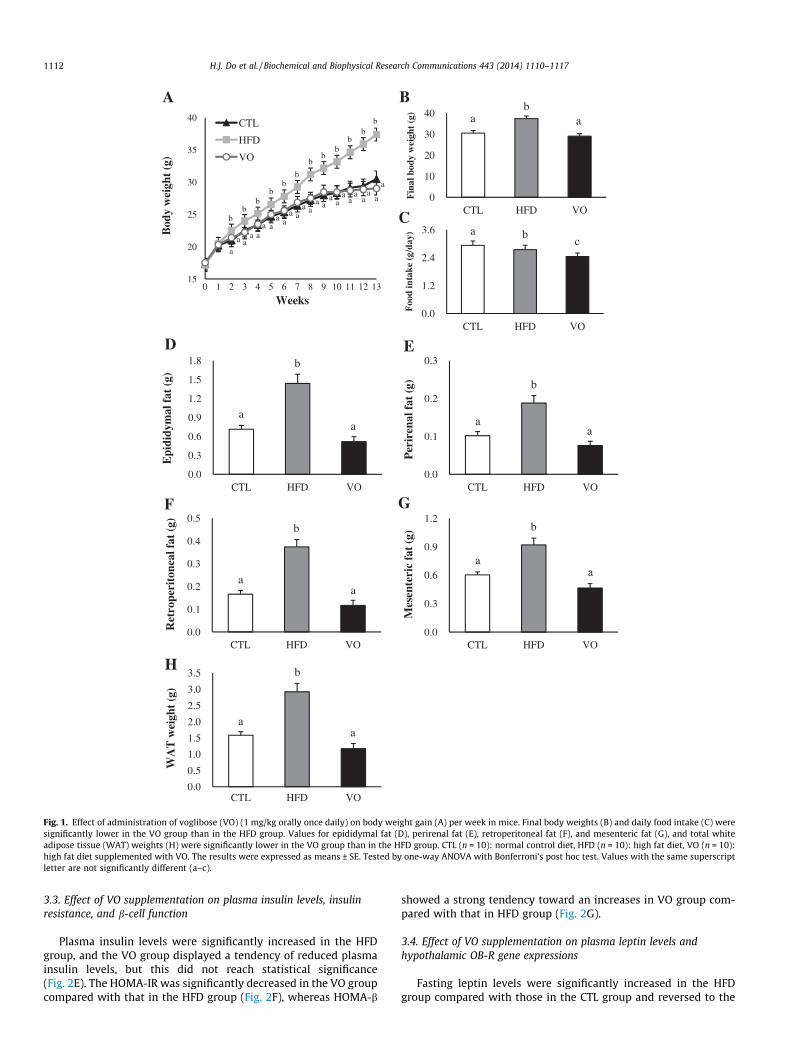
As shown in Fig. 1A, there were significant differences in bodyweight gain in mice fed different diets ad libitum over the 12-weekperiod. Mice in the HFD group gained more weight than those inthe CTL and VO (1 mg/kg orally once daily) groups, and final bodyweights were significantly higher in the HFD (37.4 ± 1.27 g) groupthan in the CTL (30.5 ± 1.23 g) and VO (29.0 ± 1.20 g) groups(Fig. 1B). The average daily food intake was significantly differentamong the 3 groups; the VO group consumed remarkably lessamount of food than the CTL and HFD groups (Fig. 1C). The weightsof epididymal and perirenal fat were significantly different amongthe groups (Fig. 1D and E) and the weights of retroperitoneal andmesenteric fat were significantly lower in the VO group than inHFD group (Fig. 1F and G). As shown in Fig. 1H, the HFD group(2.9 ± 0.26 g) had increased WAT weight compared with that inthe CTL (1.6 ± 0.11 g) and VO (1.2 ± 0.16 g) groups.
3.2. Effect of VO supplementation on plasma metabolic parametersand hepatic TG
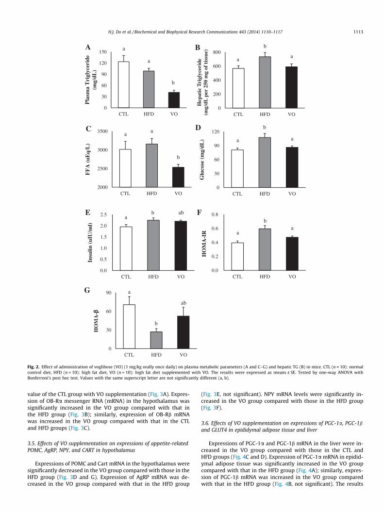
The VO group had remarkably reduced plasma TG levels(Fig. 2A). Compared with the CTL group, mice in the HFD grouphad elevated levels of TG in the liver. Hepatic TG levels were signif-icantly lower in VO group than those in HFD group (Fig. 2B). Plas-ma levels of FFA and glucose were significantly different among thegroups (Fig. 2C and D). FFA levels were lower in VO group than inthe CTL and HFD groups. Similarly, circulating glucose levels werelower in the VO group than in the HFD group.
a ab
A B
C
D E
GF
H
b
aa
b
aa
b
aa
b
aa
b
aa
a bc
aa
a aa
a
aa a a a a
a aa
aa
aa a a a a
a
bb
bb
bb
bb
bb
bb
15
20
25
30
35
40
0 1 2 3 4 5 6 7 8 9 10 11 12 13
Bod
y w
eigh
t (g
)
Weeks
CTL
HFD
VO
0.0
1.2
2.4
3.6
CTL HFD VO
Foo
d in
take
(g/
day)
0
10
20
30
40
CTL HFD VO
Fin
al b
ody
wei
ght
(g)
0.0
0.1
0.2
0.3
CTL HFD VO
Per
iren
al fa
t (g
)
0.0
0.3
0.6
0.9
1.2
CTL HFD VO
Mes
ente
ric
fat
(g)
0.0
0.1
0.2
0.3
0.4
0.5
CTL HFD VO
Ret
rope
rito
neal
fat
(g)
0.0
0.5
1.0
1.5
2.0
2.5
3.0
3.5
CTL HFD VO
WA
T w
eigh
t (g
)
0.0
0.3
0.6
0.9
1.2
1.5
1.8
CTL HFD VO
Epi
didy
mal
fat
(g)
Fig. 1. Effect of administration of voglibose (VO) (1 mg/kg orally once daily) on body weight gain (A) per week in mice. Final body weights (B) and daily food intake (C) weresignificantly lower in the VO group than in the HFD group. Values for epididymal fat (D), perirenal fat (E), retroperitoneal fat (F), and mesenteric fat (G), and total whiteadipose tissue (WAT) weights (H) were significantly lower in the VO group than in the HFD group. CTL (n = 10): normal control diet, HFD (n = 10): high fat diet, VO (n = 10):high fat diet supplemented with VO. The results were expressed as means ± SE. Tested by one-way ANOVA with Bonferroni’s post hoc test. Values with the same superscriptletter are not significantly different (a–c).
1112 H.J. Do et al. / Biochemical and Biophysical Research Communications 443 (2014) 1110–1117
3.3. Effect of VO supplementation on plasma insulin levels, insulinresistance, and b-cell function
Plasma insulin levels were significantly increased in the HFDgroup, and the VO group displayed a tendency of reduced plasmainsulin levels, but this did not reach statistical significance(Fig. 2E). The HOMA-IR was significantly decreased in the VO groupcompared with that in the HFD group (Fig. 2F), whereas HOMA-b
showed a strong tendency toward an increases in VO group com-pared with that in HFD group (Fig. 2G).
3.4. Effect of VO supplementation on plasma leptin levels andhypothalamic OB-R gene expressions
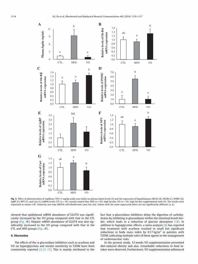
Fasting leptin levels were significantly increased in the HFDgroup compared with those in the CTL group and reversed to the
DC
FE
G
b
a
a a a
b
a ab
a a
ba
ab
b
aa
b
a
ab
bA B
Hep
atic
Tri
glyc
erid
e (m
g/dL
per
250
mg
of t
issu
e)
0
30
60
90
CTL HFD VO
HO
MA
-
0.0
0.2
0.4
0.6
0.8
CTL HFD VO
HO
MA
-IR
0.0
0.5
1.0
1.5
2.0
2.5
CTL HFD VO
Insu
lin (
uIU
/ml)
0
30
60
90
120
CTL HFD VO
Glu
cose
(m
g/dL
)
2000
2500
3000
3500
CTL HFD VO
FF
A (
uEq/
L)
0
30
60
90
120
150
CTL HFD VO
Pla
sma
Tri
glyc
erid
e (m
g/dL
)
0
200
400
600
800
CTL HFD VO
Fig. 2. Effect of administration of voglibose (VO) (1 mg/kg orally once daily) on plasma metabolic parameters (A and C–G) and hepatic TG (B) in mice. CTL (n = 10): normalcontrol diet, HFD (n = 10): high fat diet, VO (n = 10): high fat diet supplemented with VO. The results were expressed as means ± SE. Tested by one-way ANOVA withBonferroni’s post hoc test. Values with the same superscript letter are not significantly different (a, b).
H.J. Do et al. / Biochemical and Biophysical Research Communications 443 (2014) 1110–1117 1113
value of the CTL group with VO supplementation (Fig. 3A). Expres-sion of OB-Ra messenger RNA (mRNA) in the hypothalamus wassignificantly increased in the VO group compared with that inthe HFD group (Fig. 3B); similarly, expression of OB-Rb mRNAwas increased in the VO group compared with that in the CTLand HFD groups (Fig. 3C).
3.5. Effects of VO supplementation on expressions of appetite-relatedPOMC, AgRP, NPY, and CART in hypothalamus
Expressions of POMC and Cart mRNA in the hypothalamus weresignificantly decreased in the VO group compared with those in theHFD group (Fig. 3D and G). Expression of AgRP mRNA was de-creased in the VO group compared with that in the HFD group
(Fig. 3E, not significant). NPY mRNA levels were significantly in-creased in the VO group compared with those in the HFD group(Fig. 3F).
3.6. Effects of VO supplementation on expressions of PGC-1a, PGC-1band GLUT4 in epididymal adipose tissue and liver
Expressions of PGC-1a and PGC-1b mRNA in the liver were in-creased in the VO group compared with those in the CTL andHFD groups (Fig. 4C and D). Expression of PGC-1a mRNA in epidid-ymal adipose tissue was significantly increased in the VO groupcompared with that in the HFD group (Fig. 4A); similarly, expres-sion of PGC-1b mRNA was increased in the VO group comparedwith that in the HFD group (Fig. 4B, not significant). The results
b
a a
B
a
b
a
A
C
a
b
ab
aba
bE
G
F
aba
b
NS
a
b
a
D
0.0
0.2
0.4
0.6
0.8
1.0
1.2
1.4
1.6
CTL HFD VO
Rel
ativ
e le
vels
of
Ob-
Rm
RN
A e
xpre
ssio
n
0.0
0.2
0.4
0.6
0.8
1.0
1.2
1.4
1.6
CTL HFD VO
Rel
ativ
e le
vels
of
Ob-
Rm
RN
A e
xpre
ssio
n
0
3
6
9
12
CTL HFD VO
Pla
sma
lept
in (
ng/m
l)
0.0
0.2
0.4
0.6
0.8
1.0
1.2
1.4
CTL HFD VO
Rel
ativ
e le
vels
of
Car
t m
RN
A e
xpre
ssio
n
0.0
0.2
0.4
0.6
0.8
1.0
1.2
1.4
CTL HFD VO
Rel
ativ
e le
vels
of
NP
Y
mR
NA
exp
ress
ion
0.0
0.5
1.0
1.5
2.0
2.5
3.0
3.5
CTL HFD VO
Rel
ativ
e le
vels
of
AgR
P
mR
NA
exp
ress
ion
0.0
1.0
2.0
3.0
4.0
5.0
6.0
7.0
CTL HFD VO
Rel
ativ
e le
vels
of
PO
MC
m
RN
A e
xpre
ssio
n
Fig. 3. Effect of administration of voglibose (VO) (1 mg/kg orally once daily) on plasma leptin levels (A) and the expression of hypothalamus OB-Ra (B), OB-Rb (C), POMC (D),AgRP (E), NPY (F), and Cart (G) mRNA levels. CTL (n = 10): normal control diet, HFD (n = 10): high fat diet, VO (n = 10): high fat diet supplemented with VO. The results wereexpressed as means ± SE. Tested by one-way ANOVA with Bonferroni’s post hoc test. Values with the same superscript letter are not significantly different (a, b).
1114 H.J. Do et al. / Biochemical and Biophysical Research Communications 443 (2014) 1110–1117
showed that epididymal mRNA abundance of GLUT4 was signifi-cantly increased by the VO group compared with that in the CTLgroup (Fig. 4E). Hepatic mRNA abundance of GLUT4 was also sig-nificantly increased in the VO group compared with that in theCTL and HFD groups (Fig. 4F).
4. Discussion
The effects of the a-glucosidase inhibitors such as acarbose andVO on hyperglycemia and insulin sensitivity in T2DM have beenconsistently reported [3,13–17]. This is mainly attributed to the
fact that a-glucosidase inhibitors delay the digestion of carbohy-drates by inhibiting a-glucosidases within the intestinal brush bor-der, which leads to a reduction of glucose absorption [18]. Inaddition to hypoglycemic effects, a meta-analysis [4] has reportedthat treatment with acarbose resulted in small but significantreductions in body mass index by 0.17 kg/m2 in patients withT2DM, indicating multiple roles of these agents in the managementof cardiovascular risks.
In the present study, 12 weeks VO supplementation preventeddiet-induced obesity and also, remarkable reductions in food in-takes were observed. Furthermore, VO supplementation influenced

BA
CLiver D Liver
a
b
aa a
b
Epididymal adipose tissue
NSa
b
a
E
aba
b F Liver
aa
b
Epididymal adipose tissue
Epididymal adipose tissue
0.0
0.2
0.4
0.6
0.8
1.0
1.2
1.4
CTL HFD VO
Rel
ativ
e le
vels
of
PG
C-1
mR
NA
exp
ress
ion
0.0
0.2
0.4
0.6
0.8
1.0
1.2
1.4
CTL HFD VO
Rel
ativ
e le
vels
of
PG
C-1
mR
NA
exp
ress
ion
0.0
0.2
0.4
0.6
0.8
1.0
1.2
1.4
CTL HFD VO
Rel
ativ
e le
vels
of
PG
C-1
mR
NA
exp
ress
ion
0.0
0.2
0.4
0.6
0.8
1.0
1.2
1.4
1.6
CTL HFD VO
Rel
ativ
e le
vels
of
PG
C-1
mR
NA
exp
ress
ion
CTL HFD VO
Rel
ativ
e le
vels
of
GL
UT
4 m
RN
A e
xpre
ssio
n
0.0
0.5
1.0
1.5
2.0
0.0
0.5
1.0
1.5
2.0
CTL HFD VO
Rel
ativ
e le
vels
of
GL
UT
4 m
RN
A e
xpre
ssio
n
Fig. 4. Effect of administration of voglibose (VO) (1 mg/kg orally once daily) on PGC-1a (A and C), PGC-1b (B and D), and GLUT4 (E and F) mRNA expression in epididymaladipose tissue and liver. CTL (n = 10): normal control diet, HFD (n = 10): high fat diet, VO (n = 10): high fat diet supplemented with VO. The results were expressed asmeans ± SE. Tested by one-way ANOVA with Bonferroni’s post hoc test. Values with the same superscript letter are not significantly different (a, b).
H.J. Do et al. / Biochemical and Biophysical Research Communications 443 (2014) 1110–1117 1115
obesity induced metabolic abnormalities as shown by theimprovements in glucose and lipid metabolism. To elucidate theunderlying mechanism, we measured the plasma leptin levelsknown to influence body weight through the control of appetiteand energy expenditure and hypothalamic expressions of appe-tite-related genes. The results showed that the elevated levels ofcirculating leptin in mice fed a HFD were significantly reducedwith VO supplementation; furthermore, VO supplementation mod-ulated the hypothalamic expressions of OB-Rs and appetite relatedgenes. In addition, VO supplementation resulted in up-regulatedexpression of PGC-1 in the liver and epididymal adipose tissue,implicating that VO may have elicited improved mitochondrialdysfunction.
The exact mechanism by which VO reduces body weight re-mains unclear; several pathways may be involved in the observedeffects. First, weight reduction seems to result from remarkablereductions in food intake, possibly associated with changes inthe secretion of glucagon-like peptide 1 (GLP-1). GLP-1 is anincretin that is secreted from the lower gastrointestinal tract bylocal action of the unabsorbed nutrients [19] and functions
physiologically to mediate satiety and produce the cessation ofeating [20]. The action of GLP-1 to reduce hunger and increasesatiety is likely to be mediated through prolonging gastricemptying or an interaction with appetite-regulating centers inthe central nervous system [21–23]. Since body weight is nor-mally maintained by a balance between energy intake and energyexpenditure, the weight-lowering effect of VO supplementationshown in the present study might be achieved through reductionsin energy intake. Specifically, it is possible that VO increasesintestinal GLP-1 secretion and induces satiety [24] and subse-quently reduces total food intakes. This can be further supportedby previous findings [20] that short-term treatment with ana-glucosidase inhibitor showed increased GLP-1 secretion inpatients with T2DM. However, this hypothesis cannot be fullysupported in the absence of data available for postprandialGLP-1 and/or dipeptidyl peptidase-4 activity.
Alternatively, the neuroendocrine system involving leptin canbe activated. It is well known that leptin controls energy balanceand body weight by regulating neuronal activity in the hypothala-mus [25]. The action of adipose tissue-derived leptin is mediated
1116 H.J. Do et al. / Biochemical and Biophysical Research Communications 443 (2014) 1110–1117
by its receptor (OB-Rb) in the brain in regulating energy balanceand neuroendocrine function [26] and OB-Rb activates numeroussignaling pathways that act as a network to mediate the actionof leptin [25]. On the other hand, leptin resistance resulting fromdefects in leptin transport into the brain [27], leptin signaling[28], and/or the hypothalamic neural circuitry [29,30] has shownto be associated with the imbalances between energy intake andexpenditure, ultimately resulting in obesity. In the present study,the elevated plasma leptin levels in mice fed a HFD were signifi-cantly reversed by VO supplementation. Furthermore, up-regula-tions in hypothalamic OB-Ra and OB-Rb mRNA were observed inmice in the VO group. It is likely that VO improved leptin resistancein the mice fed HFD through either reductions in circulating leptinlevels or up-regulations of receptors mediating leptin’s action tothe brain. We further tested whether VO modulates hypothalamicexpression of genes regulating energy balance because leptin is re-ported to directly target hypothalamic appetite-regulating genes[31,32]. The results showed that VO reversed elevations in hypo-thalamic POMC and CART levels induced by a HFD; in contrast,NPY mRNA levels were significantly increased in mice in the VOgroup compared with that in the HFD group. This is in contrastto the facts that POMC and CART are anorexigenic genes and NYPis an orexigenic gene stimulating feeding behavior [33] of whichhypothalamic expressions are modulated by central leptin. Our re-sults imply feedback effect of hypophagia induced by VO adminis-tration rather than the direct regulation of genes involved inappetite. It seems that the satiety derived from increased GLP-1levels by VO supplementation, but not satiety from direct modula-tion of the hypothalamic genes, can explain the reduced consump-tion of food in the mice fed a HFD in this study.
Finally, we propose a beneficial effect of VO on mitochondrialdysfunction in obesity, which leads to improved energy utilizationand metabolic abnormalities. It has been accepted that mitochon-drial dysfunction could be one of the underlying defects linkingobesity to diabetes, both by decreasing insulin sensitivity and bycompromising b-cell function [34]. Along with body weight loss,in the present study, VO supplementation remarkably improvedmetabolic status measured by TG content, FFA, and glucose levelsin mice fed a HFD. In addition, improvement of peripheral insulinaction and insulin secretory dynamics was observed, as measuredby a HOMA-IR and HOMA-b cell function effect. In this study, weobserved that VO supplementation up-regulated PGC-1a andPGC-1b in the liver and epididymal adipose tissue of mice fed aHFD. PGC-1 is a central transcriptional regulator of mitochondrialbiogenesis [35], and defects in mitochondrial energy metabolismhave been suggested to cause hepatic steatosis, insulin resistance,and T2DM [36]. In line, reduced PGC-1a expression and mitochon-drial dysfunction in the adipose tissue, have been associated withobesity and insulin resistance [37–39]. Considering that PGC-1ais importantly involved in increased mitochondrial function andoxidative metabolism, the observed metabolic benefits of VO inthe present study are likely mediated by transcriptional activityof PGC-1a targeted for downstream genes in glucose transportand hepatic lipid metabolism. Indeed, enhanced expression ofGLUT4 in adipose tissue and the liver further support thishypothesis.
In summary, 12 weeks of treatment with VO, an a-glucosidaseinhibitor, remarkably regulated body weight and reduced total en-ergy intakes. In addition, beneficial metabolic effects in obesitywere achieved by VO supplementation, including plasma glucoselevels, plasma FFA levels, and insulin sensitivity. At this point,the exact underlying mechanisms are not known but they may in-volve the incretin effect of VO, the activation of neuroendocrinelinked to leptin, and stimulation of genes responsible for enhancedenergy metabolism. Further studies are required to elucidate themechanism of the observed results.
Acknowledgments
This research was supported by Basic Science Research Programthrough the National research Foundation of Korea (NRF) fundedby the Ministry of Education, Science and Technology (NRF-2013R1A1A2A10006101).
References
[1] American Diabetes Association, J. Beck, C.E. Cox, et al., Clinical practicerecommendations, Diabetes Care 36 (2013) S1–S110.
[2] Look AHEAD Research Group, R.R. Wing, P. Bolin, et al., Cardiovascular effectsof intensive lifestyle intervention in type 2 diabetes, N. Engl. J. Med. 369 (1998)145–154.
[3] K. Matsumoto, M. Yano, S. Miyake, et al., Effects of voglibose on glycemicexcursions, insulin secretion, and insulin sensitivity in non-insulin-treatedNIDDM patients, Diabetes Care 21 (1998) 256–260.
[4] F.A. van de Laar, P.L. Lucassen, R.P. Akkermans, et al., Alpha-glucosidaseinhibitors for patients with type 2 diabetes: results from a Cochranesystematic review and meta-analysis, Diabetes Care 28 (2005) 154–163.
[5] R.V. Kumar, V.R. Sinha, Newer insights into the drug delivery approaches of a-glucosidase inhibitors, Expert Opin. Drug Deliv 9 (2012) 403–416.
[6] E. Standl, O. Schnell, Alpha-glucosidase inhibitors, cardiovascularconsiderations and trial evaluation, Diab. Vasc. Dis. Res. 9 (2012) (2012)163–169.
[7] J.L. Chiasson, R.G. Josse, R. Gomis, et al., Acarbose for prevention of type 2diabetes mellitus: the STOP-NIDDM randomised trial, Lancet 359 (2002) 2072–2077.
[8] R. Kawamori, N. Tajima, Y. Iwamoto, et al., Voglibose for prevention of type 2diabetes mellitus: a randomised, double-blind trial in Japanese individualswith impaired glucose tolerance, Lancet 373 (2009) 1607–1614.
[9] Y. Liao, S. Takashima, H. Zhao, et al., Control of plasma glucose with alpha-glucosidase inhibitor attenuates oxidative stress and slows the progression ofheart failure in mice, Cardiovasc. Res. 70 (2006) 107–116.
[10] T. Narita, H. Yokoyama, R. Yamashita, et al., Comparisons of the effects of 12-week administration of miglitol and voglibose on the responses of plasmaincretins after a mixed meal in Japanese type 2 diabetic patients, DiabetesObes. Metab. 14 (2012) 283–287.
[11] J. Folch, M. Lees, G.H. Sloane-Stanley, A simple method for the isolation andpurification of total lipids from animal tissues, J. Biol. Chem. 226 (1957) 497–509.
[12] D.R. Matthews, J.P. Hosker, A.S. Rudenski, et al., Homeostasis modelassessment: insulin resistance and beta-cell function from fasting plasmaglucose and insulin concentrations in man, Diabetologia 28 (1985) 412–419.
[13] R.F. Coniff, J.A. Shapiro, T.B. Seaton, Long-term efficacy and safety of acarbosein the treatment of obese subjects with non-insulin-dependent diabetesmellitus, Arch. Intern. Med. 154 (1994) 2442–2448.
[14] P. Segal, P.U. Feig, G. Schernthaner, et al., The efficacy and safety of miglitoltherapy compared with glibenclamide in patients with NIDDM inadequatelycontrolled by diet alone, Diabetes Care 20 (1997) 687–691.
[15] T. Kawagishi, Y. Nishizawa, H. Taniwaki, et al., Relationship between gastricemptying and an alpha-glucosidase inhibitor effect on postprandialhyperglycemia in NIDDM patients, Diabetes Care 20 (1997) 1529–1532.
[16] H. Laube, T. Linn, P. Heyen, The effect of acarbose on insulin sensitivity andproinsulin in overweight subjects with impaired glucose tolerance, Exp. Clin.Endocrinol. Diabetes 106 (1998) 231–233.
[17] K. Shinozaki, M. Suzuki, M. Ikebuchi, et al., Improvement of insulin sensitivityand dyslipidemia with a new alpha-glucosidase inhibitor, voglibose, innondiabetic hyperinsulinemic subjects, Metabolism 45 (1996) 731–737.
[18] S. Matthaei, M. Stumvoll, M. Kellerer, et al., Pathophysiology andpharmacological treatment of insulin resistance, Endocr. Rev. 21 (2000)585–618.
[19] M.J. Dailey, T.H. Moran, Glucagon-like peptide 1 and appetite, Rev. Diabet.Stud. 24 (2013) 85–91.
[20] A. Lee, P. Patrick, J. Wishart, et al., The effects of miglitol on glucagon-likepeptide-1 secretion and appetite sensations in obese type 2 diabetics, DiabetesObes. Metab. 4 (2002) 329–335.
[21] R.S. McIntyre, A.M. Powell, O. Kaidanovich-Beilin, et al., The neuroprotectiveeffects of GLP-1: possible treatments for cognitive deficits in individuals withmood disorders, Behav. Brain Res. 237 (2013) 164–171.
[22] J.N. Roberge, P.L. Brubaker, Regulation of intestinal proglucagon-derivedpeptide secretion by glucose-dependent insulinotropic peptide in a novelenteroendocrine loop, Endocrinology 133 (1993) 233–240.
[23] R.A. Reimer, C. Darimont, S. Gremlich, et al., A human cellular model forstudying the regulation of glucagon-like peptide-1 secretion, Endocrinology142 (2001) 4522–4528.
[24] R. Moriya, T. Shirakura, J. Ito, et al., Activation of sodium-glucose cotransporter1 ameliorates hyperglycemia by mediating incretin secretion in mice, Am. J.Physiol. Endocrinol. Metab. 297 (2009) E1358–E1365.
[25] D.L. Morris, L. Rui, Recent advances in understanding leptin signaling andleptin resistance, Am. J. Physiol. Endocrinol. Metab. 297 (2009) E1247–E1259.
[26] S. Arora, Anubhuti, role of neuropeptides in appetite regulation and obesity – areview, Neuropeptides 40 (2006) 375–401.
H.J. Do et al. / Biochemical and Biophysical Research Communications 443 (2014) 1110–1117 1117
[27] K. El-Haschimi, D.D. Pierroz, S.M. Hileman, et al., Two defects contribute tohypothalamic leptin resistance in mice with diet-induced obesity, J. Clin.Invest. 105 (2000) 1827–1832.
[28] J.K. Howard, B.J. Cave, L.J. Oksanen, et al., Enhanced leptin sensitivity andattenuation of diet-induced obesity in mice with haploinsufficiency of Socs3,Nat. Med. 10 (2004) 734–738.
[29] I.S. Farooqi, J.M. Keogh, G.S. Yeo, et al., Clinical spectrum of obesity andmutations in the melanocortin 4 receptor gene, N. Engl. J. Med. 348 (2003)1085–1095.
[30] I.S. Farooqi, G.S. Yeo, J.M. Keogh, et al., Dominant and recessive inheritance ofmorbid obesity associated with melanocortin 4 receptor deficiency, J. Clin.Invest. 106 (2000) 271–279.
[31] C.C. Cheung, D.K. Clifton, R.A. Steiner, Proopiomelanocortin neurons are directtargets for leptin in the hypothalamus, Endocrinology 138 (1997) 4489–4492.
[32] M.L. Håkansson, A.L. Hulting, B. Meister, Expression of leptin receptor mRNA inthe hypothalamic arcuate nucleus – relationship with NPY neurons,Neuroreport 7 (1996) 3087–3092.
[33] M.W. Schwartz, R.J. Seeley, L.A. Campfield, et al., Identification of targets ofleptin action in rat hypothalamus, J. Clin. Invest. 98 (1996) 1101–1106.
[34] R.H. Eckel, S.E. Kahn, E. Ferrannini, et al., Obesity and type 2 diabetes: what canbe unified and what needs to be individualized?, J Clin. Endocrinol. Metab. 96(2011) 1654–1663.
[35] P.J. Fernandez-Marcos, J. Auwerx, Regulation of PGC-1a, a nodal regulator ofmitochondrial biogenesis, Am. J. Clin. Nutr. 93 (2011) 884S–890S.
[36] Z. Wu, P. Puigserver, U. Andersson, et al., Mechanisms controllingmitochondrial biogenesis and respiration through the thermogeniccoactivator PGC-1, Cell 98 (1999) 115–124.
[37] S. Kleiner, R.J. Mepani, D. Laznik, et al., Development of insulin resistance inmice lacking PGC-1a in adipose tissues, Proc. Natl. Acad. Sci. USA 109 (2012)9635–9640.
[38] A. Hammarstedt, P.A. Jansson, C. Wesslau, et al., Reduced expression of PGC-1and insulin-signaling molecules in adipose tissue is associated with insulinresistance, Biochem. Biophys. Res. Commun. 301 (2003) 578–582.
[39] P. Puigserver, Z. Wu, C.W. Park, et al., A cold-inducible coactivator of nuclearreceptors linked to adaptive thermogenesis, Cell 92 (1998) 829–839.


![Untitled-3 [psychocare.biz]PCHPL Psychocare Health Pvt. Ltd. (An ISO 9001 :2015 Certified co.) CARDIO/DIABETIC RANGE Composition Voglibose 0.2 mg Tablets Voglibose 0.3 mg Tablets](https://static.fdocuments.in/doc/165x107/602c36fce8449b43213b3c28/untitled-3-pchpl-psychocare-health-pvt-ltd-an-iso-9001-2015-certified-co.jpg)
