Vitrectomy: when things go wrong - Dr Nagpal's Retina ......Pars plana vitrectomy has become an...
16
Author Proof Review 10.1586/17469899.2.4.xxx © 2007 Future Drugs Ltd ISSN 1746-9899 1 www.future-drugs.com Vitrectomy: when things go wrong Manish Nagpal † and Sharang Wartikar † Author for correspondence Retina Foundation, Asopalov Eye Hospital, Near Shahibaug Underbridge, Shahibaug, Ahmedabad, India - 380004 Tel.: +91 792 286 7982 Fax: +91 792 286 6381 [email protected] KEYWORDS: complications, endophthalmitis, epithelial defects, fibrous ingrowth, oil emulsification, pars plana vitrectomy, phototoxicity, proliferative vitreoretinopathy, sclerotomy, sutureless vitrectomy, tamponade, vitreous hemorrhage Pars plana vitrectomy has become an important tool in the treatment of vitreoretinal diseases, with newer applications being considered. This has necessitated an overall improvement in the surgical expertise and the innovative supportive technology driving the machines. Frequent use has also brought about a variety of complications, especially during the learning curve that every surgeon faces. The vitreous cavity being enclosed by the retina and the crystalline lens leave little margin for error. Precise control over instrumentation is required in order to avoid inadvertent mechanical touching of these tissues. Tissue respect is of utmost importance and in this review we discuss all possible complications and ways to avoid them and manage them if they occur. Expert Rev. Ophthalmol. 2(4), xxx–xxx (2007) History The idea of vitreous surgery as conceived by Shafer in 1950 and propagated by David Kas- ner in the form of an open-sky vitrectomy in the 1960s, was rather hazardous, with many complications [1]. A major advancement in the treatment of vitreoretinal diseases was achieved by Machemer and colleagues in 1971 [2] with the introduction of pars plana vitrectomy with a single vitreous infusion suction cutter (VISC) system. The invention of divided sys- tem instrumentation by O’Malley gave rise to bimanual vitrectomy. Work carried out by Cibis with intravitreal silicone oil, by Norton with intravitreal sulfur hexafluoride gas and by Chang with heavy liquids has further simpli- fied many steps of the procedure [3]. While present-day vitrectomy has become safer and more reliable, there remains scope for improvement. Newer techniques are being introduced at a rapid pace, bringing with them newer complications. Relevant surgical anatomy Any surgical intervention should be based on a sound knowledge of the normal structures and their relations. The vitreous body is a clear gel- like fibrillar meshwork composed of 99% water, with the outer cortex being denser [4]. It contains interpenetrating networks of collagen fibrils and hyaluronic acid [5]. While the ante- rior hyaloid face is strongly attached to the crystalline lens, the posterior hyaloid face is attached at places to the inner limiting mem- brane of retina. The strongest attachment is at the vitreous base and relatively weaker attach- ments exist around the optic disc, at the mac- ula and along retinal vessels [6]. Sclerotomies made through the pars plana region avoid damage to the pars plicata anteriorly and the vitreous base posteriorly. Instrumentation Vitrectomy visualization was initially based only on the use of assistant-held irrigating lenses that neutralized the corneal curvature. While these lenses give excellent resolution to the surgeon, his field of view remains restricted. Handling of the peripheral vitreous becomes difficult and may be fraught with complications. Introduction of the stereo- scopic diagonal inverter (SDI; Spitznas and Reiver) and the binocular indirect ophthalmo- microscope (BIOM) has popularized the use of contact and noncontact wide-angle viewing systems [7,8]. These offer a simple, effective and quick method for illumination of the ora ser- rata and anterior vitreous in aphakic and pseu- dophakic eyes during vitrectomy [9,10]. Self-sta- bilizing lenses eliminate the need for suturing CONTENTS History Relevant surgical anatomy Instrumentation Complications & management Expert commentary Five-year view Financial disclosure Key issues References Affiliations
Transcript of Vitrectomy: when things go wrong - Dr Nagpal's Retina ......Pars plana vitrectomy has become an...
Author Pro
of
Review
10.1586/17469899.2.4.xxx © 2007 Future Drugs Ltd ISSN 1746-9899 1www.future-drugs.com
Vitrectomy: when things go wrongManish Nagpal† and Sharang Wartikar
†Author for correspondenceRetina Foundation, Asopalov Eye Hospital, Near Shahibaug Underbridge, Shahibaug, Ahmedabad, India - 380004Tel.: +91 792 286 7982Fax: +91 792 286 [email protected]
KEYWORDS: complications, endophthalmitis, epithelial defects, fibrous ingrowth, oil emulsification, pars plana vitrectomy, phototoxicity, proliferative vitreoretinopathy, sclerotomy, sutureless vitrectomy, tamponade, vitreous hemorrhage
Pars plana vitrectomy has become an important tool in the treatment of vitreoretinal diseases, with newer applications being considered. This has necessitated an overall improvement in the surgical expertise and the innovative supportive technology driving the machines. Frequent use has also brought about a variety of complications, especially during the learning curve that every surgeon faces. The vitreous cavity being enclosed by the retina and the crystalline lens leave little margin for error. Precise control over instrumentation is required in order to avoid inadvertent mechanical touching of these tissues. Tissue respect is of utmost importance and in this review we discuss all possible complications and ways to avoid them and manage them if they occur.
Expert Rev. Ophthalmol. 2(4), xxx–xxx (2007)
HistoryThe idea of vitreous surgery as conceived byShafer in 1950 and propagated by David Kas-ner in the form of an open-sky vitrectomy inthe 1960s, was rather hazardous, with manycomplications [1]. A major advancement in thetreatment of vitreoretinal diseases was achievedby Machemer and colleagues in 1971 [2] withthe introduction of pars plana vitrectomy witha single vitreous infusion suction cutter(VISC) system. The invention of divided sys-tem instrumentation by O’Malley gave rise tobimanual vitrectomy. Work carried out byCibis with intravitreal silicone oil, by Nortonwith intravitreal sulfur hexafluoride gas and byChang with heavy liquids has further simpli-fied many steps of the procedure [3]. Whilepresent-day vitrectomy has become safer andmore reliable, there remains scope forimprovement. Newer techniques are beingintroduced at a rapid pace, bringing with themnewer complications.
Relevant surgical anatomyAny surgical intervention should be based on asound knowledge of the normal structures andtheir relations. The vitreous body is a clear gel-like fibrillar meshwork composed of 99%water, with the outer cortex being denser [4]. Itcontains interpenetrating networks of collagen
fibrils and hyaluronic acid [5]. While the ante-rior hyaloid face is strongly attached to thecrystalline lens, the posterior hyaloid face isattached at places to the inner limiting mem-brane of retina. The strongest attachment is atthe vitreous base and relatively weaker attach-ments exist around the optic disc, at the mac-ula and along retinal vessels [6]. Sclerotomiesmade through the pars plana region avoiddamage to the pars plicata anteriorly and thevitreous base posteriorly.
InstrumentationVitrectomy visualization was initially basedonly on the use of assistant-held irrigatinglenses that neutralized the corneal curvature.While these lenses give excellent resolution tothe surgeon, his field of view remainsrestricted. Handling of the peripheral vitreousbecomes difficult and may be fraught withcomplications. Introduction of the stereo-scopic diagonal inverter (SDI; Spitznas andReiver) and the binocular indirect ophthalmo-microscope (BIOM) has popularized the use ofcontact and noncontact wide-angle viewingsystems [7,8]. These offer a simple, effective andquick method for illumination of the ora ser-rata and anterior vitreous in aphakic and pseu-dophakic eyes during vitrectomy [9,10]. Self-sta-bilizing lenses eliminate the need for suturing
CONTENTS
History
Relevant surgical anatomy
Instrumentation
Complications & management
Expert commentary
Five-year view
Financial disclosure
Key issues
References
Affiliations
Author Pro
of
Nagpal & Wartikar
2 Expert Rev. Ophthalmol. 2(4), (2007)
on the sclera or dependence on the assistant to provide place-ment [11]. Depending on the step of surgery, the lenses can beused interchangeably [12]. Use of a Chandelier illumination sys-tem during a four-port vitrectomy has allowed for bimanualityin its true sense, further simplifying many surgical steps thatwere previously better avoided [13–15]. Even in cases where themedia do not allow visualization with the standard microscopicsystems, microendoscopy provides an alternative, albeit with aloss of both resolution and the third dimension [16].
Newer cutters that are sharper and have higher cut rateshave tried to reduce the complications further [17]. A soft tipextrusion cannula allows for safe removal of fluid, hemor-rhage, gas or silicone oil from the subretinal space [18]. Closedvitrectomy is known to cause wide fluctuations in the intraoc-ular pressure (IOP) [19], and present-day machines aim to pro-vide precision and better control over fluidics, reducing thechances of intraoperative hypotony or prolonged raised IOP.
Complications & managementThe decision to perform any invasive procedure depends on itsbenefit:risk ratio. By reducing the complication rate of a pro-cedure we can decrease the risk, making the procedure applica-ble for additional indications. Patients’ general health and thestatus of the other eye may also influence the final decision.
Conjunctiva-related complications During peritomy, cutting the conjunctiva close to the limbusallows good exposure, causes less fibrosis and reduces conjunc-tival shrinkage during resurgeries. Essentially, conjunctivaldamage has to be minimized by preventing both tearing andbutton-holing.
Conjunctival closure, as in any ocular surgery, needs to beend to end so as to ensure quicker healing. Absorbable suturesare preferred even though they may produce slightly more tis-sue reaction in some patients. Fibrin glue and other acrylicadhesives have been advocated for closing conjunctival wounds,especially in elderly patients. They result in good adaptationand are time saving and effective [20,21].
With the advent of sutureless vitrectomy, many conjunctiva-and suture-related complications have been resolved. Conjunc-tival chemosis, however, is commonly seen with transconjuncti-val sclerotomies without the placement of cannulas owing tothe leakage of perfusion fluid [22]. Also, with an added risk ofthe conjunctival epithelium being inadvertently introducedinto the vitreous cavity, special attention needs to be paid toconjunctival sterilization [23].
Sclerotomy-related complicationsIntraoperatively, inadvertent damage may occur to structuresadjacent to the site of sclerotomy. Lens damage, ciliary detach-ment, vitreous base dialysis or retinal tears may occur whilemaking the sclerotomy or during insertion or removal of instru-ments [24]. The tip of the sclerotomy blade should always bedirected toward the midvitreous and it should be ensured thatthe internal opening of the infusion cannula is well within the
vitreous cavity prior to starting the infusion, especially incases with choroidal thickening. A subretinal cannula infu-sion, once started, can be a disaster for the surgeon. However,if a choroidal detachment does occur, the infusion should bestopped and the cannula freed from any superficial tissueusing a microvitreoretinal blade or a cutter.
Retinal incarceration may occur if an instrument is with-drawn from an eye with raised IOP, resulting in rapid egress offluid, which pulls the detached retina along with it [25]. Retinamay have to be reposited with a blunt instrument followed byfluid–air exchange and cryotherapy.
All sclerotomy wounds heal with an ingrowth of fibrovasculartissue from the eye wall into the vitreous cavity. Fortunately,only in unusual circumstances does this process become exag-gerated and result in what clinicians have termed fibrovascularingrowth (FVIG), with its resultant ocular problems [26]. Post-operatively, FVIG may lead to recurrent vitreous hemorrhage[27]. While episcleral tissue, scleral fibroblasts and ciliary epithe-lium all contribute, the majority of the fibroproliferative heal-ing of a sclerotomy originates from the uvea of the ciliary body[28]. Incarceration of the vitreous into the wound appears to bethe culprit and so care needs to be taken to remove any vitreousfrom the lips of the wound prior to closure [29]. Ultrasoundbiomicroscopy (UBM) is useful in detecting fibrovascular pro-liferation at sclerotomy sites [30]. Episcleral sentinel vessels seenexternally entering the wound site should raise our suspicionsof FVIG, but they neither confirm it nor does their absencerule it out. They form as a result of a high degree of metabolicactivity during the healing and similar vessels are also seenmicroscopically in the ciliary body [28]. Anterior peripheral reti-nal cryotherapy combined with cryotherapy of sclerotomy sitesmight be helpful adjunct procedures in diabetic vitrectomy forthe inhibition of FVIG and prevention of recurrent vitreoushemorrhage [31].
Construction of sutureless self-sealing sclerotomies byoblique penetration of the sclera with a 19-G MVR blade hasbeen found to be resistant to stretching and tearing, to rarelyneed suturing and to be convenient for the passage of instru-ments [32]. They also reduce the risk of intraoperative hypot-ony following removal of instruments or the infusion cannula.The technique reduces postoperative inflammation andsuture-related problems, including astigmatism, and allowsmore rapid rehabilitation [33]. In nonphakic eyes, the traumaof making a sclerotomy site may be avoided with the anteriorchamber infusion approach by way of a paracentesis [34].
Diathermy should be avoided as far as possible since it cancause undue scarring and, in rare cases, surgically inducednecrotizing scleritis (SINS) after repeat surgeries has evenbeen reported [35]. SINS requires prompt and aggressiveimmunosuppressive treatment.
Cornea-related complicationsCorneal complications, mainly in the form of epithelial distur-bance, have been found to occur in over 7% of vitrectomy sur-geries while, in diabetic patients, the incidence may be as high
Author Pro
of
Vitrectomy: when things go wrong
www.future-drugs.com 3
as 10–50% [36–38]. Use of irrigating lenses has been linked toan increase in the intraoperative corneal edema, necessitatingthe debridement of corneal epithelium, and this, in turn,seems to be a major contributor for development of cornealcomplications. Sew-on lenses with a viscoelastic cushion areless damaging, while noncontact lenses may provide the bestcorneal protection [36,39,40]. The type of viscous surface lubri-cant used with sew-on lenses may also play a significant role[41]. Corneal complications related to the use of silicone oil willbe discussed later.
Use of better instruments [42] and irrigating fluids [43], avoid-ance of epithelial debridement, and reduction in the time ofsurgery [40] and the time for which IOP is raised [26] may reducethe incidence of these complications. The degree of surgicalinvasion that occurs during the procedure also decides the cor-neal status [37]. Accidental diathermy burns are best avoided bykeeping the use of diathermy to a bare minimum. Preservationof anterior lens capsule during pars plana lensectomy has beeenshown to reduce the risk of corneal endothelial damage, espe-cially when combined with long-acting gas tamponade [44].Subsequent opacification of the anterior capsule may requireNd:YAG capsulotomy for clearing the visual axis.
Persistent corneal epithelial defects may be treated with oint-ments, pressure patches or autologous serum eye drops com-bined with artificial tears and topical antibiotics, and generallyrespond well within 3 days [45,46]. Defects that fail to heal withconventional therapies may be fitted with an extended-weargas-permeable scleral lens [47]. Re-epithelialization appears tobe aided by a combination of oxygenation, moisture and pro-tection of the fragile epithelium afforded by the scleral lens.However, albumin may be deposited on the contact lens sur-face and the risk of microbial keratitis cannot be ignored [45,47].Use of topical steroids is usually avoided; however, topical dex-amethasone administered five-times a day may decrease poten-tial stromal scarring without significantly delaying the cornealepithelial healing (BOX 1) [46].
Anterior chamber collapseAnterior chamber collapse has been found in over 3% of apha-kic eyes that underwent vitrectomy with gas tamponade. Pres-ence of preoperative shallow anterior chamber, removal of theintraocular lens (IOL) as a part of the procedure, occurrence ofintraoperative anterior chamber collapse and use of SF6 wereassociated with a higher risk of anterior chamber collapse post-operatively [48].
Iris-related complicationsDamage to the iris leading to hemorrhage or iridodialysis mayoccur occasionally due to an improper technique of making asclerotomy. Postoperative iritis may be treated with topicalsteroids.
Postoperative iris and angle neovascularization has been foundin 8–15% of eyes undergoing vitrectomy for proliferative dia-betic retinopathy (PDR) [49,50]. Preoperative presence of retinalneovascularization and absence of panretinal photocoagulation
(PRP) and postoperative retinal detachment (RD) are signifi-cant risk factors [50,51]. Recent studies have found a lower inci-dence of postoperative rubeosis iridis with vitrectomy com-bined with removal of the lens in these cases of PDR [49,52].Such rubeosis, however, has been found to be regressive ratherthan progressive [50].
In nondiabetic patients who undergo vitrectomy for RD, suc-cessful reattachment of the retina has been found to be the mostimportant factor in the prevention of iris neovascularization,while PRP has not been found to have any role in such cases [53].
Short-term results have suggested that intravitreal or intra-cameral injection of anti-VEGFs, such as bevacizumab, resultsin rapid regression of iris neovascularization secondary toPDR [54–56].
Pupil-related complicationsIn the past, most surgical dilating procedures for intraoperativemiosis involved an incision or excision of iris tissue, which waseither inadequate or irreversible [57]. Even though currentlyused wide-angle lenses allow excellent visualization even inmiotic pupils [58], it is always better to ensure proper preopera-tive dilatation with the use of adjuncts, such as flurbiprofen [59]
or ketorolac [60]. Intraoperative miosis can be adequately managed by the use
of epinephrine solution without preservatives either intracam-erally (0.1 ml of 1:10,000) or in the infusion fluid [61,62], or by
Box 1. Postvitrectomy corneal complications.
Factors that delay corneal epithelial healing
• Pre- and intraoperative topical solutions
• Median operative time
• Presence of diabetes mellitus
• Prior ocular surgeries
• Pseudophakia/aphakia
• Presence of intraocular gas or silicone oil in aphakic patients
Prevention of corneal complications
• Use of better instruments and irrigating fluids
• Avoidance of epithelial debridement
• Preservation of posterior capsule
• Use of viscoelastics in anterior chamber
• Reduced duration of raised intraocular pressure and surgery
Management of corneal complications
• Ointments
• Pressure patches
• Bandage contact lens
• Avoidance of topical steroids
Modified from [46].
Author Pro
of
Nagpal & Wartikar
4 Expert Rev. Ophthalmol. 2(4), (2007)
the use of flexible nylon iris retractors [63–65] or silicone rings [66].Sphincterectomy may be used as a final resort [67].Postoperativeinflammation may cause pupillary membranes that do notrespond to steroids and may be removed with Nd:YAG laser [68].
Lens-related complicationsDevelopment of lenticular opacities after vitrectomy is a majorcomplication, often necessitating cataract extraction. The rea-son for such a change may be related either to the overoxygena-tion of the lens after removal of the normal vitreous barrier orto diffusion of electrolytes. This is supported by the fact thatnuclear sclerosis has not been found to progress even at 5 yearsafter a nonvitrectomizing vitreous surgery [69,70].
Apart from the removal of the vitreous itself, the cataract pro-gression depends on the duration of follow-up, the use of tam-ponading agent and the age and diabetic status of the patient.Up to 26% of phakic eyes may develop cataract after vitreoussurgery for PDR [71]. The cumulative cataract extraction ratesat 2 years have been found to be 15% after diabetic vitrecto-mies and 50–66% after nondiabetic vitrectomies, suggesting alower rate of cataract formation in diabetics [72]. Patients olderthan 50 years of age may have an approximately sixfold greaterrate of increase in nuclear sclerotic cataracts than patientsyounger than 50 years of age [73].
Presence of tamponade by intraocular gas has been shown toincrease the rate of lenticular opacities to 86–96% [40,76]. Inci-dence of cataract extraction due to nuclear sclerosis 1 year post-vitrectomy with gas tamponade may be as high as 67%, whilein nontamponade vitrectomies it is 18% [73,77]. A transient pos-terior subcapsular cataract is also seen in gas tamponade cases inthe immediate postoperative period, probably due to a disrup-tion of fluid balance [77]. Tamponade with silicone oil causessome degree of cataract in almost 100% cases [74,75], and will bediscussed later.
Combined cataract and vitreoretinal surgery with intraocularair or SF6 gas tamponade also induces severe posterior capsularfibrosis in 50–66% of pseudophakic cases in the early postoper-ative period, presumably due to accumulation of fibrin andproliferation-stimulating factors in the narrow space betweenthe IOL and air/ SF6-gas bubble [78].
Extended operating times, constant irrigation of the lens andthe type of the irrigating fluid may also be responsible for thelenticular opacities [38]. Various antioxidant, anti-inflammatoryand cytostatic agents are being investigated in an attempt to sup-press such vitrectomy-induced posterior lens fiber changes [79].
Cataract formation due to direct mechanical trauma is bestavoided with careful surgical technique. Damage generallyoccurs either with the cutter during removal of the anterior vit-reous or at times with the light pipe while working in the farperiphery. Modified instruments, such as a curved vitrectomyprobe, have been designed to facilitate vitreous base excision[80]. A conservative approach may be warranted for minor lensinjuries, but if the damage is significant, especially with ruptureof the lens capsule, then it is advisable to remove the lens in thesame sitting.
Interestingly, the diabetic status of a patient may play a role indeciding whether to remove the lens as diabetic eyes have beenfound to be less likely to require additional vitreoretinal surgeryif they are rendered nonphakic before or during vitrectomy [52].
GlaucomaVarious mechanisms may contribute to a rise in the IOP aftervitrectomy. Past studies have reported raised IOP in up to 30%eyes within 2–10 days of vitrectomy for vitreous hemorrhage,mainly due to ghost cell or hemolytic glaucoma [81,82]. Such anobstruction of the trabecular meshwork, either by degeneratedred blood cells or by macrophages laden with red blood celldebris, can be prevented by thorough irrigation of the vitreouscavity at the time of vitrectomy, ensuring that no cells or debrisare left behind [82]. With the advancement in instrumentationand visualization methods, this incidence has decreased. If theIOP is not controlled with routine antiglaucoma medication,irrigation of hemolytic debris from the anterior chamber, withor without a revitrectomy, may be carried out [83,84].
After vitrectomy with air or gas tamponade, up to 43% ofcases may show elevated IOP within 24 h that responds toaqueous suppression by 72 h [86]. At 6 months, follow-up,these raised levels may be maintained in 2–5% eyes [40]. Ele-vated IOP has been found to be associated with increasingpatient age, expansile gas concentrations, use of C3F8 andcircumferential scleral buckles [86].
Inability to maintain proper prone positioning after vitrec-tomy with gas tamponade may lead to pupillary block by thebuoyant gas bubble, thus pushing the iris diaphragm anteriorly,in turn leading to permanent peripheral anterior synechiae andintractable secondary glaucoma [85]. To prevent such a pupillaryblock caused by silicone oil in aphakics, Ando has described aninferior peripheral iridectomy to provide an alternate channelfor the aqueous to flow into the anterior chamber [87] and thismay also be used alongside gas tamponade [88].
Treatment of postvitrectomy glaucoma is initially with stand-ard topical and systemic antiglaucoma medications with YAGperipheral iridotomy or with cyclodestructive procedures suchas cyclophotocoagulation, if needed.
Some 5–10% of eyes develop increased IOP secondary toangle neovascularization after vitrectomy in PDR [50,71,82,89].The presence of lens appears to have a protective role [90] and itsremoval along with vitrectomy may increase the risk of neovas-cular glaucoma (NVG) by a factor of more than four. Otherfactors associated with a significantly increased incidence ofpostoperative iris neovascularization include severe preoperativeretinal neovascularization, absence of preoperative PRP andpresence of postoperative RD [51].
Iris neovascularization in nondiabetic cases rarely progressesto NVG and so PRP is not indicated in these patients. Retinalreattachment is the most important factor in the preventionand/or resolution of postoperative iris neovascularization [53].
Intravitreal or intracameral injection of anti-VEGFs, such asbevacizumab, has shown great promise in reducing iris and reti-nal neovascularizations, thus preventing the onset of NVG
Author Pro
of
Vitrectomy: when things go wrong
www.future-drugs.com 5
[54–56]. Anterior segment fluorescein angiography may be used toevaluate the response to therapy [91]. Management of NVG isextremely challenging, often requiring multiple filtration surger-ies and destructive procedures, such as cyclocryotherapy, whichoften lead to phthisis bulbi [92]. Combined pars plana vitrectomyand placement of a glaucoma drainage implant is often a suc-cessful management option in selected patients with refractoryglaucoma, although visual outcome may still be poor [93].
Other causes of raised IOP may be related to the chronic post-operative use of topical steroids and postoperative inflammation.
While an increased risk of open-angle glaucoma (OAG) hasbeen found after vitrectomy, in patients with established OAG,the number of antiglaucoma medications needed may increaseafter surgery. Oxidative stress has been hypothesized to have arole in the pathogenesis [90].
Vitreous hemorrhageVitreous hemorrhage, primary or recurrent, is a commonoccurrence after diabetic vitrectomy. While the incidence ofpostvitrectomy diabetic vitreous hemorrhage (PDVH) in casesof PDR ranges from 29 to 75% in earlier reported series [94,95],recent studies have reported a much lower incidence of13–17% [71,96]. An incidence of 1.5% has been found aftervitrectomy for diabetic macular edema [89].
Studies have found a significant correlation of PDVH withiris neovascularization and lower extremity amputations. Whilepresence of the latter indicates a 70% risk of developingPDVH, the risk decreases to 30–40% with the use of antihyper-tensive agents within 3 months of vitrectomy [97]. PDVH has,however, not shown any correlation with the lens status [52].
PDVH has been linked to the FVIG at the sclerotomy sites,which has been noted in 57–87.5% of all eyes experiencingPDVH [27,98]. UBM has been able to detect FVIG in a highproportion of eyes and its use has been advocated as an aid inplanning resurgeries [99].
In diabetic vitrectomy, along with PRP, anterior peripheralretinal cryotherapy (ARC) with cryotherapy of sclerotomy siteshas been shown to reduce the incidence of FVIG from 36.5 to0%, with a corresponding nine-times reduction (from 37.5 to4.3%) in the incidence of PDVH [98]. These adjunct proce-dures lead to anatomic stabilization and even visual improve-ment. ARC is often feasible in cases with media opacity thatpreclude the use of endolaser or indirect PRP [100]. The use ofthese adjuncts, however, should not be made routine in all casesof PDR, but should be reserved for cases of recurrent vitreoushemorrhage [101].
Use of either the sew-on or the wide-angle lens systems seemsto make no difference to the PDVH occurrence [96].
Fluid–gas exchange and vitreous cavity lavage are popularless-invasive methods of treating this kind of recurrent vitreoushemorrhage [95] and may be needed in up to 12% of cases [27].The fluid–gas exchange, however, does not offer clear visionimmediately after the procedure and has even appeared to exac-erbate cataract formation, justifying a period of observation forPDVH [102].
Use of intravitreal injection of 30 µg of tissue plasminogenactivator (t-PA) for lysis of postvitrectomy blood clot, adminis-tered 4 days prior to the vitreous cavity lavage, has shown animmediate clearing of the vitreous cavity in 80% of eyes [95].Postvitrectomy tamponade with 10% C3F8 may also be a usefuladjunct in the reduction of early PDVH [103].
With studies showing marked regression of neovasculariza-tion and rapid resolution of vitreous hemorrhage after intravit-real injection of bevacizumab without any side-effects [104–106],this drug has also been used preparatory to diabetic vitrectomy,1–3 weeks prior to surgery.
Intraoperative bleeding may be prevented by avoiding thevascular component of proliferations while attempting toremove the surrounding traction [107] or by use of prophylacticcoagulation [108]. A temporary increase in the infusion pressureor use of endodiathermy usually controls the bleed. Completeremoval of posterior vitreous cortex reduces the risk of PDVHby removing the scaffold necessary for proliferation of new ves-sels and by eliminating vitreous traction on the existing vessels.
EndophthalmitisPostoperative endophthalmitis is a rare, albeit serious, complica-tion of vitrectomy and with the recent increased use ofintravitreal injections, has been noted in these cases as well [109].
Over the years, preoperative and intraoperative measures,such as lid hygiene, appropriate surgical draping, and improvedsurgical technique, have all decreased the incidence of postop-erative endophthalmitis [110]. Prophylaxis with topical povi-done–iodine, and possibly antibiotics, has further minimizedthe risk [109]. Recent studies have found the incidence ofpostvitrectomy endophthalmitis to be 0.04–0.07% [112–115],which is lower than with postcataract surgery [111].
Studies on vitreous aspirated immediately after sclerotomyand that aspirated before wound closure have shown that bacte-ria do enter the eye during pars plana vitrectomy but do notnecessarily lead to postoperative endophthalmitis [116].
In 80% of cases, it is the patient’s own flora that is responsi-ble for endophthalmitis. In two-thirds of cases, bacterial agentsare Gram-positive, including Staphylococcus epidermidis andS. aureus, and in a third of cases they are Gram-negative,including Pseudomonas, Proteus or Klebsiella [113,117].
In one study, while 27% recovered a visual acuity (VA) of20/50 or better, 61% had less than 5/200 VA. Approximately50–67% of eyes may end up with vision no better than per-ception of light [112,113]. Postvitrectomy bacterial endoph-thalmitis caused by organisms other than coagulase-negativestaphylococci has a poor visual prognosis [112,118]. Other pre-dictors of VA include baseline acuity of counting fingers orbetter, or culture-negative endophthalmitis [118].
Instillation of 5% povidone–iodine just prior to commence-ment of surgery is a proven method to dramatically reduce themicrobial load from the conjunctival sac [119]. Other methodsbeing empirically practiced include the use of preoperative andpostoperative topical and intraoperative intracameral, and thesubconjunctival antibiotics and use of surgical drapes [120–122].

Author Pro
of
Nagpal & Wartikar
6 Expert Rev. Ophthalmol. 2(4), (2007)
If endophthalmitis does occur in a vitrectomized eye, a needlemay be placed through pars plana to aspirate vitreous fluid forculture and sensitivity [123,124]. Treatment is based on theendophthalmitis vitrectomy study, and consists of intravitrealantibiotics with or without a revitrectomy [110].
Hazard analysis critical control points, a quality assurancesystem so far used in the food industry to ensure safety, has alsobeen adopted when conventional infection-control measureshave failed to reduce the incidence of postoperative endoph-thalmitis [125]. Hospital cleaning and healthcare-associatedinfections continue to attract adverse media attention and con-sumer concern, and so taking this type of approach could pro-vide greater transparency, reduce infection rates and increaseconsumer confidence [126]. Moreover, the introduction of infec-tion-control policies into clinical practice will make preventiondeliberate and more effective [127].
Hypotony Transient hypotony is common after vitrectomy, observed in upto 15–16% cases [38,128]. With the advent of sutureless vitrec-tomy, the problem of initial hypotony due to improper sclerot-omy is bound to be more common, especially during the learn-ing curve. However, with a tunneled sclerotomy, an airtightclosure can be obtained [129]. Chronic hypotony is extremelyrare and may be due to leakage through sclerotomy sites or to acyclodialysis cleft being formed. Steroids may help in relievingany associated inflammation [130].
Retinal complicationsCystoid macular edema
Removal of vitreous has been known not only to cause cyst-oid macular edema (CME), but also to make it disappear,showing that its pathogenesis is yet to be understood. In vit-rectomies for retained lens matter, 29% of eyes with finalvision less than 20/40 had CME [131]. The incidence of CMEin the presence of a sulcus-fixated posterior chamber IOLimplanted at cataract extraction is much lower (8%) com-pared with eyes with aphakia or an anterior chamber IOL(46%) [132]. CME has been found to develop a mean of4 months after vitrectomy. The prognosis is guarded andlong-term treatment with steroid drops, topical cyclo-oxygen-ase inhibitor and posterior sub-Tenon’s steroid injections maybe needed [132].
Vascular occlusion
Prolonged duration of raised IOP during surgery or postopera-tively may lead to a loss of central or paracentral vision due tovascular occlusion. Compromise in the choroidal circulationmay cause an outer retinal-layer ischemia [133]. Retinal arterialocclusion with resulting visual field loss has been reported12 days after an uneventful vitrectomy with gas–fluidexchange. Only a rapid resorption of the intraocular gas andtimely postoperative examination may reveal such an occlusionsince subsequent reperfusion may leave little evidence of thevascular event [134].
Retinal detachment
Iatrogenic retinal breaks leading to postoperative retinaldetachment is a serious complication of vitrectomy seen in avarying number of cases (4–8%) [71,89,135,136]. Retinal breaksmay occur following direct mechanical trauma or due tovitreous traction during introduction and removal ofinstruments or due to excessive pull on existing tractionbands [137]. Breaks are more common anterior to the equatorand the majority of these occur in relation to the sclerotomysite [138].
In patients with PDR, using a wide-angle noncontact lenssystem has been shown to reduce the incidence of postoperativeRD significantly (from 9 to 1%) [96]. Use of an external diaph-anoscopic illuminator doubling up as an indenter can help inthe removal of peripheral vitreous or incarcerated vitreous fromsclerotomies within the pars plana [139].
Traction on the retina should be minimized by using highercut rates and lower suction, especially near the vitreous base.Removal of posterior hyaloid face when needed has to be per-formed cautiously starting from the peripapillary region.Standard PRP applied in a complete encircling pattern mayprevent macular detachment by restricting the posterior pro-gression of RD due to sclerotomy-related retinal tears thatoccur after vitrectomy for PDR [140]. At the end of vitrectomy,it is necessary to inspect the peripheral retina carefully withscleral indentation for any missed out or newly created breaks.In up to 71% of eyes, the retina may be successfully reattachedwith additional surgeries [71].
Microplasmin, a truncated form of the natural human pro-tein plasmin, is currently undergoing a Phase II trial for its usein vitrectomy. Results so far have suggested that microplasmin,when injected 1 week prior to vitrectomy, induces PVD in 50%of patients without the need for either suction or mechanicalintervention [301].
The introduction of vital dyes for staining has led to bettervisibility of the internal limiting membrane (ILM), epiretinalmembranes and the posterior hyaloid, potentially making theirremoval more controllable, easier and safer [141].
Studies on cadaver eyes have shown that of all the markingagents used, triamcinolone acetonide best highlights the vitre-ous without staining the surrounding ocular structures [142].Transmission electron microscopy has been used to demon-strate that the posterior vitreous hyaloid remaining on ILMwas significantly lower when triamcinolone acetonide wasused for its removal [143]. Both triamcinolone acetonide andtrypan blue have been found to be useful adjuncts, improvingthe efficiency and safety of membrane identification andremoval [144–146].
Questions concerning the potential toxicity of indocyaninegreen (ICG) are currently being discussed [141]. ICG has beenknown to cause pigment epithelial atrophy, disc atrophy, andretinal and choroidal toxicity [147]. However, application ofICG in the air- versus fluid-filled eye, short-time staining,small volume and a lower concentration reduce these possiblecomplications [148–150].
Author Pro
of
Vitrectomy: when things go wrong
www.future-drugs.com 7
PVR & redetachment
Primary vitrectomy with or without either gas tamponade orwithout scleral buckle has become a regularly used procedure incomplicated rhegmatogenous RDs. Retinal reattachment hasbeen achieved in 64–88% of cases with a single surgery and in83–96% with one or more operations [76,151,152]. Studies havenot found a significant difference in the success rate betweeneyes that underwent vitrectomy alone and those that receivedadjunctive scleral buckling [152]. Macular pucker has been notedin 6–11% of cases, CME in 17%, and full-thickness macularholes in 2% [76,151]. Postoperative PVR causing redetachmentwas found in 6% of cases [76].
Visual field defects
Visual field defects can occur following vitrectomy andgas–fluid exchange for macular hole. Various theories have beenproposed to explain this occurrence. A dense and wedge-shapedvisual field defect involving the temporal visual field may bedue to trauma to the peripapillary retinal vasculature or nervefiber layer during elevation of the posterior hyaloid or duringaspiration at the time of air–fluid exchange, followed by com-pression and occlusion of the retinal peripapillary vessels duringgas tamponade [153]. It has been suggested that the visual fieldloss after macular surgery may be reduced if peeling of the pos-terior hyaloid is confined to the area of the macula so that thehyaloid remains attached at the optic nerve head [154].
Nasal and peripheral visual field defects have been seen withICG-assisted ILM peeling and their incidence appears todepend on the concentration of the ICG solution and/or theexposure time to the retina [155,156].
High infusion flow during air–fluid exchange may result insignificant retinal damage, sometimes seen as whitening diago-nally opposite to the infusion cannula site. The region of dam-aged retina may develop a corresponding visual field defect or,occasionally, even a retinal break and detachment [157,158]. Newkinds of infusion cannulas with closed tip and side openingshave also been devised to prevent this complication [159].
Photic injury
Retinal photic injury may arise from light exposure to the operat-ing microscope or to the fiberoptic endo-illuminator, especiallyduring prolonged surgeries for macular hole. Ultraviolet and short-wavelength visible light are more dangerous than longer wave-length light [160]. While many mild injuries may go unnoticed, flu-orescein angiography may demonstrate a previously absent para-macular lesion consistent with a photic injury [161]. It can bereduced by careful planning of vitreous surgery for epimacularmembrane removal, using filters, minimizing the length of surgery,keeping the light output low, maintaining maximal light pipe dis-tance from the retina, eccentric orientation of the light pipe, anduse of intermittent and variable site illumination techniques [161].
Choroidal complicationsSuprachoroidal hemorrhage or a choroidal effusion may be seenin up to 0.1% of eyes during or after vitrectomy in the early
postoperative period [162]. In the presence of choroidal detach-ment or endophthalmitis where the choroid is expected to bethickened, it may be wiser to place a longer infusion cannula,while keeping in mind the risk of damaging the lens in a phakiceye. It is necessary to ensure that the tip is in the vitreous cavityand not infusing into the choroid.
The risk factors for suprachoroidal hemorrhage include oldage, high myopia, aphakia or pseudophakia, RD and scleralbuckle. Postoperative suprachoroidal hemorrhage has a betterprognosis than the intraoperative type [162].
Valsalva maneuver during vitrectomy has been reported toresult in massive suprachoroidal hemorrhage and care needs tobe taken to prevent prolonged episodes of coughing duringsurgery [163]. When such a complication does occur, theinfusion needs to be stopped and perfluorocarbon liquidsused to displace the hemorrhage or fluid through anteriordrainage sclerotomies. If the visibility and tone of the eyeballpermits, the vitrectomy may be completed. Long-term tam-ponade with silicone oil is often needed, even at times withthe use of heavy liquids for the initial postoperative period upto 7 days [164,165].
Silicone oil-related complicationsIntraoperative
The main concern is of overfill of silicone oil leading to ruptureof zonules with oil entering the anterior chamber (AC). Thiscan be prevented by lowering the infusion pressure just prior toinjecting oil and stopping once oil is seen touching the poste-rior surface of the lens or refluxing up the infusion line. Sili-cone oil may, at times, enter the subretinal space through a reti-nal break [75]. This is best avoided by prior fluid–air exchangeand endodrainage of subretinal fluid. However, if it does occur,then air should be replaced with fluid and the oil aspiratedthrough the break or an anterior retinotomy [166].
Refractive changes
In a phakic eye, the silicone oil bubble attains a concave anteriorsurface and makes the eye hyperopic due to its higher refractiveindex. Aphakic eyes have a myopic shift due to the convex ante-rior surface [167]. The degree of myopia increases in the supineposition due to separation from the retina and further bulgingthrough the pupil [168].
Intraocular lenses
Silicone oil permanently coats a silicone IOL when the twocome in contact, interfering with the surgeon’s view of the retinaand reducing the patient’s VA [169,170]. It has been shown that itis nearly impossible to remove silicone oil from a silicone IOLafter short- or long-term contact, while it can be readilyremoved from an acrylic IOL [171]. Thus, it is recommended touse a soft acrylic or polymethylmethacrylate IOL in patientswho may require vitreoretinal procedures with silicone oil tam-ponade. In addition, if a silicone IOL is encountered during sili-cone oil injection, it is advisable to explant it or to use anintraocular gas instead if feasible.
Author Pro
of
Nagpal & Wartikar
8 Expert Rev. Ophthalmol. 2(4), (2007)
Another issue is trying to calculate the IOL power in a siliconeoil-filled eye. The change in sound velocity in oil has to beaccounted for during axial length measurement. While the cal-culations may be fairly accurate in eyes with normal axial length,some highly myopic eyes with posterior staphyloma show greatdeviation. Unsuitable formulae and artifacts may be the cause oferrors of deviation of refraction [172]. Various modifications ofthe standard formulae are advocated to account for the changein refractive index [173]. A conversion factor of 0.71 may be usedto correct for the apparent increase in axial length induced bysilicone oil with a viscosity of 1300 centistokes [174]. The menis-cus style or the planoconvex IOL yield the smallest differencebetween predicted and actual postoperative refraction even afterremoval of silicone oil [175].
While some have found sulcus placement of IOL to give a lesspredictable result than placement in the capsular bag [174], othershave found comparable results regardless of the technique ofIOL implantation or the type of silicone oil used [172].
In cataract surgery combined with silicone oil removal, anintraoperative biometry after removal of the oil appears to havegood predictability for the absolute postoperative refractiveerror, independent of axial length [176]. Laser interferometryappears to be a feasible and satisfactorily accurate noncontactmethod to calculate IOL power in such eyes, although it is oflesser value in the presence of advanced cataract [177,178].
Migration of oil
Long-term retention of silicone oil may lead to its migrationinto adjacent tissues. Subconjunctival migration has been notedin 3% of cases [74]. This may occur through leaking scleroto-mies or through shunt tubes placed for glaucoma [179,180].Migration of silicone oil into the anterior chamber may be seenin 6% of nonphakic eyes [74].
Cases have been reported in which silicone oil in the eyemigrated into the cerebral ventricles and the subarachnoidspace [181–183], and light microscopy has identified silicone oilbubbles in the optic nerve and the subarachnoidal space of anenucleated eye that had undergone silicone oil injection [184].
Anterior segment complications
Almost all eyes with silicone oil tamponade demonstrate somedegree of cataract formation [74,75,185], primarily due to mechan-ical obstruction to diffusion of nutrients [186]. Removal of oilmay delay the process but cannot prevent it completely [187].
Corneal complications have been encountered in up to 27%of cases with severe PVR following vitrectomy with silicone oilinjection [188]. Presence of silicone oil in the anterior chamberleads to keratopathy in almost all cases by the end of 6 months[75], the overall incidence being 3–5.5% [74,189].
Prognostic factors for development of corneal complicationsinclude preoperative aphakia or pseudophakia, preoperativerubeosis iridis, resurgery, corneal touch by silicone oil and thepresence of aqueous cells or aqueous flare [188]. Also, the physic-ochemical characteristics of the silicone oil injected may be animportant variable in long-term complications [74,75].
Corneal problems are best prevented by avoiding overfill of theoil and filling of the AC with air, by performing an inferior irid-ectomy in aphakics and by a successful surgical repair of RDwith a single operation [87,190,191]. If rubeosis iridis or severeaqueous flare is present, preoperative treatment with intense top-ical and possibly periocular steroids might reduce inflammationand, hence, corneal damage [188].
Routine performance of an inferior iridectomy in all aphakiceyes appears to lower the incidence of emulsification, keratopa-thy and secondary glaucoma [74]. When lensectomy is beingperformed in the same sitting, it is advisable to leave the ante-rior capsule intact to help prevent intraoperative and postopera-tive complications of silicone oil, simplify future IOL placementand maintain a normal iris appearance [192].
Removal of silicone oil by passive efflux has been shown tocause significant endothelial cell loss and changes in endo-thelial morphology that, nevertheless, appear to be welltolerated (TABLE 1) [193].
Intraocular pressure
Hypotony is seen in 18% of cases, while raised IOP is seen in3–11% of cases after intravitreal silicone oil injection[74,75,189,194,195]. Factors prognostic of chronic hypotony includepreoperative hypotony, diffuse contraction of the retina anteriorto the equator, rubeosis and large retinal breaks [194].
The mechanism of IOP increase could be emulsified oil inthe anterior chamber (32–53%), pupillary block with closureof inferior iridectomy (24–30%) or idiopathic angle-closureglaucoma (16–23%) [196,197]. Some patients may also presentwith OAG without silicone oil in the anterior chamber (21%)[193]. Independent predictive factors for glaucoma includerubeosis iridis, aphakia, diabetes, oil in the AC and unsettledretina [198].
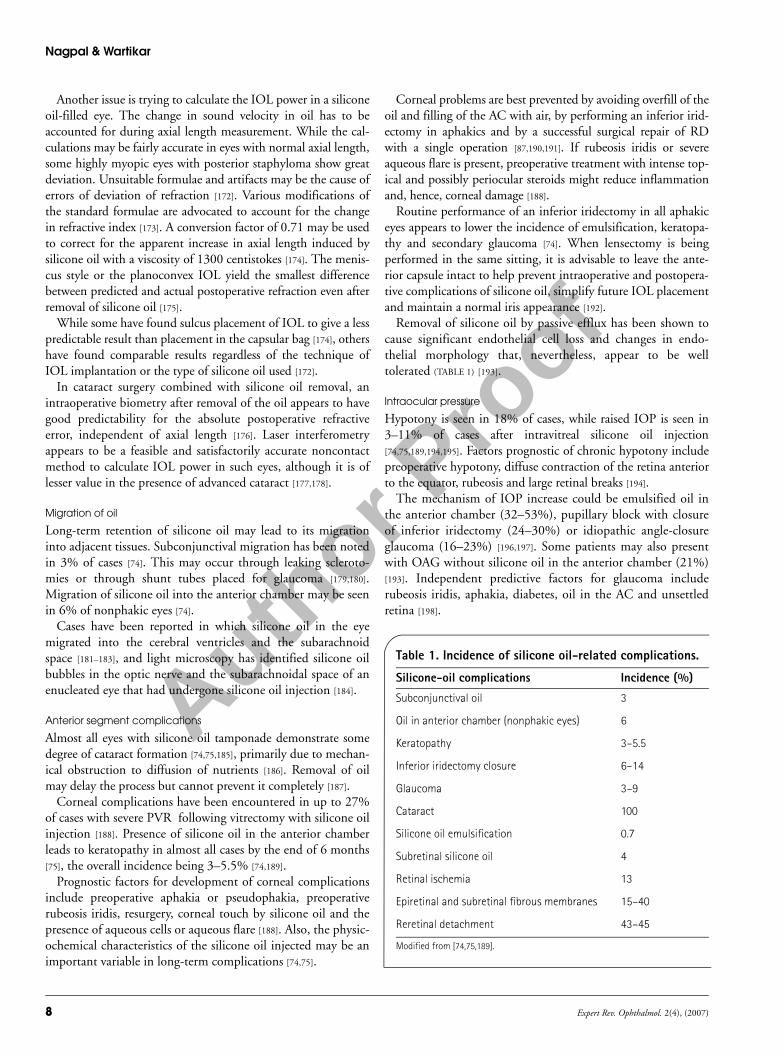
Table 1. Incidence of silicone oil-related complications.
Silicone-oil complications Incidence (%)
Subconjunctival oil 3
Oil in anterior chamber (nonphakic eyes) 6
Keratopathy 3–5.5
Inferior iridectomy closure 6–14
Glaucoma 3–9
Cataract 100
Silicone oil emulsification 0.7
Subretinal silicone oil 4
Retinal ischemia 13
Epiretinal and subretinal fibrous membranes 15–40
Reretinal detachment 43–45
Modified from [74,75,189].
Author Pro
of
Vitrectomy: when things go wrong
www.future-drugs.com 9
Postoperative glaucoma due to pupillary block and angle clo-sure in aphakics can be prevented by an inferior iridectomy [87].Blockage of trabecular meshwork by emulsified oil droplets canbe refractory to medical management and even to removal ofoil [199].
Control of glaucoma when attempted with medical manage-ment, silicone oil removal (SOR), trabeculectomy withmitomycin C, cyclocryotherapy, trans-scleral cyclophotocoagu-lation and/or anterior chamber shunting, has been shown tosucceed in 72% of eyes, while the rest remained refractory [198].Patients who undergo SOR alone to control IOP are morelikely to have persistent elevation of IOP and possibly undergoresurgery for glaucoma, whereas patients who undergo concur-rent SOR and glaucoma surgery are more likely to have hypot-ony [197]. As well as the silicone oil-related causes, all other vit-rectomy-related causes should be considered in an eye withraised IOP and silicone oil.
Emulsification
Emulsification is the formation of silicone oil droplets on thesurface of ocular tissues or at the interface between oil andintraocular fluids. It facilitates the migration of oil into ACwhere it causes further complications [200]. Almost all eyesdemonstrate some emulsification 1 year after surgery [75].Fibrin and serum are biologically active emulsifiers. In addi-tion, the lower the viscosity of silicone, the greater is its ten-dency to emulsify, particularly with viscosities from 1000 to5000 cSt [201].
Recurrent retinal detachment
Inflammatory membranes occurring at the silicone–fluidinterface may lead to macular pucker in 30% of cases and alsoto recurrent tractional RD [202,203]. Removal of silicone oil inanatomically successful eyes significantly increases the likeli-hood of improved VA [204] but also carries a risk of redetach-ment in a quarter of cases. This depends on factors such as thenumber of previously unsuccessful RD surgeries, VA before sil-icone oil removal, incomplete removal of vitreous base,absence of an encircling band in eyes with PVR and the indi-cation for pars plana vitrectomy. It is, however, independent ofthe technique of SOR and duration of silicone oil endotam-ponade [205]. Incidence rates of complications, such as keratop-athy and hypotony, have been found to be lower in eyes withthe silicone oil removed [206].
Conventional silicone oil cannot provide inferior tamponade,which is where PVR more often sets in, leading to redetach-ment. The subsequent use of high-density silicone oil (specificgravity of 1.06 g/ml) to provide support for the inferior retinais being explored as a strategy to reduce the number of reopera-tions [206,207]. However, an inflammatory response, resemblinggranulomatous uveitis and not responding to steroids, has beenseen in 37% cases after such high-density silicone oil endotam-ponade. It is likely that this vitreous substitute is an immuno-genic agent since after its removal complete resolution of theinflammation has been noted [208].
Expert commentaryVitrectomy has evolved into a high technology-based surgery.This is a unique example of treating a disease where man andmachine go hand-in-hand at every aspect. New surgical tech-niques are constantly evolving, with the resultant developmentof new technology and vice versa. The technology of cutters andits fluidics, including infusion and suction controls, has pro-vided excellent surgical control and added greater patient safetyto the surgery. Adjunct procedures using laser treatments andfragmatomes have also been refined, and are constantly evolvingto give improved precision and efficiency to the surgeon.
Over a period of years, several intraoperative surgical com-plications have been reduced through refined instrumentationand a better understanding of tissue metabolism, infusion flu-ids and fluidics. The improvement in software technology hasenhanced the foot switch control with reduced time lag, alsomaking the surgeon less dependent on support staff. Theseimprovements have drastically reduced the average vitrectomytime, resulting in a delay in the onset of cataract and othermetabolic-based sequelae. Personally, we feel that tissuerespect is of utmost importance and the way the conceptstoward sutureless vitrectomy are evolving, we are heading inthe right direction. At the moment, we are in the first genera-tion of sutureless vitrectomy systems, but almost every monthnew cutters and insertion systems are developing that aresharper and more precise than the previous ones. The next5 years should see a definite shift towards sutureless vitrec-tomy procedures with the immense benefits of reducingpatient discomfort and early rehabilitation.
Five-year viewTransconjunctival sutureless vitrectomyTransconjunctival sutureless vitrectomy (TSV) is a recentadvancement in vitreoretinal surgical techniques involving theuse of smaller gauge instruments through self-sealing scleroto-mies. The 25-G TSV is a minimally invasive technique andappears to reduce the convalescence period and the postopera-tive inflammatory response, while improving patient comfort[209,210]. It has been shown to decrease the operating time by37% compared with conventional 20-G vitrectomy [211].
In 20-G vitrectomy, repeated introduction and removal ofinstruments through the sclerotomies results in microtrauma atthe pars plana [210,212]. By contrast, the trocar and cannula sys-tem used in the 25-G vitrectomy system not only avoids theconjunctival incision and cauterization of the scleral bed, butalso facilitates easy entry, with no trauma to the sclera or thepars plana. Additionally, it has been hypothesized that theremay be less chance of vitreous and retinal herniation in asutureless scleral wound compared with one that has beensutured [213]. As the instruments do not come in contact withthe sclera or pars plana, prolonged anesthesia is not required.Sutureless vitrectomies have also been carried out under topicalanesthesia [214]. In the absence of severe congestion, chemosis orlid swelling, even cosmetically the eye looks much better fromthe first postoperative day itself.
Author Pro
of
Nagpal & Wartikar
10 Expert Rev. Ophthalmol. 2(4), (2007)
The success of 25-G vitrectomy has been well described, butits application for more complex vitreoretinal diseases, such ascomplex RD with PVR, has been limited. While its use hasbeen reported even for tractional RDs in stage 4 and 5 of retin-opathy of prematurity, modifications, including conjunctivaldissection and suturing of conjunctiva and sclerotomies, needto be used [215].
The main limiting factors of the 25-G system are the rela-tive lack of instrument rigidity, slower vitreous cutting abilityand suboptimal fluidics, and even blockage of the cutter tip,all of which are inherent to the reduced caliber of the instru-mentation [216,217]. Intraoperatively, problems, such as diffi-culty in inserting the microcannula leading to deformity andinstability of the microcannula, self-disconnection of the infu-sion tip resulting in lens damage, and the need to convert to20-G vitrectomy have been noted [128].
Factors such as the unsutured wounds, postoperative hypo-tony and lower infusion rates may contribute to the reportedincreased risk of endophthalmitis after 25-G vitrectomy[218–220,302]. An obvious wound leak needs to be sutured.Intravitreal air injection may be used to avoid immediatepostoperative hypotony but, despite partial fluid–airexchange, hypotony has been reported in up to 16–25% ofcases [128,217]. These concerns are similar to the era ofphacoemulsification undergoing a shift toward corneal tun-nel incisions. However, we feel that these are concerns of alearning curve and as the surgeon gains experience andselects cases with proper discretion, they will probably notremain major issues.
Some surgeons have suggested that the technique of 20-Gsutureless vitrectomy overcomes some of the limitations of25-G systems, but inconsistencies in the application of thistechnique have limited its widespread use [216]. Anotheroption is to combine the use of 20-G devices through themain port while keeping the other two sutureless. This mayhelp to expand the indications for the 25-G system. How-ever, postoperative low ocular tension must be addressed bycarefully considering surgical indications and preventionmeasures [221].
The 23-G vitrectomy system has been devised with a view tocombining the advantages of decreased surgical trauma andrecovery time enjoyed with 25-G sutureless vitrectomy withthe sturdier instrumentation and improved fluidics of the20-G systems [222]. These characteristics make 23-G vitrec-tomy a promising approach to tackle the complete range ofvitreoretinal surgical procedures efficiently and safely with asingle system [216].
Pharmacological vitrectomyPharmacological vitrectomy refers to the use of enzymes in aneffort both to liquefy vitreous and weaken the adhesion of vitre-ous cortex to the ILM during or before performing vitreous sur-gery. These vitreolytic enzymes may be of great value in compli-cated or office-procedure vitreoretinal surgery [223]. Plasmin,dispase and chondroitinase have been used to make the vitreoussurgery easier with fewer complications or to avoid vitrectomy.On the other hand, hyaluronidase has been used to facilitate theclearance of vitreous hemorrhage, liquefying vitreous body anddeveloping posterior vitreous detachment [224]. The highestincrease of vitreous removal when tested in enucleated pig eyeswas found with hyaluronidase and the lowest with chondroiti-nase. Damage occasionally occurred to the ILM and very rarelyto the nerve fiber layer [223]. t-PA, which converts plasminogenpresent in the vitreous cavity to plasmin, thereby inducing fibri-nolysis, has been found to simplify separation of vitreous cortexand pathological membranes without any bleeding [225]. Use ofmicroplasmin for the treatment of diabetic macular edemawithout the need for vitrectomy is also under trial [301,303].
Every new technique or equipment that is introduced shouldbe well-researched and well-learned before putting it into prac-tice. It is important to follow evidence-based medicine. Com-plications will occur. They are an integral part of every surgery.We need to realize that our best chance lies not so much in try-ing to eliminate them, but rather in detecting them earlyenough and managing them efficiently
Financial disclosureThe authors have no relevant financial interests related to thismanuscript, including employment, consultancies, honoraria,stock ownership or options, expert testimony, grants or patentsreceived or pending, or royalties.
Key issues
• Tissue respect has to be given at every step of vitrectomy.
• The technique used for making sclerotomies has an important bearing on the surgery and its outcome.
• Corneal edema, cataract and glaucoma are commonly observed complications.
• The most significant complications include retinal detachment, choroidal effusion and endophthalmitis.
• The best way to manage a complication is to avoid it.
ReferencesPapers of special note have been highlighted as:• of interest•• of considerable interest
1 Kasner D, Miller G, Taylor W, Sever R, Norton E. Surgical treatment of amyloidosis of the vitreous. Trans. Am. Acad. Ophthalmol. Otolaryngol. 72, 410–418 (1968).
2 Machemer R, Buettner H, Norton EW, Parel JM. Vitrectomy: a pars plana approach. Trans. Am. Acad. Ophthalmol. Otolaryngol. 75, 813–820 (1971).
3 Chang S. Perfluorocarbon liquids in vitreoretinal surgery. Int. Ophthalmol. Clin. 32, 153–163 (1992).
4 Peyman GA, Schulman JA. In: Intravitreal Surgery: Principles and Practice (2nd Edition). Appleton & Lange, CT, USA 1 (1994).
5 Theocharis AD, Papageorgakopoulou N, Feretis E, Theocharis DA. Occurrence and structural characterization of versican-like proteoglycan in human vitreous. Biochimie 84(12), 1237–1243 (2002).

Author Pro
of
Vitrectomy: when things go wrong
www.future-drugs.com 11
6 Mayne R, Brewton RG, Wright DW et al. Morphological and biochemical studies of the structure of the vitreous and the zonular fibres. Biochem. Soc. Trans. 19, 868–871 (1991).
7 Spitznas M, Reiver J. A stereoscopic diagonal inverter (SDI) for wide-angle vitreous surgery. Graefes Arch. Clin. Exp. Ophthalmol. 225, 9–12 (1987).
8 Spitznas M. A binocular indirect ophthalmomicroscope (BIOM) for wide-angle vitreous surgery. Graefes Arch. Clin. Exp. Ophthalmol. 225, 13–15 (1987).
9 Nakata K, Ohji M, Ikuno Y et al. Wide-angle viewing lens for vitrectomy. Am. J. Ophthalmol. 137(4), 760–762 (2004).
10 Chalam KV, Shah VA. Self-illuminated contact lens for peripheral vitreous surgery. Ophthalmic Surg. Lasers Imaging 35(1), 76–77, (2004).
11 Bovey EH, Gonvers M. A new device for noncontact wide-angle viewing of the fundus during vitrectomy. Arch. Ophthalmol. 113, 1572–1573 (1995).
12 Chalam KV, Shah VA. Two-piece, dual-purpose comprehensive contact lens for vitreous surgery. Ophthalmic Res. 37(4), 175–178, (2005).
13 Peyman GA, Canakis C, Livir-Rallatos C, Easley J. A new wide-angle endoillumination probe for use during vitrectomy. Retina 22, 242 (2002).
14 Chow DR. Shedding some light on current endoillumination. Retinal Physician 2, 37–39 (2005).
15 Oshima Y, Awh CC, Tano Y. Self-retaining 27-gauge transconjunctival chandelier endoillumination for panoramic viewing during vitreous surgery. Am. J. Ophthalmol. 143(1), 166–167 (2007).
16 de Smet MD, Mura M. Minimally invasive surgery-endoscopic retinal detachment repair in patients with media opacities. Eye (2007) DOI: 10.1038/sj.eye.6702710 (Epub ahead of print).
17 Peyman GA, Livir-Rallatos C, Canakis C, Easley J. A new high-speed pneumatic vitrectomy cutter. Am. J. Ophthalmol. 133(4), 568–569 (2002).
18 Flynn HW, Lee WG, Parel JM. Design features and surgical use of a cannulated extrusion needle. Graefes Arch. Clin. Exp. Ophthalmol. 227(4), 304–308 (1989).
19 Moorhead LC, Gardner TW, Lambert HM et al. Dynamic intraocular pressure measurements during vitrectomy. Arch. Ophthalmol. 123(11), 1514–1523 (2005).
20 Krzizok T. Fibrin glue for closing conjunctival wounds in ophthalmic surgery.
Ophthalmologe 101(10), 1006–1010 (2004).
21 Alio JL, Gomez J, Mulet E, Bujanda MM, Martinez JM, Molina Y. A new acrylic tissue adhesive for conjunctival surgery: experimental study. Ophthalmic Res. 35(6), 306–312 (2003).
22 Shimada H, Nakashizuka H, Nakajima M, Mori R, Mizutani Y. Twenty-gauge transconjunctival vitrectomy. Jpn J. Ophthalmol. 49(3), 257–260 (2005).
23 Albini TA, Evans M, Lakhanpal RR, Javaheri M, Rao NA, Chong LP. Conjunctival epithelium in pars plana vitrectomy specimens. Retina 27(1), 55–58 (2007).
24 Tardif YM, Schepens CL, Tolentino FI. Vitreous surgery. XIV. Complications from sclerotomy in 89 consecutive cases. Arch. Ophthalmol. 95(2), 229–234 (1977).
25 Liu W, Huang SY, Zhang P, Tang SB, Li JQ, Zheng HL. Bioptic significance of incarcerated contents at sclerotomy sites during vitrectomy. Retina 24(3), 407–411 (2004).
26 Kreiger A, Straatsma B Foos R. Incisional complications in pars plana vitrectomy. Mod. Prob. Ophthalmol. 18, 210–233 (1977).
27 West JF, Gregor ZJ. Fibrovascular ingrowth and recurrent haemorrhage following diabetic vitrectomy. Br. J. Ophthalmol. 84(8), 822–825 (2000).
28 Kreiger A. The pars plana incision: experimental studies, pathologic observations, and clinical experience. Trans. Am. Ophthalmol. Soc. 89, 549–621 (1991).
29 Sabti K, Kapusta M, Mansour M, Overbury O, Chow D. Ultrasound biomicroscopy of sclerotomy sites: the effect of vitreous shaving around sclerotomy sites during pars plana vitrectomy. Retina 21(5), 464–468 (2001).
30 Hotta K, Hirakata A, Ohi Y et al. Ultrasound biomicroscopy for examination of the sclerotomy site in eyes with proliferative diabetic retinopathy after vitrectomy. Retina 20(1), 52–58 (2000).
31 Yeh PT, Yang CM, Yang CH, Huang JS. Cryotherapy of the anterior retina and sclerotomy sites in diabetic vitrectomy to prevent recurrent vitreous hemorrhage: an ultrasound biomicroscopy study. Ophthalmology 112(12), 2095–2102 (2005).
32 Yeshurun I, Rock T, Bartov E. Modified sutureless sclerotomies for pars plana vitrectomy. Am. J. Ophthalmol. 138(5), 866–867 (2004).
33 Rahman R, Rosen PH, Riddell C, Towler H. Self-sealing sclerotomies for sutureless pars plana vitrectomy. Ophthalmic Surg. Lasers 31(6), 462–466 (2000).
34 Schmidt JC, Meyer CH, Mennel S. Pars-plana vitrectomy with anterior chamber infusion via a paracentesis in pseudophakic eyes. Ophthalmologe 104(3), 222–225 (2007).
35 Morley AM, Pavesio C. Surgically induced necrotising scleritis following three-port pars plana vitrectomy without scleral buckling: a series of three cases. Eye (2007) DOI: 10.1038/sj.eye.6702708 (Epub ahead of print).
36 Chiambo S, Bailez Fidalgo C, Pastor Jimeno JC et al. Corneal epithelial complications after vitrectomy: a retrospective study. Arch. Soc. Esp. Oftalmol. 79(4), 155–161 (2004).
37 Hiraoka M, Amano S, Oshika T, Kato S, Hori S. Factors contributing to corneal complications after vitrectomy in diabetic patients. Jpn J. Ophthalmol. 45(5), 492–495 (2001).
38 Scott IU, Flynn HW Jr, Murray TG, Feuer WJ. Outcomes of surgery for retinal detachment associated with proliferative vitreoretinopathy using perfluoro-n-octane: a multicenter study. Am. J. Ophthalmol. 136(3), 454–463 (2003).
39 Friberg TR, Ohji M, Scherer JJ, Tano Y. Frequency of epithelial debridement during diabetic vitrectomy. Am. J. Ophthalmol. 135(4), 553–554 (2003).
• Survey of active vitreoretinal surgeons was carried out to determine how often they intentionally debride the corneal epithelium during vitrectomy surgery for diabetic patients. Irrigating contact lenses were found to increase the need for epithelial debridement the most.
40 Virata SR, Kylstra JA, Singh HT. Corneal epithelial defects following vitrectomy surgery using hand-held, sew-on, and noncontact viewing lenses. Retina 19(4), 287–290 (1999).
41 Garcia-Valenzuela E, Abdelsalam A, Eliott D et al. Reduced need for corneal epithelial debridement during vitreo-retinal surgery using two different viscous surface lubricants. Am. J. Ophthalmol. 136(6), 1062–1066 (2003).
42 Virata SR, Kylstra JA. Postoperative complications following vitrectomy for proliferative diabetic retinopathy with sew-on and noncontact wide-angle viewing lenses. Ophthalmic Surg. Lasers 32(3), 193–197 (2001).
43 Samuel MA, Desai UR, Strassman I, Abusamak M. Intraocular irrigating solutions. A clinical study of BSS Plus and dextrose bicarbonate fortified BSS as an infusate during pars plana vitrectomy. Indian J. Ophthalmol. 51(3), 237–242 (2003).
44 Mitamura Y, Yamamoto S, Yamazaki S.

Author Pro
of
Nagpal & Wartikar
12 Expert Rev. Ophthalmol. 2(4), (2007)
Corneal endothelial cell loss in eyes undergoing lensectomy with and without anterior lens capsule removal combined with pars plana vitrectomy and gas tamponade. Retina 20(1), 59–62 (2000).
45 Schrader S, Wedel T, Moll R, Geerling G. Combination of serum eye drops with hydrogel bandage contact lenses in the treatment of persistent epithelial defects. Graefes Arch. Clin. Exp. Ophthalmol. 244(10), 1345–1349 (2006).
46 Yulek F, Ozdek S, Gurelik G, Hasanreisoglu B. Effect of topical steroids on corneal epithelial healing after vitreoretinal surgery. Acta Ophthalmol. Scand. 84(3), 319–322 (2006).
47 Rosenthal P, Cotter JM, Baum J. Treatment of persistent corneal epithelial defect with extended wear of a fluid-ventilated gas-permeable scleral contact lens. Am. J. Ophthalmol. 130(1), 33–41 (2000).
48 Gopal L, Nagpal A, Kabra S, Roy J. Anterior chamber collapse following vitreoretinal surgery with gas tamponade in aphakic eyes: incidence and risk factors. Retina 26(9), 1014–1020 (2006).
49 Kadonosono K, Matsumoto S, Uchio E, Sugita M, Akura J, Ohno S. Iris neovascularization after vitrectomy combined with phacoemulsification and intraocular lens implantation for proliferative diabetic retinopathy. Ophthalmic Surg. Lasers 32(1), 19–24 (2001).
50 Helbig H, Kellner U, Bornfeld N, Foerster MH. Rubeosis iridis after vitrectomy for diabetic retinopathy. Graefes Arch. Clin. Exp. Ophthalmol. 236(10), 730–733 (1998).
51 Rice TA, Michels RG, Maguire MG, Rice EF. The effect of lensectomy on the incidence of iris neovascularization and neovascular glaucoma after vitrectomy for diabetic retinopathy. Am. J. Ophthalmol. 95(1), 1–11 (1983).
52 Schiff WM, Barile GR, Hwang JC et al. Diabetic vitrectomy: influence of lens status upon anatomic and visual outcomes. Ophthalmology 114(3), 544–550 (2007).
53 Comaratta MR, Chang S, Sparrow J. Iris neovascularization in proliferative vitreoretinopathy. Ophthalmology 99(6), 898–905 (1992).
54 Avery RL, Pearlman J, Pieramici DJ et al. Intravitreal bevacizumab (Avastin) in the treatment of proliferative diabetic retinopathy. Ophthalmology 113(10), 1695 (2006).
• Possible therapeutic effect found in the fellow eye raises concern that systemic side effects may occur in patients receiving intravitreal bevacizumab (1.25 mg). Doses
as low as 6.2 µg may achieve a therapeutic result with lesser risk of systemic side effects.
55 Oshima Y, Sakaguchi H, Gomi F, Tano Y. Regression of iris neovascularization after intravitreal injection of bevacizumab in patients with proliferative diabetic retinopathy. Am. J. Ophthalmol. 142(1), 155–158 (2006).
56 Grisanti S, Biester S, Peters S et al. Intracameral bevacizumab for iris rubeosis. Am. J. Ophthalmol. 142(1), 158–160 (2006).
57 Federman JL, Anand R. Surgical dilation of the pupil during pars plana vitrectomy. Ophthalmic Surg. 20(1), 46–48 (1989).
58 Chalam KV, Shah VA. Optics of wide-angle panoramic viewing system-assisted vitreous surgery. Surv. Ophthalmol. 49(4), 437–445 (2004).
59 Jackson H, Patel CK, Westcott M, Thompson GM, Mathalone BM. Does topical flurbiprofen affect the pupillary response to acetylcholine? Eye 8(3), 329–331 (1994).
60 Stewart R, Grosserode R, Cheetham JK, Rosenthal A. Efficacy and safety profile of ketorolac 0.5% ophthalmic solution in the prevention of surgically induced miosis during cataract surgery. Clin. Ther. 21(4), 723–732 (1999).
61 Backstrom G, Behndig A. Redilatation with intracameral mydriatics in phacoemulsification surgery. Acta Ophthalmol. Scand. 84(1), 100–104 (2006).
62 Liou SW, Chen CC. Maintenance of mydriasis with one bolus of epinephrine injection during phacoemulsification. J. Ocul. Pharmacol. Ther. 17(3), 249–253 (2001).
63 Tognetto D, Agolini G, Grandi G, Ravalico G. Iris alteration using mechanical iris retractors. J. Cataract Refract. Surg. 27(10), 1703–1705 (2001).
64 Kadonosono K, Ohno S. New iris retractor for pupil dilatation during anterior vitrectomy: double-hook iris retractor. Ophthalmic Surg. Lasers 30(3), 241–243 (1999).
65 Chong LP. A disposable iris retractor for vitrectomy. Am. J. Ophthalmol. 112(6), 731–732 (1991).
66 Arpa P. A new device for pupillary dilatation in vitreous surgery. Retina 12(Suppl. 3), S87–S89 (1992).
67 Pham DT, Volkmer C, Leder K, Wollensak J. Partial sphincterectomy in cataract surgery. Clinical and patho-histologic results. Ophthalmologe 95(9), 635–638 (1998).
68 Kozobolis VP, Pallikaris IG, Tsambarlakis IG, Vlachonikolis IG. Nd:YAG laser removal of pupillary membranes developed after ECCE with PC-IOL implantation. Acta Ophthalmol. Scand. 75(6), 711–715 (1997).
69 Sawa M, Saito Y, Hayashi A, Kusaka S, Ohji M, Tano Y. Assessment of nuclear sclerosis after nonvitrectomizing vitreous surgery. Am. J. Ophthalmol. 132(3), 356–362 (2001).
70 Sawa M, Ohji M, Kusaka S et al. Nonvitrectomizing vitreous surgery for epiretinal membrane long-term follow-up. Ophthalmology 112(8), 1402–1408 (2005).
71 Sima P, Zoran T. Long-term results of vitreous surgery for proliferative diabetic retinopathy. Doc. Ophthalmol. 87(3), 223–232 (1994).
72 Smiddy WE, Feuer W. Incidence of cataract extraction after diabetic vitrectomy. Retina 24(4), 574–581 (2004).
• Rate of cataract extraction after vitrectomy was found to be lower in diabetics, suggesting a lower rate of cataract formation. Visual loss attributable to cataract maybe overestimated in diabetics undergoing vitrectomy.
73 Thompson JT. The role of patient age and intraocular gas use in cataract progression after vitrectomy for macular holes and epiretinal membranes. Am. J. Ophthalmol. 137(2), 250–257 (2004).
74 Riedel KG, Gabel VP, Neubauer L, Kampik A, Lund OE. Intravitreal silicone oil injection: complications and treatment of 415 consecutive patients. Graefes Arch. Clin. Exp. Ophthalmol. 228(1), 19–23 (1990).
75 Federman JL, Schubert HD. Complications associated with the use of silicone oil in 150 eyes after retina-vitreous surgery. Ophthalmology 95(7), 870–876 (1988).
76 Heimann H, Bornfeld N, Friedrichs W et al. Primary vitrectomy without scleral buckling for rhegmatogenous retinal detachment. Graefes Arch. Clin. Exp. Ophthalmol. 234(9), 561–568 (1996).
77 Hsuan JD, Brown NA, Bron AJ, Patel CK, Rosen PH. Posterior subcapsular and nuclear cataract after vitrectomy. J. Cataract Refract. Surg. 27(3), 437–444 (2001).
78 Scharwey K, Pavlovic S, Jacobi KW. Early posterior capsule fibrosis after combined cataract and vitreoretinal surgery with intraocular air/SF6 gas tamponade. Klin. Monatsbl Augenheilkd. 212(3), 149–153 (1998).
79 Kuszak JR, Sivak JG, Moran KL et al. Suppression of post-vitrectomy lens changes in the rabbit by novel benzopyranyl esters and amides. Exp. Eye Res. 75(4), 459–473
Author Pro
of
Vitrectomy: when things go wrong
www.future-drugs.com 13
(2002).
80 Chalam KV, Shah VA, Tripathi RC. A curved vitrectomy probe. Ophthalmic Surg. Lasers Imaging 35(3), 259–260 (2004).
81 Weinberg RS, Peyman GA, Huamonte FU. Elevation of intraocular pressure after pars plana vitrectomy. Graefes Arch. Klin. Exp. Ophthalmol. 200, 157–161 (1976).
82 Campbell DG, Simmons RJ, Grant WM. Ghost cells as a cause of glaucoma. Am. J. Ophthalmol. 81, 441 (1976).
83 Phelps CD, Watzke RC. Hemolytic glaucoma. Am. J. Ophthalmol. 80, 690 (1975).
84 Singh H, Grand MG. Treatment of blood-induced glaucoma by trans pars plana vitrectomy. Retina 1(3), 255–257 (1981).
85 Al-Shahwan S, Ghadhfan F, Kalantan H, Al-Torbak AA. Pupillary block glaucoma in phakic perfluoropropane gas filled eye. Br. J. Ophthalmol. 90(6), 797–798 (2006).
86 Chen PP, Thompson JT. Risk factors for elevated intraocular pressure after the use of intraocular gases in vitreoretinal surgery. Ophthalmic Surg. Lasers 28(1), 37–42 (1997).
87 Ando F. Intraocular hypertension resulting from pupillary block by silicone oil. Am. J. Ophthalmol. 99, 87–88 (1985).
88 Kumar A, Kedar S, Garodia VK, Singh RP. Angle closure glaucoma following pupillary block in an aphakic perfluoropropane gas-filled eye. Indian J. Ophthalmol. 50, 220–221 (2002).
89 Yamamoto T, Hitani K, Tsukahara I et al. Early postoperative retinal thickness changes and complications after vitrectomy for diabetic macular edema. Am. J. Ophthalmol. 135(1), 14–19 (2003).
90 Chang S. LXII Edward Jackson lecture: open angle glaucoma after vitrectomy. Am. J. Ophthalmol. 141(6), 1033–1043 (2006).
91 Sugiyama W, Ando N. Anterior segment fluorescein angiography for evaluating the effect of vitrectomy for neovascular glaucoma. Nippon Ganka Gakkai Zasshi. 109(11), 741–747 (2005).
92 Krupin T, Mitchell KB, Becker B. Cyclocryotherapy in neovascular glaucoma. Am. J. Ophthalmol. 86, 24 (1978).
93 Scott IU, Alexandrakis G, Flynn HW Jr et al. Combined pars plana vitrectomy and glaucoma drainage implant placement for refractory glaucoma. Am. J. Ophthalmol. 129(3), 334–341 (2000).
94 Schachat AP, Oyakawa RT, Michels RG et al. Complications of vitreous surgery for diabetic retinopathy. II. Postoperative complications. Ophthalmology 90, 522
(1983).
95 Wu WC, Chang SM, Chen JY, Chang CW. Management of postvitrectomy diabetic vitreous hemorrhage with tissue plasminogen activator (t-PA) and volume homeostatic fluid–fluid exchanger. J. Ocul. Pharmacol. Ther. 17(4), 363–371 (2001).
96 Virata SR, Kylstra JA. Postoperative complications following vitrectomy for proliferative diabetic retinopathy with sew-on and noncontact wide-angle viewing lenses. Ophthalmic Surg. Lasers 32(3), 193–197 (2001).
97 Soto-Pedre E, Hernaez-Ortega MC, Vazquez JA. Risk factors for postoperative hemorrhage after vitrectomy for diabetic retinopathy. Ophthalmic Epidemiol. 12(5), 335–341 (2005).
98 Yeh PT, Yang CM, Yang CH, Huang JS. Cryotherapy of the anterior retina and sclerotomy sites in diabetic vitrectomy to prevent recurrent vitreous hemorrhage: an ultrasound biomicroscopy study. Ophthalmology 112(12), 2095–2102 (2005).
• Retrospective analysis that shows a strong correlation between the presence of fibrovascular ingrowth as seen on ultrasound biomicroscopy study to the occurrence of vitreous hemorrhage after diabetic vitrectomy. It also evaluates the role of panretinal photocoagulation, anterior peripheral retinal cryotherapy and cryotherapy to ports in such cases.
99 Hershberger VS, Augsburger JJ, Hutchins RK, Raymond LA, Krug S. Fibrovascular ingrowth at sclerotomy sites in vitrectomized diabetic eyes with recurrent vitreous hemorrhage: ultrasound biomicroscopy findings. Ophthalmology 111(6), 1215–1221 (2004).
100 Neely KA, Scroggs MW, McCuen BW 2nd. Peripheral retinal cryotherapy for postvitrectomy diabetic vitreous hemorrhage in phakic eyes. Am. J. Ophthalmol. 126(1), 82–90 (1998).
101 Hajji Z, Rouillot JS, Roth P, Grange JD. Should associated intraoperative and/or postoperative photocoagulation be systematic during or after vitrectomy for proliferative diabetic vitreoretinopathy? J. Fr. Ophtalmol. 26(1), 47–53 (2003).
102 Han DP, Murphy ML, Mieler WF, Abrams GW. Outpatient fluid–air exchange for severe postvitrectomy diabetic vitreous hemorrhage. Long-term results and complications. Retina 11(3), 309–314 (1991).
103 Yang CM, Yeh PT, Yang CH. Intravitreal long-acting gas in the prevention of early postoperative vitreous hemorrhage in diabetic vitrectomy. Ophthalmology 114(4),
710–715 (2007).
104 Jorge R, Costa RA, Calucci D, Cintra LP, Scott IU. Intravitreal bevacizumab (Avastin) for persistent new vessels in diabetic retinopathy (IBEPE study). Retina 26(9), 1006–1013 (2006).
105 Spaide RF, Fisher YL. Intravitreal bevacizumab (Avastin) treatment of proliferative diabetic retinopathy complicated by vitreous hemorrhage. Retina 26(3), 275–278 (2006).
106 Mason JO 3rd, Nixon PA, White MF. Intravitreal injection of bevacizumab (Avastin) as adjunctive treatment of proliferative diabetic retinopathy. Am. J. Ophthalmol. 142(4), 685–688 (2006).
107 Peyman GA, Raichand M, Huamonte FU et al. Vitrectomy in 125 eyes with diabetic vitreous hemorrhage. Br. J. Ophthalmol. 60, 752 (1976).
108 Peyman GA, Green JL, Carroll CP. A simplified approach to the management of tangential retinal traction bands. Br. J. Ophthalmol. 65, 414 (1981).
109 Christopher N. Minimizing the risk of endophthalmitis following intravitreous injections. Retina 24(5), 699–705 (2004).
110 Mamalis N, Kearsley L, Brinton E. Postoperative endophthalmitis. Curr. Opin. Ophthalmol. 13(1), 14–18 (2002).
111 Somani S, Grinbaum A, Slomovic AR. Postoperative endophthalmitis: incidence, predisposing surgery, clinical course and outcome. Can. J. Ophthalmol. 32(5), 303–310 (1997).
112 Cohen SM, Flynn HW Jr, Murray TG, Smiddy WE. Endophthalmitis after pars plana vitrectomy. The Postvitrectomy Endophthalmitis Study Group. Ophthalmology 102(5), 705–712 (1995).
113 Eifrig CW, Scott IU, Flynn HW Jr, Smiddy WE, Newton J. Endophthalmitis after pars plana vitrectomy: incidence, causative organisms, and visual acuity outcomes. Am. J. Ophthalmol. 138(5), 799–802 (2004).
114 Joondeph BC, Blanc JP, Polkinghorne PJ. Endophthalmitis after pars plana vitrectomy: a New Zealand experience. Retina 25(5), 587–589 (2005).
115 Zhang S, Ding X, Hu J, Gao R. Clinical features of endophthalmitis after vitreoretinal surgery. Yan Ke Xue Bao 19(1), 39–43 (2003).
116 Egger SF, Scholda C, Haddad R, Vecsei P, Huber-Spitzy V. Contamination of intraocular fluid in pars plana vitrectomy. Ophthalmologe 93(2), 126–129 (1996).
117 Zaninetti M, Baglivo E, Safran AB. Morganella morganii endophthalmitis after

Author Pro
of
Nagpal & Wartikar
14 Expert Rev. Ophthalmol. 2(4), (2007)
vitrectomy: case report and review of the literature. Klin. Monatsbl Augenheilkd. 220(3), 207–209 (2003).
118 Wong TY, Chee SP. The epidemiology of acute endophthalmitis after cataract surgery in an Asian population. Ophthalmology 111(4), 699–705 (2004).
119 Speaker MG. Prophylaxis of endophthalmitis with topical povidone–iodine. Ophthalmology 98, 1769–1774 (1991).
120 Apt L, Isenberg SJ, Yoshimori R et al. The effect of povidone-iodine solution applied at the conclusion of ophthalmic surgery. Am. J. Ophthalmol. 119, 701–705 (1998).
121 Donnefeld ED, Perry HD. Cataract surgery: five ways to prevent endophthalmitis. Rev. Ophthalmol. 3, 67–72 (1996).
122 Liesegang TJ. Use of antimicrobials to prevent postoperative infection in patients with cataracts. Curr. Opin. Ophthalmol.12, 68–74 (2001).
123 Donahue SP, Kowalski RP, Jewart BH et al. Vitreous cultures in suspected endophthalmitis: biopsy or vitrectomy? Ophthalmology 100, 452–455 (1993).
124 Cuilla TA. Update on acute and chronic endophthalmitis. Ophthalmology 106, 2237–2238 (1999).
125 Baird DR, Henry M, Liddell KG, Mitchell CM, Sneddon JG. Post-operative endophthalmitis: the application of hazard analysis critical control points (HACCP) to an infection control problem. J. Hosp. Infect. 49(1), 14–22 (2001).
126 Griffith C. HACCP and the management of healthcare associated infections: are there lessons to be learnt from other industries? Int. J. Health Care Qual. Assur. Inc. Leadersh. Health Serv. 19(4–5), 351–367 (2006).
127 Sohajda Z, Orosi P, Hayfron J, Berta A. The role of infection control in the prevention of postoperative endophthalmitis. Klin. Monatsbl Augenheilkd. 223(8), 668–674 (2006).
128 Byeon SH, Chu YK, Lee SC, Koh HJ, Kim SS, Kwon OW. Problems associated with the 25-gauge transconjunctival sutureless vitrectomy system during and after surgery. Ophthalmologica 220(4), 259–265 (2006).
129 Rizzo S, Genovesi-Ebert F, Vento A, Miniaci S, Cresti F, Palla M. Modified incision in 25-gauge vitrectomy in the creation of a tunneled airtight sclerotomy: an ultrabiomicroscopic study. Graefes Arch. Clin. Exp. Ophthalmol. DOI: 10.1007/s00417-006-0533-x (2007) (Epub ahead of print).
130 Brubaker RF, Pederson JE. Ciliochoroidal detachment. Surv. Ophthalmol. 27, 281
(1983).
131 Scott IU, Flynn HW Jr, Smiddy WE et al. Clinical features and outcomes of pars plana vitrectomy in patients with retained lens fragments. Ophthalmology 110(8), 1567–1572 (2003).
132 Cohen SM, Davis A, Cukrowski C. Cystoid macular edema after pars plana vitrectomy for retained lens fragments. J. Cataract Refract. Surg. 32(9), 1521–1526 (2006).
133 Gass JDM, Parrish R. Outer retinal ischemic infarction – a newly recognized complication of cataract extraction and closed vitrectomy. Part 1. A case-report. Ophthalmology 89, 1467 (1982).
134 Malinowski SM, Pesin SR. Visual field loss caused by retinal vascular occlusion after vitrectomy surgery. Am. J. Ophthalmol. 123(5), 707–708 (1997).
135 William SE, Luis G, Rogerio P, William F. Retinal detachment rate after vitrectomy for retained lens material after phacoemulsification. Am. J. Ophthalmol. 135(2), 183–187 (2003).
136 Schrey S, Krepler K, Wedrich A. Incidence of rhegmatogenous retinal detachment after vitrectomy in eyes of diabetic patients. Retina 26(2), 149–152 (2006).
137 Faulborn J, Conway B, Machemer R. Symposium: complications of modern surgical procedures: surgical complications of pars plana vitreous surgery. Ophthalmology 85, 116 (1978).
138 Michels RG. Vitrectomy for complications of diabetic retinopathy. Arch. Ophthalmol. 96, 237 (1978).
139 Schmidt JC, Nietgen GW, Hesse L, Kroll P. External diaphanoscopic illuminator: a new device for visualization in pars plana vitrectomy. Retina 20(1), 103–106 (2000).
140 Hudson HL, Chong LP, Frambach DA, Valencia M, Green RL, Lopez PF. Encircling panretinal laser photocoagulation may prevent macular detachment after vitrectomy for proliferative diabetic retinopathy. Int. Ophthalmol. 18(2), 101–104 (1994).
141 Haritoglou C, Kampik A. Staining techniques in macular surgery. Ophthalmologe 103(11), 927–934 (2006).
142 Guo S, Tutela AC, Wagner R, Caputo AR. A comparison of the effectiveness of four biostains in enhancing visualization of the vitreous. J. Pediatr. Ophthalmol. Strabismus 43(5), 281–284 (2006).
143 Matsumoto H, Yamanaka I, Hisatomi T et al. Triamcinolone acetonide-assisted pars plana vitrectomy improves residual posterior vitreous hyaloid removal: ultrastructural analysis of the inner limiting membrane.
Retina 27(2), 174–179 (2007).
144 Feron EJ, Veckeneer M, Parys-Van Ginderdeuren R, Van Lommel A, Melles GR, Stalmans P. Trypan blue staining of epiretinal membranes in proliferative vitreoretinopathy. Arch. Ophthalmol. 120(2), 141–144 (2002).
145 Da Mata AP, Burk SE, Riemann CD et al. Indocyanine green-assisted peeling of the retinal internal limiting membrane during vitrectomy surgery for macular hole repair. Ophthalmology 108(7), 1187–1192 (2001).
146 Zheng Y, Sun N, Xiong Q, Wang X, Quan Y. Triamcinolone-assisted pars plana vitrectomy for retinal disease. Yan Ke Xue Bao 21(3), 142–146 (2005).
147 Uemoto R, Yamamoto S, Takeuchi S. Changes in retinal pigment epithelium after indocyanine green-assisted internal limiting lamina peeling during macular hole surgery. Am. J. Ophthalmol. 140(4), 752–755 (2005).
148 Cheng SN, Yang TC, Ho JD, Hwang JF, Cheng CK. Ocular toxicity of intravitreal indocyanine green. J. Ocul. Pharmacol. Ther. 21(1), 85–93 (2005).
149 Schmidt JC, Rodrigues EB, Meyer CH, Hoerle S, Kroll P. A modified technique to stain the internal limiting membrane with indocyanine green. Ophthalmologica 218(3), 176–179 (2004).
150 Mennel S, Thumann G, Peter S, Meyer CH, Kroll P. Influence of vital dyes on the function of the outer blood–retinal barrier in vitro. Klin. Monatsbl Augenheilkd. 223(7), 568–576 (2006).
151 Campo RV, Sipperley JO, Sneed SR et al. Pars plana vitrectomy without scleral buckle for pseudophakic retinal detachments. Ophthalmology 106(9), 1811–1815 (1996).
152 Hakin KN, Lavin MJ, Leaver PK. Primary vitrectomy for rhegmatogenous retinal detachment. Graefes Arch. Clin. Exp. Ophthalmol. 231(6), 344–346 (1993).
153 Boldt HC, Munden PM, Folk JC, Mehaffey MG. Visual field defects after macular hole surgery. Am. J. Ophthalmol. 122(3), 371–381 (1996).
154 Cullinane AB, Cleary PE. Prevention of visual field defects after macular hole surgery. Br. J. Ophthalmol. 84(4), 372–377 (2000).
155 Kanda S, Uemura A, Yamashita T, Kita H, Yamakiri K, Sakamoto T. Visual field defects after intravitreous administration of indocyanine green in macular hole surgery. Arch. Ophthalmol. 122(10), 1447–1451 (2004).
156 Ando F, Sasano K, Ohba N, Hirose H, Yasui O. Anatomic and visual outcomes after indocyanine green-assisted peeling of the
Author Pro
of
Vitrectomy: when things go wrong
www.future-drugs.com 15
retinal internal limiting membrane in idiopathic macular hole surgery. Am. J. Ophthalmol. 137(4), 609–614 (2004).
157 Hirata A, Yonemura N, Hasumura T, Murata Y, Negi A. Effect of infusion air pressure on visual field defects after macular hole surgery. Am. J. Ophthalmol. 130(5), 611–616 (2000).
158 Yang SS, McDonald R, Everett AI, Johnson RN, Jumper JM, Fu AD. Retinal damage caused by air–fluid exchange during pars plana vitrectomy. Retina 26(3), 334–338 (2006).
159 Hirata A, Yonemura N, Hasumura T, Murata Y, Negi A, Tanihara H. New infusion cannula for prevention of retinal damage by infusion air during vitrectomy. Retina 23(5), 682–685 (2003).
160 Azzolini C, Brancato Y, Venturi G, Bandello F, Pece A, Santoro P. Updating on intraoperative light-induced retinal injury. Int. Ophthalmol. 269–276 (1994).
161 Kuhn F, Morris R, Massey M. Photic retinal injury from endoillumination during vitrectomy. Am. J. Ophthalmol. 111(1), 42–46 (1991).
162 Ghoraba HH, Zayed AI. Suprachoroidal hemorrhage as a complication of vitrectomy. Ophthalmic Surg. Lasers 32(4), 281–288 (2001).
163 Pollack AL, McDonald HR, Ai E et al. Massive suprachoroidal hemorrhage during pars plana vitrectomy associated with Valsalva maneuver. Am. J. Ophthalmol. 132(3), 383–387 (2001).
164 Becquet F, Caputo G, Mashhour B, Chauvaud D, Pouliquen Y. Management of delayed massive suprachoroidal hemorrhage: a clinical retrospective study. Eur. J. Ophthalmol. 6(4), 393–397 (1996).
165 Lee SC, Lee I, Koh HJ, Kim SH, Kwon OW. Massive suprachoroidal hemorrhage with retinal and vitreous incarceration; a vitreoretinal surgical approach. Korean J. Ophthalmol. 14(1), 41–44 (2000).
166 Peyman GA, Schulman JA. In: Vitreous Substitutes. Appleton & Lange, CT, USA 1–52 (1995).
167 Smith RC, Smith GC, Wong D. Refractive changes in silicone filled eyes. Eye 4, 230–234 (1990).
168 Dick HB, Schwenn O, Pavlovic S et al. Effect of head position on refraction in aphakic and phakic silicone-filled eyes. Retina 17, 397–402 (1997).
169 Kusaka S, Kodama T, Ohashi Y. Condensation of silicone oil on the posterior surface of a silicone intraocular lens during vitrectomy. Am. J. Ophthalmol. 121, 574–575 (1996).
170 Horgan SE, Cooling RJ. Irreversible silicone oil adhesion. Ophthalmology 104, 898–900 (1997).
171 Khawly JA, Lambert RJ, Jaffe GJ. Intraocular lens changes after short- and long-term exposure to intraocular silicone oil. An in vivo study. Ophthalmology 105(7), 1227–1233 (1998).
172 Ghoraba HH, El-Dorghamy AA, Atia AF, Ismail Yassin Ael A. The problems of biometry in combined silicone oil removal and cataract extraction: a clinical trial. Retina 22(5), 589–596 (2002).
173 Takei K, Sekine Y, Okamoto F, Hommura S. Measurement of axial length of eyes with incomplete filling of silicone oil in the vitreous cavity using x ray computed tomography. Br. J. Ophthalmol. 86, 47–50 (2002).
174 Murray D, Durrani O, Good P, Benson M, Kirkby G. Biometry of the silicone oil-filled eye: II. Eye 16(6), 727–730 (2002).
175 Grinbaum A, Treister G, Moisseiev J. Predicted and actual refraction after intraocular lens implantation in eyes with silicone oil. J. Cataract Refract. Surg. 22, 726–729 (1996).
176 el-Baha SM, el-Samadoni A, Idris HF, Rashad KM. Intraoperative biometry for intraocular lens (IOL) power calculation at silicone oil removal. Eur. J. Ophthalmol. 13(7), 622–626 (2003).
177 Habibabadi HF, Hashemi H, Jalali KH, Amini A, Esfahani MR. Refractive outcome of silicone oil removal and intraocular lens implantation using laser interferometry. Retina 25(2), 162–166 (2005).
178 Nepp J, Krepler K, Jandrasits K et al. Biometry and refractive outcome of eyes filled with silicone oil by standardized echography and partial coherence interferometry. Graefes Arch. Clin. Exp. Ophthalmol. 243(10), 967–972 (2005).
179 Friberg TR, Fanous MM. Migration of intravitreal silicone oil through a Baerveldt tube into the subconjunctival space. Semin. Ophthalmol. 19(3–4), 107–108 (2004).
180 Nazemi PP, Chong LP, Varma R, Burnstine MA. Migration of intraocular silicone oil into the subconjunctival space and orbit through an Ahmed glaucoma valve. Am. J. Ophthalmol. 132(6), 929–931 (2001).
181 Fangtian D, Rongping D, Lin Z, Weihong Y. Migration of intraocular silicone into the cerebral ventricles. Am. J. Ophthalmol. 140(1), 156–158 (2005).
182 Kuhn F, Kover F, Szabo I, Mester V. Intracranial migration of silicone oil from an eye with optic pit. Graefes Arch. Clin. Exp. Ophthalmol. 244(10), 1360–1362 (2006).
183 Yu JT, Apte RS. A case of intravitreal silicone oil migration to the central nervous system. Retina 25(6), 791–793 (2005).
184 Papp A, Toth J, Kerenyi T, Jackel M, Suveges I. Silicone oil in the subarachnoidal space – a possible route to the brain? Pathol. Res. Pract. 200(3), 247–252 (2004).
185 Moisseiev J, Bartov E, Cahane M et al. Cataract extraction in eyes filled with silicone oil. Arch. Ophthalmol. 110, 1649–1651 (1992).
186 Lucke K, Laqua H. In: Silicone Oil in the Treatment of Complicated Retinal Detachments: Techniques, Results and Complications. Springer-Verlag, NY, USA 21–24 (1990).
187 Franks WA, Leaver PK. Removal of silicone oil: rewards and penalties. Eye 5, 333–337 (1991).
188 Abrams GW, Azen SP, Barr CC et al. The incidence of corneal abnormalities in the Silicone Study. Silicone Study Report 7. Arch. Ophthalmol. 113(6), 764–769 (1995).
• Probably the first study to document that the incidence rates of corneal abnormalities are equivalent with the use of either oil or gas tamponade. Successful surgical repair of the retinal detachment and prevention of corneal touch by oil were thought to prevent corneal abnormalities.
189 Karel I, Kalvodova B. Long-term results of pars plana vitrectomy and silicone oil for complications of diabetic retinopathy. Eur. J. Ophthalmol. 4(1), 52–58 (1994).
190 Kertes PJ, Peyman GA, Chou F et al. The use of tissue plasminogen activator in silicone oil-filled eyes. Can. J. Ophthalmol. 33, 28–29 (1998).
191 Madreperla SA, McCuen BW. Rate of closure of surgical inferior peripheral iridectomy and effect on silicone oil position in eyes treated with silicone oil extended tamponade. Invest. Ophthalmol. Vis. Sci. 35(Suppl.), 2071 (1994).
192 MacCumber MW, Packo KH, Civantos JM, Greenberg JB. Preservation of anterior capsule during vitrectomy and lensectomy for retinal detachment with proliferative vitreoretinopathy. Ophthalmology 109(2), 329–333 (2002).
193 Boscia F, Cardascia N, Sborgia L et al. Evaluation of corneal damage by combined phacoemulsification and passive efflux of silicone oil in vitrectomized eyes. J. Cataract Refract. Surg. 29(6), 1120–1126 (2003).
194 Barr CC, Lai MY, Lean JS et al. Postoperative intraocular pressure abnormalities in the Silicone Study. Silicone Study Report 4. Ophthalmology 100, 1629–1635 (1993).

Author Pro
of
Nagpal & Wartikar
16 Expert Rev. Ophthalmol. 2(4), (2007)
195 Al-Jazzaf AM, Netland PA, Charles S. Incidence and management of elevated intraocular pressure after silicone oil injection. J. Glaucoma 14(1), 40–46 (2005).
196 Popovic Suic S, Sikic J, Pokupec R. Intraocular pressure values following vitrectomy with silicone oil tamponade. Acta Med. Croatica 59(2), 143–146 (2005).
197 Budenz DL, Taba KE, Feuer WJ et al. Surgical management of secondary glaucoma after pars plana vitrectomy and silicone oil injection for complex retinal detachment. Ophthalmology 108(9), 1628–1632 (2001).
198 Honavar SG, Goyal M, Majji AB, Sen PK, Naduvilath T, Dandona L. Glaucoma after pars plana vitrectomy and silicone oil injection for complicated retinal detachments. Ophthalmology 106(1), 169–176 (1999).
199 Moisseiev J, Barrack A, Manaim T, Treister G. Removal of silicone oil in the management of glaucoma in eyes with emulsified silicone. Retina 13, 290–295 (1993).
200 Nakamura K, Refojo MF, Crabtree DV. Factors contributing to the emulsification of intraocular silicone and fluorosilicone oils. Invest. Ophthalmol. Vis. Sci. 31, 647–656 (1990).
201 Heidenkummer HP, Kampik A, Theirfelder S. Emulsification of silicone oils with specific physiochemical characteristics. Graefes Arch. Clin. Exp. Ophthalmol. 229, 88–94 (1991).
• Eight different silicone oils in terms of their rate of emulsification with viscosities between 1000 and 10,000 cSt were tested with different emulsifiers. Purified polydimethylsiloxane of high viscosity showed least emulsification.
202 Charles S. In: Vitreous Microsurgery (2nd Edition). Williams & Wilkins, MD, USA 146–147 (1987).
203 Fredman Jl, Schubert HD. Complications associated with the use of silicone oil in 150 eyes after retina–vitreous surgery. Ophthalmology 95, 870–876 (1988).
204 Hutton WL, Azen SP, Blumenkranz MS et al. The effects of silicone oil removal. Silicone Study Report 6. Arch. Ophthalmol. 112, 778–785 (1994).
205 Jonas JB, Knorr HLJ, Rank RM, Budde WM. Retinal redetachment after removal of intraocular silicone oil tamponade. Br. J. Ophthalmol. 85, 1203–1207 (2001).
206 Wong D, Cazabon S, Ali H et al. Can the sequential use of conventional silicone oil and heavy oil be a strategy for the management of proliferative vitreoretinopathy? Ann. Acad. Med.
Singapore 35(3), 181–184 (2006).
207 Sandner D, Engelmann K. First experiences with high-density silicone oil (Densiron) as an intraocular tamponade in complex retinal detachment. Graefes Arch. Clin. Exp. Ophthalmol. 244(5), 609–619 (2006).
208 Theelen T, Tilanus M, Klevering JB. Intraocular inflammation following endotamponade with high-density silicone oil. Graefes Arch. Clin. Exp. Ophthalmol. 242(7), 617–620 (2004).
209 Yanyali A, Celik E, Horozoglu F, Oner S, Nohutcu AF. 25-gauge transconjunctival sutureless pars plana vitrectomy. Eur. J. Ophthalmol. 16(1), 141–147 (2006).
210 Fujii GY, De Juan E Jr, Humayun MS et al. Initial experience using the tranconjunctival sutureless vitrectomy system for vitreoretinal surgery. Ophthalmology 109(10), 1814–1820 (2002).
211 Romero P, Salvat M, Almena M, Baget M, Mendez I. Experience with 25-gauge transconjunctival vitrectomy compared to a 20-gauge system. Analysis of 132 cases. J. Fr. Ophtalmol. 29(9), 1025–1032 (2006).
212 Lakhanpal RR, Humayun MS, de Juan E Jr et al. Outcomes of 140 consecutive cases of 25-gauge transconjunctival surgery for posterior segment disease. Ophthalmology 112, 817–824 (2005).
213 Kwok AK, Tham CC, Loo AV, Fan DS, Lam DS. Ultrasound biomicroscopy of conventional and sutureless pars plana sclerotomies: a comparative and longitudinal study. Am. J. Ophthalmol. 132(2), 172–177 (2001).
214 Raju B, Raju NSD, Raju AS. 25 gauge vitrectomy under topical anesthesia: a pilot study. Ind. J. Ophthalmol. 54(3), 185–188 (2006).
215 Gonzales CR, Boshra J, Schwartz SD. 25-gauge pars plicata vitrectomy for stage 4 and 5 retinopathy of prematurity. Retina 26(7), S42–S46 (2006).
216 Bhatnagar P, Fine HF, Van Ho I. Evolutionary and revolutionary trends in vitreoretinal surgery. Retinal Physician 2, 37–39 (2005).
217 O’Reilly P, Beatty S. Transconjunctival sutureless vitrectomy: initial experience and surgical tips. Eye (2006) (Epub ahead of print).
218 Stewart JM. Wound integrity and the conjunctiva in prevention of endophthalmitis following sutureless 25-gauge vitrectomy. Eye 20(12), 1490 (2006).
219 Taban M, Ufret-Vincenty RL, Sears JE. Endophthalmitis after 25-gauge transconjunctival sutureless vitrectomy.
Retina 26(7), 830–831 (2006).
220 Taylor SR, Aylward GW. Endophthalmitis following 25-gauge vitrectomy. Eye 19(11), 1228–1229 (2005).
221 Shimada H, Nakashizuka H, Mori R, Mizutani Y. Expanded indications for 25-gauge transconjunctival vitrectomy. Jpn J. Ophthalmol. 49(5), 397–401 (2005).
222 Eckardt C. Transconjunctival sutureless 23-gauge vitrectomy. Retina 25(2), 208–211 (2005).
223 Staubach F, Nober V, Janknecht P. Enzyme-assisted vitrectomy in enucleated pig eyes: a comparison of hyaluronidase, chondroitinase, and plasmin. Curr. Eye Res. 29(4–5), 261–268 (2004).
224 Tanaka M, Qui H. Pharmacological vitrectomy. Semin. Ophthalmol. 15(1), 51–61 (2000).
225 Hesse L, Chofflet J, Kroll P. Tissue plasminogen activator as a biochemical adjuvant in vitrectomy for proliferative diabetic vitreoretinopathy. Ger. J. Ophthalmol. 4(6), 323–327 (1995).
Websites
301 Thrombogenics www.thrombogenics.com/html/products1eye.html
302 Lynda Charters. Is there more risk with 25-gauge vitrectomy? (As discussed with Kunimoto DY). Ophthalmology Times/Special Report. Mar 1, 2007 www.ophthalmologytimes.com/ophthalmologytimes/article/articledetail.jsp?id=410646.
• A 12-fold higher incidence of endophthalmitis found after 25-G vitrectomy compared with 20-G vitrectomy in a study of 8601 eyes.
303 Promising Microplasmin Phase IIa results www.thrombogenics.com/documents/06_tg_12mivi-icapetownfinalen.pdf
Affiliations
• Manish Nagpal, MS, DO, FRCS (UK)
Consultant, Retina Foundation, Asopalov Eye Hospital, Near Shahibaug Underbridge, Shahibaug, Ahmedabad, India - 380004Tel.: +91 792 286 7982Fax: +91 792 286 [email protected]
• Sharang Wartikar, MS, DNB
Vitreo-Retinal Fellow, Retina Foundation, Asopalov Eye Hospital, Near Shahibaug Underbridge, Shahibaug, Ahmedabad, India - 380004Tel.: +91 922 323 5039Fax: +91 792 286 [email protected]