Vitellogenin Levels in Male Fathead Minnow (Pimephales promelas
29
Oestrogenic Activity of Tropical Fish Food Can Alter Baseline Vitellogenin Levels in Male Fathead Minnow (Pimephales promelas) Nicola Beresford*, Jayne V. Brian, Tamsin J. Runnalls, John P. Sumpter, Susan Jobling Institute for the Environment, Brunel University, Uxbridge, Middlesex, UB8 3PH, United Kingdom *To whom correspondence may be addressed ([email protected] ).
Transcript of Vitellogenin Levels in Male Fathead Minnow (Pimephales promelas

Oestrogenic Activity of Tropical Fish Food Can Alter Baseline Vitellogenin
Levels in Male Fathead Minnow (Pimephales promelas)
Nicola Beresford*, Jayne V. Brian, Tamsin J. Runnalls, John P. Sumpter,
Susan Jobling
Institute for the Environment, Brunel University, Uxbridge, Middlesex, UB8 3PH,
United Kingdom
*To whom correspondence may be addressed ([email protected]).

Abstract–Vitellogenin (VTG) is a precursor of egg-yolk protein and is therefore
present at high concentrations in the plasma of female fish. In male fish, VTG
concentrations are usually undetectable or low, but can be induced upon exposure to
oestrogenic substances either via the water or the diet. This work was carried out to
determine the reason for apparently elevated VTG concentrations in unexposed
stock male fathead minnow maintained in our laboratory. The results showed clearly
that some of the food given to the fish was oestrogenic and that replacement of this
with non-oestrogenic food led to a significant reduction in the basal VTG levels
measured in male fish after a 6 month period. This reduction in male VTG
concentrations drastically increased the sensitivity of the VTG test in further studies
carried out with these fish. Moreover, a review of published concentrations of VTG in
unexposed male fathead minnow suggests that this problem may exist in other
laboratories. The fathead minnow is a standard ecotoxicological fish test species and
so these findings will be of interest to any laboratory carrying out fish tests on
endocrine disrupting chemicals.
Keywords–Vitellogenin, Fathead minnow, Fish diet, Oestrogenic food
INTRODUCTION
Vitellogenin (VTG) is a precursor of egg-yolk protein, present in high
concentrations (typically from 1 – 15 mg/ml [1-3]) in the plasma of sexually mature
female fish. It is produced in the liver in response to oestrogens from the ovaries.
Under normal circumstances, VTG levels in male fish are either undetectable or low
(approximately 10,000 to 100,000 times lower than typical female concentrations
[4,5]). However, when male fish are exposed to oestrogenic substances, they are
capable of producing a large amount of VTG and blood concentrations can reach
values similar to those in females. Thus VTG induction in male fish is commonly
used as a specific biomarker for the detection of oestrogenic endocrine disruptors in
the environment and in Organisation for Economic Cooperation and Development
tests [6] of suspected oestrogenic chemicals [4,7].
A number of direct and indirect methods exist for measuring VTG; enzyme-linked
immunosorbent assays (ELISAs) are most commonly used nowadays because they
are easy to perform, highly sensitive, and amenable to high throughput. However,
because laboratories often use slightly different ELISAs, results are not always
comparable between laboratories [8]. The need for this investigation arose when we
noticed an increase in the VTG concentrations measured in unexposed male fathead
minnows maintained in our laboratory. The increase coincided with a change from
using a heterologous carp competitive ELISA employing a polyclonal antibody to a
commercial fathead minnow sandwich ELISA employing a monoclonal antibody.
Independent analysis of the samples with a fathead minnow ELISA utilizing a
polyclonal antibody [9], however, produced the same elevated results (Fig. 1A).

Moreover, the concentrations of VTG found in our male stock fish further increased
with time (see Fig. 1B). Taken together, these results raised concern about the
sensitivity of our ecotoxicity test in which VTG induction in male fish is used as a
biomarker of exposure to oestrogenic chemicals. The presence of higher than
normal basal levels of VTG in male fish may mask the effects of weakly oestrogenic
compounds on VTG induction, and thus, desensitise any assay used to measure
these effects [10].
A review of the literature revealed that published basal concentrations of VTG in
fathead minnow vary greatly; from undetectable/very low levels [11,12] to levels in
the µg/ml range [3,13]. There is also disagreement over the levels of VTG that one
would expect to find in unexposed male fathead minnow, and some researchers
believe that where VTG is detected in male fish, this is due to an exposure to
oestrogens via the water or food [14]. Vitellogenin is also highly fragile, and physical
or proteolytic degradation can lead to an overestimation of the amount of VTG
present in stored samples due to breakdown of the molecule and an increase in the
number of antigenic sites [15]. Other researchers argue that the VTG levels
determined using heterologous ELISAs are too low compared with those data
obtained using homologous ELISAs because the antibodies are not specific enough
[16,17], yet they have been shown to have good cross-reactivity with VTG in other
species [18,19].
When taken together, the review of the literature and the results of our
investigations suggested that VTG concentrations found in our male stock fathead
minnows may be abnormal and could have arisen due to oestrogenic contamination

of either the food or the water, or both. Contamination of the water could occur via
exposure to xenoestrogens (e.g., from plastic pipe work and tubing [20]) or via
exposure to natural oestrogens released by female fish also present in the same
water as the males [21]. This may be more apparent when fish are kept at high
density with a recirculating water supply (e.g., in stock tanks) or in experiments with
slow flow rates and thus long water replacement times [22]. This is further
exacerbated by the fact that males lack the ability to readily eliminate VTG. Hence,
concentrations can remain elevated for a period of weeks after exposure to an
oestrogen and may at that time not reflect the effect of treatment [23].
Commercial fish diet is a known source of oestrogens. Matsumoto et al. [24]
surveyed fish foods for oestrogenic activity using a yeast oestrogen screen and
found all the foods tested to have activity. Commercial fish diets contain high levels
of protein, in the form of fish meal and soybean. Fish meal, which usually represents
15 to 40% of the diet [21], is prepared from several sources, including whole fish and
fish viscera. Depending on the fish species and the sexual stage of the fish available,
fish meal can contain high levels of sex steroid hormones, and Feist and Schreck
[25] found that sex steroid levels in fish diets were correlated with the amount of fish
by-products incorporated into them. Fish diets are also prepared using soybean (up
to 20-30%), which is known to contain several kinds of phytoestrogens, such as
genistein and daidazein, and these have been shown to have oestrogenic activity
[24,26,27]. Furthermore, Miyahara et al. [26] tested a variety of animal diets using an
in vitro yeast 2 hybrid assay, and found a correlation between the phytoestrogen
content and oestrogenic activities of the diets.

Pelissero et al. [28] observed that both a commercial and a soya-based synthetic
diet stimulated significant levels of plasma VTG in Siberian sturgeon compared with
a diet based on casein. Other species of fish have also been shown to experience
diet-induced increases in the plasma VTG concentration, caused by sex steroids
and/or phytoestrogens in the diet, including tilapia [10], goldfish [29,30], medaka [31],
and carp [32]. No studies have, however, been carried out on commonly available
tropical fish diets or with fathead minnows. Although our tropical fish food had tested
negative for environmental contaminants, oestrogenic activity could also have arisen
from natural hormones present in fish meal or phytoestrogens.
In this study, we aimed to determine whether either endogenous sex steroids in
the water or sex steroids/phytoestrogens in the fish diet were responsible for the
raised VTG levels in our unexposed male fathead minnow.
MATERIALS AND METHODS
Test organisms
Fathead minnows were hatched and reared at Brunel University. Adult fish (12±1
months, unless otherwise stated) were maintained in dechlorinated tap water at
25±1oC and with a photoperiod of 16:8 h light:dark. Fish were fed frozen brine shrimp
twice daily and flake food once daily. All pipe work and casings were glass, silicone
or Durapipe ABS (Durapipe UK, Cannock, UK).
Experiment 1: effect of natural steroids in the water system on VTG levels

Experimental design. At time zero, 16 male fish were sampled. Granulated activated
charcoal was then added to the recirculating water system (Tropical Marine Centre,
Chorleywood, UK) to remove any organic chemicals, including steroid oestrogens,
and the charcoal in this system was renewed every 2 weeks. Fish were placed in
25L mixed sex tanks (8 males and 8 females) and 25L male only tanks (16 males) at
flows of 40L/h. In addition to the tanks set up with recirculating water, tanks were
similarly set up under flow-through conditions. Male fish only were sampled after 6
weeks and 12 weeks.
At each sampling point, 1L tank water samples were collected from all tanks for
determination of oestrogenic activity. 5ml methanol was added to each water
sample to prevent bacterial growth and aid subsequent solid-phase extraction [33]
prior to storage at 4oC.
Preparation of water samples for the analysis of oestrogenic activity. After overnight
storage at 4oC, the 1L water samples were extracted by passing them through
preconditioned Sep-Pak C18 cartridges (Waters Ltd., Elstree, UK). The extract was
then eluted with 5ml methanol, dried under nitrogen and dissolved in 0.5ml ethanol.
Negative controls and oestradiol-spiked positive controls were included with every
set of extractions. Samples were stored at 4oC until tested for oestrogenicity using
the yeast oestrogen screen (YES assay; [34]). Each sample was tested in duplicate
and every assay was carried out at least twice. The water samples had been
concentrated 2000 times, and the resulting detection limit for oestradiol in these
water samples was 0.01ng/L.
Experiment 2: effect of fish diet on VTG levels

Preparation of fish food samples for the analysis of oestrogenic activity. Seven
commercial fry/juvenile diets and 7 adult diets were tested for oestrogenic activity.
The fry/juvenile diets were as follows: Tetramin Baby (Tetra, Southampton, UK),
ZM100, ZM200, ZM300, ZM400 (Zebrafish Management, Winchester, UK), Liquifry
No. 1 (Interpet Ltd., Dorking, UK) and Liquifry Baby Plus (Interpet Ltd.). The adult
diets tested were Brine shrimp (San Francisco Bay Brand, Newark, CA, USA),
Ecostart 17 1mm pellets (Biomar, Brande, Denmark) and the following tropical flake
foods: Tetramin (Tetra), King British (King British, Gainsborough, UK), Aquarian
(Mars Fishcare Europe, Southall, UK), Nutrafin Max (Rolf C. Hagen, Castleford, UK)
and New Era (New Era Aquaculture, Thorne, UK). In addition, for two adult diets
(King British flake food and Tetramin flake food), 4 different batches of each were
tested.
One gram of each fish food was mixed with 25ml double glass-distilled water in a
50ml polypropylene tube. This was homogenised for a total of 4 mins using a
Polytron (Kinematica AG, Lucerne, Switzerland) electronic homogeniser at setting
25. Then 20ml ethyl acetate was added, and the samples were stored overnight at
4oC with periodic mixing. Tubes were centrifuged for 1 h at 4oC and 700rpm. The
extract was then removed to a 15ml polypropylene tube, dried under vacuum
centrifugation, and dissolved in 1ml ethanol. A negative control without the addition
of fish food and an oestradiol-spiked positive control were also included. Samples
were stored at 4oC until tested for oestrogenicity using the YES assay [34]. Each
sample was tested in duplicate and every assay was carried out at least twice.
Experimental design. At time zero, the fish diet was changed from King British
tropical flake food to Tetramin tropical flake food. These fish were maintained in a
recirculating water system. Adult male fish only (n=8) from 100L mixed sex tanks
were sampled after 3 months and 6 months on the new diet. In addition, six month
old fish (n=8) fed all their lives on Tetramin flake food, were sampled from a 100L
mixed sex tank.
Fish sampling
Fish were sacrificed with a lethal dose of buffered MS-222 (Sigma-Aldrich, Poole,
UK) and blood was collected from the caudal vein/artery using a heparinised
microhaemocrit tube. Plasma was collected by centrifugation at 7000g for 5mins at
4oC, and stored at -80oC until use. Fork length and wet weight were measured and
the condition factor was calculated by expressing the cube of the length as a
percentage of the weight. The gonads were dissected and weighed, and
gonadosomatic indices (GSIs) calculated by expressing the gonad weight as a
percentage of the total weight.
Vitellogenin analysis
Plasma VTG concentrations were measured using a homologous VTG kit
designed specifically for fathead minnow (Biosense Laboratories AS, Bergen,
Norway). The detection limit for this assay was 5ng/ml (Experiment 1) and 2.5ng/ml
(Experiment 2). Plasma samples were diluted 1:50, 1:5000 and 1:500,000 and
assayed in duplicate according to the manufacturer’s protocol. Interassay variation
was 15.6% (R2 values for the standard curves in Experiment 1 were greater than
0.99 and for Experiment 2, 0.995.).
Statistical analyses

Data sets were examined for normal distribution and homogeneity of variance,
and vitellogenin data sets were log10 transformed. One-way analysis of variance was
then carried out, followed by Tukey’s pairwise comparisons, which made it possible
to identify differences between groups. Statistical analyses were carried out with the
program SigmaStat 3.5 (Systat Software, USA). Differences were considered
significant at p≤0.05.
RESULTS
Experiment 1: effect of natural steroids in the water system on VTG levels
Water analysis. The positive control recoveries were ≥90% and the detection limit for
the yeast oestrogen screens was 21ng/L (data not shown). No oestrogenic activity
was detected in any of the water samples from the fish tanks.
Biological results. There were no significant differences between any of the
biological measurements (fork length, wet wt, condition factor, gonad wt, and GSI) in
any of the groups of fish, at each time point.
When charcoal was added to the recirculation water system, there was no reduction
in the basal VTG concentrations with time, and these remained very high for the 12
week duration of the study (mean values remained between 10,000 and
100,000ng/ml; only 100 to 1000 times less than typical female VTG concentrations
[2]). Similarly, there was no difference between the VTG concentrations in the fish
kept under recirculating water conditions and those in a flow-through system, or

between those kept in single sex tanks and those kept in tanks with females (data
not shown).
Experiment 2: effect of fish diet on VTG levels
Food analysis. For the fish food extractions, the positive control recoveries were
≥83% and the detection limit for the yeast oestrogen screens was 21ng/L. Of the
juvenile fish diets, only the Liquifry had any oestrogenic activity (Fig. 2A). At the
higher Liquifry concentrations, there was a reduction in the absorbance (apparent
reduction in oestrogenic activity) measured in the YES assay, which was
concomitant with a reduction in the yeast turbidity and it is therefore difficult to
determine whether the reduction in absorbance was due to toxicity or to anti-
oestrogenicity. Three of the adult fish foods (King British, Nutrafin and Ecostart) had
oestrogenic activity, with the most oestrogenic being the ‛King British’ flake food (Fig.
2B). The different batches of Tetramin flake food were all non-oestrogenic (Fig. 3A),
whereas an equal number of batches of King British flake food were consistently
oestrogenic (Fig. 3B).
Biological results. There were no treatment-related changes in the weights or lengths
of the fish, although the fish only ever fed Tetramin were smaller than the other
groups of fish at the end of the study because they were 6 months younger than
these groups at the start of the study. The condition factor of the fish initially fed King
British flake increased slightly 3 months after the change in diet to Tetramin
(p=0.038), but by 6 months the condition factor was not significantly different. There

were no significant differences in the gonad weight or GSI between the different
groups of fish.
Fathead minnow fed on King British flake food until the start of this experiment
had mean and median VTG concentrations of approximately 100,000 and
10,000ng/ml, respectively (Fig. 4). Three months after the change in diet to Tetramin,
VTG concentrations had reduced to a mean value of 1970ng/ml and a median value
of 621ng/ml, although this reduction was not statistically significant at the 95% level
(p=0.249). After 6 months on Tetramin flake food, VTG concentrations had
significantly reduced to mean and median values of 4507 (this higher mean VTG
value was due to one fish with a level of 34,550ng/ml) and 114ng/ml, respectively
(p=0.006). Fish that hatched after the change in diet, and therefore had only ever
been fed Tetramin, had even lower mean and median concentrations, of 172 and
41ng/ml (p<0.001), than those fish that had only ever been fed on King British flake
food.
DISCUSSION
The results of this study confirmed our hypothesis that the VTG concentrations in
male fish at the start of our study were abnormally high as a result of the ingestion of
commercial fish diet contaminated with oestrogenic substances. Considering that the
fish receive the food continuously, any oestrogenic activity of the fish diet is likely to
affect the baseline VTG concentration measured in male fish and reduce the
sensitivity of experiments designed to assess the oestrogenicity of chemicals. As
male fish lack the ability to remove VTG from their blood via uptake into oocytes,
VTG concentrations remain high in the plasma for many weeks after exposure
[1,35,36]. This probably explains why there was only a very slow reduction in the

plasma VTG concentration after ceasing to feed the fish with oestrogenic food. As
mentioned briefly in the introduction, the King British fish food did not contain
detectable levels of over 200 environmental contaminants, from numerous groups
including those from organochlorine, organophosphate, organonitrogen, triazine,
carbamate, triazole, dicarboximide, strobiluron, and pyrethroid groups, but this does
not rule out the possibility of additive effects of undetectable levels of contaminants.
Moreover, the King British flake food was the most oestrogenic, but as this food
contains higher amounts of both fish meal and soya, it is unclear whether one or
both of these components were the reason for this activity, and identifying the
oestrogenic culprit(s) remains outside the scope of this present study.
Our data provide evidence to refute the theory that the presence of VTG in the
plasma of our unexposed male fish was a result of method artefacts. After 6 months
on a non-oestrogenic diet, the VTG concentrations in our stock fish were
approximately 100ng/ml, and comparable to many of the lower levels reported in the
literature. We cannot say, however, that these levels are now normal, as there are
very few baseline data to know how our present levels would compare with those in
wild fathead minnows or whether they may still be elevated compared with
concentrations determined using a more sensitive assay, e.g., a luminometric
immunoassay [37].
The VTG concentrations measured in the fish, even within the same treatment
group, were highly variable. Other researchers have also reported large individual
variation in the VTG concentration of control fish [38] and in the VTG response
following oestrogen exposure [39]. This variation in plasma VTG concentration
between fish seems unlikely to be due to the physiological status of the fish, as all
fish within one tank were of the same age and kept under identical conditions. Also,
it is not likely to be due to different amounts of oestrogenic activity in fish diet
consumed by individual fish, as even after the change to non-oestrogenic food, the
VTG concentrations remained variable. Therefore, it could be due to genetic
variation in the degree of the response of the fish. Biales et al. [40] examined the
transcriptional responses of the VTG gene in fathead minnows and found high
biological variability between identically treated individuals, even under controlled
laboratory conditions. The variability was not seen in other genes from the same
RNA preparations, suggesting that it was specific to the VTG response.
The drastic reduction in the plasma VTG concentrations of unexposed male
fathead minnows led to an increase in the sensitivity of further assays that used VTG
concentrations as an endpoint. For example, we carried out an exposure study 7
months after our feeding regime changed and, in that study, control male plasma
VTG concentrations were found to be 130±258ng/ml (see Table 1) when the fish
were maintained in single sex tanks (8 male fish per 20L tank), and 1482±1513ng/ml
when male fish were paired with female fish in 6L tanks. These results further
confirmed that the raised concentrations of VTG seen previously were not an artefact
of the commercial ELISA kit that we had used. With this increased sensitivity, it was
possible to measure statistically significant higher VTG concentrations in male fish
kept in tanks with female fish (these were mirrored by the higher levels of steroids in
these tanks; see Table 1) than in those maintained only with other males.

The results of this work will be relevant to any laboratory that carries out exposure
studies with any species of fish. Contaminated food may be the reason for other
laboratories reporting VTG concentrations in control male fish in the µg/ml range.
Our study strongly suggests that such concentrations are abnormal, and perhaps not
due to technical problems or artefacts with the assays used to measure VTG.
Acknowledgement–We would like to thank the Brunel University fish husbandry
team. The data published in Table 1 were from a study funded by Thames Water,
and we thank them for allowing us to use these results here. We would also like to
thank staff at the University of Florida - Center for Environmental and Human
Toxicology, for their independent VTG analysis of our plasma samples.

REFERENCES
1. Thorpe KL, Benstead R, Hutchinson TH, Tyler CR. 2007. Associations between
altered vitellogenin concentrations and adverse health effects in fathead
minnow (Pimephales promelas). Aquat Toxicol 85:176-183.
2. Watanabe KH, Jensen KM, Orlando EF, Ankley GT. 2007. What is normal? A
characterization of the values and variability in reproductive endpoints of the
fathead minnow, Pimephales promelas. Comp Biochem Physiol C 146:348-
356.
3. Ankley GT, Kuehl DW, Kahl MD, Jensen KM, Linnum A, Leino RL, Villeneuve
DA. 2005. Reproductive and developmental toxicity and bioconcentration of
perfluorooctanesulfonate in a partial life-cycle test with the fathead minnow
(Pimephales promelas) Environ Toxicol Chem 24:2316-2324.
4. Sumpter JP, Jobling, S. 1995. Vitellogenesis as a biomarker for estrogenic
contamination of the aquatic environment. Environ Health Perspect 103:173-
178.
5. Ankley GT, Johnson RD. 2004. Small fish models for identifying and assessing
the effects of endocrine-disrupting chemicals. ILAR J 45:469-483.
6. Organisation for Economic Co-operation and Development. 2006. Report of the
validation of the 21-day fish screening assay for the detection of endocrine
substances (Phase 1B). Paris, France.
7. Jobling S, Sheahan D, Osborne JA, Matthiessen P, Sumpter JP. 1996.
Inhibition of testicular growth in rainbow trout (Oncorhynchus mykiss) exposed
to estrogenic alkylphenolic chemicals. Environ Toxicol Chem 15:194-202.

8. Jensen KM, Ankley GT. 2006. Evaluation of a commercial kit for measuring
vitellogenin in the fathead minnow (Pimephales promelas). Ecotoxicol Environ
Saf 64:101:105.
9. Denslow ND, Chow MC, Kroll KJ, Green L. 1999. Vitellogenin as a biomarker of
exposure for estrogen or estrogen mimics. Ecotoxicology 8:385-398.
10. Davis LK, Fox BK, Lim C, Hiramatsu N, Sullivan CV, Hirano T, Grau EG.
2009. Induction of vitellogenin production in male tilapia (Oreochromis
mossambicus) by commercial fish diets. Comp Biochem Physiol A 154:249-
254.
11. Parks LG,Cheek AO,Denslow ND, Heppell SA, McLachlan JA, LeBlanc GA,
Sullivan CV. 1999. Fathead minnow (Pimephales promelas) vitellogenin:
purification, characterization and quantitative immunoassay for the detection
of estrogenic compounds. Comp Biochem Physiol C 123:113-125.
12. Pawlowski S, van Aerle R, Tyler CR, Braunbeck T. 2004. Effects of 17 α-
ethinylestradiol in a fathead minnow (Pimephales promelas) gonadal
recrudescence assay. Ecotoxicol Environ Saf 57:330-345.
13. Kunz PY, Gries T, Fent K. 2006. The ultraviolet filter 3-benzylidene camphor
adversely affects reproduction in fathead minnow (Pimephales promelas).
Toxicol Sci 93:311-321.
14. Tyler CR, van Aerle R, Hutchinson TH, Maddix S, Trip H. 1999. An in vivo
testing system for endocrine disruptors in fish early life stages using induction
of vitellogenin. Environ Toxicol Chem 18:337-347.

15. Tyler CR, van Aerle R, Santos EM. 2004. ELISAs for detecting vitellogenin in
the fathead minnow (Pimephales promelas) - a critical analysis. Response to
Mylchreest et al. Comp Biochem Physiol C 138:531-532.
16. Mylchreest, E, Snajdr, S, Korte, JJ, Ankley, GT. 2003. Comparison of ELISAs
for detecting vitellogenin in the fathead minnow (Pimephales promelas).
Comp Biochem Physiol C 134:251-257.
17. Korte JJ, Mylchreest E, Ankley GT. 2004. Comparative evaluation of ELISAs
for detecting vitellogenin in the fathead minnow (Pimephales promelas) - a
response to Tyler et al. Comp Biochem Physiol C 138:533-536.
18. Tyler CR, van der Eerden B, Jobling S, Panter G, Sumpter JP. 1996.
Measurement of vitellogenin, a biomarker for exposure to oestrogenic
chemicals, in a wide variety of cyprinid fish. J Comp Physiol B 166:418-426.
19. Nilsen BM, Berg K, Arukwe A, Goksoyr A. 1998. Monoclonal and polyclonal
antibodies against fish vitellogenin for use in pollution monitoring. Mar Environ
Res 46:153-157.
20. Harries JE, Runnalls T, Hill E, Harris CA, Maddix S, Sumpter JP, Tyler CR.
2000. Development of a reproductive performance test for endocrine
disrupting chemicals using pair-breeding fathead minnows (Pimephales
promelas). Environ Sci Technol 34:3003-3011.
21. Pelissero C, Sumpter JP. 1992. Steroids and ‘steroid-like’ substances in fish
diets. Aquaculture 107:283-301.

22. Thorpe KL, Benstead R, Eccles P, Maack G, Williams T, Tyler CR. 2008. A
practicable laboratory flow-through exposure system for assessing the health
effects of effluents in fish. Aquat Toxicol 88:164-172.
23. Panter GH, Thompson RS, Sumpter JP. 2000. Intermittent exposure of fish to
estradiol. Environ Sci Technol 34:2756-2760.
24. Matsumoto T, Kobayashi M, Moriwaki T, Kawai S, Watabe S. 2004. Survey of
estrogenic activity in fish feed by yeast estrogen-screen assay. Comp
Biochem Physiol C 139:147-152.
25. Feist G, Schreck CB. 1990. Hormonal content of commercial fish diets and of
young coho salmon (Oncorhynchus-Kisutch) fed these diets. Aquaculture
86:63-75.
26. Miyahara M, Ishibashi H, Inudo M, Nishijima H, Iguchi T, Guillette LJ, Arizono
K. 2003. Estrogenic activity of a diet to estrogen receptors -alpha and -beta in
an experimental animal. J Health Sci 49:481-491.
27. Matsuoka M, Ishibashi H, Ushijima M, Inudo M, Honda E, Iwahara M, Cho
HS, Ishibashi Y, Arizono K. 2005. The potential contribution of phytoestrogens
and organochlorine pesticides in an experimental fish diet to estrogenic
activity. J Health Sci 51: 212-219.
28. Pelissero C, Le Menn F, Kaushick S. 1991. Estrogenic effect of dietary soya
bean meal on vitellogenesis in cultured siberian sturgeon Acipenser baeri.
Gen Comp Endocr 83:447-457.
29. Ishibashi H, Kobayashi M, Koshiishi T, Moriwaki T, Tachibana K, Tsuchimoto
M, Soyano K, Iguchi T, Mori C, Arizono K. 2002. Induction of plasma

vitellogenin synthesis by the commercial fish diets in male goldfish (Carassius
auratus) and dietary phytoestrogens. J Health Sci 48:427-434.
30. Ishibashi H, Tachibana K, Tsuchimoto M, Soyano K, Tatarazako N,
Matsumura N, Tomiyasu Y, Tominaga N, Arizono K. 2004. Effects of
nonylphenol and phytoestrogen-enriched diet on plasma vitellogenin, steroid
hormone, hepatic cytochrome P450 1A, and glutathione-S-transferase values
in goldfish (Carassius auratus). Comp Med 54:54-62.
31. Inudo M, Ishibashi H, Matsumura N, Matsuoka, Mori T, Taniyama S,
Kadokami K, Koga M, Shinohara R, Hutchinson TH, Iguchi T, Arizono K.
2004. Effect of estrogenic activity, and phytoestrogen and organochlorine
pesticide contents in an experimental fish diet on reproduction and hepatic
vitellogenin production in medaka (Oryzias latipes).Comp Med 54:673-680.
32. Turker H, Bozcaarmutlu A. 2009. Effect of total isoflavones found in soybean
on vitellogenin production in common carp. Kafkas Universitesi Veteriner
Fakultesi Dergisi 15:561-568.
33. Desbrow C, Routledge EJ, Brighty GC, Sumpter JP, Waldock M. 1998.
Identification of estrogenic chemicals in STW effluent. 1. Chemical
fractionation and in vitro biological screening. Environ Sci Technol 32:1549-
1558.
34. Routledge EJ, Sumpter JP. 1996. Estrogenic activity of surfactants and some
of their degradation products assessed using a recombinant yeast screen.
Environ Toxicol Chem 15:241-248.

35. Schmid T, Gonzalez-Valero J, Rufli H, Dietrich DR. 2002. Determination of
vitellogenin kinetics in male fathead minnows (Pimephales promelas). Toxicol
Lett 131:65-74.
36. Korte JJ, Kahl MD, Jensen KM, Pasha MS, Parks LG, LeBlanc GA, Ankley
GT. 2000. Fathead minnow vitellogenin: Complementary DNA sequence and
messenger RNA and protein expression after 17 beta-estradiol treatment.
Environ Toxicol Chem 19:972-981.
37. Bessho H, Iwakami S, Hiramatsu N, Hara A, Hashimoto S. 2000.
Development of a sensitive luminometric assay for determining baseline
seasonal changes in serum vitellogenin levels in male flounder (Pleuronectes
yokohamae). Int J Environ Anal Chem 76:155-166.
38. Williams TD, Caunter JE, Lillicrap AD, Hutchinson TH, Gillings EG, Duffell S.
2007. Evaluation of the reproductive effects of tamoxifen citrate in partial and
full life-cycle studies using fathead minnows (Pimephales promelas). Environ
Toxicol Chem 26:695-707.
39. Pickford KA, Thomas-Jones RE, Wheals B, Tyler CR, Sumpter JP. 2003.
Route of exposure affects the oestrogenic response of fish to 4-tert-
nonylphenol. Aquat Toxicol 65:267-279.
40. Biales AD, Bencic DC, Flick RW, Lazorchak J, Lattier DL. 2007. Quantification
and associated variability of induced vitellogenin gene transcripts in fathead
minnow (Pimephales promelas) by quantitative real-time polymerase chain
reaction assay. Environ Toxicol Chem 26:287-296.
Legends
Figure 1. (A) Plasma vitellogenin (VTG) concentrations in male fathead minnow
determined using the Biosense fathead minnow VTG enzyme-linked immunosorbent
assay (ELISA) (black bars) and a non-commercial fathead minnow VTG ELISA (grey
bars). Baseline fish were sampled prior to a 3-week exposure to no chemical (control
tank 1 and control tank 2) or to oestradiol at 100ng/L (E2 tank 1 and E2 tank 2).
Values are expressed as means ± standard deviation (n=16). Pearson’s product
moment correlation coefficient was r=0.766. (B) Plasma VTG concentrations in
control male fathead minnow determined using the Biosense fathead minnow VTG
ELISA. The data are control values from exposure studies carried out at Brunel
University between 2003 and 2008. Many of these control values are solvent
controls, and the solvent used is indicated in the parentheses (etOH = ethanol; DMF
= dimethylformamide). The box represents the 25th and 75th percentiles, the
whiskers the 5th and 95th percentiles, and the dots are the outliers.
Figure 2. Oestrogenic activities of (A) Juvenile fish foods, and (B) Adult fish foods.
The inset on 2B shows in more detail, the lower end of the scale of the same adult
food plots. Each point is the mean of duplicate values and the data are
representative of the experiments carried out.

Figure 3. Oestrogenic activities of four different batches of (A) Tetramin tropical flake
food, and (B) King British tropical flake food. Each point is the mean of duplicate
values and the data are representative of the experiments carried out.
Figure 4. Plasma vitellogenin (VTG) concentrations in male fathead minnow after a
change in diet from King British flake food to Tetramin flake food. The boxes
represent the 25th and 75th percentiles, the whiskers the 5th and 95th percentiles,
and the dots are the outliers. Significantly different from the VTG levels in the fish fed
only King British flake food at p<0.01 (**) and p<0.005 (***).
Table 1. The data are from an exposure study carried out after the change in diet to
non-oestrogenic Tetramin flake food. The first three rows show concentrations of
oestrone (E1), 17 β-oestradiol (E2) and oestradiol equivalents (E2EQ, measured by
the yeast oestrogen screen) in the water of control fish tanks containing either male
or mixed sex fish. The final row shows plasma vitellogenin (VTG) concentrations in
control male fathead minnow from these tanks, determined using the Biosense
fathead minnow VTG enzyme-linked immunosorbent assay (n=8).

Figure 1A. P
lasm
a V
TG
(n
g/m
l)
103
104
105
106
107
108
Baseline E2tank 1
Control tank 2
Control tank 1
E2tank 2
(A)

Figure 1B.
Pla
sm
a V
TG
(n
g/m
l)
10-1
100
101
102
103
104
105
106
107
2003 (etOH)
2007(DMF)
2008 (DMF)
2008 (water)
(B)
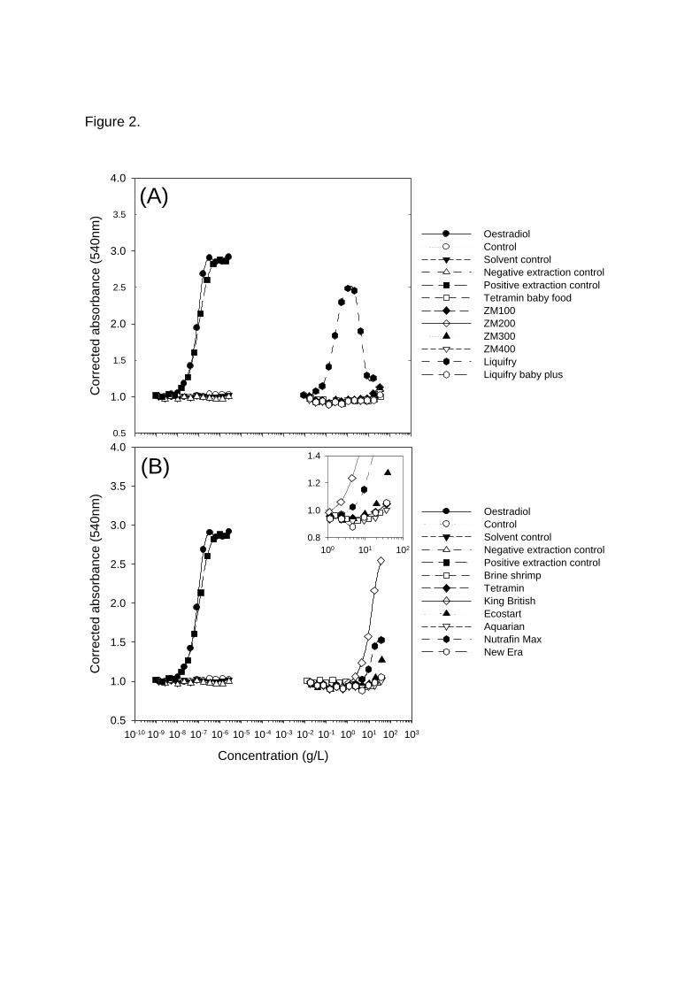
Figure 2.
Concentration (g/L)
10-10 10-9 10-8 10-7 10-6 10-5 10-4 10-3 10-2 10-1 100 101 102 103
Corr
ecte
d a
bsorb
ance (
540
nm
)
0.5
1.0
1.5
2.0
2.5
3.0
3.5
4.0
Oestradiol
Control
Solvent control
Negative extraction control
Positive extraction control
Brine shrimp
Tetramin
King British
Ecostart
Aquarian
Nutrafin Max
New Era
(B)
Corr
ecte
d a
bsorb
ance (
540
nm
)
0.5
1.5
2.5
3.5
1.0
2.0
3.0
4.0
Oestradiol
Control
Solvent control
Negative extraction control
Positive extraction control
Tetramin baby food
ZM100
ZM200
ZM300
ZM400
Liquifry
Liquifry baby plus
(A)
100 101 102
0.8
1.0
1.2
1.4

Figure 3.
Corr
ecte
d a
bsorb
ance (
540
nm
)
0.5
1.0
1.5
2.0
2.5
3.0
3.5
Oestradiol
Control
Solvent control
negative extraction control
positive extraction control
Tetramin batch 1
Tetramin batch 2
Tetramin batch 3
Tetramin batch 4
Concentration (g/L)
10-10 10-9 10-8 10-7 10-6 10-5 10-4 10-3 10-2 10-1 100 101 102 103
Corr
ecte
d a
bsorb
ance (
540
nm
)
0.5
1.0
1.5
2.0
2.5
3.0
3.5
Oestradiol
Control
Solvent control
negative extraction control
positive extraction control
King British batch 1
King British batch 2
King British batch 3
King British batch 4
(A)
(B)

Figure 4.
Pla
sm
a V
TG
(ng/m
l)
10-1
100
101
102
103
104
105
106
107
*****
only King British flake food
Tetramin for3 months
Tetramin for 6 months
only Tetramin flake food
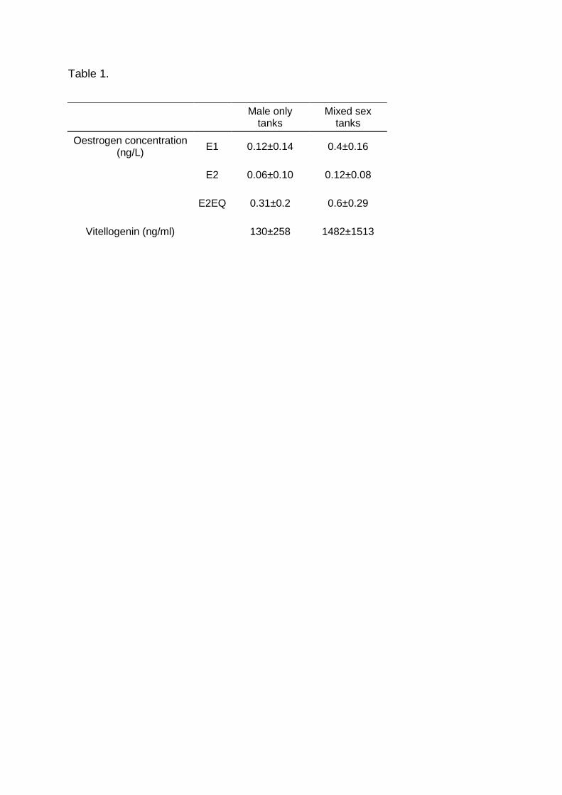
Table 1.
Male only tanks
Mixed sex tanks
Oestrogen concentration (ng/L)
E1 0.12±0.14 0.4±0.16
E2 0.06±0.10 0.12±0.08
E2EQ 0.31±0.2 0.6±0.29
Vitellogenin (ng/ml)
130±258 1482±1513