Vitamins UNIT V: Integration of Metabolism. I. Overview Vitamins are chemically unrelated organic...
102
Vitamins UNIT V: Integration of Metabolism
-
date post
20-Dec-2015 -
Category
Documents
-
view
215 -
download
1
Transcript of Vitamins UNIT V: Integration of Metabolism. I. Overview Vitamins are chemically unrelated organic...

Vitamins
UNIT V:
Integration of Metabolism

I. Overview
• Vitamins are chemically unrelated organic compounds that cannot be synthesized in adequate quantities by humans and, therefore, must be supplied by the diet.
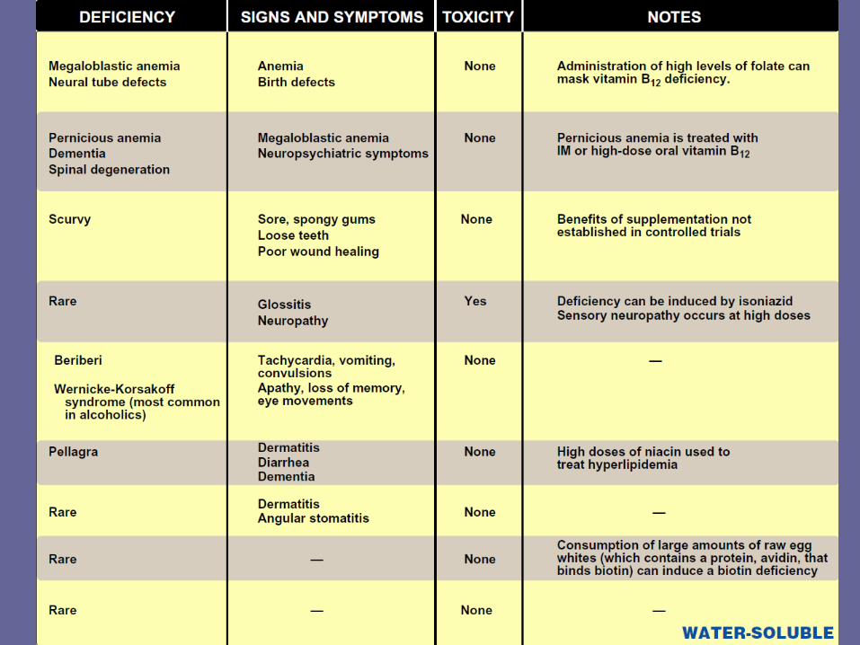
• Nine vitamins (folic acid, cobalamin, ascorbic acid, pyridoxine, thiamine, niacin, riboflavin, biotin, and pantothenic acid) are classified as water-soluble, whereas four vitamins (vitamins A, D, K, and E) are termed fat-soluble (Figure 28.1).
• Vitamins are required to perform specific cellular functions, for example, many of the water-soluble vitamins are precursors of coenzymes for the enzymes of intermediary metabolism.

• In contrast to the water-soluble vitamins, only one fat soluble vitamin (vitamin K) has a coenzyme function. These vitamins are released, absorbed, and transported with the fat of the diet.
• They are not readily excreted in the urine, and significant quantities are stored in the liver and adipose tissue.
• In fact, consumption of vitamins A and D in excess of the recommended dietary allowances can lead to accumulation of toxic quantities of these compounds.
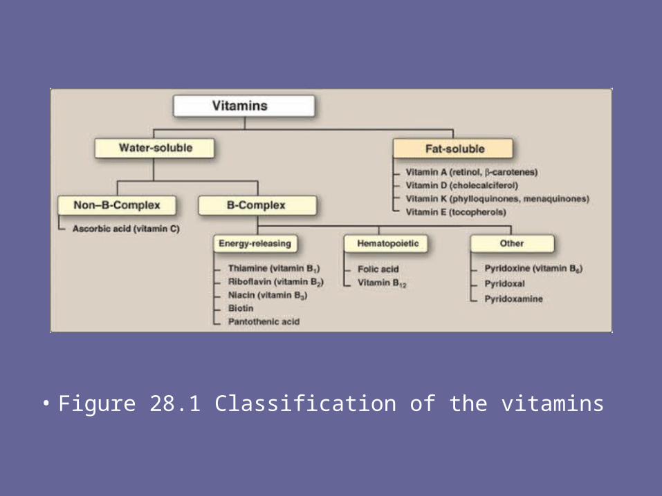
• Figure 28.1 Classification of the vitamins
II. Folic Acid• Folic acid (or folate), which plays a key role in
one-carbon metabolism, is essential for the biosynthesis of several compounds.
• Folic acid deficiency is probably the most common vitamin deficiency in the United States, particularly among pregnant women and alcoholics.
A. Function of folic acid• Tetrahydrofolate receives one-carbon fragments
from donors such as serine, glycine, and histidine and transfers them to intermediates in the synthesis of amino acids, purines, and thymidine monophosphate (TMP).
B. Nutritional anemias• Anemia is a condition in which the blood
has a lower than normal concentration of hemoglobin, which results in a reduced ability to transport oxygen.
• Nutritional anemias—those caused by inadequate intake of one or more essential nutrients—can be classified according to the size of the red blood cells or mean corpuscular volume observed in the individual (Figure 28.2).
• Microcytic anemia, caused by lack of iron, is the most common form of nutritional anemia.
• The second major category of nutritional anemia, macrocytic, results from a deficiency in folic acid or vitamin B12. [Note: These macrocytic anemias are commonly called megaloblastic because a deficiency of folic acid or vitamin B12 causes accumulation of large, immature red cell precursors, known as megaloblasts, in the bone marrow and the blood.]

• Figure 28.2 Classification of nutritional anemias by cell size. The normal mean corpuscular volume (MCV) for people older than age 18 is between 80 and 100 µm3.
1. Folate and anemia: • Inadequate serum levels of folate can be caused by:
– increased demand (for example, pregnancy and lactation), – poor absorption caused by pathology of the small intestine,
alcoholism, or– treatment with drugs that are dihydrofolate reductase inhibitors,
for example, methotrexate (Figure 28.3). – A folate-free diet can cause a deficiency within a few weeks.
• A primary result of folic acid deficiency is megaloblastic anemia (Figure 28.4), caused by diminished synthesis of purines and TMP, which leads to an inability of cells to make DNA and, therefore, they cannot divide. [Note: It is important to evaluate the cause of the megaloblastic anemia prior to instituting therapy, because vitamin B12 deficiency indirectly causes symptoms of this disorder.]

• Figure 28.3 Inhibition of tetrahydrofolate synthesis by sulfonamides and trimethoprim.
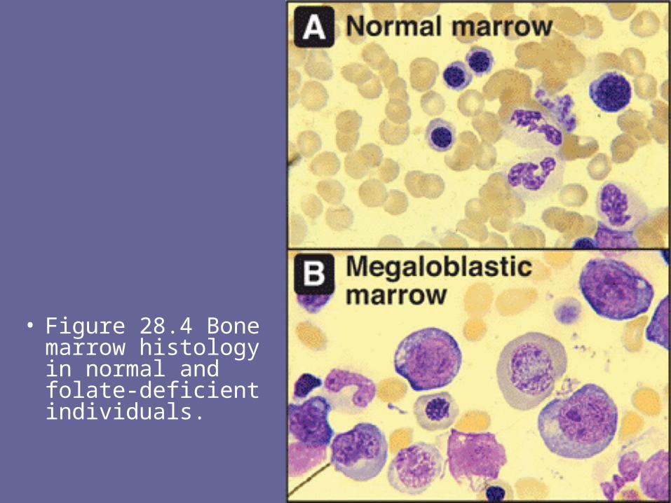
• Figure 28.4 Bone marrow histology in normal and folate-deficient individuals.

2. Folate and neural tube defects in the fetus: • Spina bifida and anencephaly, the most
common neural tube defects, affect approximately 4,000 pregnancies in the United State annually.
• Folic acid supplementation before conception and during the first trimester has been shown to significantly reduce the defects.
• Therefore, all women of childbearing age are advised to consume 0.4 mg/day of folic acid to reduce the risk of having a pregnancy affected by neural tube defects.
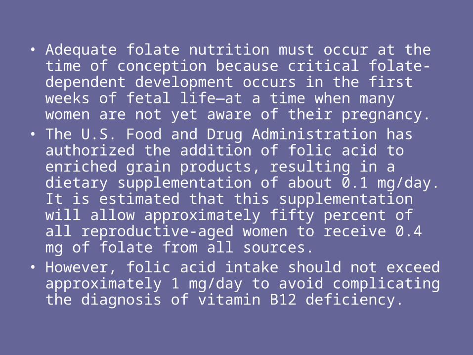
• Adequate folate nutrition must occur at the time of conception because critical folate-dependent development occurs in the first weeks of fetal life—at a time when many women are not yet aware of their pregnancy.
• The U.S. Food and Drug Administration has authorized the addition of folic acid to enriched grain products, resulting in a dietary supplementation of about 0.1 mg/day. It is estimated that this supplementation will allow approximately fifty percent of all reproductive-aged women to receive 0.4 mg of folate from all sources.
• However, folic acid intake should not exceed approximately 1 mg/day to avoid complicating the diagnosis of vitamin B12 deficiency.
III. Cobalamin (Vitamin B12)• Vitamin B12 is required in humans for two
essential enzymatic reactions: the remethylation of homocysteine to methionine and the isomerization of methylmalonyl coenzyme A (CoA) that is produced during the degradation of some amino acids, and fatty acids with odd numbers of carbon atoms (Figure 28.5).
• When the vitamin is deficient, abnormal fatty acids accumulate and become incorporated into cell membranes, including those of the nervous system. This may account for some of the neurologic manifestations of vitamin B12 deficiency.
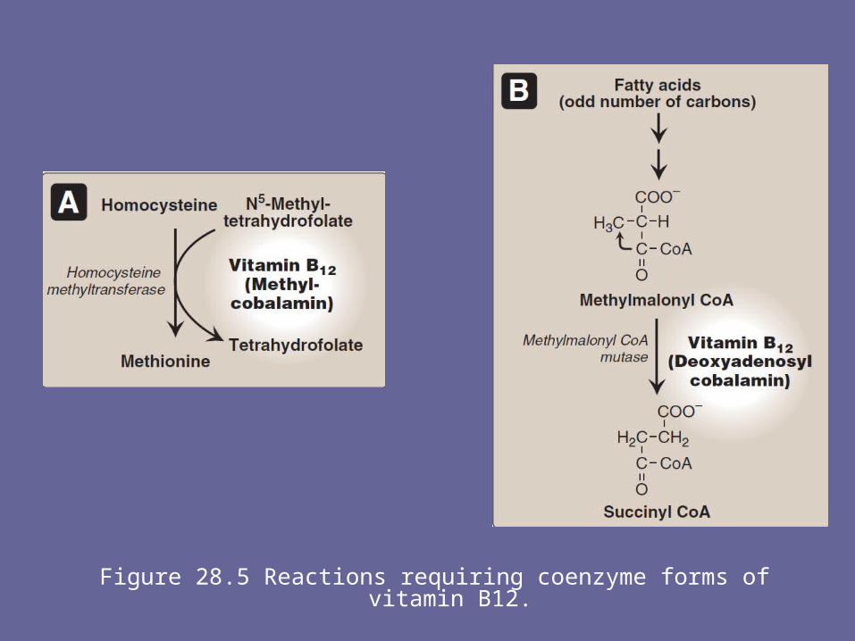
Figure 28.5 Reactions requiring coenzyme forms of vitamin B12.

A. Structure of cobalamin and its coenzyme forms• Cobalamin contains a corrin ring system that differs from
the porphyrins in that two of the pyrrole rings are linked directly rather than through a methene bridge.
• Cobalt is held in the center of the corrin ring by four coordination bonds from the nitrogens of the pyrrole groups.
• The remaining coordination bonds of the cobalt are with the nitrogen of 5,6-dimethylbenzimidazole and with cyanide in commercial preparations of the vitamin in the form of cyanocobalamin (Figure 28.6).
• The coenzyme forms of cobalamin are 5′-deoxyadenosylcobalamin, in which cyanide is replaced with 5′-deoxyadenosine (forming an unusual carbon–cobalt bond), and methylcobalamin, in which cyanide is replaced by a methyl group (see Figure 28.6).
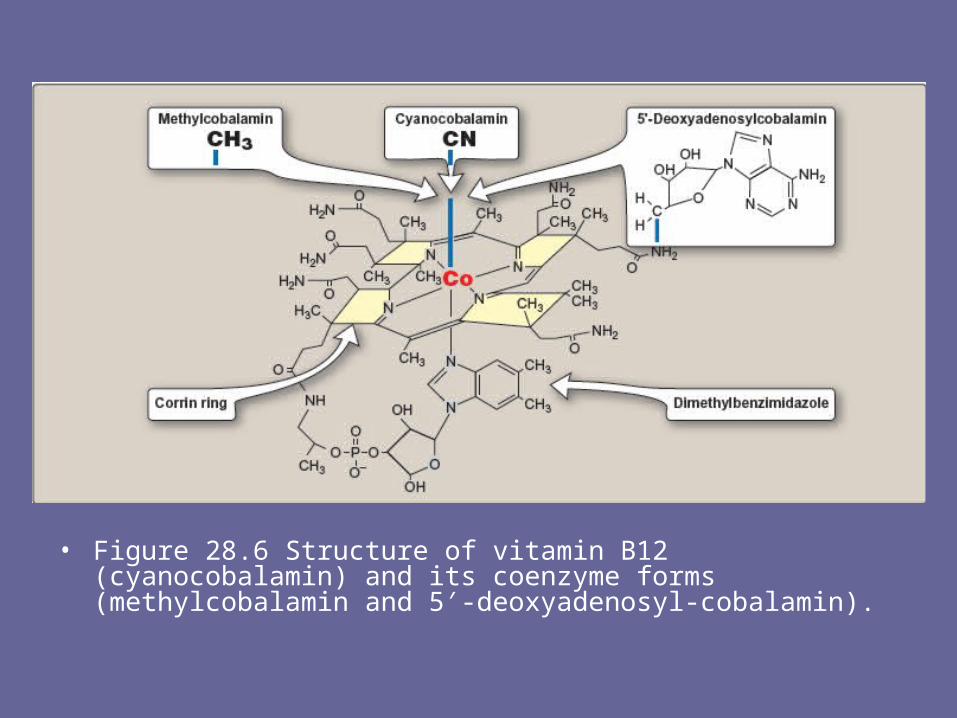
• Figure 28.6 Structure of vitamin B12 (cyanocobalamin) and its coenzyme forms (methylcobalamin and 5′-deoxyadenosyl-cobalamin).
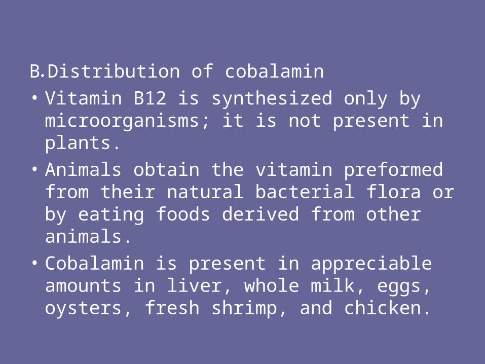
B. Distribution of cobalamin
• Vitamin B12 is synthesized only by microorganisms; it is not present in plants.
• Animals obtain the vitamin preformed from their natural bacterial flora or by eating foods derived from other animals.
• Cobalamin is present in appreciable amounts in liver, whole milk, eggs, oysters, fresh shrimp, and chicken.

C. Folate trap hypothesis• The effects of cobalamin deficiency are most
pronounced in rapidly dividing cells, such as the erythropoietic tissue of bone marrow and the mucosal cells of the intestine.
• Such tissues need both the N5-N10-methylene and N10-formyl forms of tetrahydrofolate for the synthesis of nucleotides required for DNA replication.
• However, in vitamin B12 deficiency, the utilization of the N5-methyl form of tetrahydrofolate is impaired. Because the methylated form cannot be converted directly to other forms of tetrahydrofolate, folate is trapped in the N5-methyl form, which accumulates.
• The levels of the other forms decrease. Thus, cobalamin deficiency is hypothesized to lead to a deficiency of the tetrahydrofolate forms needed in purine and TMP synthesis, resulting in the symptoms of megaloblastic anemia.

D. Clinical indications for vitamin B12• In contrast to other water-soluble vitamins,
significant amounts (4–5 mg) of vitamin B12 are stored in the body.
• As a result, it may take several years for the clinical symptoms of B12 deficiency to develop in individuals who have had a partial or total gastrectomy (who, therefore, become intrinsic factor-deficient, and can no longer absorb the vitamin.

1. Pernicious anemia: • Vitamin B12 deficiency is rarely a result of an absence
of the vitamin in the diet. • It is much more common to find deficiencies in patients
who fail to absorb the vitamin from the intestine, resulting in pernicious anemia.
• The disease is most commonly a result of an autoimmune destruction of the gastric parietal cells that are responsible for the synthesis of a glycoprotein called intrinsic factor.
• Normally, vitamin B12 obtained from the diet binds to intrinsic factor in the intestine (Figure 28.7). The cobalamin–intrinsic factor complex travels through the gut and eventually binds to specific receptors on the surface of mucosal cells of the ileum.

• The bound cobalamin is transported into the mucosal cell and, subsequently, into the general circulation, where it is carried by B12-binding proteins.
• Lack of intrinsic factor prevents the absorption of vitamin B12, resulting in pernicious anemia.
• Patients with cobalamin deficiency are usually anemic, but later in the development of the disease they show neuropsychiatric symptoms.
• However, central nervous system (CNS) symptoms may occur in the absence of anemia.
• The CNS effects are irreversible and occur by mechanisms that appear to be different from those described for megaloblastic anemia.
• The disease is treated by giving high-dose B12 orally, or intramuscular injection of cyanocobalamin. Therapy must be continued throughout the lives of patients with pernicious anemia.
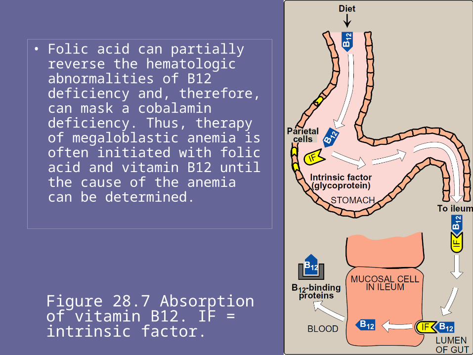
Figure 28.7 Absorption of vitamin B12. IF = intrinsic factor.
• Folic acid can partially reverse the hematologic abnormalities of B12 deficiency and, therefore, can mask a cobalamin deficiency. Thus, therapy of megaloblastic anemia is often initiated with folic acid and vitamin B12 until the cause of the anemia can be determined.
IV. Ascorbic Acid (Vitamin C)• The active form of vitamin C is ascorbic acid.• The main function of ascorbate is as a reducing
agent in several different reactions. • Vitamin C has a well-documented role as a
coenzyme in hydroxylation reactions, for example, hydroxylation of prolyl and lysyl residues of collagen.
• Vitamin C is, therefore, required for the maintenance of normal connective tissue, as well as for wound healing.
• Vitamin C also facilitates the absorption of dietary iron from the intestine.
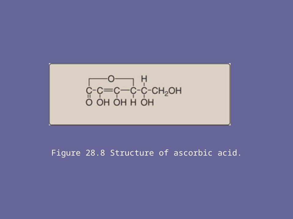
Figure 28.8 Structure of ascorbic acid.
A. Deficiency of ascorbic acid• A deficiency of ascorbic acid results in scurvy, a disease
characterized by sore and spongy gums, loose teeth, fragile blood vessels, swollen joints, and anemia.
• Many of the deficiency symptoms can be explained by a deficiency in the hydroxylation of collagen, resulting in defective connective tissue.
B. Prevention of chronic disease• Vitamin C is one of a group of nutrients that includes
vitamin E and β-carotene, which are known as antioxidants.
• Consumption of diets rich in these compounds is associated with a decreased incidence of some chronic diseases, such as coronary heart disease and certain cancers.
• However, clinical trials involving supplementation with the isolated antioxidants have failed to determine any convincing beneficial effects.

• Figure 28.9 Hemorrhage and swollen gums of a patient with scurvy.
V. Pyridoxine (Vitamin B6)• Vitamin B6 is a collective term for pyridoxine, pyridoxal,
and pyridoxamine, all derivatives of pyridine. • They differ only in the nature of the functional group
attached to the ring. • Pyridoxine occurs primarily in plants, whereas pyridoxal
and pyridoxamine are found in foods obtained from animals.
• All three compounds can serve as precursors of the biologically active coenzyme, pyridoxal phosphate.
• Pyridoxal phosphate functions as a coenzyme for a large number of enzymes, particularly those that catalyze reactions involving amino acids.

Reaction type Example
Transamination Oxaloacetate + glutamate ⇔ aspartate + α-ketoglutarate
Deamination Serine → pyruvate + NH3
Decarboxylation Histidine → histamine + CO2
Condensation Glycine + succinyl CoA → δ-aminolevulinic acid

• Figure 28.10 Structures of vitamin B6 and and the antituberculosis drug isoniazid.
A. Clinical indications for pyridoxine:• Isoniazid (isonicotinic acid hydrazide), a drug
frequently used to treat tuberculosis, can induce a vitamin B6 deficiency by forming an inactive derivative with pyridoxal phosphate.
• Dietary supplementation with B6 is, thus, an adjunct to isoniazid treatment.
• Otherwise, dietary deficiencies in pyridoxine are rare but have been observed in newborn infants fed formulas low in B6, in women taking oral contraceptives, and in alcoholics.
B. Toxicity of pyridoxine
• Neurologic symptoms have been observed at intakes of greater than 2 g/day.
• Substantial improvement, but not complete recovery, occurs when the vitamin is discontinued.

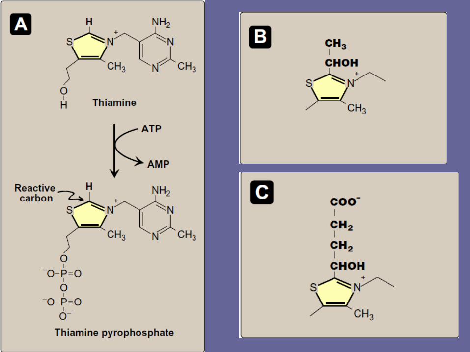
VI. Thiamine (Vitamin B1)• Thiamine pyrophosphate is the biologically
active form of the vitamin, formed by the transfer of a pyrophosphate group from adenosine triphosphate (ATP) to thiamine (Figure 28.11).
• Thiamine pyrophosphate serves as a coenzyme in the formation or degradation of α-ketols by transketolase (Figure 28.12A), and in the oxidative decarboxylation of α-keto acids (Figure 28.12B).
A. Clinical indications for thiamine• The oxidative decarboxylation of pyruvate and α-
ketoglutarate, which plays a key role in energy metabolism of most cells, is particularly important in tissues of the nervous system.
• In thiamine deficiency, the activity of these two dehydrogenase reactions is decreased, resulting in a decreased production of ATP and, thus, impaired cellular function.
• [Note: Thiamine deficiency is diagnosed by an increase in erythrocyte transketolase activity observed on addition of thiamine pyrophosphate.]

• Figure 28.12 Reactions that use thiamine pyro-phosphate (TPP) as coenzyme. A. Transketolase. B. Pyruvate dehydrogenase and α-ketoglutarate dehydrogenase. Note that TPP is also used by branched-chain α-keto acid dehydrogenase.

1. Beriberi: • This is a severe thiamine-deficiency syndrome
found in areas where polished rice is the major component of the diet.
• Signs of infantile beriberi include tachycardia, vomiting, convulsions, and, if not treated, death.
• The deficiency syndrome can have a rapid onset in nursing infants whose mothers are deficient in thiamine.
• Adult beriberi is characterized by dry skin, irritability, disorderly thinking, and progressive paralysis.
2. Wernicke-Korsakoff syndrome: • In the United States, thiamine deficiency, which is seen
primarily in association with chronic alcoholism, is due to dietary insufficiency or impaired intestinal absorption of the vitamin.
• Some alcoholics develop Wernicke-Korsakoff syndrome—a thiamine deficiency state characterized by apathy, loss of memory, and a rhythmical to-and-fro motion of the eyeballs (nystagmus).
• The neurologic consequences of Wernicke's syndrome are treatable with thiamine supplementation.
• Intravenous thiamine administration typically is initiated at 50 mg/day until the same dose is tolerated orally.

VII. Niacin• Niacin, or nicotinic acid, is a substituted pyridine
derivative. • The biologically active coenzyme forms are
nicotinamide adenine dinucleotide (NAD+) and its phosphorylated derivative, nicotinamide adenine dinucleotide phosphate (NADP+, Figure 28.13).
• Nicotinamide, a derivative of nicotinic acid that contains an amide instead of a carboxyl group, also occurs in the diet.
• Nicotinamide is readily deaminated in the body and, therefore, is nutritionally equivalent to nicotinic acid.

• Figure 28.13 Structure and biosynthesis of NAD+ and NADP+. Note that a metabolite of tryptophan (quinolinate) can also be used in the synthesis of NAD+.

• NAD+ and NADP+ serve as coenzymes in oxidation-reduction reactions in which the coenzyme undergoes reduction of the pyridine ring by accepting a hydride ion (hydrogen atom plus one electron, Figure 28.14).
• The reduced forms of NAD+ and NADP+ are NADH and NADPH, respectively.

• Figure 28.14 Reduction of NAD+ to NADH.

A. Distribution of niacin• Niacin is found in unrefined and enriched grains and
cereal, milk, and lean meats, especially liver.
B. Clinical indications for niacin1. Deficiency of niacin: • A deficiency of niacin causes pellagra, a disease
involving the skin, gastrointestinal tract, and CNS. • The symptoms of pellagra progress through the three
Ds: dermatitis, diarrhea, dementia—and, if untreated, death.
2. Treatment of hyperlipidemia: • Niacin (at doses of 1.5 g/day or 100 times the
Recommended Dietary Allowance or RDA) strongly inhibits lipolysis in adipose tissue—the primary producer of circulating free fatty acids.
• The liver normally uses these circulating fatty acids as a major precursor for triacylglycerol synthesis.
• Thus, niacin causes a decrease in liver triacylglycerol synthesis, which is required for very-low-density lipoprotein (VLDL) production.
• Low-density lipoprotein (LDL, the cholesterol-rich lipoprotein) is derived from VLDL in the plasma. Thus, both plasma triacylglycerol (in VLDL) and cholesterol (in VLDL and LDL) are lowered.
• Therefore, niacin is particularly useful in the treatment of Type IIb hyperlipoproteinemia, in which both VLDL and LDL are elevated.
VIII. Riboflavin (Vitamin B2)• The two biologically active forms are flavin
mononucleotide (FMN) and flavin adenine dinucleotide (FAD), formed by the transfer of an adenosine monophosphate moiety from ATP to FMN (Figure 28.15).
• FMN and FAD are each capable of reversibly accepting two hydrogen atoms, forming FMNH2 or FADH2.
• FMN and FAD are bound tightly—sometimes covalently—to flavoenzymes that catalyze the oxidation or reduction of a substrate.
• Riboflavin deficiency is not associated with a major human disease, although it frequently accompanies other vitamin deficiencies.
• Deficiency symptoms include dermatitis, cheilosis (fissuring at the corners of the mouth), and glossitis (the tongue appearing smooth and purplish).

• Figure 28.15 Structure and biosynthesis of flavin mononucleotide and flavin adenine dinucleotide.
IX. Biotin• Biotin is a coenzyme in carboxylation reactions,
in which it serves as a carrier of activated carbon dioxide (see Figure 10.3, p. 119, for the mechanism of biotin-dependent carboxylations).
• Biotin is covalently bound to the ε-amino groups of lysine residues of biotin-dependent enzymes (Figure 28.16).
• Biotin deficiency does not occur naturally because the vitamin is widely distributed in food.
• Also, a large percentage of the biotin requirement in humans is supplied by intestinal bacteria.

• Figure 28.16 A. Structure of biotin. B. Biotin covalently bound to a lysyl residue of a biotin-dependent enzyme.
• However, the addition of raw egg white to the diet as a source of protein induces symptoms of biotin deficiency, namely, dermatitis, glossitis, loss of appetite, and nausea.
• Raw egg white contains a glycoprotein, avidin, which tightly binds biotin and prevents its absorption from the intestine.
• However, with a normal diet, it has been estimated that 20 eggs/day would be required to induce a deficiency syndrome.
• Thus, inclusion of an occasional raw egg in the diet does not lead to biotin deficiency. However, eating raw eggs is generally not recommended due to the possibility of salmonella infection.
• Multiple carboxylase deficiency results from a defect in the ability to link biotin to carboxylases or to remove it from carboxylases during their degradation. Treatment is biotin supplementation.

X. Pantothenic Acid• Pantothenic acid is a component of CoA, which functions
in the transfer of acyl groups (Figure 28.17). • Coenzyme A contains a thiol group that carries acyl
compounds as activated thiol esters.• Examples of such structures are succinyl CoA, fatty acyl
CoA, and acetyl CoA. • Pantothenic acid is also a component of fatty acid
synthase (see p. 184). • Eggs, liver, and yeast are the most important sources of
pantothenic acid, although the vitamin is widely distributed.
• Pantothenic acid deficiency is not well characterized in humans, and no RDA has been established.

• Figure 28.17 Structure of coenzyme A

XI. Vitamin A• The retinoids, a family of molecules that
are related to retinol (vitamin A), are essential for vision, reproduction, growth, and maintenance of epithelial tissues.
• Retinoic acid, derived from oxidation of dietary retinol, mediates most of the actions of the retinoids, except for vision, which depends on retinal, the aldehyde derivative of retinol.

A. Structure of vitamin A• Vitamin A is often used as a collective term for
several related biologically active molecules (Figure 28.18).
• The term retinoids includes both natural and synthetic forms of vitamin A that may or may not show vitamin A activity.
1.Retinol: A primary alcohol containing a β-ionone ring with an unsaturated side chain, retinol is found in animal tissues as a retinyl ester with long-chain fatty acids.
2.Retinal: This is the aldehyde derived from the oxidation of retinol. Retinal and retinol can readily be interconverted.

3. Retinoic acid: This is the acid derived from the oxidation of retinal. Retinoic acid cannot be reduced in the body, and, therefore, cannot give rise to either retinal or retinol.
4. β-Carotene: Plant foods contain β-carotene, which can be oxidatively cleaved in the intestine to yield two molecules of retinal.
• In humans, the conversion is inefficient, and the vitamin A activity of β-carotene is only about one twelfth that of retinol.
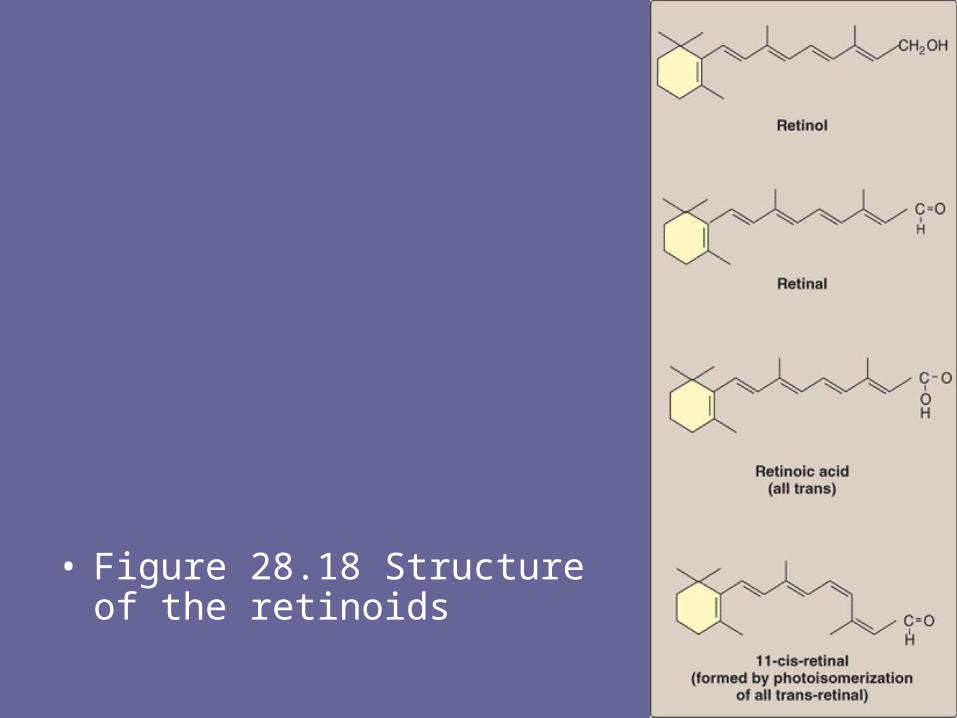
• Figure 28.18 Structure of the retinoids
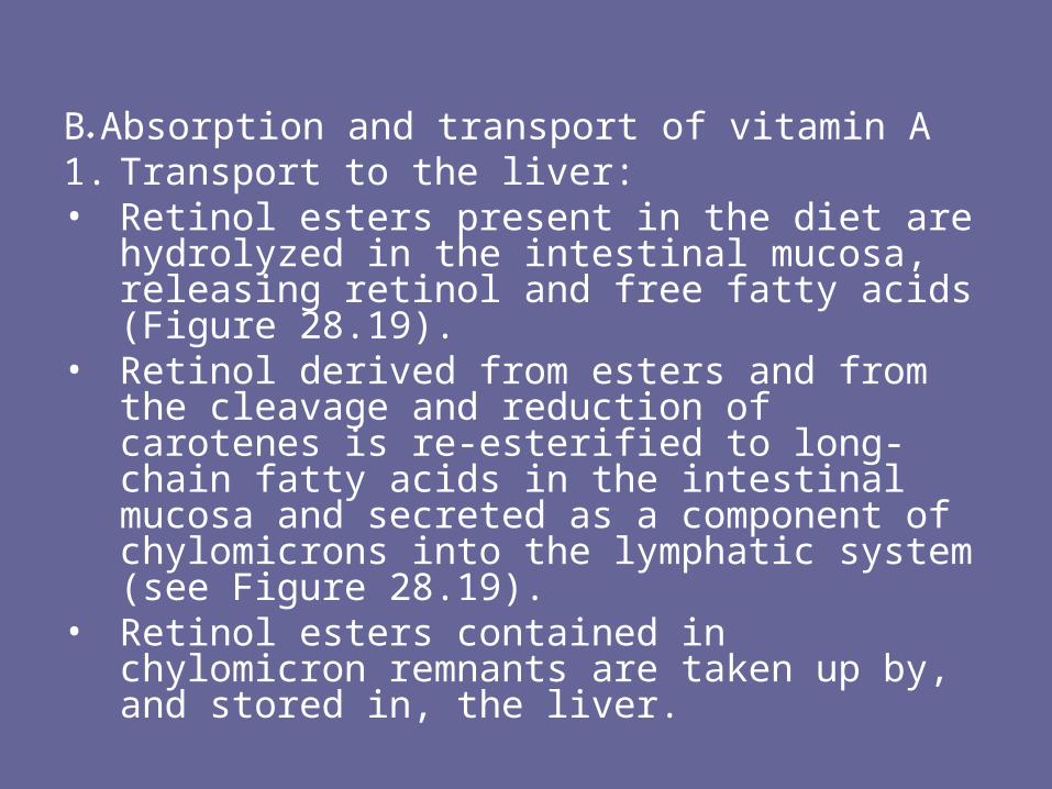
B. Absorption and transport of vitamin A1. Transport to the liver: • Retinol esters present in the diet are
hydrolyzed in the intestinal mucosa, releasing retinol and free fatty acids (Figure 28.19).
• Retinol derived from esters and from the cleavage and reduction of carotenes is re-esterified to long-chain fatty acids in the intestinal mucosa and secreted as a component of chylomicrons into the lymphatic system (see Figure 28.19).
• Retinol esters contained in chylomicron remnants are taken up by, and stored in, the liver.
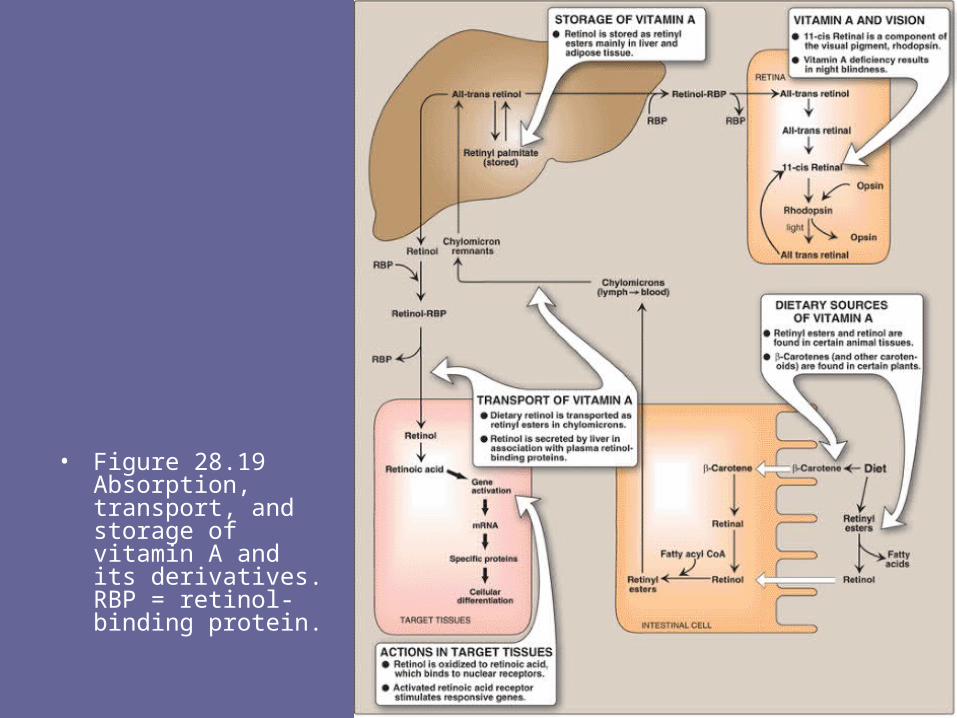
• Figure 28.19 Absorption, transport, and storage of vitamin A and its derivatives. RBP = retinol-binding protein.
2. Release from the liver: • When needed, retinol is released from the liver
and transported to extrahepatic tissues by the plasma retinol-binding protein (RBP).
• The retinol–RBP complex attaches to specific receptors on the surface of the cells of peripheral tissues, permitting retinol to enter.
• Many tissues contain a cellular retinol-binding protein that carries retinol to sites in the nucleus where the vitamin acts in a manner analogous to that of steroid hormones.

C. Mechanism of action of vitamin A• Retinoic acid binds with high affinity to specific receptor
proteins present in the nucleus of target tissues, such as epithelial cells (Figure 28.20).
• The activated retinoic acid–receptor complex interacts with nuclear chromatin to stimulate retinoid-specific RNA synthesis, resulting in the production of specific proteins that mediate several physiologic functions.
• For example, retinoids control the expression of the keratin gene in most epithelial tissues of the body.
• The specific retinoic acid–receptor proteins are part of the superfamily of transcriptional regulators that includes the steroid and thyroid hormones and 1,25-dihydroxycholecalciferol, all of which function in a similar way.

• Figure 28.20 Action of retinoids Note: Retinoic acid-receptor complex is a dimer, but is shown as monomer for simplicity. [RBP = retinol-binding protein.]

D. Functions of vitamin A1. Visual cycle: • Vitamin A is a component of the visual pigments
of rod and cone cells. • Rhodopsin, the visual pigment of the rod cells in
the retina, consists of 11-cis retinal specifically bound to the protein opsin.
• When rhodopsin is exposed to light, a series of photochemical isomerizations occurs, which results in the bleaching of the visual pigment and release of all trans retinal and opsin.
• This process triggers a nerve impulse that is transmitted by the optic nerve to the brain.

• Regeneration of rhodopsin requires isomerization of all trans retinal back to 11-cis retinal.
• Trans retinal, after being released from rhodopsin, is isomerized to 11-cis retinal, which spontaneously combines with opsin to form rhodopsin, thus completing the cycle. Similar reactions are responsible for color vision in the cone cells.
2.Growth: Vitamin A deficiency results in a decreased growth rate in children. Bone development is also slowed.

3. Reproduction: • Retinol and retinal are essential for normal
reproduction, supporting spermatogenesis in the male and preventing fetal resorption in the female.
• Retinoic acid is inactive in maintaining reproduction and in the visual cycle, but promotes growth and differentiation of epithelial cells; thus, animals given vitamin A only as retinoic acid from birth are blind and sterile.
4. Maintenance of epithelial cells: • Vitamin A is essential for normal differentiation
of epithelial tissues and mucus secretion.

E. Distribution of vitamin A• Liver, kidney, cream, butter, and egg yolk are good
sources of preformed vitamin A. Yellow and dark green vegetables and fruits are good dietary sources of the carotenes, which serve as precursors of vitamin A.
F. Requirement for vitamin A• The RDA for adults is 1,000 retinol activity equivalents
(RAE) for males and 800 RAE for females. In comparison, 1 RAE = 1 mg of retinol, 12 mg of β-carotene, or 24 mg of other carotenoids.
G. Clinical indications• Although chemically related, retinoic acid and retinol
have distinctly different therapeutic applications. Retinol and its precursor are used as dietary supplements, whereas various forms of retinoic acid are useful in dermatology.

1. Dietary deficiency: • Vitamin A, administered as retinol or retinyl
esters, is used to treat patients who are deficient in the vitamin (Figure 28.21).
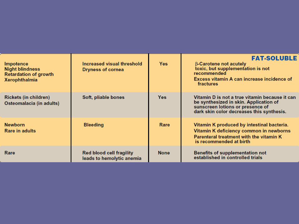
• Night blindness is one of the earliest signs of vitamin A deficiency. The visual threshold is increased, making it difficult to see in dim light.
• Prolonged deficiency leads to an irreversible loss in the number of visual cells.

• Severe vitamin A deficiency leads to xerophthalmia, a pathologic dryness of the conjunctiva and cornea.
• If untreated, xerophthalmia results in corneal ulceration and, ultimately, in blindness because of the formation of opaque scar tissue.
• The condition is most frequently seen in children in developing tropical countries.
• Over 500,000 children worldwide are blinded each year by xerophthalmia caused by insufficient vitamin A in the diet.
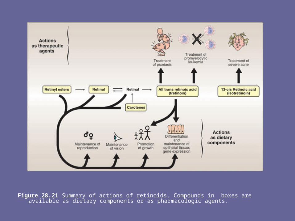
Figure 28.21 Summary of actions of retinoids. Compounds in boxes are available as dietary components or as pharmacologic agents.

2. Acne and psoriasis: • Dermatologic problems such as acne and psoriasis are
effectively treated with retinoic acid or its derivatives (see Figure 28.21).
• Mild cases of acne, Darier disease (keratosis follicularis), and skin aging are treated with topical application of tretinoin (all-trans retinoic acid), as well as benzoyl peroxide and antibiotics.
• [Note: Tretinoin is too toxic for systemic administration and is confined to topical application.]
• In patients with severe recalcitrant cystic acne unresponsive to conventional therapies, the drug of choice is isotretinoin (13-cis retinoic acid) administered orally.
H. Toxicity of retinoids1. Vitamin A: • Excessive intake of vitamin A produces a toxic
syndrome called hypervitaminosis A. • Amounts exceeding 7.5 mg/day of retinol should be
avoided. • Early signs of chronic hypervitaminosis A are reflected
in the skin, which becomes dry and pruritic, the liver, which becomes enlarged and can become cirrhotic, and in the nervous system, where a rise in intracranial pressure may mimic the symptoms of a brain tumor.
• Pregnant women particularly should not ingest excessive quantities of vitamin A because of its potential for causing congenital malformations in the developing fetus.
2. Isotretinoin: • The drug is teratogenic and absolutely
contraindicated in women with childbearing potential unless they have severe, disfiguring cystic acne that is unresponsive to standard therapies.
• Pregnancy must be excluded before initiation of treatment, and adequate birth control must be used.
• Prolonged treatment with isotretinoin leads to hyperlipidemia and an increase in the LDL/HDL ratio, providing some concern for an increased risk of cardiovascular disease.
XII. Vitamin D
• The D vitamins are a group of sterols that have a hormone-like function.
• The active molecule, 1,25-dihydroxycholecalciferol (1,25-diOH-D3), binds to intracellular receptor proteins.
• The 1,25-diOH-D3–receptor complex interacts with DNA in the nucleus of target cells in a manner similar to that of vitamin A (see Figure 28.20), and either selectively stimulates gene expression or specifically represses gene transcription.
• The most prominent actions of 1,25-diOH-D3 are to regulate the plasma levels of calcium and phosphorus.

A. Distribution of vitamin D1. Diet: Ergocalciferol (vitamin D2), found in plants, and
cholecalciferol (vitamin D3), found in animal tissues, are sources of preformed vitamin D activity (Figure 28.22).
• Ergocalciferol and cholecalciferol differ chemically only in the presence of an additional double bond and methyl group in the plant sterol.
2. Endogenous vitamin precursor: 7-Dehydrocholesterol, an intermediate in cholesterol synthesis, is converted to cholecalciferol in the dermis and epidermis of humans exposed to sunlight.
• Preformed vitamin D is a dietary requirement only in individuals with limited exposure to sunlight.
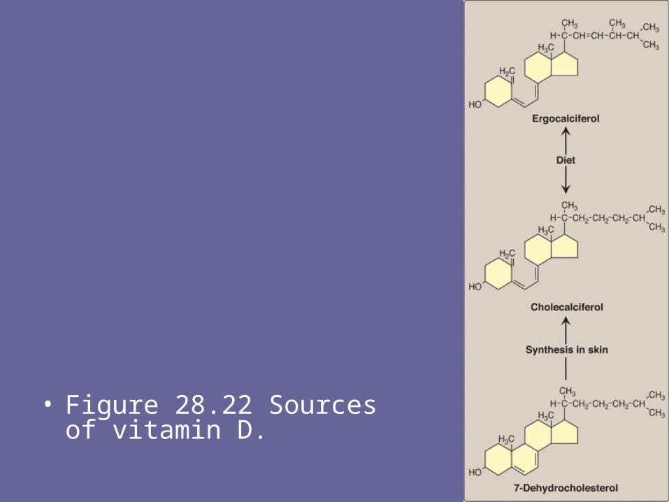
• Figure 28.22 Sources of vitamin D.

B. Metabolism of vitamin D1. Formation of 1,25-diOH-D3: Vitamins D2 and D3 are
not biologically active, but are converted in vivo to the active form of the D vitamin by two sequential hydroxylation reactions (Figure 28.23).
• The first hydroxylation occurs at the 25-position, and is catalyzed by a specific hydroxylase in the liver.
• The product of the reaction, 25-hydroxycholecalciferol (25-OH-D3), is the predominant form of vitamin D in the plasma and the major storage form of the vitamin.
• 25-OH-D3 is further hydroxylated at the one position by a specific 25-hydroxycholecalciferol 1-hydroxylase found primarily in the kidney, resulting in the formation of 1,25-diOH-D3. [Note: This hydroxylase, as well as the liver 25-hydroxylase, employ cytochrome P450, molecular oxygen, and NADPH.]
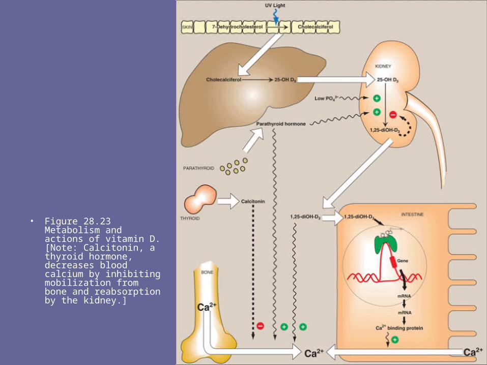
• Figure 28.23 Metabolism and actions of vitamin D. [Note: Calcitonin, a thyroid hormone, decreases blood calcium by inhibiting mobilization from bone and reabsorption by the kidney.]

2. Regulation of 25-hydroxycholecalciferol 1-hydroxylase: • 1,25-diOH-D3 is the most potent vitamin D metabolite.• Its formation is tightly regulated by the level of plasma
phosphate and calcium ions (Figure 28.24). • 25-Hydroxycholecalciferol 1-hydroxylase activity is
increased directly by low plasma phosphate or indirectly by low plasma calcium, which triggers the release of parathyroid hormone (PTH).
• Hypocalcemia caused by insufficient dietary calcium thus results in elevated levels of plasma 1,25-diOH-D3.
• 1-Hydroxylase activity is also decreased by excess 1,25-diOH-D3, the product of the reaction.
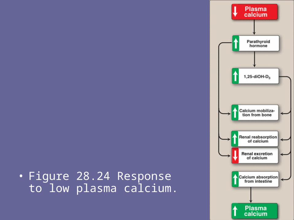
• Figure 28.24 Response to low plasma calcium.

C. Function of vitamin D• The overall function of 1,25-diOH-D3 is to
maintain adequate plasma levels of calcium. It performs this function by: 1) increasing uptake of calcium by the intestine, 2) minimizing loss of calcium by the kidney, and 3) stimulating resorption of bone when necessary (see Figure 28.23).
1. Effect of vitamin D on the intestine: • 1,25-diOH-D3 stimulates intestinal absorption of
calcium and phosphate. • 1,25-diOH-D3 enters the intestinal cell and binds
to a cytosolic receptor.
• The 1,25-diOH-D3–receptor complex then moves to the nucleus where it selectively interacts with the cellular DNA.
• As a result, calcium uptake is enhanced by an increased synthesis of a specific calcium-binding protein.
• Thus, the mechanism of action of 1,25-diOH- D3 is typical of steroid hormones (see p. 240).
2. Effect of vitamin D on bone: • 1,25-diOH-D3 stimulates the mobilization of calcium and
phosphate from bone by a process that requires protein synthesis and the presence of PTH.
• The result is an increase in plasma calcium and phosphate. Thus, bone is an important reservoir of calcium that can be mobilized to maintain plasma levels.

D. Distribution and requirement of vitamin D
• Vitamin D occurs naturally in fatty fish, liver, and egg yolk. Milk, unless it is artificially fortified, is not a good source of the vitamin.
• The RDA for adults is 5 mg of cholecalciferol, or 200 international units (IU) of vitamin D.
E. Clinical indications1. Nutritional rickets: • Vitamin D deficiency causes a net
demineralization of bone, resulting in rickets in children and osteomalacia in adults (Figure 28.25).
• Rickets is characterized by the continued formation of the collagen matrix of bone, but incomplete mineralization, resulting in soft, pliable bones.
• In osteomalacia, demineralization of pre-existing bones increases their susceptibility to fracture.

• Insufficient exposure to daylight and/or deficiencies in vitamin D consumption occur predominantly in infants and the elderly.
• Vitamin D deficiency is more common in the northern latitudes, because less vitamin D synthesis occurs in the skin as a result of reduced exposure to ultraviolet light. [Note: The RDA of 200 IU/day (which corresponds to 5 µg of cholecalciferol) may be insufficient, because higher doses of 800 IU/day have been shown to reduce the incidence of osteoporotic fractures.]
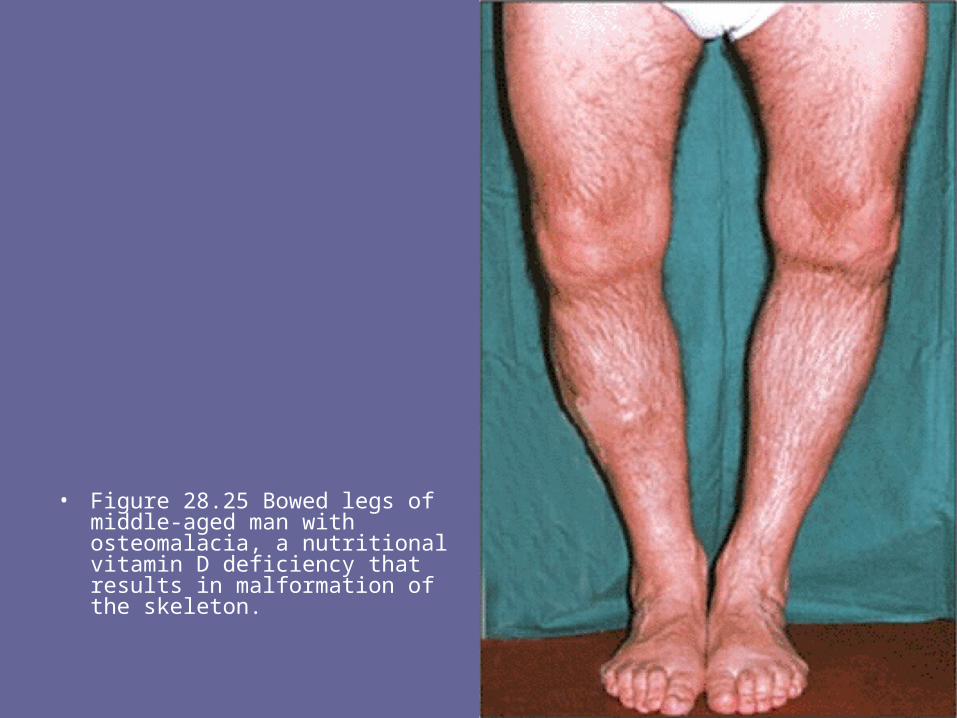
• Figure 28.25 Bowed legs of middle-aged man with osteomalacia, a nutritional vitamin D deficiency that results in malformation of the skeleton.
2.Renal rickets (renal osteodystrophy): This disorder results from chronic renal failure and, thus, the decreased ability to form the active form of the vitamin. 1,25-diOH cholecalciferol (calcitriol) administration is effective replacement therapy.
3.Hypoparathyroidism: Lack of parathyroid hormone causes hypocalcemia and hyperphosphatemia. These patients may be treated with any form of vitamin D, together with parathyroid hormone.

F. Toxicity of vitamin D• Vitamin D is the most toxic of all vitamins. Like
all fat-soluble vitamins, vitamin D can be stored in the body and is only slowly metabolized.
• High doses (100,000 IU for weeks or months) can cause loss of appetite, nausea, thirst, and stupor.
• Enhanced calcium absorption and bone resorption results in hypercalcemia, which can lead to deposition of calcium in many organs, particularly the arteries and kidneys.

XIII. Vitamin K• The principal role of vitamin K is in the
posttranslational modification of various blood clotting factors, in which it serves as a coenzyme in the carboxylation of certain glutamic acid residues present in these proteins.
• Vitamin K exists in several forms, for example, in plants as phylloquinone (or vitamin K1), and in intestinal bacterial flora as menaquinone (or vitamin K2).
• For therapy, a synthetic derivative of vitamin K, menadione, is available.
A. Function of vitamin K1. Formation of γ-carboxyglutamate (Gla): • Vitamin K is required in the hepatic synthesis of
prothrombin and blood clotting factors II, VII, IX, and X.• These proteins are synthesized as inactive precursor
molecules. Formation of the clotting factors requires the vitamin K–dependent carboxylation of glutamic acid residues (Figure 28.26).
• This forms a mature clotting factor that contains Gla and is capable of subsequent activation.
• The reaction requires O2, CO2, and the hydroquinone form of vitamin K.
• The formation of Gla is sensitive to inhibition by dicumarol, an anticoagulant occurring naturally in spoiled sweet clover, and by warfarin, a synthetic analog of vitamin K.

• Figure 28.26 Carboxylation of glutamate to form γ-carboxyglutamate (Gla).

2. Interaction of prothrombin with platelets: The Gla residues of prothrombin are good chelators of positively charged calcium ions, because of the two adjacent, negatively charged carboxylate groups.
• The prothrombin–calcium complex is then able to bind to phospholipids essential for blood clotting on the surface of platelets.
• Attachment to the platelet increases the rate at which the proteolytic conversion of prothrombin to thrombin can occur (Figure 28.27).
3. Role of Gla residues in other proteins: Gla is also present in other proteins (for example, osteocalcin of bone, and in proteins involved in the degradation of blood clots).
4. However, the physiologic role of these proteins and the function of vitamin K in their synthesis is not yet understood.
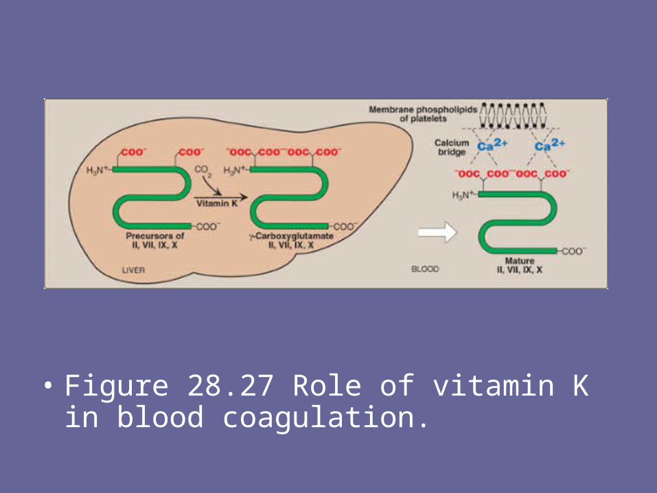
• Figure 28.27 Role of vitamin K in blood coagulation.
B. Distribution and requirement of vitamin K• Vitamin K is found in cabbage, cauliflower,
spinach, egg yolk, and liver. • There is also extensive synthesis of the vitamin
by the bacteria in the gut. • There is no RDA for vitamin K, but 70 to 140
mg/day is recommended as an adequate level.• The lower level assumes one half of the
estimated requirement comes from bacterial synthesis, whereas the upper figure assumes no bacterial synthesis.
C. Clinical indications1. Deficiency of vitamin K: A true vitamin K deficiency is
unusual because adequate amounts are generally produced by intestinal bacteria or obtained from the diet.
• If the bacterial population in the gut is decreased, for example, by antibiotics, the amount of endogenously formed vitamin is depressed, and this can lead to hypoprothrombinemia in the marginally malnourished individual, for example, a debilitated geriatric patient.
• This condition may require supplementation with vitamin K to correct the bleeding tendency.
• In addition, certain second-generation cephalosporins, for example, cefoperazone, cefamandole, and moxalactam cause hypoprothrombinemia, apparently by a warfarin-like mechanism. Consequently, their use in treatment is usually supplemented with vitamin K.

2. Deficiency of vitamin K in the newborn:• Newborns have sterile intestines and so initially
lack the bacteria that synthesize vitamin K. • Because human milk provides only about one
fifth of the daily requirement for vitamin K, it is recommended that all newborns receive a single intramuscular dose of vitamin K as prophylaxis against hemorrhagic disease.
D. Toxicity of vitamin K• Prolonged administration of large doses of
vitamin K can produce hemolytic anemia and jaundice in the infant, due to toxic effects on the membrane of red blood cells.
XIV. Vitamin E
• The E vitamins consist of eight naturally occurring tocopherols, of which α-tocopherol is the most active (Figure 28.28).
• The primary function of vitamin E is as an antioxidant in prevention of the nonenzymic oxidation of cell components, for example, polyunsaturated fatty acids, by molecular oxygen and free radicals.
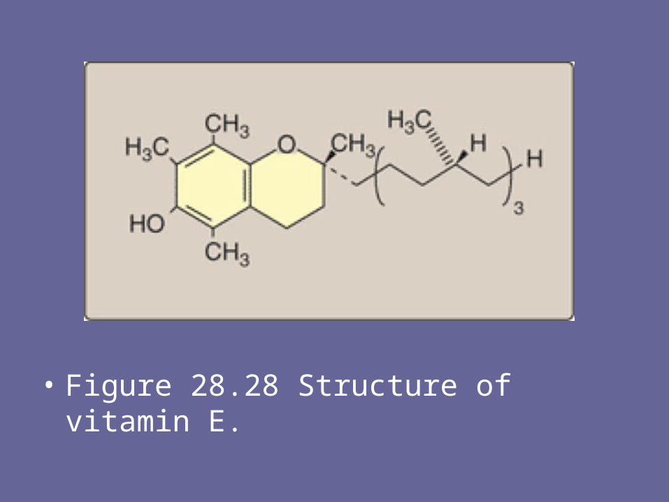
• Figure 28.28 Structure of vitamin E.

A. Distribution and requirements of vitamin E• Vegetable oils are rich sources of vitamin E, whereas
liver and eggs contain moderate amounts. • The RDA for α-tocopherol is 10 mg for men and 8 mg for
women. • The vitamin E requirement increases as the intake of
polyunsaturated fatty acid increases.B. Deficiency of vitamin E• Vitamin E deficiency is almost entirely restricted to
premature infants. • When observed in adults, it is usually associated with
defective lipid absorption or transport. • The signs of human vitamin E deficiency include
sensitivity of erythrocytes to peroxide, and the appearance of abnormal cellular membranes.
• C. Clinical indications• Vitamin E is not recommended for the prevention of
chronic disease, such as coronary heart disease or cancer.
• Clinical trials using vitamin E supplementation have been uniformly disappointing. For example, subjects in the Alpha-Tocopherol, Beta-Carotene Cancer Prevention Study trial who received high doses of vitamin E not only lacked cardiovascular benefit but also had an increased incidence of stroke.
• D. Toxicity of vitamin E• Vitamin E is the least toxic of the fat-soluble vitamins,
and no toxicity has been observed at doses of 300 mg/day.