VITAMIN D DEFICIENCY CAUSES MUSCULOSKELETAL ...
118
VITAMIN D DEFICIENCY CAUSES MUSCULOSKELETAL HYPERSENSITIVITY: THE ROLE OF NOCICEPTOR HYPERINNERVATION BY C2011 Sarah Elizabeth Tague Submitted to the graduate degree program in Molecular and Integrative Physiology and the Graduate Faculty of the University of Kansas in partial fulfillment of the requirements for the degree of Doctor of Philosophy _______________________________ Peter G. Smith, Ph.D., Chairman _______________________________ Kenneth McCarson, Ph.D. _______________________________ Douglas Wright, Ph.D. _______________________________ Nancy Berman, Ph.D. _______________________________ Leslie Heckert, Ph.D. Date defended:____________
Transcript of VITAMIN D DEFICIENCY CAUSES MUSCULOSKELETAL ...

VITAMIN D DEFICIENCY CAUSES MUSCULOSKELETAL HYPERSENSITIVITY:
THE ROLE OF NOCICEPTOR HYPERINNERVATION
BY
C2011
Sarah Elizabeth Tague
Submitted to the graduate degree program in Molecular and Integrative Physiology and the Graduate Faculty of the University of Kansas in partial fulfillment of the requirements for the
degree of Doctor of Philosophy
_______________________________
Peter G. Smith, Ph.D., Chairman
_______________________________
Kenneth McCarson, Ph.D.
_______________________________
Douglas Wright, Ph.D.
_______________________________
Nancy Berman, Ph.D.
_______________________________
Leslie Heckert, Ph.D.
Date defended:____________

ii
The Dissertation Committee for Sarah Elizabeth Tague certifies that this is the approved version of the following dissertation:
VITAMIN D DEFICIENCY CAUSES MUSCULOSKELETAL HYPERSENSITIVITY:
THE ROLE OF NOCICEPTOR HYPERINNERVATION
Committee:
_______________________________
Peter G. Smith, Ph.D., Chairman
_______________________________
Kenneth McCarson, Ph.D.
_______________________________
Douglas Wright, Ph.D.
_______________________________
Nancy Berman, Ph.D.
_______________________________
Leslie Heckert, Ph.D.
Date approved:____________

iii
ABSTRACT
Clinical studies link vitamin D deficiency and musculoskeletal pain, both of which occur more frequently
in women. However, a causal relationship has been difficult to establish and it is not clear whether
vitamin D metabolites directly influence nociceptors (‘pain-sensing’ neurons). It was shown here, via
immunohistochemistry and western blot, that rat putative nociceptors contain vitamin D receptors (VDRs)
and metabolic enzymes, whose expression is regulated by ovarian hormones. In ovariectomized rats a
vitamin D deficient diet induces balance deficits and deep tissue mechanical hyperalgesia, concurrent
with muscle hyperinnervation by presumed nociceptors. Balance deficits, muscle mechanical
hypersensitivity, and hyperinnervation are not corrected by elevated dietary calcium. In primary sensory
cultures, VDR is enriched in c-fiber growth cones and regulates neurite outgrowth through VDR rapid
response pathways. Therefore, vitamin D metabolites act directly on nociceptive neurons to inhibit
axonal sprouting, accounting for hypovitaminosis D-induced muscle hyperinnervation, and possibly
contributing to hypersensitivity.

iv
This work is dedicated to my parents, Susan D. and Walter C. Tague, for making my education a priority. Without their continued support, this work would not have been possible.

v
ACKNOWLEDGEMENTS
This work was partially funded by the KUMC Biomedical Research Training Program and a
NIH, National Institute on Aging, Kirschstein National Research Service Predoctoral Award.
I have had exceptional mentors over my academic career who have helped lay the groundwork
for these studies. I learned the basics of working in a lab during my undergraduate education at the
University of Kansas in the labs of Dr. Victoria Corbin and Dr. Elizabeth Boyce. During my master’s
degree studies at the University of Notre Dame, I learned invaluable molecular and cell biology
techniques in the lab of Dr. Crislyn D’Souza-Schorey. Most notably, she showed me how to develop and
follow through with plausible hypotheses. Finally, my work in Dr. H. Clarke Anderson’s lab at the
University of Kansas Medical Center introduced me to translational bench-animal-clinical research.
While so much of science is focused on the details, he taught me to always think about the larger picture.
During my studies at the University of Kansas Medical Center, I am grateful to have worked with
a wonderful and diverse group of scientists. I thank our lab technicians Zhaohui Liao and Elza
Kharatyan, who generally keep the lab efficient and organized, allowing me to focus on science. My
Ph.D. experience was enhanced by my fellow students, Aritra Bhattacherjee, Gwenaelle Clarke, Tim
Donohue, Argenia Doss, and Eva Selfridge. I owe a special thanks to Eva Selfridge for her excellent
proofreading skills. The senior lab members Dr. Dora Krizsan-Agbas and Dr. Anuradha Chakrabarty
have provided both guidance and friendship. Both Anuradha and Dora taught me dissection techniques,
and Anuradha helped teach me surgical procedures, culture techniques, and behavior analysis. Finally I
am especially grateful to Gwenaelle Clarke, an amazing friend who also helped me with the muscle
compression analysis and sympathetic culture techniques described here.
I owe much gratitude to the wonderful members of the Kansas Intellectual and Developmental
Disabilities Research Center, Dr. Don Warn, Phil Shafer, Jing Huang, Doug Brownyard, Michelle Winter,

vi
Tina Darrow and Beth Van Luchene. They work hard to keep everything running smoothly. I would like
to thank Michelle Winter and Dr. Ken McCarson in particular. Michelle helped with animal surgeries, set
up behavioral equipment, and performed the serum 25(OH)D assays described here, and Ken helped me
design the behavioral experiments.
Dr. Anthony Norman at the University of California Riverside kindly provided the vitamin D
analog, JN, which was used in the culture studies described here. Dr. H. Clarke Anderson provided the
muscles from rachitic rats for our preliminary experiments.
The Department of Molecular and Integrative Physiology created a wonderful environment for
students, thanks to the chair, Dr. Paul Cheney, the staff, and the Physiology Society.
As sometimes happens with the best laid plans, life gets in the way. I owe my deepest gratitude
to my friends and family who took care of me when my studies were interrupted by illness. To my
mother and father who drove me to each and every doctor’s appointment, brought me groceries, and
generally took care of me; to my friends, Merritt Engel, Amanda Dewoody, Gwenaelle Clarke, Eva
Selfridge, and Rachel Rempfer who offered emotional support, checked in on me regularly, made me
food, and cleaned my apartment; to my lab mates who sent me flowers and were incredibly supportive;
and to my mentor, Peter Smith, for his patience and understanding, I don’t know what I would have done
without all of you.
Finally I would like to extend my deepest gratitude to Dr. Peter Smith, a gifted scientist and
outstanding leader. In addition to his scientific guidance, he has taught me much about how to develop
leadership qualities, grant writing skills, and lab management strategies. His insights into the details of
careers in academia and beyond are invaluable. I am grateful that he encourages individual thought and
allowed me to pursue my scientific interests. His breadth of knowledge is impressive and I will continue
to admire him for his honesty and strength of character. It has been an honor to work with Dr. Smith.

vii
TABLE OF CONTENTS
ACCEPTANCE PAGE ................................................................................................................................. ii ABSTRACT ................................................................................................................................................. iii DEDICATION ............................................................................................................................................. iv ACKNOWLEDGEMENTS .......................................................................................................................... v TABLE OF CONTENTS ............................................................................................................................ vii LIST OF TABLES AND FIGURES .......................................................................................................... viii CHAPTER 1. GENERAL INTRODUCTION .............................................................................................. 1
TABLE .................................................................................................................................................... 10
FIGURE .................................................................................................................................................. 11
CHAPTER 2. VITAMIN D RECEPTOR AND ENZYME EXPRESSION IN DORSAL ROOT GANGLIA OF ADULT FEMALE RATS: MODULATION BY OVARIAN HORMONES ................... 13
ABSTRACT ............................................................................................................................................ 14
INTRODUCTION .................................................................................................................................. 15
MATERIALS AND METHODS ............................................................................................................ 16
RESULTS ............................................................................................................................................... 21
DISCUSSION ......................................................................................................................................... 26
TABLES ................................................................................................................................................. 34
FIGURES ................................................................................................................................................ 35
CHAPTER 3. VITAMIN D DEFICIENCY PROMOTES SENSORY AXON SPROUTING AND MUSCLE HYPERSENSITIVITY .............................................................................................................. 49
ABSTRACT ............................................................................................................................................ 50
INTRODUCTION .................................................................................................................................. 51
MATERIALS AND METHODS ............................................................................................................ 52
RESULTS AND DISCUSSION ............................................................................................................. 57
TABLES ................................................................................................................................................. 65
FIGURES ................................................................................................................................................ 67
CHAPTER 4. GENERAL CONCLUSIONS AND DISCUSSION ............................................................ 83 REFERENCES ........................................................................................................................................... 93

viii
LIST OF TABLES AND FIGURES
CHAPTER 1
Table
1. Factors that contribute to both musculoskeletal pain and vitamin D deficiency…………………10
Figure
1. Increased peripherin content in muscles from vitamin D deficient rats……………………….....11
CHAPTER 2
Table
1. Quantitative analysis of neuronal markers and effect of ovariectomy…………………………....34
Figures
1. VDR, CYP27B1, and CYP24 proteins are present in adult DRG………………………………. 35
2. Cellular localization of VDR, CYP27B1, and CYP24 in adult DRG…………………………….37
3. Cellular colocalization of VDR with the neural markers peripherin, IB4, and CGRP…………...39
4. Cellular colocalization of CYP27B1 with the neural markers peripherin, IB4, and CGRP……...41
5. Cellular colocalization of CYP24 with the neural markers peripherin, IB4, and CGRP………....43
6. Cellular colocalization of VDR with the enzymes CYP27B1 or CYP24………………………...45
7. DRG expression of VDR and CYP27B1, but not CYP24 is reduced following ovariectomy…...47
CHAPTER 3
Tables
1. Diet, weight, and serum analysis………………………………………………………………....65
2. No changes in bone parameters were found in rats fed a vitamin D deficient diet for four
weeks……………………………………………………………………………………………...66

ix
Figures
1. Experimental timeline…………………………………………………………………………….67
2. Modification of the digital Randall Selitto instrument for muscle compressions………………...69
3. Behavioral comparisons of control, VD-/+Ca, and VD- rats……………………………………..71
4. Representative images of tibial growth plates from rats after four weeks on control, VD-/+Ca, or
VD- diets………………………………………………………………………………………....73
5. Comparisons of innervation densities of the lateral gastrocnemius muscles of rats after four weeks
on control, VD-/+Ca, or VD- diets……………………………………………………………….75
6. Representative images of the immunoreactive nerves in calf muscle…………………………....77
7. 1,25(OH)2D3 acts directly on c-fiber sensory nerves to regulate axonal growth……………….79
8. Sympathetic neurons express VDRs, but 1,25(OH)2D3 has no effect on neurite outgrowth…..81

1
CHAPTER 1. GENERAL INTRODUCTION

2
General Background
Accumulating evidence supports a role for vitamin D in certain types of pain. For example, vitamin
D deficiency is associated with chronic musculoskeletal pain and acroparaesthesia (numbness, burning, or
tingling in the extremities) (Benson, Wilson et al. 2006; de Torrente de la Jara, Pecoud et al. 2006; Glerup
and Eriksen 1999; Gloth, Lindsay et al. 1991; Heidari, Shirvani et al. 2010; Macfarlane, Palmer et al.
2005; McBeth, Pye et al. 2010; Plotnikoff and Quigley 2003). Up to 93% of patients who report chronic
musculoskeletal pain are vitamin D deficient (Plotnikoff and Quigley 2003). Several studies have shown
that this pain may be relieved by vitamin D supplementation (de Torrente de la Jara, Pecoud et al. 2006;
Glerup and Eriksen 1999; Gloth, Lindsay et al. 1991). However, a direct causal relationship has not been
established and potential mechanisms underlying this phenomenon have remained unexplored.
Musculoskeletal pain
Musculoskeletal pain in general has enormous socioeconomic impacts on our society. It can
significantly decrease the quality of life for an individual by preventing entry into the workforce and
rendering many incapable of completing simple daily activities. The council for disability awareness
estimates that in 2006, of the $79.9 billion in social security disability insurance payouts and $7.2 billion
in long-term disability payouts from private companies, musculoskeletal disorders accounted for 25.7% of
all disability claims making it the number one cause for long-term disability (Awareness 2006).
Musculoskeletal pain also substantially affects the load on our health care system. It is estimated that the
prevalence of chronic musculoskeletal pain (pain lasting longer than 3 months) in adolescents is between
27.5-36% (De Inocencio 2004) and 35-50% in adults (Bergman 2007). In 10-22% of adults this pain is
widespread (Abusdal, Hagen et al. 1997; Bergman, Herrstrom et al. 2001; Croft, Rigby et al. 1993; Hunt,
Silman et al. 1999; Schochat and Raspe 2003; White, Speechley et al. 1999; Wolfe, Ross et al. 1995). In
addition, 20-40% of all primary health care visits are due to musculoskeletal complaints (Andersson,
Ejlertsson et al. 1999; De Inocencio 2004). Despite these statistics the mechanisms and pathology that
lead to chronic musculoskeletal pain are poorly understood.

3
Vitamin D
The secosteroid vitamin D is generally considered a prosteroid hormone and is not technically a
vitamin because humans can synthesize it; however, most individuals also acquire vitamin D from natural
dietary sources (i.e. fatty fish), fortified foods, or nutritional supplements (Yetley 2008). Synthesis
begins upon exposure to UVB light, when 7-dehydroxycholesterol is converted to previtamin D within
the keratinocyte cell membrane of the skin (Holick 2005). Previtamin D spontaneously isomerizes into
vitamin D during the course of several hours and enters the blood stream through mass action, due to the
presence of vitamin D binding proteins within the blood (Holick 2005). Whether synthesized or from
nutritional sources, vitamin D is converted in the liver to the main circulating form, 25-hydroxyvitamin D
(25(OH)D) (Horst, Reinhardt et al. 2005). 25(OH)D can be further transformed by 25-hydroxyvitamin D
1α-hydroxylase/cytochrome P450, family 27, subfamily B, polypeptide 1 (CYP27B1) into 1,25-
dihydroxyvitamin D (1,25(OH)2D), which is the most well understood active metabolite (Horst, Reinhardt
et al. 2005). Conversion of 25(OH)D into 1,25(OH)2D occurs largely in kidney, but other cell types
express CYP27B1 and can locally convert circulating precursor to active hormone (Jones 2007).
Hydroxylation of 1,25(OH)2D to form 1,24,25-trihydroxyvitamin D by the widely expressed enzyme, 25-
hydroxyvitamin D 24-hydroxylase/cytochrome P450, family 24, subfamily A, polypeptide 1 (CYP24), is
an initial step in the pathway of 1,25(OH)2D degradation, but CYP24 also produces other vitamin D
metabolites (i.e. 24,25-dihydroxyvitamin D), whose actions are just beginning to be understood (Omdahl,
Bobrovnikova et al. 2003; Sakaki, Kagawa et al. 2005). CYP27B1 and CYP24 therefore act as opposing
forces in regulating the concentration of 1,25(OH)2D. This active hormone binds to vitamin D receptors
(VDRs), which are spatially dynamic and may be localized within the nucleus where they influence gene
transcription, or within the cytoplasm and membrane where they modulate ion flux and the generation of
second messengers (Mizwicki and Norman 2009).
Vitamin D deficiency is prevalent worldwide. It is thought that the best predictor of vitamin D
status is serum 25(OH)D levels, because 1,25(OH)2D has a relatively short half life and is more tightly

4
regulated (Jones 2008). The NIH recently revised its standards for serum 25(OH)D levels according to a
recent Institute of Medicine committee. Previously anything >37.5nmol/L was considered sufficient,
<37.5nmol was insufficient, and below 25nmol/L was deficient. Now >50nmol/L (20ng/ml) is
considered sufficient for 97.5% of the population, and < 30nmol/L (12ng/ml) is considered deficient
(Institute of Medicine 2010). However, a number of researchers feel that these levels should be adjusted
even higher (Dawson-Hughes, Heaney et al. 2005; Hollis 2005). Peak calcium absorption from the
intestines occurs when serum 25(OH)D is approximately 80pmol/L (32ng/ml) (Heaney, Dowell et al.
2003) and levels >80pmol/L may be required to alleviate secondary hyperparathyroidism in the elderly
(Vieth, Ladak et al. 2003), so some have suggested that above 80nmol/L (32ng/ml) should be considered
replete and below this level should be considered insufficient. Some researchers believe that serum
25(OH)D concentrations below 50nmol/L (20ng/ml) should be considered deficient, because there is a 5-
15% increase in parathyroid hormone and higher bone turnover (Chapuy, Preziosi et al. 1997; Holick
2009; Need 2006). When concentrations fall below 25nmol/L (10ng/ml) there is an increased fracture
risk (Cummings, Black et al. 1993; Ooms, Roos et al. 1995) and below 12.5nmol/L (5ng/ml) is when
overt osteomalacia or rickets becomes apparent (Need 2006). While the data available concerning the
upper levels are not clear, serum levels above 125nmol/L (50ng/ml) may begin to raise concerns (Institute
of Medicine 2010), however toxicity rarely occurs below 500nmol/L (Heaney 2008). Based on the
National Health and Nutrition Examination Survey (NHANES), approximately 70% of the population has
serum 25(OH)D concentrations that are less than 80nmol/L (32ng/ml), 30% has levels less than 50nmol/L
(20ng/ml), and 5% has levels less than 27.5nmol/L (11ng/ml) (Yetley 2008).
Vitamin D and musculoskeletal pain
A significant proportion of patients who report chronic musculoskeletal pain are vitamin D deficient,
with serum 25(OH)D concentrations that are less than 50nmol/L (20ng/ml) (Benson, Wilson et al. 2006;
Macfarlane, Palmer et al. 2005; Plotnikoff and Quigley 2003). Depending on the population studied and
the cut-off values of 25(OH)D used to determine deficiency, the reported percentage of patients with

5
musculoskeletal pain that are vitamin D deficient varies from 25-100%. Gloth et al. first described five
patients with severe pain triggered by minimal movement or touch. All five patients had 1,25-
dihydroxyvitamin D3 (1,25(OH)2D3) levels below 45pmol/L (normal range 50-190pmol/L) and four of
the five patients had serum 25(OH)D levels below 50nmol/L (20ng/ml). All patients recovered after
vitamin D supplementation (Gloth, Lindsay et al. 1991). A study of Australian Aborigenes showed that
while 100% of Aborigenes with muscle pain had a levels of 25(OH)D below 50nmol/L (20ng/ml), only
12.5% of Aborigines without muscle pain were vitamin D deficient (Benson, Wilson et al. 2006). Of 150
people who reported to a US community clinic with non-specific musculoskeletal pain, 93% had
25(OH)D levels below 50nmol/L (20ng/ml) (Plotnikoff and Quigley 2003). A study in Norway of 276
patients with musculoskeletal pain and 229 controls found that 81.6% of patients with leg pain and 60%
with chronic widespread pain were vitamin D deficient (<20ng/ml) compared to only 36.1% of controls.
They also show that average serum 25(OH)D levels were significantly lower in patients with leg pain
(14.5ng/ml) or widespread pain (20.6ng/ml) than in control patients (33.1ng/ml) (Heidari, Shirvani et al.
2010). Results from a recent European Male Ageing Study analyzing 3075 men age 49-79, found that
25.5% of men with chronic widespread pain had 25(OH)D levels below 37.5nmol/L (15ng/ml), compared
to only 18.6% in men with no pain (McBeth, Pye et al. 2010). Conversely a study in Switzerland of
asylum seekers with hypovitaminosis D (<21nmol/L) found that up to 91% experienced somatic pain,
and 85% reported complete or partial resolution of symptoms after vitamin D treatment (de Torrente de la
Jara, Pecoud et al. 2006). In addition to pain, it has also been reported that 59% of Arab women with
hypovitaminosis D experience acroparaesthesia. After vitamin D supplementation, 92% displayed
improvement and 81% reported a complete remission of symptoms (Glerup and Eriksen 1999). In
addition to the growing number of studies linking vitamin D deficiency to musculoskeletal pain, there
have also been several conflicting reports, which appear to stem from a focus on vitamin D deficiency and
fibromyalgia (Block 2004; de Rezende Pena, Grillo et al. 2010; Tandeter, Grynbaum et al. 2009; Warner
and Arnspiger 2008). However, this issue may have been cleared up by Heidari et. al, who recently
showed that while low vitamin D levels are linked to chronic widespread pain in general, vitamin D status

6
is not associated with chronic widespread pain subtype, fibromyalgia (Heidari, Shirvani et al. 2010). All
in all there is a significant clinical link between vitamin D deficiencies and non-specific musculoskeletal
pain, but an understanding of how vitamin D affects the pain pathways of the nervous system is severely
lacking.
Vitamin D and the nervous system
Vitamin D is becoming recognized as an important hormone in nervous system health. VDR is
expressed by neurons, oligodendrocytes, and astrocytes within the limbic system, basal ganglia, spinal
cord, cerebellum, and cerebral cortex (Baas, Prufer et al. 2000; Eyles, Smith et al. 2005; Glaser, Veenstra
et al. 1999; Neveu, Naveilhan et al. 1994; Perez-Fernandez, Alonso et al. 1997; Prufer, Veenstra et al.
1999; Walbert, Jirikowski et al. 2001). Studies have also shown that neurons and glia in the brain express
CYP27B1 (Eyles, Smith et al. 2005; Zehnder, Bland et al. 2001), and CYP24 mRNA has been detected in
glioma and primary glial cell culture (Naveilhan, Neveu et al. 1993), suggesting that the brain can
regulate local 1,25(OH)2D concentrations. Vitamin D signaling has been linked to cell survival,
proliferation, and differentiation, and may be especially important during brain development (Eyles,
Brown et al. 2003; Levenson and Figueiroa 2008). In the adult, vitamin D supplementation has been
proposed to improve outcomes in multiple sclerosis, Parkinson’s disease, and traumatic brain injury
(Cekic, Sayeed et al. 2009; Myhr 2009; Newmark and Newmark 2007). Expression of VDR has also
been identified in fetal (E12-E21) DRG sensory neurons (Johnson, Grande et al. 1996; Veenstra, Prufer et
al. 1998), but it was not established whether expression continues post-natally. Thus, accumulating
evidence suggests that vitamin D signaling plays important roles in the nervous system. However, little is
known about how vitamin D may affect pain pathways.
Sex differences in musculoskeletal pain and vitamin D deficiency
In all age groups after the age of five, vitamin D deficiency is more prevalent in women than men
(Yetley 2008) . Women are also twice as likely to develop chronic widespread pain (Bergman 2007).

7
This has led us to consider a role for ovarian hormones in the regulation of the vitamin D system and the
development of pain. While testosterone levels do not appear to affect serum 25(OH)D levels or VDR
expression (Tiwari, Gupta et al. 2002), VDR expression is up-regulated by 17β-estradiol in some cell
types, including osteoblasts (Duque, El Abdaimi et al. 2002; Liel, Kraus et al. 1992; Mahonen and
Maenpaa 1994), breast cancer cells (Escaleira, Sonohara et al. 1993; Gilad, Bresler et al. 2005),
colonocytes and duodenocytes (Gilad, Bresler et al. 2005; Liel, Shany et al. 1999; Schwartz, Smirnoff et
al. 2000), liver (Duncan, Glass et al. 1991), and uterus (Levy, Zuili et al. 1984; Walters 1981). Gilad et.
al. showed that the enhanced expression of VDR by 17β-estradiol is not through classical estrogen
response elements, but through 1,25(OH)2D binding to membrane-localized estrogen receptors that
rapidly activate transcription of VDR through a Ras-Raf-MEK-ERK-cJUN pathway (Gilad, Bresler et al.
2005). However, not all cell types respond the same, as 17β-estradiol inhibits VDR expression in the
kidney (Duncan, Glass et al. 1991). Estrogens can also increase 1,25(OH)2D concentrations locally by
increasing CYP27B1 and decreasing CYP24 levels (Lechner, Bajna et al. 2006). In fact, in women
shortly after the estrogen surge of the menstrual cycle, 1,25(OH)2D serum levels are reportedly double
what they are on day one of the menstrual cycle (Gray, McAdoo et al. 1982). In addition, progesterone
has been reported to enhance estrogen-induced CYP27B1 expression (Tanaka, Castillo et al. 1978). Thus
it appears that in general, vitamin D signaling is up-regulated by ovarian hormones. This is interesting
because musculoskeletal pain is primarily associated with low estrogen status. For instance, a major side-
effect of aromatase inhibitors, which prevent the production of estrogens, is musculoskeletal pain (Felson
and Cummings 2005; Garreau, Delamelena et al. 2006; Morales, Pans et al. 2006; Mouridsen 2006). In
addition, Leuprolide, a gonadotropin-releasing hormone agonist, which causes hypoestrogenemia, induces
musculoskeletal pain in 25% of women who use it (Felson and Cummings 2005; Friedman, Juneau-
Norcross et al. 1993). Furthermore, the prevalence for chronic widespread pain peaks in the 50’s and
60’s, corresponding with the age of menopause onset, a time when hormone levels are naturally dropping.
In fact, it is well known that generalized musculoskeletal pain is a symptom of menopause (Dugan,
Powell et al. 2006; Greendale, Reboussin et al. 1998). Asian women, who tend to have particularly low

8
estrogen levels during menopause (Cauley, Gutai et al. 1989; Randolph, Sowers et al. 2004) and are
especially susceptible to vitamin D deficiency (Ford, Graham et al. 2006; Pal, Marshall et al. 2003; Shaw
and Pal 2002), appear to be more susceptible to menopausal musculoskeletal pain, with one study
reporting it as their most common menopausal symptom (Ho, Chan et al. 1999). Hormone replacement
therapy, which is known to increase serum 1,25(OH)2D3 levels (van Hoof, van der Mooren et al. 1994;
van Hoof, van der Mooren et al. 1999), has been shown to relieve and prevent the generalized pain
associated with menopause (Dugan, Powell et al. 2006). In this study we examine whether the loss of
ovarian hormones causes specific changes in the expression of vitamin D-related proteins in neurons that
are part of the pain pathway.
The Need for an Animal Model
While clinical links between chronic musculoskeltal pain and vitamin D deficiency exist,
confounding factors in humans make vitamin D deficiency difficult to analyze in isolation. Factors that
contribute to a vitamin D deficiency, such as sex, skin color, smoking, body weight, socioeconomic
status, age, and physical activity are also associated with musculoskeletal pain (Table 1). These factors
may enhance susceptibility to vitamin D deficiency, which in turn cause musculoskeletal pain.
Alternatively, vitamin D deficiency may be more likely to occur in individuals already susceptible to
chronic musculoskeletal pain. An animal model of hypovitaminosis D-induced musculoskeletal pain will
allow analysis without confounding factors. The studies described here will use a rat model, because rats
are extensively used to study pain-related behavior and have been used to assess features of vitamin D
deficiency for nearly 90 years (McCollum, Simmonds et al. 1922). Female rats will be used, because of
the high prevalence of vitamin D deficiency and musculoskeletal pain in women (Andersson, Ejlertsson et
al. 1993; Bergman, Herrstrom et al. 2001; Bolland, Grey et al. 2007; Dawson-Hughes, Harris et al. 1997;
Rollman and Lautenbacher 2001).

9
Pain Associated with Hyperinnervation of Tissue
Increased peripheral nerve density is associated with several painful conditions (Di Sebastiano, Fink
et al. 1995; Di Sebastiano, Fink et al. 1997; Fink, Di Sebastiano et al. 1994; Pang, Marchand et al. 1995;
Reinert, Kaske et al. 1998; Reynolds and Fitzgerald 1995; Schubert, Weidler et al. 2005; Shinoda, Honda
et al. 2003), and is known to occur in muscles, joints, and tendons (Reinert, Kaske et al. 1998; Schubert,
Weidler et al. 2005; Shinoda, Honda et al. 2003). Steroid hormones other than vitamin D are known to
regulate axonal sprouting, leading us to speculate whether vitamin D metabolites also had this potential
(Blacklock, Johnson et al. 2005). Preliminary data were obtained, revealing that muscles from rachitic rats
(a severe vitamin D deficiency model) had increased levels of the c-fiber specific intermediate filament
protein, peripherin, suggesting that the muscles from vitamin D deficient might be hyperinnervated
(Figure 1). Therefore, the focus of these studies was on how a vitamin D deficiency affects nociceptors,
the first order neurons of the pain and temperature pathway, whose distal axons innervate peripheral
tissue. In the first study described here, putative nociceptors were examined to determine whether they
contain the machinery needed to metabolize and directly respond to vitamin D metabolites. It the second
study, vitamin D status in rats was altered in order to determine whether a vitamin D deficiency can result
in deep tissue hypersensitivity and muscle hyperinnervation.

10
TABLE
Table 1. Factors that contribute to both musculoskeletal pain and vitamin D deficiency.
Vitamin D levels Musculoskeletal Pain
Sex Vitamin D deficiency is more prevalent in women (Bolland, Grey et al. 2007; Dawson-Hughes, Harris et al. 1997).
Chronic widespread pain is more prevalent in women (Andersson, Ejlertsson et al. 1993; Bergman, Herrstrom et al. 2001; Rollman and Lautenbacher 2001).
Skin color
Due to the competition of melanin for UVB rays, synthesis of vitamin D in the skin is less efficient as skin pigmentation increases (Armas, Dowell et al. 2007; Clemens, Adams et al. 1982).
Chronic widespread pain prevalence is increased in African American women compared to Caucasian women (Gansky and Plesh 2007). In addition, black chronic pain patients experience a higher severity of pain (Edwards, Doleys et al. 2001; McCracken, Matthews et al. 2001).
Smoking Smokers have a higher prevalence of vitamin D deficiency (Sowers, Wallace et al. 1986)
Smokers have a higher prevalence of musculoskeletal pain (Andersson, Ejlertsson et al. 1998; Brage and Bjerkedal 1996; Palmer, Syddall et al. 2003; Yunus, Arslan et al. 2002).
Body Weight
There is a negative correlation of vitamin D levels and body mass index (Arunabh, Pollack et al. 2003; Bischof, Heinze et al. 2006; Bolland, Grey et al. 2007; Need, Morris et al. 1993; Wortsman, Matsuoka et al. 2000).
There is a positive correlation between body mass index and musculoskeletal pain (Salaffi, De Angelis et al. 2005; Sievert and Goode-Null 2005; Yunus, Arslan et al. 2002).
Physical Activity
Exercise improves vitamin D activity (Bell, Godsen et al. 1988; Klausen, Breum et al. 1993; Nelson, Meredith et al. 1988).
Exercise alleviates chronic widespread pain (Jones, Adams et al. 2006; Linton and van Tulder 2001)
Age
Serum 25(OH)D levels decrease with age (Holick, Matsuoka et al. 1989; Need, Morris et al. 1993; Sowers, Wallace et al. 1986).
The prevalence of musculoskeletal pain increases with age, peaking between the ages of 55-65 and then decreasing slightly thereafter (Andersson, Ejlertsson et al. 1993; Croft, Rigby et al. 1993; Hunt, Silman et al. 1999; White, Speechley et al. 1999; Wolfe, Ross et al. 1995).

11
FIGURE Figure 1.

12
Figure 1. Increased peripherin content in muscles from vitamin D deficient rats. Western blots of lysates
prepared from the lateral gastrocnemius muscles of control or rachitic vitamin D deficient rats. β actin
was used to normalize intensity values, which are expressed in graphical form as fold change compared to
control.

13
CHAPTER 2. VITAMIN D RECEPTOR AND ENZYME EXPRESSION IN DORSAL ROOT GANGLIA OF ADULT FEMALE RATS: MODULATION BY OVARIAN HORMONES

14
ABSTRACT Vitamin D insufficiency impacts sensory processes including pain and proprioception, but little is known
regarding vitamin D signaling in adult sensory neurons. We analyzed female rat dorsal root ganglia
(DRG) for vitamin receptor (VDR) and the vitamin D metabolizing enzymes CYP27B1 and CYP24.
Western blots and immunofluorescence revealed the presence of these proteins in sensory neurons.
Nuclear VDR immunoreactivity was present within nearly all neurons, while cytoplasmic VDR was
found preferentially in unmyelinated calcitonin gene-related peptide (CGRP)-positive neurons,
colocalizing with CYP27B1 and CYP24. These data suggest that 1,25(OH)2D may affect sensory
neurons through nuclear or extranuclear signaling pathways. In addition, local vitamin D metabolite
concentrations in unmyelinated sensory neurons may be controlled through expression of CYP27B1 and
CYP24. Because vitamin D deficiency appears to exacerbate some peri-menopausal pain syndromes, we
assessed the effect of ovariectomy on vitamin D-related proteins. Two weeks following ovariectomy,
total VDR expression in DRG dropped significantly, owing to a slight decrease in the percentage of total
neurons expressing nuclear VDR and a large drop in unmyelinated CGRP-positive neurons expressing
cytoplasmic VDR. Total CYP27B1 expression dropped significantly, predominantly due to decreased
expression within unmyelinated CGRP-positive neurons. CYP24 expression remained unchanged.
Therefore, unmyelinated CGRP-positive neurons appear to have a distinct vitamin D phenotype with
hormonally-regulated ligand and receptor levels. These findings imply that vitamin D signaling may play
a specialized role in a neural cell population that is primarily nociceptive.

15
INTRODUCTION
Vitamin D metabolites have widespread roles in human health. In addition to well known actions on
calcium homeostasis and bone remodeling, vitamin D has been linked to immunity, cardiovascular
disease, and cancer (Holick and Chen 2008). Accumulating evidence supports a role for vitamin D in
certain types of pain. For example, vitamin D deficiency is associated with chronic musculoskeletal pain
and acroparaesthesia (numbness, burning, or tingling in the extremities) (Glerup and Eriksen 1999; Gloth,
Lindsay et al. 1991; Masood, Narang et al. 1989; Plotnikoff and Quigley 2003). Over 85% of patients
who report chronic musculoskeletal pain have levels of serum 25-hydroxyvitamin D3 (25OHD3) that are
less than 20ng/ml (50nmol/L) (Benson, Wilson et al. 2006; Macfarlane, Palmer et al. 2005; Plotnikoff and
Quigley 2003), which is relieved by vitamin D supplementation (de Torrente de la Jara, Pecoud et al.
2006; Glerup and Eriksen 1999; Gloth, Lindsay et al. 1991). However, the mechanism by which vitamin
D deficiency leads to altered sensation is unknown.
Vitamin D is derived from diet or exposure to sunlight, converted in the liver to the main circulating
form, 25-hydroxyvitamin D (25OHD), which is further transformed by 25-hydroxyvitamin D 1α-
hydroxylase/cytochrome P450, family 27, subfamily B, polypeptide 1 (CYP27B1) into 1,25-
dihydroxyvitamin D (1,25(OH)2D), which is the most well understood active metabolite. Conversion of
25(OH)D into 1,25(OH)2D occurs largely in kidney, but other cell types express CYP27B1 and can
locally convert circulating precursor to active hormone (Jones 2007). 1,25(OH)2D binds to vitamin D
receptors (VDRs), which are spatially dynamic and may be localized within the nucleus where they
influence gene transcription, or within the cytoplasm and membrane where they modulate ion flux and the
generation of second messengers (Mizwicki and Norman 2009). Hydroxylation of 1,25(OH)2D to form
1,24,25-trihydroxyvitamin D by the widely expressed enzyme, 25-hydroxyvitamin D 24-
hydroxylase/cytochrome P450, family 24, subfamily A, polypeptide 1 (CYP24), is an initial step in the
pathway of 1,25(OH)2D degradation, but CYP24 also produces other vitamin D metabolites, whose
actions are just beginning to be understood (Omdahl, Bobrovnikova et al. 2003; Sakaki, Kagawa et al.

16
2005). The extent to which these vitamin D-related proteins are present within adult sensory neurons is
unknown.
A number of factors can influence VDR signaling, including ovarian hormones. For instance, VDR
expression is generally up-regulated by 17β-estradiol (Gilad, Bresler et al. 2005). Further, estrogens can
increase 1,25(OH)2D concentrations locally by increasing CYP27B1 and decreasing CYP24 levels
(Lechner, Bajna et al. 2006). In addition, progesterone has been reported to enhance estrogen-induced
CYP27B1 expression (Tanaka, Castillo et al. 1978). While it is not known whether ovarian hormones
and vitamin D signaling mechanisms interact in sensory nerve pathways, peripheral sensory neurons do
express estrogen and progesterone receptors (Chan, Rodriguez-Waitkus et al. 2000; Sohrabji, Miranda et
al. 1994), and anecdotal evidence suggests that vitamin D and ovarian hormones may interact to influence
pain sensation. For example, musculoskeletal pain is more prevalent in females and frequently
exacerbated by menopause or pharmacological estrogen suppression (Andersson, Ejlertsson et al. 1993;
Bergman, Herrstrom et al. 2001; Croft, Rigby et al. 1993; Dugan, Powell et al. 2006; Felson and
Cummings 2005; Friedman, Juneau-Norcross et al. 1993; Garreau, Delamelena et al. 2006; Greendale,
Reboussin et al. 1998; Morales, Pans et al. 2006; Mouridsen 2006; White, Speechley et al. 1999; Wolfe,
Ross et al. 1995). Moreover, musculoskeletal pain induced by hypoestrogenemia is inversely correlated
with serum 25(OH)D levels and can be partially ameliorated by high-dose vitamin D3 therapy (Khan,
Reddy et al. 2009; Waltman, Ott et al. 2009). To assess the potential for vitamin D signaling in peripheral
sensory neurons and the possible influence of reproductive hormones, we examined expression patterns of
VDR, CYP27B1, and CYP24 in adult dorsal root ganglia (DRG) in intact and ovariectomized female rats.
MATERIALS AND METHODS Experimental preparations
Five female Sprague Dawley (Harlan Laboratories, Indianapolis, IN) rats at six weeks of age
were ovariectomized by bilateral hind flank incision following anesthesia with 60mg/kg ketamine (Pfizer,

17
New York, New York), 0.4mg/kg atropine (Baxter, Deerfield, IL), 8mg/kg xylazine (Lloyd Laboratories,
Shenandoah, Iowa). Four age-matched female rats were allowed to cycle normally and estrous cycle
stage was tracked daily by vaginal lavage with an eyedropper and 0.9% saline (Becker, Arnold et al.
2005). After two weeks, all rats were euthanized by i.p. injection of sodium pentobarbital (150mg/kg,
Ovation) followed by decapitation; cycling rats cycled normally during this two week period and were
euthanized at estrus, as determined by an opaque vaginal smear consisting predominantly of cornified
epithelial cells. All rats were exposed to a 14hr light/ 10hr dark cycle and sacrificed 4-6 hr into the light
cycle. All procedures were reviewed by the University of Kansas Medical Center Institutional Animal
Care and Use Committee and conformed to all local and federal guidelines.
DRGs from C3-S1 were removed and all left ganglia from each animal were pooled and stored in
RNAlater (Ambion, Austin, Texas) at -20°C for western blot protein analysis. The right DRGs were
embedded in tissue chilled freezing medium (Electron Microscopy Sciences, Hatfield, PA), snap frozen,
and stored at -80°C for immunofluorescence studies.
Antibody Characterization
In preliminary experiments several anti-VDR antibodies were tested by immunohistochemistry on
rat DRG sections. Antibodies GTX73019 (GeneTex, Irvine, CA), and Santa Cruz antibodies C-20, H81,
and D-6 (Santa Cruz Biotechnology Inc., Santa Cruz, CA) all had very similar staining patterns, we chose
to use the C-20 antibody, because a blocking peptide was available to confirm specificity. The rabbit
anti-VDR antibody C-20 was raised against a region within the last 50 amino acids on the C-terminal end
of the rat VDR protein. The antibody recognized a band of the expected size (50-55kDa) as well as
several bands that have previously been shown with this antibody and other VDR antibodies(Gonzalez
Pardo, Boland et al. 2008; Nangia, Butcher et al. 1998; Wang, Becklund et al. 2010). The sheep anti-
CYP27B1 (The Binding Site, Birmingham, UK) was raised against a murine CYP27B1 peptide
(RHVELREGEAAMRNQGKPEEDMPS) and recognized two bands, corresponding to sizes expected for
full length CYP27B1 (56kDa) and a known splice variant that has no enzymatic activity (25kDa) (Diesel,

18
Radermacher et al. 2005). The goat anti-CYP24 G-15 antibody (Santa Cruz Biotechnology Inc., Santa
Cruz, Ca) was raised against 15 amino acids between amino acids 400-450 of the human CYP24a protein
and recognized a single band corresponding to the expected size (55kDa). All western blot bands and
immunofluorescent staining with the above antibodies was almost completely abolished after overnight
preincubation of the primary antibodies with the following blocking peptides (1:5 antibody:blocking
peptide): VDR C-20 (Santa Cruz Biotechnology Inc., Santa Cruz, Ca), CYP27B1 G-20 (Santa Cruz
Biotechnology Inc., Santa Cruz, Ca), CYP27B1 G-15 (Santa Cruz Biotechnology Inc., Santa Cruz, Ca).
Peripherin selectively identifies unmyelinated neurons (Goldstein, House et al. 1991). Because of
potential interactions with double staining, two different peripherin antibodies were used. Chicken IgY
anti-peripherin AB9282 (Millipore, Billerica, MA) was raised against recombinant peripherin protein and
rabbit anti-peripherin AB1530 (Millipore, Billerica, MA) was raised against a trp-E fusion protein
containing all but the four N-terminal amino acids. The rabbit anti-peripherin antibody has been
previously characterized (Tseng, Chau et al. 2008), and the percentage of DRG neurons expressing
peripherin was similar to other accounts (Table 1) (Goldstein, House et al. 1991). Double staining of
DRG with both the chicken and rabbit antibodies revealed identical staining patterns.
Calcitonin gene-related peptide (CGRP) resides within NGF-dependent sensory neurons (Lawson
1992), and again two separate antibodies were used. Goat anti-CGRP P01256 (AbD Serotec, Raleigh,
NC) was raised against synthetic rat CGRP conjugated to gamma globulin, and sheep anti-CGRP CA1137
(Enzo Life Sciences, Plymouth Meeting, PA) was raised against synthetic rat CGRP conjugated to bovine
serum albumin. These two antibodies had identical staining patterns, which have been previously
characterized (Ruscheweyh, Forsthuber et al. 2007; Yasuhara, Aimi et al. 2008), and the percentage of
DRG neurons expressing CGRP was similar to that reported by others (Table 1) (Lawson 1992).
Western Blots
Pooled left DRGs were homogenized in 350μl of RP1 buffer (Machery-Nagel, Düren, Germany)
with 3.5μl of β-mercaptoethanol (Sigma-Aldrich Corp, St. Louis, MO) and proteins extracted using a

19
Protein/RNA Nucleospin kit (Machery-Nagel, Düren, Germany). Proteins were separated side by side on
4-12% Bis-Tris precast gels (Invitrogen, Carlsbad, Ca) and transferred to PVDF membrane (Bio-Rad,
Hercules, CA). Membranes were blocked with 5% nonfat milk, 0.1% bovine serum albumin, and 2%
normal serum from the secondary antibody host. Blots were probed overnight at 4°C using anti-VDR C-
20 (1:50 rabbit IgG, Santa Cruz), anti-CYP27B1 (1:50 sheep IgG, The Binding Site), or anti-CYP24 G-15
(1:50 goat IgG, Santa Cruz). Secondary antibodies conjugated to alkaline phosphatase or horseradish
peroxidase were added for 2hr at room temperature. Blots were developed with either Biorad Immuno-
Star AP substrate (Bio-Rad, Hercules, CA) or Thermo Scientific SuperSignal Chemiluminescent substrate
(Rockford, IL). Blots were stripped (Restore Western Blot Stripping Buffer, Thermo Scientific,
Rockford, IL) and reprobed with 1:2000 mouse anti-GAPDH (Millipore, Billerica, MA). Images were
acquired on a Molecular Imager ChemiDoc XRS system (Bio-Rad, Hercules, CA)and analyzed with
Quantity One software (Bio-Rad, Hercules, CA). Adjusted volumes (Intensity*mm2) of bands of interest
were normalized to GAPDH in the same lane, and values for animal replicates averaged. Significance
(p<0.05) was determined by student’s t-test. The images presented here have been cropped to show
representative lanes/bands and adjusted for brightness and contrast.
Immunofluorescence
DRGs from each animal were sectioned at a thickness of 10μm and stored at -80oC. Thawed
sections were post-fixed 30 min in 4% formaldehyde freshly-prepared from paraformaldehyde (Sigma-
Aldrich, St. Louis, MO) in phosphate buffered saline (PBS). Slides were incubated 20 min in blocker
containing 30% normal serum (Equitech-Bio Inc., Kerrville, TX) from the secondary antibody host, 0.5%
triton X-100 (Sigma-Aldrich, St. Louis, MO), 0.54% ammonium chloride (Sigma-Aldrich, St. Louis,
MO), and 0.07% porcine gelatin (Sigma-Aldrich, St. Louis, MO). Sections were incubated overnight at
room temperature with primary antibody at the following dilutions: 1:50 rabbit anti-VDR C-20 , 1:25
Sheep anti-CYP27B1 , 1:25 goat anti-CYP24 G-15 , 1:1000 chicken IgY (or 1:400 rabbit anti-peripherin,
1:200 goat or Sheep (Biomol) anti-CGRP. Isolectin IB4 (IB4) conjugated to Alexa Flour 488
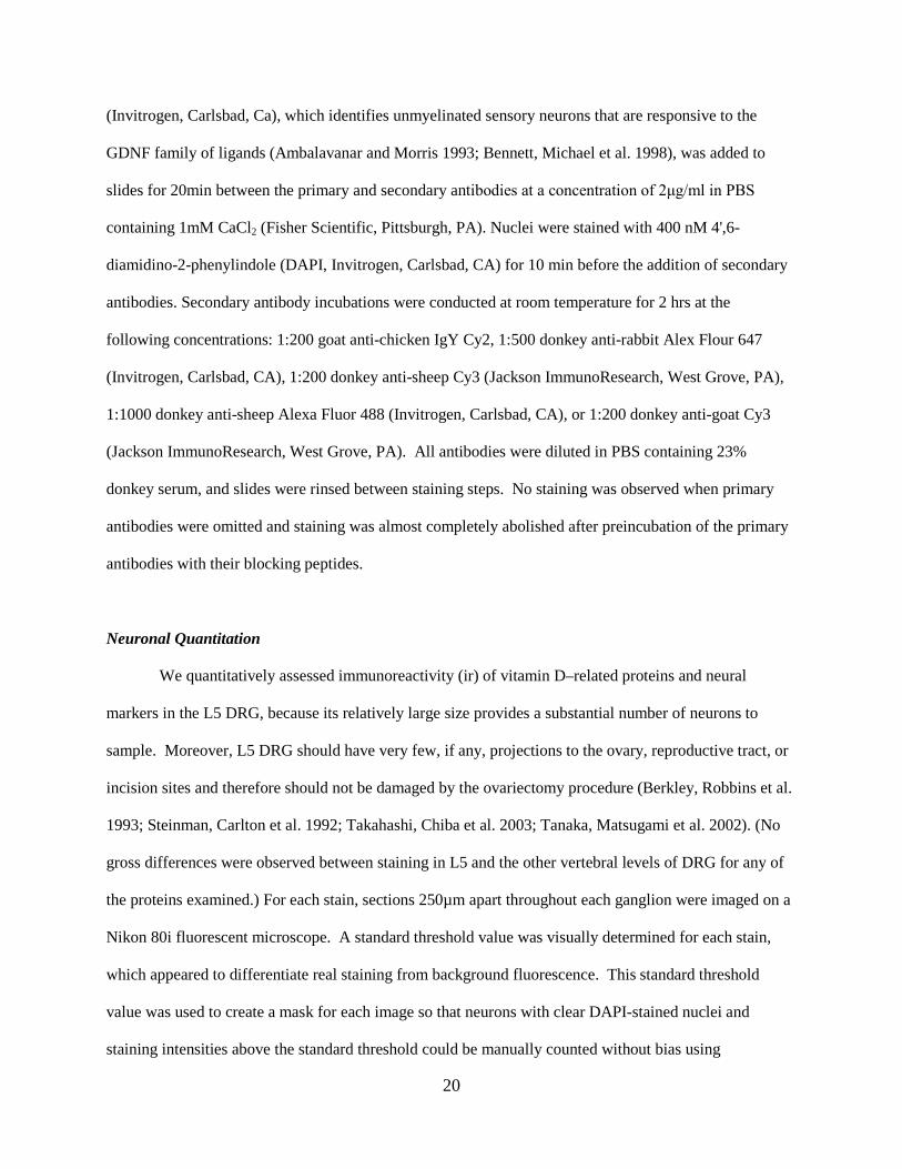
20
(Invitrogen, Carlsbad, Ca), which identifies unmyelinated sensory neurons that are responsive to the
GDNF family of ligands (Ambalavanar and Morris 1993; Bennett, Michael et al. 1998), was added to
slides for 20min between the primary and secondary antibodies at a concentration of 2μg/ml in PBS
containing 1mM CaCl2 (Fisher Scientific, Pittsburgh, PA). Nuclei were stained with 400 nM 4',6-
diamidino-2-phenylindole (DAPI, Invitrogen, Carlsbad, CA) for 10 min before the addition of secondary
antibodies. Secondary antibody incubations were conducted at room temperature for 2 hrs at the
following concentrations: 1:200 goat anti-chicken IgY Cy2, 1:500 donkey anti-rabbit Alex Flour 647
(Invitrogen, Carlsbad, CA), 1:200 donkey anti-sheep Cy3 (Jackson ImmunoResearch, West Grove, PA),
1:1000 donkey anti-sheep Alexa Fluor 488 (Invitrogen, Carlsbad, CA), or 1:200 donkey anti-goat Cy3
(Jackson ImmunoResearch, West Grove, PA). All antibodies were diluted in PBS containing 23%
donkey serum, and slides were rinsed between staining steps. No staining was observed when primary
antibodies were omitted and staining was almost completely abolished after preincubation of the primary
antibodies with their blocking peptides.
Neuronal Quantitation
We quantitatively assessed immunoreactivity (ir) of vitamin D–related proteins and neural
markers in the L5 DRG, because its relatively large size provides a substantial number of neurons to
sample. Moreover, L5 DRG should have very few, if any, projections to the ovary, reproductive tract, or
incision sites and therefore should not be damaged by the ovariectomy procedure (Berkley, Robbins et al.
1993; Steinman, Carlton et al. 1992; Takahashi, Chiba et al. 2003; Tanaka, Matsugami et al. 2002). (No
gross differences were observed between staining in L5 and the other vertebral levels of DRG for any of
the proteins examined.) For each stain, sections 250µm apart throughout each ganglion were imaged on a
Nikon 80i fluorescent microscope. A standard threshold value was visually determined for each stain,
which appeared to differentiate real staining from background fluorescence. This standard threshold
value was used to create a mask for each image so that neurons with clear DAPI-stained nuclei and
staining intensities above the standard threshold could be manually counted without bias using

21
Metamorph software. Counting sections 250µm apart throughout the ganglion (3-5 sections) yielded an
average of 428±42 total neurons with clearly stained nuclei for each DRG. We further assessed cell types
in which vitamin D-associated proteins reside by co-immunostaining for peripherin, CGRP, or IB4 and
using the same color thresholding techniques for merged images as described above. We also
characterized the intracellular localization of VDR to determine if;1) Intensely stained VDR-
immunoreactive aggregates were present within the DAPI-stained nuclear area, or 2) VDR-
immunoreactive-staining above the standard threshold was found outside the DAPI-stained nuclear area
for each neuron. For each animal and stain, percentages of labeled neurons were computed from averages
of all sections analyzed, and data are presented as the mean + standard error. Significance (P<0.05) was
determined by ANOVA followed by post-hoc analysis using Student-Newman-Keuls (SNK) or student’s
t-test. For publication images were pseudocolorized and adjusted for brightness and contrast.
RESULTS Vitamin D-related proteins in dorsal root ganglia of rats at estrus
Vitamin D receptor
We assessed DRGs from intact female rats in estrus for the presence of VDRs. Western blots of
total DRG protein revealed a strong band at approximately 60kDa. Two fainter bands were also
consistently observed at 41kDa, and 24kDa and three very faint bands at 74kDa, 65kDa, and 50kDa could
also be identified (Fig. 1). Immunofluorescence of sectioned DRG revealed VDR-ir in neurons, but no
VDR-ir surrounding the neurons or within central nerve bundles where satellite glia and Schwann cells
are located (Fig. 2A). Most neurons displayed VDR-ir localized to small nuclear aggregates; however,
some neurons also had diffuse staining throughout the cell body or patchy membrane-localized staining.
For this study we will refer to diffuse VDR-ir as cytoplasmic, although some of this staining may be
confined to vesicles dispersed throughout the cytoplasm.

22
The percentage of total neurons with nuclear (nVDR) or cytoplasmic (cVDR) VDR-ir was
quantified in L5 DRG (Fig. 2G). About 71% of all neurons express nVDR, while 10% had cVDR
staining and 8% had both nVDR and cVDR-ir.
To determine whether VDR is differentially expressed by small or large fiber neurons, DRG
sections were co-immunostained for VDR and the small fiber specific intermediate filament peripherin
(Fig. 3A). Peripherin-positive and peripherin-negative neurons with clear DAPI stained nuclei were
counted (DAPI-staining is not shown). In intact rats, the percentage of neurons with nVDR did not differ
between the peripherin-negative and peripherin-positive neuronal subpopulations (Fig. 3G). On the other
hand, cVDR was preferentially expressed by small fiber peripherin-positive neurons (Fig. 3H). The
percentage of peripherin-ir neurons expressing cVDR was more than twice that of peripherin-negative
neurons (p=0.002).
To determine whether VDR is differentially expressed by a specific subset of small fiber neurons,
DRG sections were co-labeled for VDR and IB4 (Fig. 3B), which stains small fiber neurons responsive to
the GDNF family of ligands, or CGRP (Fig. 3C), which is found in NGF-dependent neurons. nVDR
expression did not differ between the two neuronal sub-types (Fig. 3I), while the percentage of CGRP
neurons that expressed cVDR was almost three times that of IB4-positive neurons (Fig. 3J; p=0.016).
CYP27B1
We assessed whether DRG from intact female rats in estrus express CYP27B1, the enzyme that
converts circulating 25(OH)D into active 1,25(OH)2D. Western blots revealed two bands that migrated at
approximately 56kDa and 25kDa (Fig. 1).
Immunofluorescent-staining of sectioned DRG revealed CYP27B1-ir in DRG neurons but not
glia (Fig. 2B). CYP27B1-ir was diffuse throughout neural cell bodies. The percentage of total neurons
expressing CYP27B1 was about 22% (Fig. 2H).
DAPI-stained DRG sections were immunofluorescently-labeled for CYP27B1 and peripherin to
determine whether CYP27B1 is differentially expressed by small or large fiber neurons (Fig. 4A, DAPI-

23
staining is not shown). The percentage of peripherin-ir neurons expressing CYP27B1 was more than
three times that of peripherin-negative neurons (Fig 4G; p<0.001).
DRG sections were immunofluorescently-labeled for CYP27B1 and IB4 (Fig. 4B) or CGRP (Fig.
4C) to determine whether expression occurs in specific neuronal subsets. The percentage of CGRP-ir
neurons that expressed CYP27B1 was more than two times the percentage of IB4-positive expressing
CYP27B1 (Fig. 4H; p=0.003).
CYP24
Western Blot was used to determine whether DRG from intact female rats in estrus express
CYP24, an enzyme involved in the degradation of 1,25(OH)2D and the production of other vitamin D
metabolites. A single faint band of the expected size of 55kDa was identified on western blots run with
total DRG protein (Fig. 1).
Immunofluorescence of sectioned DRG revealed CYP24-ir in DRG neurons but not glia (Fig.
2C). Like CYP27B1-ir, CYP24-ir was diffuse throughout neural cell bodies. The percentage of total
neurons with CYP24 was quantified in the L5 DRG of intact female rats in estrus. About 17% of all
neurons express CYP24 (Fig. 2I).
To determine whether CYP24 is differentially expressed by large or small fiber neuronal cell
types, DRG sections were co-labeled for CYP24 and peripherin (Fig. 5A). CYP24 was preferentially
expressed by small fiber peripherin-positive neurons (Fig. 5G). The percentage of peripherin-ir neurons
expressing CYP24 was one and a half times that of peripherin-negative neurons.
We assessed the extent to which CYP24 is expressed by peptidergic or non-peptidergic small
fiber neurons. DRG sections were immunofluorescently-labeled for CYP24 and CGRP (Fig. 5C) or IB4
(Fig. 5B). The percentage of CGRP-positive neurons expressing CYP24 was about four times more than
the percentage of IB4-positive neurons expressing CYP24 (Fig. 5H; p<0.001).

24
cVDR colocalizes with CYP27B1 and CYP24
Because cVDR, CYP27B1, and CYP24 were all preferentially expressed by peripherin-ir, CGRP-
ir neurons, we co-stained DRG for cVDR and CYP27B1 (Fig. 6A) or CYP24 (Fig. 6B) to determine if
they are expressed within the same neurons. Most neurons with cVDR-ir were observed to have
CYP27B-ir and CYP24-ir, showing that neurons with cVDR typically have proteins necessary to locally
regulate 1,25(OH)2D concentrations.
Effect of ovariectomy (OVX) on Vitamin D-related proteins in dorsal root ganglia
VDR
Having verified that DRG neurons from intact female rats express VDR, CYP27B1, and CYP24,
we assessed whether expression of these proteins is affected by OVX. We began by examining whether
OVX alters VDR protein levels in total DRG lysates (Fig. 7A). The only VDR isoform to change
significantly following OVX was the 60kDa band, which was reduced by 51% compared to lysates from
intact rats in estrus (p=0.037).
Immunohistochemistry revealed that, as in intact rats, VDR-ir in ovariectomized rats was
distributed in nuclear and cytoplasmic compartments in neurons, but not glia (Fig. 2D). Approximately
66% of neurons in DRG from ovariectomized rats express nVDR, 8% express cVDR, and 6% express
both (Fig. 2G); only the modest reduction in nVDR was significant (p=0.038).
DAPI-stained DRG sections from ovariectomized rats were co-labeled for VDR and peripherin
(Fig. 3D). The percentage of peripherin-ir and peripherin-negative DRG neurons expressing nVDR in
ovariectomized rats did not change (Fig. 3G). However, the percentage of peripherin-ir neurons with
cVDR was reduced, so that there was no longer any difference in the percentage of peripherin-ir and
peripherin-negative neurons expressing cVDR in ovariectomized rats (Fig. 3H).
DAPI-stained DRG sections from ovariectomized rats were co-labeled for VDR and IB4 (Fig.
3E) or CGRP (Fig. 3F). The percentage of DRG neurons expressing nVDR in ovariectomized rats was

25
comparable in CGRP-ir and IB4-positive neurons (Fig. 3I). However, there was a 63% decrease in the
percentage of CGRP-ir expressing cVDR following OVX (Fig. 3J; p=0.009).
CYP27B1
Western blots of total DRG protein showed that while there was no change in expression of the
25kDa CYP27B1 band, expression of the 56kDa CYP27B1 band dropped by 31% following OVX (Fig.
7B; p=0.042).
CYP27B1-ir in DRG from ovariectomized rats was found in neurons, but not glia (Fig. 2E). The
average percentage of total neurons expressing CYP27B1 dropped from 22±4% to 10±2% following
OVX, but this change did not achieve statistical significance (Fig. 2H; p=0.063).
DAPI-stained DRG sections from ovariectomized rats were co-labeled for peripherin and
CYP27B1 (Fig. 4D). Ovariectomy did not affect the small proportion of peripherin-negative neurons
expressing this enzyme. (Fig. 4G). However, OVX resulted in a 57% decrease in proportion of
peripherin-ir neurons expressing CYP27B1 (p=0.003) to a level comparable to that of peripherin-negative
neurons (Fig. 4G).
DAPI-stained DRG sections from ovariectomized rats were co-labeled for CYP27B1 and IB4
(Fig. 4E) or CGRP (Fig. 4F). Ovariectomy did not significantly alter the proportion of IB4-positive
neurons expressing CYP27B1 (Fig. 4H). In contrast, there was a 61% drop in the proportion of CGRP-ir
DRG neurons with CYP27B1-ir after OVX (Fig. 4H; p<0.001), such that percentages of neurons
expressing this enzyme were comparable in IB4 and CGRP-ir subpopulations.
CYP24
Western blots of total DRG protein showed that there was no significant change in total CYP24
levels following OVX (Fig. 7C). As in rats at estrus, CYP24-ir was present exclusively in neurons (Fig.
2F), and immunostaining showed no change in the percentage of total neurons expressing CYP24
following OVX (Fig. 2I).

26
DAPI-stained DRG sections from ovariectomized rats were co-labeled for peripherin and CYP24
(Fig. 5D). As with rats in estrus, CYP24-ir was preferentially expressed by small fiber peripherin-
positive neurons in OVX rats (Fig. 5G). Ovariectomy did not alter the proportion of peripherin-ir or
peripherin negative neurons displaying CYP24-ir (Fig. 5G).
DAPI-stained DRG sections from OVX rats were co-labeled for CYP24 and IB4 (Fig. 5E) or
CGRP (Fig. 5F). As in estrus, CYP24-ir was present predominantly within CGRP-ir neurons, with only
small numbers of IB4-positive neurons expressing this enzyme (Fig. 5H). Ovariectomy did not alter
numbers of either CGRP-ir or IB4-positive neurons expressing CYP24 (Fig. 5H).
Changes in neural markers following ovariectomy
We assessed whether changes in neuronal protein expression following OVX could contribute to
observed differences in expression of vitamin D-related proteins. The percentage of total neurons
expressing peripherin was 63% in intact rats and underwent a small but significant decline to 59%
following OVX (p=0.042, Table 1). Within the peripherin-positive population of DRG neurons, OVX
resulted in a 38% decline in the percentage of neurons expressing CGRP-ir, while the IB4-positive
population was unaffected (Table 1).
The small decrease in peripherin-positive neurons resulted in a proportional increase in
peripherin-negative neurons , but the small numbers of these neurons expressing CGRP or IB4 were
unaffected (Table 1). The percentages of IB4-positive and CGRP-ir neurons that expressed peripherin-ir
were unaltered by OVX (Tables 1).
DISCUSSION The central finding of this study is that adult rat sensory neurons express proteins associated with
vitamin D signaling pathways. Nuclear VDR is widely distributed within all types of DRG neurons
suggesting broad transcriptional actions of this hormone. Membrane and cytoplasmic VDR, perhaps
indicative of rapid signaling pathways, is present selectively in unmyelinated, CGRP-ir neurons. This

27
same subpopulation contains vitamin D metabolizing enzymes, implying close regulation of vitamin D
metabolite levels. Ovarian hormones may play an important role in regulating neuronal responsiveness to
1,25(OH)2D by up-regulating both its activating enzyme and its cytoplasmic receptor. Because vitamin D
deficiency is strongly associated with musculoskeletal pain and this is exacerbated by the loss of ovarian
hormones, it is important to consider the role of these convergent systems in regulating nociceptor
function.
Vitamin D signaling in the central nervous system
Vitamin D is becoming recognized as an important hormone in nervous system health. VDR is
expressed by neurons, oligodendrocytes, and astrocytes within the limbic system, basal ganglia, spinal
cord, cerebellum, and cerebral cortex (Baas, Prufer et al. 2000; Eyles, Smith et al. 2005; Glaser, Veenstra
et al. 1999; Neveu, Naveilhan et al. 1994; Perez-Fernandez, Alonso et al. 1997; Prufer, Veenstra et al.
1999; Walbert, Jirikowski et al. 2001). VDR localization within both neuronal nucleus and cytoplasm has
been described (Glaser, Veenstra et al. 1999; Prufer, Veenstra et al. 1999; Racz and Barsony 1999).
Studies have also shown that neurons and glia in brain express CYP27B1 (Eyles, Smith et al. 2005;
Zehnder, Bland et al. 2001), and CYP24 mRNA has been detected in glioma and primary glial cell culture
(Naveilhan, Neveu et al. 1993), suggesting that the brain can regulate local 1,25(OH)2D concentrations.
Vitamin D signaling has been linked to cell survival, proliferation, and differentiation, and is especially
important during brain development (Levenson and Figueiroa 2008). In fact, gestational vitamin D
deficiency is thought to be associated with long term cognitive disorders such as anxiety and
schizophrenia (Levenson and Figueiroa 2008). In the adult, vitamin D supplementation has been
proposed to improve outcomes in multiple sclerosis, Parkinson’s disease, and traumatic brain injury
(Cekic, Sayeed et al. 2009; Myhr 2009; Newmark and Newmark 2007). Thus, accumulating evidence
suggests that vitamin D signaling plays important roles in the central nervous system.

28
Vitamin D-related proteins in adult rat DRG
Much less is known regarding the role of vitamin D in the peripheral nervous system. VDR has
been identified in fetal (E12-E21) DRG neurons but not glia (Johnson, Grande et al. 1996; Veenstra,
Prufer et al. 1998) (although VDR mRNA was reported in cultured sciatic nerve Schwann cells (Cornet,
Baudet et al. 1998)). However, the presence of VDRs and related proteins in the adult peripheral nervous
system is not well documented.
Our western blot findings show that VDR protein is present within adult rat DRG, and
immunocytochemical analyses show that it is found in most neurons. VDR can regulate transcription by
forming homodimers or heterodimers with 9-cis retinoid X receptor (RXR) which then translocate to the
nucleus (Shaffer and Gewirth 2004). Consistent with a transcriptional role, nuclear VDR was observed
in the majority of DRG neurons as small discrete foci (Glaser, Veenstra et al. 1999; Prufer, Veenstra et al.
1999); because these may be absent from a given section plane of a given neuronal nucleus, we may have
underestimated their actual prevalence. These foci are believed to represent binding of ligand-activated
RXR/VDR heterodimers to DNA, thus corresponding to sites of transcription (Prufer, Racz et al. 2000).
Because nuclear VDR was distributed widely throughout all DRG neural subtypes, VDR is likely to act as
a classical transcription factor in most adult DRG neurons.
We also observed a subset of neurons where VDR-ir was localized to the cytoplasm or plasma
membrane. While some of the cytosolic VDR could represent de novo synthesis or ligand-deficient VDR
dimers (Prufer, Racz et al. 2000; Racz and Barsony 1999), another explanation is that this VDR fraction is
involved in rapid signaling. The most prominent band disclosed in DRG homogenates had a mass of
60kDa, and this isoform reportedly is exclusive to membrane and cytosolic subcellular fractions
(Gonzalez Pardo, Boland et al. 2008). In cardiomyocytes, cVDR serves as a reserve which, upon
1,25(OH)2D binding, translocates to cardiac t-tubules (Tishkoff, Nibbelink et al. 2008). The patchy VDR-
ir pattern localized to the plasma membrane of some DRG neurons suggests a distribution to distinct
membrane microdomains, and is consistent with a role for VDR in rapid signaling in sensory neurons
(Huhtakangas, Olivera et al. 2004). For example, 1,25(OH)2D can influence calcium channel activity,

29
leading to rapid increases in intracellular calcium concentration and production of second messengers
(Losel, Falkenstein et al. 2003). Thus, in addition to its transcriptional role within all DRG neuronal
populations, VDR may exert rapid signaling in a selected subpopulation of neurons.
VDR signaling in DRG neuronal subpopulations
Selective neuronal markers provide insight as to how VDR signaling may vary as a function of
neuronal subtype. Neurons lacking the intermediate filament protein peripherin are predominantly large
myelinated neurons (Goldstein, House et al. 1991), which are involved in tactile sensation and
proprioception (Casellini and Vinik 2007; Muller, Ryals et al. 2008; Nardone, Galante et al. 2007).
These neurons contained nVDR but were mostly devoid of both CYP27B1-ir and CYP24-ir. This implies
that large sensory neurons may be influenced by VDR transcriptional signaling, but that VDR activation
is determined passively by levels of circulating 1,25(OH)2D, as they likely lack the ability to regulate
local 1,25(OH)2D levels. The functional significance of this signaling pathway is unclear, but it is
noteworthy that vitamin D deficiency in humans is associated with increased incidence in falls (Bischoff-
Ferrari, Dawson-Hughes et al. 2004; Faulkner, Cauley et al. 2006), which may suggest altered
proprioceptive function of large sensory fibers.
Neurons containing peripherin-ir represent unmyelinated C-fibers, which are associated with
chemoreception, thermal sensation, and pain, burning and itching. As with large myelinated neurons, the
majority of these also showed VDR-ir localized to the nucleus. However, these peripherin-ir neurons also
include the subgroup in which VDR is localized to the cytoplasm, most often in conjunction with nuclear
staining. It is well known that C-fiber neurons can be further characterized based on their neurotrophin
dependencies and peptide content, and our colocalization studies show that the cytoplasmic form of VDR
is distributed selectively in the NGF-dependent, CGRP-ir neurons, with approximately one third
displaying cVDR; these neurons conduct information regarding pain and temperature, and antidromically
release neuropeptides that promote vasodilation and extravasation, thus contributing to inflammation

30
(Birklein and Schmelz 2008). These findings suggest that vitamin D can activate multiple signaling
pathways, which may functionally influence a subpopulation of neurons with pain-sensing capabilities.
Our observations regarding VDR distribution in the DRG suggest that some neurons may be more
strongly influenced by vitamin D than others. If so, then local vitamin D metabolite levels for those
neurons may require closer regulation. Findings concerning vitamin D-related proteins provide evidence
that this is the case. Hence, the prohormone converting enzyme CYP27B1 is enriched specifically in
peripherin-ir, CGRP-ir neurons, as is CYP24, which could be involved in the inactivation of 1,25(OH)2D
or perhaps the formation of other metabolites such as 24,25(OH)2D3. Further, these enzymes co-localize
selectively within neurons displaying cytoplasmic VDR. Collectively, these findings are consistent with
the hypothesis that DRG peptidergic neurons possess vitamin D signaling pathways, whose gain can be
modulated by altering levels of cVDR, or by regulating levels of vitamin D metabolic enzymes.
Regulation of VDR and CYP27B1by ovarian hormones
One likely factor in regulating vitamin D-related proteins in DRG is female reproductive
hormone status. Ovarian hormones have been found to promote the formation of 1,25(OH)2D by
enhancing CYP27B1 expression and by inhibiting the expression of CYP24 (Lechner, Bajna et al. 2006;
Tanaka, Castillo et al. 1978). They are also reported to promote the expression of VDR (Gilad, Bresler et
al. 2005). In cancer models there has been some progress in uncovering how estradiol regulates VDR
expression. Estradiol binds membrane bound ERβ receptors, which signal through ERK1/2 to increase
transcripton of VDR (Gilad, Bresler et al. 2005). Research from another group has shown that estrogen
may regulate VDR promoter activity of exon 1C through SP1 sites, which can be mediated through ERα
or ERβ (Wietzke, Ward et al. 2005). To determine whether ovarian hormones regulate VDR or enzyme
expression in DRG neurons, we compared vitamin D-related protein expression in DRG from cycling rats
at estrus to that of ovariectomized rats. Estrus occurs several hours after the natural surge in reproductive
hormones including estradiol, progesterone, and prolactin (Freeman 1994), when hormone-responsive
changes in protein expression are most apt to be evident. In contrast, rats following OVX have sustained

31
depression of ovarian hormones similar to that which occurs naturally in menopause, albeit occurring at a
younger age and more abruptly than would occur normally.
Consistent with reported findings for other tissues, OVX resulted in decreased expression of VDR
and CYP27B1. In protein blots, the cytoplasm/membrane-specific 60kDa isoform of VDR was reduced
by 51%, and the 56kDa full length CYP27B1 band was reduced by 31%. Immunofluorescence
examination showed that the numbers of neurons with cVDR and CYP27B1 expression were both
diminished, and this decrease occurred exclusively within the peripherin-ir, CGRP-ir population. In
contrast, no significant changes occurred in nVDR or CYP24. Therefore, based on these findings in OVX
rats, a decline in ovarian hormones would not only down-regulate cVDR expression, but would also
decrease the amount of CYP27B1 available to convert prohormone to active 1,25(OH)2D. At the same
time, levels of the CYP24 remain unchanged, thereby favoring a reduction in levels of 1,25(OH)2D.
Accordingly, low ovarian hormone levels may impair vitamin D signaling in peptidergic nociceptor
neurons by diminishing levels of both cyotplasmic receptor and ligand.
While these observations are consistent with a reduction in the numbers of neurons in which
cVDR signaling occurs, a caveat is that changes in reproductive hormones could influence marker
expression, thereby introducing error to our counts of neuronal subpopulations containing vitamin D-
related proteins. While hormone-related changes in IB4-positive neurons have not been reported (and
were not detected here), it is known that estrogens increase CGRP expression (Gangula, Lanlua et al.
2000; Mowa, Usip et al. 2003). Consistent with these reports, we found that OVX resulted in a reduction
of roughly 1/3 in the number of neurons expressing CGRP-ir. Of those retaining the CGRP phenotype,
cVDR and activating enzyme expression were reduced by over 60%. This occurred without concomitant
increases in cVDR or CYP27B1 in other cell phenotypes that might imply changes due to altered marker
expression. It therefore appears that ovarian hormone deficiency selectively affects a subpopulation of
DRG neurons, where both peptide phenotype and VDR signaling are dramatically reduced.

32
Vitamin D, ovarian hormones, and pain
While an association between pain and vitamin D deficiency has been observed for at least 20
years (Masood, Narang et al. 1989), this is often attributed to bone and muscle pathologies. Muscle
symptoms, including pain, precede the appearance of biochemical indicators of bone pathology (Glerup,
Mikkelsen et al. 2000; Masood, Narang et al. 1989), suggesting that bone is not the only source of
musculoskeletal pain. Vitamin D deficiency does lead to atrophy of type II muscle fibers (Glerup,
Mikkelsen et al. 2000); however, type II muscle fiber atrophy is non-specific and does not in itself cause
pain (Mastaglia and Hilton-Jones 2007). Based on our findings, it seems likely that vitamin D may act
directly on sensory nerves to modulate pain. Moreover, the extent to which vitamin D deficiency
produces musculoskeletal pain appears to be strongly linked to estrogen status. For example,
musculoskeletal pain occurs commonly in hypoestrogenemia resulting from naturally occurring
menopause (Andersson, Ejlertsson et al. 1993; Bergman, Herrstrom et al. 2001; Croft, Rigby et al. 1993;
Dugan, Powell et al. 2006; Greendale, Reboussin et al. 1998; White, Speechley et al. 1999; Wolfe, Ross
et al. 1995) or pharmacotherapy using aromatase inhibitors or gonadotropin-releasing hormone agonists
(Felson and Cummings 2005; Friedman, Juneau-Norcross et al. 1993; Garreau, Delamelena et al. 2006;
Morales, Pans et al. 2006; Mouridsen 2006). Musculoskeletal pain associated with menopause is
ameliorated by hormone replacement therapy (Dugan, Powell et al. 2006), implying a direct link between
pain and hormone levels. It is not clear why musculoskeletal pain occurs with hypoestrogenemia.
However, low estrogen levels following aromatase inhibition are accompanied by low serum 25(OH)D
levels, and high dose vitamin D3 supplementation partially reverses the discomfort (Khan, Reddy et al.
2009; Waltman, Ott et al. 2009), consistent with a link among estrogens, vitamin D and pain. However, it
should be noted that high dose vitamin D3 treatment does not fully alleviate pain symptoms. This may
not be surprising given our findings that low ovarian hormone levels are also associated with diminished
DRG neuronal expression of VDR and CYP27B1. Thus, hypoestrogenemia might hinder conversion of
the therapeutic supplement to the active hormone and limit receptor-mediated signaling, thereby
rendering dietary supplementation less effective. Collectively, these studies and the present findings

33
underscore a need for additional research clarifying the relationships among vitamin D, steroid hormones,
and sensory pathways.

34
TABLES
Table 1. Quantitative analysis of neuronal markers and effect of ovariectomy. This table shows the
percentages of total neurons, peripherin-positive neurons, peripherin-negative neurons,
CGRP-positive neurons, or IB4-positive neurons that express peripherin, CGRP, or IB4. %
Estrus (% OVX). There was a significant drop in the percentage of total neurons expressing
peripherin and peripherin-positive neurons co-expressing CGRP following ovariectomy,
p=*0.042 and **0.049 by student’s t-test.
Total neurons
%
Peripherin-positive neurons
%
Peripherin-negative neurons
%
IB4 neurons
%
CGRP neurons
%
Peripherin *63±1 *(59±1)
100 (100)
0 (0)
90±2 (85±3)
85±2 (76±4)
IB4 35±2 (36±2)
54±4 (57±1)
9±2 (11±2)
100 (100)
Not determined
CGRP 43±7 (28±3)
**55±9 **(34±3)
18±2 (19±4)
Not determined
100 (100)

35
FIGURES Figure 1.

36
Figure 1. VDR, CYP27B1, and CYP24 proteins are present in adult DRG. Western blots of
extracted DRG proteins from intact rats in estrus were probed for VDR, CYP27B1, or CYP24.
Samples were run alongside molecular markers in order to estimate protein size. Images are cropped
to show representative lanes.

37
Figure 2.

38
Figure 2. Cellular localization of VDR, CYP27B1, and CYP24 in adult DRG. Cryosections of L5
DRG from intact rats in estrus (A-C) or ovariectomized (OVX) rats (D-F) were immunofluorescently
labeled for VDR (A&D; red), CYP27B1 (B&E; red), or CYP24 (C&F; red), and nuclei were stained
with DAPI (shown only in the insets of A and D; blue). Examples of neurons with nuclear-localized
VDR are indicated in A and D by arrow heads, and neurons with intense cytoplasmic VDR staining
are indicated by arrows. The insets show magnified examples of neurons with nuclear or nuclear and
cytoplasmic VDR immunoreactivity. In B& E, examples of neurons with intense labeling for
CYP27B1 are indicated by arrows. In C & F, neurons with intense labeling for CYP24 are indicated
by arrows. In A-F staining is not consistent with glial expression of VDR, CYP27B1, or CYP24 (ie.
No staining surrounding neurons in satellite glia or in the Schwann cells of outlined nerve bundles
(NB)). In G, the total proportion of neurons expressing VDR in estrus or after OVX is presented.
The shaded area within each bar represents the proportion of neurons with both cytoplasmic and
nuclear VDR staining. Significant differences between individual groups are shown by bracketing,
p= *<0.001 and **0.038. The overall difference between Estrus and OVX expressing VDR was also
significant, p=0.028. H and I present quantitative analysis of the percentages of neurons
immunostained for CYP27B1 and CYP24, respectively. Scale bar = 100μm (10μm in insets).

39
Figure 3.

40
Figure 3. Cellular colocalization of VDR with the neural markers peripherin, IB4, and CGRP.
Cryosections of L5 DRG from intact rats in estrus (A-C) or ovariectomized rats (D-F) were
immunofluorescently-labeled for VDR (A-F; red) and peripherin (A&D; green), IB4 (B&E; green),
or CGRP (C&F; green), and the nuclei were stained with DAPI (A-F not shown). Neurons with
nuclear VDR and neural markers are indicated by arrowheads, and neurons with cytoplasmic VDR
and neural markers are indicated by arrows. G-J: The mean percentages of neurons with neural
marker immunoreactivity that express nVDR or cVDR are presented in graphical form. Significant
differences between individual groups are shown by bracketing, p= H) *0.002, J) *0.016 and
**0.009. Overall differences between neural markers were significant for H) cVDR peripherin (+)
vs. peripherin (-) (p=0.001) and J) cVDR IB4 vs. CGRP (p=0.039). Overall differences between
Estrus and OVX were also significant J) p=0.027. Scale bar = 50μm.

41
Figure 4.

42
Figure 4. Cellular colocalization of CYP27B1 with the neural markers peripherin, IB4, and CGRP.
Cryosections of L5 DRG from intact rats in estrus (A-C) or ovariectomized rats (D-F) were
immunostained for CYP27B1 (A-F; red) and peripherin (A&D; green), IB4 (B&E; green), or CGRP
(C&F; green), and the nuclei were stained with DAPI (A-F not shown). Neurons with intense
CYP27B1 and neural marker staining are indicated by arrows. G-J: The mean percentages of neurons
with neural marker immunoreactivity that express CYP27B1 are illustrated in graphical form.
Significant differences between individual groups are shown by bracketing, p= G) *<0.001 and
**0.003, H) *0.003 and <0.001. Overall differences between neural markers was significant for G)
CYP27B1 peripherin (+) vs. peripherin (-) (p<0.001) and J) CYP27B1 IB4 vs. CGRP (p=0.005).
Overall differences between Estrus and OVX were also significant G) p=0.005 and J) p=0.001.
Scale bar = 50μm.

43
Figure 5.

44
Figure 5. Cellular colocalization of CYP24 with the neural markers peripherin, IB4, and CGRP.
Cryosections of L5 DRG from intact rats in estrus (A-C) or ovariectomized rats (D-F) were
immunostained for CYP24 (A-F; red) and peripherin (A&D; green), IB4 (B&E; green), or CGRP
(C&F; green), and the nuclei were stained with DAPI (A-F not shown). Neurons with intense CYP24
and neural marker staining are indicated by arrows. G&H: The mean percentages of neurons with
neural marker immunoreactivity that express CYP24 are illustrated in graphical form. Significant
differences between individual groups are shown by bracketing, p= G) *0.024 and **0.005, J)
*<0.001 and **0.001. Overall differences among neural markers were significant for G) CYP24
peripherin (+) vs. peripherin (-) (p=0.005) and J) CYP24 IB4 vs. CGRP (p<0.001). Scale bar = 50μm

45
Figure 6.

46
Figure 6. Cellular colocalization of VDR with the enzymes CYP27B1 or CYP24. Cryosections of
L5 DRG from rats in estrus were immunostained for VDR (red) and the enzymes CYP27B1 (A) or
CYP24 (B) (green) and nuclei were stained with DAPI (not shown). Neurons co-labeled for each
enzyme and cVDR are indicated by arrows. Scale bar = 50 μm.

47
Figure 7.

48
Figure 7. DRG expression of VDR and CYP27B1, but not CYP24 is reduced following
ovariectomy. Western blots of proteins extracted from DRG of intact rats in estrus or ovariectomized
rats were probed for VDR (A), CYP27B1 (B), or CYP24 (C) and GAPDH (A-C) as a loading
control. A) The 60kDa band of VDR showed a significant decrease in intensity following
ovariectomy. B) The 56kDa band of CYP27B1 also showed a significant drop in intensity following
ovariectomy. C) There was no significant change in CYP24 after ovariectomy. OVX n=5, Estrus
n=4, 2-3 replicates per animal were averaged. Significance determined by student’s t-test.

49
CHAPTER 3. VITAMIN D DEFICIENCY PROMOTES SENSORY AXON SPROUTING AND MUSCLE HYPERSENSITIVITY

50
ABSTRACT
Musculoskeletal pain affects 35-50% of adults, most of whom are vitamin D deficient. Previous findings
from this laboratory have demonstrated that putative nociceptors express vitamin D receptors (VDRs),
suggesting that vitamin D metabolites can act directly on sensory nerves. In this study, 2-4 weeks of a
vitamin D deficient diet produces deep muscle mechanical hypersensitivity in rats that is not corrected by
dietary calcium supplementation. This hypersensitivity to deep pressure is accompanied by nociceptor
hyperinnervation of the muscle. In culture, VDRs are enriched in small fiber sensory growth cones and
rapid response receptors regulate neurite growth consistent with in vivo findings. Because vitamin D
metabolites directly inhibit axonal growth of sensory c-fibers, vitamin D deficiency leads to muscle
hyperinnervation, which may contribute to musculoskeletal pain.

51
INTRODUCTION
Vitamin D recently has been proposed to play wide ranging roles in human health, and
deficiencies in this secosteroid are implicated in chronic musculoskeletal pain (Benson, Wilson et al.
2006; de Torrente de la Jara, Pecoud et al. 2006; Glerup and Eriksen 1999; Gloth, Lindsay et al. 1991;
Heidari, Shirvani et al. 2010; Macfarlane, Palmer et al. 2005; McBeth, Pye et al. 2010; Plotnikoff and
Quigley 2003) and an increased risk of falling (Annweiler, Montero-Odasso et al. 2010). 27.5-36% of
adolescents and 35-50% adults suffer from chronic musculoskeletal pain (Bergman 2007; De Inocencio
2004) and up to 93% of them may be vitamin D deficient, with levels of serum 25-hydroxyvitamin D
(25(OH)D) less than 20ng/ml (50nmol/L) (Plotnikoff and Quigley 2003). Some studies show that this
pain may be relieved by vitamin D supplementation (de Torrente de la Jara, Pecoud et al. 2006; Glerup
and Eriksen 1999; Gloth, Lindsay et al. 1991). However, it is difficult to establish a causal relationship, as
the incidence of musculoskeletal pain is increased in people that are older, have a higher body mass
index, and spend less time out-of-doors, all factors contributing to vitamin D deficiency (Bergman 2007;
Yetley 2008).
While vitamin D could affect pain sensitivity at multiple sites, we found recently that small
unmyelinated dorsal root ganglion (DRG) sensory neurons express both nuclear and
membrane/cytoplasmic vitamin D receptors (VDRs) (Tague and Smith 2010), suggesting that sensory
neurons, including putative nociceptors, are direct targets of an active vitamin D hormone, 1,25-
dihydroxyvitamin D (1,25(OH)2D). In order to further investigate the relationship between vitamin D and
musculoskeletal pain, a rat model in which vitamin D deficiency is reliably induced was used. Reducing
dietary intake of vitamin D can induce persistent vitamin D deficiency in laboratory rats (Weishaar and
Simpson 1987). However, vitamin D deficiency, in turn, leads to reductions in serum calcium that can
affect many physiological functions independently (Johnson and DeLuca 2002; Uhland, Kwiecinski et al.
1992; Weishaar and Simpson 1987), although this can be corrected by administering a diet high in
calcium and phosphate (Weishaar and Simpson 1987).

52
MATERIALS AND METHODS
Animals and diets
All animal protocols and procedures were in accordance with the NIH guidelines for the care and use of
laboratory animals and were approved by the University of Kansas Medical Center Animal Care and Use
Committee. Fifteen weaned (24 day-old) female Sprague Dawley (Harlan) rats were housed 2-3 per cage
in forced-air-ventilated micro-isolation cages with a 14hr light/10hr dark light cycle and fed ad libitum a
normal chow diet. At the age of 31 days, rats were anaesthetized with ketamine 70mg/kg (Pfizer)/
xylazine 6mg/kg i.p. (Lloyd Laboratories) and ovariectiomized via bilateral hindflank incision. Upon
waking, and at 24hr and 48hr post-operation, rats were administered ketoprofen 5mg/kg s.c. (Ketofen;
Fort Dodge Animal Health) as an analgesic. At 48 days of age, rats were randomly separated into three
groups and fed one of three diets (blinded); Control: 2.2 IU/g vitamin D (cholecalciferol), 0.47% Ca,
0.3% P (Harlan Teklad, TD.07370), VD-/+ Ca: No vitamin D, 2.5% Ca, 1.5% P (Harlan Teklad,
TD.07541), VD-: No vitamin D, 0.47% Ca, 0.3% P (Harlan Teklad, TD.89123). Food was provided in a
ceramic bowl on the cage floor and replaced twice weekly. Rats were weighed weekly and food was
weighed before and after changing (only data for week 0, 2, and 4 are presented in Table 1). Rats were
maintained on their assigned diet for four weeks. After two weeks on the diet, one to two ml of blood
was drawn from the tail vein. Serum was collected and stored at -20 ºC. At the end of the study, rats were
anaesthetized with ketamine 70mg/kg / xylazine 6mg/kg (i.p.), thoracic cavities were opened, blood was
removed via cardiac puncture, and the rats were perfused with 50ml of cold 0.9% saline containing
10units/ml heparin (APP Pharmaceuticals) at a rate of 40ml/min, followed by 150-200ml of 4%
formaldehyde (prepared in PBS from paraformaldehyde (Sigma-Aldrich)). Serum was collected and
stored at -20C. See S1 for an overview of the experimental timeline.
Behavioral testing
All behavioral testing was performed in a quiet room under normal fluorescent lighting six to eight hours
into the light cycle. The rat cages were moved to the behavior room 1hr before testing. All statistical

53
analysis of behavior was conducted using two way repeated measures ANOVA with Student-Newman-
Keuls multiple comparisons.
Muscle mechanical sensitivity- The muscle compression test used in this study is similar to that of models
previously described (Sharma, Ryals et al. 2009; Skyba, Radhakrishnan et al. 2005). A digital Randall
Selitto instrument (IITC Life Science Inc) with a plastic cap with a 0.5cm wide surface area (S2) was
used. During the week prior to diet manipulation, rats were habituated to a rat sling (IITC Life Science,
Inc) for 5min on two separate days. The day prior to starting the diets, and each week thereafter, rats
were stabilized in the rat sling to allow free access to the hind limbs. While holding the left hind paw, the
flat side of the probe was placed against the inner left calf, while the capped pressure sensor was placed
on the center of the left exterior calf muscle. The instrument was slowly compressed until the rat
attempted a limb withdrawal (or vocalized). The calf was compressed three times and the maximum
force applied each time was recorded and averaged. To minimize stress, rats were restrained in the sling
for no longer than 3min/week. To ensure that the same part of the calf muscle was compressed each time,
the left calf was shaved each week 3 days before muscle compression under brief isoflurane anesthesia.
During week four, after initial testing, 2.5% lidocaine/2.5% prilocaine cream (TOLMAR Inc.) was
applied to the calf skin for 30 minutes and muscle sensitivity measurements were repeated. Similar to
previous reports (Skyba, Radhakrishnan et al. 2005), there were no significant differences in muscle
sensitivity before and after cutaneous lidocaine treatment (data not shown), verifying that this test
measures deep tissue pressure sensitivity and not cutaneous mechanical sensitivity.
Cutaneous mechanical sensitivity- Cutaneous mechanical sensation was measured using a calibrated 4g
monofilament. Rats were placed on a wire-mesh grid with plexiglass dividers and allowed to habituate for
20min. The monofilament was applied to the plantar surface of the hind paw five times per trial for a
total of three trials. The number of positive responses was recorded, and the percent withdrawal response
for each paw was calculated. A response was considered positive when the rat retracted its hind paw in

54
response to the monofilament. The results shown are averages from the left and right hind paws.
Cutaneous sensitivity was measured two days before starting diet manipulation and each week thereafter.
Balance and motor co-ordination - An elevated, 2.5 cm wide round dowel was suspended between two
support stands with an enclosed plastic platform at the one end. Rats were trained at the ages of 28, 39,
and 41 days to walk across the beam into the platform box, by placing them on the beam at increasing
distances from the box, until they voluntarily walked the full 110cm of the beam. Two days before
starting the diets, and at week 2 and 4, rats were videotaped for three beam walk trials. The videos were
analyzed to determine the number of hind paw footfall slips per beam walk, which were then averaged.
Muscle weakness - - Three days before starting the diets, and at weeks 2 and 4, rats were placed on a wire
grid attached to a digital force gauge (San Diego Instruments). The maximum grip force applied by the
forelimbs was measured as the rats were gently pulled by the base of their tail until their grip was broken.
This was repeated three times for each rat and the maximum force was recorded.
General locomotor activity - Rats were placed in a Force Plate Actimeter (BASi) (Fowler, Birkestrand et
al. 2001) and allowed to move freely for 5min. Movements were detected by force transducers and
recorded by a computer and rearing events were recorded by the investigator.
Serum Measurements
Serum 25-hydroxyvitamin D was measured by direct ELISA (Immunodiagnostik) according to
manufacturer instructions. Serum calcium and phosphorous was measured by an outside laboratory
(Physicians Reference Laboratory, L.L.C) by spectrophotometry.
Tissue processing
The left calf muscles (gastrocnemius and soleus) were removed and post-fixed in Zamboni’s fixative
overnight at 4ºC. Weight and volume (determined by fluid displacement in a graduated cylinder) were

55
recorded. The muscles were washed for two weeks and cryoprotected at 4ºC in phosphate buffered saline
containing 30% sucrose changed daily. The entire calf was cut in the transverse plane at 25%, 50%, and
75% of the way through the muscle. The muscle was embedded in tissue freezing media (Electron
Microscopy Science), frozen in liquid nitrogen-cooled isopentane, and stored at -80oC.
The left tibia was removed and post-fixed in Zamboni’s fixative overnight at 4ºC. The bones were
washed at 4ºC in PBS changed daily for three days, then decalcified in PBS containing 10% EDTA for
two weeks at 4ºC. The decalcified bones were then cryoprotected overnight at 4ºC in PBS containing
30% sucrose. The bone was cut in half along the transverse plane. The proximal portion was cut in half
along the sagittal plane and the medial portion was embedded in tissue freezing media for sagittal
sectioning of the epiphyseal plate. The distal half of the bone was embedded in tissue freezing media for
transverse sections of the diaphysis. Bones were frozen on dry ice and stored at -80oC.
Immunostaining of nerve fibers in muscle
Thawed 20µm cryosections of muscle were immunoflourescently labeled. Slides were incubated in pre-
incubation solution (1.5% donkey serum (Jackson ImmunoResearch), 0.5% gelatin (Sigma-Aldrich), and
0.5% Triton X-100 (Sigma-Aldrich) prepared in Superblock (Thermo Scientific) for 1hr at room
temperature. Primary and secondary antibodies were diluted in incubation solution (50% preincubation
solution, 50% Superblock). Primary antibodies were incubated overnight at room temperature and
secondary antibodies incubated for two hours. Before and after the secondary antibody, slides were
washed in PBS containing 0.25% Triton-X 100. Slides were then dipped in distilled water and mounted
with coverslips using 50% glycerol in PBS. Primary and secondary antibodies were diluted as follows:
1:500 chicken anti-peripherin (Chemicon), 1:200 sheep anti-CGRP (Biomol), 1:1,000 rabbit anti-
VMAT2 (Chemicon), or 1:1,500 rabbit anti-NFH (Sigma), 1:1,500 donkey anti-chicken DyLight 488
(Jackson), 1:750 Donkey anti-sheep Dylight 649 (Jackson), 1:1,000 donkey anti-rabbit 647 (Invitrogen).

56
Quantitation of innervation density
Images were taken (blinded) from transverse sections of the left lateral gastrocnemius muscle, which is
located on the outside of the calf where most of the force was applied during muscle compressions. A
total of 18 images were taken per animal, 6 each from three levels at 25%, 50%, and 75% through the
proximo-distal extent of the muscle using a Nikon 80i epifluorescent microscope. A stereological grid
was superimposed over the images, and numbers of grid intersections overlying immunoflourescently-
labeled axons was counted and divided by the total number of tissue intersections, to provide the apparent
percentage area occupied by nerves (Clarke, Bhattacherjee et al. 2010). Statistical analysis was
conducted using one way ANOVA and Student-Newman-Keuls multiple comparisons.
Bone measurements
Hematoxylin and eosin-stained 20µm sections of bone were analyzed. For growth plate analysis, three
fully visible chondrocyte columns in the center of each section were analyzed for height and cell number
and averaged per section. The transverse bone sections of the diaphysis were analyzed for bone
circumference and bone marrow area. Statistical analysis was completed with one way ANOVA and
Student-Newman-Keuls multiple comparisons.
Primary neuronal cell cultures
Sensory: DRG were removed from six week old female Sprague Dawley rats, dissociated with 2 units/ml
dispase (ICN Biomicals Inc) and 2mg/ml collagenase type 1A (Sigma) and plated on glass coverslips
(Bellco) coated with poly-D-lysine (Sigma) and laminin (Invitrogen) in 24-well culture dishes. Cultures
were maintained in Neurobasal A media (Invitrogen) containing 1mM glutamine (Sigma), 2% B27
(Gibco), 100µg/ml primocin (InvivoGen), 20µM FrdU (Sigma) and 20µM uridine (Sigma) for 4 days,
with concentrations of 1,25(OH)2D3 or JN that ranged from 0-100pM. Cultures were incubated at 37ºC
with 5% CO2 and media was changed after 24hrs and 72hrs. Cultures were fixed in 4% formaldehyde for

57
one hour, DAPI stained, and immunofluorescently-labeled for peripherin (chicken 1:1000, Chemicon),
NFH (mouse 1:200, Sigma), or VDR (rabbit 1:25, GeneTex) peripherin and GAP43 (mouse 1:240,
Zymed). Cultures were grown in triplicate and repeated twice.
Sympathetic: Superior cervical ganglia were removed from one day-old rats, dissociated with 0.25%
trypsin-EDTA (Gibco) and collagenase 1A (Sigma) and plated on poly-D-lysine and laminin-coated
coverslips. Cultures were maintained in Neurobasal A media as described above with the addition of
10µg/ml NGF (Alomone Labs) for 48 hours, with 0, 20pM or 100pM of 1,25(OH)2D3. Cultures were
fixed in 4% formaldehyde, DAPI stained and immunofluorescently-labeled for peripherin and VDR.
Cultures were grown in triplicate and repeated twice.
Neurite area quantitation
A stereological grid was superimposed over culture images and the number of times a grid intersection
crossed an immunofluorescently-labeled nerve fiber was counted and divided by the total number of
intersections within each field to provide neurite area; this was divided by the number of viable neurons,
determined by DAPI staining, to provide an estimate of neurite area per neuron. Statistical analysis was
completed with one way ANOVA and Student-Newman-Keuls multiple comparisons.
RESULTS AND DISCUSSION
To assess how vitamin D deficiency alters deep muscle sensitivity to noxious mechanical stimuli,
Sprague Dawley rats were fed chow deficient in vitamin D, with or without high levels of calcium.
Starting at seven weeks of age, rats were fed ad libitum, for four weeks, one of three blinded diets:
control, vitamin D deficient with increased calcium and phosphate (VD-/+Ca), or vitamin D deficient
with normal levels of calcium and phosphate (VD-) (Table 1). The VD-/+Ca diet was based on previously
published reports that increasing dietary Ca from 0.47% to 2.5% normalizes serum Ca in vitamin D

58
deficiency (1.5% P is needed as a counterbalance)(Weishaar and Simpson 1987). Female rats were
ovariectomized 2.5 weeks prior to initiation of the diet to remove the influence of cycling ovarian
hormones on behavioral sensitivity testing (Craft 2007) and VDR expression in sensory nerves (Tague
and Smith 2010). Additionally, the incidences of vitamin D deficiency and musculoskeletal pain are high
in women and tend to be associated with suppressed ovarian hormones (i.e. post-menopause,
pharmacologically-induced estrogen suppression) (Alexander, Dennerstein et al. 2007; Gaugris, Heaney
et al. 2005; Khan, Reddy et al. 2010). Rats were housed under normal fluorescent lighting with a 14 hr
light/10 hr dark cycle. Rats fed vitamin D-deficient diets showed weight gains comparable to control
subjects over the 4-week study (Table 1). However, food consumption was increased in the VD-/+Ca
group throughout the study and in the VD- group at week four (Table 1). To confirm vitamin D
deficiency, blood was drawn at 2 weeks from the tail vein and at 4 weeks via cardiac puncture. 25(OH)D
serum concentrations in both VD-/+Ca and VD- rats were reduced below 25nmol/L by week 2 and below
10nmol/L by week 4 (Table 1). In addition, serum Ca and P levels at week four were not reduced even
though Ca is reported to be decreased in VD- rats after longer intervals (Weishaar and Simpson 1987).
To determine whether vitamin D deficiency causes increased mechanical muscular sensitivity, we
used a behavioral testing paradigm developed for this purpose (Sharma, Ryals et al. 2009; Skyba,
Radhakrishnan et al. 2005). Rats were placed weekly in a restraining sling allowing free access to their
limbs. While holding the left foot, the left calf muscle was compressed using a modified digital Randall-
Selitto testing device (S2) until the rat attempted to withdraw the limb or vocalized withdrew (or
vocalized), and the maximum force applied by the testing device was recorded. Between 0 and 2 weeks,
the force required to elicit a response increased in control rats, a finding consistent with increased body
mass occurring in this period (Table 1), and plateaued thereafter (Fig. 1A). In VD- rats, mechanical
sensitivity was normal through week 2 of the diet, but was increased by week 3 and was significantly
higher than controls by week 4 (Fig 1A). Rats receiving the vitamin D-deficient diet with elevated Ca
showed normal sensitivity at week 1, but marked hypersensitivity by week 2 and thereafter (Fig. 1A).
Thus, the high calcium diet not only failed to reverse the hyperalgesic phenotype; it accelerated the onset
of deep tissue hypersensitivity. These findings support the hypothesis that dietary vitamin D deficiency
produces muscle hypersensitivity within 2-3 weeks in rats.

59
Gloth et. al first described patients with vitamin D deficiency attributed pain, which was triggered
by minimal movement or touch (Gloth, Lindsay et al. 1991). Accumulating evidence supports a causal
role for vitamin D deficiency in some (Benson, Wilson et al. 2006; de Torrente de la Jara, Pecoud et al.
2006; Glerup and Eriksen 1999; Gloth, Lindsay et al. 1991; Heidari, Shirvani et al. 2010; Macfarlane,
Palmer et al. 2005; McBeth, Pye et al. 2010; Plotnikoff and Quigley 2003), but not all (e.g. fibromyalgia
(Block 2004; de Rezende Pena, Grillo et al. 2010; Heidari, Shirvani et al. 2010; Tandeter, Grynbaum et
al. 2009; Warner and Arnspiger 2008)) types of musculoskeletal pain. However, it is unclear whether
hypersensitivity is generalized. To determine if hypersensitivity was apparent in other modalities, we
evaluated cutaneous sensitivity of the hind paw plantar surface using a von Frey calibrated 4g
monofilament. The percentage of applied mechanical stimuli that elicited a withdrawal response was
assessed on a weekly basis. There were no differences between groups in cutaneous sensitivity at any
week (Fig 1B). Therefore, while a vitamin D deficiency resulted in deep tissue mechanical
hypersensitivity, this does not appear to be due to wide-spread changes in sensory threshold that affect
cutaneous mechanical withdrawal latencies.
Changes in muscle sensitivity could affect other types of behavior, including balance and
coordination. Accordingly, vitamin D deficient rats were tested for normal balance in a beam-walk test.
Rats were trained to walk along a 110cm long, 2.5cm diameter elevated beam to an enclosed platform. At
week 0, week 2, and week 4, this activity was video-recorded and analyzed to determine the number of
times that their footfalls slipped off the beam. Rats receiving a normal diet showed substantial
improvement in traversing the beam between 0 and 2 weeks, and slips remained low at 4 weeks (Fig. 1C).
Rats receiving the VD- diet, however, failed to show improvement at 2 weeks or 4 weeks. Rats receiving
the vitamin D deficient diet supplemented with Ca performed worse than either control or VD- groups at
week 2 and failed to improve during the course of the study. Because the vitamin D deficient rats,
especially the VD-/+Ca rats, tended to sit lower on the beam, use more caution, and make more balance
corrections, these data may underestimate the true extent of motor coordination deficits evoked by
vitamin D deficiency.
The balance deficits observed in vitamin D deficient rats could be secondary to alterations in
sensory innervation, but may also be due to myopathic changes in muscle that occur with protracted

60
vitamin D deficiency (Schott and Wills 1976). To assess any overt muscle dysfunction, we conducted
grip strength analyses (Fig. 1D). Forelimb grip strength increased with age in all groups, and vitamin D-
deficient rats showed no deficits relative to rats on normal diets, indicating an absence of gross muscle
dysfunction. To determine whether the apparent balance deficits were due to problems in mobility, the
general locomotor activity of each rat was analyzed in a force-plate actimeter (BASi). There were no
significant differences in the overall locomotor activity between treatment groups, including total distance
traveled (Fig.1E) and number of rearing events (Fig. 1F). Thus the balance deficits do not appear to result
from grossly limited locomotor mobility or overt muscle weakness.
Because vitamin D deficiency and deep muscle hypersensitivity are well established by four
weeks after the initiation of vitamin D deficient diets, we examined tissues at this time. Calf muscle
volume (Fig. 2A) and weight (not shown), were essentially identical in all groups, corroborating
measurements of muscle strength and confirming an absence of measurable muscle atrophy in the vitamin
D-deficient animals at this stage. It has been argued that musculoskeletal pain in vitamin D deficiency is
caused by the expansion of uncalcified bone matrix, resulting in pressure on the richly innervated
periosteum (Mascarenhas and Mobarhan 2004). Despite normal serum calcium, prolonged vitamin D
deficiency in rats can result in shortened and bent tibia with enlarged and disorganized epiphyseal growth
plates (Lester, VanderWiel et al. 1982). However, in the current study, tibiae from the vitamin D-
deficient rats showed no overt abnormalities or differences in size or shape relative to controls.
Histological analyses of decalcified tibial sections also showed organized growth plates with no
expansion, either by height or number of chondrocytes per cartilage column (S3 and S4). No changes in
diaphysis circumference or marrow area were present (S4). These findings comport with observations
that muscle symptoms in vitamin D deficiency occur prior to bone pathology (Masood, Narang et al.
1989), and suggest that gross changes in muscle or bone integrity do not underlie deep muscle pain in the
early stages of vitamin D deficiency.
There is growing evidence that many clinical pain syndromes are accompanied by nociceptor
axon sprouting within affected peripheral tissues (Alfredson, Ohberg et al. 2003; Bohm-Starke, Hilliges et
al. 1999; Schubert, Weidler et al. 2005). Because steroid hormones other than vitamin D can influence
sensory axonal outgrowth (Blacklock, Johnson et al. 2005), we examined innervation of control and

61
vitamin D deficient muscle. Sections were immunostained for peripherin, a marker used to identify intact
unmeylinated axons (Goldstein, House et al. 1991). VD-/+Ca and VD- rats showed nearly a twofold
increase in peripherin-immunoreactive (-ir) nerve density relative to controls (Fig. 2B, 3A-C). To
identify which populations of c-fibers are increased in vitamin D-deficiency, sections of muscle were
immunostained for calcitonin gene related peptide (CGRP) as a marker for ‘peptidergic nociceptors’
(Lawson 1992), and vesicular monoamine transporter 2 (VMAT2) as a marker for noradrenergic
sympathetic axons (Headley, Suhan et al. 2007). CGRP-ir axon density was increased approximately
twofold (Fig. 2C, 3D-F) while that of VMAT2-ir was not significantly changed (Fig 2D and 3G-I). To
address whether the myelinated fibers innervating the muscle are affected by vitamin D deficiency,
muscle sections were immunostained for neurofilament H (NFH), a marker of large myelinated motor
and proprioceptive fibers, and bungarotoxin, which reveals terminal muscle innervation by staining motor
end plates. We found no significant changes in sectional area of NFH-ir (Fig 2E and 3J-L) or
bungarotoxin between control and vitamin D deficient muscles. These findings indicate that vitamin D
deficiency leads to selective muscle hyperinnervation by unmyelinated fibers, which are most likely
peptidergic nociceptors.
We recently reported that adult sensory dorsal root ganglion neurons contain vitamin D receptors
and metabolic enzymes (Tague and Smith 2010). Accordingly, vitamin D deficiency could affect muscle
innervation through a direct action on DRG neurons. To test this hypothesis, adult DRG neurons were
grown in vitro. The expression of VDRs by cultured neurons was first verified; results reveal VDR
localized to the nucleus and somal cytoplasm (Fig. 4A, B). Interestingly, VDR was concentrated within
growth cones of peripherin-ir neurites, and co-localized with the growth associated protein GAP43 (Fig.
4C-E), suggesting a role for VDR in growth cone kinetics.
To determine if vitamin D levels influence unmyelinated sensory axonal architecture, DRG
neurons were cultured with various concentrations of 1,25(OH)2D3. The normal mean serum
concentration of 1,25(OH)2D is approximately 80pM (Lund and Sorensen 1979), and patients with severe
musculoskeletal pain are reported to have levels ranging from 12pM to 45pM (Gloth, Lindsay et al.
1991). To mimic this range, 1,25(OH)2D3 concentrations between 0 and 100pM were added to primary
DRG cultures. DRG neurons grown in concentrations of 1,25(OH)2D3 between 60pM and 100pM,
62
(corresponding to normal serum levels) showed typical axon outgrowth (Fig. 4F&I). Below 60pM there
was a steady increase in neurite outgrowth until 20pM, where outgrowth peaked at 60% greater than
controls (Fig. 4G, I). These findings indicate that 1,25(OH)2D3 can have direct effects on the outgrowth
of sensory unmyelinated neurons. At normal physiological levels it inhibits neurite outgrowth, and
therefore correlates with the in vivo increases in muscle innervation during vitamin D deficiency.
We assessed whether vitamin D deficiency promotes growth of other peripheral nerve axons by
examining whether superior cervical ganglion (SCG) neurons are affected by 1,25(OH)2D3. The
expression of VDR in sympathetic neurons in vivo and in culture was first assessed. Similar to small-
fiber DRG neurons, VDR-ir was present in both cytoplasmic and nuclear compartments of SCG neurons
(S5 A&C). However, while VDR was concentrated at the growth cone of sensory neurites, it appeared
evenly distributed throughout sympathetic neurites, and was not enriched in the axon terminal (S5 C&E).
SCG neurons were cultured with 0pM, 20pM, or 100pM 1,25(OH)2D3. In contrast to small-diameter
sensory neurons, 1,25(OH)2D3 had no effect on peripherin-ir neurite outgrowth from sympathetic neurons
(S5 F). This is consistent with in vivo findings demonstrating that VMAT2-ir axon density did not
change in muscles of rats with vitamin D deficiency, and supports the hypothesis that vitamin D
deficiency produces selective effects on c-fiber sensory neurons.
The vitamin D-induced outgrowth of sensory but not sympathetic axons suggests that vitamin D
signaling may differ between these two types of neurons. VDRs can signal through either classical
genomic or rapid response pathways via membrane-bound/cytoplasmic receptors that modulate ion flux
and generation of second messengers (Mizwicki and Norman 2009). The enrichment of VDR in growth
cones of sensory but not sympathetic axons is consistent with the latter rapid signaling mechanism in
regulating axonal outgrowth. To determine if sensory axon outgrowth is regulated by rapid
membrane/cytoplasmic signaling mechanisms, we used 6-s-cis lumisterol (JN), which selectively binds to
and activates membrane VDRs but has no effect on nuclear VDR signaling (Norman, Okamura et al.
1997). JN was applied to DRG cultures in the same concentrations as 1,25(OH)2D3 in the previous
experiment. Immunostaining for large diameter NFH-ir neurons, which in vivo primarily express only
nuclear VDRs (Tague and Smith 2010), showed no effect of JN on outgrowth, indicating that large
diameter neurons fail to respond to membrane/cytoplasmic VDR activation with increased outgrowth

63
(Fig. 4J). However, JN did elicit neurite outgrowth of peripherin-ir fibers at concentrations similar to
those of 1,25(OH)2D3, with peak outgrowth also occurring around 20pM (Fig. 4J). Therefore, VDR rapid
response mechanisms likely regulate neurite outgrowth from sensory c-fiber neurons.
Musculoskeletal pain affects up to 50% of adolescents and adults (Bergman 2007; De Inocencio
2004) and is the second leading cause of lost productivity due to pain (Stewart, Ricci et al. 2003), making
this an important clinical problem. Vitamin D supplementation is reported to be a safe and effective
approach for reversing this pain in some individuals (de Torrente de la Jara, Pecoud et al. 2006; Glerup
and Eriksen 1999; Gloth, Lindsay et al. 1991; Heaney 2008). Our findings show that rats fed a vitamin D-
deficient diet also have deep muscle pain, providing evidence for a role of vitamin D in some muscle pain
syndromes.
Hypovitaminosis D-induced mechanical hypersensitivity was not secondary to, nor exacerbated
by hypocalcemia in our model. In fact, high dietary calcium concentrations appeared to accelerate
vitamin D deficiency-induced increases in deep tissue sensitivity. In vitamin D deficiency, hypocalcemia
can lead to increased parathyroid hormone release, which increases CYP27B1 activity resulting in greater
conversion of 25(OH)D to 1,25(OH)2D. In fact, when serum 25(OH)D levels drop below 40nM,
1,25(OH)2D levels often increase (Need, Horowitz et al. 2000). However, this is not always the case, as
all patients with pain in the study by Gloth et. al. had 1,25-dihydroxyvitamin D3 serum concentrations
below 45pM (Gloth, Lindsay et al. 1991). All of the patients tested in that study had normal serum
calcium levels, and most were reportedly taking calcium supplements or multivitamins. In rats, elevated
dietary calcium inhibits CYP27B1 expression and attenuates the initial spike in serum 1,25(OH)2D, and
accelerates its reduction (Anderson, Lee et al. 2010; Mallon, Boris et al. 1981), and may well account for
the more rapid onset of muscle sensitivity in our rats receiving calcium-enriched chow. In view of how
common dietary calcium supplementation is, this may be a clinically important factor modulating muscle
sensitivity in vitamin D-deficient patients.
The finding that the affected tissue also shows selective hyperinnervation by putative nociceptors
is in accord with reports in which hypersensitivity is accompanied by abnormal increases in numbers of
presumptive pain-sensing nerves (Alfredson, Ohberg et al. 2003; Bohm-Starke, Hilliges et al. 1999;
Schubert, Weidler et al. 2005). While the contribution of increased innervation to pain sensitivity remains

64
unclear, actively growing axons show greater excitability than do quiescent axons (Janig, Grossmann et
al. 2009), as do neurons with more complex axonal geometries (Janse, Peretz et al. 1999). Similarly,
more extensive axonal branching is likely to result in greater summation of locally evoked depolarizing
potentials. Accordingly, the finding that low vitamin D levels induce sensory c-fiber sprouting is
consistent with the increased muscular sensitivity observed in vitamin D deficient rats. Regarding
locomotor activity and balance, the current observations are consistent with a clinical study, showing that
while low vitamin D levels were associated with increased fall risk, they were not associated with
decreased grip strength or gait speed (Faulkner, Cauley et al. 2006). Whether increased mechanical
muscle sensitivity also contributes to the balance deficits and increased risk of falling in vitamin D-
deficient humans (Annweiler, Montero-Odasso et al. 2010) remains to be determined.
Vitamin D metabolites are known to regulate nervous system development (Levenson and
Figueiroa 2008), but a role in postnatal sensory axon growth has not been reported previously. In this
regard, vitamin D deficiency appears to be selective to c-fiber sensory neurons, as reduced 1,25(OH)2D3
levels did not increase neurite growth by sympathetic neurons or large fiber sensory neurons.
Hypovitaminosis D-induced sensory axon sprouting appears to involve signaling by
membrane/cytoplasmic rapid response VDRs. Rapid VDR signaling can affect intracellular calcium
mobilization (Norman, Okamura et al. 1997), and growth cone calcium homeostasis is known to influence
growth cone kinetics (Gomez and Zheng 2006). Thus, VDR-mediated regulation of growth cone calcium
channels represents a likely mechanism mediating sensory axon outgrowth. While we are just beginning
to explore the details of this phenomenon, the implications are exciting, as unlike many other pain
therapies, proper vitamin D supplementation is safe, inexpensive, and has virtually no side-effects
(Heaney 2008).

65
TABLES
Table 1.
Diet Food Intake Weight Serum 25D Serum
Ca Serum
P Vitamin
D3 (IU/g)
Ca (%)
P (%)
Week 2 (kcal/day)
Week 4 kcal/day
Week 0
(g)
Week 2
(g)
Week 4
(g)
Week 2 (nmol/L)
Week 4 (nmol/L)
Week 4 (mg/dL)
Week 4 (mg/dL)
Control 2.2 0.47 0.3 58.9±0.4 63.7±0.1 164±4 224±5 264±5 51.8±6.0 70.8±6.3 11.0±0.1 8.5±0.4
VD-/+ Ca - 2.5 1.5 64.1±1.3* 67.8±1.9* 168±4 225±6 262±9 24.6±5.5* 6.6±0.7* 11.0±0.1 8.3±0.4
VD- - 0.47 0.3 59.9±0.3 66.4±0.6* 166±3 224±2 260±5 18.3±2.5* 4.2±1.5* 11.0±0.1 8.8±0.4
*p<0.05 compared to control. Values shown are means±SE
Table 1. Diet, weight, and serum analysis.

66
Table 2.
Growth Plate Diaphysis
Thickness (µm)
Cell #/ column
Circumference (mm)
Marrow area (mm2)
Control 282±20 20.8±0.9 6.9±0.1 3.5±0.1 VD-/+Ca 274±25 20.0±1.0 7.1±0.4 3.6±0.3
VD- 279±14 21.1±1.0 6.8±0.4 3.5±0.2 Table 2. No changes in bone parameters were found in rats fed a vitamin D deficient diet for four weeks.
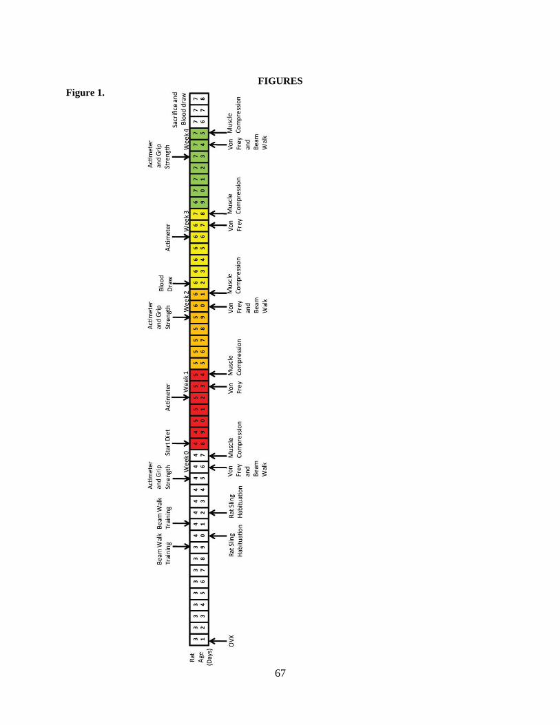
67
FIGURES Figure 1.

68
Figure 1. Experimental timeline. Experimental procedures are indicated on a timeline of rat age. .

69
Figure 2.

70
Figure 2. Modification of the digital Randall Selitto instrument (IITC Life Science Inc). A plastic cap
with a 0.5cm diameter surface area was placed over the pressure sensor.

71
Figure 3.

72
Figure 3 Behavioral comparisons of control (○), VD-/+Ca (■), and VD- (▼) rats. (A) Changes in deep
tissue pressure sensitivity, measured as the maximum force (g) required to elicit an attempted limb
withdrawal or vocalization to mechanical compression of the calf muscle. (B) There were no changes in
cutaneous mechanical sensitivity as measured by the percentage of withdrawal responses to the
application of a 4g monofilament to the plantar surface of the hind paw. (C) Changes in balance were
measured by the number of hind paw slips while traversing an elevated 2.5cm diameter 110cm long
beam. VD- and VD-/+Ca rats failed to improve, while control rats improved compared to initial
performance. (D) There were no changes in forelimb grip strength, as measured by the force (g) required
to break the rats grip. (E-F) Assessment of locomotor activity during a five minute recording using a
force-plate actimeter revelealed no changes in the (E) distance traveled or (F) number of rearing events.
(*p<0.05 compared to control in same week, #p<0.05 compared to VD- in same week, ## p<0.5
compared to week 0 within the same group).

73
Figure 4.

74
Figure 4. Representative images of tibial growth plates from rats after four weeks on (A) control, (B)
VD-/+Ca, or (C) VD- diets. Sagittal sections (20µm) of the growth plate stained with hematoxylin and
eosin.

75
Figure 5.

76
Figure 5. Comparisons of innervation densities of the lateral gastrocnemius muscles of rats after four
weeks on control, VD-/+Ca, or VD- diets. (A) There were no changes in the volumes of the calf muscles
as measured by fluid displacement. (A-E) There were significant increases in the percentage of nerve
area per field area of (B) peripherin-immunoreactive (ir) (c-fibers) and (C) CGRP-ir (sensory
peptidergic/nociceptor) nerves, but not (D) VMAT2-ir (sympathetic) or (E) NFH-ir (large fiber) nerves.
(F) There were also no changes in neuromuscular junction (NMJ) area as measured by bungarotoxin
binding.

77
Figure 6.

78
Figure 6. Representative images of the immunoreactive nerves in calf muscle. Transverse sections
(20µm) of the lateral gastrocnemius of rats receiving (A,D,G,J) control, (B,E,H,K) VD-/+Ca, or
(C,F,I,L) VD- diets were immunoflourescently-labeled for (A-C) peripherin (green), (D-F) CGRP (red),
(G-I) VMAT2 (red), or (J-L) NFH (red) and Bungarotoxin (green). (Scale 50µM)

79
Figure 7.

80
Figure 7. 1,25(OH)2D3 acts directly on c-fiber sensory nerves to regulate axonal growth. (A-B) Primary
DRG cultures immunoflourescently stained for (A) peripherin and (B) vitamin D receptors (VDR),
showing VDR is expressed by cultured sensory neurons, and localized to both the nucleus and the
cytoplasm (Scale 50µm). (C-E) At a higher magnification it is possible to see that (E) VDR is localized
to the tip of (C) peripherin-ir neurites, along with the growth cone marker, (D) growth associated protein
43 (GAP43) (Scale 5µm). (F-H) Representative images of neurons treated with (F) 0, (G) 20, or (H)
100pM 1,25(OH)2D3 (Scale 50µm). (I) Fold change in peripherin-ir neurite area per neuron at the
indicated 1,25(OH)2D3 concentrations. Results reveal a biphasic curve with the peak at 20pM
1,25(OH)2D3. The shaded area corresponds to reported 1,25(OH)2D3 levels in patients with severe pain
(Gloth, Lindsay et al. 1991). (J) DRG cultures treated with JN, a selective agonist for
membrane/cytoplasm-localized rapid response VDRs, and stained for peripherin (●) or NFH (□). While
there was no significant change in NFH-ir neurite area /neuron, there was a biphasic response to JN, with
a peak at 20pM. (*p<0.05)

81
Figure 8.

82
Figure 8. Sympathetic neurons express VDRs, but 1,25(OH)2D3 has no effect on neurite outgrowth. (A)
Sympathetic neurons of superior cervical ganglia (SCG) were immunoflourescently labeled for VDR.
VDR was localized to the nucleus and cytoplasm of selected neurons. (B-E) Primary SCG cultures
immunoflourescently-stained for (B&D) peripherin and (C&E) VDR (Scale B&C 50µm, D&E 5µm).
VDR was localized throughout the (C) cell body, (E) neurite, and growth cone. (F) Primary SCG cultures
were grown for 48 hours with 0pM, 20pM, or 100pM 1,25(OH)2D3 and immunoflourescently labeled
with peripherin. There were no significant changes in neurite area/neuron.

83
CHAPTER 4. GENERAL CONCLUSIONS AND DISCUSSION

84
While the evidence linking vitamin D deficiency and musculoskeletal pain has been
accumulating, these are the first studies to show a direct causal relationship. In the rat model described
here, vitamin D deficiency leads to deep tissue hypersensitivity, which occurred concurrently with muscle
hyperinnervation by putative nociceptors. This hyperinnervation likely contributes to hypovitaminosis D-
induced musculoskeletal pain and neither hypersensitivity nor hyperinnervation is related to serum
calcium levels.
These are also the first studies to show direct actions of vitamin D metabolites on sensory
neurons. Both vitamin D receptors and metabolic enzymes are found in c-fiber neurons, indicating the
importance of tightly regulating vitamin D signaling in this cell population. At normal physiological
concentrations, 1,25(OH)2D3 appears to directly act on membrane/cytoplasmic rapid response vitamin D
receptors, possibly within the growth cone, to inhibit axonal sprouting from c-fiber sensory neurons. A
vitamin D deficiency therefore reduces this inhibition, resulting in tissue hyperinnervation. These effects
are cell-type specific, as hypovitaminosis D-induced axonal growth is not apparent with large fiber
sensory neurons or sympathetic neurons either in vivo or in culture. Expression of both the ligand and
cytoplasmic VDR is modulated by ovarian hormones, with the overall effect of reducing vitamin D
signaling when ovarian hormones are suppressed. This in itself could lead to tissue hyperinnervation and
hypersensitivity or perhaps exacerbate the effects of a vitamin D deficiency, accounting for the increased
incidence of musculoskeletal pain in women with suppressed ovarian hormones.
Musculoskeletal Pain Management
With an estimated 27.5-36% of adolescents and 30-50% of adults suffering from chronic
musculoskeletal pain (Bergman 2007; De Inocencio 2004), our current pain prevention and pain
management strategies are clearly not effective. The most common drug therapies for chronic pain are
NSAIDS (44%), weak opioids (i.e. tramadol, codeine) (23%), and acetaminophen (18%) (Breivik, Collett
et al. 2006), all of which can have significant safety concerns. For example, 20% of patients taking

85
NSAIDS longer than two months will develop an ulcer and 1:1200 will die from gastroduodenal
complications (Tramer, Moore et al. 2000); patients taking tramadol are at increasesd risk for developing
seizures and life threatening serotonin syndrome (Sansone and Sansone 2009); and acetaminophen is
reported to cause acute liver and renal failure if not taken properly (Graham, Scott et al. 2005). In
addition to the potential side effects, in many cases these drugs are not efficient in managing pain. In fact
only 36% of chronic pain patients report that their current therapies are adequate for managing pain
(Breivik, Collett et al. 2006). Non-pharmacological pain management strategies on the other hand often
have limited or short term effects. Massage, manipulation, or mobilization therapies may have short-term
benefits, but there is no evidence that they have a positive long-term impact on chronic musculoskeletal
pain (Airaksinen, Brox et al. 2006). There is also no evidence that hyperstimulation analgesia (i.e.
acupuncture or transcutaneous electrical nerve stimulation) or relaxation techniques have lasting effects in
studies analyzing chronic lower back pain (Airaksinen, Brox et al. 2006; Carroll and Seers 1998).
Cognitive behavioral therapies, designed to change how patients perceive and react to their pain, have had
positive results, but outcomes may be highly dependent on the social and physcological variables of the
patient (McCracken and Turk 2002). Regular exercise may also help treat and prevent chronic pain, but
patient adherence is low (Jordan, Holden et al. 2010). It therefore appears that neither current
pharmacological nor alternative therapies for managing chronic musculoskeletal pain are sufficient.
This study is novel because it indicates that maintaining a healthy vitamin D status could prevent
the development of some forms of muscle pain. The number of people suffering from hypovitaminosis
D-induced musculoskeletal pain may be substantial, as up to 93% of patients with non-specific
musculoskeletal pain are vitamin D deficient (Plotnikoff and Quigley 2003). While the model described
here does not show whether vitamin D supplementation can reverse musculoskeletal pain once
established, a number of clinical studies suggest that it can (de Torrente de la Jara, Pecoud et al. 2006;
Glerup and Eriksen 1999; Gloth, Lindsay et al. 1991). Proper vitamin D supplementation may therefore
be a relatively easy, inexpensive, and safe way to treat and prevent non-specific musculoskeletal pain.

86
Tissue Hyperinnervation and increased sensitivity
Nociceptor hyperinnervation could increase peripheral input of pain signals to the spinal dorsal horn
in a number of ways. It is known that actively growing axons show greater excitability than do quiescent
axons (Janig, Grossmann et al. 2009). Neurons with more complex axonal geometries also show greater
excitability (Janse, Peretz et al. 1999), perhaps because of greater spatial summation on the nociceptor
itself. Similarly, more extensive axonal branching is likely to increase the number of nociceptors
activated, resulting in greater spatial summation on postsynaptic terminals. It is also known that
nociceptors can couple in the periphery (Meyer, Raja et al. 1985). In other words, the action potential of
one neuron can initiate an action potential in adjacent neurons. If innervation density is increased, each
neuron may come in contact with a larger population of neurons with which to couple, which could
further increase the number of nociceptors activated per stimulus. Through any or all of these
mechanisms, increased neurite outgrowth may enhance nociception.
Transition to Chronic Pain
Results from this study indicate that vitamin D deficiency causes peptidergic nociceptor
hyperinnervation of the muscle, which could lower the nociceptive threshold and account for the observed
deep tissue mechanical hypersensitivity. However, this hyperinnervation also has the potential to lead to
chronic (unevoked) pain. In addition to their afferent actions, the peptidergic c-fibers, can antidromically
release neuropeptides that promote neurogenic inflammation (Maggi 1995; Richardson and Vasko 2002).
Once activated, the nerve endings release CGRP and tachykinins, which cause vasodilation, protein
extravasation, mast cell degranulation, and infiltration and activation of immune cells, leading to tissue
inflammation (Maggi 1995). This comes full circle, as deep tissue inflammation in turn further increases
the production of CGRP and tachykinins (Ambalavanar, Moritani et al. 2006) and results in the release of
cytokines, growth factors and other substances from the tissue, which sensitize nociceptors (Richardson
and Vasko 2002). If peptidergic c-fibers are found at a high density in vitamin D deficient patients,
neurogenic inflammation could be amplified as there are more nerve endings to release pro-inflammatory

87
substances. CGRP content itself may also be higher in the neurons of vitamin D deficient patients.
1,25(OH)2D is a well-established repressor of cAMP-induced CT/CGRP gene transcription and an
inhibitor of CT and CGRP secretion from thyroid C-cells, thus with the removal of this repression, the
loss of vitamin D may enhance CGRP expression and secretion and promote neurogenic inflammation
(Baier, Grauer et al. 1994; Collignon, Laborie et al. 1992; Cote, Rogers et al. 1987; Lamari, Tahri et al.
1994; Lazaretti-Castro, Grauer et al. 1995; Lazaretti-Castro, Grauer et al. 1990; Naveh-Many, Raue et al.
1992; Naveh-Many and Silver 1988; Raue, Deutschle et al. 1984; Raue, Deutschle et al. 1983).
Furthermore, vitamin D deficient patients may be particularly susceptible to inflammation, as vitamin D
metabolites act as inflammatory inhibitors (Baeke, Gysemans et al. 2010). Once sensitized by
inflammation, the nociceptors may spontaneously discharge (Xiao and Bennett 2008). Prolonged firing
of nociceptors, this can cause glutamate release in the spinal cord, which acts on NMDA receptors,
leading to central sensitization (Bennett 2000). In addition, if CGRP levels are elevated in peptidergic
nociceptors due to vitamin D deficiency, central sensitization may be further enhanced, as the central
release of CGRP has been shown to potentiate the response of NMDA and non-NMDA receptors in the
spinal dorsal horn, facilitating wind-up (Leem, Gwak et al. 2001). It is therefore possible that vitamin D
deficient patients may transition from a state of deep tissue hypersensitivity to a chronic pain state
through inflammation and central sensitization. At this point it is unclear whether vitamin D deficiency
may play a role in establishing or maintaining central sensitization within the brain, but this possibility
cannot be ruled out, as VDR is expressed in both neurons and glia throughout the brain (Baas, Prufer et al.
2000; Eyles, Smith et al. 2005; Glaser, Veenstra et al. 1999; Neveu, Naveilhan et al. 1994; Perez-
Fernandez, Alonso et al. 1997; Prufer, Veenstra et al. 1999; Walbert, Jirikowski et al. 2001).
Gender differences and the role of sex hormones
Although both musculoskeletal pain and vitamin D deficiency are more common in women,
vitamin D deficiency is also associated with musculoskeletal pain in men. Results from the European
Male Ageing Study analyzing 3075 men ages 49-79, found that 8.6% of all men had chronic widespread
88
pain. 25.5% of men with chronic widespread pain had 25(OH)D levels below 15ng/ml (37.5nmol/L),
compared to only 18.6% in men with no pain (McBeth, Pye et al. 2010), suggesting that vitamin D
deficiency may cause musculoskeletal pain in men. However, chronic musculoskeletal pain in general is
two times more prevalent in women than men (Bergman 2007). It seems to be associated with reduced
ovarian hormones, as musculoskeletal pain is a common menopausal symptom and a major side-effect for
estrogen lowering drugs (i.e. aromatase inhibitors, GnRH agonists) (Alexander, Dennerstein et al. 2007;
Gaugris, Heaney et al. 2005; Khan, Reddy et al. 2010). This could simply be due to the increased
incidence of vitamin D deficiency within this population; however, this study indicates that altered
vitamin D signaling pathways within the nociceptors themselves may play a role. Sustained suppression
of ovarian hormones results in suppression of both local ligand and receptor concentrations within
peptidergic neurons, which might decrease vitamin D signaling and exacerbate the effects of a vitamin D
deficiency. The clinical study by Plotnikoff and Quigley (Plotnikoff and Quigley 2003) supports this
hypothesis. They examined 150 patients who sought clinical treatment for non-specific musculoskeletal
pain. Reflecting the increased incidence of musculoskeletal pain in women, the study was comprised of
107 women and 43 men. Most of the women were over the age of 50 and therefore likely to be post-
menopause. Almost all of the patients were vitamin D deficient (93%), but interestingly the women over
the age of 50 had serum 25(OH)D levels that were significantly higher than either the women of
childbearing age (<36 years old) or the men. Given our findings, this data may suggest that suppression of
ovarian hormones in these postmenopausal women leads to reduced vitamin D signaling, causing
musculoskeletal pain at earlier stages of a vitamin D deficiency. The reduced vitamin D signaling in
putative nociceptors after hormone suppression may provide insight into another clinical situation.
Patients taking aromatase inhihitors tend to be vitamin D deficient and have musculoskeletal pain (Khan,
Reddy et al. 2010; Waltman, Ott et al. 2009). While vitamin D supplementation has been shown to be
effective at reducing musculoskeletal pain in most vitamin D deficient patients, vitamin D
supplementation only minimally reduced musculoskeletal pain in patients taking aromatase inhibitors,
despite the fact that the supplementation increased serum levels of 25(OH)D (Khan, Reddy et al. 2010).

89
It is possible that aromatase inhibitor-induced hypoestrogenemia might severely hinder conversion of the
therapeutic supplement to the active hormone, 1,25(OH)2D, and thereby limit receptor-mediated
signaling, rendering dietary supplementation less effective.
When vitamin D supplementation is not sufficient
The good news is that many people with musculoskeletal pain may find relief with vitamin D
supplementation, which when done correctly has no side effects (Heaney 2008). In fact, a number of
studies have shown this to be the case (de Torrente de la Jara, Pecoud et al. 2006; Glerup and Eriksen
1999; Gloth, Lindsay et al. 1991). However, understanding the mechanism may prove important for
some populations who may not improve with simple vitamin D supplementation. Patients taking estrogen
lowering drugs as described above may be one population. In addition, patients on dialysis, who have a
compromised ability to convert 25(OH)D to 1,25(OH)2D and who often suffer from musculoskeletal pain
(Golan, Haggiag et al. 2009), may not benefit from vitamin D supplementation either, because they
cannot produce adequate levels of active ligand. One option is Vitamin D receptor activators (i.e.
calcitriol); however, these carry a higher risk than traditional vitamin D supplements, because of their
ability to promote hypercalcemia. Alternatively, based on our findings that vitamin D deficiency causes
an increase in the density of petidergic c-fibers within the muscle, it is possible that therapies that
desensitize these peripheral nerve endings (i.e. TRPV1 antagonists) may prove beneficial for
musculoskeletal pain.
Dietary calcium and hypovitaminosis D-induced musculoskeletal pain
Some of the effects of a vitamin D deficiency are not due to the direct actions of this hormone,
but rather due to secondary hypocalcemia (i.e. reduced fertility and increased vascular muscle
contractility) (Johnson and DeLuca 2002; Uhland, Kwiecinski et al. 1992; Weishaar and Simpson 1987).
However, it appears that hypovitaminosis D-induced muscle pain is not due to secondary hypocalcemia.

90
In fact, the results from this study suggest that high dietary calcium concentrations in vitamin D
deficiency may actually exacerbate this condition. It is interesting to note that all patients with severe
pain in the study by Gloth et. al. had low 1,25(OH)2D serum concentrations, normal serum calcium
concentrations, and most were taking a calcium supplement (Gloth, Lindsay et al. 1991). Dietary calcium
concentrations are important to consider, because they may affect 1,25(OH)2D concentrations.
1,25(OH)2D does not always decrease in a vitamin D deficiency, and in fact when serum 25D levels drop
below 40nM, 1,25(OH)2D levels often increase (Need, Horowitz et al. 2000). This is because as serum
Ca concentrations drop in a vitamin D deficiency, parathyroid hormone levels increase, which increases
expression of CYP27B1, the enzyme responsible for converting 25(OH)D to 1,25(OH)2D. It is possible
that high dietary calcium may prevent the drop in serum calcium that leads to increased PTH levels,
thereby preventing a spike in 1,25(OH)2D levels. This appears to be the case in rats, because increased
dietary calcium inhibits expression of CYP27B1, abrogates the 1,25(OH)2D spike, and accelerates the
decrease of 1,25(OH)2D in a vitamin D deficiency (Anderson, Lee et al. 2010; Mallon, Boris et al. 1981).
If high dietary calcium reduces the levels of active hormone, this could be the reason why increasing
dietary calcium accelerated the onset of deep tissue hypersensitivity in this study. In view of how
common dietary calcium supplementation is (43% of population use Ca supplements) (Bailey, Dodd et al.
2010), it may be an import aspect for a doctor to consider when suspecting hypovitaminosis D-induced
musculoskeletal pain.
Beyond Pain
It is unlikely that nociceceptor-sprouting in response to vitamin D deficiency evolved for the sole
purpose of causing pain, so it is interesting to consider the evolutionary benefits of this process. One
potential benefit could be increased regulation of blood flow to skeletal muscle. Vitamin D deficiency is
already known to have profound effects on the cardiovascular system. As vitamin D levels drop, blood
pressure rises (Scragg, Sowers et al. 2007). This alone could increase tissue blood flow in vitamin D
deficiency. This may be important for individuals living at higher latitudes, who experience dramatic

91
seasonal variations in temperature, have low serum 25(OH)D during the winter months (Holick 2005),
and are known to have elevated blood pressures in the winter (Woodhouse, Khaw et al. 1993). If this
drop in vitamin D levels results in increased blood flow to peripheral tissues, it could be important for
maintaining heat in the extremities during cold winters. In addition, the increased c-fiber innervation of
skeletal muscle could also promote thermogenesis. Shivering thermogenesis is important for maintaining
basal body temperatures in cold environments and involves the involuntary, rhythmic contractions of
skeletal muscles. Shivering requires a dramatic increase in oxygen consumption (~5 times the resting
metabolic rate). An increase in peptidergic c-fibers in skeletal muscle, could locally increase oxygen
delivery, by priming the blood vessels in the muscle for vasodilation. Upon activation of these nerves,
peptidergic nociceptors antidromically release CGRP, which promotes vasodilation. For example,
electrical stimulation of the saphenous nerve results in muscle arteriolar dilation and increased blood flow
to the gracilis muscle, which can be prevented by the CGRP receptor antagonist, CGRP8-37 (Loaiza,
Yamaguchi et al. 2002). The hypovitaminosis D-induced increases in the innervation of muscle by
CGRP-containing nerves may therefore play a role in further increasing blood flow to the skeletal muscle,
providing thermogenic advantages in colder climates. In fact, it is known that individuals from higher
latitude cold weather climates (e.g. Alaskans), who are known to have lower vitamin D levels (Holick
2005) in the winter, are able to maintain higher temperatures in their extremities when exposed to cold
than individuals from warmer climates, making them less susceptible to frost-bite (Miller and Irving
1962).
Potential Mechanisms
The results from this study indicate that 1,25(OH)2D3 is capable of acting directly on sensory c-
fibers to regulate neurite outgrowth. 1,25(OH)2D3 likely acts through membrane/cytoplasmic VDRs,
which typically initiate rapid response signaling cascades. The rapid response specific agonist, JN, has
similar dose dependent effects on neurite growth as 1,25(OH)2D3. These rapid response cascades could
be initiated locally within the growth cone, as VDR appears to be enriched there. (Mizwicki and Norman

92
2009). The rapid responses known to be mediated by 1,25(OH)2D are likely cell type specific, but
include PKC, MAPK, PLC, PLA2, Src, and Raf activation, opening Ca2+ and Cl- channels, and
sphingomyelin hydrolysis (Mizwicki and Norman 2009). Any or all of these downstream signals could
play a role in neurite outgrowth. One of the best characterized VDR rapid responses is regulation of the
flux of ions such as calcium. Like VDR activation, calcium modulation has a biphasic curve on neurite
outgrowth (Gomez and Zheng 2006). In other cell types, membrane VDR regulates intracellular calcium
concentrations through modulation of L-type calcium channels (Zanello and Norman 2004). This is
interesting, because L-type voltage gated channels are the primary source for inward calcium flux in c-
fiber neurons (Scroggs and Fox 1992) and they are known regulators of sensory neurite outgrowth
(Archer, Doherty et al. 1999). Therefore, while serum calcium concentrations were not a factor in the
development of muscle pain, 1,25(OH)2D may regulate intracellular calcium concentrations within the
axon.
Conclusion
Collectively, these results show that vitamin D deficiency does cause deep tissue hypersensitivity
in rats. 1,25(OH)2D3 acts directly on membrane/cytoplasmic rapid response VDRs to regulate axonal
growth from a sensory subtype considered to be nociceptive. During a vitamin D deficiency, the reduced
signaling from these rapid response VDR receptors leads to sensory hyperinnervation of muscle, which
may contribute to the development of deep tissue hypersensitivity in rats. Suppression of ovarian
hormones may further promote hypovitaminosis D-induced musculoskeletal pain by reducing vitamin D
signaling within nociceptors. This may contribute to the high prevalence of musculoskeletal pain in
women with low levels of ovarian hormones. Preventing the development of non-specific
musculoskeletal pain may be as simple as supplementing the diet with vitamin D.

93
REFERENCES Abusdal UG, Hagen KB, et al. (1997). [Self-reported chronic muscle pain among women in
Oslo]. Tidsskr Nor Laegeforen 117(11): 1606-1610. Airaksinen O, Brox JI, et al. (2006). Chapter 4. European guidelines for the management of
chronic nonspecific low back pain. Eur Spine J 15 Suppl 2: S192-300. Alexander JL, Dennerstein L, et al. (2007). Arthralgias, bodily aches and pains and somatic
complaints in midlife women: etiology, pathophysiology and differential diagnosis. Expert Rev Neurother 7(11 Suppl): S15-26.
Alfredson H, Ohberg L, et al. (2003). Is vasculo-neural ingrowth the cause of pain in chronic
Achilles tendinosis? An investigation using ultrasonography and colour Doppler, immunohistochemistry, and diagnostic injections. Knee Surg Sports Traumatol Arthrosc 11(5): 334-338.
Ambalavanar R, Moritani M, et al. (2006). Deep tissue inflammation upregulates neuropeptides
and evokes nociceptive behaviors which are modulated by a neuropeptide antagonist. Pain 120(1-2): 53-68.
Ambalavanar R and Morris R (1993). An ultrastructural study of the binding of an alpha-D-
galactose specific lectin from Griffonia simplicifolia to trigeminal ganglion neurons and the trigeminal nucleus caudalis in the rat. Neuroscience 52(3): 699-709.
Anderson PH, Lee AM, et al. (2010). The effect of dietary calcium on 1,25(OH)2D3 synthesis
and sparing of serum 25(OH)D3 levels. J Steroid Biochem Mol Biol 121(1-2): 288-292. Andersson H, Ejlertsson G, et al. (1998). Widespread musculoskeletal chronic pain associated
with smoking. An epidemiological study in a general rural population. Scand J Rehabil Med 30(3): 185-191.
Andersson HI, Ejlertsson G, et al. (1993). Chronic pain in a geographically defined general
population: studies of differences in age, gender, social class, and pain localization. Clin J Pain 9(3): 174-182.
Andersson HI, Ejlertsson G, et al. (1999). Musculoskeletal chronic pain in general practice.
Studies of health care utilisation in comparison with pain prevalence. Scand J Prim Health Care 17(2): 87-92.
Annweiler C, Montero-Odasso M, et al. (2010). Fall prevention and vitamin D in the elderly: an
overview of the key role of the non-bone effects. J Neuroeng Rehabil 7: 50. Archer FR, Doherty P, et al. (1999). CAMs and FGF cause a local submembrane calcium signal
promoting axon outgrowth without a rise in bulk calcium concentration. Eur J Neurosci 11(10): 3565-3573.

94
Armas LA, Dowell S, et al. (2007). Ultraviolet-B radiation increases serum 25-hydroxyvitamin D levels: The effect of UVB dose and skin color. J Am Acad Dermatol.
Arunabh S, Pollack S, et al. (2003). Body fat content and 25-hydroxyvitamin D levels in healthy
women. J Clin Endocrinol Metab 88(1): 157-161. Awareness TCFD (2006). The 2006 CDA Long-Term Disability Claims Review. 1-3. Baas D, Prufer K, et al. (2000). Rat oligodendrocytes express the vitamin D(3) receptor and
respond to 1,25-dihydroxyvitamin D(3). Glia 31(1): 59-68. Baeke F, Gysemans C, et al. (2010). Vitamin D insufficiency: implications for the immune
system. Pediatr Nephrol 25(9): 1597-1606. Baier R, Grauer A, et al. (1994). Differential effects of 1,25-dihydroxyvitamin D3 on cell
proliferation and calcitonin gene expression. Endocrinology 135(5): 2006-2011. Bailey RL, Dodd KW, et al. (2010). Estimation of total usual calcium and vitamin D intakes in
the United States. J Nutr 140(4): 817-822. Becker JB, Arnold AP, et al. (2005). Strategies and methods for research on sex differences in
brain and behavior. Endocrinology 146(4): 1650-1673. Bell NH, Godsen RN, et al. (1988). The effects of muscle-building exercise on vitamin D and
mineral metabolism. J Bone Miner Res 3(4): 369-373. Bennett DL, Michael GJ, et al. (1998). A distinct subgroup of small DRG cells express GDNF
receptor components and GDNF is protective for these neurons after nerve injury. J Neurosci 18(8): 3059-3072.
Bennett GJ (2000). Update on the neurophysiology of pain transmission and modulation: focus
on the NMDA-receptor. J Pain Symptom Manage 19(1 Suppl): S2-6. Benson J, Wilson A, et al. (2006). Muscle pain as an indicator of vitamin D deficiency in an
urban Australian Aboriginal population. Med J Aust 185(2): 76-77. Bergman S (2007). Public health perspective--how to improve the musculoskeletal health of the
population. Best Pract Res Clin Rheumatol 21(1): 191-204. Bergman S, Herrstrom P, et al. (2001). Chronic musculoskeletal pain, prevalence rates, and
sociodemographic associations in a Swedish population study. J Rheumatol 28(6): 1369-1377.
Berkley KJ, Robbins A, et al. (1993). Functional differences between afferent fibers in the
hypogastric and pelvic nerves innervating female reproductive organs in the rat. J Neurophysiol 69(2): 533-544.

95
Birklein F and Schmelz M (2008). Neuropeptides, neurogenic inflammation and complex
regional pain syndrome (CRPS). Neurosci Lett 437(3): 199-202. Bischof MG, Heinze G, et al. (2006). Vitamin D status and its relation to age and body mass
index. Horm Res 66(5): 211-215. Bischoff-Ferrari HA, Dawson-Hughes B, et al. (2004). Effect of Vitamin D on falls: a meta-
analysis. Jama 291(16): 1999-2006. Blacklock AD, Johnson MS, et al. (2005). Estrogen increases sensory nociceptor neuritogenesis
in vitro by a direct, nerve growth factor-independent mechanism. Eur J Neurosci 21(9): 2320-2328.
Block SR (2004). Vitamin D deficiency is not associated with nonspecific musculoskeletal pain
syndromes including fibromyalgia. Mayo Clin Proc 79(12): 1585-1586; author reply 1586-1587.
Bohm-Starke N, Hilliges M, et al. (1999). Neurochemical characterization of the vestibular
nerves in women with vulvar vestibulitis syndrome. Gynecol Obstet Invest 48(4): 270. Bolland MJ, Grey AB, et al. (2007). Age-, gender-, and weight-related effects on levels of 25-
hydroxyvitamin D are not mediated by vitamin D binding protein. Clin Endocrinol (Oxf) 67(2): 259-264.
Brage S and Bjerkedal T (1996). Musculoskeletal pain and smoking in Norway. J Epidemiol
Community Health 50(2): 166-169. Breivik H, Collett B, et al. (2006). Survey of chronic pain in Europe: prevalence, impact on daily
life, and treatment. Eur J Pain 10(4): 287-333. Carroll D and Seers K (1998). Relaxation for the relief of chronic pain: a systematic review. J
Adv Nurs 27(3): 476-487. Casellini CM and Vinik AI (2007). Clinical manifestations and current treatment options for
diabetic neuropathies. Endocr Pract 13(5): 550-566. Cauley JA, Gutai JP, et al. (1989). The epidemiology of serum sex hormones in postmenopausal
women. Am J Epidemiol 129(6): 1120-1131. Cekic M, Sayeed I, et al. (2009). Combination treatment with progesterone and vitamin D
hormone may be more effective than monotherapy for nervous system injury and disease. Front Neuroendocrinol 30(2): 158-172.

96
Chan JR, Rodriguez-Waitkus PM, et al. (2000). Progesterone synthesized by Schwann cells during myelin formation regulates neuronal gene expression. Mol Biol Cell 11(7): 2283-2295.
Chapuy MC, Preziosi P, et al. (1997). Prevalence of vitamin D insufficiency in an adult normal
population. Osteoporos Int 7(5): 439-443. Clarke GL, Bhattacherjee A, et al. (2010). Beta-adrenoceptor blockers increase cardiac
sympathetic innervation by inhibiting autoreceptor suppression of axon growth. J Neurosci 30(37): 12446-12454.
Clemens TL, Adams JS, et al. (1982). Increased skin pigment reduces the capacity of skin to
synthesise vitamin D3. Lancet 1(8263): 74-76. Collignon H, Laborie C, et al. (1992). Effects of dexamethasone, calcium and 1,25-
dihydroxycholecalciferol on calcitonin and calcitonin gene-related peptide mRNA levels from the CA-77 C cell line. Thyroid 2(4): 361-365.
Cornet A, Baudet C, et al. (1998). 1,25-Dihydroxyvitamin D3 regulates the expression of VDR
and NGF gene in Schwann cells in vitro. J Neurosci Res 53(6): 742-746. Cote GJ, Rogers DG, et al. (1987). The effect of 1,25-dihydroxyvitamin D3 treatment on
calcitonin and calcitonin gene-related peptide mRNA levels in cultured human thyroid C-cells. Biochem Biophys Res Commun 149(1): 239-243.
Craft RM (2007). Modulation of pain by estrogens. Pain 132 Suppl 1: S3-12. Croft P, Rigby AS, et al. (1993). The prevalence of chronic widespread pain in the general
population. J Rheumatol 20(4): 710-713. Cummings SR, Black DM, et al. (1993). Bone density at various sites for prediction of hip
fractures. The Study of Osteoporotic Fractures Research Group. Lancet 341(8837): 72-75.
Dawson-Hughes B, Harris SS, et al. (1997). Plasma calcidiol, season, and serum parathyroid
hormone concentrations in healthy elderly men and women. Am J Clin Nutr 65(1): 67-71. Dawson-Hughes B, Heaney RP, et al. (2005). Estimates of optimal vitamin D status. Osteoporos
Int 16(7): 713-716. De Inocencio J (2004). Epidemiology of musculoskeletal pain in primary care. Arch Dis Child
89(5): 431-434. De Rezende Pena C, Grillo LP, et al. (2010). Evaluation of 25-hydroxyvitamin d serum levels in
patients with fibromyalgia. J Clin Rheumatol 16(8): 365-369.

97
De Torrente De La Jara G, Pecoud A, et al. (2006). Female asylum seekers with musculoskeletal pain: the importance of diagnosis and treatment of hypovitaminosis D. BMC Fam Pract 7: 4.
Di Sebastiano P, Fink T, et al. (1995). Changes of protein gene product 9.5 (PGP 9.5)
immunoreactive nerves in inflamed appendix. Dig Dis Sci 40(2): 366-372. Di Sebastiano P, Fink T, et al. (1997). Immune cell infiltration and growth-associated protein 43
expression correlate with pain in chronic pancreatitis. Gastroenterology 112(5): 1648-1655.
Diesel B, Radermacher J, et al. (2005). Vitamin D(3) metabolism in human glioblastoma
multiforme: functionality of CYP27B1 splice variants, metabolism of calcidiol, and effect of calcitriol. Clin Cancer Res 11(15): 5370-5380.
Dugan SA, Powell LH, et al. (2006). Musculoskeletal pain and menopausal status. Clin J Pain
22(4): 325-331. Duncan WE, Glass AR, et al. (1991). Estrogen regulation of the nuclear 1,25-dihydroxyvitamin
D3 receptor in rat liver and kidney. Endocrinology 129(5): 2318-2324. Duque G, El Abdaimi K, et al. (2002). Estrogens (E2) regulate expression and response of 1,25-
dihydroxyvitamin D3 receptors in bone cells: changes with aging and hormone deprivation. Biochem Biophys Res Commun 299(3): 446-454.
Edwards RR, Doleys DM, et al. (2001). Ethnic differences in pain tolerance: clinical
implications in a chronic pain population. Psychosom Med 63(2): 316-323. Escaleira MT, Sonohara S, et al. (1993). Sex steroids induced up-regulation of 1,25-(OH)2
vitamin D3 receptors in T 47D breast cancer cells. J Steroid Biochem Mol Biol 45(4): 257-263.
Eyles D, Brown J, et al. (2003). Vitamin D3 and brain development. Neuroscience 118(3): 641-
653. Eyles DW, Smith S, et al. (2005). Distribution of the vitamin D receptor and 1 alpha-hydroxylase
in human brain. J Chem Neuroanat 29(1): 21-30. Faulkner KA, Cauley JA, et al. (2006). Higher 1,25-dihydroxyvitamin D3 concentrations
associated with lower fall rates in older community-dwelling women. Osteoporos Int 17(9): 1318-1328.
Felson DT and Cummings SR (2005). Aromatase inhibitors and the syndrome of arthralgias with
estrogen deprivation. Arthritis Rheum 52(9): 2594-2598.
98
Fink T, Di Sebastiano P, et al. (1994). Growth-associated protein-43 and protein gene-product 9.5 innervation in human pancreas: changes in chronic pancreatitis. Neuroscience 63(1): 249-266.
Ford L, Graham V, et al. (2006). Vitamin D concentrations in an UK inner-city multicultural
outpatient population. Ann Clin Biochem 43(Pt 6): 468-473. Fowler SC, Birkestrand BR, et al. (2001). A force-plate actometer for quantitating rodent
behaviors: illustrative data on locomotion, rotation, spatial patterning, stereotypies, and tremor. J Neurosci Methods 107(1-2): 107-124.
Freeman ME (1994). The Neuroendocrine Control of the Ovarian Cycle of the Rat. The
Physiology of Reproduction. J. D. N. E. Knobil. New York, Raven Press: 613-658. Friedman AJ, Juneau-Norcross M, et al. (1993). Adverse effects of leuprolide acetate depot
treatment. Fertil Steril 59(2): 448-450. Gangula PR, Lanlua P, et al. (2000). Regulation of calcitonin gene-related peptide expression in
dorsal root ganglia of rats by female sex steroid hormones. Biol Reprod 62(4): 1033-1039.
Gansky SA and Plesh O (2007). Widespread pain and fibromyalgia in a biracial cohort of young
women. J Rheumatol 34(4): 810-817. Garreau JR, Delamelena T, et al. (2006). Side effects of aromatase inhibitors versus tamoxifen:
the patients' perspective. Am J Surg 192(4): 496-498. Gaugris S, Heaney RP, et al. (2005). Vitamin D inadequacy among post-menopausal women: a
systematic review. QJM 98(9): 667-676. Gilad LA, Bresler T, et al. (2005). Regulation of vitamin D receptor expression via estrogen-
induced activation of the ERK 1/2 signaling pathway in colon and breast cancer cells. J Endocrinol 185(3): 577-592.
Glaser SD, Veenstra TD, et al. (1999). Distribution of 1,25-dihydroxyvitamin D3 receptor
immunoreactivity in the rat olfactory system. Cell Mol Neurobiol 19(5): 613-624. Glerup H and Eriksen EF (1999). Acroparaesthesia--a typical finding in vitamin D deficiency.
Rheumatology (Oxford) 38(5): 482. Glerup H, Mikkelsen K, et al. (2000). Hypovitaminosis D myopathy without biochemical signs
of osteomalacic bone involvement. Calcif Tissue Int 66(6): 419-424. Gloth FM, 3rd, Lindsay JM, et al. (1991). Can vitamin D deficiency produce an unusual pain
syndrome? Arch Intern Med 151(8): 1662-1664.

99
Golan E, Haggiag I, et al. (2009). Calcium, parathyroid hormone, and vitamin D: major determinants of chronic pain in hemodialysis patients. Clin J Am Soc Nephrol 4(8): 1374-1380.
Goldstein ME, House SB, et al. (1991). NF-L and peripherin immunoreactivities define distinct
classes of rat sensory ganglion cells. J Neurosci Res 30(1): 92-104. Gomez TM and Zheng JQ (2006). The molecular basis for calcium-dependent axon pathfinding.
Nat Rev Neurosci 7(2): 115-125. Gonzalez Pardo V, Boland R, et al. (2008). Vitamin D receptor levels and binding are reduced in
aged rat intestinal subcellular fractions. Biogerontology 9(2): 109-118. Graham GG, Scott KF, et al. (2005). Tolerability of paracetamol. Drug Saf 28(3): 227-240. Gray TK, Mcadoo T, et al. (1982). Fluctuation of serum concentration of 1,25-dihydroxyvitamin
D3 during the menstrual cycle. Am J Obstet Gynecol 144(8): 880-884. Greendale GA, Reboussin BA, et al. (1998). Symptom relief and side effects of postmenopausal
hormones: results from the Postmenopausal Estrogen/Progestin Interventions Trial. Obstet Gynecol 92(6): 982-988.
Headley DB, Suhan NM, et al. (2007). Different subcellular distributions of the vesicular
monoamine transporter, VMAT2, in subclasses of sympathetic neurons. Brain Res 1129(1): 156-160.
Heaney RP (2008). Vitamin D: criteria for safety and efficacy. Nutr Rev 66(10 Suppl 2): S178-
181. Heaney RP, Dowell MS, et al. (2003). Calcium absorption varies within the reference range for
serum 25-hydroxyvitamin D. J Am Coll Nutr 22(2): 142-146. Heidari B, Shirvani JS, et al. (2010). Association between nonspecific skeletal pain and vitamin
D deficiency. Int J Rheum Dis 13(4): 340-346. Ho SC, Chan SG, et al. (1999). Menopausal symptoms and symptom clustering in Chinese
women. Maturitas 33(3): 219-227. Holick MF (2005). Photobiology of Vitamin D. Vitamin D. D. Feldman, F. H. Glorieux and J.
W. Pike. Amsterdam ; Boston, Elsevier Academic Press. 1: 37-43. Holick MF (2005). Photobiology of Vitamin D. Vitamin D. D. Feldman, F. H. Glorieux and J.
W. Pike. Amsterdam ; Boston, Elsevier Academic Press. 1: 37-45. Holick MF (2009). Vitamin D status: measurement, interpretation, and clinical application. Ann
Epidemiol 19(2): 73-78.

100
Holick MF and Chen TC (2008). Vitamin D deficiency: a worldwide problem with health
consequences. Am J Clin Nutr 87(4): 1080S-1086S. Holick MF, Matsuoka LY, et al. (1989). Age, vitamin D, and solar ultraviolet. Lancet 2(8671):
1104-1105. Hollis BW (2005). Circulating 25-hydroxyvitamin D levels indicative of vitamin D sufficiency:
implications for establishing a new effective dietary intake recommendation for vitamin D. J Nutr 135(2): 317-322.
Horst RL, Reinhardt TA, et al. (2005). Vitamin D Metabolism. Vitamin D. D. Feldman, F. H.
Glorieux and J. W. Pike. Amsterdam ; Boston, Elsevier Academic Press. Huhtakangas JA, Olivera CJ, et al. (2004). The vitamin D receptor is present in caveolae-
enriched plasma membranes and binds 1 alpha,25(OH)2-vitamin D3 in vivo and in vitro. Mol Endocrinol 18(11): 2660-2671.
Hunt IM, Silman AJ, et al. (1999). The prevalence and associated features of chronic widespread
pain in the community using the 'Manchester' definition of chronic widespread pain. Rheumatology (Oxford) 38(3): 275-279.
Institute of Medicine FaNB (2010). Dietary Reference Intakes for Calcium and Vitamin D.
Washington, DC. Janig W, Grossmann L, et al. (2009). Mechano- and thermosensitivity of regenerating cutaneous
afferent nerve fibers. Exp Brain Res 196(1): 101-114. Janse C, Peretz B, et al. (1999). Excitability and branching of neuroendocrine cells during
reproductive senescence. Neurobiol Aging 20(6): 675-683. Johnson JA, Grande JP, et al. (1996). 1,25-Dihydroxyvitamin D(3) receptors in developing
dorsal root ganglia of fetal rats. Brain Res Dev Brain Res 92(1): 120-124. Johnson LE and Deluca HF (2002). Reproductive defects are corrected in vitamin d-deficient
female rats fed a high calcium, phosphorus and lactose diet. J Nutr 132(8): 2270-2273. Jones G (2007). Expanding role for vitamin D in chronic kidney disease: importance of blood
25-OH-D levels and extra-renal 1alpha-hydroxylase in the classical and nonclassical actions of 1alpha,25-dihydroxyvitamin D(3). Semin Dial 20(4): 316-324.
Jones G (2008). Pharmacokinetics of vitamin D toxicity. Am J Clin Nutr 88(2): 582S-586S. Jones KD, Adams D, et al. (2006). A comprehensive review of 46 exercise treatment studies in
fibromyalgia (1988-2005). Health Qual Life Outcomes 4: 67.

101
Jordan JL, Holden MA, et al. (2010). Interventions to improve adherence to exercise for chronic musculoskeletal pain in adults. Cochrane Database Syst Rev(1): CD005956.
Khan QJ, Reddy PS, et al. (2009). Effect of vitamin D supplementation on serum 25-hydroxy
vitamin D levels, joint pain, and fatigue in women starting adjuvant letrozole treatment for breast cancer. Breast Cancer Res Treat.
Khan QJ, Reddy PS, et al. (2010). Effect of vitamin D supplementation on serum 25-hydroxy
vitamin D levels, joint pain, and fatigue in women starting adjuvant letrozole treatment for breast cancer. Breast Cancer Res Treat 119(1): 111-118.
Klausen T, Breum L, et al. (1993). Plasma levels of parathyroid hormone, vitamin D, calcitonin,
and calcium in association with endurance exercise. Calcif Tissue Int 52(3): 205-208. Lamari Y, Tahri EH, et al. (1994). Steroid hormones and retinoic acid interact in the regulation
of calcitonin and calcitonin gene-related peptide secretion and messenger ribonucleic acid levels in CA-77 C cells. Cell Mol Biol (Noisy-le-grand) 40(4): 541-550.
Lawson SN (1992). Morphological and biochemical cell types of sensory neurons. Sensory
neurons: diversity, development, and plasticity. S. A. Scott. New York, Oxford Press: 27-59.
Lazaretti-Castro M, Grauer A, et al. (1995). 1,25-Dihydroxyvitamin D3 stimulates growth and
inhibits calcitonin secretion in a human C cell carcinoma cell line. Braz J Med Biol Res 28(9): 1013-1018.
Lazaretti-Castro M, Grauer A, et al. (1990). 1,25-Dihydroxyvitamin D3 suppresses
dexamethasone effects on calcitonin secretion. Mol Cell Endocrinol 71(3): R13-18. Lechner D, Bajna E, et al. (2006). Genistein and 17beta-estradiol, but not equol, regulate vitamin
D synthesis in human colon and breast cancer cells. Anticancer Res 26(4A): 2597-2603. Leem JW, Gwak YS, et al. (2001). Effects of iontophoretically applied substance P, calcitonin
gene-related peptide on excitability of dorsal horn neurones in rats. Yonsei Med J 42(1): 74-83.
Lester GE, Vanderwiel CJ, et al. (1982). Vitamin D deficiency in rats with normal serum
calcium concentrations. Proc Natl Acad Sci U S A 79(15): 4791-4794. Levenson CW and Figueiroa SM (2008). Gestational vitamin D deficiency: long-term effects on
the brain. Nutr Rev 66(12): 726-729. Levy J, Zuili I, et al. (1984). Induction of cytosolic receptors for 1 alpha, 25-dihydroxyvitamin
D3 in the immature rat uterus by oestradiol. J Endocrinol 100(3): 265-269.

102
Liel Y, Kraus S, et al. (1992). Evidence that estrogens modulate activity and increase the number of 1,25-dihydroxyvitamin D receptors in osteoblast-like cells (ROS 17/2.8). Endocrinology 130(5): 2597-2601.
Liel Y, Shany S, et al. (1999). Estrogen increases 1,25-dihydroxyvitamin D receptors expression
and bioresponse in the rat duodenal mucosa. Endocrinology 140(1): 280-285. Linton SJ and Van Tulder MW (2001). Preventive interventions for back and neck pain
problems: what is the evidence? Spine 26(7): 778-787. Loaiza LA, Yamaguchi S, et al. (2002). Vasodilatation of muscle microvessels induced by
somatic afferent stimulation is mediated by calcitonin gene-related peptide release in the rat. Neurosci Lett 333(2): 136-140.
Losel RM, Falkenstein E, et al. (2003). Nongenomic steroid action: controversies, questions, and
answers. Physiol Rev 83(3): 965-1016. Lund B and Sorensen OH (1979). Measurement of circulating 1,25-dihydroxyvitamin D in man.
Changes in serum concentrations during treatment with 1 alpha-hydroxycholecalciferol. Acta Endocrinol (Copenh) 91(2): 338-350.
Macfarlane GJ, Palmer B, et al. (2005). An excess of widespread pain among South Asians: are
low levels of vitamin D implicated? Ann Rheum Dis 64(8): 1217-1219. Maggi CA (1995). Tachykinins and calcitonin gene-related peptide (CGRP) as co-transmitters
released from peripheral endings of sensory nerves. Prog Neurobiol 45(1): 1-98. Mahonen A and Maenpaa PH (1994). Steroid hormone modulation of vitamin D receptor levels
in human MG-63 osteosarcoma cells. Biochem Biophys Res Commun 205(2): 1179-1186.
Mallon JP, Boris A, et al. (1981). Decrease in serum levels of 1,25-dihydroxycholecalciferol in
rats and chickens fed a vitamin D-deficient diet. J Nutr 111(4): 665-667. Mascarenhas R and Mobarhan S (2004). Hypovitaminosis D-induced pain. Nutr Rev 62(9): 354-
359. Masood H, Narang AP, et al. (1989). Persistent limb pain and raised serum alkaline phosphatase
the earliest markers of subclinical hypovitaminosis D in Kashmir. Indian J Physiol Pharmacol 33(4): 259-261.
Mastaglia FL and Hilton-Jones D (2007). Myopathies. Edinburgh ; New York, Elsevier. Mcbeth J, Pye SR, et al. (2010). Musculoskeletal pain is associated with very low levels of
vitamin D in men: results from the European Male Ageing Study. Ann Rheum Dis 69(8): 1448-1452.

103
Mccollum EV, Simmonds N, et al. (1922). An experimental demonstration of the existence of a
vitamin which promotes calcium deposition. J Biol Chem 53: 293-298. Mccracken LM, Matthews AK, et al. (2001). A comparison of blacks and whites seeking
treatment for chronic pain. Clin J Pain 17(3): 249-255. Mccracken LM and Turk DC (2002). Behavioral and cognitive-behavioral treatment for chronic
pain: outcome, predictors of outcome, and treatment process. Spine (Phila Pa 1976) 27(22): 2564-2573.
Meyer RA, Raja SN, et al. (1985). Coupling of action potential activity between unmyelinated
fibers in the peripheral nerve of monkey. Science 227(4683): 184-187. Miller LK and Irving L (1962). Local reactions to air cooling in an Eskimo population. J Appl
Physiol 17: 449-455. Mizwicki MT and Norman AW (2009). The vitamin D sterol-vitamin D receptor ensemble
model offers unique insights into both genomic and rapid-response signaling. Sci Signal 2(75): re4.
Morales L, Pans S, et al. (2006). Debilitating musculoskeletal pain and stiffness with letrozole
and exemestane: associated tenosynovial changes on magnetic resonance imaging. Breast Cancer Res Treat.
Mouridsen HT (2006). Incidence and management of side effects associated with aromatase
inhibitors in the adjuvant treatment of breast cancer in postmenopausal women. Curr Med Res Opin 22(8): 1609-1621.
Mowa CN, Usip S, et al. (2003). The effects of pregnancy and estrogen on the expression of
calcitonin gene-related peptide (CGRP) in the uterine cervix, dorsal root ganglia and spinal cord. Peptides 24(8): 1163-1174.
Muller KA, Ryals JM, et al. (2008). Abnormal muscle spindle innervation and large-fiber
neuropathy in diabetic mice. Diabetes 57(6): 1693-1701. Myhr KM (2009). Vitamin D treatment in multiple sclerosis. J Neurol Sci 286(1-2): 104-108. Nangia AK, Butcher JL, et al. (1998). Association of vitamin D receptors with the nuclear matrix
of human and rat genitourinary tissues. J Steroid Biochem Mol Biol 66(4): 241-246. Nardone A, Galante M, et al. (2007). Balance control in Sensory Neuron Disease. Clin
Neurophysiol 118(3): 538-550. Naveh-Many T, Raue F, et al. (1992). Regulation of calcitonin gene expression by hypocalcemia,
hypercalcemia, and vitamin D in the rat. J Bone Miner Res 7(10): 1233-1237.

104
Naveh-Many T and Silver J (1988). Regulation of calcitonin gene transcription by vitamin D
metabolites in vivo in the rat. J Clin Invest 81(1): 270-273. Naveilhan P, Neveu I, et al. (1993). Expression of 25(OH) vitamin D3 24-hydroxylase gene in
glial cells. Neuroreport 5(3): 255-257. Need AG (2006). Bone resorption markers in vitamin D insufficiency. Clin Chim Acta 368(1-2):
48-52. Need AG, Horowitz M, et al. (2000). Vitamin D status: effects on parathyroid hormone and 1,
25-dihydroxyvitamin D in postmenopausal women. Am J Clin Nutr 71(6): 1577-1581. Need AG, Morris HA, et al. (1993). Effects of skin thickness, age, body fat, and sunlight on
serum 25-hydroxyvitamin D. Am J Clin Nutr 58(6): 882-885. Nelson ME, Meredith CN, et al. (1988). Hormone and bone mineral status in endurance-trained
and sedentary postmenopausal women. J Clin Endocrinol Metab 66(5): 927-933. Neveu I, Naveilhan P, et al. (1994). 1,25-dihydroxyvitamin D3 regulates the synthesis of nerve
growth factor in primary cultures of glial cells. Brain Res Mol Brain Res 24(1-4): 70-76. Newmark HL and Newmark J (2007). Vitamin D and Parkinson's disease--a hypothesis. Mov
Disord 22(4): 461-468. Norman AW, Okamura WH, et al. (1997). Comparison of 6-s-cis- and 6-s-trans-locked analogs
of 1alpha,25-dihydroxyvitamin D3 indicates that the 6-s-cis conformation is preferred for rapid nongenomic biological responses and that neither 6-s-cis- nor 6-s-trans-locked analogs are preferred for genomic biological responses. Mol Endocrinol 11(10): 1518-1531.
Omdahl JL, Bobrovnikova EV, et al. (2003). Expression, structure-function, and molecular
modeling of vitamin D P450s. J Cell Biochem 88(2): 356-362. Ooms ME, Roos JC, et al. (1995). Prevention of bone loss by vitamin D supplementation in
elderly women: a randomized double-blind trial. J Clin Endocrinol Metab 80(4): 1052-1058.
Pal BR, Marshall T, et al. (2003). Distribution analysis of vitamin D highlights differences in
population subgroups: preliminary observations from a pilot study in UK adults. J Endocrinol 179(1): 119-129.
Palmer KT, Syddall H, et al. (2003). Smoking and musculoskeletal disorders: findings from a
British national survey. Ann Rheum Dis 62(1): 33-36.

105
Pang X, Marchand J, et al. (1995). Increased number of substance P positive nerve fibres in interstitial cystitis. Br J Urol 75(6): 744-750.
Perez-Fernandez R, Alonso M, et al. (1997). Vitamin D receptor gene expression in human
pituitary gland. Life Sci 60(1): 35-42. Plotnikoff GA and Quigley JM (2003). Prevalence of severe hypovitaminosis D in patients with
persistent, nonspecific musculoskeletal pain. Mayo Clin Proc 78(12): 1463-1470. Prufer K, Racz A, et al. (2000). Dimerization with retinoid X receptors promotes nuclear
localization and subnuclear targeting of vitamin D receptors. J Biol Chem 275(52): 41114-41123.
Prufer K, Veenstra TD, et al. (1999). Distribution of 1,25-dihydroxyvitamin D3 receptor
immunoreactivity in the rat brain and spinal cord. J Chem Neuroanat 16(2): 135-145. Racz A and Barsony J (1999). Hormone-dependent translocation of vitamin D receptors is linked
to transactivation. J Biol Chem 274(27): 19352-19360. Randolph JF, Jr., Sowers M, et al. (2004). Change in estradiol and follicle-stimulating hormone
across the early menopausal transition: effects of ethnicity and age. J Clin Endocrinol Metab 89(4): 1555-1561.
Raue F, Deutschle I, et al. (1984). Reversible diminished calcitonin secretion in the rat during
chronic hypercalcemia. Endocrinology 115(6): 2362-2367. Raue F, Deutschle I, et al. (1983). Acute effect of 1,25-dihydroxy-vitamin D3 on calcitonin
secretion in rats. Horm Metab Res 15(4): 208-209. Reinert A, Kaske A, et al. (1998). Inflammation-induced increase in the density of neuropeptide-
immunoreactive nerve endings in rat skeletal muscle. Exp Brain Res 121(2): 174-180. Reynolds ML and Fitzgerald M (1995). Long-term sensory hyperinnervation following neonatal
skin wounds. J Comp Neurol 358(4): 487-498. Richardson JD and Vasko MR (2002). Cellular mechanisms of neurogenic inflammation. J
Pharmacol Exp Ther 302(3): 839-845. Rollman GB and Lautenbacher S (2001). Sex differences in musculoskeletal pain. Clin J Pain
17(1): 20-24. Ruscheweyh R, Forsthuber L, et al. (2007). Modification of classical neurochemical markers in
identified primary afferent neurons with Abeta-, Adelta-, and C-fibers after chronic constriction injury in mice. J Comp Neurol 502(2): 325-336.

106
Sakaki T, Kagawa N, et al. (2005). Metabolism of vitamin D3 by cytochromes P450. Front Biosci 10: 119-134.
Salaffi F, De Angelis R, et al. (2005). Health-related quality of life in multiple musculoskeletal
conditions: a cross-sectional population based epidemiological study. II. The MAPPING study. Clin Exp Rheumatol 23(6): 829-839.
Sansone RA and Sansone LA (2009). Tramadol: seizures, serotonin syndrome, and
coadministered antidepressants. Psychiatry (Edgmont) 6(4): 17-21. Schochat T and Raspe H (2003). Elements of fibromyalgia in an open population. Rheumatology
(Oxford) 42(7): 829-835. Schott GD and Wills MR (1976). Muscle weakness in osteomalacia. Lancet 1(7960): 626-629. Schubert TE, Weidler C, et al. (2005). Achilles tendinosis is associated with sprouting of
substance P positive nerve fibres. Ann Rheum Dis 64(7): 1083-1086. Schwartz B, Smirnoff P, et al. (2000). Estrogen controls expression and bioresponse of 1,25-
dihydroxyvitamin D receptors in the rat colon. Mol Cell Biochem 203(1-2): 87-93. Scragg R, Sowers M, et al. (2007). Serum 25-hydroxyvitamin D, ethnicity, and blood pressure in
the Third National Health and Nutrition Examination Survey. Am J Hypertens 20(7): 713-719.
Scroggs RS and Fox AP (1992). Calcium current variation between acutely isolated adult rat
dorsal root ganglion neurons of different size. J Physiol 445: 639-658. Shaffer PL and Gewirth DT (2004). Vitamin D receptor-DNA interactions. Vitam Horm 68: 257-
273. Sharma NK, Ryals JM, et al. (2009). Acidic saline-induced primary and secondary mechanical
hyperalgesia in mice. J Pain 10(12): 1231-1241. Shaw NJ and Pal BR (2002). Vitamin D deficiency in UK Asian families: activating a new
concern. Arch Dis Child 86(3): 147-149. Shinoda M, Honda T, et al. (2003). Nerve terminals extend into the temporomandibular joint of
adjuvant arthritic rats. Eur J Pain 7(6): 493-505. Sievert LL and Goode-Null SK (2005). Musculoskeletal pain among women of menopausal age
in Puebla, Mexico. J Cross Cult Gerontol 20(2): 127-140. Skyba DA, Radhakrishnan R, et al. (2005). Characterization of a method for measuring primary
hyperalgesia of deep somatic tissue. J Pain 6(1): 41-47.

107
Sohrabji F, Miranda RC, et al. (1994). Estrogen differentially regulates estrogen and nerve growth factor receptor mRNAs in adult sensory neurons. J Neurosci 14(2): 459-471.
Sowers MR, Wallace RB, et al. (1986). Parameters related to 25-OH-D levels in a population-
based study of women. Am J Clin Nutr 43(4): 621-628. Steinman JL, Carlton SM, et al. (1992). The segmental distribution of afferent fibers from the
vaginal cervix and hypogastric nerve in rats. Brain Res 575(1): 25-31. Stewart WF, Ricci JA, et al. (2003). Lost productive time and cost due to common pain
conditions in the US workforce. Jama 290(18): 2443-2454. Tague SE and Smith PG (2010). Vitamin D receptor and enzyme expression in dorsal root
ganglia of adult female rats: Modulation by ovarian hormones. J Chem Neuroanat. Takahashi Y, Chiba T, et al. (2003). Dermatomes and the central organization of dermatomes
and body surface regions in the spinal cord dorsal horn in rats. J Comp Neurol 462(1): 29-41.
Tanaka K, Matsugami T, et al. (2002). The origin of sensory innervation of the peritoneum in the
rat. Anat Embryol (Berl) 205(4): 307-313. Tanaka Y, Castillo L, et al. (1978). Synergistic effect of progesterone, testosterone, and estradiol
in the stimulation of chick renal 25-hydroxyvitamin D3-1alpha-hydroxylase. Endocrinology 103(6): 2035-2039.
Tandeter H, Grynbaum M, et al. (2009). Serum 25-OH vitamin D levels in patients with
fibromyalgia. Isr Med Assoc J 11(6): 339-342. Tishkoff DX, Nibbelink KA, et al. (2008). Functional vitamin D receptor (VDR) in the t-tubules
of cardiac myocytes: VDR knockout cardiomyocyte contractility. Endocrinology 149(2): 558-564.
Tiwari S, Gupta SK, et al. (2002). Short-term androgen deprivation does not alter CaR and VDR
mRNA expression in duodenal mucosa in male rats. Indian J Exp Biol 40(7): 780-784. Tramer MR, Moore RA, et al. (2000). Quantitative estimation of rare adverse events which
follow a biological progression: a new model applied to chronic NSAID use. Pain 85(1-2): 169-182.
Tseng KW, Chau YP, et al. (2008). Abnormal cellular translocation of alpha-internexin in spinal
motor neurons of Dystonia musculorum mice. J Comp Neurol 507(1): 1053-1064. Uhland AM, Kwiecinski GG, et al. (1992). Normalization of serum calcium restores fertility in
vitamin D-deficient male rats. J Nutr 122(6): 1338-1344.

108
Van Hoof HJ, Van Der Mooren MJ, et al. (1994). Hormone replacement therapy increases serum 1,25-dihydroxyvitamin D: A 2-year prospective study. Calcif Tissue Int 55(6): 417-419.
Van Hoof HJ, Van Der Mooren MJ, et al. (1999). Female sex hormone replacement therapy
increases serum free 1,25-dihydroxyvitamin D3: a 1-year prospective study. Clin Endocrinol (Oxf) 50(4): 511-516.
Veenstra TD, Prufer K, et al. (1998). 1,25-Dihydroxyvitamin D3 receptors in the central nervous
system of the rat embryo. Brain Res 804(2): 193-205. Vieth R, Ladak Y, et al. (2003). Age-related changes in the 25-hydroxyvitamin D versus
parathyroid hormone relationship suggest a different reason why older adults require more vitamin D. J Clin Endocrinol Metab 88(1): 185-191.
Walbert T, Jirikowski GF, et al. (2001). Distribution of 1,25-dihydroxyvitamin D3 receptor
immunoreactivity in the limbic system of the rat. Horm Metab Res 33(9): 525-531. Walters MR (1981). An estrogen-stimulated 1,25-dihydroxyvitamin D3 receptor in rat uterus.
Biochem Biophys Res Commun 103(2): 721-726. Waltman NL, Ott CD, et al. (2009). Vitamin D insufficiency and musculoskeletal symptoms in
breast cancer survivors on aromatase inhibitor therapy. Cancer Nurs 32(2): 143-150. Wang Y, Becklund BR, et al. (2010). Identification of a highly specific and versatile vitamin D
receptor antibody. Arch Biochem Biophys 494(2): 166-177. Warner AE and Arnspiger SA (2008). Diffuse musculoskeletal pain is not associated with low
vitamin D levels or improved by treatment with vitamin D. J Clin Rheumatol 14(1): 12-16.
Weishaar RE and Simpson RU (1987). Involvement of vitamin D3 with cardiovascular function.
II. Direct and indirect effects. Am J Physiol 253(6 Pt 1): E675-683. Weishaar RE and Simpson RU (1987). Vitamin D3 and cardiovascular function in rats. J Clin
Invest 79(6): 1706-1712. White KP, Speechley M, et al. (1999). The London Fibromyalgia Epidemiology Study: the
prevalence of fibromyalgia syndrome in London, Ontario. J Rheumatol 26(7): 1570-1576.
Wietzke JA, Ward EC, et al. (2005). Regulation of the human vitamin D3 receptor promoter in
breast cancer cells is mediated through Sp1 sites. Mol Cell Endocrinol 230(1-2): 59-68. Wolfe F, Ross K, et al. (1995). The prevalence and characteristics of fibromyalgia in the general
population. Arthritis Rheum 38(1): 19-28.
109
Woodhouse PR, Khaw KT, et al. (1993). Seasonal variation of blood pressure and its relationship to ambient temperature in an elderly population. J Hypertens 11(11): 1267-1274.
Wortsman J, Matsuoka LY, et al. (2000). Decreased bioavailability of vitamin D in obesity. Am
J Clin Nutr 72(3): 690-693. Xiao WH and Bennett GJ (2008). C-fiber spontaneous discharge evoked by chronic
inflammation is suppressed by a long-term infusion of lidocaine yielding nanogram per milliliter plasma levels. Pain 137(1): 218-228.
Yasuhara O, Aimi Y, et al. (2008). Distribution of a splice variant of choline acetyltransferase in
the trigeminal ganglion and brainstem of the rat: comparison with calcitonin gene-related peptide and substance P. J Comp Neurol 509(4): 436-448.
Yetley EA (2008). Assessing the vitamin D status of the US population. Am J Clin Nutr 88(2):
558S-564S. Yunus MB, Arslan S, et al. (2002). Relationship between body mass index and fibromyalgia
features. Scand J Rheumatol 31(1): 27-31. Yunus MB, Arslan S, et al. (2002). Relationship between fibromyalgia features and smoking.
Scand J Rheumatol 31(5): 301-305. Zanello LP and Norman AW (2004). Rapid modulation of osteoblast ion channel responses by
1alpha,25(OH)2-vitamin D3 requires the presence of a functional vitamin D nuclear receptor. Proc Natl Acad Sci U S A 101(6): 1589-1594.
Zehnder D, Bland R, et al. (2001). Extrarenal expression of 25-hydroxyvitamin d(3)-1 alpha-
hydroxylase. J Clin Endocrinol Metab 86(2): 888-894.