Vitamin B Maternal Micronutrients (Folic Acid, 12 ) and...
39
Chapter 3 Maternal Micronutrients (Folic Acid, Vitamin B 12 ) and Homocysteine across Gestation in Preeclampsia
Transcript of Vitamin B Maternal Micronutrients (Folic Acid, 12 ) and...

Chapter 3 Maternal Micronutrients (Folic Acid,
Vitamin B12) and Homocysteine across
Gestation in Preeclampsia

84
3.1 Introduction
Both human and animal studies carried out in our department have
adequately demonstrated a link between DHA, micronutrients (folate and
vitamin B12) and homocysteine in the one carbon cycle. In Chapter 2, we have
elaborately discussed altered LCPUFA levels from early pregnancy in
preeclampsia. Therefore, this study aimed at examining the levels of maternal
plasma folate, vitamin B12 and homocysteine across gestation and also to
examine their association with maternal fatty acid levels in normotensive control
and women with preeclampsia. This will help in understanding the temporal
relationship of micronutrients with the pathophysiology of preeclampsia.
3.1.1 Micronutrients
Pregnant women have to meet their own nutritional requirements and also
supply nutrients to the growing fetus and the infant (reviewed by Ramachandran,
2002). Both macro- and micro-nutrients are essential during this critical period as it
has now been recognized that poor growth results not only from a deficiency of
macronutrients but also due to inadequate intake of micronutrients (reviewed by Abu-
Saad and Fraser, 2010). Micronutrient deficiencies among pregnant women are
associated with poor outcomes for both the mother (preeclampsia) and the baby (fetal
growth restriction, preterm delivery and low neonatal micronutrient stores) (reviewed
by Owens and Fall, 2008; Pathak et al., 2007).
Micronutrients such as folate and vitamin B12 are crucial during pregnancy.
These two vitamins play a critical role in nucleic acid synthesis and one carbon
metabolism (Yajnik et al., 2008). The next section therefore describes micronutrients
like folate and vitamin B12 and their association with pregnancy outcome.

85
3.1.2 Folate
Structure
Folate is the generic term used for vitamin B9 (Furness et al., 2012). Folates
are a group of heterocyclic compounds based on the 4-[(pteridin-6-ylmethyl) amino]
benzoic acid skeleton conjugated with one or more L-glutamate units. Folate is a
naturally occurring form of the vitamin found in food, while folic acid is synthetically
produced and used in fortified foods and supplements. Folate is considered an
essential nutrient, since it cannot be synthesized in the human body (reviewed by
Abu-Saad and Fraser, 2010). Most naturally occurring folates in food contain one to
six additional glutamate molecules that are linked through a peptide bond to the
gamma-carboxyl group of glutamine. Practically all tissue folates are polyglutamate
forms in which the glutamate tail is extended via the gamma-carboxyl of glutamate
and these glutamate chain lengths can vary from about 4 to 10 in human tissues.
Metabolism of folates to polyglutamate forms is essential for their biological activity
and for effective retention of folate by tissues (reviewed by Shane, 2008). They
comprise a family of chemically related compounds based on the folic acid structure
(reviewed by Shane, 2008). Folic acid on the other hand consists of a p-aminobenzoic
acid molecule joined at one end to a pteridine ring and at the other to a single
glutamic acid molecule (Fig. 21). It is the parent compound of the folate family which
is not found in nature and is prepared by chemical synthesis and is the form which is
used for supplements and fortified foods (reviewed by Selhub and Rosenberg, 2008).

86
Figure 21: Structure of Folic Acid
Source: reviewed by Shane, 2008, J Pregnancy. 29:S5-16.
Sources
Green leafy vegetables like spinach and broccoli, orange juice, legumes (e.g.,
black beans, kidneybeans), nuts, asparagus and strawberries are among the rich
sources of folate (reviewed by Simpson et al., 2010). Food folates are relatively
unstable to oxidation and heat and therefore large losses are known to occur during
food preparation and cooking (reviewed by Allen, 2008).
Absorption, Transport and Bioavailability
Folic acid and dietary folate lack the ability to act as a substrate until they
have been absorbed from the gastrointestinal tract and hepatically converted to the
metabolically active form (reviewed by Pietrzik et al., 2010). Before absorption, they
are cleaved to their monoglutamyl forms by a brush border glutamyl hydrolase,
sometimes called intestinal folate conjugase (Devlin et al., 2000). Folates are
generally reduced to dihydrofolate or to tetrahydrofolate and they have a one carbon
unit (methyl, methylene, methenyl, formyl or formimino) at the 5 or 10 positions, or
both (Kim et al., 2012). They are then absorbed in the proximal small intestine by a
saturable, pH-sensitive transporter that transports oxidized and reduced folates (Qiu et
87
al., 2006b). It is known that folic acid is absorbed two-fold better than folates
(Milman, 2012).
The reduced folate is transported across the apical brush border and the
basolateral membranes of the enterocytes by the reduced folate carrier (reviewed by
Halsted, 2013). A distinct high affinity folate transporter known as folate-binding
protein or the folate receptor are highly expressed in the choroid plexus, kidney
proximal tubes and placenta while lower levels have been found in a variety of other
tissues (reviewed by Shane, 2008). These receptors are responsible for reabsorption of
folate in the kidney by a receptor-mediated endocytotic process and are believed to
play a similar role in folate transport in other tissues (Stabler et al., 1991).
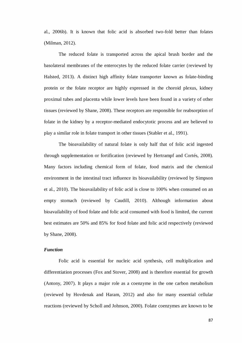
The bioavailability of natural folate is only half that of folic acid ingested
through supplementation or fortification (reviewed by Hertrampf and Cortés, 2008).
Many factors including chemical form of folate, food matrix and the chemical
environment in the intestinal tract influence its bioavailability (reviewed by Simpson
et al., 2010). The bioavailability of folic acid is close to 100% when consumed on an
empty stomach (reviewed by Caudill, 2010). Although information about
bioavailability of food folate and folic acid consumed with food is limited, the current
best estimates are 50% and 85% for food folate and folic acid respectively (reviewed
by Shane, 2008).
Function
Folic acid is essential for nucleic acid synthesis, cell multiplication and
differentiation processes (Fox and Stover, 2008) and is therefore essential for growth
(Antony, 2007). It plays a major role as a coenzyme in the one carbon metabolism
(reviewed by Hovdenak and Haram, 2012) and also for many essential cellular
reactions (reviewed by Scholl and Johnson, 2000). Folate coenzymes are known to be

88
involved in amino acid metabolism that involves the donation of a methyl group to
homocysteine to form methionine, an essential amino acid that is converted to SAM
which participates in the methylation of over 100 different compounds (reviewed by
Simpson et al., 2010) (Fig. 22). It is also known to influence antioxidant defences
through its role as a superoxide scavenger (Doshi et al., 2001) and can protect bio-
constituents such as cellular membranes or DNA from free radical damage (Joshi et
al., 2001). It also has a role in promoting immune function (Troen et al., 2006) and
has beneficial effects on the endothelial function by improving nitric oxide
availability (Antoniades et al., 2006).
Figure 22: Functions of Folate
DHF: Dihydrofolate; THF: Tetrahydrofolate; SAM: S-adenosylmethionine; SAH: S-denosylhomocysteine
Source: Modified from Water Soluble Vitamins Clinical Research and Future Application. Editors: Olaf
Stanger. Subcellular Biochemistry, Volume 56, 2012.
89
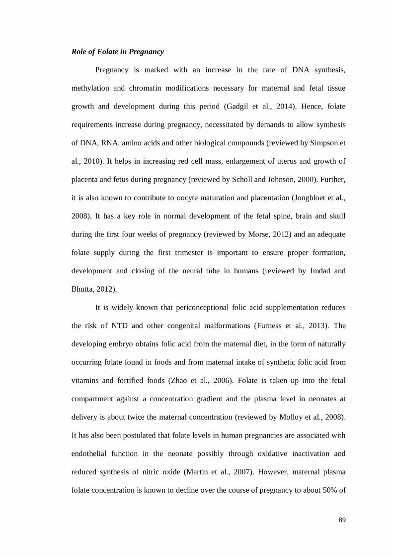
Role of Folate in Pregnancy
Pregnancy is marked with an increase in the rate of DNA synthesis,
methylation and chromatin modifications necessary for maternal and fetal tissue
growth and development during this period (Gadgil et al., 2014). Hence, folate
requirements increase during pregnancy, necessitated by demands to allow synthesis
of DNA, RNA, amino acids and other biological compounds (reviewed by Simpson et
al., 2010). It helps in increasing red cell mass, enlargement of uterus and growth of
placenta and fetus during pregnancy (reviewed by Scholl and Johnson, 2000). Further,
it is also known to contribute to oocyte maturation and placentation (Jongbloet et al.,
2008). It has a key role in normal development of the fetal spine, brain and skull
during the first four weeks of pregnancy (reviewed by Morse, 2012) and an adequate
folate supply during the first trimester is important to ensure proper formation,
development and closing of the neural tube in humans (reviewed by Imdad and
Bhutta, 2012).
It is widely known that periconceptional folic acid supplementation reduces
the risk of NTD and other congenital malformations (Furness et al., 2013). The
developing embryo obtains folic acid from the maternal diet, in the form of naturally
occurring folate found in foods and from maternal intake of synthetic folic acid from
vitamins and fortified foods (Zhao et al., 2006). Folate is taken up into the fetal
compartment against a concentration gradient and the plasma level in neonates at
delivery is about twice the maternal concentration (reviewed by Molloy et al., 2008).
It has also been postulated that folate levels in human pregnancies are associated with
endothelial function in the neonate possibly through oxidative inactivation and
reduced synthesis of nitric oxide (Martin et al., 2007). However, maternal plasma
folate concentration is known to decline over the course of pregnancy to about 50% of

90
nonpregnant levels (Cikot et al., 2001). Maternal folate status is also known to
attenuate some of the adverse effects of protein restriction (reviewed by Christian and
Stewart, 2010). Thus, it is essential to have an adequate supply of folate during
pregnancy (reviewed by Xu et al., 2009).
During pregnancy the recommended dietary allowance (RDA) for folate is
almost 50% higher than that of the nonpregnant woman which is considered sufficient
to maintain red blood cell folate concentrations (tissue stores) in the normal range
(reviewed by Simpson et al., 2010). The National Anaemia Prophylaxis Programme in
India recommends folic acid and iron (0.5 mg and 60 mg, respectively)
supplementation to all pregnant mothers (reviewed by Kalaivani, 2009; ICMR, 2000).
Folate Deficiency
Folate deficiency is known to occur due to various reasons such as inadequate
dietary intake, intestinal malabsorption, altered hepatic uptake and metabolism and
reduced reabsorption by renal tubular cells (reviewed by Halsted, 2013). Inadequate
folate availability results in profound adverse effects in rapidly dividing cells such as
those of the developing conceptus thereby contributing to restricted fetal growth
(Xiao et al., 2005). Low folate status is also known to cause hyperhomocysteinemia,
hypercoagulability and venous thrombosis (reviewed by Hovdenak and Haram, 2012).
Dietary folate deficiency is most common in developing countries with about
25% of pregnant women in India being folate deficient (reviewed by Hovdenak and
Haram, 2012). Compromised folate intake or status in mothers is shown to be
associated with LBW, abruptio placentae and risk for spontaneous abortions, NTD,
preeclampsia, stillbirth and preterm delivery (reviewed by Molloy et al., 2008). The
major clinical concern for folate deficiency is increased incidence of NTD (reviewed
by Simpson et al., 2010).
91
Widespread acknowledgement of the negative effects of folate deficiency has
resulted in worldwide folate fortification of foods in order to prevent deficiency in
pregnant women (reviewed by Berry et al., 2010). Studies have reported a reduced
risk of NTD which includes spina bifida (malformation of the spinal column) and
anencephaly (absence of major portion of the brain, skull and scalp) being associated
with both increased maternal folate intake and higher maternal red blood cell folate
concentration (reviewed by Morse, 2012).
Folate in Pregnancy Complications
Pregnancy disorders such as preeclampsia, small for gestational age and
preterm birth are known to be associated with folate deficiency (Timmermans et al.,
2009; Bodnar et al., 2006; reviewed by Scholl and Johnson, 2000). Folate deficiency
along with hyperhomocysteinemia has been implicated as risk factors for the
subsequent development of preeclampsia and other placenta-mediated diseases
(reviewed by Ray and Laskin, 1999). Emerging evidence suggests that folic acid-
containing multivitamins reduces the risk of gestational hypertension or preeclampsia
(Hernández-Díaz et al., 2002). It has also been suggested that during the
periconceptional period, folic acid from food intake and routine supplementation may
be adequate, however larger doses must be provided in early gestation, particularly for
women with a higher risk of adverse pregnancy outcomes (e.g., preeclampsia)
(reviewed by Xu et al., 2009). Folic acid supplementation at 1 mg or higher daily is
reported to be associated with a reduced risk of preeclampsia (Wen et al., 2008).
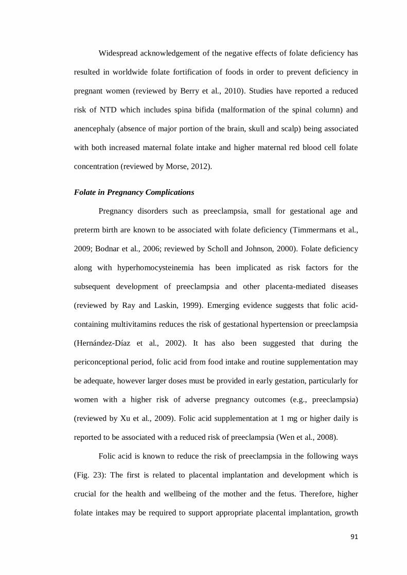
Folic acid is known to reduce the risk of preeclampsia in the following ways
(Fig. 23): The first is related to placental implantation and development which is
crucial for the health and wellbeing of the mother and the fetus. Therefore, higher
folate intakes may be required to support appropriate placental implantation, growth

92
and development in early pregnancy (Wen et al., 2013). The second is related to the
effect of folic acid on lowering blood homocysteine levels (Bernasconi et al., 2006;
Chuang et al., 2006), as hyperhomocysteinemia is a risk factor for preeclampsia
(Fayed et al., 2004). The third is related to the effect of folic acid supplementation in
improving the function of endothelial cells (Antoniades et al., 2006) and therefore
reducing the risk of preeclampsia.
Figure 23: Schematic Representation of Different Proposed Mechanisms through
which Folic Acid Reduces the Risk of Developing Preeclampsia
Source: reviewed by Wen et al., 2013, J Pregnancy. 2013:294312.
3.1.3 Vitamin B12
Structure
Vitamin B12 is a large organometallic molecule, ~1300-1500 Da in size and is
the most chemically complex vitamin known (reviewed by Froese and Gravel, 2010).
A more specific name for vitamin B12 is cobalamin and is the largest of the B complex
93
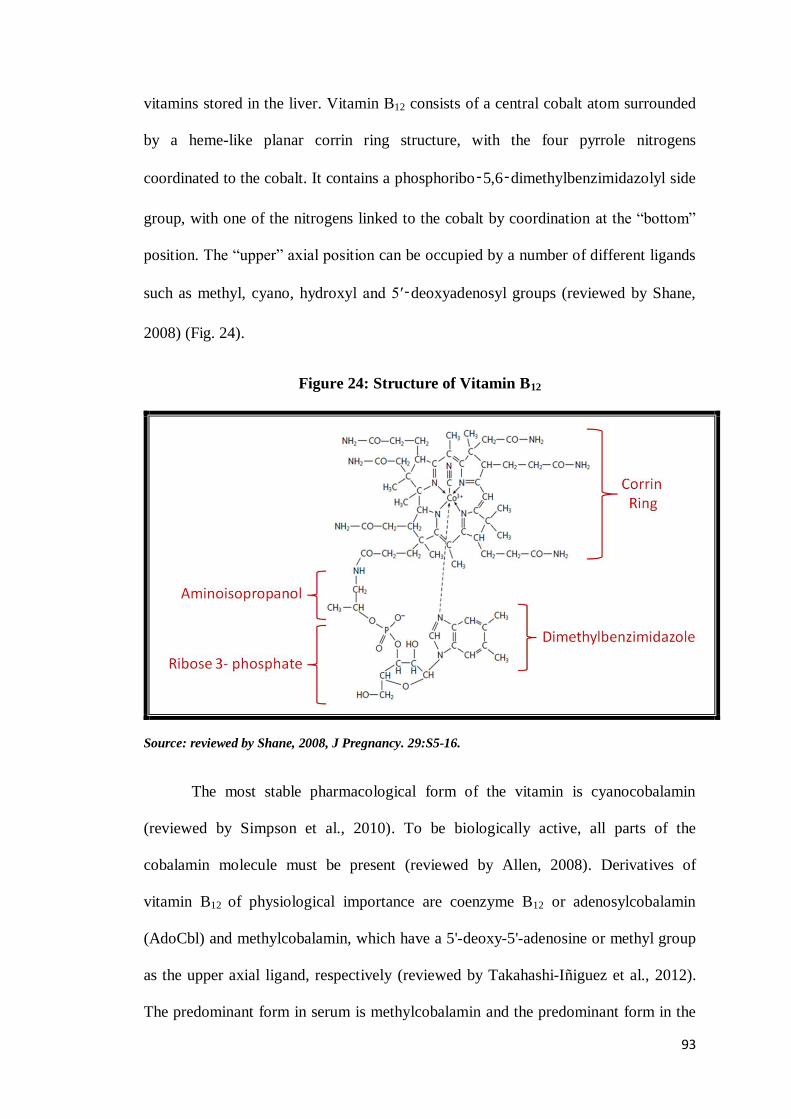
vitamins stored in the liver. Vitamin B12 consists of a central cobalt atom surrounded
by a heme-like planar corrin ring structure, with the four pyrrole nitrogens
coordinated to the cobalt. It contains a phosphoribo‑5,6‑dimethylbenzimidazolyl side
group, with one of the nitrogens linked to the cobalt by coordination at the “bottom”
position. The “upper” axial position can be occupied by a number of different ligands
such as methyl, cyano, hydroxyl and 5′‑deoxyadenosyl groups (reviewed by Shane,
2008) (Fig. 24).
Figure 24: Structure of Vitamin B12
Source: reviewed by Shane, 2008, J Pregnancy. 29:S5-16.
The most stable pharmacological form of the vitamin is cyanocobalamin
(reviewed by Simpson et al., 2010). To be biologically active, all parts of the
cobalamin molecule must be present (reviewed by Allen, 2008). Derivatives of
vitamin B12 of physiological importance are coenzyme B12 or adenosylcobalamin
(AdoCbl) and methylcobalamin, which have a 5'-deoxy-5'-adenosine or methyl group
as the upper axial ligand, respectively (reviewed by Takahashi-Iñiguez et al., 2012).
The predominant form in serum is methylcobalamin and the predominant form in the
94
cytosol is deoxyadenosylcobalamin (reviewed by Klee, 2000). There are three
important, interrelated factors contributing to cobalamin reactivity and function: i) the
oxidation state of cobalt; ii) whether the 5,6-dimethylbenzimidazole is coordinated to
cobalt in the lower axial position; and iii) the identity of the R-group bound in the
upper axial position (reviewed by Froese and Gravel, 2010).
Sources
Vitamin B12 is generally found only in foods of animal origin (reviewed by
Simpson et al., 2010). Major dietary sources include animal products such as liver,
beef, kidney, chicken, fish such as salmon, halibut and tuna, yogurt, milk, cheese and
eggs (reviewed by Anyanwu and Kanu, 2007). Synthetic crystalline vitamin B12 is
used as a fortificant in cereals or as supplements (reviewed by Dror and Allen, 2012).
Vitamin B12 deficiency is known to be prevalent when intake of these foods is low
due to their high cost, lack of availability and cultural or religious beliefs. However,
deficiency is certainly more prevalent in strict vegetarians (reviewed by de Benoist,
2008).
Absorption, Transport and Bioavailability
Absorption of vitamin B12 is a complex process, involving a series of steps
that can be affected adversely by intestinal disease, infections and medications
(reviewed by Allen, 2008). Vitamin B12 in food is bound to protein and is released in
the stomach by the acid environment and by proteolysis of binders by pepsin
(reviewed by Shane, 2008). It then subsequently binds to haptocorrin (HC) to form
the complex HC-cobalamin (reviewed by Randaccio et al., 2010). In humans, the
stomach contains specialized parietal cells that secrete a 50 kDa glycoprotein called
intrinsic factor (IF) that can bind cobalamin (reviewed by Shane, 2008). In the small
intestine, pancreatic proteases cleave HC and released cobalamin then binds to the IF

95
to form an IF-cobalamin complex (reviewed by Allen, 2008). The IF-receptor
(cubulin) located at the distal ileum at the end of the small intestine recognizes this
IF-cobalamin complex, not cobalamin or unligated IF and the complex is internalized
by a receptor-mediated endocytotic process (Fedosov et al., 2005). Inside the
enterocytes, the IF is degraded and the free cobalamin binds to a 38 kDa protein
called transcobalamin II (TC-II) forming a TC-II-cobalamin complex that is released
into plasma, where it is endocytosed by membrane receptors, R-TC-cobalamin
(Quadros et al., 2009). Once endocytosed, the TC-II-cobalamin complex is degraded
in the lysosome and the free cobalamin is transported out of the lysosome to the
cytosol. Plasma also contains two additional vitamin B12-binding glycoproteins or R-
binders called haptocorrin (transcobalamin I, TC-I) and transcobalamin III (TC-III)
which are less specific than TC-II and also bind vitamin B12 analogs (reviewed by
Shane, 2008). Once in the cytosol, cobalamin is processed by many proteins to
produce the cofactors 5'-deoxyadenosylcobalamin in mitochondrion and
methylcobalaminin in cytosol (reviewed by Froese and Gravel, 2010). The vitamin is
excreted via the urine and the bile (Fig. 25).
The bioavailability of vitamin B12 from the diet is approximately 50%
(reviewed by Simpson et al., 2010) whereas synthetic (crystalline) vitamin B12 is
known to be more efficiently absorbed; approximately 60% (reviewed by Allen,
2008). Normal body stores are about 1 to 3 mg; the turnover of the vitamin B12 in
healthy persons is about 0.1% per day; and signs of deficiency appear when the pool
drops below 300 μg (reviewed by Allen, 2008).

96
Figure 25: Absorption and Cellular Transport Cycle of Vitamin B12
THF: Tetrahydrofolate; TC-II-R: Transcobalamine II Receptor; TC-II-Cobalamine: Transcobalamine
II-Cobalamine
Source: Modified from review of Carmel, 2008, Food Nutr Bull. 29:S177-87.
Function
Vitamin B12 is known to play a vital role in one carbon metabolism (Katre et
al., 2010). There are two major metabolic roles for vitamin B12 where it acts as a
cofactor: (a) synthesis of methionine from homocysteine; and (b) conversion of
methylmalonyl coenzyme A to succinyl coenzyme A (reviewed by Klee, 2000). It is
also critical for nucleotide synthesis and amino acid metabolism (Ronnenberg et al.,
97
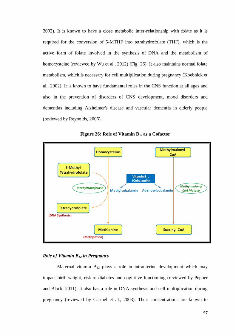
2002). It is known to have a close metabolic inter-relationship with folate as it is
required for the conversion of 5-MTHF into tetrahydrofolate (THF), which is the
active form of folate involved in the synthesis of DNA and the metabolism of
homocysteine (reviewed by Wu et al., 2012) (Fig. 26). It also maintains normal folate
metabolism, which is necessary for cell multiplication during pregnancy (Koebnick et
al., 2002). It is known to have fundamental roles in the CNS function at all ages and
also in the prevention of disorders of CNS development, mood disorders and
dementias including Alzheimer's disease and vascular dementia in elderly people
(reviewed by Reynolds, 2006).
Figure 26: Role of Vitamin B12 as a Cofactor
Role of Vitamin B12 in Pregnancy
Maternal vitamin B12 plays a role in intrauterine development which may
impact birth weight, risk of diabetes and cognitive functioning (reviewed by Pepper
and Black, 2011). It also has a role in DNA synthesis and cell multiplication during
pregnancy (reviewed by Carmel et al., 2003). Their concentrations are known to
98
decline during pregnancy due to haemodilution (Metz et al., 1995). Vitamin B12 is
concentrated in the placenta and transferred to the fetus, with newborn vitamin B12
concentrations approximately double than those of the mother (reviewed by Dror and
Allen, 2012). A strong association is known to exist between maternal and infant
plasma vitamin B12 concentrations at delivery, indicating that maternal vitamin B12
status affects the fetal vitamin status at birth (reviewed by Hovdenak and Haram,
2012). In India, the RDA of vitamin B12 for pregnancy and lactation is 1.2 and 1.5
μg/day respectively (ICMR, 2009).
Vitamin B12 Deficiency
Vitamin B12 deficiency is mainly seen in pregnant women consuming a
predominantly vegetarian diet (reviewed by Hovdenak and Haram, 2012). Studies in
India have shown a low dietary consumption of vitamin B12 due to dietary pattern of
vegetarianism and poor consumption of milk and milk products resulting in
deficiency of vitamin B12 although concentrations of folate are adequate. Thus, this
leads to an imbalance of plasma folate and vitamin B12, which might have
physiological consequences during pregnancy (Gadgil et al., 2014).
Elevated methylmalonic acid and total homocysteine concentrations are
considered as sensitive metabolic markers for vitamin B12 deficiency (Herrmann et al.,
2000). Vitamin B12 deficiency primarily in the elderly occurs due to malabsorption
(reviewed by Allen, 2008). Deficiency of vitamin B12 is also known to cause
pernicious anemia (reviewed by Pepper and Black, 2011). Myelopathy and
neuropathy are known to be main clinical manifestations of vitamin B12 deficiency
(reviewed by Cetin et al., 2010). Symptoms of vitamin B12 deficiency include
megaloblastic anaemia, tingling and numbness of the extremities, gait abnormalities,
visual disturbances, memory loss and dementia. Furthermore, folic acid fortification

99
or supplementation with >1 mg/day is known to „mask‟ clinical symptoms of vitamin
B12 deficiency (reviewed by Dror and Allen, 2012). Thus, vitamin B12
supplementation may be especially needed in women on vegetarian diets, in
malabsorption disorders and in communities or countries where undernutrition is
common (reviewed by Hovdenak and Haram, 2012).
Vitamin B12 in Pregnancy Complications
Maternal vitamin B12 deficiency is known to be associated with increased risk
for several adverse pregnancy outcomes for both mother and fetus (Vanderjagt et al.,
2011) such as NTD (Molloy et al., 2009), IUGR (Muthayya et al., 2006),
preeclampsia (reviewed by Allen, 2005) and early miscarriage (Hübner et al., 2008).
Poor maternal B-vitamin status has been a major global cause of
hyperhomocysteinemia and poor pregnancy outcomes (reviewed by Allen, 2005).
Therefore, the next section describes homocysteine and its role in several pregnancy
complications.
3.1.4 Homocysteine
Structure
Homocysteine is a non-protein forming, thiol-containing, four-carbon amino
acid derived from the demethylation of the essential amino acid methionine (reviewed
by Holmes, 2003) (Fig. 27). It is at the intersection of two metabolic pathways; (1) the
remethylation cycle that includes remethylation to methionine, which requires folate,
vitamin B12 or betaine; and (2) transsulfuration to cystathionine, which requires
pyridoxal-5'-phosphate (Miller et al., 2003). Thus, homocysteine may be either
remethylated to methionine or eliminated from the cycle by formation of cysteine in
the transsulfuration pathway (Mislanova et al., 2011). The methionine cycle takes

100
place in all cell types, whereas transsulfuration occurs in a limited number of tissues
including liver, kidney, small intestine and pancreas (reviewed by Finkelstein, 2006).
Figure 27: Structure of Homocysteine and Homocystine
Source: Nelson DL and Cox MM. Lehninger Principles of Biochemistry,
2004, 4th
Edition, Freeman WH, USA.
Structurally, homocysteine closely resembles methionine and cysteine and all
three amino acids contain sulfur. Homocystine is a dimer composed of two oxidized
molecules of homocysteine linked by a disulfide bond (reviewed by Klee, 2000) (Fig.
27). Only 1 to 2% of total homocysteine circulate in blood freely in its reduced form,
while 70 to 90% are protein-bound and the remaining are disulfides, homocystine and
the mixed disulfide homocysteine-cysteine (reviewed by Herrmann, 2001). The sum
of all the forms of homocysteine present in plasma is usually referred as total
homocysteine (reviewed by Medina et al., 2001). It is an essential amino acid required
for the growth of cells and tissues in the human body. The only source of

101
homocysteine in the human organism comes from the methionine in dietary proteins.
Methionine is the only essential, sulfur containing amino acid in mammalian diets
which are mainly of animal origin (reviewed by de la Calle et al., 2003).
Homocysteine metabolism is strongly linked to its function as a methyl group donor
in transmethylation reactions (Poirier, 2002). There are four key enzymes involved in
homocysteine metabolism: cystathionine β-synthase, 5,10-methylentetrahydrofolate
reductase, methionine synthase and methionine synthase reductase (Also-Rallo et al.,
2005).
It is important to note that plasma homocysteine can also serve as a functional
indicator of vitamin status and therefore is useful as a monitoring tool to measure the
efficacy of a food-fortification program with B-vitamins (reviewed by Selhub, 2008).
Circulating total homocysteine concentrations are a useful integral marker of one
carbon metabolism and are known to be influenced by genetic and dietary factors
(Katre et al., 2010).
Homocysteine is an oxidant that can generate reactive oxygen species thereby
damaging macromolecules, including DNA, proteins and lipids (reviewed by Wu et
al., 2012). Homocysteine is highly cytotoxic and the intracellular homocysteine
concentration is kept low by catabolism and by a cellular homocysteine export
mechanism into plasma (reviewed by Herrmann, 2001). Plasma homocysteine is
mainly metabolized in the kidney (about 70%) but very little is excreted into the urine
(reviewed by Yeun, 1998).
Hyperhomocysteinemia
Hyperhomocysteinemia either results from a genetic defect in the enzymes
that participate in homocysteine synthesis and metabolism or a deficiency of folic
acid, vitamins B6 and B12 (Makedos et al., 2007). Elevated homocysteine levels are a

102
sign of disturbed remethylation of homocysteine (Geisel et al., 2005). Higher plasma
total homocysteine concentrations are also associated with deficits in cognition,
arterial and/or venous thrombosis, vascular dementia, neuropathies, risk of stroke and
myocardial infarction (reviewed by Cetin et al., 2010). Studies have shown that
hyperhomocysteinemia usually can be corrected by supplementation with folic acid
(Yamamoto et al., 2012; Scorsatto et al., 2011).
Homocysteine and Pregnancy
In normal pregnancy, homocysteine concentrations fall with advancing
gestational age (Guven et al., 2009) and are linked to many factors such as:
physiological response to pregnancy, increase in oestrogen, hemodilution from
increased plasma volume or increased demand for methionine by both the mother and
the fetus (Davari-Tanha et al., 2008).
Elevated levels of homocysteine are associated with a greater risk of adverse
pregnancy outcomes. The proposed mechanisms are: increase in oxygen free radical
concentrations and reduction in nitrous oxide concentrations, leading to endothelial
dysfunction. It also increases oxidative stress and results in placental ischemia,
increases inflammatory response that is cytotoxic to endothelial cells and also leads to
apoptosis of endothelial cells (reviewed by Allen, 2005). Further, placental
development in early pregnancy is also known to be negatively influenced by
increased maternal homocysteine concentrations (Steegers-Theunissen and Steegers,
2003).
Homocysteine and Preeclampsia
A number of studies have reported elevated levels of homocysteine in women
with preeclampsia (Mujawar et al., 2011; Napolitano et al., 2008; Braekke et al.,
2007). Hyperhomocysteinemia is suggested to play a key role in the pathogenesis of
103
preeclampsia through oxidative stress and endothelial cell dysfunction which is the
central theme ultimately leading to hypertension and proteinuria during gestation
(reviewed by Steegers et al., 2010; Falcao et al., 2009). Homocysteine is known to
injure the vascular endothelium by generating hydrogen peroxide and by impairing
basal nitric oxide production (Stamler et al., 1993). Further, hyperhomocysteinemia
increases oxidative stress which is caused by an increase in the concentration of
fibronectin, lipid peroxides and plasma triglycerides (reviewed by de la Calle et al.,
2003). Elevated homocysteine levels which are associated with preeclampsia are also
reported to be due to genetic abnormalities (reviewed by Selhub, 1999). A recent
study demonstrates correlation between folate, vitamin B12 and homocysteine that are
required for the remethylation of homocysteine to methionine in the one carbon cycle
(Mujawar et al., 2011).
3.1.5 One Carbon Cycle
The growing field of epigenetic research has highlighted the role of one
carbon metabolites on the developmental programming of chronic disease (reviewed
by Waterland and Michels, 2007). One carbon metabolism is a network of interrelated
biochemical reactions that involve the transfer of one carbon groups from one site to
another (reviewed by Choi and Mason, 2000). It is compartmentalized in the cell,
occurring primarily within the cytoplasm and the mitochondria (reviewed by Beaudin
and Stover, 2007). These reactions are significant for the production of DNA bases,
the conversion of homocysteine into methionine, neurological and immunological
function, growth and development and the formation of red blood cells (reviewed by
Wu et al., 2012).
The vitamins, folate and B12 serve as coenzymes in the one carbon metabolism
(reviewed by Selhub et al., 2008). One carbon metabolism in mitochondria primarily

104
functions to generate one carbon units in the form of formate for cytoplasmic one
carbon metabolism. In the cytoplasm, folate activated one carbon units act in an
interdependent anabolic network comprised of 3 biosynthetic pathways: de novo
purine biosynthesis; de novo thymidylate biosynthesis and the remethylation of
homocysteine to methionine (reviewed by Stover, 2009). In this metabolism, a carbon
unit from serine or glycine reacts with THF to form methylene-THF (reviewed by
Selhub et al., 2008). This form of folate can be used for the synthesis of thymidine;
oxidized to formyltetrahydrofolate for the synthesis of purines or reduced to 5-MTHF
and used to methylate homocysteine to form methionine (reviewed by Ross, 2003).
Methionine is formed via the vitamin B12-dependent transfer of a methyl
group from 5-MTHF to homocysteine in the methionine synthase reaction (James et
al., 2004). Methionine is then activated by methionine adenosyltransferase to form
SAM, the primary methyl donor (Geisel et al., 2005). It is known to donate its labile
methyl groups to more than 80 biological methylation reactions, including the
methylation of DNA, RNA, proteins, phospholipids and neurotransmitters (reviewed
by Choi and Mason, 2002). The transfer of the methyl group from SAM to the various
methyl acceptors via numerous methyltransferases results in the formation of S-
adenosylhomocysteine (SAH) (James et al., 2004). SAH is subsequently cleaved to
homocysteine, which lies at an important metabolic branch point. Alternatively,
homocysteine can be remethylated to reform methionine via either betaine-
homocysteine methyltransferase or methionine synthase or can be exported to the
extracellular space (Stead et al., 2006).
In some cell types and tissues, homocysteine can also be degraded by the
transsulfuration pathway, through which homocysteine irreversibly condenses with
serine to form cystathionine via a vitamin B6-dependent enzyme cystathionine β-
105
synthase (reviewed by Beaudin and Stover, 2007). Pregnancy involves a marked
acceleration in one carbon transfer reactions, particularly those required for nucleotide
synthesis and thus cell division, which is the basis for the substantial increase in folate
requirements during pregnancy (reviewed by Bailey, 2000). Impairments at various
steps that affect remethylation of homocysteine to methionine or degradation of
homocysteine to cysteine, including inadequate concentrations of folate or vitamin
B12, will result in elevated homocysteine concentrations (reviewed by Selhub et al.,
2008).
One of the major methyl acceptors in the one carbon cycle are the
phospholipids. Phosphatidylethanolamine-N-methyltransferase (PEMT) catalyzes the
sequential methylation of phosphatidylethanolamine to phosphatidylcholine (reviewed
by Vance et al., 1997). PEMT preferentially utilizes phosphatidylethanolamine that
contains DHA molecule generating a phosphatidylcholine molecule containing DHA
(DeLong et al., 1999). It has thus been suggested that the methylation of
phosphatidylethanolamine to phosphatidylcholine by PEMT plays an important role
in the transport of PUFA like DHA from the liver to the plasma and other tissues
(Pynn et al., 2011; Selley, 2007). Further, it has been hypothesized that B-vitamin
(folate, vitamin B12) availability can directly modify liver PEMT activity and PEMT-
dependent PUFA secretion. Thus, adequate dietary intake of these vitamins would be
necessary to maintain normal plasma DHA concentrations and thus tissue availability
(van Wijk et al., 2012). Further, animal studies in our department have also
extensively demonstrated that alterations in maternal levels of folate and vitamin B12
affect the levels of plasma, brain, milk and placental DHA (Roy et al., 2012; Dangat
et al., 2011; Kulkarni et al., 2011a). Besides, our studies on pregnant women have
also discussed that micronutrients such as folic acid, vitamin B12 and DHA are

106
interlinked in the one carbon cycle (reviewed by Dhobale and Joshi, 2012; reviewed
by Sundrani et al., 2011; Kulkarni et al., 2011b) (Fig. 28).
Figure 28: One Carbon Cycle
5,10-MTHF: 5,10-Methylenetetrahydrofolate; 5-MTHF: 5-Methyltetrahydrofolate; COMT: Catecholamine-O-Methyltransferase; DA: Dopamine; DHA: Docosahexaenoic Acid; DNA:
Deoxyribonucleic Acid; EP: Epinephrine; MS: Methionine Synthase; MT: Methyltransferase;
MTHFR: Methylenetetrahydrofolate Reductase; NE: Nor-Epinephrine; PC-DHA:
Phosphatidylcholine-DHA; PE-DHA: Phosphatidylethanolamine-DHA; PEMT:
Phosphatidylethanolamine Methyltransferase; RNA: Ribonucleic Acid; SAH: S-Adenosyl
Homocysteine; SAM: S-Adenosyl Methionine;THF: Tetrahydrofolate;
Earlier cross-sectional studies carried out in our department in women with
pregnancy complications such as preeclampsia indicate altered levels of LCPUFA and
higher levels of homocysteine at delivery, which are associated with poor birth
outcome (Dhobale et al., 2012; Kulkarni et al., 2011b, 2011c; Dangat et al., 2010;
Mehendale et al., 2008). Furthermore, the department also reported a negative
association between erythrocyte DHA and plasma homocysteine concentrations in
preeclampsia (Kulkarni et al., 2011b). However, all these observations were made at

107
the end of pregnancy when the pathology had progressed. Thus, it would be very
useful to analyze these levels in early pregnancy to examine changes over time, to
understand their role in various pregnancy complications. There is also a need to
examine longitudinally these associations during pregnancy as they are important
determinants of the one carbon cycle, which play an important role in fetal
programming and increase the risk of developing NCD such as type 2 diabetes
(reviewed by Yajnik and Deshmukh, 2008) and CVD (reviewed by Martinelli et al.,
2009; reviewed by Erkkilä et al., 2008) in later life.
This chapter therefore examines maternal plasma folate, vitamin B12 and
homocysteine levels at three different time points during gestation and their
association with maternal fatty acid levels (plasma and erythrocyte, discussed
earlier in Chapter 2) in women with preeclampsia and compare them with
normotensive control women.
3.2 Methods and Materials
3.2.1 Study Subjects
The criteria for recruitment of study population and the inclusion and
exclusion criteria are as mentioned in Chapter 2 (Section 2.2.1).
3.2.2 Sample Collection and Processing
Blood samples were collected and processed as described in Chapter 2
(Section 2.2.2). Folate in plasma is known to be unstable with long storage times,
therefore analysis for folate, vitamin B12 and homocysteine was carried out
immediately. The storage cut-off was 3 months. Care was taken not to perform
analysis on samples that were stored for a longer duration. Furthermore, hemolyzed
samples were not used for analysis. Figure 29 shows the number of maternal plasma

108
samples analyzed for folate, vitamin B12 and homocysteine levels at various time
points.
Figure 29: Flow Chart Showing Number of Maternal Plasma Samples Analyzed for
Folate, Vitamin B12 and Homocysteine Levels at Various Time Points
NC: Normotensive Control; PE: Preeclampsia
3.2.3 Neonatal Measurements
Neonatal measurements were recorded as described in Chapter 2 (Section
2.2.3).
3.2.4 Folate, Vitamin B12 and Homocysteine Estimations
Folate, vitamin B12 and homocysteine levels were estimated by the
chemiluminescent microparticle immunoassay (CMIA) technology (Abbott
Diagnostics,Abbott Park, IL, USA) (reviewed by Lee and Griffiths, 1985) and
described by our department earlier (Dhobale et al., 2012). Briefly, 100 µl of plasma
109
was used for analysis of each folate, vitamin B12 and homocysteine. The folate,
vitamin B12 and homocysteine assay was a two-step assay with an automated sample
pre-treatment for determining the presence of folate, vitamin B12 and homocysteine in
human plasma. The reference range for plasma folate assay was 2.34-17.56 ng ml-1
,
for plasma vitamin B12 assay was 187-883 pg ml-1
and for homocysteine assay it was
5.08-15.39 µmol L-1
.
Low plasma folate and vitamin B12 concentrations were defined as <10 ng ml-1
and <150 pg ml-1
respectively and elevated plasma total homocysteine concentrations
as a concentration >10 µmol L-1
(Kulkarni et al., 2011b).
3.2.5 Statistical Analysis
The data was analyzed using the SPSS/PC+ package (Version 20, Chicago, IL,
USA). Values are reported as mean ± SD. Skewed variables were transformed to
normality using the log to the base 10 transformation (plasma folate, vitamin B12,
homocysteine). Independent t-test was used to compare mean values of the various
parameters between normotensive control and preeclampsia (p<0.05) after adjusting
for gestation and socioeconomic status. Correlation between variables was studied
using Pearson‟s correlation analysis after adjusting for gestation and socioeconomic
status. The variable sample number (n) in different measures was due to insufficient
sample volume available.
3.3 Results
3.3.1 Frequency of Consumption of Folate and Vitamin B12 Rich Foods
In our cohort, the major source of folate was green leafy vegetables (e.g.,
spinach, ambat chukka) and legumes (e.g., cowpea, bengalgram, red gram). The rich
sources of vitamin B12 included dairy products and non-vegetarian food items. Dairy

110
products included whole milk and milk products (milk in tea and other beverages,
yoghurt, buttermilk, ghee, ice cream and other milk based preparations). The non-
vegetarian foods included meat, fish and eggs. The frequency of consumption of
folate rich foods was similar in both the normotensive control and preeclampsia
groups at T1 (p=0.134), T2 (p=0.995) and T3 (p=0.616). Similarly, the frequency of
consumption of vitamin B12 rich foods was similar in both the groups at T1 (p=0.990),
T2 (p=0.484) and T3 (p=0.364). The percent women consuming folate and vitamin
B12 rich foods in both normotensive control and preeclampsia groups are in Table 12.
Table 12: Frequency of Consumption of Foods Rich in Folate and Vitamin B12 at
Three Time Points during Pregnancy
T1 T2 T3
Food Group
n (%)
NC
(n=143)
PE
(n=52)
p NC
(n=114)
PE
(n=37)
p NC
(n=131)
PE
(n=42)
p
Folate Rich Foods
Never 7(4.9) 7(13.5) 0.040 6(5.3) 2(5.4) 0.973 8(6.1) 2(4.8) 0.745
Weekly twice 82(57.3) 29(55.8) 0.844 61(53.5) 19(51.4) 0.819 66(50.4) 17(40.5) 0.264
Weekly 2-4 times 40(28) 14(26.9) 0.884 36(31.6) 12(32.4 ) 0.923 37(28.2) 16(38.1) 0.228
More than 4 times
in a week
14(9.8)
2(3.8)
0.181
11(9.6)
4(10.8)
0.837
20(15.3)
7(16.7)
0.828
Vitamin B12 Rich Foods
Never 3(2.1) 1(1.9) 0.938 1(0.9) 0(0) 0.568 1(0.8) 2(2.4) 0.394
Weekly once 37(25.9) 14(26.9) 0.883 23(20.2) 5(13.5) 0.365 22(16.8) 26(9.5) 0.257
Weekly twice 53(37.1) 18(34.6) 0.753 41(36) 11(29.7) 0.488 46(35.1) 58(28.6) 0.434
More than 2 times
in a week
50(35)
19(36.5)
0.839
49(43)
21(56.8)
0.144
62(47.3)
87(59.5)
0.169
NC - Normotensive control; PE - Preeclampsia; n - Number of subjects; p - Significance; T1=16th-20th
week; T2= 26th-30th week; T3= at delivery
3.3.2 Levels of Maternal Plasma Folate, Vitamin B12 and Homocysteine across
Gestation
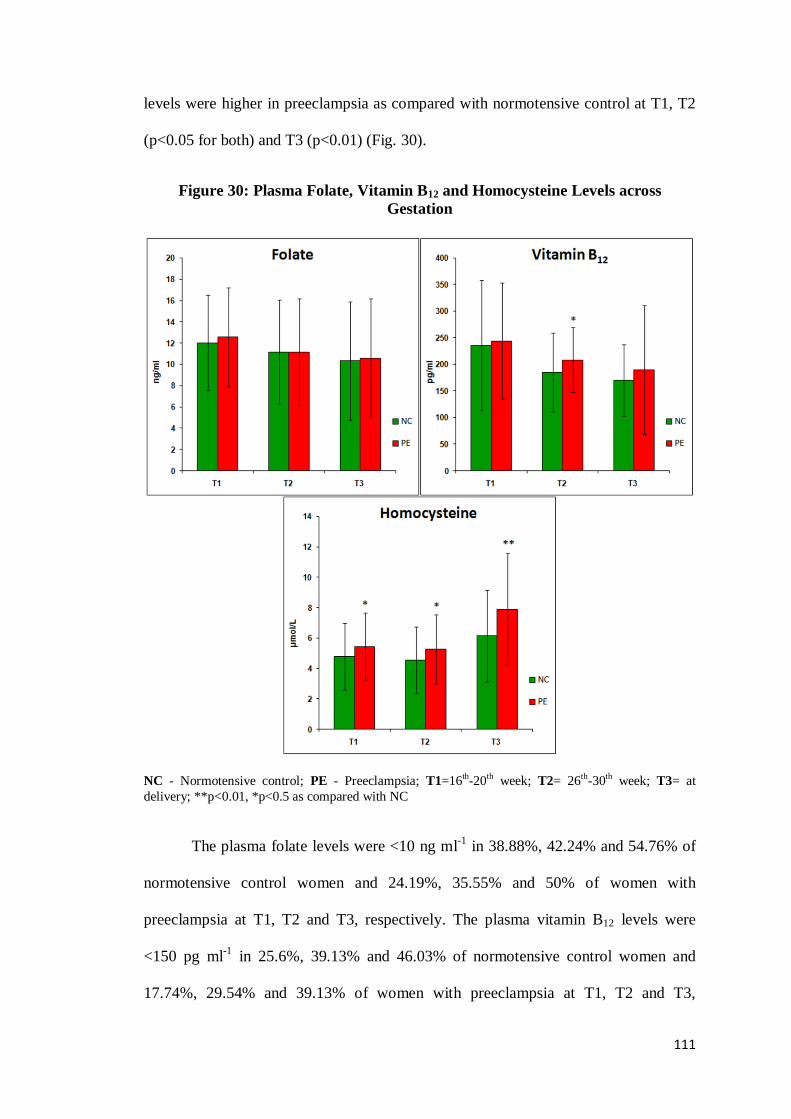
Maternal plasma folate levels were similar between preeclampsia and
normotensive control groups at all time points across gestation. Maternal plasma
vitamin B12 levels were significantly higher in preeclampsia (p<0.05) as compared
with normotensive control at T2. On the other hand, maternal plasma homocysteine
111
levels were higher in preeclampsia as compared with normotensive control at T1, T2
(p<0.05 for both) and T3 (p<0.01) (Fig. 30).
Figure 30: Plasma Folate, Vitamin B12 and Homocysteine Levels across
Gestation
NC - Normotensive control; PE - Preeclampsia; T1=16th-20th week; T2= 26th-30th week; T3= at
delivery; **p<0.01, *p<0.5 as compared with NC
The plasma folate levels were <10 ng ml-1
in 38.88%, 42.24% and 54.76% of
normotensive control women and 24.19%, 35.55% and 50% of women with
preeclampsia at T1, T2 and T3, respectively. The plasma vitamin B12 levels were
<150 pg ml-1
in 25.6%, 39.13% and 46.03% of normotensive control women and
17.74%, 29.54% and 39.13% of women with preeclampsia at T1, T2 and T3,

112
respectively, whereas the plasma homocysteine levels were >10 µmol L-1
in 4.03%,
3.47% and 16% of normotensive control women and 6.55%, 4.54% and 23.91% of
women with preeclampsia at T1, T2 and T3, respectively.
3.3.3 Associations between Folate, Vitamin B12 and Homocysteine in Maternal
Plasma
There was a negative association between maternal plasma folate and maternal
plasma homocysteine at T2 (r = -0.189, p = 0.045, n =115) in normotensive control.
Similar negative association was observed between maternal plasma vitamin B12 and
maternal plasma homocysteine at T1 and T2 (r = -0.258, p = 0.004, n =123; r = -0.264,
p = 0.005, n =114) in normotensive control. However, no associations were observed
in the preeclampsia group.
3.3.4 Associations of Maternal Plasma Fatty Acids with Maternal Plasma
Folate, Vitamin B12 and Homocysteine
There was a positive association between maternal plasma folate and maternal
plasma AA at T3 (r = 0.271, p = 0.003, n = 125) in normotensive control. While in
preeclampsia there was a positive association between maternal plasma folate and
maternal plasma DHA at T1 (r = 0.402, p = 0.003, n = 54).
A positive association was observed between maternal plasma vitamin B12 and
maternal plasma AA at T1 in normotensive control and preeclampsia (r = 0.203, p =
0.031, n =122; r = 0.295, p = 0.035, n = 54 respectively). In preeclampsia, there was a
positive association of maternal plasma vitamin B12 with maternal plasma DHA and
total omega-3 fatty acids at T1 (r = 0.327, p = 0.019, n = 54; r = 0.388, p = 0.005, n =
54 respectively).
There was a negative association of maternal plasma homocysteine with
maternal plasma AA at T1 and T3 (r = -0.235, p = 0.013, n = 121; r = -0.180, p =
113
0.050, n = 124 respectively) and maternal plasma DHA at T3 (r = -0.185, p = 0.044, n
=124) in normotensive control. In preeclampsia, a negative association was observed
between maternal plasma homocysteine and maternal plasma DHA at T2 (r = -0.447,
p = 0.006, n = 44). Similar negative association was seen between maternal plasma
homocysteine and maternal plasma total omega-3 fatty acids at T1 (r = -0.274, p =
0.054, n = 53) in preeclampsia.
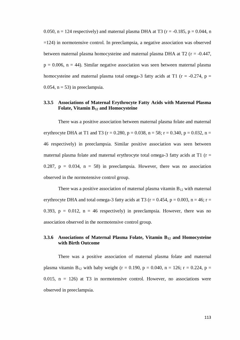
3.3.5 Associations of Maternal Erythrocyte Fatty Acids with Maternal Plasma
Folate, Vitamin B12 and Homocysteine
There was a positive association between maternal plasma folate and maternal
erythrocyte DHA at T1 and T3 (r = 0.280, p = 0.038, n = 58; r = 0.340, p = 0.032, n =
46 respectively) in preeclampsia. Similar positive association was seen between
maternal plasma folate and maternal erythrocyte total omega-3 fatty acids at T1 (r =
0.287, p = 0.034, n = 58) in preeclampsia. However, there was no association
observed in the normotensive control group.
There was a positive association of maternal plasma vitamin B12 with maternal
erythrocyte DHA and total omega-3 fatty acids at T3 (r = 0.454, p = 0.003, n = 46; r =
0.393, p = 0.012, n = 46 respectively) in preeclampsia. However, there was no
association observed in the normotensive control group.
3.3.6 Associations of Maternal Plasma Folate, Vitamin B12 and Homocysteine
with Birth Outcome
There was a positive association of maternal plasma folate and maternal
plasma vitamin B12 with baby weight (r = 0.190, p = 0.040, n = 126; r = 0.224, p =
0.015, n = 126) at T3 in normotensive control. However, no associations were
observed in preeclampsia.
114
3.4 Discussion
The present prospective study examined the levels of maternal plasma
folate, vitamin B12 and homocysteine across gestation in normotensive control
women and women with preeclampsia. Further, their associations with maternal
fatty acids (plasma and erythrocyte) were also examined. Our results for the first
time indicate several interesting observations in preeclampsia which are as
follows:
1) No change in the maternal plasma levels of folate at all time points
2) A significant increase in the maternal plasma levels of vitamin B12 only at T2
3) A significant increase in the maternal plasma levels of homocysteine as
gestation advances
4) Positive association of maternal plasma folate with maternal plasma and
erythrocyte DHA at T1
5) Positive association of maternal plasma vitamin B12 with maternal plasma
DHA and total omega-3 fatty acids at T1
6) Negative association of maternal plasma homocysteine with maternal plasma
DHA at T2 and total omega-3 fatty acids at T1
7) In addition, a positive association of maternal plasma folate and vitamin B12
with baby weight at T3 was observed only in normotensive control
3.4.1 Maternal Plasma Folate Levels
In the present study, maternal plasma folate levels were not significantly
different at all time points between the preeclampsia and normotensive control
groups. These results are similar to earlier studies which did not find any significant
difference in the folate levels between control and preeclampsia (Li et al., 2013;
Thériault et al., 2013; Furness et al., 2012; Guven et al., 2009; Makedos et al., 2007;
115
Braekke et al., 2007; Rajkovic et al., 1997). It has been suggested that there may be
some other factors in addition to this vitamin deficiency which play a role in the
etiopathogenesis of preeclampsia (Acilmis et al., 2011).
In contrast, few other studies have reported either lower levels of folate
(Salehi-PourMehr et al., 2012; Bergen et al., 2012; Mujawar et al., 2011; Patrick et
al., 2004; Sanchez et al., 2001) or higher levels (Also-Rallo et al., 2005; López-
Quesada et al., 2003; Powers et al., 2003) in preeclampsia as compared to controls.
The discrepancy of these reports may be a result of various factors, including the
timing of the intervention, the dose of folic acid, the other components in the
supplements and the population (Li et al., 2013). As per the National Anemia
Prophylaxis Programme all women in our cohort were routinely given iron (60 mg)
and folic acid (500 µg) tablets during the first trimester of pregnancy. One possible
explanation for similar levels could also be due to this Prophylaxis Programme, as in
our study the first sample was collected at the end of first trimester. Similar
observation has been reported by others in a cohort of pregnant women benefiting
from a national policy of folic acid food fortification along with a high adherence to
folic acid supplementation (Thériault et al., 2013).
3.4.2 Maternal Plasma Vitamin B12 Levels
In the current study, maternal plasma vitamin B12 levels were comparable
between preeclampsia and normotensive control group at T1 and at T3. However, at
T2, levels of maternal plasma vitamin B12 were higher in preeclampsia as compared to
normotensive control. Studies carried out in preeclampsia are contradictory, with few
reporting no change (Bergen et al., 2012; Acilmis et al., 2011; Guven et al., 2009;
Makedos et al., 2007; López-Quesada et al., 2003; Sanchez et al., 2001; Rajkovic et
al., 1997) and others reporting lower levels (Mujawar et al., 2011; Laivuori et al.,
116
1999). There is one study which reports significantly higher vitamin B12 levels found
in patients with preeclampsia bearing the wild type genotype of MTHFR gene
involved in the folate-homocysteine metabolism. However, they suggest that these
results are difficult to explain and should be confirmed in a larger group of patients
(Also-Rallo et al., 2005).
In addition to the above studies, there is one report suggesting that vitamin B12
concentrations are specific to different races and do not differ by pregnancy outcome
(Patrick et al., 2004). It has also been suggested that transcobalamin I is ubiquitous in
most body fluids and its major importance is the problem it may cause with false
positive increased vitamin B12 measurements (reviewed by Klee, 2000). Further, it has
also been reported that because serum vitamin B12 levels are often influenced by
factors unrelated to vitamin B12 intake, stores or deficiency, it is unclear whether the
differences in concentrations actually reflect vitamin B12 status (Patrick et al., 2004).
Elevated levels of plasma cobalamin are known to be associated with
functional cobalamin deficiency (Ermens et al., 2003). Increased levels of vitamin B12
are reported to be associated with inflammatory diseases (Geissbühler et al., 2000)
and inflammation is known to contribute to the development of preeclampsia (López-
Jaramillo et al., 2008). Thus, this may possibly have contributed to the increased
levels of vitamin B12 observed in the present study and our earlier departmental cross-
sectional study (Dhobale et al., 2012).
Despite similar frequency of consumption of folate and vitamin B12 rich foods
between both the groups in our study, maternal vitamin B12 levels were higher in
preeclampsia at T2. However, maternal vitamin B12 levels in Indians are known to be
lower than that reported in western subjects (reviewed by Muthayya, 2009) and
findings from the study support the same. This is because of low dietary consumption
117
of vitamin B12 as vegetarianism has been widely practiced in India (reviewed by
Antony, 2003).
3.4.3 Maternal Plasma Homocysteine Levels
The current prospective study reports higher maternal plasma homocysteine
levels across pregnancy starting from 16th week of gestation till delivery in women
with preeclampsia as compared with normotensive control women. Our results are in
accordance with studies reporting higher levels of homocysteine at 11th-16
th week
(Bergen et al., 2012), at the 11th-14
th week (Kaymaz et al., 2011), at the 24
th and 34
th
week of gestation (López-Quesada et al., 2003). In addition, others too report that
elevated homocysteine levels in early pregnancy are known to be associated with the
later development of mild preeclampsia (Cotter et al., 2003). A study suggests that
elevations in homocysteine levels precede the clinical manifestation of preeclampsia
by approximately 8-16 weeks (Sorenson et al., 1999). In contrast, others indicate that
homocysteine measured in second trimester cannot be used as a screening test for
preeclampsia (Hietala et al., 2001). A number of other studies have also found higher
levels of homocysteine in preeclampsia as compared with normotensive control (Kim
et al., 2012; Acilmis et al., 2011; Mujawar et al., 2011; Guven et al., 2009; Makedos
et al., 2007; Patrick et al., 2004; López-Quesada et al., 2003; Sanchez et al., 2001).
However, the above mentioned studies were either carried out during early pregnancy,
third trimester or at delivery.
Elevated plasma total homocysteine is known to arise from inadequate folate
or vitamin B12 status (reviewed by Molloy et al., 2008). In the current study there was
a negative association of maternal plasma folate and vitamin B12 with homocysteine
in normotensive control group although these observations were not observed in the
preeclampsia group. Reports indicate that elevated serum homocysteine levels were
118
not associated with deficiency of folic acid and vitamin B12 in preeclampsia (Acilmis
et al., 2011). Our findings support studies which report that vitamin B12 is a predictor
for maternal total homocysteine for the control group, but not in the preeclampsia
group (Braekke et al., 2007).
Studies suggest that increased homocysteine levels may cause oxidative stress
and endothelial dysfunction that would ultimately lead to hypertension and proteinuria
during gestation (Falcao et al., 2009). Few studies however believe that, in most
cases, hyperhomocysteinemia may be a consequence rather than a cause of
hypertensive disorders of pregnancy (Bergen et al., 2012; Steegers-Theunissen et al.,
2004). Hyperhomocysteinemia observed in preeclampsia could also be explained
partially by the pathologic process of hemoconcentration observed in preeclampsia
(Acilmis et al., 2011). Thus, these elevated levels of homocysteine in preeclampsia
observed in the current study support the dysregulation of the one carbon cycle in
preeclampsia.
3.4.4 Associations of Maternal Fatty Acids (Plasma and Erythrocyte) with
Maternal Plasma Folate, Vitamin B12 and Homocysteine
The current study for the first time reports a positive association of maternal
plasma folate and vitamin B12 with maternal plasma and erythrocyte DHA during
pregnancy in preeclampsia. Furthermore, there was a negative association between
maternal plasma DHA and homocysteine in preeclampsia, suggesting the associations
of folate, vitamin B12 and homocysteine with DHA in the one carbon cycle as has
been reported by us earlier in our departmental human and animal studies (Roy et al.,
2012; Sable et al., 2012; Kulkarni et al., 2011a, 2011b; Dangat et al., 2011; Kale et
al., 2010). Our findings indicate that homocysteine levels in preeclampsia are possibly
influenced by DHA. It is well known that homocysteine itself is known to generate

119
reactive oxygen species and induce lipid peroxidation (Loscalzo, 1996). A number of
animal and human studies have also suggested a link between hyperhomocysteinemia,
lipid peroxidation and a decrease in omega-3 fatty acids (Assies et al., 2004). It has
been suggested that by reducing homocysteine concentrations, folate may reduce the
generation of reactive oxygen species and thus spare DHA, which is a target for lipid
peroxidation (Durand et al., 1996). Furthermore, intervention studies and recent meta-
analysis document that the high consumption of omega-3 fatty acids decreases plasma
homocysteine levels (Huang et al., 2011).
3.4.5 Associations of Maternal Plasma Folate and Vitamin B12 with Birth
Outcome
A positive association was observed between maternal plasma folate and baby
weight at T3 in the normotensive control group in the current study. It is well known
that due to the role of folate in DNA synthesis and cell replication it can influence
fetal growth (reviewed by Scholl and Johnson, 2000). Further, a number of
observational studies suggest a possible beneficial effect of good maternal folate
status on birth weight (Relton et al., 2005; Neggers et al., 1997; Frelut et al., 1995).
The current study also reports a positive association of maternal plasma vitamin B12
and baby weight at T3. Thus, our findings suggest that supplementation of both folic
acid and vitamin B12 may be useful to improve baby weight.
Summary
The present prospective data indicates altered levels of vitamin B12,
homocysteine and DHA, key components of one carbon cycle in preeclampsia.
This study for the first time demonstrates that a disturbed one carbon cycle from
early pregnancy may be the primary mechanism underlying pregnancy outcome.

120
Therefore, this study suggests that a balanced dietary supplementation of folate,
vitamin B12 and DHA during pregnancy may be beneficial.
As discussed in Chapter 2, we found altered levels of LCPUFA in
preeclampsia. The dietary intake of normotensive control women and women
with preeclampsia were similar suggesting that these alterations could be due to
disturbed fatty acid metabolism. Therefore, the next chapter examines fatty acid
desaturases and fatty acid transport proteins which are known to influence fatty
acid metabolism.
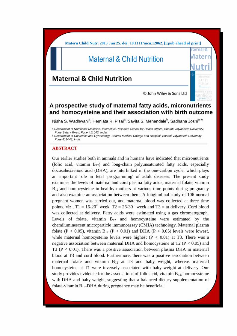
A prospective study of maternal fatty acids, micronutrients and homocysteine and their association with birth outcome Nisha S. Wadhwani
a, Hemlata R. Pisal
a, Savita S. Mehendale
b, Sadhana Joshi
a,*
a Department of Nutritional Medicine, Interactive Research School for Health Affairs, Bharati Vidyapeeth University, Pune Satara Road, Pune 411043, India
b Department of Obstetrics and Gynecology, Bharati Medical College and Hospital, Bharati Vidyapeeth University, Pune 411043, India
ABSTRACT
Our earlier studies both in animals and in humans have indicated that micronutrients
(folic acid, vitamin B12) and long-chain polyunsaturated fatty acids, especially
docosahexaenoic acid (DHA), are interlinked in the one-carbon cycle, which plays
an important role in fetal 'programming' of adult diseases. The present study
examines the levels of maternal and cord plasma fatty acids, maternal folate, vitamin
B12 and homocysteine in healthy mothers at various time points during pregnancy
and also examine an association between them. A longitudinal study of 106 normal
pregnant women was carried out, and maternal blood was collected at three time
points, viz., T1 = 16-20th week, T2 = 26-30th week and T3 = at delivery. Cord blood
was collected at delivery. Fatty acids were estimated using a gas chromatograph.
Levels of folate, vitamin B12 and homocysteine were estimated by the
chemiluminescent microparticle immunoassay (CMIA) technology. Maternal plasma
folate (P < 0.05), vitamin B12 (P < 0.01) and DHA (P < 0.05) levels were lowest,
while maternal homocysteine levels were highest (P < 0.01) at T3. There was a
negative association between maternal DHA and homocysteine at T2 (P < 0.05) and
T3 (P < 0.01). There was a positive association between plasma DHA in maternal
blood at T3 and cord blood. Furthermore, there was a positive association between
maternal folate and vitamin B12 at T3 and baby weight, whereas maternal
homocysteine at T1 were inversely associated with baby weight at delivery. Our
study provides evidence for the associations of folic acid, vitamin B12, homocysteine
with DHA and baby weight, suggesting that a balanced dietary supplementation of
folate-vitamin B12-DHA during pregnancy may be beneficial.
aternal &
Matern
NutriMaternal & Child Nutrition
Maternal & Child Nutrition
Matern Child Nutr. 2013 Jun 25. doi: 10.1111/mcn.12062. [Epub ahead of print]
© John Wiley & Sons Ltd