TURNER SYNDROME BY STEVEN MOORE. OTHER NAMES Ullrich-Turner syndrome Monosomy X.

doi101093brainawl046 Brain (2006) 129 1125ndash1136
Visuospatial executive function inTurner syndrome functional MRI andneurocognitive findings
Sarah J Hart1 Marsha L Davenport2 Stephen R Hooper34 and Aysenil Belger45
Departments of 1Psychology and 2Pediatrics 3Clinical Center for the Study of Development and Learning Department of4Psychiatry School of Medicine University of North Carolina at Chapel Hill Chapel Hill and 5Duke-UNC Brain Imagingand Analysis Center Duke University Durham NC USA
Correspondence to Aysenil Belger Department of Psychiatry CB 7160 University of North Carolina atChapel Hill Chapel Hill NC 27599 USAE-mail aysenil_belgermeduncedu
Turner syndrome is a genetic disorder that results from an abnormal or missing X chromosome in femalesand is typically associated with impairments in visuospatial but not verbal information processing Thesevisuospatial processing impairments may be exacerbated with increased task demands such as those engagedduring working memory (WM) While previous studies have examined spatial WM function in Turner syn-drome none have directly compared the neural correlates of spatial and verbal WM processes across theencoding maintenance and retrieval phases We employed both neurocognitive assessments and functionalMRI (fMRI) to examine the neural circuitry underlying both verbal and visuospatial WM functions in indivi-duals with Turner syndrome and normal controls We furthermore examined the vulnerability of task-relatedfMRI activation to distracters presented duringWMmaintenance Fifteen healthy female volunteers and eightindividuals with Turner syndrome performed a delayed-response WM task during fMRI scanning Neurocog-nitive tests revealed impaired performance across both verbal and spatial domains in Turner syndrome withgreater impairment on tasks with WM demands Frontoparietal regions in controls showed significantly sus-tained levels of activation during visuospatial WM This sustained activation was significantly reduced in thegroup with Turner syndrome Domain-specific activation of temporal regions in contrast did not differbetween the two groups Sensory distraction during the WM maintenance phase did not differentially alterfrontoparietal activation between the two groups The results reveal impaired frontoparietal circuitry recru-itment during visuospatial executive processing in Turner syndrome suggesting a significant role for theX chromosome in the development of these pathways
Keywords executive functions fMRI spatial working memory Turner syndrome verbal working memory
Abbreviations fMRI = functional MRI IFG = inferior frontal gyri IPS = intraparietal sulci ITG = inferior temporal gyriMFG = middle frontal gyri ROI = regions of interest RT = reaction time WM = working memory
Received August 26 2005 Revised January 26 2006 Accepted January 27 2006 Advance Access publication February 27 2006
IntroductionTurner syndrome is a genetic disorder that affects 1 in
2500 live-born girls (Lippe 1996 Saenger 1996 Ranke
and Saenger 2001) and results from an abnormal or missing
second sex (ie X) chromosome Individuals with Turner
syndrome typically demonstrate an uneven profile of
cognitive strengths and weaknesses with greater difficulties
in visuospatial processing accompanied by relative preserva-
tion of verbal skills (McCauley et al 1987) suggestive of right
hemisphere syndrome (Rovet 1995 Buchanan et al 1998)
Cognitive deficits in Turner syndrome appear to become
The Author 2006 Published by Oxford University Press All rights reserved For Permissions please email journalspermissionsoxfordjournalsorg
The online version of this article has been published under an open access model Users are entitled to use reproduce disseminate or display the open accessversion of this article for non-commercial purposes provided that the original authorship is properly and fully attributed the Journal and Oxford UniversityPress are attributed as the original place of publication with the correct citation details given if an article is subsequently reproduced or disseminated not in itsentirety but only in part or as a derivative work this must be clearly indicated For commercial re-use please contact journalspermissionsoxfordjournalsorg
Dow
nloaded from httpsacadem
icoupcombrainarticle12951125327080 by guest on 26 February 2022
most pronounced under high task demand conditions such
as working memory (WM) particularly in visuospatial
domains (Buchanan et al 1998) These deficits may reflect
a selective impairment in the engagement of higher-order
executive control regions such as the prefrontal cortex
with parietal regions during processing of visuospatial
information
Structural imaging studies have provided evidence of pre-
frontal and parietal pathology associated with higher-order
visuospatial processing in Turner syndrome Reduced parie-
tal parietal-occipital (Murphy et al 1993 Reiss et al 1995)
and prefrontal volumes have been reported in Turner syn-
drome (Ross and Zinn 1999) Furthermore impaired per-
formance on higher-order cognitive tasks in individuals
with Turner syndrome such as arithmetic calculation has
recently been linked to selective structural pathology in the
intraparietal sulcus (Molko et al 2003) Functional imaging
studies using PET and functional MRI (fMRI) methods have
also reported reduced activation in parietal regions both at
rest (Clark et al 1990) and during abnormal engagement
of parietal and prefrontal areas in more challenging tasks
(Tamm et al 2003 Kesler et al 2004) Haberecht et al
(2001) found that subjects with Turner syndrome showed
decreased activation in the dorsolateral prefrontal cortex
caudate and inferior parietal lobes during the high-load
but not low-load condition of a visuospatial WM task
The consistent abnormalities across functional and struct-
ural neuroimaging studies suggest deficient engagement of
frontoparietal circuits in association with visuospatial execu-
tive dysfunction in Turner syndrome
In the present study we employed multiple assessment
strategies to determine the integrity of WM functions in
individuals with Turner syndrome when compared with a
control group Neurocognitive measurement and fMRI
were used to determine the integrity of visual discrimination
visuospatial functions and working memory For the fMRI
we employed a visuospatial delayed-recognition task to
examine haemodynamic responses during the encoding
maintenance and retrieval phases of WM both with and
without distraction As previous studies have suggested
more significant deficits in visuospatial than verbal proces-
sing domains we also designed a verbal WM task with a
distracter condition to enable comparison of processing
domains We hypothesized that subjects with Turner syn-
drome would perform less capably than controls on the
visuospatial tasks across assessment methods We further
hypothesized that differential activations between the
group with Turner syndrome and controls would be demon-
strated during the visuospatial but not verbal WM task and
that these differences would involve the frontoparietal circui-
try Finally given the postulated executive function impair-
ments in Turner syndrome we predicted that the differences
between the groups would become more pronounced in the
presence of distracters The distracter condition requires the
ability to suppress task-irrelevant perceptual information
that interferes with WM maintenance
Material and methodsSubjectsTen females with monosomic (45X) Turner syndrome (age
range 14ndash29 years mean = 214 years all right-handed) and 15
control female subjects (age range 19ndash26 years mean = 223 years
14 right-handed) were recruited for this study All individuals
with Turner syndrome were recruited through the UNC Pediatric
Endocrinology Turner Syndrome Clinic All participants with
Turner syndrome had begun oestrogen replacement therapy Volun-
teers with Turner syndrome were excluded if they had a karyotype
other than 45X Individuals with Turner syndrome were also
excluded from participation if they had a history of a significant
neurological disorder or injury a history of drug or alcohol abuse
disorders an estimated verbal IQ less than 85 as measured by the
Wechsler Abbreviated Scales of Intelligence (WASI) Vocabulary
Subtest (The Psychological Corporation 1998) or a major medical
condition not typically associated with Turner syndrome For the
fMRI one participant with Turner syndrome was excluded because
of excessive head motion (gt5 mm) during the scan and one addi-
tional participant with Turner syndrome was excluded owing to
artefact from a metal dental appliance Therefore eight volunteers
with Turner syndrome participated in the fMRI portion of the
study All participants included in the fMRI analyses had average
head movement displacement values within one voxel (4 mm in
either direction) All 10 volunteers with Turner syndrome partici-
pated in the neurocognitive assessments in addition to 10 healthy
controls Female control subjects were excluded for a history of a
significant neurological disorder or injury estimated verbal IQ less
than 85 history of treatment for a major psychiatric illness history
of chronic drug or alcohol abuse or a significant chronic medical
condition This study was approved by the Duke University
Medical Center Institutional Review Board and the University of
North Carolina at Chapel Hill Committee on the Protection of
the Rights of Human Subjects All participants provided informed
consent according to the Declaration of Helsinki
Stimuli and tasksNeurocognitive assessmentVisual processing was assessed in all subjects with a battery of
tasks with demonstrated reliability and validity Tasks were selected
to determine the integrity of visual discrimination functions visuo-
spatial functions and WM In addition to providing evidence for
the presence of the characteristic phenotypical presentation in
Turner syndrome these tasks were selected to provide a clinical
correlate to measures employed in the fMRI paradigm
Visual discrimination ability was tested using the Benton Face
Recognition Test which tests the subjectrsquos ability to identify faces
while visuospatial functions were tested using the Benton Judgment
of Line Orientation Test (Benton et al 1983) Visuospatial WM
was assessed using the Wechsler Memory ScalemdashIII Spatial Span
Subtest which consists of a board with nine blocks attached to it
in no clear identifiable pattern (The Psychological Corporation
1998) The examiner pointed to a sequence of blocks starting
from a span of two and then instructed the individual to point
to the same blocks in the same order If successful the spans
increased by one block each time If not successful a second trial
of the same span was administered Testing was discontinued if
both trials at any particular span length failed For standardization
purposes both the forward and backward recall conditions of
1126 Brain (2006) 129 1125ndash1136 S J Hart et al
Dow
nloaded from httpsacadem
icoupcombrainarticle12951125327080 by guest on 26 February 2022
the Spatial Span Subtest were assessed in this study but only the
backward recall Spatial Span component was employed as the
measure of WM given the increased demands on visual WM and
executive functions
Verbal WM was assessed using the WoodcockndashJohnson-III
Numbers Reversed Subtest in which participants were asked to
repeat a series of increasingly longer digit sequences in reverse
order The score was the total number of correct responses
(Woodcock et al 2001) The backwards digit span test measures
the ability to encode a series of verbally presented digits maintain
them in WM transform the order of the items and recall them
Recalling the digits in backwards order demands a greater WM load
than in the forward recall condition requiring an additional execu-
tive component This verbal WM test provides a complementary
measure to the spatial WM component of the spatial span task
In addition the Vocabulary Subtest from the WASI was admi-
nistered to estimate verbal IQ This measure was used to screen
participants with low verbal IQ in accordance with our inclu-
sionexclusion criteria and was examined as a possible covariate
in subsequent analyses
Functional MRI working memory taskFollowing Jha and McCarthy (2000) a delayed-response task was
developed in which a visual stimulus array (S1) was presented for
4 s followed by a delay interval of 17 s followed then by a probe
stimulus (S2) presented for 4 s to which the subject responded
with a button press A total of 10 S1ndashS2 trials were presented in
each run lasting 8 min The inter-trial interval (ITI) as measured
from the end of the S2 period to the beginning of the S1 period in
the following trial was 185 s An experimental session consisted of
eight runs and lasted no longer than 2 h
Stimuli were presented using an LCD projector and were back-
projected upon a 10-inch wide screen located within the magnet
bore directly behind the subjectrsquos head All stimuli were presented
using the CIGAL display environment (Vovyodic 1999) and were
viewed using mirrored glasses Responses were acquired using a
fibre-optic button box Accuracy and reaction times (RTs) were
recorded by the experimental control software Within each experi-
mental session both the neural correlates of spatial and verbal
WM function (manipulation of domain specificity) and the effects
of distracters on the maintenance period activity (manipulation of
distractibility) were assessed
Domain specificity of WM was manipulated using a delayed-
response design by presenting two different types of stimuli to be
remembered verbal and spatial In condition 1 (verbal WM) four
5-letter words were presented at S1 for 1 s each (Fig 1) S2 consisted
of a single 5-letter word presented for 4 s that was or was not
present in the S1 memory set Using a forced response paradigm
subjects indicated whether the S2 stimulus was present in the S1
memory set by pressing a button on the response box Words were
chosen that could not be easily associated with a visual or object
representation In condition 2 (spatial WM) S1 was composed of
4 squares presented for 1 s each appearing randomly in 1 of 12
possible locations (Fig 1) At S2 the square was presented for 4 s
and appeared in 1 of the 12 possible locations Subjects indicated
by button press whether the S2 stimulus appeared in one of the same
locations as the S1 stimuli
We also examined the effects of distractibility upon verbal and
spatial WM Distracters occurred on half of the trials and were
randomly distributed The tasks were identical to those described
above as conditions 1 and 2 with the addition of distracters pre-
sented during the maintenance period Accordingly condition 3
(verbal distracters) was identical to condition 1 with the addition
of a series of briefly presented words appearing on the screen during
the delay interval A total of nine distracters were presented for
a duration of 1 s each with 500 ms interstimulus intervals Subjects
were asked to simply monitor the words and were not required to
produce a response (perceptual interference) In condition 4 (spatial
distracters) subjects were presented with squares that appeared in a
series of random locations on the screen during the delay period
Again the goal of the secondary monitoring task presented during
the maintenance interval was to introduce a perceptual distractibility
or interference condition
Neurocognitive data analysisThe neurocognitive testing procedures were obtained on 20 of the
subjects 10 with Turner syndrome and 10 controls Two individuals
with Turner syndrome were included in these neurocognitive
testing results but not in the fMRI analyses because of excessive
head motion and artefacts Group differences in age race and voca-
bulary were examined and subsequent univariate analyses were
performed to examine group differences on the four neurocognitive
measures
Imaging behavioural data analysisRT and accuracy ( correct) measures were obtained from all 8 sub-
jects with Turner syndrome and from 14 control subjects Repeated-
measures analysis of variance (ANOVA) tests were run on RT and
Fig 1 Verbal and spatial delayed-response tasks The memoryarray (S1) was presented for 4 s and a delay of 17 s ensuedfollowed by a probe stimulus (S2) for 4 s On half of the trialsdistracters were presented during the delay period
Visuospatial executive function in TS Brain (2006) 129 1125ndash1136 1127
Dow
nloaded from httpsacadem
icoupcombrainarticle12951125327080 by guest on 26 February 2022
correct measurements to assess the effects of domain distraction and
group (control versus Turner syndrome)
Functional MRI acquisition and analysesThe scans were performed on a General Electric 15 T NVi system
Imaging began with the acquisition of a T1-weighted sagittal loca-
lizer series to identify the anterior (AC) and posterior commissures
(PC) and to prescribe contiguous oblique slices parallel to the
ACndashPC plane These series were followed by the acquisition of
68 slices of high-resolution T1-weighted structural images [repeti-
tion time (TR) = 123 ms echo time (TE) = 54 ms field of view
(FOV) = 24 cm 256 middot 256 matrix slice thickness 19 mm] oriented
parallel to the ACndashPC Thirty-four contiguous functional images
sensitive to BOLD contrast were acquired at the same slice loca-
tions as the structural images (TE = 30 ms 24 cm FOV 64 middot 64
image matrix 90 flip angle TR = 15 s slice thickness = 38 mm
yielding 38 mm3 isotropic voxels)
The time courses of the voxels in each slice were realigned with the
onset of the TR in order to compensate for the interleaved slice
acquisition order Images were preprocessed using SPM99 (http
wwwfilionuclacukspm) Volumes were spatially aligned to a
reference volume using translation and rigid rotation in order to
correct for head movements Individual subjectsrsquo functional images
were co-registered with their high-resolution anatomical images
The data were normalized to standard Talairach coordinates using
the standard SPMMNI T1 template image Images were resampled
using sinc interpolation and smoothed with an 8 mm Gaussian
kernel to improve the signal-to-noise ratio
For the voxel-based analysis epochs beginning 4 time points prior
to S1-onset and continuing for 29 time points following S1 were
excised from the continuous time series of non-normalized
co-registered raw images for each condition Signal averaging was
performed on these excised epochs with separate average epochs
created for each experimental condition The average MR signal
values were then converted to per cent signal change relative to a
pre-S1 baseline defined as the four time points preceding S1 These
time course analyses were performed on the raw TR-aligned func-
tional data that had not undergone smoothing or normalization
For the anatomical regions of interest (ROI) analysis selected
structures in frontoparietal and frontotemporal regions associated
with functional and structural abnormalities in Turner syndrome
were drawn by a single observer on each individual subjectrsquos high-
resolution structural MRIs (see Supplementary Fig 1S available at
Brain online) Selected ROIs included the left and right middle
frontal gyri (MFG) inferior frontal gyri (IFG) intraparietal sulci
(IPS) and inferior temporal gyri (ITG) ROIs were drawn using a
three-dimensional interactive image segmentation program (IRIS
SNAP) (Ho et al 2002) using landmarks from the Duvernoyrsquos
Human Brain Atlas (1999) All ROI analyses were performed on
individual subjectsrsquo non-normalized brains No ROIs were found to
differ significantly in size (ie number of voxels) between controls
and the group with Turner syndrome
Using custom MATLAB software ROIs were interrogated with a
function that returned the number of voxels within the selected ROI
that correlated significantly with an ideal haemodynamic response
waveform (t gt 196 P lt 005) (Jha and McCarthy 2000) This
measurement of per cent activated voxels reflected the spatial extent
of activation relative to ROI size Relative per cent signal change was
calculated for each ROI by interrogating them with a function that
returned the average time-varying signal change of the voxels within
the selected regions Differences between the waveforms evoked
by the four different experimental conditions were tested by mea-
suring the signal over particular time periods allowing for the sepa-
rate assessment of condition and population effects in the S1 period
delay period and S2 period The per cent signal change at four
different latencies in each epoch (time points 9 12 18 and 24 relative
to epoch onset) were selected as measures of peak haemodynamic
responses associated with the encoding (time points 9 and 12)
maintenance (time point 18) and retrieval (time point 24) phases
of the WM trials
Repeated-measures ANOVAs were performed on the peak ampli-
tudes at the selected time points to examine group anatomical
structure hemisphere and experimental condition differences
Quantitative statistical analyses were performed on the dependent
measure ( signal change) with group as a between-subjects vari-
able and task phase (encode maintain and retrieve) and hemisphere
as within-subject repeated measures Post hoc analyses were then
conducted to assess the direction and pairwise effects at each
level Repeated-measures ANOVAs were used to identify group
differences in the peak measures
ResultsNeurocognitive testing resultsInitial group comparisons indicated that individuals with
Turner syndrome and controls had significantly different
verbal IQs [t(18) = 697 P lt 00001] with the controls having
a higher level of vocabulary (Turner syndrome mean
T-score = 5420 SD = 588 controls mean T-score =
6840 SD = 560) No significant differences were found
for age (Turner syndrome mean = 198 controls mean =
229) or race although there was a strong trend for the Turner
syndrome group to be younger [t(18) = 205 P lt 006] Given
these findings subsequent analyses were co-varied for esti-
mated IQ and chronological age
Univariate ANCOVAs (analysis of covariance) controlling
for estimated IQ and age revealed that the group with Turner
syndrome performed more poorly than controls on every task
except for Facial Recognition (Table 1) On the Judgment
of Line Orientation Test WJ-III Numbers Reversed and
WMS-III Spatial Span Reversed the Turner syndrome
group performed significantly lower than the control
group Effect sizes were moderate to large ranging from
063 for WMS-III Spatial Span Reversed to 072 for WJ-III
Numbers Reversed These findings suggest that while visuo-
spatial processing may be disrupted in Turner syndrome
consistent with the classic Turner syndrome phenotype
WM functionsmdashboth verbal and visuospatialmdashmay also be
significantly impaired
Imaging behavioural resultsANOVA tests were performed on the dependent variables of
accuracy ( correct) and RT to assess the between-subject
factor of group (controls versus Turner syndrome) and the
within-subject factors of domain (verbal versus spatial)
and distraction Behavioural results for accuracy and RT
are displayed in Fig 2A and 2B Participants with Turner
1128 Brain (2006) 129 1125ndash1136 S J Hart et al
Dow
nloaded from httpsacadem
icoupcombrainarticle12951125327080 by guest on 26 February 2022
syndrome were less accurate (83 correct SD = 1569) than
controls [91 correct SD = 1088 F(121) = 1339 P lt 0002]
and had slower response times (1865 ms SD = 465 ms) than
controls [1349 ms SD = 442 ms F(121) = 1436 P = 0001]
For both groups accuracy was better for the verbal WM
condition (96 correct SD = 522) than the spatial condition
(79 correct SD = 1347) and RTs were faster for the verbal
(1267 ms SD = 434 ms) than the spatial WM conditions
(1807 ms SD = 440 ms) [domain effect on accuracy F(121)
= 481 P lt 00001 and RT F(121) = 5523 P lt 00001]
Although the individuals with Turner syndrome were less
accurate in the spatial WM task than in the verbal WM
task this effect did not reach significance [group by domain
interaction F(121) = 249 P = 013] There was also no
significant group by domain interaction in the RT data
[F(121) = 03 P = 059]
Distraction had a significant overall effect on RT [F(121) =
80 P = 001] but had no significant effect on accuracy No
significant interactions were found between group and dis-
traction for either accuracy or RT Accuracy on the spatial
task was more vulnerable to distraction than the verbal task
[domain by distracter interaction F(121) = 48 P lt 004] In
addition RTs on the spatial task were more vulnerable to
distraction than the verbal task [domain by distracter inter-
action F(121) = 498 P = 0037]
Functional MRI resultsWe performed time-course analyses on the significantly acti-
vated voxels in each ROI to compare the haemodynamic
changes between groups and conditions throughout the
WM trials Group and condition effects were assessed for
the encoding (time points 9 and 12) maintenance (time
point 18) and retrieval (time point 24) phases of the trials
for each ROI All significant statistics for each time point
including the effects of group and domain are displayed in
Table 2 with hemispheric laterality effects displayed in Table 3
(see Supplementary Figs 2S and 3S for group average
activation maps) Statistics for the distracter condition are
displayed in Supplementary Table 1S
Intraparietal sulcusConsistent with posterior parietal cortex involvement in spa-
tial processing the IPS was activated significantly more
strongly by the spatial domain than by the verbal domain
at all time points (domain effect Table 2) with significantly
greater right-hemisphere dominance during the spatial task
(hemisphere effect Table 3) The average time course of
activation in the IPS during each WM task is displayed in
Fig 3 for both the control and Turner syndrome groups
Table 1 Performance on neurocognitive tests
Turner syndrome Normal controls
Tasks Mean SD Mean SD F(3 15) Effect size
Facial recognition 2222 264 2470 149 290 037Judgement of line orientation 2067 743 2670 226 1175 070WMS-III spatial span reverse 556 213 950 184 850 063WJ-III numbers reversed 1411 558 2430 417 1277 072
Comparisons of the Turner syndrome group versus the control group on the neurocognitive measures controlling for age and estimatedverbal IQ (P lt 001 P lt 00001) All scores are reported as raw scores
Fig 2 Behavioural results for spatial and verbal WM with andwithout distraction (Dist versus NoDist) (A) Mean per centcorrect with standard error bars (B) Mean RT with standarderror bars
Visuospatial executive function in TS Brain (2006) 129 1125ndash1136 1129
Dow
nloaded from httpsacadem
icoupcombrainarticle12951125327080 by guest on 26 February 2022
As reflected in the time-course graph of the controlsrsquo
activation of the IPS (Fig 3A) during the visuospatial
WM task they displayed highly sustained levels of activation
throughout the maintenance period The maintenance-phase
activation in the group with Turner syndrome in contrast
was significantly decreased relative to controls (Fig 3B)
During the verbal WM condition however the two
groups showed similar patterns of haemodynamic change
throughout the task The two groups activated the IPS
differentially according to domain during the maintenance
phase as measured by time point 18 activation (group
by domain interaction Table 2) Consistent with our
hypothesis the group with Turner syndrome therefore
showed different parietal activation patterns on the spatial
but not verbal task
Controlling for age had no effect on the group by domain
interaction in the IPS [F(120) = 65 P = 002] but when
accuracy was controlled for the interaction did not reach
significance [F(117) = 222 P = 015] suggesting that
group differences in IPS maintenance levels were partially
explained by their differences in performance Contrary to
our hypothesis that group differences would be enhanced in
the presence of distracters no group by distracter interactions
were found
Middle frontal gyrusThe MFG was activated significantly more strongly by the
spatial task than the verbal task (domain effect Table 2) at
time points 12 and 24 with significantly greater right-
hemisphere dominance during the spatial task (Table 3)
The average time course of activation in the MFG during
each WM task is displayed in Fig 4 for both groups Controls
displayed sustained activation during the maintenance phase
of the spatial WM task while activation during the verbal
WM task returned to baseline between the encoding and
retrieval phases (Fig 4A) In the group with Turner syn-
drome the MFG showed less differentiation in activation
related to domain and showed slightly greater activation
during verbal WM maintenance compared with the spatial
task (Fig 4B) The two groups therefore activated the MFG
differentially according to domain during the maintenance
Table 2 Effects of group and domain on fMRI per cent signal change
9 12 18 24
Test ROI F P F P F P F P
Group ITG 571 0026 656 0018IPS 59 0024
Domain ITG 941 0006 487 0039IPS 4445 lt00001 4404 lt00001 2177 lt00001 8406 lt00001MFG 91 0007 816 0009
Group by domain IPS 727 0014MFG 671 0017 64 0019 425 0052IFG 636 002 1578 00007 471 0042
F and P values for the encode (time point 9 and 12) maintain (time point 18) and retrieve (time point 24) periods of the task for significantlyactive voxels in each ROI ROIs that were not active for a given test at any time point are not displayed
Table 3 Hemispheric effects on fMRI per cent signal change
9 12 18 24
Test ROI F P F P F P F P
Hemisphere ITG 439 0048 421 0053IPS 1476 0001MFG 138 0001IFG 1403 0001
Hemisphere by group ITG 554 0028 721 0014 653 0018 428 0051IFG 513 0034
Hemisphere by domain ITG 2404 lt00001 2285 lt00001 897 0007IPS 1474 0001MFG 1614 0001 1803 00004 1096 0003IFG 3971 lt00001 2265 lt00001 525 0032 594 0024
Laterality effects F and P values for the encode (time point 9 and 12) maintain (time point 18) and retrieve (time point 24) periods of thetask for significantly active voxels in each ROI ROIs that were not active for a given test at any time point are not displayed Only thoseinteractions that were significant at any time point are displayed
1130 Brain (2006) 129 1125ndash1136 S J Hart et al
Dow
nloaded from httpsacadem
icoupcombrainarticle12951125327080 by guest on 26 February 2022
and retrieval phases (group by domain interaction at time
points 12 18 and 24 Table 2)
No significant effects of age or performance were found on
MFG activation Also no significant group by distracter
interactions were found
Inferior frontal gyrusThe IFG did not show overall significantly different activation
patterns depending on the task domain (Table 2) The aver-
age time course of activation in the IFG during each WM task
is displayed in Fig 5 for both groups While controls showed
a more similar pattern of activation for both verbal and
spatial tasks with slightly greater activation to the spatial
task (Fig 5A) the individuals with Turner syndrome showed
larger activation to the verbal task relative to the spatial task
(Fig 5B) Although controls and subjects with Turner syn-
drome did not differ in their overall IFG activation levels the
two groups activated the IFG differentially according to
domain for time points 12 18 and 24 (group by domain
interaction Table 2)
No significant effects of age or performance were found on
IFG activation A significant group by distraction effect was
found (see supplementary Table 1S) Finally the IFG activa-
tion patterns indicated that controls showed more right-
lateralized activation at encoding whereas subjects with
Turner syndrome showed more left-lateralized activation
(hemisphere by group interaction Table 3)
Inferior temporal gyrusConsistent with temporal cortex involvement in verbal pro-
cessing the ITG activated more strongly to the verbal than
spatial task for both groups at encoding (Table 2) and
showed significantly greater left-hemisphere dominance dur-
ing the verbal task (hemisphere by domain Table 3) The
average time course of activation in the ITG during each WM
task is displayed in Fig 6 for both groups The group with
Turner syndrome showed overall greater activation in the
ITG compared with controls (group effect Table 2) Both
the controls and individuals with Turner syndrome showed
greater activation at encoding to the verbal task but the
Intraparietal sulcus (IPS)
Fig 3 Mean per cent signal change within active voxels for the intraparietal sulcus in (A) controls and (B) Turner syndrome Grey linesindicate onset of S1 and S2 periods Asterisks indicate those tested time points (ie 9 12 18 24) that had significant group by domaininteractions
Visuospatial executive function in TS Brain (2006) 129 1125ndash1136 1131
Dow
nloaded from httpsacadem
icoupcombrainarticle12951125327080 by guest on 26 February 2022
group with Turner syndrome showed a larger increase in this
encoding-related verbal activation (Fig 6A and B) Despite
the greater activation during the verbal task in subjects with
Turner syndrome relative to controls the group by domain
interaction did not reach significance
No significant effects of age or performance were found on
ITG activation A significant group by distraction effect was
found which differed by task domain (see Supplementary
Table 1S) Hemispheric effects showed a bias in the ITG at
encoding and maintenance during the verbal WM task for
both groups but the subjects with Turner syndrome showed
significantly greater left-side bias on the verbal task relative to
controls (hemisphere by group interaction Table 3)
DiscussionIn the present study we examined neurobehavioural corre-
lates of verbal and spatial WM processes in healthy controls
and in individuals with Turner syndrome with further
examination of vulnerability to distraction in both groups
Our results produced the classic phenotypical profile of
visuospatial weaknesses that has been proliferated in the lit-
erature on children and adults with Turner syndrome How-
ever our findings also revealed significant WM differences
suggestive of executive dysfunction across methods of mea-
surement that may co-exist with or underlie the visuospatial
weaknesses in this population More specifically our findings
demonstrated changes in prefrontal and posterior parietal
function in Turner syndrome which were particularly pro-
nounced during the period of visuospatial WM maintenance
The patterns of activation in frontoparietal regions indi-
cated that the subjects with Turner syndrome engaged these
regions to a lesser degree particularly during spatial WM
performance whereas they were able to engage these regions
normally during verbal WM Several additional studies have
similarly found deficient engagement of these regions in
Turner syndrome during spatial WM (eg Haberecht et al
2001 Kesler et al 2004) The data in the current study
suggests that the inability of individuals with Turner syn-
drome to engage prefrontal and posterior parietal regions
may be primarily explained by dysfunction within the main-
tenance phase whereas these regions may function normally
Middle frontal gyrus (MFG)
Fig 4 Mean per cent signal change within active voxels for the middle frontal gyrus in (A) controls and (B) Turner syndrome Grey linesindicate onset of S1 and S2 periods Asterisks indicate those tested time points that had significant group by domain interactions
1132 Brain (2006) 129 1125ndash1136 S J Hart et al
Dow
nloaded from httpsacadem
icoupcombrainarticle12951125327080 by guest on 26 February 2022
in Turner syndrome during WM encoding and retrieval pro-
cesses Although the present study is limited by the relatively
small sample size of participants with Turner syndrome this
lack of sustained engagement of posterior parietal regions was
observed consistently across individuals in the Turner syn-
drome group Within the group with Turner syndrome only
2 out of 8 individuals activated posterior parietal regions
above baseline levels during the WM delay whereas 13 out
of 15 control participants showed sustained activity While
additional studies will certainly be needed to support these
findings of abnormal spatial WM delay activity this pattern
showing lack of parietal engagement appears to be relatively
robust across individuals
Because the participants with Turner syndrome performed
more poorly than controls on the WM tasks it is possible that
group differences in brain activation may be partially
explained by task performance Indeed a covariance analysis
of our data indicated that WM performance significantly
predicted activation in the intraparietal sulcus although it
did not predict activation in the other ROI Analyses com-
paring correct versus incorrect trials indicated that while the
group with Turner syndrome was able to adequately engage
parietal regions during their correct trials in a similar manner
to controls their incorrect trials were associated with abnor-
mally low levels of delay period activation during the spatial
WM delay phase Posterior parietal dysfunction during spa-
tial WM maintenance in Turner syndrome may therefore be
an important factor underlying their visuospatial cognitive
difficulties As there is considerable cognitive variability
among individuals with Turner syndrome future studies
may further clarify the relationship between individual
visuospatial abilities and the degree to which these parietal
mechanisms are engaged
Analyses of the behavioural results indicated that although
the individuals with Turner syndrome showed the expected
differences on the spatial neurocognitive tasks and on the
spatial WM task completed in the fMRI scanner the
group with Turner syndrome also had lower estimated verbal
IQ and concomitant weaker verbal WM On the surface these
findings would seem to contrast with the Turner syndrome
literature that typically characterizes their deficits as primarily
in the visuospatial domain A potential reason for this finding
could be that our sample of participants with Turner syn-
drome was biased towards having greater verbal deficits than
Inferior frontal gyrus (IFG)
Fig 5 Mean per cent signal change within active voxels for the inferior frontal gyrus in (A) controls and (B) Turner syndrome Grey linesindicate onset of S1 and S2 periods Asterisks indicate those tested time points that had significant group by domain interactions
Visuospatial executive function in TS Brain (2006) 129 1125ndash1136 1133
Dow
nloaded from httpsacadem
icoupcombrainarticle12951125327080 by guest on 26 February 2022
controls related to the sample size However these deficits
would support the presence of higher-order executive
dysfunctions (eg working memory) that may in turn con-
tribute to the significantly poorer verbal performance in the
group with Turner syndrome when compared with controls
Other studies of Turner syndrome have indeed found retrie-
val impairments during verbal fluency tasks with relatively
large executive demands (Temple 2002) and poorer perfor-
mance on verbal executive tasks requiring cognitive flexibility
and WM (Kirk et al 2005) It is possible therefore that a
global executive function deficit in Turner syndrome may be
responsible for poorer performance on a multitude of cogni-
tively demanding tasks that require planning attention shift-
ing and WM This executive dysfunction in Turner syndrome
may explain multiple aspects of the neurocognitive profile
such as arithmetic deficits (eg Rovet et al 1993) attentional
abilities (eg Romans et al 1998) and inhibitory control
(Tamm et al 2003) The executive function deficit in Turner
syndrome may therefore only be apparent on tasks with suf-
ficient complexity to engage prefrontal mechanisms
We directly manipulated executive requirements of our
task by including a distracter condition where irrelevant sti-
muli were presented during the WM delay However con-
trary to our hypotheses our results indicated that the
presence of distracters on both tasks did not significantly
affect performance in either group nor did the distracters
largely affect the Turner syndrome grouprsquos activation pat-
terns more than controls It is possible that because the dis-
tracters involved passive viewing of perceptual stimuli they
may not have provided significant interference to require the
engagement of the executive attention component of WM
While lower level sensory processing mechanisms may be
intact in Turner syndrome it is possible that increasing
the executive load of the task to a level sufficient to interfere
with WM rehearsal could lead to greater sensitivity in Turner
syndrome
In addition to frontoparietal dysfunction in the group
with Turner syndrome our results also revealed a pattern
of over-engagement of inferior frontal and temporal regions
in subjects with Turner syndrome compared with controls
Inferior temporal gyrus (ITG)
Fig 6 Mean per cent signal change within active voxels for the inferior temporal gyrus in (A) controls and (B) Turner syndrome Grey linesindicate onset of S1 and S2 periods
1134 Brain (2006) 129 1125ndash1136 S J Hart et al
Dow
nloaded from httpsacadem
icoupcombrainarticle12951125327080 by guest on 26 February 2022
suggesting that the neurophysiological differences in Turner
syndrome may not be restricted to frontoparietal regions
Several structural imaging studies have found specific tem-
poral lobe abnormalities in Turner syndrome such as altera-
tions in temporal lobe fibre tracts (Molko et al 2004) and
larger superior temporal gyri suggesting the disruption of
neural pruning mechanisms during development (Kesler
et al 2003) These temporal lobe abnormalities have been
suggested to possibly underlie deficits in language tasks
such as semantic fluency in Turner syndrome (Rae et al
2004) which may require greater executive demand than
reading tasks where individuals with Turner syndrome
have been found to perform normally (Temple and
Carney 1996) Structural and functional changes may there-
fore be apparent in multiple brain regions in Turner syn-
drome which may possibly underlie some of their executive
deficits or potentially reflect compensatory mechanisms
Because of the observed changes in brain structure and
function in Turner syndrome the X chromosome has been
proposed to play a particularly significant role in the devel-
opment of circuitry underlying visuospatial and executive
function (Skuse 2005) It has been hypothesized that the
brains of individuals with Turner syndrome develop abnor-
mally during gestation as a result of missing genetic material
responsible for neural pruning mechanisms (Rae et al 2004)
The cognitive deficits in Turner syndrome may arise from a
reduced dosage of a gene or genes on the X chromosome
(Buchanan et al 1998) as evidenced by studies showing that
individuals with a higher percentage of normal 46XX cells
perform better on visuospatial processing tasks than those
with the 45X karyotype (Murphy et al 1993) Furthermore
mapping of deletions using molecular markers has shown
that a 2 Mb critical region of the X chromosome is associa-
ted with visuospatial deficits in Turner syndrome (Ross et al
2000) Cognitive assessments of women with fragile X syn-
drome have also suggested specific deficits in visuospatial
ability and executive function (Bennetto et al 2001) affect-
ing the development of complex cognitive functions
(Cornish et al 2004) Future studies combining neuroima-
ging with molecular genetic techniques will help in elucidat-
ing the relationships between neurobiological and genetic
markers of the cognitive deficits associated with X chromo-
some abnormalities
Our findings support the characteristic phenotypic
description of visuospatial dysfunction in Turner syndrome
found in the literature but they also extend this description
by providing evidence of frontal-temporal and frontal-
parietal involvement as well thus contributing to the com-
plex pathophysiology inherent in Turner syndrome The
results suggest that executive function impairments in Turner
syndrome may affect a multitude of cognitive tasks that
have a sufficient level of complexity or difficulty However
the frontoparietal circuitry may be particularly vulnerable to
dysfunction during visuospatial WM in Turner syndrome
and is characterized by a relative lack of sustained engage-
ment during the maintenance of spatial information
Supplementary materialSee supplementary material available at Brain online
AcknowledgementsWe thank V Allen Santos for assistance in patient recruit-
ment and Dr Joseph Piven for valuable contributions to the
research This research was supported by the National
Institutes of Health grant 1R03HD40249 Funding to pay
the Open Access publication charges for this article was
provided by the University of North Carolina at Chapel
Hill Department of Psychiatry
References
Bennetto L Pennington BF Porter D Taylor AK Hagerman RJ Profile of
cognitive functioning in women with the fragile X mutation Neuropsy-
chology 2001 15 290ndash9
Benton AL Hamsher K Varney NR Spreen O Contributions to neuropsy-
chological assessment a clinical manual New York Oxford University
Press 1983
Buchanan L Pavlovic J Rovet J A rexamination of the visuospatial deficit in
turner syndrome contributions of working memory Dev Neuropsychol
1998 14 341ndash67
Clark C Klonoff H Hayden M Regional cerebral glucose metabolism in
Turner syndrome Can J Neurol Sci 1990 17 140ndash4
Cornish KM Turk J Wilding J Sudhalter V Munir F Kooy F et al Annota-
tion deconstructing the attention deficit in fragile X syndrome a devel-
opmental neuropsychological approach J Child Psychol Psychiatry 2004
45 1042ndash53
Duvernoy HM The human brain surface blood supply and three-
dimensional sectional anatomy New York Springer-VerlagWien 1999
Haberecht MF Menon V Warsofsky IS White CD Dyer-Friedman J Glover
GH et al Functional neuroanatomy of visuo-spatial working memory in
Turner syndrome Hum Brain Mapp 2001 14 96ndash107
Ho S Bullit E Gerig G Level set evolution with region competition auto-
matic 3-D segmentation of brain tumors Proceedings of the 16th Inter-
national Conference On Pattern Recognition ICPR IEEE Computer
Society 2002
Jha AP McCarthy G The influence of memory load upon delay-interval
activity in a working-memory task an event-related functional MRI
study J Cogn Neurosci 2000 12 (Suppl 2) 90ndash105
Kesler SR Blasey CM Brown WE Yankowitz J Zeng SM Bender BG et al
Effects of X-monosomy and X-linked imprinting on superior temporal
gyrus morphology in Turner syndrome Biol Psychiatry 2003 54 636ndash46
Kesler SR Haberecht MF Menon V Warsofsky IS Dyer-Friedman J Neely
EK et al Functional neuroanatomy of spatial orientation processing in
Turner syndrome Cereb Cortex 2004 14 174ndash80
Kirk JW Mazzocco MM Kover ST Assessing executive dysfunction in girls
with fragile X or Turner syndrome using the Contingency Naming Test
(CNT) Dev Neuropsychol 2005 28 755ndash77
Lippe BM Turner syndrome In Sperling MA editor Pediatric endocrinol-
ogy Philadelphia PA WB Saunders Co 1996 p 387ndash421
McCauley E Kay T Ito J Treder R The Turner syndrome cognitive deficits
affective discrimination and behavior problems Child Dev 1987 58
464ndash73
Molko N Cachia A Riviere D Mangin JF Bruandet M Le Bihan D et al
Functional and structural alterations of the intraparietal sulcus in a devel-
opmental dyscalculia of genetic origin Neuron 2003 40 847ndash58
Molko N Cachia A Riviere D Mangin JF Bruandet M LeBihan D et al
Brain anatomy in Turner syndrome evidence for impaired social and
spatial-numerical networks Cereb Cortex 2004 14 840ndash50
Murphy DG DeCarli C Daly E Haxby JV Allen G White BJ et al
X-chromosome effects on female brain a magnetic resonance imaging
study of Turnerrsquos syndrome Lancet 1993 342 1197ndash200
Visuospatial executive function in TS Brain (2006) 129 1125ndash1136 1135
Dow
nloaded from httpsacadem
icoupcombrainarticle12951125327080 by guest on 26 February 2022
Rae C Joy P Harasty J Kemp A Kuan S Christodoulou J et al Enlarged
temporal lobes in Turner syndrome an X-chromosome effect Cereb
Cortex 2004 14 156ndash64
Ranke MB Saenger P Turnerrsquos syndrome Lancet 2001 358 309ndash14
Reiss AL Mazzocco MM Greenlaw R Freund LS Ross JL Neurodevelop-
mental effects of X monosomy a volumetric imaging study Ann Neurol
1995 38 731ndash8
Romans SM Stefanatos G Roeltgen DP Kushner H Ross JL Transition to
young adulthood in Ullrich-Turner syndrome neurodevelopmental
changes Am J Med Genet 1998 79 140ndash7
Ross JL Zinn A Turner syndrome potential hormonal and genetic influences
on the neurocognitive profile In Tager-Flusberg editor Neurodevelop-
mental disorders 1999 251ndash68
Ross JL Roeltgen D Kushner H Wei F Zinn AR The Turner syndrome-
associated neurocognitive phenotype maps to distal Xp Am J Hum Genet
2000 67 672ndash81
Rovet JF Behavioral manifestations of Turner syndrome in children a
unique phenotype In Albertsson-Wikland KA Ranke MB editors Turner
syndrome in a life span perspective research and clinical aspects Amster-
dam Elsevier 1995 p 285ndash95
Rovet JF Ehrlich RM Czuchta D Akler M Psychoeducational characteristics
of children with Turner syndrome J Learn Disabil 1993 26 333ndash41
Saenger P Turnerrsquos Syndrome N Engl J Med 1996 335 1749ndash54
Skuse DH X-linked genes and mental functioning HumMol Genet 2005 14
R27ndash32
Tamm L Menon V Reiss AL Abnormal prefrontal cortex function during
response inhibition in Turner syndrome functional magnetic resonance
imaging evidence Biol Psychiatry 2003 53 107ndash11
Temple CM Oral fluency and narrative production in children with Turnerrsquos
syndrome Neuropsychologia 2002 40 1419ndash27
Temple CM Carney R Reading skills in children with Turnerrsquos syndrome an
analysis of hyperplexia Cortex 1996 32 335ndash45
Voyvodic JTReal-time fMRIparadigmcontrolphysiology andbehaviorcom-
bined with near real-time statistical analysis Neuroimage 1999 10 91ndash106
Woodcock WR McGrew KS Mather N Woodcock-Johnson III Tests of
cognitive abilities Itasca FL Riverside Publishing 2001
1136 Brain (2006) 129 1125ndash1136 S J Hart et al
Dow
nloaded from httpsacadem
icoupcombrainarticle12951125327080 by guest on 26 February 2022
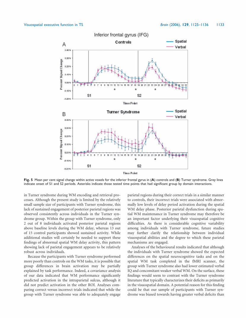